Article

Long-Term Outcome and Role of Biology within Risk-Adapted Treatment Strategies: The Austrian Neuroblastoma Trial A-NB94

by
Stefan Fiedler
1,2,
Inge M. Ambros
2,
Evgenia Glogova
2,
Martin Benesch
3,
Christian Urban
3,
Marlene Mayer
3,
Georg Ebetsberger-Dachs
4,
Edit Bardi
1,4,
Neil Jones
5,
Agnes Gamper
5,
Bernhard Meister
6,
Roman Crazzolara
6,
Gabriele Amann
7,
Karin Dieckmann
8,
Ernst Horcher
9,
Reinhold Kerbl
10,
Bettina Brunner-Herglotz
2,
Andrea Ziegler
2,
Peter F. Ambros
2 and
Ruth Ladenstein
1,2,11,*
1
St. Anna Children’s Hospital, 1090 Vienna, Austria
2
CCRI, Children’s Cancer Research Institute, 1090 Vienna, Austria
3
Department of Pediatrics and Adolescent Medicine, Division of Pediatric Hematology and Oncology, Medical University of Graz, 8036 Graz, Austria
4
Med Campus IV, University Clinic for Pediatrics, Kepler University Hospital, 4020 Linz, Austria
5
Department of Pediatrics, Paracelsus Medical University, 5020 Salzburg, Austria
6
Department of Pediatric Hematology, Oncology, and Stem-Cell Transplantation, Medical University of Innsbruck, 6020 Innsbruck, Austria
7
Department of Pathology, Medical University of Vienna, 1090 Vienna, Austria
8
Department of Radiotherapy, Medical University of Vienna, 1090 Vienna, Austria
9
Department of Pediatric Surgery, Medical University of Vienna, 1090 Vienna, Austria
10
Department of Pediatric Medicine, Landeskrankenhaus Leoben, 8700 Leoben, Austria
11
Department of Pediatric Medicine, Medical University of Vienna, 1090 Vienna, Austria
*
Author to whom correspondence should be addressed.
Cancers 2021, 13(3), 572; https://doi.org/10.3390/cancers13030572
Submission received: 23 November 2020 / Revised: 8 January 2021 / Accepted: 28 January 2021 / Published: 2 February 2021
(This article belongs to the Section Clinical Research of Cancer)

Abstract

:

Simple Summary

Neuroblastoma, the most common extracranial malignancy of childhood, shows a highly variable course of disease ranging from spontaneous regression or maturation into a benign tumor to an aggressive and intractable cancer in up to 60% of patients. To adapt treatment intensity, risk staging at diagnosis is of utmost importance. The A-NB94 trial was the first in Austria to stratify therapy intensity according to tumor staging, patient’s age, and MYCN amplification status, the latter being a biologic marker turning otherwise low-risk tumors into high-risk disease. Recent publications showed a prognostic impact of various genomic features including segmental chromosomal aberrations (SCAs). We retrospectively investigated the relevance of SCAs within this risk-adapted treatment strategy. The A-NB94 approach resulted in an excellent long-term survival for the majority of patients with acceptable long-term morbidity. An age- and stage-dependent frequency of SCAs was confirmed and SCAs should always be considered in future treatment decision making processes.

Abstract

We evaluated long-term outcome and genomic profiles in the Austrian Neuroblastoma Trial A-NB94 which applied a risk-adapted strategy of treatment (RAST) using stage, age and MYCN amplification (MNA) status for stratification. RAST ranged from surgery only to intensity-adjusted chemotherapy, single or multiple courses of high-dose chemotherapy (HDT) followed by autologous stem cell rescue depending on response to induction chemotherapy, and irradiation to the primary tumor site. Segmental chromosomal alterations (SCAs) were investigated retrospectively using multi- and pan-genomic techniques. The A-NB94 trial enrolled 163 patients. Patients with localized disease had an excellent ten-year (10y) event free survival (EFS) and overall survival (OS) of 99 ± 1% and 93 ± 2% whilst it was 80 ± 13% and 90 ± 9% for infants with stage 4S and for infants with stage 4 non-MNA disease both 83 ± 15%. Stage 4 patients either >12 months or ≤12 months but with MNA had a 10y-EFS and OS of 45 ± 8% and 47 ± 8%, respectively. SCAs were present in increasing frequencies according to stage and age: in 29% of localized tumors but in 92% of stage 4 tumors (p < 0.001), and in 39% of patients ≤ 12 months but in 63% of patients > 12 months (p < 0.001). RAST successfully reduced chemotherapy exposure in low- and intermediate-risk patients with excellent long-term results while the outcome of high-risk disease met contemporary trials.

1. Introduction

Neuroblastoma is the most common extracranial malignancy of childhood and originates from the sympathetic nervous system. It shows a highly heterogeneous behavior ranging from spontaneous regression or maturation into a benign ganglioneuroma to an aggressive and intractable disease. Risk classification systems are using clinical and biological characteristics to predict survival and adapt treatment intensity [1,2].
At this study’s initiation, recognized risk driving factors included stage defined by the International Neuroblastoma Staging System (INSS), age at diagnosis, and MYCN oncogene amplification (MNA) status [3]. MNA, a strong biologic marker associated with rapid tumor growth [4,5], transforms otherwise favorable risk profiles of infants [6,7] and children with localized resectable [8,9] or unresectable [10] disease into high-risk [11]. Metastatic disease in children older than 18 months constitutes per se an unfavorable risk group regardless of MYCN status [12]. Intratumoral heterogeneous MNA (hetMNA) refers to the coexistence of clustered or scattered single MNA cells and non-MYCN-amplified (non-MNA) tumor cells [13], a phenomenon that was largely unexplored at the initiation of A-NB94. A recent study highlights the importance of viewing it separately from the MNA profile and its unfavorable risk implication, however, prognostication and therapy allocation are still unsolved issues [14,15].
Here, we present long-term outcomes of the Austrian neuroblastoma trial A-NB94, initiated in 1994 to apply a risk-adapted strategy of treatment (RAST) based on age (≤/>12 months), INSS stage and MYCN Status (Table 1). Encouraging results of the Lyon-Marseille-Curie-Est cooperative group (LMCE2) [16] using tandem high-dose chemotherapy (HDT) followed by autologous stem cell rescue (ASCR) prompted the adoption of a similar approach for patients with incomplete response to induction therapy. In addition, we show a post hoc genomic analysis to investigate pattern and potential influence of biomarkers on long-term outcomes.

2. Results

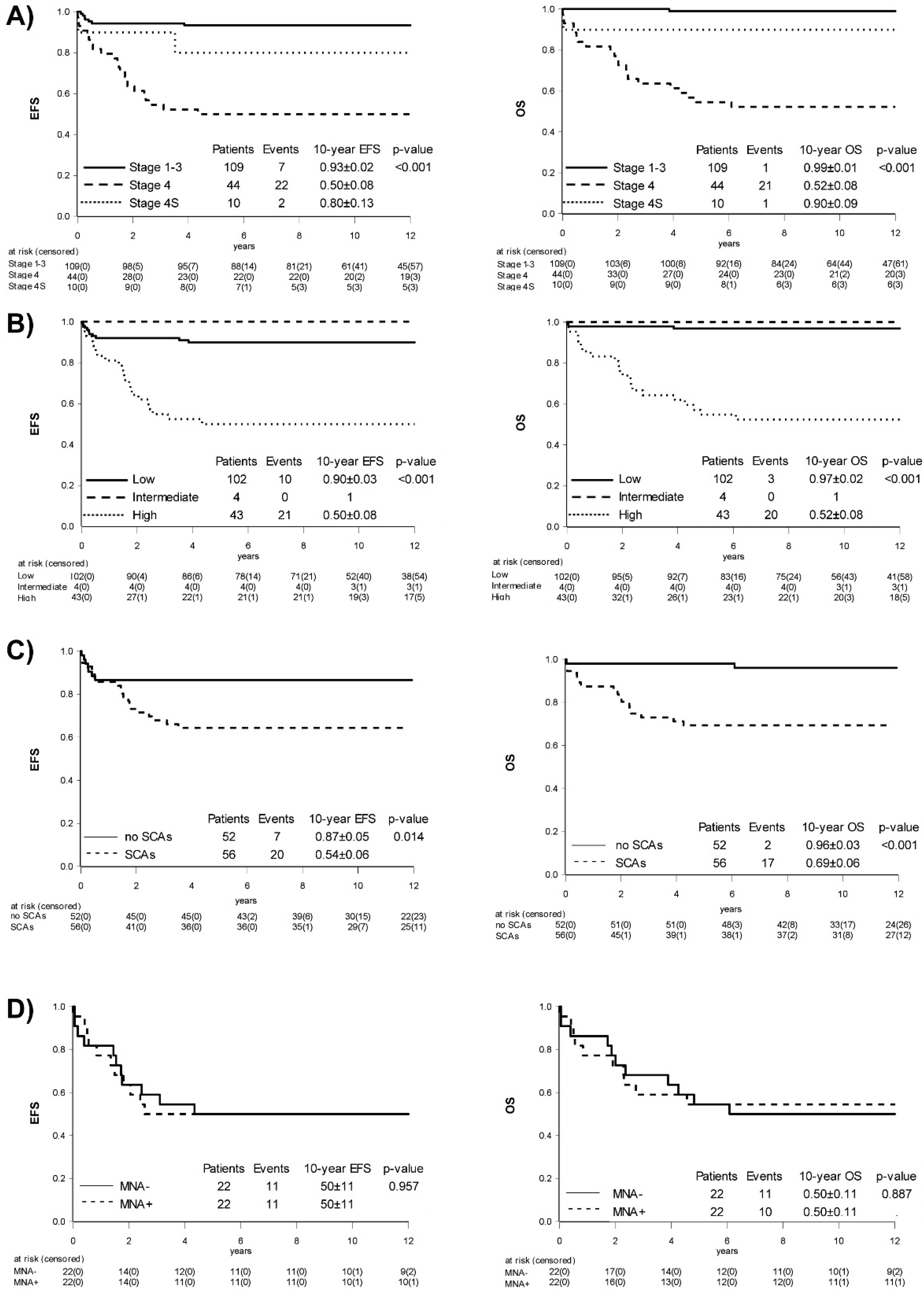
2.1. Trial Population and Overall Outcome

Between June 1994 and March 2006, a total of 163 patients were enrolled to the A-NB94 trial (Table 2) with most patients (n = 153) treated in five major Austrian centers. Histopathology revealed ganglioneuroblastoma (GNB) in 24/109 localized disease patients. Primary tumor locations were retroperitoneal-adrenal (n = 126; 77%), thoracic (n = 28; 17%), lumbar-pelvic (n = 5; 3%), and cervical (n = 4; 3%). The median age at diagnosis was 17 months (range: 4 days to 20 years); 63 patients were ≤12 months (39%), 22 patients between 12-18 months (13%) and 78 patients ≥ 18 months (48%). There were 86 males and 77 females. The 10-year (10y) EFS and OS were 80 ± 3% and 85 ± 3% for the whole trial population. The median observation time was twelve years.

2.2. Influence of Stage

Patients presenting with localized (stage 1–3) neuroblastoma (n = 109) had a 10y-EFS and OS of 93 ± 2% and 99 ± 1% (Figure 1A), respectively. Six relapses were reported and all affected non-MNA patients ≤ 12 months of age (n = 39). Four of these patients had loco-regional relapses: two were salvaged by six cycles cyclophosphamide/vincristine (CV), one by second surgery, and one was only observed as the parents declined further chemotherapy and the tumor ultimately regressed. Two infants had a relapse in infant age developing liver metastases, very much in line with a stage 4S pattern. They revealed no adverse genomic features at relapse, were closely observed, and ultimately showed spontaneous regression without further therapy. GNB was only found in localized non-MNA tumors and included the nodular (n = 4) or intermixed (n = 20) subtype. All patients with localized disease became long-term survivors, apart from one GNB patient with underlying neurofibromatosis type 1 dying later outside and unrelated to the A-NB94 trial, resulting in a 10y-EFS of 93 ± 3% for localized neuroblastoma versus 96 ± 4% for GNB (p = 0.584) with a 10y-OS of 100% versus 96 ± 4% (p = 0.062), respectively.
Infants with stage 4S neuroblastoma (n = 10) had a 10y-EFS and OS of 80 ± 13% and 90 ± 9% (Figure 1A), respectively. The tumors of four patients regressed without intervention while four patients underwent surgery of the primary tumor. Chemotherapy was given to two infants with clinical symptoms: one received electively four cycles of vincristine monotherapy and surgery whereas the other one, suffering from congenital neuroblastoma, showed uncontrollable disease progression, and died of respiratory failure despite chemotherapy escalation.
The 10y-EFS and OS of patients with metastasized neuroblastoma (n = 44) were 50 ± 8% and 52 ± 8% (Figure 1A), respectively. Six patients ≤ 12 months had non-MNA disease, a constellation associated with favorable outcome [6,7], but one patient died of progression during induction therapy. The subgroup at high risk for poor outcome including patients > 12 months with stage 4 (n = 33) and ≤ 12 months with MNA disease (n = 5) had a 10y-EFS and OS of only 45 ± 8% and 47 ± 8%.
In order to compare risk stratification of the A-NB94 trial with a more contemporary system, a retrospective assessment according to the International Neuroblastoma Risk Group (INRG) [2] was performed on this population. While patients with localized MNA tumors were upstaged to high-risk, localized, stage 4S, or stage 4 ≤ 12 months non-MNA patients remained low- or intermediate-risk, and stage 4 > 12 months and/or MNA patients remained in a high-risk group (Table A1). Outcome analysis resulted in 10y-EFS of 90 ± 3% for low- (n = 102), 100% for intermediate (n = 4), and 50 ± 8% for the high-risk group (n = 43) (p < 0.001) while 10y-OS was 97 ± 2%, 100%, and 52 ± 8% (p < 0.001) (Figure 1B), respectively.
Anti-GD2 monoclonal antibody ch14.18/SP0/2 became available in 1996 for compassionate use in 12 stage 4 patients. The landmark time identified the median time between HDT and initiation of immunotherapy as 87 days. When comparing the anti-GD2 monoclonal antibody pilot population (n = 12) to the pre-immunotherapy population accrued in the A-NB94 trial and considering only stage 4 patients without progressive disease at the landmark time point of 87 days after HDT, no difference in outcome was observed shown by a 10y-EFS of 67 ± 14% versus 64 ± 13% (p = 0.77) and a 10y-OS of 67 ± 14% versus 71 ± 12% (p = 0.907).

2.3. Role of Age

Using a cutoff at 12 months as part of RAST, 10y-OS was significantly better with 94 ± 3% for patients ≤ 12 months (n = 63) as compared to 80 ± 4% for patients > 12 months (n = 100) (p = 0.035); EFS was 83 ± 5% and 79 ± 4% (p = 0.717), respectively. An ad hoc cutoff at 18 months showed similar results with a 10y-OS of 93 ± 3% for patients ≤ 18 months (n = 85) versus 77 ± 5% for patients > 18 months (n = 78) (p = 0.009) and a 10y-EFS of 85 ± 4% versus 75 ± 5% (p = 0.201).

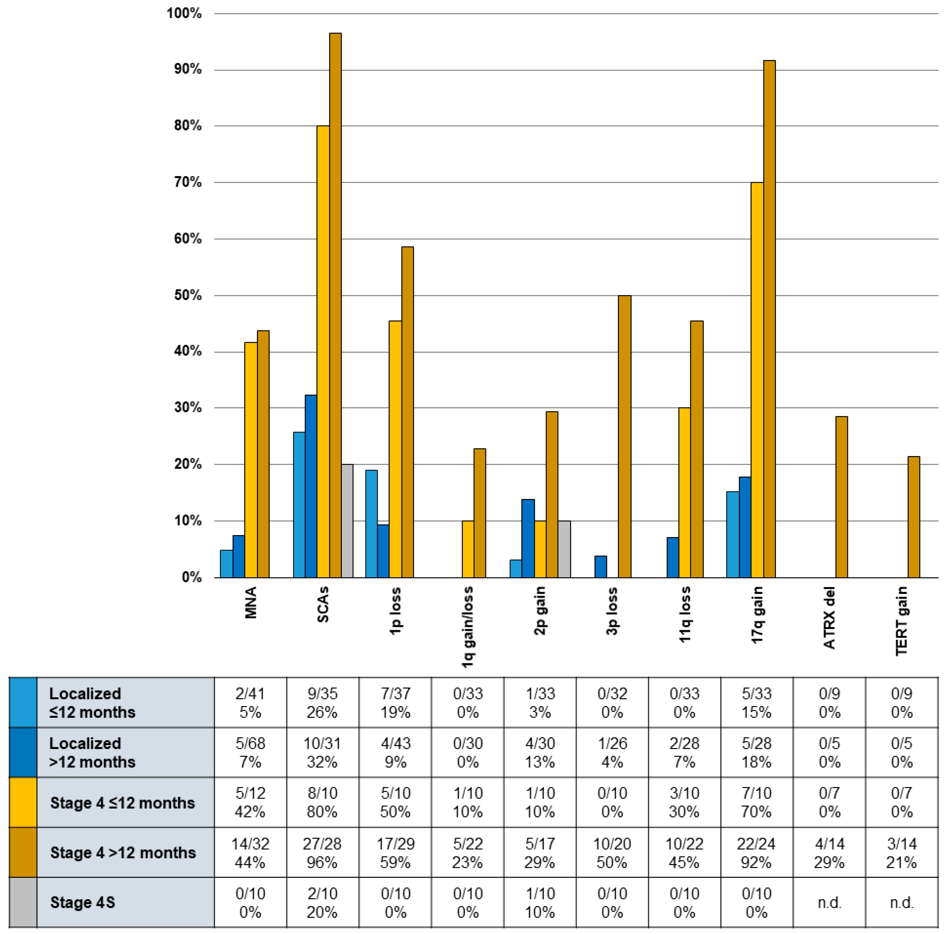
2.4. Occurrence and Influence of Biomarkers

MYCN status was evaluated for all patients prospectively during the risk stratification process. MNA was observed in 6% (7/109) of localized, in 43% (20/44) of stage 4, and in none of the stage 4S tumors (p < 0.001). We found four infants harboring hetMNA; one of them was only recognized in a later tumor sample and this patient was treated according to the original result as per MNA protocol. The other three patients were treated in the non-MNA arm.
MYCN retained predictive power in the total trial population showing a 10y-EFS of 84 ± 3% for non-MNA (n = 133), 60 ± 10% for MNA (n = 26), and 100% for hetMNA (n = 4) (p = 0.034), and a 10y-OS of 64 ± 10%, 89 ± 3%, and 100% (p = 0.008), respectively. However, MYCN amplification did not have any additional stratifying effect on outcome in patients with stage 4 neuroblastoma as 10y-EFS and OS was 50 ± 11% for both subgroups (Figure 1D).
1ploss was recorded prospectively during the active trial period and data were available for 122 patients. Quantity and quality of frozen tumor samples allowed for post hoc genomic analysis of 108 patients (multiplex ligation-dependent probe amplification (MLPA), n = 68; single nucleotide polymorphism (SNP) array, n = 32; interphase fluorescent in-situ hybridization (iFISH), n = 8) including SCAs for 1qgain/1qloss, 2pgain, 3ploss, 4ploss, 5ploss, 6qloss, 9ploss, 11qloss, 14qloss, 17ploss, 17qgain, 19qloss, and 22qloss. Only in two cases of GNB, genomic analysis was interpretable and revealed no SCAs (data included in mentioned numbers); in the other Schwann cell stroma-rich GNB tumors, the neoplastic clone was masked by the normal Schwann cells [17].
SCAs were observed in 92% of stage 4 but only in 29% of localized tumors (p < 0.001). Patients > 12 months showed tumor SCAs in 63% while SCAs were found in only 39% of patients ≤ 12 months (p < 0.001) (Figure 2).
This analysis found patients with tumors showing one or more SCAs (n = 56) with a 10y-EFS and OS of 64 ± 6% and 69 ± 6% while it was 87 ± 5% (p = 0.014) and 96 ± 3% (p < 0.001) for patients without SCAs (n = 52) (Figure 1C). In univariate analysis, presence of 1ploss, 1qgain/1qloss, 3ploss, 11qloss, or 17qgain had significant predictive power for worse 10y-EFS and/or OS (Table 3). To investigate the potential added impact of segmental chromosomal alterations (SCAs), we performed a multivariate analysis (MVA) corrected by the risk-stratifying factors of age (≤/>12 months), INSS stage and MYCN amplification status. Neither model A nor model B (Table 4) was able to identify an added risk for SCAs in the investigated trial population. ATRXdel (n = 3) and TERTgain (n = 4) were found only in stage 4 patients > 18 months of age and were mutually exclusive. All but one patient with TERTgain died with progressing disease.

2.5. Role of Treatment Elements to Achieve Remission Induction

Low- to intermediate-dose chemotherapy during first-line treatment was given to 18% (20/109) of patients with localized disease, 20% (2/10) of patients with stage 4S, and 100% (6/6) of patients ≤ 12 months with stage 4 non-MNA tumors. The overall response rate to cytotoxic treatment was 93% (26/28) in this cohort with 50% (14/28) entering a complete clinical remission (CR). One patient with stage 4 non-MNA and one with stage 4S disease did not respond, and both died of disease progression despite chemotherapy escalation.
Stage 4 patients > 12 months (n = 32) and ≤12 months with MNA tumors (n = 6) received dose-intensive induction therapy with a metastatic response rate of 74% (28/38) and a CR rate of 16% (6/38) including the effects of surgery. In this group, three patients progressed during induction. 2/38 patients (5%) experienced an infection-related septic shock during induction therapy and died with multi-organ failure.

2.5.1. Surgery

Surgical resection of the primary tumor was performed in 93% (152/163) of patients. In case of adrenal primary, 5% (6/113) underwent unilateral total nephrectomy while other patients only had unilateral adrenalectomy. Two patients needed revision surgery due to rebleeding.

2.5.2. High-Dose Therapy (HDT)

Patients eligible for HDT (Table 1) received one (n = 18), or, in case of mIBG metastatic incomplete response, two (n = 11) or three (n = 2) courses of HDT. In addition, two patients with stage 3 MNA but post-surgical macroscopic tumor residues, and one patient 11 months of age with stage 4 non-MNA but post-induction unresectable disease received a single course of HDT. Seven patients with single HDT were in metastatic CR (mCR) prior HDT; 93% (13/14) of other patients responded to HDT with a mCR rate of 50% (7/14). Within the multiple HDT group, response rate was 77% (10/13), CR rate 54% (7/13).
In the first attempt of stem cell apheresis in 27 evaluable patients, median yield was 4.5 × 10⁶ CD34 positive cells per kilogram body weight (range 0.45 to 30.9 × 10⁶). Six patients needed a second (median yield 3.76 × 10⁶, range 1.26 to 8.5 × 10⁶), and two patients a third apheresis (median 4.99 × 10⁶, range 4.97 to 5 × 10⁶) in order to reach the attempted minimum of 3 × 10⁶ cells per kilogram body weight for each of the planned HDT courses according to their respective response status.

2.6. Acute Toxicities and Long-Term Morbidity

Acute toxicities and intervention related morbidity was related to respective treatments (Table A2) and clearly showed a higher acute toxicity burden with increased treatment intensity.
Of surviving patients receiving low- and intermediate-dose chemotherapy (localized, stage 4S, or stage 4 patients ≤12 months with non-MNA disease) but no HDT (n = 24), only two patients had treatment-associated long-term disabilities manifesting as reduced renal glomerular filtration rate (GFR). Treatment-unrelated morbidity was persistent paraplegia of the lower limbs already present at diagnosis (n = 1) and subsequent craniopharyngioma (n = 1).
Of long-term survivors receiving intensive chemotherapy including HDT (n = 21), common long-term morbidity involved permanent hearing loss after cisplatin therapy in 43% (n = 9). Other disabilities in this group included reduced left-ventricular cardiac output (n = 4), hypothyroidism (n = 3), reduced GFR (n = 2), hypertension (n = 1), testosterone deficiency (n = 1), growth hormone deficiency (n = 1), dental damage (n = 1), peripheral polyneuropathy (n = 1), and osteochondroma (n = 1). One patient died from secondary acute myeloid leukemia.

3. Discussion

The A-NB94 trial was the first in Austria to apply a risk-adapted treatment strategy for children with neuroblastoma considering INSS stage, age, and MYCN amplification status [3]. Risk stratification was implemented in 1994 and results need to be viewed from this perspective. Only 15 years later, the INRG established staging based on pre-surgical image-defined factors [18], and included additional biologic makers of histopathology, ploidy, and 11q status [2]. We retrospectively stratified A-NB94 patients according to the INRG classification system which resulted in an upstaging of patients with localized MNA tumors to high-risk while other patients remained within a risk group similar to the A-NB94 staging system based on the INSS. Additionally, outcome by INRG classification matched published results, considering the high survival of stage 3 patients and small numbers within the intermediate-risk group. Patients being not stageable by INRG classification related to missing 11qloss data.
In retrospect, the A-NB94 study included a high proportion of patients with localized neuroblastoma, possibly related to the then ongoing neuroblastoma screening with 13% of the trial population being part of this program. In addition, the high-risk arm of A-NB94 closed for accrual earlier to allow for participation in the SIOPEN High-Risk trial (HR-NBL1/SIOPEN; ClinicalTrials.gov number NCT01704716) which opened already in 2002 while the SIOPEN Low- and Intermediate-Risk trial (LINES; ClinicalTrials.gov number NCT01728155) only opened in 2007.
Compared with published data, the Austrian screening program detected a comparatively high number of localized tumors harboring unfavorable biologic features (MNA, 1ploss, diploidy) [19], suggesting early adverse clonal evolution. However, the A-NB94 trial was not designed for answering the question if screening helped increasing the detection of high-risk disease at an early stage [20,21]. Overall, our approach resulted in excellent survival for these patients as all became long-term survivors apart from one GNB patient dying from complications after surgery not related to neuroblastoma treatment.
Notably, the very favorable outcome of the localized subgroup also includes 25 stage 3 patients, marking a major improvement compared to the preceding A-NB87 trial which applied an intensive chemotherapy for children with Evan’s Stage 3 disease (especially when >2 years of age, increased neuron-specific enolase, and/or ferritin (Table A3), resulting in a 28% (8/29) toxic death rate and a 5y-OS of only 50% [22]. However, detailed comparison is hampered by differing staging criteria and MYCN not being a stratifying marker in A-NB87. In A-NB94, 82% (89/109) of patients with localized neuroblastoma received little or no chemotherapy in first line treatment. These numbers include six patients with non-MNA stage 3 tumors defined by tumors crossing the midline which could otherwise be surgically removed upfront. While two relapses were treated successfully with chemotherapy, potential long-term side effects following intense therapy could be circumvented in other patients by using a surgery only approach.
While our data support the notion of improved outcome despite therapy reduction in patients with intermediate-risk neuroblastoma [23,24], contemporary trials would submit patients with stage 2 or 3 MNA [25] or diploid [26] tumors to receive HDT/ASCR. Furthermore, the presence of certain SCAs was reported to identify patients of higher risk for relapse, especially in unresectable tumors [27]. While SCAs, especially 1ploss and 11qloss, may lower EFS but not OS in children ≤18 months, they also diminish OS in older children [28]. In A-NB94, 15 patients with localized tumors matched these criteria (MNA, n = 7; diploidy, n = 5; >18 months and 1ploss, n = 1; or 11qloss, n = 2) and all survived without relapse after receiving low-/intermediate-dose chemotherapy apart from two already described patients that received HDT/ASCR. The decision for adding HDT in the latter two patients was based on the presence of post-surgical residual tumors. These small numbers might suggest that patients with residual tumors benefited from therapy escalation while others were adequately treated by conventional chemotherapy and irradiation. In this context, it may be assumed that the remarkable good outcome in A-NB94 of patients with localized tumors including especially stage 3 patients relates to the surprisingly low prevalence of SCAs observed in this subgroup.
Outcome of infantile stage 4 metastatic disease is related to MNA, diploidy/tetraploidy, 1ploss, 11qloss, or 17qgain [29]. While most stage 4S tumors are treated adequately by observation only [30,31], certain markers including 11qloss and diploidy might predict less favorable outcomes even in stage 4S disease [32]. However, disease progression of two A-NB94 stage 4S patients clearly could not be explained by these biologic factors, as neither MNA, diploidy or 11qloss were detected. Stage 4S poses threats independent of tumor biology as extensive hepatomegaly causes a variety of clinical complications.
Of 44 stage 4 patients, six infants without MNA received moderate chemotherapy without HDT or radiation according to RAST. While survival was good, one of three patients with tumors showing SCAs involving 11qloss and 17qgain progressed and died during induction, in line with reports that these features are often associated with higher risk for relapse [33].
Stage 4 patients > 12 months and stage 4 infants with MNA [34,35] underwent an induction protocol similar to the “N6” protocol previously published to achieve fast cytoreduction [36], following the notion that increased response rates precede higher survival rates [37]. Furthermore, A-NB94 successfully introduced tandem or triple HDT/ASCR to children with incomplete metastatic response to induction therapy. While benefits of multiple cycle HDT have been reported consistently by European groups [16,38], recent reports demonstrated clearly improved survival in contemporary trials [39,40].
In 1996, anti-GD2 monoclonal antibody ch14.18/SP0/2 became available for compassionate use in stage 4 patients. The identified landmark time identified between HDT and start of immunotherapy was 87 days; thus, only stage 4 patients without progressive disease were included in the pre-immunotherapy control population. Comparing these two cohorts did not result in an advantage for the small immunotherapy population. However, overall outcome data are quite in line with later publications on the use of ch14.18 monoclonal antibody in first-line maintenance treatment [41,42]. Considering the rather small population in both cohorts, we only may hypothesize that RAST, using repetitive HDT adapted to response in this high-risk population, was probably a major contributor to the observed favorable outcomes and that immunotherapy applied with a less dose intensive monotherapy schedule did not result in a measurable added benefit.
In addition, while the prognostic impact of hetMNA remains unclear [43,44], recent data suggests it has to be viewed in light of the overall genetic background in order to determine the importance of the MNA cell clone [45]. Localized tumors usually show a favorable background with reported improved outcomes compared to homogeneous MNA. In A-NB94, hetMNA tumors showed only additional 1ploss in two cases but no other SCA or other unfavorable biologic markers. One of these cases was only discovered in a later tumor sample as the original sample suggested homogeneous MNA; this patient with stage 4 disease received high-dose therapy, which may, together with the age of ≤12 months and lack of additional high-risk features, have resulted in over-treatment.
SCAs were present in higher frequency in stage 4 disease, especially in patients > 12 months, when compared to children with localized tumors of any age (Figure 2), supporting the notion of SCAs being accumulated during tumor development [44] and an unfavorable biology being associated with overall poor prognosis [45]. This study confirms SCAs are commonly involving chromosome arms 1p, 11q, and 17q [46], however, high-risk metastasized tumors often show a combination of various chromosomal defects [47].
The acute toxicity burden as well as long-term morbidity is clearly related to RAST, showing the therapy burden of the curative efforts mainly in the high-intensity treatment groups. Particularly after additional HDT/ASCR, which is in line with previous reports [48,49], although and in contrast to previous approaches [50], no total body irradiation (TBI) was used. Observed surgery-related morbidities were in line with previous reports [51]. Irreversible inner ear hearing loss, a known side-effect following cisplatin therapy [52], affected 29% (10/34) of long-term survivors receiving cisplatin. The understanding of the mechanism of uptake and accumulation in the stria vascularis of the cochlea provides now an important target for preventing ototoxicity in future trials [53]. Overall, organ-related long-term side-affects were low as well as incidence of secondary malignancies.

4. Patients and Methods

4.1. Patients

Children with histologically confirmed, previously untreated neuroblastoma or GNB up to 20 years of age were eligible for enrolment on A-NB94. Staging followed INSS guidelines and treatment response was assessed by the 1993 International Neuroblastoma Response Criteria (INRC) [3]. Written informed consent for treatment and data procession was obtained in compliance with institutional review board rules and in accordance with the Declaration of Helsinki [54].

4.2. Treatment Concepts

RAST was adjusted for age (≤/>12 months), stage, and MYCN status (Table 1). Stage 1 or 2 non-MNA tumors were planned for surgery only; incompletely resected MNA tumors received chemotherapy adapted to stage and radiotherapy. Stage 4S infants were observed up to 3 months and then underwent surgery but received chemotherapy in case of life-threatening symptoms or progression. Stage 3 and 4 were treated with neoadjuvant intensity-adjusted chemotherapy with surgery planned after four cycles. MNA tumors were irradiated age-adapted with 24 Gray (≤12 months) or 30 Gray (>12 months).
Stem cell harvest, aiming at >3 × 10⁶ CD34 positive cells per kilogram body weight per high-dose treatment (HDT), was first attempted after 4 chemotherapy cycles if cytomorphological and histological bone marrow (BM) remission was achieved, or was otherwise postponed following cycle 6 or after an in vivo purge following the first or second HDT, eventually.
Depending on metastatic response, patients were eligible for a single (complete response, CR) or repetitive HDT (partial/minor response, PR/MR). In case of CR after second HDT, the third HDT was omitted. In 1996, the anti-GD2 monoclonal antibody ch14.18/SP0/2 became accessible as compassionate use (provided by the University of Tübingen, Prof. Dr. Rupert Handgretinger) to optimize treatment of HR disease after HDT and radiotherapy. Treatment consisted of three anti-GD2 ch14.18/SP0/2 cycles (20 mg/m2 over 5 days as 8-h infusions). Supportive care and treatment of infections followed institutional guidelines.

4.3. Disease and Response Assessment

Disease evaluation was planned at diagnosis, after 2, 4, and 6 courses of chemotherapy, before and after each HDT, and before and after immunotherapy. Bone marrow examination included aspirates/trephines obtained from two sites and used, apart from cytomorphology, an automated image analysis system (MetaSystems GmbH, Altlußheim, Germany) to quantify GD2/CD56-positive cells [55,56]. Skeletal disease was defined by pathologic iodine-123 meta-iodobenzylguanidine (mIBG) or, in mIBG non-avid tumors, by tecnetium-99m-hydroxymethylenediphosphanate (Tc99m) scans [57]. Primary tumors were investigated by MRI/CT scan. Tumor marker evaluation included urinary catecholamine metabolites, lactate dehydrogenase (LDH), and neuron specific enolase (NSE). Response was assessed based on local institution reporting following INRC guidelines [3].

4.4. Toxicity Evaluation

Acute and long-term toxicities were evaluated using case report forms (CRF) documenting organ-specific toxicities according to the Common Terminology Criteria for Adverse Events (CTCAE) Version 5.0 above grade 2, secondary malignancies, and other adverse events (AE). Acute toxicity evaluation focused on patients with intense induction chemotherapy and HDT.

4.5. Biologic Studies

Frozen tumor specimens were evaluated using high-density SNP arrays (CytoScan HD Array) [47], MLPA, or iFISH for the detection of MNA or SCAs. Recording/interpretation of data were done according to international standards [58].

4.6. Statistical Analysis

Event-free survival (EFS) was calculated from the time of diagnosis until first occurrence of relapse, progressive disease, secondary malignancy, or death from any cause, or until last contact with patients. Overall survival (OS) was calculated from time of diagnosis to death from any cause. The median time between HDT and initiation of immunotherapy was 87 days, therefore only patients without progressive disease at this landmark time point were included in the pre-immunotherapy cohort. Categorical data were analyzed by chi-square test or, in case of an estimated case-count of ≤5 per field, Fisher’s exact test. Data were analyzed with SAS 9.4 program. All statistical tests were two-sided.

5. Conclusions

The risk-adapted approach resulted in an excellent long-term survival for the majority of patients with acceptable long-term morbidity. An age- and stage-dependent frequency of SCAs was confirmed and should be considered in future treatment decision-making processes.

Author Contributions

Conceptualization: S.F., R.L., I.M.A., P.F.A.; Study design: R.L.; Data curation: S.F., E.G., local coordinators; Quality control of data and algorithms: S.F., R.L., E.G., I.M.A., P.F.A.; Data analysis and interpretation: S.F., R.L., E.G.; Formal analysis: E.G.; Biologic data curation and interpretation: I.M.A., P.F.A., A.Z.; Writing—original draft preparation: S.F., R.L.; Writing—review and editing: S.F., R.L.; Local coordinators: M.B., C.U., M.M., G.E.-D., E.B., N.J., A.G., B.M., R.C., G.A., K.D., E.H., R.K. Investigation: B.B.-H. All authors have read and agreed to the published version of the manuscript.

Funding

St. Anna Kinderkrebsforschung, Vienna, Austria; Austrian National Bank (OeNB), grant no. 16611, Vienna, Austria; Austrian Science Fund (FWF), grant no. I 2799-B28, Vienna, Austria.

Institutional Review Board Statement

The study was conducted according to the guidelines of the Declaration of Helsinki, and approved by the institutional review board prior trial initiation as well as by the Lead Ethics Committee for Austria at the Medical University, Vienna. (EK Nr: 2450/2020, 26 January 2021: Relevance of modern biomarkers in patients after risk-adapted treatment strategy in the Austrian Neuroblastoma Trial A-NB94–a retrospective data analyses).

Informed Consent Statement

Informed consent was obtained from all subjects involved in the study.

Data Availability Statement

Biomarker data as presented in this cohort study are available in anonymized form on request at the CCRI by addressing the corresponding author.

Conflicts of Interest

The following authors declare conflict of interest: Georg Ebetsberger-Dachs–Consulting or advisory role: Novartis International AG, Amgen Inc.; Travel, accommodations, expenses: Shire PLC., Servier Laboratories. The other authors declare no conflict of interest.

Appendix A

Table A1. Retrospective correlation of A-NB94 risk groups with INRG stratification.
A-NB94 Staging CriteriaRetrospective INRG Risk Classification
MYCNStageAgeLowIntermediateHighn.e.Total
amplified1≤12 1 1
>12 1 1
2≤12
>12
3≤12 1 1
>12 4 4
4≤12 5 5
>12 14 14
non-amplified1≤1215 15
>1246 46
2≤129 9
>1210 10
3≤128 412
>1233 17
4≤1241 16
>12 17118
4S≤124 610
heterogeneously amplified1≤121 1
>12
2≤121 1
>12
3≤121 1
>12
4≤12 11
>12
Total 10244314163
Retrospective correlation of A-NB94 risk stratification with the International Neuroblastoma Risk Group (INRG) classification [2].
Table A2. Summary of acute treatment-related toxicities.
Type of ToxicityInduction Phase SurgeryHDT Phase
LD/IDHD
infection410413
gastrointestinal 3 8
hepatic injury 3
venous occlusive disease 1
cardiac23 4
renal failure 2
hemorrhagic cystitis 1
respiratory insufficiency 1
oral-intestinal mucositis 5 12
tumor lysis syndrome 1
capillary leak syndrome 1 1
hemolytic uremic syndrome 1
autoimmune hemolytic anemia 1
non-febrile seizures 1
intraoperative tumor rupture 2
severe bleeding 4
lymphatic fistula 4
Horner’s syndrome 3
pleural effusion 2
pneumothorax 1
cava vein thrombosis 1
Total6262146
Summary of acute treatment-related toxicities according to induction treatment split by chemotherapy intensity with low- and intermediate-dose (LD/ID) and high-dose (HD) induction schedules, the latter including stage 4 > 12 months and stage 4 ≤ 12 months but MYCN amplified tumors, the former all other treatment groups; surgical complications; and high-dose therapy (HDT).
Table A3. Chemotherapy elements and treatment regimens used in the A-NB87 trial.
StageTherapy
ISurgery
IIASurgeryIf macroscopic residual tumor: second surgery, or radiotherapy
IIBSurgeryCVRe-surgeryRadiotherapy
IIIASurgery3–4 × alternating DAMO/MVDOCRe-surgeryRadiotherapy(Re-surgery)
IIIBSurgery3–5 × alternating DAMO/MVDOC/IPERe-surgeryRadiotherapyRe-surgery
IVSurgery4 × alternating MVDOC/IPERe-surgeryHDT/TBI/ASCRRadiotherapy
Details on Chemotherapy
ChemotherapyAbbreviationSubstanceDosageDays Given
DAMODdacarbazine850 mg/m²1
Adoxorubicin30 mg/m²1, 2
Mmustargen6 mg/m²1
Ovincristine1.5 mg/m²1 + 5
MVDOCMmustargen6 mg/m²1
Vteniposide150 mg/m²1
Ddacarbacine850 mg/m²1
Ovincristine1.5 mg/m²1
Ccyclophosphamide850 mg/m²1
IPEIifosfamide3 g/m²1, 2
Pcisplatin40 mg/m²1–5
Eetoposide150 mg/m²3–5
Chemotherapy elements and treatment regimens according to the Austrian Neuroblastoma Trial A-NB87. Staging was done according to Evans criteria: stage I (localized without macroscopic lymphatic node involvement), stage IIA (macroscopic lymphatic node involvement), IIB (macroscopic residual tumor with ipsilateral lymphatic node involvement), IIIA (<2 years of age, ferritin < 300 µg/mL, neuron-specific enolase < 100 ng/mL), IIIB (any positivity of the markers described for stage IIIA), stage 4 (distant metastasized disease).

References

  1. Brodeur, G.M. Neuroblastoma: Biological insights into a clinical enigma. Nat. Rev. Cancer 2003, 3, 203–216. [Google Scholar] [CrossRef] [PubMed]
  2. Cohn, S.L.; Pearson, A.D.J.; London, W.B.; Monclair, T.; Ambros, P.F.; Brodeur, G.M.; Faldum, A.; Hero, B.; Iehara, T.; Machin, D.; et al. The International Neuroblastoma Risk Group (INRG) classification system: An INRG Task Force report. J. Clin. Oncol. 2009, 27, 289–297. [Google Scholar] [CrossRef] [PubMed]
  3. Brodeur, G.M.; Pritchard, J.; Berthold, F.; Carlsen, N.L.; Castel, V.; Castelberry, R.P.; De Bernardi, B.; Evans, A.E.; Favrot, M.; Hedborg, F. Revisions of the international criteria for neuroblastoma diagnosis, staging, and response to treatment. J. Clin. Oncol. 1993, 11, 1466–1477. [Google Scholar] [CrossRef] [PubMed]
  4. Seeger, R.C.; Brodeur, G.M.; Sather, H.; Dalton, A.; Siegel, S.E.; Wong, K.Y.; Hammond, D. Association of multiple copies of the N-myc oncogene with rapid progression of neuroblastomas. N. Engl. J. Med. 1985, 313, 1111–1116. [Google Scholar] [CrossRef]
  5. Cohn, S.L.; Rademaker, A.W.; Salwen, H.R.; Franklin, W.A.; Gonzales-Crussi, F.; Rosen, S.T.; Bauer, K.D. Analysis of DNA ploidy and proliferative activity in relation to histology and N-myc amplification in neuroblastoma. Am. J. Pathol. 1990, 136, 1043–1052. [Google Scholar] [CrossRef] [Green Version]
  6. Jereb, B.; Bretsky, S.S.; Vogel, R.; Helson, L. Age and prognosis in neuroblastoma. Review of 112 patients younger than 2 years. Am. J. Pediatr. Hematol. Oncol. 1984, 6, 233–243. [Google Scholar] [CrossRef]
  7. Paul, S.R.; Tarbell, N.J.; Korf, B.; Kretschmar, C.S.; Lavally, B.; Grier, H.E. Stage IV neuroblastoma in infants. Long-term survival. Cancer 1991, 67, 1493–1497. [Google Scholar] [CrossRef]
  8. Nitschke, R.; Smith, E.I.; Shochat, S.; Altshuler, G.; Travers, H.; Shuster, J.J.; Hayes, F.A.; Patterson, R.; McWilliams, N. Localized neuroblastoma treated by surgery: A Pediatric Oncology Group Study. J. Clin. Oncol. 1988, 6, 1271–1279. [Google Scholar] [CrossRef]
  9. Matthay, K.K.; Sather, H.N.; Seeger, R.C.; Haase, G.M.; Hammond, G.D. Excellent outcome of stage II neuroblastoma is independent of residual disease and radiation therapy. J. Clin. Oncol. 1989, 7, 236–244. [Google Scholar] [CrossRef]
  10. Garaventa, A.; De Bernardi, B.; Pianca, C.; Donfrancesco, A.; Cordero di Montezemolo, L.; Di Tullio, M.T.; Bagnulo, S.; Mancini, A.; Carli, M.; Pession, A.; et al. Localized but unresectable neuroblastoma: Treatment and outcome of 145 cases. Italian Cooperative Group for Neuroblastoma. J. Clin. Oncol. 1993, 11, 1770–1779. [Google Scholar] [CrossRef]
  11. Perez, C.A.; Matthay, K.K.; Atkinson, J.B.; Seeger, R.C.; Shimada, H.; Haase, G.M.; Stram, D.O.; Gerbing, R.B.; Lukens, J.N. Biologic variables in the outcome of stages I and II neuroblastoma treated with surgery as primary therapy: A children’s cancer group study. J. Clin. Oncol. 2000, 18, 18–26. [Google Scholar] [CrossRef] [PubMed]
  12. Ambros, P.F.; Ambros, I.M.; Strehl, S.; Bauer, S.; Luegmayr, A.; Kovar, H.; Ladenstein, R.; Fink, F.M.; Horcher, E.; Printz, G. Regression and progression in neuroblastoma. Does genetics predict tumour behaviour? Eur. J. Cancer 1995, 31A, 510–515. [Google Scholar] [CrossRef]
  13. Ambros, P.F.; Ambros, I.M.; Kerbl, R.; Luegmayr, A.; Rumpler, S.; Ladenstein, R.; Amann, G.; Kovar, H.; Horcher, E.; De Bernardi, B.; et al. Intratumoural heterogeneity of 1p deletions and MYCN amplification in neuroblastomas. Med. Pediatr. Oncol. 2001, 36, 1–4. [Google Scholar] [CrossRef]
  14. Berbegall, A.; Villamón, E.; Piqueras, M.; Tadeo, I.; Djos, A.; Ambros, P.; Martinsson, T.; Ambros, I.; Cañete, A.; Castel, V.; et al. Comparative genetic study of intratumoral heterogenous MYCN amplified neuroblastoma versus aggressive genetic profile neuroblastic tumors. Oncogene 2015. [Google Scholar] [CrossRef] [PubMed]
  15. Berbegall, A.P.; Bogen, D.; Pötschger, U.; Beiske, K.; Bown, N.; Combaret, V.; Defferrari, R.; Jeison, M.; Mazzocco, K.; Varesio, L.; et al. Heterogeneous MYCN amplification in neuroblastoma: A SIOP Europe Neuroblastoma Study. Br. J. Cancer 2018, 118, 1502–1512. [Google Scholar] [CrossRef] [PubMed]
  16. Philip, T.; Ladenstein, R.; Zucker, J.M.; Pinkerton, R.; Bouffet, E.; Louis, D.; Siegert, W.; Bernard, J.L.; Frappaz, D.; Coze, C.; et al. Double megatherapy and autologous bone marrow transplantation for advanced neuroblastoma: The LMCE2 study. Br. J. Cancer 1993, 67, 119–127. [Google Scholar] [CrossRef] [Green Version]
  17. Ambros, I.M.; Zellner, A.; Roald, B.; Amann, G.; Ladenstein, R.; Printz, D.; Gadner, H.; Ambros, P.F. Role of ploidy, chromosome 1p, and Schwann cells in the maturation of neuroblastoma. N. Engl. J. Med. 1996, 334, 1505–1511. [Google Scholar] [CrossRef]
  18. Monclair, T.; Brodeur, G.M.; Ambros, P.F.; Brisse, H.J.; Cecchetto, G.; Holmes, K.; Kaneko, M.; London, W.B.; Matthay, K.K.; Nuchtern, J.G.; et al. The International Neuroblastoma Risk Group (INRG) staging system: An INRG Task Force report. J. Clin. Oncol. 2009, 27, 298–303. [Google Scholar] [CrossRef] [Green Version]
  19. Kerbl, R.; Urban, C.E.; Ambros, I.M.; Dornbusch, H.J.; Schwinger, W.; Lackner, H.; Ladenstein, R.; Strenger, V.; Gadner, H.; Ambros, P.F. Neuroblastoma mass screening in late infancy: Insights into the biology of neuroblastic tumors. J. Clin. Oncol. 2003, 21, 4228–4234. [Google Scholar] [CrossRef]
  20. Schilling, F.H.; Spix, C.; Berthold, F.; Erttmann, R.; Fehse, N.; Hero, B.; Klein, G.; Sander, J.; Schwarz, K.; Treuner, J.; et al. Neuroblastoma screening at one year of age. N. Engl. J. Med. 2002, 346, 1047–1053. [Google Scholar] [CrossRef]
  21. Fritsch, P.; Kerbl, R.; Lackner, H.; Urban, C. “Wait and see” strategy in localized neuroblastoma in infants: An option not only for cases detected by mass screening. Pediatr. Blood Cancer 2004, 43, 679–682. [Google Scholar] [CrossRef]
  22. Ladenstein, R.; Ambros, P.F.; Urban, C.; Ambros, I.M.; Fink, F.M.; Zoubek, A.; Grienberger, H.; Schmitt, K.; Kerbl, R.; Horcher, E.; et al. Value of prognostic factors in the Austrian A-NB87 Neuroblastoma Study. Klin. Pädiatrie 1996, 208, 210–220. [Google Scholar] [CrossRef]
  23. Baker, D.L.; Schmidt, M.L.; Cohn, S.L.; Maris, J.M.; London, W.B.; Buxton, A.; Stram, D.; Castleberry, R.P.; Shimada, H.; Sandler, A.; et al. Outcome after Reduced Chemotherapy for Intermediate-Risk Neuroblastoma. N. Engl. J. Med. 2010, 363, 1313–1323. [Google Scholar] [CrossRef] [Green Version]
  24. Bagatell, R.; Beck-Popovic, M.; London, W.B.; Zhang, Y.; Pearson, A.D.J.J.; Matthay, K.K.; Monclair, T.; Ambros, P.F.; Cohn, S.L.; International Neuroblastoma Risk Group. Significance of MYCN Amplification in International Neuroblastoma Staging System Stage 1 and 2 Neuroblastoma: A Report From the International Neuroblastoma Risk Group Database. J. Clin. Oncol. 2009, 27, 365–370. [Google Scholar] [CrossRef] [Green Version]
  25. Laprie, A.; Michon, J.; Hartmann, O.; Munzer, C.; Leclair, M.-D.D.; Coze, C.; Valteau-Couanet, D.; Plantaz, D.; Carrie, C.; Habrand, J.-L.L.; et al. High-dose chemotherapy followed by locoregional irradiation improves the outcome of patients with international neuroblastoma staging system Stage II and III neuroblastoma with MYCN amplification. Cancer 2004, 101, 1081–1089. [Google Scholar] [CrossRef]
  26. Bagatell, R.; Rumcheva, P.; London, W.B.; Cohn, S.L.; Look, A.T.; Brodeur, G.M.; Frantz, C.; Joshi, V.; Thorner, P.; Rao, P.V.; et al. Outcomes of Children with Intermediate-Risk Neuroblastoma After Treatment Stratified by MYCN Status and Tumor Cell Ploidy. J. Clin. Oncol. 2005, 23, 8819–8827. [Google Scholar] [CrossRef]
  27. Defferrari, R.; Mazzocco, K.; Ambros, I.M.; Ambros, P.F.; Bedwell, C.; Beiske, K.; Bénard, J.; Berbegall, A.P.; Bown, N.; Combaret, V.; et al. Influence of segmental chromosome abnormalities on survival in children over the age of 12 months with unresectable localised peripheral neuroblastic tumours without MYCN amplification. Br. J. Cancer 2015, 112, 290–295. [Google Scholar] [CrossRef] [Green Version]
  28. Ambros, I.M.; Tonini, G.-P.; Pötschger, U.; Gross, N.; Mosseri, V.; Beiske, K.; Berbegall, A.P.; Bénard, J.; Bown, N.; Caron, H.; et al. Age Dependency of the Prognostic Impact of Tumor Genomics in Localized Resectable MYCN -Nonamplified Neuroblastomas. Report from the SIOPEN Biology Group on the LNESG Trials and a COG Validation Group. J. Clin. Oncol. 2020, 38, 3685–3697. [Google Scholar] [CrossRef]
  29. Lavarino, C.; Cheung, N.-K.V.; Garcia, I.; Domenech, G.; de Torres, C.; Alaminos, M.; Rios, J.; Gerald, W.L.; Kushner, B.; LaQuaglia, M.; et al. Specific gene expression profiles and chromosomal abnormalities are associated with infant disseminated neuroblastoma. BMC Cancer 2009, 9, 44. [Google Scholar] [CrossRef] [Green Version]
  30. Iehara, T.; Hiyama, E.; Tajiri, T.; Yoneda, A.; Hamazaki, M.; Fukuzawa, M.; Hosoi, H.; Sugimoto, T.; Sawada, T. Is the prognosis of stage 4s neuroblastoma in patients 12 months of age and older really excellent? Eur. J. Cancer 2012, 48, 1707–1712. [Google Scholar] [CrossRef]
  31. Kushner, B.H.; Cheung, N.K.; LaQuaglia, M.P.; Ambros, P.F.; Ambros, I.M.; Bonilla, M.A.; Gerald, W.L.; Ladanyi, M.; Gilbert, F.; Rosenfield, N.S.; et al. Survival from locally invasive or widespread neuroblastoma without cytotoxic therapy. J. Clin. Oncol. 1996, 14, 373–381. [Google Scholar] [CrossRef]
  32. Taggart, D.R.; London, W.B.; Schmidt, M.L.; DuBois, S.G.; Monclair, T.F.; Nakagawara, A.; De Bernardi, B.; Ambros, P.F.; Pearson, A.D.J.; Cohn, S.L.; et al. Prognostic Value of the Stage 4S Metastatic Pattern and Tumor Biology in Patients with Metastatic Neuroblastoma Diagnosed Between Birth and 18 Months of Age. J. Clin. Oncol. 2011, 29, 4358–4364. [Google Scholar] [CrossRef]
  33. Schleiermacher, G.; Michon, J.; Ribeiro, A.; Pierron, G.; Mosseri, V.; Rubie, H.; Munzer, C.; Bénard, J.; Auger, N.; Combaret, V.; et al. Segmental chromosomal alterations lead to a higher risk of relapse in infants with MYCN-non-amplified localised unresectable/disseminated neuroblastoma (a SIOPEN collaborative study). Br. J. Cancer 2011, 105, 1940–1948. [Google Scholar] [CrossRef]
  34. Canete, A.; Gerrard, M.; Rubie, H.; Castel, V.; Di Cataldo, A.; Munzer, C.; Ladenstein, R.; Brichard, B.; Bermúdez, J.D.; Couturier, J.; et al. Poor survival for infants with MYCN-amplified metastatic neuroblastoma despite intensified treatment: The International Society of Paediatric Oncology European Neuroblastoma Experience. J. Clin. Oncol. 2009, 27, 1014–1019. [Google Scholar] [CrossRef]
  35. Katzenstein, H.M.; Bowman, L.C.; Brodeur, G.M.; Thorner, P.S.; Joshi, V.V.; Smith, E.I.; Look, A.T.; Rowe, S.T.; Nash, M.B.; Holbrook, T.; et al. Prognostic significance of age, MYCN oncogene amplification, tumor cell ploidy, and histology in 110 infants with stage D(S) neuroblastoma: The pediatric oncology group experience—A pediatric oncology group study. J. Clin. Oncol. 1998, 16, 2007–2017. [Google Scholar] [CrossRef]
  36. Kushner, B.H.; LaQuaglia, M.P.; Bonilla, M.A.; Lindsley, K.; Rosenfield, N.; Yeh, S.; Eddy, J.; Gerald, W.L.; Heller, G.; Cheung, N.K. Highly effective induction therapy for stage 4 neuroblastoma in children over 1 year of age. J. Clin. Oncol. 1994, 12, 2607–2613. [Google Scholar] [CrossRef]
  37. Cheung, N.V.; Heller, G. Chemotherapy dose intensity correlates strongly with response, median survival, and median progression-free survival in metastatic neuroblastoma. J. Clin. Oncol. 1991, 9, 1050–1058. [Google Scholar] [CrossRef]
  38. Philip, T.; Ladenstein, R.; Lasset, C.; Hartmann, O.; Zucker, J.M.; Pinkerton, R.; Pearson, A.D.; Klingebiel, T.; Garaventa, A.; European Group for Blood and Marrow Transplant Registry Solid Tumour Working Party; et al. 1070 myeloablative megatherapy procedures followed by stem cell rescue for neuroblastoma: 17 years of European experience and conclusions. Eur. J. Cancer 1997, 33, 2130–2135. [Google Scholar] [CrossRef]
  39. George, R.E.; Li, S.; Medeiros-Nancarrow, C.; Neuberg, D.; Marcus, K.; Shamberger, R.C.; Pulsipher, M.; Grupp, S.A.; Diller, L. High-risk neuroblastoma treated with tandem autologous peripheral-blood stem cell-supported transplantation: Long-term survival update. J. Clin. Oncol. 2006, 24, 2891–2896. [Google Scholar] [CrossRef]
  40. Granger, M.; Grupp, S.A.; Kletzel, M.; Kretschmar, C.; Naranjo, A.; London, W.B.; Diller, L. Feasibility of a tandem autologous peripheral blood stem cell transplant regimen for high risk neuroblastoma in a cooperative group setting: A Pediatric Oncology Group study: A report from the Children’s Oncology Group. Pediatr. Blood Cancer 2012, 59, 902–907. [Google Scholar] [CrossRef] [Green Version]
  41. Yu, A.L.; Gilman, A.L.; Ozkaynak, M.F.; London, W.B.; Kreissman, S.G.; Chen, H.X.; Smith, M.; Anderson, B.; Villablanca, J.G.; Matthay, K.K.; et al. Anti-GD2 antibody with GM-CSF, interleukin-2, and isotretinoin for neuroblastoma. N. Engl. J. Med. 2010, 363, 1324–1334. [Google Scholar] [CrossRef] [Green Version]
  42. Ladenstein, R.; Pötschger, U.; Valteau-Couanet, D.; Luksch, R.; Castel, V.; Yaniv, I.; Laureys, G.; Brock, P.; Michon, J.M.; Owens, C.; et al. Interleukin 2 with anti-GD2 antibody ch14.18/CHO (dinutuximab beta) in patients with high-risk neuroblastoma (HR-NBL1/SIOPEN): A multicentre, randomised, phase 3 trial. Lancet Oncol. 2018, 19, 1617–1629. [Google Scholar] [CrossRef]
  43. Bogen, D.; Brunner, C.; Walder, D.; Ziegler, A.; Abbasi, R.; Ladenstein, R.L.; Noguera, R.; Martinsson, T.; Amann, G.; Schilling, F.H.; et al. The genetic tumor background is an important determinant for heterogeneous MYCN-amplified neuroblastoma. Int. J. Cancer 2016. [Google Scholar] [CrossRef]
  44. Schleiermacher, G.; Janoueix-Lerosey, I.; Ribeiro, A.; Klijanienko, J.; Couturier, J.; Pierron, G.; Mosseri, V.; Valent, A.; Auger, N.; Plantaz, D.; et al. Accumulation of segmental alterations determines progression in neuroblastoma. J. Clin. Oncol. 2010, 28, 3122–3130. [Google Scholar] [CrossRef]
  45. Janoueix-Lerosey, I.; Schleiermacher, G.; Michels, E.; Mosseri, V.; Ribeiro, A.; Lequin, D.; Vermeulen, J.; Couturier, J.; Peuchmaur, M.; Valent, A.; et al. Overall genomic pattern is a predictor of outcome in neuroblastoma. J. Clin. Oncol. 2009, 27, 1026–1033. [Google Scholar] [CrossRef]
  46. Attiyeh, E.F.; London, W.B.; Mossé, Y.P.; Wang, Q.; Winter, C.; Khazi, D.; McGrady, P.W.; Seeger, R.C.; Look, A.T.; Shimada, H.; et al. Chromosome 1p and 11q Deletions and Outcome in Neuroblastoma. N. Engl. J. Med. 2005, 353, 2243–2253. [Google Scholar] [CrossRef]
  47. Ambros, I.M.; Brunner, C.; Abbasi, R.; Frech, C.; Ambros, P.F. Ultra-High Density SNParray in Neuroblastoma Molecular Diagnostics. Front. Oncol. 2014, 4, 202. [Google Scholar] [CrossRef] [Green Version]
  48. Valteau-Couanet, D.; Le Deley, M.-C.; Bergeron, C.; Ducassou, S.; Michon, J.; Rubie, H.; Le Teuff, G.; Coze, C.; Plantaz, D.; Sirvent, N.; et al. Long-term results of the combination of the N7 induction chemotherapy and the busulfan-melphalan high dose chemotherapy. Pediatr. Blood Cancer 2014, 61, 977–981. [Google Scholar] [CrossRef]
  49. Rubino, C.; Adjadj, E.; Guérin, S.; Guibout, C.; Shamsaldin, A.; Dondon, M.-G.; Valteau-Couanet, D.; Hartmann, O.; Hawkins, M.; de Vathaire, F. Long-term risk of second malignant neoplasms after neuroblastoma in childhood: Role of treatment. Int. J. Cancer 2003, 107, 791–796. [Google Scholar] [CrossRef]
  50. Flandin, I.; Hartmann, O.; Michon, J.; Pinkerton, R.; Coze, C.; Stephan, J.L.; Fourquet, B.; Valteau-Couanet, D.; Bergeron, C.; Philip, T.; et al. Impact of TBI on late effects in children treated by megatherapy for Stage IV neuroblastoma. A study of the French Society of Pediatric oncology. Int. J. Radiat. Oncol. Biol. Phys. 2006, 64, 1424–1431. [Google Scholar] [CrossRef]
  51. Irtan, S.; Brisse, H.J.; Minard-Colin, V.; Schleiermacher, G.; Canale, S.; Sarnacki, S. Minimally invasive surgery of neuroblastic tumors in children: Indications depend on anatomical location and image-defined risk factors. Pediatr. Blood Cancer 2015, 62, 257–261. [Google Scholar] [CrossRef] [PubMed]
  52. Bertolini, P.; Lassalle, M.; Mercier, G.; Raquin, M.A.; Izzi, G.; Corradini, N.; Hartmann, O. Platinum Compound-Related Ototoxicity in Children: Long-Term Follow-Up Reveals Continuous Worsening of Hearing Loss. J. Pediatr. Hematol. Oncol. 2004, 26, 649–655. [Google Scholar] [CrossRef] [PubMed]
  53. Breglio, A.M.; Rusheen, A.E.; Shide, E.D.; Fernandez, K.A.; Spielbauer, K.K.; McLachlin, K.M.; Hall, M.D.; Amable, L.; Cunningham, L.L. Cisplatin is retained in the cochlea indefinitely following chemotherapy. Nat. Commun. 2017, 8, 1654. [Google Scholar] [CrossRef] [PubMed] [Green Version]
  54. World Medical Association World Medical Association Declaration of Helsinki: Ethical principles for medical research involving human subjects. JAMA 2013, 310, 2191–2194. [CrossRef] [PubMed] [Green Version]
  55. Méhes, G.; Luegmayr, A.; Ambros, I.M.; Ladenstein, R.; Ambros, P.F. Combined automatic immunological and molecular cytogenetic analysis allows exact identification and quantification of tumor cells in the bone marrow. Clin. Cancer Res. 2001, 7, 1969–1975. [Google Scholar] [PubMed]
  56. Ambros, P.F.; Méhes, G.; Hattinger, C.; Ambros, I.M.; Luegmayr, A.; Ladenstein, R.; Gadner, H. Unequivocal identification of disseminated tumor cells in the bone marrow by combining immunological and genetic approaches-functional and prognostic information. Leukemia 2001, 15, 275–277. [Google Scholar] [CrossRef] [Green Version]
  57. Ladenstein, R.; Philip, T.; Lasset, C.; Hartmann, O.; Garaventa, A.; Pinkerton, R.; Michon, J.; Pritchard, J.; Klingebiel, T.; Kremens, B.; et al. Multivariate analysis of risk factors in stage 4 neuroblastoma patients over the age of one year treated with megatherapy and stem-cell transplantation: A report from the European Bone Marrow Transplantation Solid Tumor Registry. J. Clin. Oncol. 1998, 16, 953–965. [Google Scholar] [CrossRef]
  58. Ambros, P.F.; Ambros, I.M.; Brodeur, G.M.; Haber, M.; Khan, J.; Nakagawara, A.; Schleiermacher, G.; Speleman, F.; Spitz, R.; London, W.B.; et al. International consensus for neuroblastoma molecular diagnostics: Report from the International Neuroblastoma Risk Group (INRG) Biology Committee. Br. J. Cancer 2009, 100, 1471–1482. [Google Scholar] [CrossRef] [Green Version]
Figure 1. EFS and OS by (A) prospective International Neuroblastoma Staging System (INSS) as outlined in the A-NB94 trial; (B) post hoc grouping according to the International Neuroblastoma Risk Group (INRG) Staging System; (C) presence or absence of segmental chromosomal aberrations (SCAs); (D) effect of MYCN amplification (MNA) status in INSS stage 4 patients only.
Cancers 13 00572 g001
Figure 2. Presence of SCAs, ATRX deletion, and TERT gain within groups by age and stage. Presence of segmental chromosomal alterations (SCAs) that were associated with a significantly lower event-free (EFS) and overall survival (OS) in a univariate analysis within groups differentiated by age (≤/>12 months) and stage by International Neuroblastoma Staging System (INSS) (localized/4S or stage 4).
Cancers 13 00572 g002
Table 1. Overview of the A-NB94 risk-adapted strategy of treatment.
Risk-Adapted Strategy of Treatments in the A-NB94 Trial
StageAgeMYCNNon-AmplifiedMYCNAmplified
1, 2≤12surgerysurgery
if microscopic incomplete resection:
6 × CAV, radiotherapy
>12surgery
if microscopic incomplete resection:
3 × alternating CAV + CBDCA/VP16, radiotherapy
3≤124–6 × CV
surgery
3 × alternating CAV + CBDCA/VP16
surgery
radiotherapy
>123 × alternating CAV + CBDCA/VP16
surgery
3 × alternating HD-CAV + CDDP/VP16
surgery
radiotherapy
4≤123 × alternating HD-CAV + CDDP/VP16
surgery
HDT/ASCR (single or multiple)
radiotherapy
>123 × alternating HD-CAV + CDDP/VP16
surgery
HDT/ASCR (single or multiple)
radiotherapy
4S≤12observation (up to 3 months)
if progression: surgery
if life-threatening symptoms: CV
option to escalation: CBDCA/VP16
-
Details on Chemotherapy
ChemotherapyAbbreviationSubstanceDosageDays Given
CVCYCcyclophosphamide5 mg/kg1–5
VCRvincristine0.05 mg/kg1
CAVCYCcyclophosphamide300 mg/m21–5
ADRdoxorubicin60 mg/m25
VCRvincristine1.5 mg/m21, 5
CBDCA/VP16CBDCAcarboplatin200 mg/m21–3
VP16etoposide150 mg/m21–3
HD-CAVCCYcyclophosphamide70 mg/kg1, 2
ADRdoxorubicin25 mg/m21–3
VCRvincristine1 (1.5) mg/m21–3 (9)
CDDP/VP16CDDPcisplatin40 mg/m21–5
VP16etoposide150 mg/m23–5
single HDTVP16etoposide60 mg/kg−4
CBDCAcarboplatin500 mg/m2−4–2
MELmelphalan180 mg/m2−2
multiple HDT1st courseTHIOthiotepa200 mg/m2−5–3
CBDCAcarboplatin500 mg/m2−5–3
2nd courseTHIOthiotepa200 mg/m2−5–3
CYCcyclophosphamide1500 mg/m2−4–2
3rd courseVP16etoposide40 mg/kg−3
MELmelphalan140 mg/m2−2
Therapy intensity was adapted by the risk-stratifying factors of age (≤/>12 months), disease stage according to the International Neuroblastoma Staging System (INSS), and MYCN amplification (MNA) status. In treatment arms including neoadjuvant chemotherapy, surgery was attempted after 4 cycles. Radiotherapy to the primary tumor was 24 Gray (Gy) for patients ≤ 12 months and 30 Gy for patients ≥ 12 months. Dosage for infants was calculated according to their bodyweight instead of body surface area.
Table 2. Characteristics of the A-NB94 study cohort.
TotalNon-MNAMNAhetMNA
Age<12≥12Total%<12≥12Total <12≥12Total <12≥12Total
INSS StageGNB2222415%22224
116304628%1429431121 1
2971610%8715 1 1
31492314%125171451 1
412324427%61824514191 1
4S10 106%10 10
Total63100163100%5281133719264 4
%39%61%100% 32%50%82%4%12%16%2% 2%
Characteristics of the A-NB94 study cohort including risk-stratifying factors of disease stage according to the International Neuroblastoma Staging System (INSS), age (≤ or >12 months), and MYCN oncogene amplification (MNA) status including heterogeneous MNA (hetMNA). To appreciate differences in histopathology, we separated ganglioneuroblastoma (GNB) from other localized disease.
Table 3. Summary of 10-year outcomes with reference to biologic markers.
Biologic CriterionEFSOS
MarkernEvents10ypDeaths10yp
MYCNnormal1332184 ± 30.0351489 ± 30.008
MNA261060 ± 10964 ± 10
hetMNA401000100
SCAsabsent52787 ± 50.014296 ± 3<0.001
present562064 ± 61769 ± 6
1pnormal891484 ± 40.0151089 ± 30.004
loss331264 ± 81069 ± 8
1qnormal911881 ± 4<0.0011089 ± 3<0.001
gain/loss66060
2pnormal801878 ± 50.7021186 ± 50.562
gain12283 ± 11283 ± 11
3pnormal831878 ± 50.0071187 ± 40.002
loss10640 ± 15550 ± 16
4pnormal872176 ± 50.5121487 ± 50.119
loss7186 ± 130100
11qnormal801581 ± 40.002890 ± 3<0.001
loss17947 ± 12947 ± 12
14qnormal701775 ± 50.9711283 ± 50.982
loss12375 ± 13283 ± 15
17qnormal58984 ± 50.006395 ± 3<0.001
gain381658 ± 81561 ± 8
10-year (10y) event-free survival (EFS) and overall survival (OS) according to the univariate analysis of biologic markers including MYCN amplification (MNA) and heterogeneous MNA (hetMNA), and presence or absence of segmental chromosomal alterations (SCAs).
Table 4. Multivariate model of SCAs corrected by stage, age, and MNA.
ModelMarkerEFSOS
HR95% CLpHR95% CLp
ASCAs0.670.15−2.910.591.060.16−6.930.95
B1 ploss0.830.19−3.630.800.380.07−1.970.25
1 qgain/loss1.680.43−6.530.462.450.56−10.70.23
3 ploss0.950.26−3.520.940.620.15−2.660.52
11 qloss1.690.46−6.230.432.080.50−8.740.32
17 qgain0.300.03−3.520.342.590.26−26.20.42
Multivariate analysis (MVA) showing the hazard ratio (HR) with 95% confidence level (CL), event-free survival (EFS), and overall survival (OS) of segmental chromosomal alterations (SCAs). Model A showing MVA of all SCAs combined (including 1ploss, 1qgain/1qloss, 2pgain, 3ploss, 4ploss, 5ploss, 6qloss, 9ploss, 11qloss, 14qloss, 17ploss, 17qgain, 19qloss, and 22qloss). Model B showing MVA of specific SCAs univariately significant for a lower EFS and OS.
Publisher’s Note: MDPI stays neutral with regard to jurisdictional claims in published maps and institutional affiliations.

