Review

Diving into the Pleural Fluid: Liquid Biopsy for Metastatic Malignant Pleural Effusions

1
Research Group of Cancer Biomarkers, Biomedical Research Institute of Lleida (IRBLleida), 25198 Lleida, Spain
2
Department of Medical Oncology, Arnau de Vilanova University Hospital (HUAV), 25198 Lleida, Spain
3
Pleural Medicine Unit, Department of Internal Medicine, Arnau de Vilanova University Hospital (HUAV), 25198 Lleida, Spain
*
Author to whom correspondence should be addressed.
These authors contributed equally as first authors.
Cancers 2021, 13(11), 2798; https://doi.org/10.3390/cancers13112798
Submission received: 26 April 2021 / Revised: 3 June 2021 / Accepted: 3 June 2021 / Published: 4 June 2021

Abstract

:

Simple Summary

Malignant pleural effusion is a common complication arising as the natural progression of many tumors, such as lung cancer. When this occurs, the common protocol consists of analyzing the pleural fluid for the presence of malignant cells. However, on many occasions no malignant cells are found despite a clear suspicion of cancer. Thus, the current diagnostic methodology is imperfect and more precise methods for the identification of malignancy are needed. Nonetheless, these methods are often invasive, which may be counterproductive, especially for patients with poor health condition. These concerns have made clinicians consider alternative non-invasive strategies to diagnose cancer using the generally abundant pleural fluid (e.g., liquid biopsy). Thus, a liquid sample can be analyzed for the presence of cancer footprints, such as circulating malignant cells and tumor nucleic acids. Herein, we review the literature for studies considering pleural fluid as a successful source of liquid biopsy.

Abstract

Liquid biopsy is emerging as a promising non-invasive diagnostic tool for malignant pleural effusions (MPE) due to the low sensitivity of conventional pleural fluid (PF) cytological examination and the difficulty to obtain tissue biopsies, which are invasive and require procedural skills. Currently, liquid biopsy is increasingly being used for the detection of driver mutations in circulating tumor DNA (ctDNA) from plasma specimens to guide therapeutic interventions. Notably, malignant PF are richer than plasma in tumor-derived products with potential clinical usefulness, such as ctDNA, micro RNAs (miRNAs) and long non-coding RNAs (lncRNAs), and circulating tumor cells (CTC). Tumor-educated cell types, such as platelets and macrophages, have also been added to this diagnostic armamentarium. Herein, we will present an overview of the role of the preceding biomarkers, collectively known as liquid biopsy, in PF samples, as well as the main technical approaches used for their detection and quantitation, including a proper sample processing. Technical limitations of current platforms and future perspectives in the field will also be addressed. Using PF as liquid biopsy shows promise for use in current practice to facilitate the diagnosis and management of metastatic MPE.

1. Introduction

There are over 60 recognized causes of pleural effusions (PEs), among which cancer occupies a prominent place [1]. In fact, malignant pleural effusions (MPEs) are the leading cause of exudates in patients who undergo thoracentesis [2]. Cytological examination of the pleural fluid (PF), using stained smears and cell blocks preparation [3], is able to provide the diagnosis in around 55% of MPEs, thus leaving a significant proportion of undiagnosed cases [4]. Invasive procedures (e.g., pleuroscopic biopsy) may be necessary to uncover those MPEs with a false negative cytological result. In addition to the low sensitivity of PF cytology, the clinician is confronted with the need to characterize the molecular profile of the metastatic tumor to identify patients who can benefit from personalized therapies. Liquid biopsy applied to PFs, as a minimally invasive technology to capture fluid cancer biomarkers, may potentially overcome the limitations of conventional PF examination. It may allow not only to detect circulating tumor nucleic acids in cytologically-negative MPEs (an irrefutable proof of malignancy), but also to interrogate the tumor genome to detect driver and resistance molecular alterations with direct therapeutic implications. This narrative review addresses the current role of liquid biopsy approaches for MPE, with a particular focus on technological advances in the field.

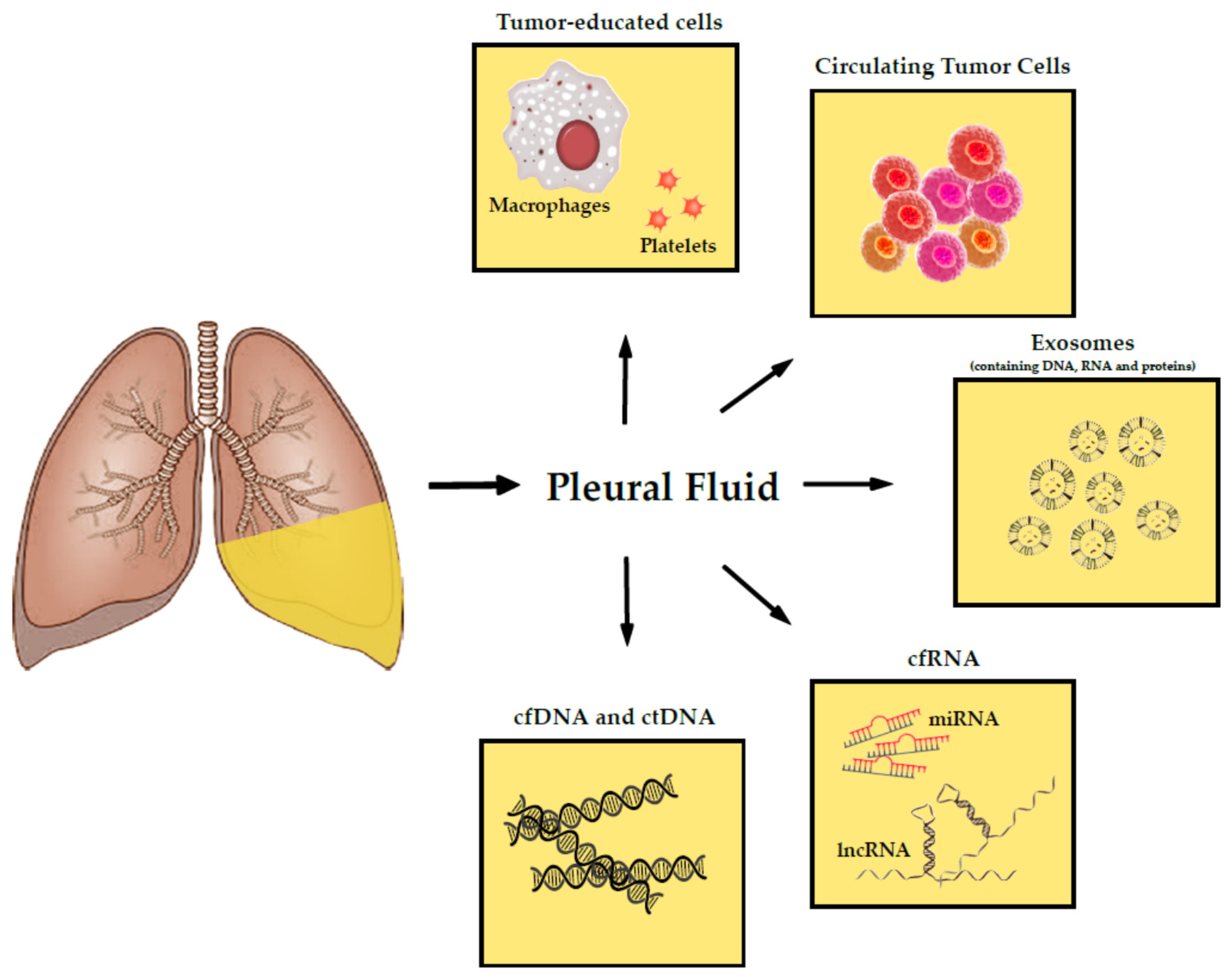
2. Tumor-Derived Products in the Pleural Fluid

Tumors release a myriad of tumor-derived products into the PF as a result of their growth within the pleural cavity. These products are molecular indicators of tumor development and represent a source of detectable biomarkers, which are useful for diagnosis, prognosis, and therapeutical intervention. Essentially, they are nucleic acids such as ctDNA and non-coding RNA (ncRNA); cells entities that include CTCs and tumor-educated cells; and extracellular vesicles, also named as exosomes (Figure 1). The purpose of this article is to undergo an in-depth review about the reported presence of these specific molecules and cells on PF, as well as the identification of the major challenges and limitations for their detection and clinical implementation. We have excluded other molecules that are also present in PF such as proteins or metabolites, as they are not generally considered a part of liquid biopsy strategies.

2.1. Nucleic Acids

Essentially, liquid biopsy is based on the analysis of circulating nucleic acids. In 1953, Mandel and Metais reported for the first time the presence of cell free DNA (cfDNA) and RNA in human blood [5] and later, in 1977, individuals with cancer were found to exhibit high levels of cfDNA in their serum [6]. The increase in cfDNA in cancer patients is driven by ctDNA released from tumor cells. Detectable concentrations of ctDNA have been found in other biofluids, such as PF [7], urine [8], cerebrospinal fluid [9], and saliva [10]. Regarding circulating RNA, several species of RNA molecules, either free or packed in extracellular vesicles, have been reported to be present in plasma and serum, and used as liquid biopsy cancer biomarkers. These are mainly non-coding RNA (miRNA being the most known), lncRNAs, and transfer RNAs, among other species [11].

2.1.1. Cell-Free DNA and Circulating Tumor DNA

CfDNA and ctDNA are passively released into the bloodstream by healthy and tumor cells, respectively, once cells die by necrosis or apoptosis. In addition, active release has also been hypothesized, particularly in tumor cells, as a strategy to promote malignancy on non-tumor cells [12]. Whilst typical concentrations of cfDNA in plasma of healthy individuals vary from 1 to 10 ng/mL, this concentration can substantially increase in certain conditions, such as acute cerebral infarction, exercise, transplantation, and infection [13]. Regarding PF, the quantification of cfDNA has demonstrated to be useful to differentiate exudative from transudative PEs [14]. A transudate is a plasma ultrafiltrate that usually results from a disequilibrium of the Starling forces (hydrostatic and oncotic pressures) in which a healthy pleura acts as a passive membrane [15]. Therefore, the small amounts of cfDNA detected in transudates derive from plasma rather than local production [14]. In contrast, an exudate implies inflammatory and cellular responses into the pleural cavity due to an increased microvessel permeability and/or lymphatic blockage [16]. The infiltration of leukocytes in infectious PE constitutes a source of cfDNA from dying cells [17], whereas the presence of a pleural tumor increases ctDNA concentrations. MPEs, which are typically exudative, showed a significant increase in cfDNA compared to transudative PEs. Considering only exudative etiologies, differences in cfDNA content between MPE and infectious PEs were also observed as significant, though less robust [14].
Besides quantitative differences on cfDNA content between PE etiologies, other authors highlight the measurement of parameters like the cfDNA fragmentation pattern, as it varies according to the cell death mechanism. Apoptotic cell death generates uniform small DNA fragmentation in plasma (~166 base pairs, bp), whereas necrotic cell death, occurring predominantly in tumor cells, generates a wide spectrum of longer fragments due to incomplete digestion of genomic DNA [18]. This phenomenon has been recently observed in PF, were cfDNA from lung associated-MPE contains much longer DNA fragments with sizes over 500 bp, compared to plasma [19]. Other authors emphasize the usefulness of the determination of DNA integrity index on PF, defined as the ratio between long and short cfDNA fragments, for diagnostic purposes. In one study, cytology and DNA integrity index taken together, offered 81 sensitivity and 87% specificity in distinguishing benign and malignant PE [20]. Interestingly, an elevated PF DNA integrity index had a positive predictive value of 81% in predicting cytology-negative MPE.
cfDNA content can be significantly increased in cancer patients, correlating with tumor volume size [21] and stage [22]. This is the reason why cfDNA, or more precisely ctDNA analysis, has become a promising tool for cancer monitoring: from the early detection of mutations on ctDNA to prognosis assessment, through the identification of minimal residual disease and prediction of disease recurrences [23]. First attempts for the identification of point mutations on ctDNA began in the early 1990s with the progress made by the Human Genome Project. In 1994, Vasioukhin et al. detected point mutations in NRAS in the leukemia patients’ blood [24]; and KRAS mutated sequences were identified in plasma and serum from patients with pancreatic adenocarcinoma [25]. Nevertheless, it was not until the past decade that researchers began to consider PF as a suitable source of detectable mutations on ctDNA. In this regard, the presence of a metastatic mass growing over the pleural membranes is undoubtedly an abundant source of ctDNA in PF. A study conducted by Porcel et al. determined that lung (37%) and breast cancer (16%) were the most common primary tumors causative of MPE, followed by hematological (10%) and gastrointestinal tumors (10%) [2]. Thus, it is not surprising that most studies on PF genomics are focused on lung cancer and, more specifically, non-small cell lung cancer (NSCLC).
One of the most studied genes in PF samples is Epidermal Growth Factor Receptor (EGFR), as it is the most common actionable target in NSCLC. Approximately, 50% of NSCLC cases in Asia [26] and around 10% in Caucasian European population [27] carry activating mutations on the tyrosine kinase domain of EGFR. Tyrosine kinase inhibitors (TKI) are the cornerstone of treatment for NSCLC, but resistances frequently occur, which have been attributed to secondary mutations in EGFR, being the T790M base substitution the most widely reported. Other relevant mutations in NSCLC involve KRAS, MET, and PIK3CA, with an overall prevalence of 23.0, 6.8, and 4.9%, respectively [28].

Genomic Strategies for Mutation Profiling in Pleural Fluid

In 2005, Huang et al. reported for the first time the detection of a specific EGFR mutation (exon 19: delE746—A750), using polymerase chain reaction (PCR) followed by direct DNA sequencing of the pleural cell pellet, in one NSCLC patient who responded to gefitinib treatment [29]. Another research showed that PF supernatants were comparable to tissue biopsy and superior to PF pellets (i.e., cell blocks) in successfully identifying EGFR point mutations on exons 19–21 by high-resolution melting and direct DNA sequencing in NSCLC patients samples [30]. Similarly, EGFR and KRAS mutations and RET and ALK rearrangements were identified by direct DNA sequencing in pre-amplified products from PF supernatants and pellets of 722 patients with metastatic lung adenocarcinoma with cytology-proven MPE [31]. Moreover, direct PCR sequencing was useful in identifying EGFR and KRAS mutations in formalin-fixed and paraffin-embedded (FFPE) samples from primary tumors and pleural metastasis, this latter including PF cell blocks and pleural biopsies of 37 lung adenocarcinoma patients [32]. Nevertheless, the authors claimed that there was a substantial discordance (16.2%) on EGFR status between the primary tumor and their corresponding metastasis. In these discordant cases, the responsiveness to EGFR TK (gefitinib or erlotinib) inhibitors was more likely to be correlated with EGFR mutations in metastatic lesions than in primary tumors. In another study, mutant-enrichment PCR-based on PCR-restriction fragment length polymorphism showed successful detection of EGFR mutations in both PF supernatants and pellets from 26 patients who were pathologically diagnosed with advanced NSCLC [33]. PCR and direct sequencing were applied to PF supernatants for the detection of mutations on EGFR (exons 18–21) in 11 out of 43 NSCLC patients. Interestingly, patients showing partial response or stable disease after treatment with gefitinib harbored EGFR mutations [34].
Another study successfully identified copy number variations (CNV) on EGFR, NOTCH1, NF1, CCDN1, and MEN1, among others; as well as point mutations (PIK3CA E542K) on ctDNA from ascites and PF supernatants of cancer patients by using genomic microarray hybridization [35]. Interestingly, ctDNA isolated from PF supernatants and extracellular vesicles (EV) presented 88 and 91% of concordance with tissue, respectively, regarding EGFR mutation detection by the peptide nucleic acid (PNA)-mediated PCR clamping method [36]. Moreover, ctDNA from EV seemed to be the best sample type for the detection of EGFR T790M mutations. These kind of mutations, which confer resistance to TKI, were detected in 72% of EV, whereas the detection rate in ctDNA from PF supernatants and cell blocks were 61 and 17%, respectively. By using the same methodology, Yang et al. detected 47 different mutations in the EGFR exons 18–24 in 72.5% of the 40 cytopathology-positive PF cell blocks examined. When looking at the clinical data, the study revealed that patients treated with TKI whose PF pellets carried EGFR mutations had better progression-free survival compared to those that were wild-type (7.33 versus 2.07 months, p = 0.032). In addition, the objective response rate was better in the EGFR-mutated patients compared to the non-mutated individuals (80.8% versus 10%, p < 0.001) [37]. Similarly, one additional investigation assessed EGFR status in 37 histologically diagnosed NSCLC patients with available tumor tissue, PF (supernatants and cell blocks), and serum [38]. It was found that there was a high concordance between PNA clamping methodology and direct PCR sequencing across all sample types. Nevertheless, the diagnostic performance of PE was higher using the PNA clamping method than direct PCR sequencing (89% sensitivity versus 100% specificity). Another study further supported the superiority of PNA clamping for the detection of EGFR mutation in PF cell blocks from lung adenocarcinoma patients with a low proportion of tumor cells [39].
A different methodology for the identification of mutations in the EGFR gene is the real-time quantitative PCR (RTqPCR). Shin et al. demonstrated the feasibility of this method by using an allele-specific PCR test (The Cobas EGFR Mutation Test from Roche), designed for the detection of 41 hotspot mutations in exons 18–21 [40]. In this study, all analyzed samples showed valid real-time PCR results from both PF supernatants and pellets DNA. Interestingly, the concordance rate between RTqPCR and Sanger sequencing plus PNA-clamping was 98.7%. Another study showed the efficacy of the detection of EGFR mutations by RTqPCR in serum and PF of 88 patients with advanced NSCLC cytologically-proven [41]. Interestingly, tumors carrying mutant EGFR showed higher response rate to gefitinib compared with EGFR wild-type counterparts (90.9 versus 9.1%, p < 0.01). An additional work reported successful determination of EGFR mutations (exons 18–21) by RTqPCR and direct sequencing in 136 lung adenocarcinoma patients [42]. The authors observed a slight discrepancy in mutation rate between PF and resected tissue. PE harbored higher EGFR mutation rate than tissues (68.4 versus 50.5%, p = 0.007), which was especially relevant for the L858R mutation (36.8% versus 20.9%, p = 0.011). Notably, survival analysis revealed that the median overall survival was longer for TKI-treated patients with EGFR mutations than for patients with wild-type EGFR (21.4 months versus 11.5 months, p = 0.005).
Along with the development of multiplexed detection methods, new works reported promising results with the combination of pyrosequencing, capillary electrophoresis, and RTqPCR [43]. For instance, Akamatsu et al. found genetic abnormalities in 48% of the cytologically-positive PF samples analyzed, and 18% of the cytologically-negative ones. In addition, the concordance rate regarding the mutations found between PE and matched FFPE samples was 88%. Similarly, multiplex RTqPCR detected the oncogenic fusions KIF5BRET and CCDC6RET in 2.4% of MPE specimens derived from metastatic lung adenocarcinoma patients [31]. Another work showed that the combination of several techniques, such as fluorescence in situ hybridization (FISH) for detecting ALK rearrangements, Sanger sequencing for EGFR, pyrosequencing for KRAS and BRAF, and next generation sequencing (NGS) using a lung cancer panel was capable to detect genetic abnormalities in 59% of the PF cell blocks from cytologically-proven NSCLC. Remarkably, authors demonstrated that PF volume was not associated with overall cellularity or tumor cellularity and, therefore, low volume of PF should not discourage further molecular testing [44].
Droplet digital PCR (ddPCR) is a highly sensitive method that allows the detection of very low amounts of DNA, which is the typical scenario for liquid biopsies. PF has been confirmed to be a good specimen for the detection of EGFR mutations by ddPCR [45]. In one paper, the mutation detection rate was significantly higher using ddPCR compared with direct DNA sequencing (75.4% versus 43.8%, p < 0.0001) and amplification-refractory mutation system (61.3% versus 38.7%, p = 0.016) and, more importantly, some patients were redefined for their EGFR status. Other authors demonstrated that ddPCR provides high concordance rates of EGFR mutations between PF supernatants and cell pellets of lung cancer patients [46].
Buttitta et al. reported the superiority of NGS over Sanger sequencing to assess EGFR status in bronchoalveolar lavage and PF from 830 lung cancer patients [47]. For instance, EGFR mutations were detectable in 81 and 16% of the cytologically-positive cases for NGS and Sanger sequencing, respectively. Interestingly, while cytological examinations and Sanger sequencing dictated negativity for EGFR mutations, 42% of cases turned to be positive according to NGS. Another work compared the performance of NGS with RTqPCR on the detection of EGFR, PIK3CA, and KRAS mutations. Although the concordance was high, NGS was more advantageous than RTqPCR in detecting non-hotspot mutations and providing accurate information about allele sequence and mutation frequency in PF pellets of lung adenocarcinoma patients [48]. Despite good results in PF cell pellets, other studies suggest that NGS on body fluid supernatants possesses higher performance for detecting EGFR mutations in cfDNA compared to body fluid sedimentary tumor cells and plasma cfDNA specimens [49]. In addition, targeted hybrid capture deep sequencing for the analysis of 130 cancer-related genes confirmed the presence of mutations in 15 PF supernatants where median depth of target was >1000× [50]. High concordance was seen between PF and FFPE samples.
Later, NGS was shown to successfully identify multiple mutations in 17 genes in 108 PF cell pellets from lung cancer patients [51]. The authors claimed that their NGS approach was currently the most successful allowing a sequencing depth of target of nearly 3492X, 10 times greater than others previously published [52]. Due to the high depth, EGFR mutations were detected in 86% of the MPE analyzed, and 66.7% of which were associated with sensitivity to known therapeutic agents. The different genomic approaches utilized in liquid biopsy of PF are summarized in Table 1.
The limit of detection or the sensitivity of the different aforementioned techniques is the main factor that determines the method of choice. Whilst Sanger sequencing and pyrosequencing are capable of providing high versatility on the detection of different genomic alterations, they have the lowest sensitivity [53]. Methods with intermediate sensitivity include high-resolution melting, PNA clamping, RTqPCR, and amplification-refractory mutation system [54], which are especially useful for the detection of single nucleotide variations and small insertions and deletions. Finally, ddPCR and NGS offer the highest sensitivity among all available technologies [55,56]. Limit of detection of different approaches are summarized in Table 2.

Approved Clinical Tests for Routine Testing

Although the utility of PF as a source of detectable genomic aberrations seems to be unquestionable, the implementation of the aforementioned techniques in the clinical routine testing is still limited. Recommendations on the development and implementation of safe and effective genetic tests have been suggested by the Evaluation of Genomic Applications in Practice and Prevention (EGAPP). Analytical validity, clinical validity, and clinical utility are the three major criteria taken into account for the adoption of tumor biomarker tests in clinical care [57]. Nevertheless, many commercially available tests have never been submitted to the US Food and Drug Administration (FDA) for approval and, even more, FDA-approved tests have not necessarily needed to demonstrate clinical utility [58].
To date, there are only 3 FDA-approved tests with clinical utility in NSCLC patients, namely Cobas® EGFR Mutation Test v2 (Roche Molecular Systems, Inc., Pleasanton, CA, USA), Guardant360® CDx test (Guardant Health, Inc., Redwood City, CA, USA), and The FoundationOne Liquid® CDx test (Foundation Medicine, Inc., Cambridge, MA, USA).
The clinical utility of the Cobas® EGFR Mutation Test v2 (Roche Molecular Systems, Inc.) has clearly been demonstrated [59]. This test is a real-time PCR-based method that allows for qualitative identification of 42 EGFR mutations in exons 19–21. This test granted FDA approval on 1 June 2016, being the first test employing liquid biopsy for the detection of EGFR exon 19 deletions or exon 21 (L858R) mutations in patients with metastatic NSCLC eligible for treatment with erlotinib. This was based on the results from the ENSURE study where patients who carried EGFR mutations in plasma were treated with erlotinib presented a higher progression-free survival (PFS) compared to those treated with chemotherapy [60]. The second test, Guardant360® CDx test (Guardant Health, Inc.), is the first test deploying NGS technology in liquid biopsy. It was approved by the FDA on 11 August 2020 for two purposes: (i) as a comprehensive genomic profiling in patients with any solid malignancy, and (ii) as a companion diagnostic to identify NSCLC patients with EGFR alterations who may benefit from treatment with osimertinib, based on the evidence of two phase II clinical trials: AURA3 [61] and FLAURA [62,63]. This test consists of the analysis of a 73-gene panel and possesses an average ctDNA detection rate across cancer types of 86%. The third test used in liquid biopsy, the NGS-based FoundationOne® Liquid CDx test (Foundation Medicine, Inc.), was approved by the FDA on 6 November 2020 as a companion diagnostic tool for the detection of genetic aberration in 324 genes by hybridization-based capture technology using ctDNA isolated from plasma specimens. One of the diagnostic indications is the identification of ALK rearrangements in patients with NSCLC eligible for treatment with alectinib.

Limitations of the Detection of Mutations on ctDNA
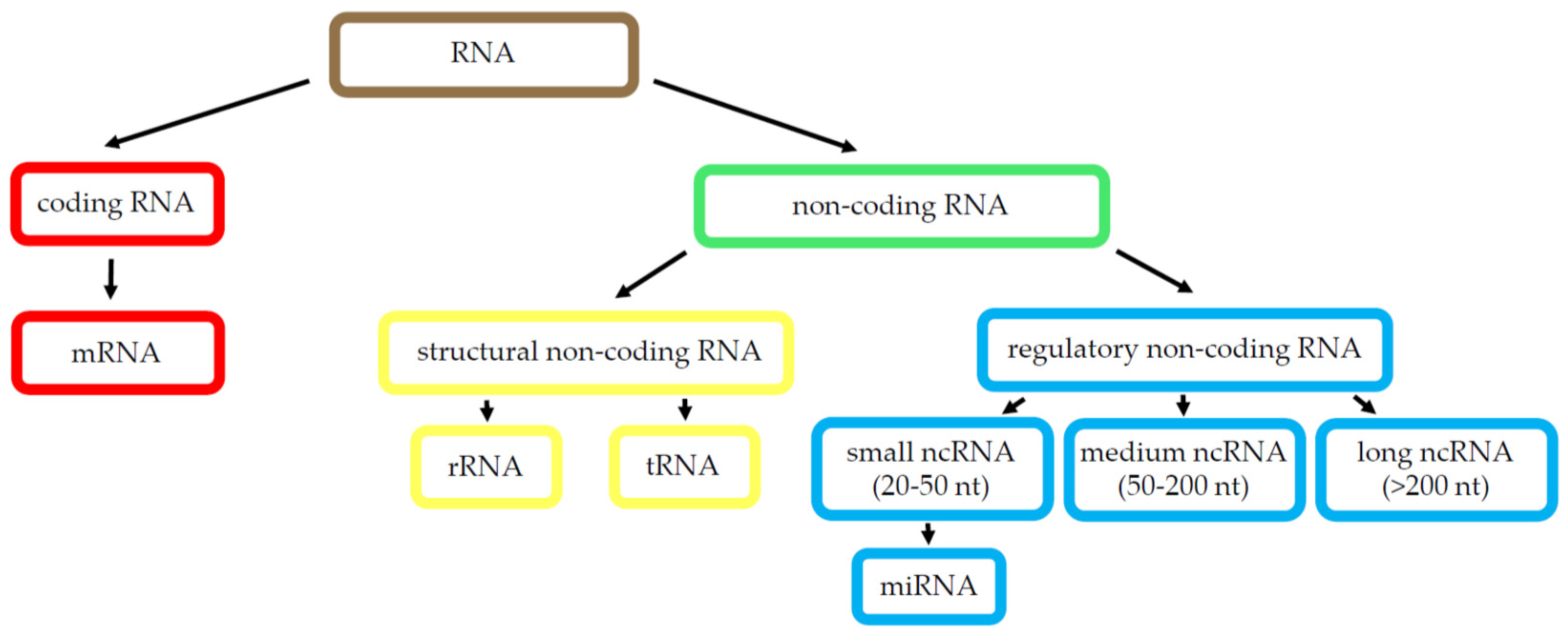
The major challenge of liquid biopsy is the detection of infrequent mutation variants, which are typically very close to the limits of sensitivity of the available methodologies. Thus, the quantity of initial input material is crucial to ensure proper detection of all variants. However, ctDNA amount in plasma is low [22], which represents a major concern. Typically, 1 mL of plasma from a cancer patient contains 1500 diploid genome equivalents (10 ng of DNA) with a total quantity of molecules per region of interest of 3000 [64]. This means a theoretical limit of sensitivity of 0.03%, implying the detection of at least one molecule of our target of interest in 3000. If the frequency of mutations is lower, for example 0.01%, we will need more material, which can be sometimes difficult despite the accessibility of blood. So far, the most sensitive techniques, such as CAPP-Seq deep sequencing, are offering sensitivities around 0.02% [65]. In the case of PF, the extraction of several tens of milliliters of PF can be generally guaranteed, which represents a clear advantage over other matrices.
The estimation of tumor burden has received considerable attention as a potential predictive biomarker for treatment response with immune checkpoint inhibitors. However, studies addressing tumor burden through the use of cfDNA from PF are scarce [66].

2.1.2. RNA Species

Although over 75% of the human genome is transcribed, only about 2% are protein-coding genes. The other 98% of transcripts are non-coding RNA (ncRNA), which are grouped into two main categories: housekeeping ncRNAs (such as transfer RNAs and ribosomal RNAs, among others) and regulatory ncRNAs, which can be separated into two groups based on size: small ncRNAs, to which microRNA (miRNA) belong, participating on gene expression regulation; and long non-coding RNAs (lncRNAs), which comprise 82% of the total ncRNAs and participate on tissue homeostasis and cell-type determination [67]. Figure 2 shows a schematic representation of known RNA species.

MicroRNA

The presence of miRNAs in blood is widely documented and alterations in their expression have been observed in various solid tumors [68,69], including lung cancer [70,71,72]. Moreover, recent studies have shown that miRNAs are also present in PE, and their characterization could be considered as a diagnostic tool to discriminate between benign and MPE [73,74,75]. Bao et al. identified miRNAs that were differentially expressed in MPE and tuberculous effusions, and three of them became potential diagnostic biomarkers. Furthermore, certain miRNA signatures have been associated with different types of primary lung tumors. For example, the expression of miR-134, miR-185, and miR-22 in PF has been linked to NSCLC [76], as well as miR-198 [77]. In another set of analysis, Wang et al. correlated the differential expression of several miRNAs with poor survival rates in NSCLC [78].

Long Non-Coding RNA

Regarding lncRNAs, mounting evidence indicates that they are also promising diagnostic and prognostic biomarkers for lung cancer, especially when detected in body fluids. The diagnostic and prognostic potentials of lncRNAs in different lung cancer types have been explored [79]. For instance, many studies showed MALAT1 to be upregulated in lung tumor tissues compared with the normal ones, suggesting its usefulness as biomarker in different body fluids for early detection of lung cancer [80]. In addition, Wang et al. observed that lncRNAs can also be detected in PF and can be potentially used as tumor markers [81]. In particular, they showed that three lncRNAs (MALAT1, H19, and CUDR) have potential as diagnostic markers in lung cancer-associated MPE. Moreover, baseline MALAT1 expression in PEs was inversely correlated with chemotherapy response, and the combination of MALAT1 and carcinoembryonic antigen was even more reliable for diagnostic purposes.

Limitations of the ncRNA Implementation

Despite the preceding studies, the use of ncRNAs as diagnostic biomarkers may have certain limitations. For example, the main techniques to detect ncRNA are qRT-PCR-based, and although straightforward and robust, there is still a lack of agreement in the normalization approach. This means that it is difficult to find a suitable internal control to standardize the results. Furthermore, a standardized protocol is needed to guarantee the reproducibility of results on different biological samples (blood and PF). In addition, each ncRNA could regulate more than one messenger RNA (mRNA) or gene, making the determination of a target molecule difficult; while on the other hand, one specific mRNA may be controlled by several miRNAs and lncRNAs; not to mention the implication of the same marker in entirely different diseases [75]. Finally, successful RNA detection is strictly dependent on proper sample handling, where the action of activated RNAses could degrade the substrate, hampering further analysis [82,83].

2.2. Exosomes

Extracellular vesicles (EVs), also known as exosomes, are bilayer lipid vesicles of 30–150 nm in size that participate in cell-to-cell communication (e.g., between the stroma and cancer cells) [84]. EVs can contain DNA, mRNA, miRNA, lncRNA, receptors, transcription factors, enzymes, lipids, and extracellular matrix proteins, which serve as communication molecules [84]. It is hypothesized that EVs packaged with those molecules intervene in crucial tumor progression processes such as extracellular matrix remodeling, hypoxia, epithelial to mesenchymal transition, immunosuppression, and vascular leakiness [84]. Interestingly, tumor-derived EVs isolated from serum recapitulate the genetic aberrations present in primary tumors, as the presence of the mutant EGFR and EGFRvIII in glioblastoma [85] or MYC amplification in medulloblastoma [86]. Moreover, circulating tumor-derived EVs have been considered to be promising biomarkers for cancer diagnostic and prognosis. For example, exosomal miR-21 has been associated with esophageal cancer recurrence and distant metastasis [87].
Andre et al. carried out the first characterization of exosomes in MPE. They were 80 nm in size, rich in antigen-presenting molecules and tumor antigens, and the ones derived from melanoma contained Mart1, which is necessary for the presentation to cytotoxic T lymphocytes [88]. Concerning other immunological properties, Wada et al. found that MPE-derived exosomes bound to TGF-β increase regulatory T-cell number, which could lead to tumor immune evasion [89]. Alegre et al. detected the presence of ubiquitinated human leukocyte antigen-G (HLA-G) in exosomes from pleural exudates, the function of which is to be elucidated [90]. Exosomes from MPEs have also been characterized proteomically [91,92]. Regarding miRNA content, one study found that high expression of miR-21, miR-23b, and miR-29 in exosomes from MPEs of ovarian carcinoma were associated with poor progression-free survival, whereas high miR-21 was correlated with poor overall survival [93]. Mice treated with these exosomes harbored more aggressive tumors [93]. The same exosomal miR-21 was found in PFs and it identified malignancy better than cytology [94].
The authors pointed out the value of EV-miR-21 as a diagnostic and prognostic factor of pleural invasions. Moreover, they suggested the involvement of EV-miR-21 in the mesothelial-to-mesenchymal transition, leading to the phenotypic switch of mesothelial cells to cancer-associated fibroblasts [94]. Other exosomal miRNAs, miR-205-5p and miR-200b, were increased in lung cancer PEs compared to pneumonia and tuberculosis-related PEs, indicating that these two differentially expressed miRNAs could possess diagnostic value [95]. Similarly, exosomal miR-182 and miR-210 were found to be significantly more expressed in MPE from lung adenocarcinoma patients compared to benign PEs [96]. A more extensive RNA profiling study of exosomes derived from PEs identified differential expression of 17 miRNAs and 17 mRNAs between patients with lung adenocarcinoma and benign inflammatory processes. In particular, the miR-200 family and the mRNA transcript lipocalin-2 presented the strongest diagnostic power [97]. Another study identified the EV-associated miRNAs miRNA-1-3p, miRNA-144-5p, and miRNA-150-5p in PEs through interrogation of 754 miRNAs and Machine Learning as the most accurate lung cancer diagnostic biomarkers [98]. Regarding DNA species, Song et al. studied the feasibility of using exosomal DNA from PEs due to lung adenocarcinoma patients for genetic testing. The study revealed that 78% of the mutations found in PE-derived exosomal DNA matched with those found in PE-derived ctDNA [99], supporting its reliability for genetic testing.
Undoubtedly, EV-miRNAs are informative biomarkers in liquid biopsies with a proven valuable diagnostic and prognostic potential. Some limitations that EV have to overcome are the technicalities related to their isolation, which could hamper implementation in the routine clinical testing. Isolation involves ultracentrifugation and some hospital services may not have access to it. Briefly, pleural exudates are centrifuged a few times at increasing centrifuge forces, usually from 300× g to 10,000× g for 30 min, to discard dead cells and cell debris. Then, supernatants are added in a 30% sucrose/D2O cushion and ultracentrifuged two times at around 100,000× g for at least 1 h. The resulting pellets are the exosomes which can be resuspended in PBS and stored at −80 °C. Exosomes can be quantified for protein content following, for example, the Bradford method. Also, researchers can proceed to the lysis using protein and RNA specific reagents for protein and RNA extraction, respectively.
Apart from being cancer biomarkers, one interesting applicability of exosomes is their use as delivery vehicles for bioactive compounds such as small molecules, siRNA, and cDNA [100]. They are less immunogenic than nanoparticles, could potentially improve the pharmacokinetic properties of nanoparticles, and also deliver gene therapy, viruses, and antibodies inside the specific target cell more efficiently [100].

2.3. Cell Entities

PEs contain diverse cell types in different proportions depending on whether they are benign or malignant [101]. Unfortunately, the sensitivity of the cytology in the diagnosis of a MPE is low, around 60%, mostly because nuclei atypia can be confounded with reactive mesothelial cells even for experienced cytopathologists [102]. The detection of certain cell types in the PF, such as CTCs, can be a promising tool that can serve for MPE diagnosis and, at the same time, inform about cancer progression and prognosis. Nevertheless, the isolation of CTCs can be tedious. In this section, we will review the clinical value of CTCs together with the current stage of identification and characterization techniques, and their limitations and challenges.

2.4. Circulating Tumor Cells

CTCs are defined as carcinoma cells that have shed into the blood or lymphatic circulation from an existing primary tumor or metastatic lesion. It is estimated that growing tumors shed 3.2 × 10⁶ cells daily per gram of tissue into efferent blood [103]. CTCs are believed to generate from specific tumor subclones that have acquired survival advantage during dissemination and have been able to escape immune surveillance. Altogether, this makes them prone to “seed” in other organs and originate metastasis [104]. Interestingly, Liu et al. reported that the CTCs present in the 4T1 breast cancer model retained the epithelial pheno-type less than the disseminated tumor cells found in the bone marrow [105]. The first accurate characterization of CTCs was reported in 1998 by Racila et al. In this work, the authors detected and isolated CTCs in the blood of patients with breast and prostate cancer using magnetic ferrofluids coupled with an epithelial cell adhesion molecule (EpCAM) antibody followed by cell cytometry [106]. Since then, new technical approaches have emerged for the detection of CTC in PE (Table 3). A general review on the approaches of isolation and characterization of CTC can be found elsewhere [107].
CTCs have been considered to have a clear clinical diagnostic and prognostic value in several malignancies, such as metastatic breast cancer [118], NSCLC [119], colorectal cancer [120], and prostate cancer [121]. Apart from blood, CTCs can be found in rarer human fluids such as PF, cerebrospinal fluid, or urine. An accumulation of PF within the pleural space can be the consequence of cancer progression, usually NSCLC, breast cancer, Kaposi sarcoma, and lymphoma [102]. The detection of malignant cells in the PE denotes advanced stage of the disease usually coursing with metastasis [122]. It also indicates poor prognosis and uselessness of curative treatment. Therefore, the identification of CTCs in the PF represents a valuable approach for the diagnosis of malignancy and can provide guidance for treatment decision. Whilst the detection and characterization of CTCs and the significance of their abundance in peripheral blood have extensively been studied, only few studies have addressed these points in the PF.
One study found that CTCs in PF matched with CTCs in blood from the same patients with NSCLC, regarding cytokeratin expression and absence of EpCAM [108]. The CTC identification was done in two steps: a first CD45 depletion to discard the leukocytes and a subsequent immunostaining with anti-cytokeratin and anti-EpCAM. In another work, the authors established a CTC line from a MPE (SCLC26A) and another from the peripheral blood of a SCLC patient. They reported expression of chitinase-3-like-1/YKL-40 (CHI3L1), a secreted glycoprotein present in cancers with poor outcome, in SCLC26A cells, which correlated with higher expression of several tyrosine kinase receptors responsible for motility, invasion, and poor prognosis [109]. The CTC line SCLC26A also showed to be more sensitive to the combined treatment with camptothecin analogues compared to the SCLC cell lines NCI-H417 and DMS153 [110]. Moreover, SCLC26A expressed E-cadherin at high levels and presented lower expression of stem cell markers compared to other blood CTCs from primary tumors and metastases [111]. In addition, Ge et al. attempted the characterization of a CTC line isolated by anti-EpCAM immunocapture from a MPE secondary to pancreatic cancer. The study revealed polyploidy according to FISH of chromosome 8 [114]. A more complex study was performed by Tang et al. in which they detected CTCs from a MPE by high-throughput fluorescent screening using a fluorescent glucose analogue based on the fact that cancer cells show higher glucose consumption. The validation of malignancy was done by single-cell sequencing [115]. In another study, researchers were able to discriminate between CTCs and other cell types from MPEs, such as leukocytes, with the utilization of a live-cell fluorescent dye, which was able to bind to mitochondria, considering the increased number of mitochondria and higher activity of cancer cells [116]. Such procedure of CTC detection did not disrupt downstream single-cell analysis.
The preceding identification methodologies are mainly indicated for the detection of big CTCs or EpCAM+/CK+. However, tumors are highly heterogenous and so can be the CTCs, namely highly heterogenous rare CTCs (CRCs) that include circulating endothelial (CECs) and CTCs. Lin et al. proposed an integrative program for the isolation and enrichment of CRCs, which includes subtraction enrichment immunostaining-fluorescence in situ hybridization (SE-iFISH), without hypotonic damage nor anti-EpCAM detection, to enable proper karyotyping for the observation of chromosomal aneuploidy, detection of multiple protein expression and cytogenetic arrangements. Altogether, it can be very informative of the CTC-CEC crosstalk in relation to tumor development processes, such as tumor angiogenesis, drug resistance, and metastasis [123]. The same authors identified individual aneuploid CD31+ cells as well as fusion clusters of endothelial-epithelial aneuploid tumor cells with SE-iFISH technology in several biofluids including PE [117].
Despite all these promising techniques, CELLSEARCH® still remains the only clinically approved technology to detect and enumerate CTCs of epithelial origin. It was approved in 2014 by the FDA for the detection of CTCs in peripheral blood and prediction of clinical outcome in patients with metastatic breast cancer [124]. It consists of magnetic nanoparticles coating an anti-EpCAM antibody for the capture and further immunolabelling with cytokeratin 8, 18, and 19 to identify epithelial-differentiating CTCs, with CD45 to discard leucocytes and DAPI to detect nucleated cells. The potential of CELLSEARCH® for the diagnosis of MPE has been explored and compared with traditional cytology [112]. However, its clinical suitability is not clear and more studies are required. A variation of CELLSEARCH® was used by Beije et al. for the identification and enumeration of CTCs in PFs of malignant mesothelioma patients. The methodological variation includes the immunodetection by flow cytometry of the melanoma cell adhesion molecule (MCAM), which is specific for malignant cells [125].
Studies including analysis of exosomes and CTC in PF are scarce. This explains why it is difficult to refute results from different reports or make a contrasted review about these two biomarkers.

3. Future Perspectives: Tumor-Educated Cells

Apart from ctDNA, RNA species, and CTCs, there are other cellular entities present in the blood that have started receiving interest as diagnostic and prognostic cancer biomarkers. Eventually, they will start getting studied in MPE. These are tumor-associated cells such as tumor-educated platelets (TEPs) and tumor-associated macrophages (TAM). Platelets emerge from the fragmentation of megakaryocytes in the bone marrow and are anucleated. Primarily known to be involved in wound healing processes, their newly discovered role in tumor dissemination has awaken the interest of researchers. Tumors are in crosstalk with platelets and the exchange and sequestration of tumor-associated molecules by platelets contribute to their “education” [126]. Platelets have the capacity to undergo splicing of mRNAs when activated via surface receptors and lipopolysaccharides, or by signals emitted by cancer cells and the tumor microenvironment [127]. Interestingly, the different abundances of mRNA, as a result of platelets activity, can serve as a cancer diagnostic tool [128]. For example, a study revealed a platelet-based gene expression signature of metastatic lung cancer characterized by a decreased expression of 200 genes, with the most altered levels between metastatic and healthy subjects [129]. Another study has proposed the analysis of the TEP mRNA repertoire in blood as a promising early pan-cancer diagnosis tool, based on the results obtained with RNA Sequencing in TEPs [130]. Notably, TEP RNA has been considered to be a more advantageous biomarker for the detection of early-stage cancers compared to the ctDNA in plasma due to the low abundance of the latter [128]. Despite the high accuracy of TEP RNA to differentiate between malignant and benign nature of diseases, there are still no works in the literature studying their potential to discriminate between benign and MPE, which we believe is nonetheless worth exploring.
Another understudied field in MPEs is the identification of TAMs. TAMs are the most abundant cell type of the tumor microenvironment. TAMs, like CTCs, derive from the primary tumor and are believed to support CTCs dissemination to distant sites [131]. For that, cell-to-cell communication is needed via production of chemoattractants by TAMs, including MMP-1 and CXCL12 [131]. It is hypothesized that the presence of TAMs in plasma is a biomarker of advanced cancer. In this line, Adams et al. isolated TAMs by microfiltration from peripheral blood of patients with breast, pancreatic and prostate cancer, but these cells were not present in healthy individuals [132]. TAMs expressed epithelial, monocytic and endothelial markers and were bound to CTCs. In another study, it was shown that the lncRNA LINC00662 could enhanced hepatocellular growth and metastasis through Wnt/β-catenin signaling and M2 TAM polarization [133]. Like TEPs, little is known about the potential of TAMs as biomarkers of MPEs, though they could provide valuable diagnostic and predictive information.
Among all strategies evaluated in this review, the choice of one technique over the others will depend on factors such as the sample quality and quantity, prior molecular knowledge on specific tumor gene characteristics, available budget, and equipment requirements. For example, PCR-based approaches, including direct PCR sequencing, PNA-PCR, RTqPCR, and ddPCR, offer a reasonable performance; sensitivity being the main parameter that differentiates them. They are in general affordable but require some previous molecular biology background. Other technologies like NGS-based approaches are quite expensive, but they allow the detection of multiple mutations and less sample volume is generally necessary. In addition, prior knowledge is not needed, though bioinformatics skills are essential to perform the variant calling. Regarding RNA analysis, sample quality is the major concern. RNA detection is quite simple and affordable; however, it requires some specialized training. Exosome studies are very innovative and, in our opinion, will be crucial for further advances in the field. However, exosome extraction is tedious, and the identification of their cargo relies on extremely sensitive techniques. Finally, cell-based research is technically quite complex because of the low abundance of CTC. Moreover, it is imperative to reproduce physiological conditions to obtain good results, a specialized equipment is mandatory, and test reproducibility is a major challenge.

4. Conclusions

PF provides a valuable opportunity for cancer detection, which is especially useful in those cases where tissue biopsy is not available. In this regard, genomic analysis of PFs has a positive impact on diagnostic performance and faithfully recapitulates the molecular profiles found in tissue. It is worth noting that PF is a very suitable matrix for the detection of actionable mutations of the most studied oncogenes and can be used as a guide for therapeutic decisions. Interestingly, cell-free specimens such as plasma and PF supernatants have revolutionized the way we understand the genomic landscape of tumors. Indeed, they are the samples of choice for state-of-art genomic characterization approaches as they ensure sample quality and consistency, unlike other matrices like tissue, still considered as the gold standard. Nevertheless, there are yet major concerns to be addressed related to the implementation of the liquid biopsy in the clinical practice. In the case of PFs, it is still to be considered as a valid sample.
Unlike DNA molecules, RNA species are far away from the routine clinical practice; however, they are promising and detectable on PE specimens. Regarding CTCs, their detection opens an avenue of new opportunities to understand tumor biology processes and infer clinical interventions in the future. Although not yet detected in PFs, the exploration of educated cells represents a new paradigm, which could provide valuable information added to the global tumoral picture and improve our understanding about the dynamic networks existing between tumor and stroma. Undoubtedly, PF has begun to stand out above other matrices and will be a key biological sample to understand MPE and cancers coursing with MPE.

Author Contributions

J.M.P. was responsible of the conceptualization; J.M.P., M.A.S., A.S. (Anabel Sorolla) and E.P. wrote the original draft of the review; E.P. performed the figures; J.M.P. made a critical review and edited the manuscript. Finally, A.S. (Antonieta Salud) was responsible for project administration and funding. All authors have read and agreed to the published version of the manuscript.

Funding

This research was funded by the Instituto de Salud Carlos III of Ministerio de Ciencia e Innovación, PI20-01500 and by Spanish Society of Respiratory Pathology (SEPAR), 678-2018, given to JMP; by the University and Research Grants Management Agency (AGAUR) of Catalonia Government emergent research group recognition award, 2017SRG1620, given to MAS; by Merck Salud Foundation, 2018 project award, given to A. Salud and the Instituto de Salud Carlos III of Ministerio de Ciencia e Innovación Miguel Servet award (CP20/00039), given to A. Sorolla.

Conflicts of Interest

The authors declare no conflict of interest. The funders had no role in the design of the study; in the collection, analyses, or interpretation of data; in the writing of the manuscript, or in the decision to publish the results.

References

  1. Porcel, J.M.; Light, R.W. Pleural effusions. Dis. Mon. 2013, 59, 29–57. [Google Scholar] [CrossRef]
  2. Porcel, J.M.; Esquerda, A.; Vives, M.; Bielsa, S. Etiology of Pleural Effusions: Analysis of More Than 3000 Consecutive Thoracenteses. Arch. Bronconeumol. 2014, 50, 161–165. [Google Scholar] [CrossRef]
  3. Porcel, J.M.; Quirós, M.; Gatius, S.; Bielsa, S. Examination of cytological smears and cell blocks of pleural fluid: Complementary diagnostic value for malignant effusions. Revista Clínica Española 2017, 217, 144–148. [Google Scholar] [CrossRef]
  4. Porcel, J.M. Diagnosis and characterization of malignant effusions through pleural fluid cytological examination. Curr. Opin. Pulm. Med. 2019, 25, 362–368. [Google Scholar] [CrossRef] [PubMed]
  5. Mandel, P.; Metais, P. Nuclear Acids in Human Blood Plasma. Available online: https://pubmed.ncbi.nlm.nih.gov/18875018/ (accessed on 23 January 2021).
  6. Leon, S.A.; Shapiro, B.; Sklaroff, D.M.; Yaros, M.J. Free DNA in the Serum of Cancer Patients and the Effect of Therapy. Cancer Res. 1977, 37, 646–650. [Google Scholar] [PubMed]
  7. Benlloch, S.; Martí-Ciriquián, J.L.; Galbis-Caravajal, J.M.; Martín, C.; Sánchez-Payá, J.; Rodríguez-Paniagua, J.M.; Romero, S.; Massutí, B. Cell-free DNA concentration in pleural fluid and serum: Quantitative approach and potential prognostic factor in patients with cancer and pleural effusions. Clin. Lung Cancer 2006, 8, 140–145. [Google Scholar] [CrossRef] [PubMed]
  8. Salvi, S.; Martignano, F.; Molinari, C.; Gurioli, G.; Calistri, D.; De Giorgi, U.; Conteduca, V.; Casadio, V. The potential use of urine cell free DNA as a marker for cancer. Expert Rev. Mol. Diagn. 2016, 16, 1283–1290. [Google Scholar] [CrossRef]
  9. Wang, Y.; Springer, S.; Zhang, M.; McMahon, K.W.; Kinde, I.; Dobbyn, L.; Ptak, J.; Brem, H.; Chaichana, K.; Gallia, G.L.; et al. Detection of tumor-derived DNA in cerebrospinal fluid of patients with primary tumors of the brain and spinal cord. Proc. Natl. Acad. Sci. USA 2015, 112, 9704–9709. [Google Scholar] [CrossRef] [Green Version]
  10. Hyun, K.A.; Gwak, H.; Lee, J.; Kwak, B.; Jung, H. Il Salivary exosome and cell-free DNA for cancer detection. Micromachines 2018, 9, 340. [Google Scholar] [CrossRef] [PubMed] [Green Version]
  11. Umu, S.U.; Langseth, H.; Bucher-Johannessen, C.; Fromm, B.; Keller, A.; Meese, E.; Lauritzen, M.; Leithaug, M.; Lyle, R.; Rounge, T.B. A comprehensive profile of circulating RNAs in human serum. RNA Biol. 2018, 15, 242–250. [Google Scholar] [CrossRef] [Green Version]
  12. de Souza, A.G.; Bastos, V.A.F.; Fujimura, P.T.; Ferreira, I.C.C.; Leal, L.F.; da Silva, L.S.; Laus, A.C.; Reis, R.M.; Martins, M.M.; Santos, P.S.; et al. Cell-free DNA promotes malignant transformation in non-tumor cells. Sci. Rep. 2020, 10. [Google Scholar] [CrossRef]
  13. Wan, J.C.M.; Massie, C.; Garcia-Corbacho, J.; Mouliere, F.; Brenton, J.D.; Caldas, C.; Pacey, S.; Baird, R.; Rosenfeld, N. Liquid biopsies come of age: Towards implementation of circulating tumour DNA. Nat. Rev. Cancer 2017, 17, 223–238. [Google Scholar] [CrossRef] [PubMed]
  14. Chan, M.H.M.; Chow, K.M.; Chan, A.T.C.; Leung, C.B.; Chan, L.Y.S.; Chow, K.C.K.; Lam, C.W.; Lo, Y.M.D. Quantitative analysis of pleural fluid cell-free DNA as a tool for the classification of pleural effusions. Clin. Chem. 2003, 49, 740–745. [Google Scholar] [CrossRef] [Green Version]
  15. Chetty, K. Transudative pleural effusions. Clin. Chest Med. 1985, 6, 49–54. [Google Scholar] [CrossRef]
  16. Porcel, J.M.; Light, R.W. Diagnostic approach to pleural effusion in adults. Am. Fam. Physician 2006, 73, 1211–1220. [Google Scholar]
  17. Santotoribio, J.D.; Cabrera-Alarcón, J.L.; Batalha-Caetano, P.; Macher, H.C.; Guerrero, J.M. Pleural fluid cell-free DNA in parapneumonic pleural effusion. Clin. Biochem. 2015, 48, 1003–1005. [Google Scholar] [CrossRef] [PubMed]
  18. Schwarzenbach, H.; Hoon, D.S.B.; Pantel, K. Cell-free nucleic acids as biomarkers in cancer patients. Nat. Rev. Cancer 2011, 11, 426–437. [Google Scholar] [CrossRef]
  19. Yu, Y.; Qian, J.; Shen, L.; Ji, W.; Lu, S. Distinct profile of cell-free DNA in malignant pleural effusion of non-small cell lung cancer and its impact on clinical genetic testing. Int. J. Med. Sci. 2021, 18, 1510–1518. [Google Scholar] [CrossRef]
  20. Sriram, K.B.; Relan, V.; Clarke, B.E.; Duhig, E.E.; Windsor, M.N.; Matar, K.S.; Naidoo, R.; Passmore, L.; McCaul, E.; Courtney, D.; et al. Pleural fluid cell-free DNA integrity index to identify cytologically negative malignant pleural effusions including mesotheliomas. BMC Cancer 2012. [Google Scholar] [CrossRef] [Green Version]
  21. Abbosh, C.; Birkbak, N.J.; Wilson, G.A.; Jamal-Hanjani, M.; Constantin, T.; Salari, R.; Le Quesne, J.; Moore, D.A.; Veeriah, S.; Rosenthal, R.; et al. Phylogenetic ctDNA analysis depicts early-stage lung cancer evolution. Nature 2017, 545, 446–451. [Google Scholar] [CrossRef]
  22. Bettegowda, C.; Sausen, M.; Leary, R.J.; Kinde, I.; Wang, Y.; Agrawal, N.; Bartlett, B.R.; Wang, H.; Luber, B.; Alani, R.M.; et al. Detection of circulating tumor DNA in early- and late-stage human malignancies. Sci. Transl. Med. 2014, 6. [Google Scholar] [CrossRef] [PubMed] [Green Version]
  23. Crowley, E.; Di Nicolantonio, F.; Loupakis, F.; Bardelli, A. Liquid biopsy: Monitoring cancer-genetics in the blood. Nat. Rev. Clin. Oncol. 2013, 10, 472–484. [Google Scholar] [CrossRef] [PubMed]
  24. Vasioukhin, V.; Anker, P.; Maurice, P.; Lyautey, J.; Lederrey, C.; Stroun, M. Point mutations of the N-ras gene in the blood plasma DNA of patients with myelodysplastic syndrome or acute myelogenous leukaemia. Br. J. Haematol. 1994, 86, 774–779. [Google Scholar] [CrossRef] [PubMed]
  25. Sorenson, G.D.; Pribish, D.M.; Valone, F.H.; Memoli, V.A.; Bzik, D.J.; Yao, S.L. Soluble normal and mutated DNA sequences from single-copy genes in human blood. Cancer Epidemiol. Prev. Biomarkers 1994, 3, 67–71. [Google Scholar]
  26. Shi, Y.; Au, J.S.K.; Thongprasert, S.; Srinivasan, S.; Tsai, C.M.; Khoa, M.T.; Heeroma, K.; Itoh, Y.; Cornelio, G.; Yang, P.C. A prospective, molecular epidemiology study of EGFR mutations in Asian patients with advanced non-small-cell lung cancer of adenocarcinoma histology (PIONEER). J. Thorac. Oncol. 2014, 9, 154–162. [Google Scholar] [CrossRef] [PubMed] [Green Version]
  27. Gahr, S.; Stoehr, R.; Geissinger, E.; Ficker, J.H.; Brueckl, W.M.; Gschwendtner, A.; Gattenloehner, S.; Fuchs, F.S.; Schulz, C.; Rieker, R.J.; et al. EGFR mutational status in a large series of Caucasian European NSCLC patients: Data from daily practice. Br. J. Cancer 2013, 109, 1821–1828. [Google Scholar] [CrossRef] [Green Version]
  28. Kerr, K.M.; Dafni, U.; Schulze, K.; Thunnissen, E.; Bubendorf, L.; Hager, H.; Finn, S.; Biernat, W.; Vliegen, L.; Losa, J.H.; et al. Prevalence and clinical association of gene mutations through multiplex mutation testing in patients with NSCLC: Results from the ETOP Lungscape Project. Ann. Oncol. 2018, 29, 200–208. [Google Scholar] [CrossRef]
  29. Huang, M.J.; Lim, K.H.; Tzen, C.Y.; Hsu, H.S.; Yen, Y.; Huang, B.S. EGFR mutations in malignant pleural effusion of non-small cell lung cancer: A case report. Lung Cancer 2005, 49, 413–415. [Google Scholar] [CrossRef]
  30. Lin, J.; Gu, Y.; Du, R.; Deng, M.; Lu, Y.; Ding, Y. Detection of EGFR mutation in supernatant, cell pellets of pleural effusion and tumor tissues from non-small cell lung cancer patients by high resolution melting analysis and sequencing. Int. J. Clin. Exp. Pathol. 2014, 7, 8813–8822. [Google Scholar]
  31. Tsai, T.H.; Wu, S.G.; Hsieh, M.S.; Yu, C.J.; Yang, J.C.H.; Shih, J.Y. Clinical and prognostic implications of RET rearrangements in metastatic lung adenocarcinoma patients with malignant pleural effusion. Lung Cancer 2015, 88, 208–214. [Google Scholar] [CrossRef]
  32. Han, H.S.; Eom, D.W.; Kim, J.H.; Kim, K.H.; Shin, H.M.; An, J.Y.; Lee, K.M.; Choe, K.H.; Lee, K.H.; Kim, S.T.; et al. EGFR mutation status in primary lung adenocarcinomas and corresponding metastatic lesions: Discordance in pleural metastases. Clin. Lung Cancer 2011, 12, 380–386. [Google Scholar] [CrossRef]
  33. Zhang, X.; Zhao, Y.; Wang, M.; Yap, W.S.; Chang, A.Y.C. Detection and comparison of epidermal growth factor receptor mutations in cells and fluid of malignant pleural effusion in non-small cell lung cancer. Lung Cancer 2008, 60, 175–182. [Google Scholar] [CrossRef]
  34. Kimura, H.; Fujiwara, Y.; Sone, T.; Kunitoh, H.; Tamura, T.; Kasahara, K.; Nishio, K. EGFR mutation status in tumour-derived DNA from pleural effusion fluid is a practical basis for predicting the response to gefitinib. Br. J. Cancer 2006, 95, 1390–1395. [Google Scholar] [CrossRef]
  35. Husain, H.; Nykin, D.; Bui, N.; Quan, D.; Gomez, G.; Woodward, B.; Venkatapathy, S.; Duttagupta, R.; Fung, E.; Lippman, S.M.; et al. Cell-Free DNA from Ascites and Pleural Effusions: Molecular Insights into Genomic Aberrations and Disease Biology. Mol. Cancer Ther. 2017. [Google Scholar] [CrossRef] [Green Version]
  36. Lee, J.S.; Hur, J.Y.; Kim, I.A.; Kim, H.J.; Choi, C.M.; Lee, J.C.; Kim, W.S.; Lee, K.Y. Liquid biopsy using the supernatant of a pleural effusion for EGFR genotyping in pulmonary adenocarcinoma patients: A comparison between cell-free DNA and extracellular vesicle-derived DNA. BMC Cancer 2018, 18. [Google Scholar] [CrossRef] [PubMed] [Green Version]
  37. Yang, J.; Lee, O.J.; Son, S.M.; Woo, C.G.; Jeong, Y.; Yang, Y.; Kwon, J.; Lee, K.H.; Han, H.S. EGFR mutation status in lung adenocarcinoma-associated malignant pleural effusion and efficacy of egfr tyrosine kinase inhibitors. Cancer Res. Treat. 2018, 50, 908–916. [Google Scholar] [CrossRef] [PubMed] [Green Version]
  38. Yeo, C.D.; Kim, J.W.; Kim, K.H.; Ha, J.H.; Rhee, C.K.; Kim, S.J.; Kim, Y.K.; Park, C.K.; Lee, S.H.; Park, M.S.; et al. Detection and comparison of EGFR mutations in matched tumor tissues, cell blocks, pleural effusions, and sera from patients with NSCLC with malignant pleural effusion, by PNA clamping and direct sequencing. Lung Cancer 2013, 81, 207–212. [Google Scholar] [CrossRef]
  39. Han, H.S.; Lim, S.N.; An, J.Y.; Lee, K.M.; Choe, K.H.; Lee, K.H.; Kim, S.T.; Son, S.M.; Choi, S.Y.; Lee, H.C.; et al. Detection of EGFR mutation status in lung adenocarcinoma specimens with different proportions of tumor cells using two methods of differential sensitivity. J. Thorac. Oncol. 2012, 7, 355–364. [Google Scholar] [CrossRef] [PubMed] [Green Version]
  40. Shin, S.; Kim, J.; Kim, Y.; Cho, S.M.; Lee, K.A. Assessment of real-time PCR method for detection of EGFR mutation using both supernatant and cell pellet of malignant pleural effusion samples from non-small-cell lung cancer patients. Clin. Chem. Lab. Med. 2017, 55, 1962–1969. [Google Scholar] [CrossRef]
  41. Jian, G.; Songwen, Z.; Ling, Z.; Qinfang, D.; Jie, Z.; Liang, T.; Caicun, Z. Prediction of epidermal growth factor receptor mutations in the plasma/pleural effusion to efficacy of gefitinib treatment in advanced non-small cell lung cancer. J. Cancer Res. Clin. Oncol. 2010, 136, 1341–1347. [Google Scholar] [CrossRef]
  42. Wu, S.G.; Gow, C.H.; Yu, C.J.; Chang, Y.L.; Yang, C.H.; Hsu, Y.C.; Shih, J.Y.; Lee, Y.C.; Yang, P.C. Frequent epidermal growth factor receptor gene mutations in malignant pleural effusion of lung adenocarcinoma. Eur. Respir. J. 2008, 32, 924–930. [Google Scholar] [CrossRef] [Green Version]
  43. Akamatsu, H.; Koh, Y.; Kenmotsu, H.; Naito, T.; Serizawa, M.; Kimura, M.; Mori, K.; Imai, H.; Ono, A.; Shukuya, T.; et al. Multiplexed molecular profiling of lung cancer using pleural effusion. J. Thorac. Oncol. 2014, 9, 1048–1052. [Google Scholar] [CrossRef] [Green Version]
  44. Carter, J.; Miller, J.A.; Feller-Kopman, D.; Ettinger, D.; Sidransky, D.; Maleki, Z. Molecular profiling of malignant pleural effusion in metastatic non-small-cell lung carcinoma the effect of preanalytical factors. Ann. Am. Thorac. Soc. 2017, 14, 1169–1176. [Google Scholar] [CrossRef] [PubMed]
  45. Li, X.; Liu, Y.; Shi, W.; Xu, H.; Hu, H.; Dong, Z.; Zhu, G.; Sun, Y.; Liu, B.; Gao, H.; et al. Droplet digital PCR improved the EGFR mutation diagnosis with pleural fluid samples in non-small-cell lung cancer patients. Clin. Chim. Acta 2017, 471, 177–184. [Google Scholar] [CrossRef] [PubMed]
  46. Asaka, S.; Yoshizawa, A.; Saito, K.; Kobayashi, Y.; Yamamoto, H.; Negishi, T.; Nakata, R.; Matsuda, K.; Yamaguchi, A.; Honda, T. Rapid point-of-care testing for epidermal growth factor receptor gene mutations in patients with lung cancer using cell-free DNA from cytology specimen supernatants. Int. J. Oncol. 2018, 52, 2110–2118. [Google Scholar] [CrossRef] [Green Version]
  47. Buttitta, F.; Felicioni, L.; Del Grammastro, M.; Filice, G.; Di Lorito, A.; Malatesta, S.; Viola, P.; Centi, I.; D’Antuono, T.; Zappacosta, R.; et al. Effective assessment of egfr mutation status in bronchoalveolar lavage and pleural fluids by next-generation sequencing. Clin. Cancer Res. 2013, 19, 691–698. [Google Scholar] [CrossRef] [PubMed] [Green Version]
  48. Zhang, Y.; Li, J.; Hua, P.; Liu, N.; Li, Q.; Zhu, X.; Jiang, L.; Zheng, K.; Su, X. Targeted next-generation sequencing in cytology specimens for molecular profiling of lung adenocarcinoma. Int. J. Clin. Exp. Pathol. 2018, 11, 3647–3655. [Google Scholar]
  49. Zhang, P.; Wu, X.; Tang, M.; Nie, X.; Li, L. Detection of EGFR gene mutation status from pleural effusions and other body fluid specimens in patients with lung adenocarcinoma. Thorac. Cancer 2019, 10, 2218–2224. [Google Scholar] [CrossRef] [PubMed] [Green Version]
  50. Yang, S.R.; Mooney, K.L.; Libiran, P.; Jones, C.D.; Joshi, R.; Lau, H.D.; Stehr, H.; Berry, G.J.; Zehnder, J.L.; Long, S.R.; et al. Targeted deep sequencing of cell-free DNA in serous body cavity fluids with malignant, suspicious, and benign cytology. Cancer Cytopathol. 2020, 128, 43–56. [Google Scholar] [CrossRef] [PubMed]
  51. Ruan, X.; Sun, Y.; Wang, W.; Ye, J.; Zhang, D.; Gong, Z.; Yang, M. Multiplexed molecular profiling of lung cancer with malignant pleural effusion using next generation sequencing in Chinese patients. Oncol. Lett. 2020, 19, 3495–3505. [Google Scholar] [CrossRef]
  52. Liu, L.; Shao, D.; Deng, Q.; Tang, H.; Wang, J.; Liu, J.; Guo, F.; Lin, Y.; Peng, Z.; Mao, M.; et al. Next generation sequencing-based molecular profiling of lung adenocarcinoma using pleural effusion specimens. J. Thorac. Dis. 2018, 10, 2631–2637. [Google Scholar] [CrossRef]
  53. Vanni, I.; Tanda, E.T.; Spagnolo, F.; Andreotti, V.; Bruno, W.; Ghiorzo, P. The Current State of Molecular Testing in the BRAF-Mutated Melanoma Landscape. Front. Mol. Biosci. 2020, 7, 113. [Google Scholar] [CrossRef] [PubMed]
  54. Ye, P.; Cai, P.; Xie, J.; Wei, Y. The diagnostic accuracy of digital PCR, ARMS and NGS for detecting KRAS mutation in cell-free DNA of patients with colorectal cancer: A systematic review and meta-analysis. PLoS ONE 2021, 16. [Google Scholar] [CrossRef]
  55. Chakrabarti, S.; Xie, H.; Urrutia, R.; Mahipal, A. The promise of circulating tumor DNA (ctDNA) in the management of early-stage colon cancer: A critical review. Cancers 2020, 12, 2808. [Google Scholar] [CrossRef] [PubMed]
  56. Postel, M.; Roosen, A.; Laurent-Puig, P.; Taly, V.; Wang-Renault, S.F. Droplet-based digital PCR and next generation sequencing for monitoring circulating tumor DNA: A cancer diagnostic perspective. Expert Rev. Mol. Diagn. 2018, 18, 7–17. [Google Scholar] [CrossRef]
  57. Heitzer, E.; Haque, I.S.; Roberts, C.E.S.; Speicher, M.R. Current and future perspectives of liquid biopsies in genomics-driven oncology. Nat. Rev. Genet. 2019, 20, 71–88. [Google Scholar] [CrossRef] [PubMed]
  58. Hayes, D.F. Precision medicine and testing for tumor biomarkers-are all tests born equal? JAMA Oncol. 2018, 4, 773–774. [Google Scholar] [CrossRef]
  59. Leighl, N.B.; Rekhtman, N.; Biermann, W.A.; Huang, J.; Mino-Kenudson, M.; Ramalingam, S.S.; West, H.; Whitlock, S.; Somerfield, M.R. Molecular testing for selection of patients with lung cancer for epidermal growth factor receptor and anaplastic lymphoma kinase tyrosine kinase inhibitors: American Society of Clinical Oncology endorsement of the college of American pathologists/international association for the study of lung cancer/association for molecular pathology guideline. J. Clin. Oncol. 2014, 32, 3673–3679. [Google Scholar] [CrossRef]
  60. Wu, Y.L.; Zhou, C.; Liam, C.K.; Wu, G.; Liu, X.; Zhong, Z.; Lu, S.; Cheng, Y.; Han, B.; Chen, L.; et al. First-line erlotinib versus gemcitabine/cisplatin in patients with advanced EGFR mutation-positive non-small-cell lung cancer: Analyses from the phase III, randomized, open-label, ENSURE study. Ann. Oncol. 2015, 26, 1883–1889. [Google Scholar] [CrossRef]
  61. Mok, T.S.; Wu, Y.-L.; Ahn, M.-J.; Garassino, M.C.; Kim, H.R.; Ramalingam, S.S.; Shepherd, F.A.; He, Y.; Akamatsu, H.; Theelen, W.S.M.E.; et al. Osimertinib or Platinum–Pemetrexed in EGFR T790M–Positive Lung Cancer. N. Engl. J. Med. 2017, 376, 629–640. [Google Scholar] [CrossRef] [Green Version]
  62. Soria, J.-C.; Ohe, Y.; Vansteenkiste, J.; Reungwetwattana, T.; Chewaskulyong, B.; Lee, K.H.; Dechaphunkul, A.; Imamura, F.; Nogami, N.; Kurata, T.; et al. Osimertinib in Untreated EGFR -Mutated Advanced Non–Small-Cell Lung Cancer. N. Engl. J. Med. 2018, 378, 113–125. [Google Scholar] [CrossRef]
  63. Ramalingam, S.S.; Vansteenkiste, J.; Planchard, D.; Cho, B.C.; Gray, J.E.; Ohe, Y.; Zhou, C.; Reungwetwattana, T.; Cheng, Y.; Chewaskulyong, B.; et al. Overall Survival with Osimertinib in Untreated, EGFR -Mutated Advanced NSCLC. N. Engl. J. Med. 2020, 382, 41–50. [Google Scholar] [CrossRef] [PubMed]
  64. Newman, A.M.; Lovejoy, A.F.; Klass, D.M.; Kurtz, D.M.; Chabon, J.J.; Scherer, F.; Stehr, H.; Liu, C.L.; Bratman, S.V.; Say, C.; et al. Integrated digital error suppression for improved detection of circulating tumor DNA. Nat. Biotechnol. 2016, 34, 547–555. [Google Scholar] [CrossRef]
  65. Newman, A.M.; Bratman, S.V.; To, J.; Wynne, J.F.; Eclov, N.C.W.; Modlin, L.A.; Liu, C.L.; Neal, J.W.; Wakelee, H.A.; Merritt, R.E.; et al. An ultrasensitive method for quantitating circulating tumor DNA with broad patient coverage. Nat. Med. 2014, 20, 548–554. [Google Scholar] [CrossRef] [PubMed]
  66. Yu, Y.; Shen, L.; Ji, W.; Lu, S. Standardization of pleural effusion-based tumor mutation burden (TMB) estimation using capture-based targeted sequencing. Ann. Transl. Med. 2021, 9, 140. [Google Scholar] [CrossRef] [PubMed]
  67. Saw, P.E.; Xu, X.; Chen, J.; Song, E.W. Non-coding RNAs: The new central dogma of cancer biology. Sci. China Life Sci. 2021, 64, 22–50. [Google Scholar] [CrossRef] [PubMed]
  68. Mitchell, P.S.; Parkin, R.K.; Kroh, E.M.; Fritz, B.R.; Wyman, S.K.; Pogosova-Agadjanyan, E.L.; Peterson, A.; Noteboom, J.; O’Briant, K.C.; Allen, A.; et al. Circulating microRNAs as stable blood-based markers for cancer detection. Proc. Natl. Acad. Sci. USA 2008, 105, 10513–10518. [Google Scholar] [CrossRef] [Green Version]
  69. Zen, K.; Zhang, C.Y. Circulating MicroRNAs: A novel class of biomarkers to diagnose and monitor human cancers. Med. Res. Rev. 2012, 32, 326–348. [Google Scholar] [CrossRef]
  70. Bianchi, F.; Nicassio, F.; Marzi, M.; Belloni, E.; Dall’Olio, V.; Bernard, L.; Pelosi, G.; Maisonneuve, P.; Veronesi, G.; Di Fiore, P.P. A serum circulating miRNA diagnostic test to identify asymptomatic high-risk individuals with early stage lung cancer. EMBO Mol. Med. 2011, 3, 495–503. [Google Scholar] [CrossRef]
  71. Boeri, M.; Verri, C.; Conte, D.; Roz, L.; Modena, P.; Facchinetti, F.; Calabrò, E.; Croce, C.M.; Pastorino, U.; Sozzi, G. MicroRNA signatures in tissues and plasma predict development and prognosis of computed tomography detected lung cancer. Proc. Natl. Acad. Sci. USA 2011, 108, 3713–3718. [Google Scholar] [CrossRef] [Green Version]
  72. Shen, J.; Todd, N.W.; Zhang, H.; Yu, L.; Lingxiao, X.; Mei, Y.; Guarnera, M.; Liao, J.; Chou, A.; Lu, C.L.; et al. Plasma microRNAs as potential biomarkers for non-small-cell lung cancer. Lab. Investig. 2011, 91, 579–587. [Google Scholar] [CrossRef] [Green Version]
  73. Bao, Q.; Xu, Y.; Ding, M.; Chen, P. Identification of differentially expressed miRNAs in differentiating benign from malignant pleural effusion. Hereditas 2020, 157. [Google Scholar] [CrossRef] [PubMed] [Green Version]
  74. Han, H.S.; Jo, Y.N.; Lee, J.Y.; Choi, S.Y.; Jeong, Y.; Yun, J.; Lee, O.J. Identification of suitable reference genes for the relative quantification of microRNAs in pleural effusion. Oncol. Lett. 2014, 8, 1889–1895. [Google Scholar] [CrossRef]
  75. Wojczakowski, W.; Kobylarek, D.; Lindner, J.; Limphaibool, N.; Kaczmarek, M. MicroRNAs—Novel biomarkers for malignant pleural effusions. Wspolczesna Onkol. 2019, 23, 133–140. [Google Scholar] [CrossRef] [PubMed] [Green Version]
  76. Shin, Y.M.; Yun, J.; Lee, O.J.; Han, H.S.; Lim, S.N.; An, J.Y.; Lee, K.H.; Lee, K.M.; Choe, K.H. Diagnostic Value of Circulating Extracellular miR-134, miR-185, and miR-22 Levels in Lung Adenocarcinoma-Associated Malignant Pleural Effusion. Cancer Res. Treat. 2014, 46, 178–185. [Google Scholar] [CrossRef] [PubMed]
  77. Han, H.S.; Yun, J.; Lim, S.N.; Han, J.H.; Lee, K.H.; Kim, S.T.; Kang, M.H.; Son, S.M.; Lee, Y.M.; Choi, S.Y.; et al. Downregulation of cell-free miR-198 as a diagnostic biomarker for lung adenocarcinoma-associated malignant pleural effusion. Int. J. Cancer 2013, 133, 645–652. [Google Scholar] [CrossRef] [PubMed]
  78. Wang, T.; Lv, M.; Shen, S.; Zhou, S.; Wang, P.; Chen, Y.; Liu, B.; Yu, L.; Hou, Y. Cell-free microRNA expression profiles in malignant effusion associated with patient survival in non-small cell lung cancer. PLoS ONE 2012, 7. [Google Scholar] [CrossRef] [Green Version]
  79. Peng, W.; Wang, J.; Shan, B.; Peng, Z.; Dong, Y.; Shi, W.; He, D.; Cheng, Y.; Zhao, W.; Zhang, C.; et al. Diagnostic and Prognostic Potential of Circulating Long Non-Coding RNAs in Non Small Cell Lung Cancer. Cell. Physiol. Biochem. 2018, 49, 816–827. [Google Scholar] [CrossRef] [PubMed]
  80. Gutschner, T.; Hämmerle, M.; Eißmann, M.; Hsu, J.; Kim, Y.; Hung, G.; Revenko, A.; Arun, G.; Stentrup, M.; Groß, M.; et al. The noncoding RNA MALAT1 is a critical regulator of the metastasis phenotype of lung cancer cells. Cancer Res. 2013, 73, 1180–1189. [Google Scholar] [CrossRef] [PubMed] [Green Version]
  81. Wang, W.W.; Zhou, X.L.; Song, Y.J.; Yu, C.H.; Zhu, W.G.; Tong, Y.S. Combination of long noncoding RNA MALAT 1 and carcinoembryonic antigen for the diagnosis of malignant pleural effusion caused by lung cancer. Onco. Targets. Ther. 2018, 11, 2333–2344. [Google Scholar] [CrossRef] [PubMed] [Green Version]
  82. Greillier, L.; Roll, P.; Barlesi, F.; Robaglia-Schlupp, A.; Fraticelli, A.; Cau, P.; Astoul, P. Apport des puces à ADN dans le diagnostic étiologique des pleurésies: Étude de faisabilité. Rev. Mal. Respir. 2007, 24, 859–867. [Google Scholar] [CrossRef]
  83. Michael, C.W.; Davidson, B. Pre-analytical issues in effusion cytology. Pleura Peritoneum 2016, 1, 45–56. [Google Scholar] [CrossRef] [PubMed] [Green Version]
  84. Becker, A.; Thakur, B.K.; Weiss, J.M.; Kim, H.S.; Peinado, H.; Lyden, D. Extracellular Vesicles in Cancer: Cell-to-Cell Mediators of Metastasis. Cancer Cell 2016, 30, 836–848. [Google Scholar] [CrossRef] [Green Version]
  85. Skog, J.; Würdinger, T.; van Rijn, S.; Meijer, D.H.; Gainche, L.; Curry, W.T.; Carter, B.S.; Krichevsky, A.M.; Breakefield, X.O. Glioblastoma microvesicles transport RNA and proteins that promote tumour growth and provide diagnostic biomarkers. Nat. Cell Biol. 2008, 10, 1470–1476. [Google Scholar] [CrossRef]
  86. Balaj, L.; Lessard, R.; Dai, L.; Cho, Y.J.; Pomeroy, S.L.; Breakefield, X.O.; Skog, J. Tumour microvesicles contain retrotransposon elements and amplified oncogene sequences. Nat. Commun. 2011, 2. [Google Scholar] [CrossRef]
  87. Liao, J.; Liu, R.; Shi, Y.J.; Yin, L.H.; Pu, Y.P. Exosome-shuttling microRNA-21 promotes cell migration and invasion-targeting PDCD4 in esophageal cancer. Int. J. Oncol. 2016, 48, 2567–2579. [Google Scholar] [CrossRef] [PubMed] [Green Version]
  88. Andre, F.; Schartz, N.E.C.; Movassagh, M.; Flament, C.; Pautier, P.; Morice, P.; Pomel, C.; Lhomme, C.; Escudier, B.; Le Chevalier, T.; et al. Malignant effusions and immunogenic tumour-derived exosomes. Lancet 2002, 360, 295–305. [Google Scholar] [CrossRef]
  89. Wada, J.; Onishi, H.; Suzuki, H.; Yamasaki, A.; Nagai, S.; Morisaki, T.; Katano, M. Surface-bound TGF-beta1 on effusion-derived exosomes participates in maintenance of number and suppressive function of regulatory T-cells in malignant effusions. Anticancer. Res. 2010, 30, 3747–3757. [Google Scholar]
  90. Alegre, E.; Rebmann, V.; Lemaoult, J.; Rodriguez, C.; Horn, P.A.; Díaz-Lagares, A.; Echeveste, J.I.; González, A. In vivo identification of an HLA-G complex as ubiquitinated protein circulating in exosomes. Eur. J. Immunol. 2013, 43, 1933–1939. [Google Scholar] [CrossRef] [PubMed] [Green Version]
  91. Bard, M.P.; Hegmans, J.P.; Hemmes, A.; Luider, T.M.; Willemsen, R.; Severijnen, L.A.A.; Van Meerbeeck, J.P.; Burgers, S.A.; Hoogsteden, H.C.; Lambrecht, B.N. Proteomic analysis of exosomes isolated from human malignant pleural effusions. Am. J. Respir. Cell Mol. Biol. 2004, 31, 114–121. [Google Scholar] [CrossRef]
  92. Park, J.O.; Choi, D.Y.; Choi, D.S.; Kim, H.J.; Kang, J.W.; Jung, J.H.; Lee, J.H.; Kim, J.; Freeman, M.R.; Lee, K.Y.; et al. Identification and characterization of proteins isolated from microvesicles derived from human lung cancer pleural effusions. Proteomics 2013, 13, 2125–2134. [Google Scholar] [CrossRef]
  93. Vaksman, O.; Tropé, C.; Davidson, B.; Reich, R. Exosome-derived miRNAs and ovarian carcinoma progression. Carcinogenesis 2014, 35, 2113–2120. [Google Scholar] [CrossRef]
  94. Watabe, S.; Kikuchi, Y.; Morita, S.; Komura, D.; Numakura, S.; Kumagai-Togashi, A.; Watanabe, M.; Matsutani, N.; Kawamura, M.; Yasuda, M.; et al. Clinicopathological significance of microRNA-21 in extracellular vesicles of pleural lavage fluid of lung adenocarcinoma and its functions inducing the mesothelial to mesenchymal transition. Cancer Med. 2020, 9, 2879–2890. [Google Scholar] [CrossRef] [PubMed]
  95. Lin, J.; Wang, Y.; Zou, Y.Q.; Chen, X.; Huang, B.; Liu, J.; Xu, Y.M.; Li, J.; Zhang, J.; Yang, W.M.; et al. Differential miRNA expression in pleural effusions derived from extracellular vesicles of patients with lung cancer, pulmonary tuberculosis, or pneumonia. Tumor Biol. 2016, 37, 15835–15845. [Google Scholar] [CrossRef] [PubMed]
  96. Tamiya, H.; Mitani, A.; Saito, A.; Ishimori, T.; Saito, M.; Isago, H.; Jo, T.; Yamauchi, Y.; Tanaka, G.; Nagase, T. Exosomal MicroRNA expression profiling in patients with lung adenocarcinoma-associated malignant pleural effusion. Anticancer Res. 2018, 38, 6707–6714. [Google Scholar] [CrossRef] [PubMed]
  97. Hydbring, P.; De Petris, L.; Zhang, Y.; Brandén, E.; Koyi, H.; Novak, M.; Kanter, L.; Hååg, P.; Hurley, J.; Tadigotla, V.; et al. Exosomal RNA-profiling of pleural effusions identifies adenocarcinoma patients through elevated miR-200 and LCN2 expression. Lung Cancer 2018, 124, 45–52. [Google Scholar] [CrossRef]
  98. Roman-Canal, B.; Moiola, C.P.; Gatius, S.; Bonnin, S.; Ruiz-Miró, M.; González, E.; Ojanguren, A.; Recuero, J.L.; Gil-Moreno, A.; Falcón-Pérez, J.M.; et al. EV-associated miRNAs from pleural lavage as potential diagnostic biomarkers in lung cancer. Sci. Rep. 2019, 9. [Google Scholar] [CrossRef] [Green Version]
  99. Song, Z.; Cai, Z.; Yan, J.; Shao, Y.W.; Zhang, Y. Liquid biopsies using pleural effusion-derived exosomal DNA in advanced lung adenocarcinoma. Transl. Lung Cancer Res. 2019, 8, 392–400. [Google Scholar] [CrossRef]
  100. Cully, M. Exosome-based candidates move into the clinic. Nat. Rev. Drug Discov. 2021, 20, 6–7. [Google Scholar] [CrossRef]
  101. Carneiro, F.P.; Muniz-Junqueira, M.I.; Pittella-Silva, F.; Vasconcelos Carneiro, M.D.; Takano, G.H.S.; De Sousa Vianna, L.M.; De Andrade, L.B.; De Castro, T.M.M.L.; Peres, I.; Dos Santos Borges, T.K.; et al. A panel of markers for identification of malignant and non-malignant cells in culture from effusions. Oncol. Rep. 2017, 38, 3538–3544. [Google Scholar] [CrossRef]
  102. Light, R.W. Clinical practice. Pleural effusion. N. Engl. J. Med. 2002, 346, 1971–1977. [Google Scholar] [CrossRef]
  103. Butler, T.P.; Gullino, P.M. Quantitation of Cell Shedding into Efferent Blood of Mammary Adenocarcinoma. Cancer Res. 1975, 35, 512–516. [Google Scholar]
  104. Keller, L.; Pantel, K. Unravelling tumour heterogeneity by single-cell profiling of circulating tumour cells. Nat. Rev. Cancer 2019, 19, 553–567. [Google Scholar] [CrossRef] [PubMed] [Green Version]
  105. Liu, X.; Li, J.; Cadilha, B.L.; Markota, A.; Voigt, C.; Huang, Z.; Lin, P.P.; Wang, D.D.; Dai, J.; Kranz, G.; et al. Epithelial-type systemic breast carcinoma cells with a restricted mesenchymal transition are a major source of metastasis. Sci. Adv. 2019, 5. [Google Scholar] [CrossRef] [Green Version]
  106. Racila, E.; Euhus, D.; Weiss, A.J.; Rao, C.; McConnell, J.; Terstappen, L.W.M.M.; Uhr, J.W. Detection and characterization of carcinoma cells in the blood. Proc. Natl. Acad. Sci. USA 1998, 95, 4589–4594. [Google Scholar] [CrossRef] [PubMed] [Green Version]
  107. Yu, M.; Stott, S.; Toner, M.; Maheswaran, S.; Haber, D.A. Circulating tumor cells: Approaches to isolation and characterization. J. Cell Biol. 2011, 192, 373–382. [Google Scholar] [CrossRef] [PubMed]
  108. Liu, Z.; Fusi, A.; Klopocki, E.; Schmittel, A.; Tinhofer, I.; Nonnenmacher, A.; Keilholz, U. Negative enrichment by immunomagnetic nanobeads for unbiased characterization of circulating tumor cells from peripheral blood of cancer patients. J. Transl. Med. 2011, 9. [Google Scholar] [CrossRef] [PubMed] [Green Version]
  109. Hamilton, G.; Rath, B.; Klameth, L.; Hochmair, M. Receptor tyrosine kinase expression of circulating tumor cells in small cell lung cancer. Oncoscience 2015, 2, 629–634. [Google Scholar] [CrossRef] [PubMed] [Green Version]
  110. Hamilton, G.; Klameth, L.; Rath, B.; Thalhammer, T. Synergism of cyclin-Dependent kinase inhibitors with camptothecin derivatives in small cell lung cancer cell lines. Molecules 2014, 19, 2077–2088. [Google Scholar] [CrossRef] [Green Version]
  111. Hamilton, G.; Hochmair, M.; Rath, B.; Klameth, L.; Zeillinger, R. Small cell lung cancer: Circulating tumor cells of extended stage patients express a mesenchymal-epithelial transition phenotype. Cell Adhes. Migr. 2016, 10, 360–367. [Google Scholar] [CrossRef] [Green Version]
  112. Lustgarten, D.E.S.; Thompson, J.; Yu, G.; Vachani, A.; Vaidya, B.; Rao, C.; Connelly, M.; Udine, M.; Tan, K.S.; Heitjan, D.F.; et al. Use of circulating tumor cell technology (CELLSEARCH) for the diagnosis of malignant pleural effusions. Ann. Am. Thorac. Soc. 2013, 10, 582–589. [Google Scholar] [CrossRef] [Green Version]
  113. Beije, N.; Kraan, J.; den Bakker, M.A.; Maat, A.P.W.M.; van der Leest, C.; Cornelissen, R.; Van, N.M.; Martens, J.W.M.; Aerts, J.G.J.V.; Sleijfer, S. Improved diagnosis and prognostication of patients with pleural malignant mesothelioma using biomarkers in pleural effusions and peripheral blood samples—A short report. Cell. Oncol. 2017, 40, 511–519. [Google Scholar] [CrossRef] [PubMed] [Green Version]
  114. Ge, F.; Zhang, H.; Wang, D.D.; Li, L.; Lin, P.P. Enhanced detection and comprehensive in situ phenotypic characterization of circulating and disseminated heteroploid epithelial and glioma tumor cells. Oncotarget 2015, 6, 27049–27064. [Google Scholar] [CrossRef] [Green Version]
  115. Tang, Y.; Wang, Z.; Li, Z.; Kim, J.; Deng, Y.; Li, Y.; Heath, J.R.; Wei, W.; Lu, S.; Shi, Q. High-throughput screening of rare metabolically active tumor cells in pleural effusion and peripheral blood of lung cancer patients. Proc. Natl. Acad. Sci. USA 2017, 114, 2544–2549. [Google Scholar] [CrossRef] [Green Version]
  116. Situ, B.; Ye, X.; Zhao, Q.; Mai, L.; Huang, Y.; Wang, S.; Chen, J.; Li, B.; He, B.; Zhang, Y.; et al. Identification and Single-Cell Analysis of Viable Circulating Tumor Cells by a Mitochondrion-Specific AIE Bioprobe. Adv. Sci. 2020, 7. [Google Scholar] [CrossRef] [PubMed] [Green Version]
  117. Lin, P.P.; Gires, O.; Wang, D.D.; Li, L.; Wang, H. Comprehensive in situ co-detection of aneuploid circulating endothelial and tumor cells. Sci. Rep. 2017, 7. [Google Scholar] [CrossRef] [PubMed] [Green Version]
  118. Bidard, F.C.; Peeters, D.J.; Fehm, T.; Nolé, F.; Gisbert-Criado, R.; Mavroudis, D.; Grisanti, S.; Generali, D.; Garcia-Saenz, J.A.; Stebbing, J.; et al. Clinical validity of circulating tumour cells in patients with metastatic breast cancer: A pooled analysis of individual patient data. Lancet Oncol. 2014, 15, 406–414. [Google Scholar] [CrossRef]
  119. Krebs, M.G.; Sloane, R.; Priest, L.; Lancashire, L.; Hou, J.M.; Greystoke, A.; Ward, T.H.; Ferraldeschi, R.; Hughes, A.; Clack, G.; et al. Evaluation and prognostic significance of circulating tumor cells in patients with non-small-cell lung cancer. J. Clin. Oncol. 2011, 29, 1556–1563. [Google Scholar] [CrossRef]
  120. Cohen, S.J.; Punt, C.J.A.; Iannotti, N.; Saidman, B.H.; Sabbath, K.D.; Gabrail, N.Y.; Picus, J.; Morse, M.; Mitchell, E.; Miller, M.C.; et al. Relationship of circulating tumor cells to tumor response, progression-free survival, and overall survival in patients with metastatic colorectal cancer. J. Clin. Oncol. 2008, 26, 3213–3221. [Google Scholar] [CrossRef] [PubMed]
  121. Danila, D.C.; Heller, G.; Gignac, G.A.; Gonzalez-Espinoza, R.; Anand, A.; Tanaka, E.; Lilja, H.; Schwartz, L.; Larson, S.; Fleisher, M.; et al. Circulating tumor cell number and prognosis in progressive castration-resistant prostate cancer. Clin. Cancer Res. 2007, 13, 7053–7058. [Google Scholar] [CrossRef] [Green Version]
  122. Psallidas, I.; Kalomenidis, I.; Porcel, J.M.; Robinson, B.W.; Stathopoulos, G.T. Malignant pleural effusion: From bench to bedside. Eur. Respir. Rev. 2016, 25, 189–198. [Google Scholar] [CrossRef]
  123. Lin, P. Aneuploid CTC and CEC. Diagnostics 2018, 8, 26. [Google Scholar] [CrossRef] [Green Version]
  124. Cristofanilli, M.; Broglio, K.R.; Guarneri, V.; Jackson, S.; Fritsche, H.A.; Islam, R.; Dawood, S.; Reuben, J.M.; Kau, S.W.; Lara, J.M.; et al. Circulating tumor cells in metastatic breast cancer: Biologic staging beyond tumor burden. Clin. Breast Cancer 2007, 7, 34–42. [Google Scholar] [CrossRef]
  125. Fukuoka, K.; Tanaka, F.; Tsujimura, T.; Hashimoto-Tamaoki, T.; Hasegawa, S.; Nakano, T. [Exploratory study on the detection of markers for diagnosing early-stage malignant mesothelioma]. Nihon Eiseigaku Zasshi. 2011, 66, 553–557. [Google Scholar] [CrossRef] [Green Version]
  126. Klement, G.L.; Yip, T.T.; Cassiola, F.; Kikuchi, L.; Cervi, D.; Podust, V.; Italiano, J.E.; Wheatley, E.; Abou-Slaybi, A.; Bender, E.; et al. Platelets actively sequester angiogenesis regulators. Blood 2009, 113, 2835–2842. [Google Scholar] [CrossRef] [PubMed]
  127. Denis, M.M.; Tolley, N.D.; Bunting, M.; Schwertz, H.; Jiang, H.; Lindemann, S.; Yost, C.C.; Rubner, F.J.; Albertine, K.H.; Swoboda, K.J.; et al. Escaping the nuclear confines: Signal-dependent pre-mRNA splicing in anucleate platelets. Cell 2005, 122, 379–391. [Google Scholar] [CrossRef] [PubMed] [Green Version]
  128. Wurdinger, T.; In’t Veld, S.G.J.G.; Best, M.G. Platelet RNA as pan-tumor biomarker for cancer detection. Cancer Res. 2020, 80, 1371–1373. [Google Scholar] [CrossRef] [PubMed] [Green Version]
  129. Calverley, D.C.; Phang, T.L.; Choudhury, Q.G.; Gao, B.; Oton, A.B.; Weyant, M.J.; Geraci, M.W. Significant downregulation of platelet gene expression in metastatic lung cancer. Clin. Transl. Sci. 2010, 3, 227–232. [Google Scholar] [CrossRef] [PubMed]
  130. Best, M.G.; Sol, N.; Kooi, I.; Tannous, J.; Westerman, B.A.; Rustenburg, F.; Schellen, P.; Verschueren, H.; Post, E.; Koster, J.; et al. RNA-Seq of Tumor-Educated Platelets Enables Blood-Based Pan-Cancer, Multiclass, and Molecular Pathway Cancer Diagnostics. Cancer Cell 2015, 28, 666–676. [Google Scholar] [CrossRef] [PubMed] [Green Version]
  131. Condeelis, J.; Pollard, J.W. Macrophages: Obligate partners for tumor cell migration, invasion, and metastasis. Cell 2006, 124, 263–266. [Google Scholar] [CrossRef] [Green Version]
  132. Adams, D.L.; Martin, S.S.; Alpaugh, R.K.; Charpentier, M.; Tsai, S.; Bergan, R.C.; Ogden, I.M.; Catalona, W.; Chumsri, S.; Tang, C.M.; et al. Circulating giant macrophages as a potential biomarker of solid tumors. Proc. Natl. Acad. Sci. USA 2014, 111, 3514–3519. [Google Scholar] [CrossRef] [PubMed] [Green Version]
  133. Tian, X.; Wu, Y.; Yang, Y.; Wang, J.; Niu, M.; Gao, S.; Qin, T.; Bao, D. Long noncoding RNA LINC00662 promotes M2 macrophage polarization and hepatocellular carcinoma progression via activating Wnt/β-catenin signaling. Mol. Oncol. 2020, 14, 462–483. [Google Scholar] [CrossRef] [PubMed] [Green Version]
Figure 1. Schematic representation of the foremost tumor-derived products in pleural fluid. Abbreviations: deoxyribonucleic acid (DNA), cell-free DNA (cfDNA), cell-free RNA (cfRNA), circulating tumor DNA (ctDNA), ribonucleic acid (RNA), microRNA (miRNA), long non-coding RNA (lncRNA).
Cancers 13 02798 g001
Figure 2. RNA species. Scheme of the best-known RNA types. Abbreviations: ribonucleic acid (RNA), messenger RNA (mRNA), non-coding RNA (ncRNA), ribosomal RNA (rRNA), transfer RNA (tRNA), microRNA (miRNA), long non-coding RNA (lncRNA).
Cancers 13 02798 g002
Table 1. Genomic approaches of liquid biopsies on pleural fluid specimens.
RefYearGenomic ApproachPatients and SamplesMutations DetectedDetection RateConcordanceRelevance
[29]2005PCR and direct DNA sequencing1 NSCLC patient: 1 PFEGFR: exon 19 delE746-A750100%No EGFR status tested on tissueResponsiveness to gefitinib therapy
[30]2014HRM and direct DNA sequencing36 NSCLC patients: 36 PFs and cell blocks, some of them paired with 22 tumor tissuesEGFR: exon 19 del; exon 21 L858R and exon 20 Ins77350% in PFs
36.1% in cell blocks
59.1% in tissue
90.8% PF-tissue
72.2% PF-cell block
PF cell-free supernatant is a better source for mutation detection than cell blocks
[31]2015Multiplex RTqPCR and DNA sequencing722 lung adenocarcinoma patients: 722 PF cell blocksRET: KIF5B-RET and CCDC6-RET; EGFR: exon 19 del, exon 20, exon 21 L858R; KRAS: codons 12, 13, and 61;
ALK: EML4-ALK
2.4% RET
62.5% EGFR
1.8% KRAS
7.1% ALK
Not evaluatedMetastatic RET-rearranged lung adenocarcinoma patients have better prognosis than metastatic EGFR-mutant tumors
[32]2011Two-round PCR and DNA sequencing37 lung adenocarcinoma patients: 37 paired primary and metastatic tumors; 21 paired primary tumor and pleural metastases (PF cell blocks or pleural biopsy)EGFR: exon 19 del; exon 21 L858R
KRAS: codon 12–13
EGFR: 48.6% in primary tumor; 43.2% in metastatic tumor and 38.1% in pleural metastasis
KRAS: 2.7% in primary tumor and 5.4% in metastases
EGFR: 83.8% primary tumor-metastatic tumor; 85.7% primary tumor-pleural metastases
KRAS: 50% primary tumor-metastatic tumor
Responsiveness to EGFR TKI is more likely to be correlated with EGFR status in metastatic lesions than in primary tumors
[33]2008PCR and PCR-RFLP followed by direct DNA sequencing26 NSCLC patients: 26 PFs paired with PF cell blocksEGFR: exon 21 L858R; exon 19 delE746-A750 and E747–749delA750P; exon 20 S768IPCR: 23.1% in both cell blocks and PFs
PCR-RFLP: 50% in both cell blocks and PFs
PCR: 71.4% PF-cell blocks
PCR-RFLP: 100% PF-cell blocks
PF cell-free supernatants and cell blocks are feasible clinical specimens for EGFR mutation detection
[34]2006PCR and direct DNA sequencing43 NSCLC patients: 43 PFsEGFR: exon 19 E746_A750del, E746_T751del insA, and L747_T751del; exon 21 L858R25.6% in PFsNot evaluatedEGFR status in PF is a predictor of response to gefitinib
[35]2017Microarray hybridation and NGS11 cancer patients with different primary tumors: lung, breast, pancreas, colon, rectal, and renal. Samples included ascites, PFs and plasmaMutations on 9 gene-panel: BRAF, EGFR, IDH1, IDH2, KRAS, NRAS, PIK3CA, PTEN, and TP5363% of patients had proper material to be analyzed
1 patient with CNV’s mutation gain in PF
Not evaluatedcfDNA from ascites and PFs provide additional information not detected in tumor and/or plasma
[36]2018PNA clamping50 lung adenocarcinoma patients: PF and EV PFEGFR: exon 18 G719S, exon 19 del, exon 21 L858R59.4% in tissue
68.8% in EV PF
59.4% in PFs
88% tissue-PF
91% tissue-EV PF
91% PF-EV PF
Liquid biopsy using EV-derived DNA is promising for EGFR genotyping, including the detection of the T790M resistance mutation
[37]2018PNA clamping40 lung adenocarcinoma patients: 40 PF cell blocks paired with 23 primary tissuesEGFR: exon 19 del, exon 21 L858R, exon 21 L861R72.5% in PFs
82.6% in tissue
73.9% tissue-PFEGFR mutation status in PF cell block is highly predictive of EGFR TKI efficacy
[38]2013PNA clamping and direct DNA sequencing37 NSCLC patients: tumor tissue, PF, PF cell blocks, and serumEGFR: exon 18 G719X, exon 19 del, exon 20 S768I and L788L, exon 21 L858R, L861Q, and R832HPNA: 35.7% in tissue, 33.3% in PF cell blocks, 27% in PFs, and 2.8% in serum
Seq: 27.8% in tissue, 38.1% in PF cell blocks, 27% in PFs, and 2.8% in serum
86% PNA-Seq in tissue
95% PNA-Seq in PF cell blocks
89% PNA-Seq in PFs
94% PNA-Seq in serum
PF has good diagnostic performance and PNA clamping method offers sensitive and accurate detection of EGFR allowing a better prediction of response to EGFR TKI
[39]2012PNA clamping and direct DNA sequencingNSCLC patients: 41 PF cell blocks and 23 lung biopsies and/or resected tissuesEGFR: exon 19 del, exon 21 L858R, and L861QPNA: 39% in PF cell blocks and 69.6% in tissues
Seq: 14.6% in PF cell blocks and 52.2 in tissues
PNA: 82.6% biopsy-resected tissues
Seq: 100% biopsy-resected tissues
Both PNA clamping and DNA sequencing methods are complementary for the analysis of EGFR status in lung adenocarcinoma patients
[40]2017RTqPCR, PNA clamping, and DNA sequencing77 NSCLC patients: 77 PFs and cell blocksEGFR: exon 18 G719X, exon 19 del, exon 20 T790M E20 insertion, exon 21 L858RRTqPCR: 45.5% in PFs and/or PF cell blocks98.7% RTqPCR-PNA DNA seqRTqPCR on follow-up PF samples is a promising approach when tissue is difficult to obtain
[41]2010RTqPCR88 NSCLC patients: 32 PFs and 56 plasma specimensEGFR: exon 19 E746_A750del and L747–S752del, exon 21 L858R23.2% in plasma
28.1% in PF
Not evaluatedEGFR mutation detection by RTqPCR highly predicted the efficacy of gefitinib in advanced NSCLC
[42]2008RTqPCR and direct DNA sequencing136 lung adenocarcinoma patients: 136 cytologically-positive PF cell blocks; 91 lung adenocarcinoma resected patients: 91 tissue biopsiesEGFR: exon 21 L858R, L861Q and K861I; exon 19 del; exon 20 767–769 dupASV, 771_H773insYNP and H773Y50.5% in tumor tissue
68.4% in PF cell blocks
Not evaluatedPatients with MPE had a higher EGFR mutation rate than the surgically resected specimens suggesting EGFR TKI treatment for those patients having MPE
[43]2014Pyrosequencing and RTqPCR84 lung cancer patients: 102 PF cell blocksEGFR, KRAS, BRAF, PIK3CA, NRAS, MEK1, AKT, PTEN, and ERBB2Global mutation rate: 42%.
Per gene: EGFR: 29%, ALK: 5%, KRAS: 4%
88% PF and FFPE tissueMultiplexed molecular testing is suitable to monitor molecular profiles in PF
[44]2017FISH, Sanger seq, pyrosequencing, and NGS50 NSCLC patients: 27 PF cell blocks suitable for genetic analysisEGFR: exon 21 L858R; exon 19 E746_A750del and L747_A750del, S768I and V769; exon 20 T790M; KRAS: codon 12 and 13
PI3KCA: E542K
ALK: EML4-ALK fusion
Global mutation rate: 59%
Per gene: EGFR: 33%, KRAS: 25.9%, PI3KCA: 3.7% and ALK: 3.7%
Not evaluatedMolecular profiling of PF is a viable alternative to testing solid tissue in advanced NSCLC
[45]2017Digital droplet PCR, ARMS, and direct PCR sequencing95 NSCLC patients: PFEGFR: exon 19 A750-K757del and S752-L760del, exon 21 L858RddPCR: 75.4%
ARMS: 61.3%
Sanger seq: 43.8%
Not evaluatedddPCR is feasible to detect EGFR mutations in PF
[46]2018Digital droplet PCR90 cancer patients (74 lung cancer): 80 bronchial lavages, 9 PFs, and 1 cerebrospinal fluidEGFR: exon 19 E746-A750del, exon 20 T790M and exon 21 L858R13.3% in supernatants
16.7% in cell blocks
96.7% supernatant-cell blockCell-free supernatants are suitable for genetic analysis using ddPCR
[47]2013NGS830 lung adenocarcinoma patients: 33 bronchial lavages and 15 PF cell blocksEGFR: exon 19del, exon 2181% by NGS
16% by Sanger seq
53.3% PF cell block-tissue
78.8% bronchial lavage cell block-tissue
NGS is a sensitive method for the detection of mutations in liquid specimens
[48]2018NGS, RTqPCR, and Sanger seq18 lung adenocarcinoma patients: 8 PF cell blocks and 10 tissuesEGFR: exon 19 E746-A750del, E746-T751del, S752-I759del, and T790M, exon 21 L858R; KRAS: G13C; PIK3CA: E545K46.7% by NGS
40.0% by RTqPCR
33.3% by Sanger seq
75% PF cell block-tissueHigh quality of DNA from FFPE PF cell blocks assures successful NGS
[49]2019NGS20 lung adenocarcinoma patients: 15 PFs, 2 pericardial fluids, 2 cerebrospinal fluids, 1 ascites, and 20 plasmasEGFR: exon 19del, exon 21 L858RCell-free fluid: 100% PF, pericardial, ascites, and cerebrospinal;
Cell block: 100% in PF, 50% in pericardial fluid and cerebrospinal fluid
Plasma: 80%
100% PF-PF cell block
50% pericardial fluid-pericardial fluid cell block
50% cerebrospinal fluid-cerebrospinal fluid cell block
86.7% PF-plasma
Cell-free body fluids have a higher detection rate and sensitivity for tumor-specific mutations than body fluid sediments or plasma
[50]2020NGS21 cancer patients (7 lung adenocarcinomas): 15 PFs, 5 peritoneal fluids, 1 pericardial fluid, 8 cell blocks, and 3 tissuesMutations on 130 gene-panel of cancer related genes71.4% global mutation rate72.3% PF-tissuecfDNA testing of effusion samples allows robust detection of clinically actionable genetic biomarkers
[51]2020NGS108 lung cancer patients: PF cell blocksMutations on 17 gene-panel containing lung cancer associated genes86% EGFR, 41.7% TP53, 9% BCL2, 21.3% BRAF, 19.4% PIK3CA, 21.3% PTEN, 18.5% FGFR1, 25% MET, 27.8% RET, 5.6% KRAS, and 5.6% ALKNot evaluatedHigh capture efficiency and deep sequencing using NGS for molecular profiling of pleural effusions
[52]2018NGS and ARMS-PCR30 NSCLC patients: PF cell blocks and tissuesMutations on 9 gene-panel containing lung cancer associated genesGlobal mutation rate: 83.33% in both tissue and PF cell blocks86.7% PF cell blocks-tissueHigh concordance rate on the detection of EGFR, KRAS, and ALK between tissue and PF cell blocks
Abbreviations; ARMS: amplification-refractory mutation system; CNV: copy number variation, EV: extracellular vesicles; FISH: fluorescence in situ hybridization, FFPE: formalin-fixed and paraffin-embedded; HRM: high resolution melting, MPE, malignant pleural effusion; NGS: next generation sequencing; NSCLC: non-small cell lung cancer; PCR: polymerase chain reaction; PF: pleural fluid; PNA: peptide nucleic acid; RFLP: restriction fragment length polymorphism; TKI: tyrosine kinase inhibitor. When comparing PF with plasma or serum, it has been observed that the detection rate for EGFR mutations is higher in the former [38,41,49]. One study demonstrated a full detection rate of EGFR status both in cell-free PF and other cell-free matrices such as pericardial fluid, cerebrospinal fluid, and ascites [49].
Table 2. Limit of detection of different genomic approaches to detect mutation variants.
Genomic ApproachLimit of Detection
Sanger sequencing20–25%
Pyrosequencing5–10%
High resolution melting5%
Peptide nucleic acid clamping5%
Real-time quantitative PCR0.5–5%
Amplification-refractory mutation system1%
ddPCR0.001–0.01%
NGS-Safe seq0.1%
NGS-CAPP seq0.01%
Abbreviations: ddPCR: digital droplet PCR; NGS-CAPP seq: next generation sequencing-cancer personalized profiling by deep sequencing.
Table 3. Studies exploring the detection and characterization of CTC by different techniques in pleural effusions.
Name of the CTC LinePrimary TumorTechnique for CTCs Isolation/Enrichment/Detection/CharacterizationMajor Findings/Basic CharacterizationReference
Not mentionedNon-small cell lung cancerCD45 depletion, and EpCAM and CD45 immunostainingCD45 EpCAM CK+ CTCs were identified in PEs with CTCs detected in matched peripheral blood[108]
SCLC26APancreatic cancerNot mentionedCTC had no expression of CHI3L1 and lower expression of tyrosine kinase receptors[109]
SCLC26APancreatic cancerNot mentionedCTCs were sensitive to camptothecin analogues[110]
SCLC26APancreatic cancerNot mentionedHigh expression of E-cadherin consistent with a mesenchymal-epithelial transition.
Low expression of stem cell markers, which hamper the epithelial phenotype
[111]
Not mentionedBenign, malignant epithelial, and non-epithelial cancersAnti-EpCAM capture and CELLSEARCH®More studies are needed to implement pleural CELLSEARCH.
It could be useful in addition to traditional cytology
[112]
Not mentionedMalignant mesotheliomaVariation of CELLSEARCH® for CTC enumeration based on the detection of MCAMMCAM+ cells are malignant. Thus, they can help to identify malignancy in PE[113]
Not mentionedPancreatic cancerAnti-EpCAM capture and iFISHCTCs were non-hematopoietic (CD45) and polyploid[114]
Not mentionedLung cancerIdentification of CTC by using a fluorescent glucose analog (2-NBDG) by high-throughput screening. Confirmation of CTC by single-cell sequencingMost CTCs share the same oncogenic mutations as the primary tumors where they come from.
Detection of emerging secondary mutations responsible for drug resistance before the manifestation of resistance.
The reported method can complement traditional cytology for MPE diagnosis
[115]
Not mentionedLung and liver cancerMitochondria-targeting bioprobe with aggregation-induced emission activity. CTC characterization by single-cell sequencingWith this method of detection of CTCs there is less cell disruption; thus downstream single-cell sequencing gets less affected than using traditional cytokeratin markers[116]
Not mentionedNot mentionedAntigen-independent subtraction enrichment and immunostaining-FISH (SE-iFISH)Aneuploid circulating rare cells can be detected using the methodology described[117]
Abbreviations: CHI3L1: chitinase 3 Like 1; CTC: circulating tumor cells; CK: cytokeratin; EpCAM: epithelial cell adhesion molecule; iFISH: immunostaining fluorescent in situ hybridization; MCAM: melanoma cell adhesion molecule; SE: subtraction enrichment.
Publisher’s Note: MDPI stays neutral with regard to jurisdictional claims in published maps and institutional affiliations.

