Review

Decoding LncRNAs

by
Lidia Borkiewicz
*,
Joanna Kalafut
,
Karolina Dudziak
,
Alicja Przybyszewska-Podstawka
and
Ilona Telejko
Department of Biochemistry and Molecular Biology, Medical University of Lublin, 20-059 Lublin, Poland
*
Author to whom correspondence should be addressed.
Cancers 2021, 13(11), 2643; https://doi.org/10.3390/cancers13112643
Submission received: 7 May 2021 / Revised: 23 May 2021 / Accepted: 25 May 2021 / Published: 27 May 2021

Abstract

:

Simple Summary

Advances in RNA sequencing (RNA-seq) have led to the identification of long non-coding RNAs (lncRNAs). Molecular studies on these molecules have shown that lncRNAs act as important regulators of gene expression at the transcriptional and post-transcriptional level, in both physiological and pathological conditions, yet cell functions of many of identified lncRNAs remain unknown. Here we summarize the achievements on lncRNAs studies including identification of lncRNA interactomes, structural studies and creating reporters for lncRNA activity. We also collect recent data on the involvement of lncRNAs in diseases, the clinical applications of these molecules and discuss major problems remaining the area of lncRNAs pointing future challenges.

Abstract

Non-coding RNAs (ncRNAs) have been considered as unimportant additions to the transcriptome. Yet, in light of numerous studies, it has become clear that ncRNAs play important roles in development, health and disease. Long-ignored, long non-coding RNAs (lncRNAs), ncRNAs made of more than 200 nucleotides have gained attention due to their involvement as drivers or suppressors of a myriad of tumours. The detailed understanding of some of their functions, structures and interactomes has been the result of interdisciplinary efforts, as in many cases, new methods need to be created or adapted to characterise these molecules. Unlike most reviews on lncRNAs, we summarize the achievements on lncRNA studies by taking into consideration the approaches for identification of lncRNA functions, interactomes, and structural arrangements. We also provide information about the recent data on the involvement of lncRNAs in diseases and present applications of these molecules, especially in medicine.

1. Introduction

Non-coding RNAs (ncRNAs) were considered as superfluous by-products due to the lack of direct involvement in translation. It is now clear that these molecules play important roles in fine-tuning cellular functions. NcRNA are generally classified into two groups: those longer than 200 nucleotides, long non-coding RNAs (lncRNAs), and those below—small non-coding RNAs (sncRNA) [1,2].
Despite their extraordinary numbers, there are more than 50,000 annotated lncRNA genes in the human genome [3], lncRNAs were considered transcriptional noise until rather recently. LncRNAs are expressed at very low levels and show more cell type- or tissue-specific expression patterns than mRNAs. The biogenesis of lncRNAs is similar to that of mRNAs, where transcription, splicing and polyadenylation are mediated through RNA polymerase II [4]. The heterogenicity of lncRNAs is further enriched by the existence of isoforms through post-transcriptional alternative cleavage, alternative (or absence of) polyadenylation and/or alternative splicing [5,6,7,8]. Based on their genomic localisation, lncRNAs can be classified into intronic (transcribed from an intron within a protein-coding gene), intergenic (lincRNA; between two protein-coding genes) or enhancer (eRNA; transcribed from genomic regions distant to gene transcription start site that positively regulate nearest genes’ expression), in addition of being sense, antisense or bidirectional in reference to neighbouring genes [5,9].
In contrast to the small non-coding RNAs (sncRNAs), lncRNAs are poorly evolutionarily conserved (sequence-wise) and their cell functions are highly heterogenous [9]. Some lncRNAs affect chromatin structure by dexterously interacting with both DNA and chromatin-modifying proteins creating scaffolds for DNA-protein complexes [9], other lncRNAs can bind neighbouring genomic loci from their place of transcription to initiate genomic imprinting. Large lincRNA can also control gene expression by recruiting enzymes participating in histone modifications [10]. Additionally, lncRNAs can regulate translation, splicing and RNA stability through interaction with mRNAs [9]. Some lncRNAs seem to work as sponges inhibiting the activity of sncRNAs i.e., microRNAs (miRNAs) [11,12], and some bind mRNA or proteins [13,14,15] what results in becoming stabilisers/degrons, translocators or modulators of their activity [16,17,18]. The interaction of lncRNAs with all other macromolecules is achieved through structural recognition and/or base-pairing [5], making lncRNA either decoys, signals or guides [9]. Of note, some lncRNAs have also been found to encode peptides within small ORFs (smORFs; containing less than 100 codons) [19,20].
Furthermore, only around 1% of human known lncRNAs have been characterised to date. Progress in this field is difficult due to their limited expression in the cell, low level of lncRNA sequence conservation, and a large variety of mechanisms of action [21].
Furthermore, processes like regular co-transcriptional splicing [22] or post-transcriptional back-splicing [23,24] can produce another class of lncRNAs—the circular RNAs (circRNAs). The back-splicing circRNA can be formed from within an intron (ciRNA, circular intronic RNA), one or more exons and exon fragments with intron (elciRNA) [25]. The differences in biogenesis of circRNA might be important for their localization and thus functions, for example ciRNAs as well as elciRNAs mainly accumulate in the nucleus and are thought to regulate transcription [26,27], while exonic circRNAs are mostly present in the cytoplasm where they seem to act in post-transcriptional gene regulation e.g., as miRNA-sponges [25,28].
LncRNAs can also be classified by the function they perform—imprinted lncRNAs, disease-associated lncRNAs, pathogen-induced lncRNAs, miRNA sponges and bifunctional RNAs [10]. Imprinted lncRNAs have an important role in reinforcing local chromatin organisation, resulting in one of the autosomal alleles of a gene being epigenetically silenced [29]. Disease-associated lncRNAs are those whose expression is postnatally silenced in most tissues but re-activated during regeneration or pathophysiological conditions such as tumorigenesis [30]. Pathogen-induced lncRNAs are modulated as a response to invading microorganisms, such as Helicobacter pylori, and Salmonella enterica [31,32]. Bifunctional lncRNAs can have more than one role in gene expression and, in some cases, have smORFs [33].
This review outlines the available methods and tools currently used to study lncRNAs biology in terms of structure, interactome, activity and function. We have also summarized the mounting data on the potential applications of lncRNAs especially in medicine.

2. LncRNAs Structure-Functions

It is assumed that ncRNAs functions are “encoded” within their structure. In lncRNAs, numbers of local and long-range pairing of nucleotides lead to folding the strand into dynamic and flexible shapes. In this way, different structural motifs like helices, terminal or internal loops, junctions, pseudoknots, triplexes or G-quadruplexes are formed. Depending on such structural elements, RNAs are involved in cis- (within the same RNA molecule) or trans-interactions (with other molecules: RNAs, DNAs, proteins, lipids) [15,34]. Most structural analyses are based on in silico or biophysical techniques (like X-ray crystallography, NMR spectroscopy or more recently, cryo-electron microscopy). However, 3D analyses have a number of limitations for studying large molecules. First, as they require isolated and purified molecules, their assessments are based on thermodynamically stable structures, and finally the dynamic shapeshifting nature of lncRNAs is lost. In fact, in vitro or in silico studies show more lncRNA structural motifs for the same lncRNA molecule than those observed in cells [35]. This is due to the lncRNAs’ interactions with cellular factors (RNA binding proteins -RBPs, RNA helicases, ribosomes) that promote RNA conformational changes [35], often resulting in the formation of multicomponent complexes [36]. In an elegant study, Uroda and collaborators mapped the tertiary structure motifs of MEG3 (human maternally expressed gene 3), a tumor suppressor lncRNA that modulates the p53 response, and associate such motifs with function. The pseudoknots, or “kissing loops”, of MEG3 that are formed by the interaction between two distal complementary motifs, are critical to the interaction and stimulation of the p53 signalling pathway [37].
In addition to cellular context and environmental conditions such as temperature, ATP depletion, metabolite concentration and availability of partnering molecules, all known to act on the structure of RNAs [38,39,40], lncRNAs undergo a series of post-transcriptional modifications that also affect their affinity for binding partners. For example, unmethylated and methylated MALAT1 exhibits different conformations in a hairpin stem [41] whereas methylation of Xist reduces its interactions with protein partners [42].
Yet, predicting the structure-function of any RNA—this applies to lncRNAs as well—purely based on their nucleotide sequence has proven challenging as some of these molecules achieve incredible lengths and possess high plasticity. LncRNAs are diverse class of RNA including molecules that do not exceed the arbitrary threshold of 200 nt and ‘macroRNAs’ extending beyond 90 kb. The first are BC1 and snaR, which are less than or close to 200 nt but function either as primary or spliced transcripts, independent of extant known classes of small ncRNAs [43], while the second refers to 108 kb Air, the 91 kb kcnq1ot1 and enormous XACT (252 kb) [29,44,45]. These diversity is also reflected with the stucture and function of lncRNAs. Similarly to sncRNAs, some lncRNAs act via their primary structure, for example ½ sbsRNA containing Alu element necessary for binding with mRNA in STAU1-mediated mRNA decay [46] or lncRNAs sequestering miRNAs (see below).
On the other hand for some RNAs the secondary structure might be more important than the sequence. This is reflected by analyses of internal ribosome entry sites (IRES), RNA sequences responsible for cap-independent translation initiation, that show little sequence homology but similar secondary structures, supporting the idea that their plasticity and the secondary structure stability are more important than sequence [47,48]. Further, mutational analyses identified base pairs critical for IRES function, but also found that compensatory mutations that regained secondary structures could partially rescue translational efficiency [49]. Although, to make things even more complex, some IRES have no predictable secondary structure and despite that they remain functional [50,51].
The first experimentally derived secondary structure of an intact lncRNA was reported by Novikova and collaborators for human steroid receptor RNA activator (SRA) [52]. This 0.87 kb lncRNA posses an intricate and highly structured two-dimensional architecture organized into four major subdomains, 25 helices, 16 terminal loops, 15 internal loops and 5 junction regions. Generally, the longer the RNA sequence, the more alternative folding choices are present what makes structural predictions more challenging. For example to analyse partial of entire structure of XIST which is of 17 kb, an effort of many groups was undertaken [53,54,55,56] (see below). Recently, the modular domain architecture of XIST in complex with interacting proteins was revealed [57]. Interestingly, discrete XIST domains are responsible for binding of distinct sets of effector proteins nonetheless the central role of the A-repeat domain in this process is indisputable.
While the obvious and simplest manner of evaluation of lncRNAs by sequence similarity comparison didn’t reveal features that survived through generations, comparing lncRNAs’ genome locations, structure (exon-intron), the 3D structure of the resulting RNA, and their expression patterns, support that probably over 70% of 5413 human lncRNA analysed are evolutionary conserved [58]. The comparison of the structure-function of these many lncRNAs is an arduous task, yet, in one seminal study, Hezroni and collaborators analysed the most well-known human lncRNAs and concluded that more than 1000 lncRNAs have similar functions in other mammals [59]. Based on these facts, we suggest that lncRNAs should be analysed with view of structure shifting in a landscape of biological factors, similar to the way intrinsically disordered proteins are investigated [60,61].
For the above-mentioned reasons, new or adapted strategies to map the motifs and functions of lncRNAs had to be developed. Today, the general rule in lncRNA structure analysis is based on six stages: (1) in vivo/in vitro experimentation; (2) RNA denaturation/purification; (3) probing—enzymatic/chemical; (4) sequencing (High-Throughput Sequencing -HTS/Next Generation Sequencing -NGS), RNA-seq/capillary electrophoresis; (5) bioinformatic analysis; (6) determination of nucleotide position and modification frequency.

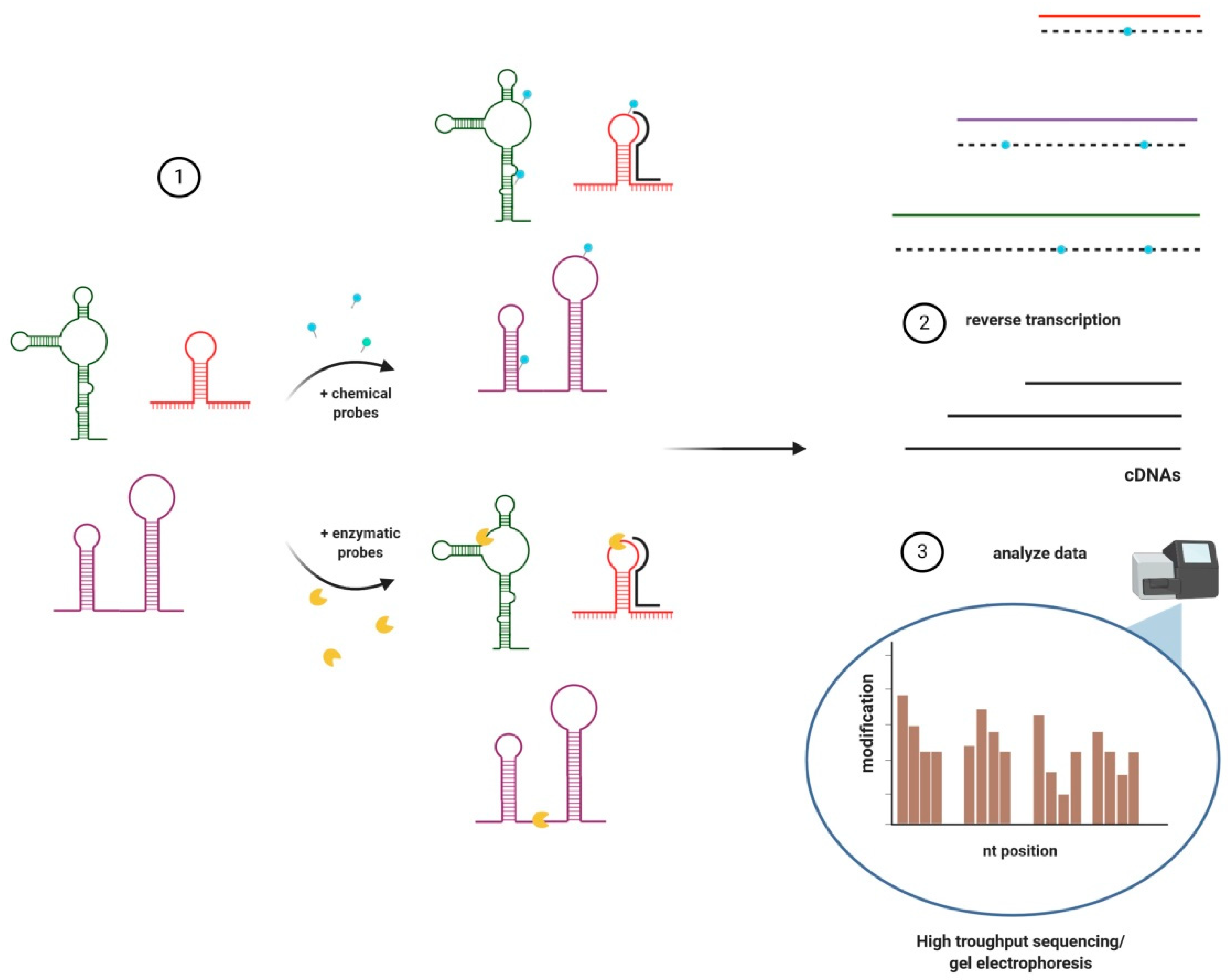
3. Structural Analyses of lncRNAs

The preferred tool to study the structure of lncRNAs is enzymatic/chemical probing, supported by computational RNA structure prediction methods (Figure 1) [62]. Although, other methods exist (see Table 1). The use of chemical probes or nucleases is based on the fact that RNA bases, whether they are free, pairing, or forming 3D structures, show different accessibility and reactivity to different types of probes. Binding of small molecule probes to RNAs leads to formation of adducts that cause reverse transcriptase (RT) to either create errors recognized as ’mutations’ or ’stop’ events during reverse transcription. The first results in modifications of newly synthesized cDNA while the latter causes the detachment of RT from the RNA fragment and a truncated cDNA [63,64]. These modifications are then positioned using capillary electrophoresis or sequencing techniques where cDNAs are compared with probed and untreated RNAs.
Enzymatic probing utilises nucleases recognising and cleaving specific sequences or structures e.g., single- or double-stranded regions (RNases such as P1 and V1, respectively), A’s (RNase A) and U’s (RNase U2) [63], which were later subjected to reverse transcription and sequencing. Both of these methods can be applied to purified lncRNA but only chemical probing is used in living cells (in vivo) due to the difficulties with the identification of lncRNAs secondary structures at the single-nucleotide level after nuclease digestion of millions of RNAs present in the cell. In general, data obtained after digestion with one nuclease shows only a structural trend (e.g., single- or double-stranded character), but in most cases, additional analysis using other nucleases or chemical probes are necessary [63].
In contrast, small molecule probes easily penetrate into living cells and provide better resolution [63,65]. In 2013, Novicova and collaborators described a new approach called 3S (Shotgun Secondary Structure) that is especially useful for analysing long RNAs [66]. It is based on parallel chemical probing of the entire lncRNA as well as multiple fragments of the lncRNA overlapping with each other. LncRNA fragments are prepared by in vitro transcription from dsDNA templates obtained from available cDNA clones or chemically synthesized by custom gene services. The obtained probing profiles of fragments are compared with the profile of the full RNA to estimate the identity/similarities. Regions with profiles similar to the full-length transcript are considered to have the same secondary structure. As the base-pairing partners within this region are not likely to occur outside the region, this method enables to map the structure of RNA through hierarchical probing of smaller and smaller fragments of the full RNA. 3S has been used for determination of the secondary structures of the best known lncRNAs: SRA, HOTAIR, COOLAIR, RepA, Braveheart and NEAT1 [66,67,68,69,70,71].
HTS is nevertheless considered the most specific technique to analyse lncRNA structures to date. The development of methodologies coupling HTS with ribonuclease cleavage or chemical probing facilitate RNA structure mapping in the context of whole transcriptome. For example, FragSeq (Fragmentation Sequencing) is based on P1 nuclease (which specifically degrades single-stranded nucleic acids) probing followed by HTS and bioinformatic analysis. The degradation in a site between two adjacent bases is characterized by a ‘cutting score’ reflecting the preference of P1 nuclease to digest comparing to other sites in the same RNA; as P1 cuts 3′ of an unpaired base, the uncleaved nucleotide targets may be engaged in base pairing or tertiary interactions [72]. The ds-RNA fragments left after nuclease-treatment are further separated according to size, cloned together with 5′ and 3′ specific adapters and sequenced. The obtained reads are used in mapping of single-stranded of RNA regions by comparing the sequence with sequencing libraries of multiple ncRNAs with known structure.

4. Identification of lncRNAs’ Binding Partners

Recently, initiatives such as lncRNA interactome mapping, single-complex mapping, and large-scale (network) mapping have been used for revealing the complexity of lncRNAs partnerships with other RNAs, proteins and/or DNA and thus provided some information about their potential functions.

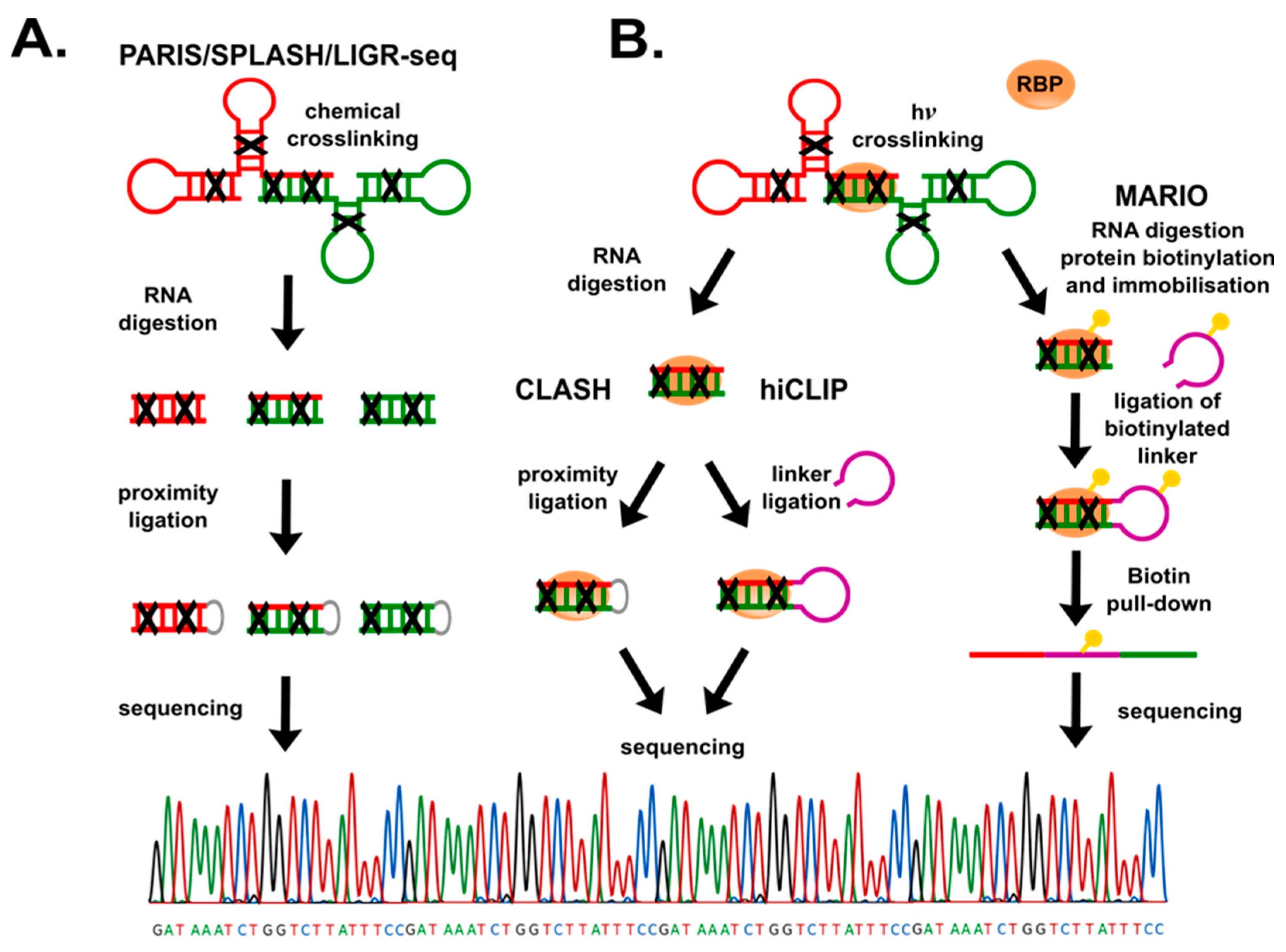
4.1. RNA-RNA Interactions

Interactions between pairing RNAs, or duplexes within the same molecule, are most commonly detected following one of two strategies: crosslinking of the two RNA strands or employing known RBPs. The former approach is used in techniques such as PARIS (Psoralen Analysis of RNA Interactions and Structures) [78], SPLASH (Sequencing of Psoralen crosslinked, Ligated, and Selected Hybrids) [79], and LIGR-seq (LIGation of interacting RNA followed by high-throughput sequencing) [80], where a photochemical reagent—psoralen, or its derivates, is used to (Figure 2A) intercalate and crosslink double stranded fragments of RNA (or DNA) molecules. Photoirradiation results in the formation of covalent adducts between psoralen and pyrimidine bases. An advantage of psoralen is that it is activated at higher wavelengths than standard UV light crosslinking (320–400 nm for psoralen vs. 260 nm for UV), causing less damage to nucleic acids.
Furthermore, the bridge formed between two RNA fragments is photoreversible by short wavelength (264 nm) UV light [81,82]. Psoralen crosslinking is followed by purification of RNA duplexes, ligation of adjacent nucleic acids termini (proximity ligation) and identification of interacting RNAs in cis and in trans, by sequencing. For instance, PARIS was used for analysis of structural organization of XIST, a nuclear lncRNA that triggers silencing of one of the X chromosomes in mammalian female cells, showing that four major coherent domains are kept by local duplexes within XIST [78]. Additionally, the authors reported the interactions between snRNA U1 and lncRNA MALAT1. The main drawback of psoralen-dependent methods is the relatively low number of uniquely mapped duplexes due to psoralen’s limited efficiency [35].
The second strategy includes pull-down of tagged RBP in complex with their interacting RNA(s). Most commonly used RBPs are Argonaute proteins [83], PUM2 (human Pumilio 2, a member of the Puf-protein family), QKI (Quaking homolog, START proteins family) [84], and small nucleolar RBPs [85] (see also Table 2), that bind sequence or structure-specific regions of RNA. The RBP-mediated methods (Figure 2B) such as CLASH (Cross-linking, Ligation And Sequencing of Hybrids) [86] and hiCLIP (RNA hybrid and individual-nucleotide resolution ultraviolet crosslinking and immunoprecipitation) [87], follow a similar strategy starting by overexpressing a known RBP that binds to RNAs duplexes. These methods have assisted the discovery of new snoRNA-rRNA interactions in yeast, and miRNA–mRNA dimers recognised by Argonaute in human cells [88], respectively. It is unknown whether overexpression of tagged-RBPs, although innovative, influence RNA–RNA–RBP interactions. MARIO (MApping RNA Interactome In vivo) [89] overcame such potential problem by UV light-crosslinking of any RNA–protein complexes, followed by RBP labelling by covalent biotin binding to amino acids such as lysine or tyrosine, or to carboxyl group on the C-terminal ends of proteins using biotin ligases. The RNA is then fragmented by RNase I, and the RNA–RBPs are then immobilised on streptavidin/avidin beads. The adjacent RNA ends are further joined by short biotin tagged RNA linkers. Next, biotin labelled complexes are purified, and RNAs are extracted for sequencing. MARIO has been used to capture the RNA interactome, including lincRNAs, based on the whole proteome [89].

4.2. RNA-DNA Interactions

The first eukaryotic lncRNA (H19) was identified in the pre-genomic era during cDNA library screens performed to study gene expression. Initially, it was considered an mRNA due to the presence of small ORFs [93]. Despite the fact that H19 does not code any protein, its dosage is important in embryonic development [94]. Shortly afterward, another lncRNA participating in dosage compensation, Xist, was identified as X-inactive-specific transcript, during the pioneering studies on X-chromosome inactivation (XCI) in mice [95]. Further studies on H19, Xist and newly identified lncRNAs revealed tight interplay between lncRNAs and DNA.
Evidence suggested that one of the most prominent roles of, at least some, lncRNAs was the regulation of chromatin structure [96], and several techniques were succesfully applied to decipher the molecular mechanisms of lncRNAs action. For example, Chromatin Isolation by RNA Purification (ChIRP), using hybridising biotinylated oligonucleotides, showed that HOTAIR acts as an active recruiter of chromatin-modifying complexes and is associated with relocalisation of PRC2 (Polycomb repressive complex 2)—a protein complex with H3K27 histone methytransferase activity that causes chromatin condensation and gene silencing. The lncRNA-dependent chromatin occupancy of PRC2 has been further confirmed by Long and collaborators [97], who reutilised rChIP (RNA-dependent Chromatin Immunoprecipitation) and coupled it with deep sequencing (rChIP-Seq).
Modifications of these techniques exist to improve specificity, for example by an initial RNase-H mapping assay to determine the most accessible sequences for probing. To map the chromatin interaction sites of lncRNAs, a ChIRP like, but more specific, method is commonly used: capture hybridization analysis of RNA targets (CHART), which uses short (22–28 nts) antisense biotinylated-oligonucleotides as probes to capture lncRNA-chromatin complexes [98]. This approach revealed that roX2, a Drosophila lncRNA, binds across gene bodies of X-linked genes exhibiting focal peaks of high occupancy at chromosomal entry sites [99]. In addition, RNA Antisense Purification (RAP) [53] uses antisense RNA probes (a bit longer than ChIRP: 120 nt for Xist), which improves specificity and minimizes background noise. RAP analyses showed that during maintenance of XCI, Xist is localized broadly across the entire X chromosome, lacking focal binding sites [53].
A significant limitation of all the above-mentioned methods is the need of knowing the sequence of the targeted nuclear lncRNA (nlncRNA). To overcome this obstacle, three new methods have been developed, namely: MARGI (MApping RNA-Genome Interactions) [100], GRID-seq (Global RNA Interactions with DNA by deep Sequencing) [101], and ChAR-seq (Chromatin-Associated RNA Sequencing) [102]. The common feature between them is the crosslinking of nlncRNAs with their DNA targets, followed by proximal ligation, fragmentation, and sequencing. The outcome is a detailed and comprehensive database of genome-wide interactions, showing that most of the nlncRNAs act as cis-regulators, and only a few notable nlncrNAs such as MALAT1, NEAT1, and roX2 are trans-regulators [101].

4.3. RNA-Protein Interactions

RNA–protein interactions can be studied either through the prism of RNA or that of protein. RNA-centric methods employ probes in similar ways as in RNA-RNA analyses [95], yet they differ in the downstream steps, usually involving mass spectrometry (MS) to determine protein identities. One such method is ChIRP-MS (Comprehensive Identification of RNA-binding Proteins by MS) [103], which captures lncRNA(s) by biotinylated oligonucleotides together with their binding proteins, and are identified by MS. The technique was successfully used to identify 81 Xist-interacting proteins, among them HnrnpU and HnrnpK emerged as the most abundant Xist-associated factors, and both functionally contribute to XCI [104]. The obvious goal of these methods is the identification of proteins interacting with a given RNA, which means to distinguish between covalently bounded RNA–protein and non-covalent interactions. In RAP-MS (RNA Antisense Purification coupled with quantitative MS) the purification of complexes is performed in denaturing and reducing conditions to disrupt the non-covalent interactions. Therefore, the mapping of specific RNA required long biotinylated probes, forming very stable RNA–DNA hybrids. Another challenge has been the quantification of interacting proteins vs. background proteins, what might be accomplished by isotopic labelling of amino acids and quantitative comparison of purified proteins by MS. At the end a short list of high confidence interactions is given, for example Xist interacts with SHARP and SMRT proteins, recruiting HDAC3 (Histone Deacetylase 3), and with PRC2 guiding the complex to the X chromosome for transcriptional silencing [105].
On the other hand, pulling down a known RBP with its lncRNA in complex with other proteins has been successfully carried out using two main approaches. The first involves the modification of the lncRNA, inserting an RNA sequence that is known to attract a specific RBP. The second option is to use a known RBP as a lure to pull down lncRNA–protein complexes. For example, the MS2 bacteriophage coat protein (MCP) tagged with the HB (histidine-biotin) tag, a peptide sequence that contains 6xHis and the 75-amino acid sequence of Propionibacterium shermanii’s transcarboxylase—which is efficiently biotinylated in eukaryotic cells by endogenous biotin ligases [106] has been used to pull down MS2 loop-tagged lncRNAs together with their interacting RBPs, a technique named MS2 in vivo Biotin Tagged RNA Affinity Purification (MS2-BioTRAP) [107]. The application of HB-tag allows to purify authentic complexes of MS2-RNA–RBPs [107]. As in all cases for protein identification, MS is the top choice.
In order to retain the RBP–lncRNA complexes, UV light cross-linking is most commonly used [108,109], followed by immunoprecipitation (CLIP) [110] and numerous derived methods such as high-throughput sequencing HITS-CLIP [111] for comprehensive identification of RBP target sequences at the transcriptome level, photoactivatable ribonucleoside enhanced PAR-CLIP where alternative ribonucleosides are used during RT at the position of cross-linking, which enables the detection of RNA–RBP interacting sites with nucleotide resolution and reduces background reads [84], or enhanced CLIP (eCLIP) where 3′ RNA linkers and further 3′ DNA linkers are ligated to increase the total number of non-PCR reads that can be obtained after HTS, often decreased due to the RT termination at the crosslink site [112]. For more information regarding the different variants of CLIP methods, we suggest the following review [17].

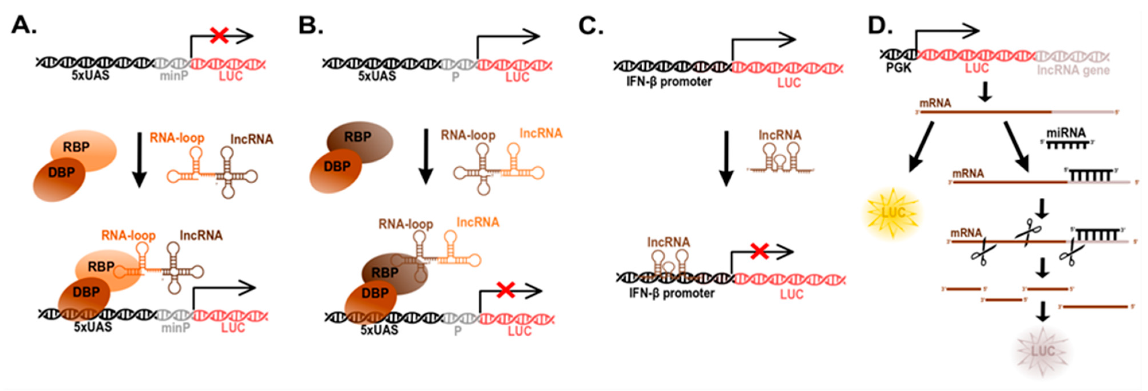
5. Determination of lncRNA Function (Reporter Systems)

Studies on the function of lncRNA have been hindered by lack of relevant tools. Thus, most lncRNA reporter systems have been created ab initio. As most of the known functions of lncRNAs have been discovered in nlncRNAs, such as XIST and HOTAIR, their functions have been assigned as gene or chromatin regulators. Such nlncRNAs often work as scaffolds of unsympathetic protein partners and/or guides of RNP complexes to particular loci—what results in local changes in transcription, chromatin structure histone modifications (methylation, acetylation) and/or DNA methylation [113,114]. Therefore, targeting nlncRNAs to synthetic promoter–reporter systems is currently the most common approach to determine the function of these enigmatic nucleic acids as epigenetic modulators.
Here, we shall separate the assays into two types: those to study gene repression and those to study gene activation (Figure 3). The practical difference between the two is that for the first a constitutively active promoter is used and thus repression is measured as the decrease of reporter signal, while for the latter a transcriptional complex must be delivered to a minimal promoter for transcriptional activation and reporter expression. Either of these promoters include a binding sequence, often in tandem repeats, for a DNA binding protein (DBP). The DBP is then engineered as a fusion with the RNA binding domain (RBD) of an RBP (Figure 3A), which might be an endogenous protein known to interact with the specific lncRNA or an artificial (exogenous) protein that binds to a specific RNA sequence/structure. As mentioned above, there are RBPs to natural or synthetic loops e.g., MCP-MS2 loops, and novel proteins that are able to bind RNA by guiding RNAs (see Table 2 for a list of the most commonly used RBPs).
As endogenous RBPs often bind to a plethora of RNAs, domesticated RBDs are far more specific and thus more commonly used. Synthetic RBPs usually bind to RNA-specific loops, these loops need to be artificially cloned in fusion with the lncRNA to be studied. Two of the most common loop binding proteins are λN and MCP, that bind to the BoxB and MS2 loops, respectively. By fusing a DBP, e.g., GAL4 or TtA, to either of these loop-binding proteins, they will drag (guide) the lncRNA to either a constitutive or a minimal promoter, depending on the desired effect (repression or transcriptional activation, respectively). Such promoters control the expression of a reporter gene (luciferase or fluorescent protein) (Figure 3). These systems have been used to verify the functions of lncRNAs such as NALT, LUNAR1, LINC00152 or HOTTIP as transcriptional activators [90,91,115,116], and HOTAIR as a transcriptional repressor [92] (Figure 3B).
It is of note that some lncRNAs bind directly to DNA sequences forming tertiary structures (Figure 3C). In some cases, the interaction lncRNA–DNA does not seem to be sequence-specific e.g., XIST or Ferre [53,117] who bind DNA in cis-, nearby its production site [118,119]; while in other cases, lncRNAs have targeted sequences. For example, the lncRNA CISAL contains a 22 nt DNA-recognition sequence (900 nts from its 3′-terminus) to the promoter of BRCA1. Binding to the BRCA1 promoter results in a triple helix structure, while another region of CISAL sequesters GABPA, a BRCA1-transcription factor, away from downstream regulatory regions, all of which uncouples BRCA1 transcription [120]. This study is of particular interest as the lncRNA-interacting sites for promoter and transcription factor were elegantly mapped by truncations and mutagenesis of all three partners (DNA, lncRNA and protein). Another example is lnc-MxA, a lncRNA that binds to the IFN-β promoter forming a triplex with the DNA and leading to interference of gene expression—as determined by a reporter bioassay (Figure 3C) [121].
LncRNAs as miRNA ‘sponges’ were first described in plants, where an Arabidopsis thaliana non-protein coding gene IPS1 (Induced by Starvation 1) harbouring a miR-399 targeting sequence was reported to sequester that miRNA away from mRNA [122]. To found more information about sponging miRNAs by lncRNAs we recommend the following article [123], here we will only focus on the development of an assay to analyse the activity of lncRNAs being ‘sponges’ of miRNAs (Figure 3D). Since miRNAs target mRNA for degradation, a miRNA sponge works by hijacking miRNAs and thus a positive regulator of mRNAs. Using a luciferase reporter system, the target region of a miRNA, often located in the 3′-untranslated region (3‘UTR) of the target’s mRNA, is cloned downstream of luciferase, thus upon miRNA expression luciferase expression is suppressed via miRNA-mRNA destabilization. Therefore, if co-expression of a lncRNA results in miRNA binding and regain expression of luciferase, it suggests that the lncRNA is a miRNA sponge. Further verification includes mutagenesis of the miRNA-binding site in the lncRNA of interest, to ensure direct effects [124].
Tools to unravel the impact of lncRNAs on chromatin condensation are still limited due to the complexity of the process. Yet, there are some groundbreaking examples such as a doxycycline (DOX)-inducible system regulating XIST that was integrated into multiple loci (different chromosomes) to investigate the effect of local XIST expression in the neighbouring DNA and chromatin environment. After DOX induction, XIST expression and localization was determined by RNA Fluorescence In-Situ Hybridization (FISH), showing that local gene silencing could be achieved by this lncRNA without the participation of other known, and thought to be essential, chromatin modifiers such as macroH2A, SMCHD1, and H3K27me3/H4K20me1. The analyses by allele-discriminating pyrosequencing assays revealed the pattern of genes silenced flanking the integration sites, while ChIP-seq showed that XIST –mediated silencing occurs at all sites tested, but the range of silencing to flanking endogenous human genes is variable [118].

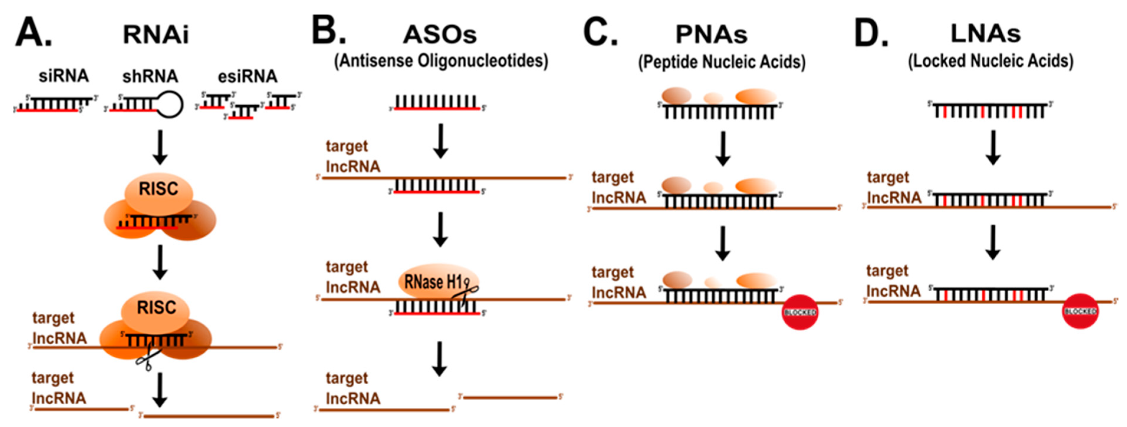
6. Mapping lncRNAs’ Functional Domains

LncRNAs, similarly to proteins, can acquire 3D structures, lncRNAs are often more dynamic shapeshifters than their amino acid counterparts, as well as having intrinsically disordered regions—similar to intrinsically disordered proteins [51]. This, turns out, allows these nucleic acids to use sequence-specific-binding to other RNAs or to DNA, to form docking structures for proteins, RNAs and DNA, and become magnets and guides for multi-component complexes between all three macromolecules. The detailed biological understanding of lncRNAs needs the identification of their functional domains in terms of specific motifs, local architecture, and/or posttranslational modifications. Analysing lncRNA is far more challenging than proteins because of the unique features of these molecules. The initial approaches to reveal lncRNA functional domains were based on the RNA sequence similarity but provided only limited useful information [125]. More informative data was obtained by analysing common structural RNA elements [126,127], which, as mentioned earlier, in many cases are evolutionary conserved [3,128]. Since the prediction of functionally relevant sequences requires careful verification by in vivo studies, a variety of methods to systematically map lncRNA functional domains are under development. Below, we summarize some of the previously used and most modern techniques.
Screening for functional molecules and/or functional domains has traditionally been performed by loss-of-function (LoF) studies. LncRNAs have been knocked-down by small interfering RNA (siRNA), endoribonuclease-prepared RNA (esiRNA) or short hairpin RNAs (shRNA) sampling for biologically relevant phenotypes (Figure 4A) e.g., stemness [4,129,130,131]. However, some of the approaches induced a large number of side-off effects, off-targeting on other transcripts, or detected only specific phenotypes in particular cell lines and/or conditions [132].
Similar to siRNA, ASOs (antisense oligonucleotides) have been applied to target certain regions of lncRNAs (Figure 4B) [133]. ASOs can hybridize with complementary RNA sequences and recruit endogenous RNase H to cleave the lncRNAs leading to either knock-down lncRNA or sterically block the access of functional domains without degradation [133]. Chemical modifications on ASOs improve their stability and reduce unwanted cell responses e.g., interferon response what makes them useful also for therapy of various diseases [134]. Importantly, ASOs can also enter the nucleus and thus effectively modulate activity of both cytoplasmic and nuclear lncRNAs [135].
Yet, the direct targeting of functional regions within a lncRNA was not done until Beletskii and co-workers developed Peptide Nucleic Acid (PNA)-Interference Mapping (P-IMP) (Figure 3C) [136]. PNAs mimic nucleic acids but having a peptide backbone, composed of charge neutral and achiral N-(2-aminoethyl) glycine units, which, unlike natural nucleic acids, protects them from nucleases [137]. Originally, this technique was used for mapping the regions of the murine Xist, revealing that a distinct repeat in the first exon is responsible for binding to genomic DNA [136].
Similarly, Sarma and collaborators used locked nucleic acid (LNA) technology to target and block key regions of Xist (Figure 3D) [138]. LNAs are nucleic acid analogues containing the ribose ring “locked” by a methylene bridge between the 2′ oxygen and the 4′ carbon, which results in higher stability and an increased affinity of LNAs to base pair with complementary RNA vs. DNA. In this way several domains of Xist engaged in its nuclear distribution were identified [138].
The advent of genome editing tools, in particular Clustered Regularly Interspaced Short Palindromic Repeats (CRISPR)/Cas9, brought several additional possibilities to the lncRNA field. First, it allowed for the permanent genetic modification of endogenous lncRNAs genes by targeting Cas9 nuclease using guide RNAs (gRNAs) to specific genetic sequences and introducing InDels (insertion or deletions) [139]. Indeed, systematic deletions of NEAT1 in a human haploid cell line resulted in the identification of a modular domain architecture of this lncRNA [140,141]. The authors depicted three functional NEAT1 domains playing distinct roles during formation of RNA–protein complexes called paraspeckles. Moreover, this approach was also effective to show three subdomains within the middle domain of NEAT1 responsible for paraspeckles assembly [140]. Since CRISPR/Cas9 is amenable for high-throughput screening, by using libraries of gRNAs, targeting unique genomic locations, single or multiple targets can be analysed downstream phenotypical screens. These screens have been very effective to identify the functional domains of proteins and recently have been adopted to non-coding genes too. For example, Tiling CRISPR, where a set of gRNAs is used to direct Cas9 to sequences covering the entire gene of interest (Figure 5), was successfully used to authenticate the region containing A-repeats as XIST silencing domain [54].

7. LncRNAs Databases and Bioinformatic Tools

The lncRNA databases and bioinformatic toolkit can greatly assist the research on lncRNAs. Some of available online tools for in silico lncRNA analysis provide rather general information, while others are more specific. The first includes ’meta-databases’ of lncRNAs such as LNCipedia, representing the most comprehensive compendia of human lncRNA [142]. It contains data from ten unique origins as of 2019, allowing embedded prognostication of coding potential, yet there is no automatic forecast of subcellular localization, correlation with disease or functional estimates. Contrary, LNCBook compiles lncRNAs from both previously established databases and experimentally verified transcripts selected by the community [143]. LNCBook tenders multi-omic data integration such as expression profiles in normal and carcinogenic tissues, function annotation and disease association, DNA methylation patterns in various gene regions, genome variability and forecasts of microRNA (miRNA) synergies using miRanda [143,144] and TargetScan algorithms [145]. Both LNCipedia and LNCBook extend embedded coding potential foresight through predictor of lncRNAs and mRNAs based on specific pattern containing k nucleotides called k-mer (PLEK) [146] and Coding Potential Alignment Tool (CPAT) [147]. As this field is evolving rapidly and numerous new transcripts are steadily being distinguished, these databases should be frequently updated to improve their potential.
The second group of tools is dedicated for analysing particular features of lncRNAs and refers to targeted databases; for example, databases of lncRNA expression in human tissues (GTEx) [148], cancers (TANRIC) [149], and plants (CANTATAdb) [150], or lncRNA localization such as LncSLdb [151] or LncATLAS [152], and the most popular predictor of subcellular localization- LncLocator [153]. In addition, essential in lncRNAs research are tools designed to separate transcripts encoding micropeptides from authentic non-coding RNAs, such as Coding Potential Prediction (CPPred) [154] as well as PhyloCSF [155] and COME [156]. Recently, a freely available mobile-friendly web server- Coding-Non-coding Identifying Tool (CNIT) [157] based on the Coding-Non-Coding Index (CNCI) [158] database, was introduced for researchers.
At present, the only available tools for structure prediction of lncRNAs are those dedicated to all RNA molecules. One of which is the RNA Mapping Database (RMDB) that facilitate access and meta-analysis of the diverse structural mapping experiments performed on ribonucleic acids [159]. To date the use of RMDB for lncRNA may be limited, however the rapid development of high-throughput techniques may contribute to increasing the functionality of this database [160]. Ultimately, RNAfold [161] or DMfold [162] can be used to predict lncRNA structure. The RNAfold predicts minimum free energy (MFE) folding for RNA secondary structure and equilibrium base-pairing probabilities, both algorithms are considering also circRNA [163]. The limitation of RNAfold with predicting pseudoknots can be solved using DMfold, which is based on deep learning [162]. Though DMfold exhibits a high accuracy in predicting lncRNA structure, it may be less accessible due to its command line utility [160].
Despite the growing demand, the availability of in silico methods and predictive tools for mapping lncRNAs functional domains is still poor. Since single bioinformatics tools are unable to truly estimate the structure: function of lncRNAs, a bioinformatic tool—Predicting LncRNA Activity through Integrative Data-driven Omics and Heuristics (PLAIDOH)—has been created to combine data from the transcriptome, subcellular localization, enhancer landscape, genome architecture, chromatin interaction, and RNA-binding (eCLIP) analyses, providing statistically defined output scores [164]. This approach also provides the interactome between individual lncRNA, coding genes, and protein pairs using enhancer, transcript cis-regulatory, and RBP data to enhance its predicting capacity.
In contrast, there are several databases for predicting lncRNA partnerships. Databases, such as IntaRNA 2.0 [165] or LncRRIsearch [166] are dedicated to predict lncRNA—RNA interactions, while TheLnChrom [167] and Triplexator [168] to predict lncRNA–DNA interactions. For their detailed comparison we recommend the following article [160]. In addition, many tools focus on lncRNA–protein interaction, such as NPInter [169,170], starBase [171] and several others [172]. Additionally, RNA Interactome Repository-RNAInter is more comprehensive tool collecting information from published data along with another 35 database resources [173].
Recently, to predict lncRNA function a new approach of sequence evaluation based on comparison of short sequence elements (k-mer) representation (SEEKR), was developed [174]. As lncRNAs with similar k-mer content have been shown to exhibit related functions despite their lack of linear homology, the SEEKR database provides very useful information about some determinants of lncRNAs function. In addition, lnChrom [167] and lncRNADisease [175] databases collect lncRNA-disease associations.

8. LncRNAs and Disease

Since multiple lncRNAs have been found playing roles as regulatory elements in gene expression, it is of little surprise that they take part in physiological and disease processes [5]. We summarize them in the section below, with the exception of their potential role as biomarkers in cancers, which we describe in a separate chapter.
LncRNAs have rather clear and unique expression patterns, which makes them good markers for health and disease conditions. Changes in expression of many lncRNAs are specific to the tissue, developmental stage and/or conditions (see Table 3).
Many reports describe the dysregulation of lncRNAs in multifactorial chronic diseases [187]. Starting with neurodegenerative diseases such as Alzheimer [188,189,190], Parkinson’s [191] and Huntington’s [177]; through autism spectrum disorders [187,192,193]; and schizophrenia [194,195], cardiovascular diseases, such as during chronic heart failure [196,197], atherosclerosis [198,199], myocardial infarction [200], and diabetic cardiomyopathy [201]. Moreover, as lncRNAs are implicated at many levels of metabolism regulation, perturbations in their expression serve as an important component for the occurrence of metabolic diseases. In a comprehensive study, Morán and collaborators identified a series of pancreatic islet lncRNAs (e.g., HI-LNC12, HI-LNC25, HI-LNC75, and HI-LNC78) dynamically regulated during β-cell differentiation and maturation, two other (KCNQ1OT1 and HILNC45) that are dysregulated in type 2 diabetes, and a set of lncRNAs that map to human diabetes genetic susceptibility loci [202]. Among other lncRNAs having diagnostic value are TUNA associated with the intensity of Huntington’s disease [177], MALAT1 upregulated in myocardial infarction [203], and Mhrt downregulated in cardiac hypertrophy [204].
In addition, data gained from the studies on infectious diseases show that lncRNAs have been associated with the regulation of both pro- and anti-inflammatory processes [205,206,207,208]. Moreover, lncRNAs seem to play critical roles in the regulation of pathways in autoimmune diseases such as rheumatoid arthritis [209,210], psoriasis [7], and Chron’s disease [211]. Expression of lncRNAs changes in response to various pathogens—likely due to deregulation of host immune responses [212,213,214]. There seems to be a footprint pattern of lncRNAs expression depending of the pathogen, as has been shown by studies with Escherichia coli [215], Salmonella typhimurium [216], Mycobacterium tuberculosis [217,218,219,220], Campylobacter oncisus [221], or Helicobacter pylori [222,223], also after infection by the human immunodeficiency virus (HIV) [224], ZIKA virus, Sendai virus, hepatitis C virus [205] and COVID-19 [225].
A recent report on Toxoplasmosa gondii infection, for the first time, suggests that protozoan infections also alter lncRNA expression [226]. The molecular mechanisms at play by these lncRNAs during infections are yet to be discovered. Virtually no information is available on lncRNA effects during protozoan invasion, which might be an interesting area of research, in particular if there are common patterns versus infectious agent or host responses—beneficial or deleterious—as they could be potential drug targets. Considering the poor sequence conservation along lncRNAs, it will be highly interesting to compare the different species responses to infections, which might not only bring new information to human medicine, but to zoonosis and veterinary medicine, as well.

9. Applications of lncRNAs

9.1. Diagnostic Biomarkers in Cancers and Targets for Therapy

It is widely known that early detection of tumorigenesis greatly increases the chances for successful treatment and survival. Numerous studies have shown that several lncRNAs are associated with the stage and prognosis of multiple tumour types [31,227,228,229,230] such as breast [231,232,233,234], lung [235,236], gastric [75], colorectal [237,238], and prostate cancers [239]. Additionally, the overexpression of multiple lncRNAs e.g., AC006050.3-003 and ODRUL in cancer cells increases their resistance to DNA damaging agents [240,241], suggesting that these lncRNAs could be used as prognosis markers. To date, current cancer diagnosis biomarkers are unreliable due to the high number of false positive and false negative results. Yet, since lncRNAs have highly specific expression patterns, their background in biological samples is virtually nonexistent and their presence can be detected in biological fluids [189,242,243,244,245,246]: whole blood [247], urine [248,249], serum [250,251], saliva [252], and gastric juice samples [253].
Their high specificity allows for the detection of cancer initiation (e.g., SPRY4-IT1) [254], progression (e.g., ATB) [255], metastasis (e.g., LINC00461, CCAT2 and H19) [256,257,258] and response to therapy (e.g., MALAT1 or HOTAIR) [77,259,260]. An additional important group of lncRNAs is the exosomal lncRNAs—secreted by cancer cells. They might play pivotal roles in tumorigenesis (e.g., H19 in cervical cancer and hepatocellular carcinoma, ZFAS1 in gastric carcinoma) and might be excellent cancer biomarkers (e.g., MALAT1, HOTAIR, both in cervical and bladder cancer). The functions, if any, of these exosomal lncRNAs are yet to be unravelled [261,262,263].
As lncRNA can be easily amplified and quantitated, this equals high sensitivity and specificity, easy and minimally invasive sample collection in contrast with conventional biopsies, and inexpensive methods [264]. A future perspective is to make them suitable for routine procedures in clinical practice and use for diagnostic purposes.
The therapeutic potential of lncRNAs is another crucial aspect. Recent reports have highlighted the relationship between lncRNA dysregulation and resistance to chemotherapy and targeted therapy [265,266,267,268], inhibition of signal transduction [15,269], and resistance to anti-hormone therapies [265,268,270]. For instance, overexpression of HOTAIR enhances the proliferation of breast cancer cells, while its depletion by shRNA considerably reduces cell survival and decreases growth during anti-hormone therapy (tamoxifen) [74]. Similarly, Adriaens and collaborators [271] demonstrated that the sensitivity of precancerous cells to DNA damaging or chemotherapy drugs was enhanced by knockout lncRNA-Neat1 in mice and NEAT1 in MCF-7 cells. Furthermore, the depletion of potentially oncogenic lncRNA SNHG15 by siRNA or CRISPR-Cas9, decreased cell proliferation and invasion, and tumorigenic capacity of CRC cells while overexpression of this lncRNA directed to opposite phenotype [272]. Similarly, deletion of LINC00538 (YIYA) in human breast cancer strongly inhibited the tumour growth and invasion in vitro and suppressed tumour growth in a mouse xenograft model [273].
Therefore, many potential oncogenic lncRNAs are currently being tested as targets for therapy (Figure 6). For example, targeting lncRNA MALAT1 with ASO induced differentiation and inhibited metastasis breast cancer in the mouse mammary tumour virus-PyMT (MMTV) carcinoma model [274,275]. The anti-metastatic effect of knocking down MALAT1 was also reported in a lung cancer xenograft model [276]. Silencing of a lncRNA called Cancer Susceptibility Candidate 9 (CASC9), who is associated with various processes in several cancer types, provided conflicting results, as it decreased the migration and invasion potential of ESCC (Esophageal Squamous Cell Carcinoma) cells while promoted their apoptotic potential of breast cancer cells MDA-MB-415 and MCF-7/DOX [277].
The comprehensive studies on lncRNAs associated with Notch oncogenic signalling in T-cell acute lymphoblastic leukaemia (T-ALL) uncovered a novel target candidate for treatment of this aggressive haematological disease, namely LUNAR1 (leukaemia-induced non-coding activator RNA 1) [115]. Silencing LUNAR1 in T-ALL cells suppressed the expression of IGF1R gene crucial for T-ALL tumour growth and caused significant cell growth retardation. Recently, LUNAR1 was found expressed in colorectal cancer (CRC), expanding the promise of LUNAR1 as a therapeutic target [278].
Targeting lncRNAs might not be straightforward; nevertheless, the removal of regulatory lncRNAs might cause complex changes in the cell. This was illustrated when the Colon Cancer-Associated Transcript 1 (CCAT1) was erased by CRISPR/Cas9, which led to the dysregulation of genes involved in several biological processes, including metabolism, cell migration, proliferation; and CCAT1-null cells lost their ability to anchorage-independent growth [279].
In addition to silencing a lncRNA, also adding or replacing it might have clinical benefits. In chronic metabolic disease such as atherosclerosis, lncRNA LeXis (Liver-expressed LXR-induced sequence) gene therapy resulted in positive outcomes. Studies on mice showed significantly reduced atherosclerotic load in mice treated with a vector expressing LeXis compared to control (the vector expressing Green Fluorescent Protein) treated mice [182]. Taking into account that there is an orthologue of LeXis in humans, such data might have clinical implications.

9.2. Silencing of Single Chromosome/Trisomy Effects

As described above, XCI is the result of a lncRNA—XIST activity. With this in mind, Yiang and collaborators developed a system to silence a single chromosome in a trisomy with the use of XIST [280]. They introduced an inducible XIST transgene to one of the three Chr21 in Down Syndrome (DS) patient-derived pluripotent stem cells using zinc finger nucleases. Induction of XIST expression resulted in the repression of virtually all genes across the autosome, and a total chromosome 21 transcriptional output near normal disomic levels which also rescued the cells phenotype [281]. That was the pioneering work on „chromosome therapy”, a technique that could have implications in many other chromosomal disorders, some of which are fatal in the first 1–2 years of life (e.g., trisomy Chr13 and Chr18).

9.3. Tissue/Muscle Regeneration

In a series of unexpected discoveries, lncRNAs have been found to modulate stem/progenitor cells physiology, including cells for engineering tissues. Growing evidence supports the importance of lncRNAs in different cellular lineages (neuronal, liver, skin, muscle and vascular tissue) growth, maintenance, proliferation, migration, and differentiation (for review see [282]), which suggests that lncRNAs can be applicable for tissue engineering. For example, lncRNA Dum (Developmental pluripotency-associated 2 (Dppa2) Upstream binding Muscle lncRNA) is induced during the early regeneration stage, when satellite cells become activated, proliferate and start to differentiate [283]. Dum acts in cis by silencing transcription of its upstream neighbour gene, Dppa2, encoding a pluripotency regulator, promoting then early differentiation. Dum interacts with and recruit DNA methyltransferases (Dnmts) to Dppa2 promoter, leading to CpG site hypermethylation and Dppa2 gene silencing. Dum knockdown in vivo impaired the injury-induced muscle regeneration, suggesting an important role of this lncRNA in the regulation of myogenesis [283]. Therefore, the careful and coordinated regulation of expression of lncRNAs, might be essential to engineer more physiologically relevant tissues for transplantation.
In terms of the bigger picture, extensive research on lncRNAs structure, partnerships and function might provide powerful outcomes in the future for different branches of medicine supporting therapeutic modalities for various pathogen infections, anticancer therapy, “chromosome therapy” and transplantology.

10. Discussion

Since their discovery, we have made great progress in our understanding of lncRNAs, as numerous studies have documented the significant contribution of lncRNAs to multiple regulatory mechanisms essential for mammalian homeostasis and development. However, the detailed functions of lncRNAs remain largely unknown. ‘Decoding’ lncRNAs in terms of revealing the key aspects determining their function is an ambitious goal due to their low expression in the cell, flexible structure, weak sequence conservation and diversity of functions.
The structural architecture of lncRNAs is still an unexplored field. With the rise of sequencing techniques such as single-cell RNA-seq (scRNA-seq), many cell-type-specific lncRNAs have been identified, and it is likely that many structural architectures and mechanisms will be revealed. Initially, the identification of common short sequence elements, similar structural features and structure-function relationships in lncRNAs might be applied. The resolving of lncRNA structure will need a combination of high-resolution global imaging techniques with secondary structural determinations. Moreover, the plasticity of all RNA molecules dynamically responding to the presence of their interacting partners, cellular context and environmental conditions are factors that cannot be ommited. While it is notoriously difficult to crystallize RNA molecules, cryoEM seems to work very well for RNA, and it negates the need for crystals, requires minimal sample and can differentiate biochemical and conformational heterogeneities of the samples. Thus, applying this powerful technique might enable the breakthrough in lncRNA studies similarly to the revolutionizing the structural biology of macromolecular protein assemblies, in the future. Structural maps can then be used to guide detailed biophysical explorations of lncRNAs cell functions and study the effect of mutations on lncRNA activity as well as elucidating their involvement in disease occurrence and designing lncRNA- targeting drugs.
Though the number of platforms for annotating lncRNA functions is growing, the highly efficient methods with enhanced targeting, higher resolution and increased maneuverability at the molecular level still need to be developed. The assessment of lncRNAs functions in cells requires a thorough analysis including gene perturbation experiments such as overexpression and downregulation, followed by real-time quantitative PCRs or deep sequencing, to observe any differential gene expressions. Importantly, not all lncRNAs exhibit functions in cells and laboratory model animals or are difficult to knock down or overexpress due to their enormous sizes and/or genomic architecture.
Understanding the structural biology and mechanism of action of lncRNAs will give the necessary foundation of the identification of therapeutics targeting these molecules with high specificity. Presently, the most popular method to target lncRNAs is through oligonucleotide-based therapies, yet CRISPR-Cas9-based techniques are also of a great potential. Importantly, the interactive network between lncRNAs and their partners, including other noncoding RNAs, particularly miRNAs, is another research field that should be explored in future studies. This complex relationship might also alter the disease’s level occurrence, course, or response to therapy. As mechanisms of action, the design of oligonucleotides and methods for targeting lncRNAs continue to be researched it is expected that a number of compounds efficient for lncRNA-based treatment of a wide variety of diseases will be obtained.

11. Conclusions

LncRNAs represent a new class of RNA that fine-tune complex physiological processes and the onset of diseases. The detailed understanding of their functions, structures and interactomes is challenging as the conventional methods used to study mRNA functions are inefficient for lncRNAs. In this review we summarized the interdisciplinary efforts undertaken to characterise lncRNAs. In many cases, a combination of several techniques was successfully applied; for example, for predicting structure of these molecules the bioinformatic predictions, followed by enzymatic or chemical probing of lncRNAs and HTS. Moreover, the ability of lncRNAs to interact with DNA, RNA, and proteins to exert their functions, is raising their complexity to a higher level. To explore the interactome of lncRNA techniques based on crosslinking RNAs with their partners, employing known RBPs, labeling RNA–protein complexes or adapting chromatin IP with oligonucleotide probing of RNA are coupled with sequencing or MS, depending on the target molecules. Yet, the nature and dynamics of such interactions need to be elucidated in the future. Functional studies on lncRNAs required new tools dedicated for their versatile activities in the cell. For the determination of lncRNAs function and map functional elements currently used methodologies target certain regions or whole lncRNAs gene through RNAi, ASOs, PNAs or CRISPR/Cas9. The improvement of these methods is of great value, as lncRNAs are connected with a wide spectrum of diseases. Exploring their biology, disease-related lncRNAs will gain greater relevance as potential biomarkers in cancers and for personalized medicine, especially for gene therapy.

Author Contributions

Conceptualization, L.B.; writing—original draft preparation, L.B., J.K., K.D., A.P.-P., I.T.; writing—review and editing, L.B., J.K., K.D., A.P.-P., I.T.; visualization, J.K., K.D.; supervision, L.B.; project administration, L.B.; funding acquisition, K.D. All authors have read and agreed to the published version of the manuscript.

Funding

This research was funded by the Polish National Science Centre (NCN): DEC-2017/25/B/NZ4/02364, and Polish National Agency for Academic Exchange (NAWA): PPI/APM/2019/1/00089/DEC/1, and Medical University of Lublin DS441 2021/2022 grant.

Acknowledgments

The authors thank Adolfo Rivero-Müller for his insightful comments in reviewing the manuscript.

Conflicts of Interest

The authors declare no conflict of interest.

References

  1. Harrow, J.; Frankish, A.; Gonzalez, J.M.; Tapanari, E.; Diekhans, M.; Kokocinski, F.; Aken, B.L.; Barrell, D.; Zadissa, A.; Searle, S.; et al. GENCODE: The reference human genome annotation for the ENCODE project. Genome Res. 2012, 22, 1760–1774. [Google Scholar] [CrossRef] [Green Version]
  2. Saghafi, T.; Ali Taheri, R.; Parkkila, S.; Emameh, R.Z. Phytochemicals as modulators of long non-coding RNAs and inhibitors of cancer-related carbonic anhydrases. Int. J. Mol. Sci. 2019, 20, 2939. [Google Scholar] [CrossRef] [Green Version]
  3. Derrien, T.; Johnson, R.; Bussotti, G.; Tanzer, A.; Djebali, S.; Tilgner, H.; Guernec, G.; Martin, D.; Merkel, A.; Knowles, D.G.; et al. The GENCODE v7 catalog of human long noncoding RNAs: Analysis of their gene structure, evolution, and expression. Genome Res. 2012, 22, 1775–1789. [Google Scholar] [CrossRef] [Green Version]
  4. Guttman, M.; Amit, I.; Garber, M.; French, C.; Lin, M.F.; Feldser, D.; Huarte, M.; Zuk, O.; Carey, B.W.; Cassady, J.P.; et al. Chromatin signature reveals over a thousand highly conserved large non-coding RNAs in mammals. Nature 2009, 458, 223–227. [Google Scholar] [CrossRef] [PubMed]
  5. Fernandes, J.C.R.; Acuña, S.M.; Aoki, J.I.; Floeter-Winter, L.M.; Muxel, S.M. Long non-coding RNAs in the regulation of gene expression: Physiology and disease. Non-Coding RNA 2019, 5, 17. [Google Scholar] [CrossRef] [PubMed] [Green Version]
  6. Niemczyk, M.; Ito, Y.; Huddleston, J.; Git, A.; Abu-Amero, S.; Caldas, C.; Moore, G.E.; Stojic, L.; Murrell, A. Imprinted chromatin around DIRAS3 regulates alternative splicing of GNG12-AS1, a long noncoding RNA. Am. J. Hum. Genet. 2013, 93, 224–235. [Google Scholar] [CrossRef] [PubMed] [Green Version]
  7. Yan, J.; Song, J.; Qiao, M.; Zhao, X.; Li, R.; Jiao, J.; Sun, Q. Long noncoding RNA expression profile and functional analysis in psoriasis. Mol. Med. Rep. 2019, 49, 3421–3430. [Google Scholar] [CrossRef] [PubMed] [Green Version]
  8. Quinn, J.J.; Chang, H.Y. Unique features of long non-coding RNA biogenesis and function. Nat. Rev. Genet. 2016, 17, 47–62. [Google Scholar] [CrossRef] [PubMed]
  9. Wang, K.C.; Chang, H.Y. Molecular Mechanisms of Long Noncoding RNAs. Mol. Cell 2011, 43, 904–914. [Google Scholar] [CrossRef] [Green Version]
  10. Mongelli, A.; Martelli, F.; Farsetti, A.; Gaetano, C. The dark that matters: Long noncoding RNAs as master regulators of cellular metabolism in noncommunicable diseases. Front. Physiol. 2019, 10, 369. [Google Scholar] [CrossRef] [Green Version]
  11. Zhao, Y.; Zhao, J.; Guo, X.; She, J.; Liu, Y. Long non-coding RNA PVT1, A molecular sponge for miR-149, contributes aberrant metabolic dysfunction and inflammation in IL-1β-simulated osteoarthritic chondrocytes. Biosci. Rep. 2018, 38, BSR20180576. [Google Scholar] [CrossRef] [PubMed] [Green Version]
  12. Chen, L.; Hu, N.; Wang, C.; Zhao, H.; Gu, Y. Long non-coding RNA CCAT1 promotes multiple myeloma progression by acting as a molecular sponge of miR-181a-5p to modulate HOXA1 expression. Cell Cycle 2018, 17, 319–329. [Google Scholar] [CrossRef] [PubMed] [Green Version]
  13. Zhang, X.; Wu, J.; Wu, C.; Chen, W.; Lin, R.; Zhou, Y.; Huang, X. The LINC01138 interacts with PRMT5 to promote SREBP1-mediated lipid desaturation and cell growth in clear cell renal cell carcinoma. Biochem. Biophys. Res. Commun. 2018, 507, 337–342. [Google Scholar] [CrossRef] [PubMed]
  14. Polycarpou-Schwarz, M.; Groß, M.; Mestdagh, P.; Schott, J.; Grund, S.E.; Hildenbrand, C.; Rom, J.; Aulmann, S.; Sinn, H.P.; Vandesompele, J.; et al. The cancer-associated microprotein CASIMO1 controls cell proliferation and interacts with squalene epoxidase modulating lipid droplet formation. Oncogene 2018, 37, 4750–4768. [Google Scholar] [CrossRef] [PubMed]
  15. Lin, A.; Hu, Q.; Li, C.; Xing, Z.; Ma, G.; Wang, C.; Li, J.; Ye, Y.; Yao, J.; Liang, K.; et al. The LINK-A lncRNA interacts with PtdIns(3,4,5)P3 to hyperactivate AKT and confer resistance to AKT inhibitors. Nat. Cell Biol. 2017, 19, 238–251. [Google Scholar] [CrossRef]
  16. Yan, C.; Chen, J.; Chen, N. Long noncoding RNA MALAT1 promotes hepatic steatosis and insulin resistance by increasing nuclear SREBP-1c protein stability. Sci. Rep. 2016, 6, 22640. [Google Scholar] [CrossRef]
  17. Li, J.; Chen, C.; Ma, X.; Geng, G.; Liu, B.; Zhang, Y.; Zhang, S.; Zhong, F.; Liu, C.; Yin, Y.; et al. Long noncoding RNA NRON contributes to HIV-1 latency by specifically inducing tat protein degradation. Nat. Commun. 2016, 7, 11730. [Google Scholar] [CrossRef]
  18. Legnini, I.; Morlando, M.; Mangiavacchi, A.; Fatica, A.; Bozzoni, I. A Feedforward Regulatory Loop between HuR and the Long Noncoding RNA linc-MD1 Controls Early Phases of Myogenesis. Mol. Cell 2014, 53, 506–514. [Google Scholar] [CrossRef] [Green Version]
  19. Kok, F.O.; Baker, A.H. The function of long non-coding RNAs in vascular biology and disease. Vasc. Pharmacol. 2019, 114, 23–30. [Google Scholar] [CrossRef]
  20. Pang, Y.; Mao, C.; Liu, S. Encoding activities of non-coding RNAs. Theranostics 2018, 8, 2496–2507. [Google Scholar] [CrossRef]
  21. Van Solingen, C.; Scacalossi, K.R.; Moore, K.J. Long noncoding RNAs in lipid metabolism. Curr. Opin. Lipidol. 2018, 29, 224–232. [Google Scholar] [CrossRef]
  22. Zhang, Y.; Zhang, X.O.; Chen, T.; Xiang, J.F.; Yin, Q.F.; Xing, Y.H.; Zhu, S.; Yang, L.; Chen, L.L. Circular Intronic Long Noncoding RNAs. Mol. Cell 2013, 51, 792–806. [Google Scholar] [CrossRef] [Green Version]
  23. Zhang, Y.; Xue, W.; Li, X.; Zhang, J.; Chen, S.; Zhang, J.L.; Yang, L.; Chen, L.L. The Biogenesis of Nascent Circular RNAs. Cell Rep. 2016, 15, 611–624. [Google Scholar] [CrossRef] [Green Version]
  24. Barrett, S.P.; Wang, P.L.; Salzman, J. Circular RNA biogenesis can proceed through an exon-containing lariat precursor. eLife 2015, 4, e07540. [Google Scholar] [CrossRef]
  25. Salzman, J.; Gawad, C.; Wang, P.L.; Lacayo, N.; Brown, P.O. Circular RNAs are the predominant transcript isoform from hundreds of human genes in diverse cell types. PLoS ONE 2012, 7, e30733. [Google Scholar] [CrossRef] [PubMed] [Green Version]
  26. Li, Z.; Huang, C.; Bao, C.; Chen, L.; Lin, M.; Wang, X.; Zhong, G.; Yu, B.; Hu, W.; Dai, L.; et al. Exon-intron circular RNAs regulate transcription in the nucleus. Nat. Struct. Mol. Biol. 2015, 22, 256–264. [Google Scholar] [CrossRef] [PubMed]
  27. Huang, S.; Yang, B.; Chen, B.J.; Bliim, N.; Ueberham, U.; Arendt, T.; Janitz, M. The emerging role of circular RNAs in transcriptome regulation. Genomics 2017, 109, 401–407. [Google Scholar] [CrossRef] [PubMed]
  28. Ragan, C.; Goodall, G.J.; Shirokikh, N.E.; Preiss, T. Insights into the biogenesis and potential functions of exonic circular RNA. Sci. Rep. 2019, 9, 2048. [Google Scholar] [CrossRef] [PubMed]
  29. Sleutels, F.; Zwart, R.; Barlow, D.P. The non-coding Air RNA is required for silencing autosomal imprinted genes. Nature 2002, 415, 810–813. [Google Scholar] [CrossRef]
  30. Matouk, I.J.; DeGroot, N.; Mezan, S.; Ayesh, S.; Abu-Lail, R.; Hochberg, A.; Galun, E. The H19 non-coding RNA is essential for human tumor growth. PLoS ONE 2007, 2, e845. [Google Scholar] [CrossRef] [Green Version]
  31. Zhou, X.; Chen, H.; Zhu, L.; Hao, B.; Zhang, W.; Hua, J.; Gu, H.; Jin, W.; Zhang, G. Helicobacter pylori infection related long noncoding RNA (lncRNA) AF147447 inhibits gastric cancer proliferation and invasion by targeting MUC2 and up-regulating miR-34c. Oncotarget 2016, 7, 82770–82782. [Google Scholar] [CrossRef] [PubMed] [Green Version]
  32. Imamura, K.; Takaya, A.; Ishida, Y.; Fukuoka, Y.; Taya, T.; Nakaki, R.; Kakeda, M.; Imamachi, N.; Sato, A.; Yamada, T.; et al. Diminished nuclear RNA decay upon Salmonella infection upregulates antibacterial noncoding RNAs. EMBO J. 2018, 37, e97723. [Google Scholar] [CrossRef] [PubMed]
  33. Hashimoto, K.; Ishida, E.; Matsumoto, S.; Shibusawa, N.; Okada, S.; Monden, T.; Satoh, T.; Yamada, M.; Mori, M. A liver X receptor (LXR)-β alternative splicing variant (LXRBSV) acts as an RNA co-activator of LXR-β. Biochem. Biophys. Res. Commun. 2009, 390, 1260–1265. [Google Scholar] [CrossRef] [PubMed]
  34. Fabbri, M.; Girnita, L.; Varani, G.; Calin, G.A. Decrypting noncoding RNA interactions, structures, and functional networks. Genome Res. 2019, 29, 1377–1388. [Google Scholar] [CrossRef] [Green Version]
  35. Qian, X.; Zhao, J.; Yeung, P.Y.; Zhang, Q.C.; Kwok, C.K. Revealing lncRNA Structures and Interactions by Sequencing-Based Approaches. Trends Biochem. Sci. 2019, 44, 33–52. [Google Scholar] [CrossRef]
  36. Busan, S.; Weeks, K.M. Visualization of RNA structure models within the Integrative Genomics Viewer. RNA 2017, 23, 1012–1018. [Google Scholar] [CrossRef] [Green Version]
  37. Uroda, T.; Anastasakou, E.; Rossi, A.; Teulon, J.M.; Pellequer, J.L.; Annibale, P.; Pessey, O.; Inga, A.; Chillón, I.; Marcia, M. Conserved Pseudoknots in lncRNA MEG3 Are Essential for Stimulation of the p53 Pathway. Mol. Cell 2019, 75, 982–995.e9. [Google Scholar] [CrossRef] [PubMed] [Green Version]
  38. Wan, Y.; Qu, K.; Ouyang, Z.; Kertesz, M.; Li, J.; Tibshirani, R.; Makino, D.L.; Nutter, R.C.; Segal, E.; Chang, H.Y. Genome-wide Measurement of RNA Folding Energies. Mol. Cell 2012, 48, 169–181. [Google Scholar] [CrossRef] [PubMed] [Green Version]
  39. Rouskin, S.; Zubradt, M.; Washietl, S.; Kellis, M.; Weissman, J.S. Genome-wide probing of RNA structure reveals active unfolding of mRNA structures in vivo. Nature 2014, 505, 701–705. [Google Scholar] [CrossRef] [Green Version]
  40. Tapsin, S.; Sun, M.; Shen, Y.; Zhang, H.; Lim, X.N.; Susanto, T.T.; Yang, S.L.; Zeng, G.S.; Lee, J.; Lezhava, A.; et al. Genome-wide identification of natural RNA aptamers in prokaryotes and eukaryotes. Nat. Commun. 2018, 9, 1289. [Google Scholar] [CrossRef]
  41. Zhou, K.I.; Parisien, M.; Dai, Q.; Liu, N.; Diatchenko, L.; Joseph, R.; Pan, T.; Program, S.T.; Division, B.S. Hairpin Predisposes Its Conformation To Protein Binding. J. Mol. Biol. 2017, 428, 822–833. [Google Scholar] [CrossRef] [PubMed] [Green Version]
  42. Pintacuda, G.; Young, A.N.; Cerase, A. Function by structure: Spotlights on xist long non-coding RNA. Front. Mol. Biosci. 2017, 4, 90. [Google Scholar] [CrossRef] [Green Version]
  43. Amaral, P.P.; Clark, M.B.; Gascoigne, D.K.; Dinger, M.E.; Mattick, J.S. LncRNAdb: A reference database for long noncoding RNAs. Nucleic Acids Res. 2011, 39 (Suppl. 1), 146–151. [Google Scholar] [CrossRef] [PubMed] [Green Version]
  44. Korostowski, L.; Raval, A.; Breuer, G.; Engel, N. Enhancer-driven chromatin interactions during development promote escape from silencing by a long non-coding RNA. Epigenetics Chromatin 2011, 4, 21. [Google Scholar] [CrossRef] [PubMed] [Green Version]
  45. Vallot, C.; Patrat, C.; Collier, A.J.; Huret, C.; Casanova, M.; Liyakat Ali, T.M.; Tosolini, M.; Frydman, N.; Heard, E.; Rugg-Gunn, P.J.; et al. XACT Noncoding RNA Competes with XIST in the Control of X Chromosome Activity during Human Early Development. Cell Stem Cell 2017, 20, 102–111. [Google Scholar] [CrossRef]
  46. Gong, C.; Maquat, L.E. ALUstrious long ncRNAs and their roles in shortening mRNA half-lives. Cell Cycle 2011, 10, 1882–1883. [Google Scholar] [CrossRef] [Green Version]
  47. Filbin, M.E.; Kieft, J.S. Towards a structural understanding of IRES RNA function. Bone 2009, 19, 267–276. [Google Scholar] [CrossRef] [Green Version]
  48. Xia, X.; Holcik, M. Strong eukaryotic IRESs have weak secondary structure. PLoS ONE 2009, 4, e4136. [Google Scholar] [CrossRef]
  49. Fernández, N.; Fernandez-Miragall, O.; Ramajo, J.; García-Sacristán, A.; Bellora, N.; Eyras, E.; Briones, C.; Martínez-Salas, E. Structural basis for the biological relevance of the invariant apical stem in IRES-mediated translation. Nucleic Acids Res. 2011, 39, 8572–8585. [Google Scholar] [CrossRef]
  50. May, J.; Johnson, P.; Saleem, H.; Simon, A.E. A Sequence-Independent, Unstructured Internal Ribosome Entry Site Is Responsible for Internal Expression of the Coat Protein of Turnip Crinkle Virus. J. Virol. 2017, 91, e02421-16. [Google Scholar] [CrossRef] [Green Version]
  51. Uversky, V.N. Intrinsically disordered proteins and their ‘Mysterious’ (meta)physics. Front. Phys. 2019, 7, 8–23. [Google Scholar] [CrossRef] [Green Version]
  52. Novikova, I.V.; Hennelly, S.P.; Sanbonmatsu, K.Y. Structural architecture of the human long non-coding RNA, steroid receptor RNA activator. Nucleic Acids Res. 2012, 40, 5034–5051. [Google Scholar] [CrossRef]
  53. Engreitz, J.M.; Pandya-Jones, A.; McDonel, P.; Shishkin, A.; Sirokman, K.; Surka, C.; Kadri, S.; Xing, J.; Goren, A.; Lander, E.S.; et al. The Xist lncRNA exploits three-dimensional genome architecture to spread across the X chromosome. Science 2013, 341, 1237973. [Google Scholar] [CrossRef] [Green Version]
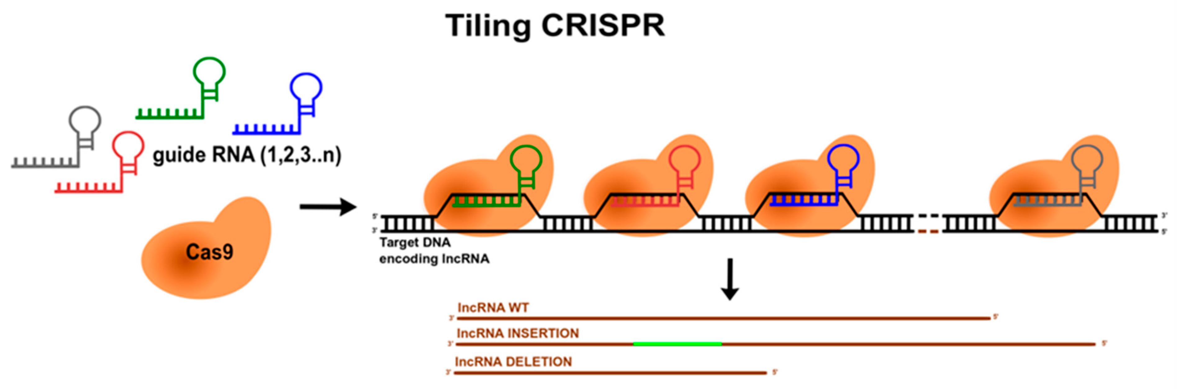
  54. Wang, Y.; Zhong, Y.; Zhou, Y.; Tanaseichuk, O.; Li, Z.; Zhao, J.C. Identification of a Xist silencing domain by Tiling CRISPR. Sci. Rep. 2019, 9, 2408. [Google Scholar] [CrossRef] [Green Version]
  55. Smola, M.J.; Christy, T.W.; Inoue, K.; Nicholson, C.O.; Friedersdorf, M.; Keene, J.D.; Lee, D.M.; Calabrese, J.M.; Weeks, K.M. SHAPE reveals transcript-wide interactions, complex structural domains, and protein interactions across the Xist lncRNA in living cells. Proc. Natl. Acad. Sci. USA 2016, 113, 10322–10327. [Google Scholar] [CrossRef] [Green Version]
  56. Duszczyk, M.M.; Wutz, A.; Rybin, V.; Sattler, M. The Xist RNA A-repeat comprises a novel AUCG tetraloop fold and a platform for multimerization. RNA 2011, 17, 1973–1982. [Google Scholar] [CrossRef] [PubMed] [Green Version]
  57. Lu, Z.; Guo, J.K.; Wei, Y.; Dou, D.R.; Zarnegar, B.; Ma, Q.; Li, R.; Zhao, Y.; Liu, F.; Choudhry, H.; et al. Structural modularity of the XIST ribonucleoprotein complex. Nat. Commun. 2020, 11, 6163. [Google Scholar] [CrossRef] [PubMed]
  58. Nitsche, A.; Rose, D.; Fasold, M.; Reiche, K.; Stadler, P.F. Comparison of splice sites reveals that long noncoding RNAs are evolutionarily well conserved. RNA 2015, 21, 801–812. [Google Scholar] [CrossRef] [Green Version]
  59. Hezroni, H.; Koppstein, D.; Schwartz, M.G.; Avrutin, A.; Bartel, D.P.; Ulitsky, I. Principles of Long Noncoding RNA Evolution Derived from Direct Comparison of Transcriptomes in 17 Species. Cell Rep. 2015, 11, 1110–1122. [Google Scholar] [CrossRef] [PubMed] [Green Version]
  60. Orosz, F.; Ovádi, J. Proteins without 3D structure: Definition, detection and beyond. Bioinformatics 2011, 27, 1449–1454. [Google Scholar] [CrossRef] [Green Version]
  61. Vishwanath, S.; de Brevern, A.G.; Srinivasan, N. Same but not alike: Structure, flexibility and energetics of domains in multi-domain proteins are influenced by the presence of other domains. PLoS Comput. Biol. 2018, 14, e1006008. [Google Scholar] [CrossRef] [Green Version]
  62. Strobel, E.J.; Yu, A.M.; Lucks, J.B. High-throughput determination of RNA structures. Nat. Rev. Genet. 2018, 19, 615–634. [Google Scholar] [CrossRef]
  63. Ziehler, W.A.; Engelke, D.R. Probing RNA Structure with Chemical Reagents and Enzymes. Curr. Protoc. Nucleic Acid Chem. 2000, 6.1.1–6.1.21. [Google Scholar] [CrossRef]
  64. Weeks, K.M. Advances in RNA secondary and tertiary structure analysis by chemical probing. Curr. Opin. Struct. Biol. 2010, 20, 295–304. [Google Scholar] [CrossRef] [PubMed] [Green Version]
  65. Martens, L.; Rühle, F.; Stoll, M. LncRNA secondary structure in the cardiovascular system. Non-Coding RNA Res. 2017, 2, 137–142. [Google Scholar] [CrossRef]
  66. Novikova, I.V.; Dharap, A.; Hennelly, S.P.; Sanbonmatsu, K.Y. 3S: Shotgun secondary structure determination of long non-coding RNAs. Methods 2013, 63, 170–177. [Google Scholar] [CrossRef] [PubMed]
  67. Somarowthu, S.; Legiewicz, M.; Chilló, I.; Marcia, M.; Liu, F.; Pyle, A.M. HOTAIR forms an intricate and modular secondary structure. Mol. Cell 2015, 58, 353–361. [Google Scholar] [CrossRef] [PubMed] [Green Version]
  68. Hawkes, E.J.; Hennelly, S.P.; Novikova, I.V.; Irwin, J.A.; Dean, C.; Sanbonmatsu, K.Y. COOLAIR Antisense RNAs Form Evolutionarily Conserved Elaborate Secondary Structures. Cell Rep. 2016, 16, 3087–3096. [Google Scholar] [CrossRef] [PubMed] [Green Version]
  69. Liu, F.; Somarowthu, S.; Pyle, A.M. Visualizing the secondary and tertiary architectural domains of lncRNA RepA. Nat. Chem. Biol. 2017, 13, 282–289. [Google Scholar] [CrossRef] [PubMed]
  70. Xue, Z.; Hennelly, S.; Doyle, B.; Gulati, A.A.; Novikova, I.V.; Sanbonmatsu, K.Y.; Boyer, L.A.; Xue, Z.; Hennelly, S.; Doyle, B.; et al. Article A G-Rich Motif in the lncRNA Braveheart Interacts with a Zinc-Finger Transcription Factor to Specify the Cardiovascular Lineage Article A G-Rich Motif in the lncRNA Braveheart Interacts with a Zinc-Finger Transcription Factor to Specify the Cardio. Mol. Cell 2016, 64, 37–50. [Google Scholar] [CrossRef] [Green Version]
  71. Lin, Y.; Schmidt, B.F.; Bruchez, M.P.; McManus, C.J. Structural analyses of NEAT1 lncRNAs suggest long-range RNA interactions that may contribute to paraspeckle architecture. Nucleic Acids Res. 2018, 46, 3742–3752. [Google Scholar] [CrossRef] [PubMed]
  72. Underwood, J.G.; Uzilov, A.V.; Katzman, S.; Onodera, C.S.; Mainzer, J.E.; Mathews, D.H.; Lowe, T.M.; Salama, S.R.; Haussler, D. FragSeq: Transcriptome-wide RNA structure probing using high-throughput sequencing. Nat. Methods 2010, 7, 995–1001. [Google Scholar] [CrossRef] [PubMed]
  73. Ilik, I.A.; Quinn, J.J.; Georgiev, P.; Tavares-Cadete, F.; Maticzka, D.; Toscano, S.; Wan, Y.; Spitale, R.C.; Luscombe, N.; Backofen, R.; et al. Tandem stem-loops in roX RNAs act together to mediate X Chromosome dosage compensation in Drosophila. Mol. Cell 2013, 51, 156–173. [Google Scholar] [CrossRef] [PubMed] [Green Version]
  74. Xue, X.; Yang, Y.A.; Zhang, A.; Fong, K.-W.; Kim, J.; Song, B.; Li, S.; Zhao, J.C.; Yu, J. LncRNA HOTAIR enhances ER signaling and confers tamoxifen resistance in breast cancer. Oncogene 2015, 77, 1571–1578. [Google Scholar] [CrossRef] [PubMed] [Green Version]
  75. Zhang, K.; Shi, H.; Xi, H.; Wu, X.; Cui, J.; Gao, Y.; Liang, W.; Hu, C.; Liu, Y.; Li, J.; et al. Genome-wide lncRNA microarray profiling identifies novel circulating lncrnas for detection of gastric cancer. Theranostics 2017, 7, 213–227. [Google Scholar] [CrossRef]
  76. Ponti, R.D.; Armaos, A.; Marti, S.; Tartaglia, G.G. A method for RNA structure prediction shows evidence for structure in lncRNAs. Front. Mol. Biosci. 2018, 5, 111. [Google Scholar] [CrossRef] [Green Version]
  77. Brown, J.A.; Bulkley, D.; Wang, J.; Valenstein, M.L.; Yario, T.A.; Steitz, T.A.; Steitz, J.A. Structural insights into the stabilization of MALAT1 noncoding RNA by a bipartite triple helix. Nat. Struct. Mol. Biol. 2014, 21, 633–640. [Google Scholar] [CrossRef]
  78. Lu, Z.; Zhang, Q.C.; Lee, B.; Flynn, R.A.; Smith, M.A.; Robinson, J.T.; Davidovich, C.; Gooding, A.R.; Goodrich, K.J.; Mattick, J.S.; et al. RNA Duplex Map in Living Cells Reveals Higher-Order Transcriptome Structure. Cell 2016, 165, 1267–1279. [Google Scholar] [CrossRef] [Green Version]
  79. Aw, J.G.A.; Shen, Y.; Wilm, A.; Sun, M.; Lim, X.N.; Boon, K.L.; Tapsin, S.; Chan, Y.S.; Tan, C.P.; Sim, A.Y.L.; et al. In Vivo Mapping of Eukaryotic RNA Interactomes Reveals Principles of Higher-Order Organization and Regulation. Mol. Cell 2016, 62, 603–617. [Google Scholar] [CrossRef] [Green Version]
  80. Sharma, E.; Sterne-Weiler, T.; O’Hanlon, D.; Blencowe, B.J. Global Mapping of Human RNA-RNA Interactions. Mol. Cell 2016, 62, 618–626. [Google Scholar] [CrossRef] [Green Version]
  81. Sastry, S.S.; Ross, B.M.; P’arraga, A. Cross-linking of DNA-binding proteins to DNA with psoralen and psoralen furan-side monoadducts: Comparison of action spectra with DNA-DNA cross- linking. J. Biol. Chem. 1997, 272, 3715–3723. [Google Scholar] [CrossRef] [Green Version]
  82. Simon, M.D. Insight into lncRNA biology using hybridization capture analyses. Biochim. Biophys. Acta-Gene Regul. Mech. 2016, 1859, 121–127. [Google Scholar] [CrossRef] [Green Version]
  83. Meister, G. Argonaute proteins: Functional insights and emerging roles. Nat. Rev. Genet. 2013, 14, 447–459. [Google Scholar] [CrossRef]
  84. Hafner, M.; Landthaler, M.; Burger, L.; Khorshid, M.; Hausser, J.; Berninger, P.; Rothballer, A.; Ascano, M.; Jungkamp, A.C.; Munschauer, M.; et al. Transcriptome-wide Identification of RNA-Binding Protein and MicroRNA Target Sites by PAR-CLIP. Cell 2010, 141, 129–141. [Google Scholar] [CrossRef] [Green Version]
  85. Granneman, S.; Kudla, G.; Petfalski, E.; Tollervey, D. Identification of protein binding sites on U3 snoRNA and pre-rRNA by UV cross-linking and high-throughput analysis of cDNAs. Proc. Natl. Acad. Sci. USA 2009, 106, 9613–9618. [Google Scholar] [CrossRef] [Green Version]
  86. Helwak, A.; Kudla, G.; Dudnakova, T.; Tollervey, D. Mapping the human miRNA interactome by CLASH reveals frequent noncanonical binding. Cell 2013, 153, 654–665. [Google Scholar] [CrossRef] [Green Version]
  87. Sugimoto, Y.; Vigilante, A.; Darbo, E.; Zirra, A.; Militti, C.; D’Ambrogio, A.; Luscombe, N.M.; Ule, J. HiCLIP reveals the in vivo atlas of mRNA secondary structures recognized by Staufen 1. Nature 2015, 519, 491–494. [Google Scholar] [CrossRef] [Green Version]
  88. Kashi, K.; Henderson, L.; Bonetti, A.; Carninci, P. Discovery and functional analysis of lncRNAs: Methodologies to investigate an uncharacterized transcriptome. Biochim. Biophys. Acta-Gene Regul. Mech. 2016, 1859, 3–15. [Google Scholar] [CrossRef] [PubMed] [Green Version]
  89. Nguyen, T.C.; Cao, X.; Yu, P.; Xiao, S.; Lu, J.; Biase, F.H.; Sridhar, B.; Huang, N.; Zhang, K.; Zhong, S. Mapping RNA-RNA interactome and RNA structure in vivo by MARIO. Nat. Commun. 2016, 7, 12023. [Google Scholar] [CrossRef] [PubMed] [Green Version]
  90. Wang, Y.; Wu, P.; Lin, R.; Rong, L.; Xue, Y.; Fang, Y. LncRNA NALT interaction with NOTCH1 promoted cell proliferation in pediatric T cell acute lymphoblastic leukemia. Sci. Rep. 2015, 5, 13749. [Google Scholar] [CrossRef] [PubMed] [Green Version]
  91. Ji, J.; Tang, J.; Deng, L.; Xie, Y.; Jiang, R.; Li, G.; Sun, B. LINC00152 promotes proliferation in hepatocellular carcinoma by targeting EpCAM via the mTOR signaling pathway. Oncotarget 2015, 6, 42813–42824. [Google Scholar] [CrossRef] [PubMed] [Green Version]
  92. Portoso, M.; Ragazzini, R.; Brenčič, Ž.; Moiani, A.; Michaud, A.; Vassilev, I.; Wassef, M.; Servant, N.; Sargueil, B.; Margueron, R. PRC 2 is dispensable for HOTAIR—Mediated transcriptional repression. EMBO J. 2017, 36, 981–994. [Google Scholar] [CrossRef]
  93. Pachnis, V.; Belayew, A.; Tilghman, S.M. Locus unlinked to α-fetoprotein under the control of the murine raf and Rif genes. Proc. Natl. Acad. Sci. USA 1984, 81, 5523–5527. [Google Scholar] [CrossRef] [PubMed] [Green Version]
  94. Brannan, C.I.; Dees, E.C.; Ingram, R.S.; Tilghman, S.M. The product of the H19 gene may function as an RNA. Mol. Cell. Biol. 1990, 10, 28–36. [Google Scholar] [CrossRef] [PubMed] [Green Version]
  95. Borsani, G.; Tonlorenzi, R.S.M. Characterization of a murine gene expressed from the inactive X chromosome. Nature 1991, 354, 56–58. [Google Scholar] [CrossRef] [PubMed]
  96. Koziol, M.J.; Rinn, J.L. RNA traffic control of chromatin complexes. Curr. Opin. Genet. Dev. 2010, 20, 142–148. [Google Scholar] [CrossRef] [Green Version]
  97. Long, Y.; Hwang, T.; Gooding, A.R.; Goodrich, K.J.; Rinn, J.L.; Cech, T.R. RNA is essential for PRC2 chromatin occupancy and function in human pluripotent stem cells. Nat. Genet. 2020, 52, 931–938. [Google Scholar] [CrossRef]
  98. Simon, M.D.; Wang, C.I.; Kharchenko, P.V.; West, J.A.; Chapman, B.A.; Alekseyenko, A.A.; Borowsky, M.L.; Kuroda, M.I.; Kingston, R.E. The genomic binding sites of a noncoding RNA. Proc. Natl. Acad. Sci. USA 2011, 108, 20497–20502. [Google Scholar] [CrossRef] [PubMed] [Green Version]
  99. Chu, C.; Qu, K.; Zhong, F.L.; Artandi, S.E.; Chang, H.Y. Genomic Maps of Long Noncoding RNA Occupancy Reveal Principles of RNA-Chromatin Interactions. Mol. Cell 2011, 44, 667–678. [Google Scholar] [CrossRef] [PubMed] [Green Version]
  100. Sridhar, B.; Rivas-Astroza, M.; Nguyen, T.C.; Chen, W.; Yan, Z.; Cao, X.; Hebert, L.; Zhong, S. Systematic Mapping of RNA-Chromatin Interactions In Vivo. Curr. Biol. 2017, 27, 602–609. [Google Scholar] [CrossRef] [Green Version]
  101. Li, X.; Zhou, B.; Chen, L.; Gou, L.T.; Li, H.; Fu, X.D. GRID-seq reveals the global RNA-chromatin interactome. Nat. Biotechnol. 2017, 35, 940–950. [Google Scholar] [CrossRef]
  102. Bell, J.C.; Jukam, D.; Teran, N.A.; Risca, V.I.; Smith, O.K.; Johnson, W.L.; Skotheim, J.M.; Greenleaf, W.J.; Straight, A.F. Chromatin-associated RNA sequencing (ChAR-seq) maps genome-wide RNA-to-DNA contacts. eLife 2018, 7, e27024. [Google Scholar] [CrossRef] [PubMed]
  103. Ramanathan, M.; Porter, D.F.; Khavari, P.A. Methods to study RNA–protein interactions. Nat. Methods 2019, 16, 225–234. [Google Scholar] [CrossRef]
  104. Chu, C.; Zhang, Q.C.; Da Rocha, S.T.; Flynn, R.A.; Bharadwaj, M.; Calabrese, J.M.; Magnuson, T.; Heard, E.; Chang, H.Y. Systematic discovery of Xist RNA binding proteins. Cell 2015, 161, 404–416. [Google Scholar] [CrossRef] [PubMed] [Green Version]
  105. McHugh, C.A.; Chen, C.K.; Chow, A.; Surka, C.F.; Tran, C.; McDonel, P.; Pandya-Jones, A.; Blanco, M.; Burghard, C.; Moradian, A.; et al. The Xist lncRNA interacts directly with SHARP to silence transcription through HDAC3. Nature 2015, 521, 232–236. [Google Scholar] [CrossRef] [PubMed]
  106. Tagwerker, C.; Flick, K.; Cui, M.; Guerrero, C.; Dou, Y.; Auer, B.; Baldi, P.; Huang, L.; Kaiser, P. A tandem affinity tag for two-step purification under fully denaturing conditions: Application in ubiquitin profiling complex identification combined with in vivo cross-linking. Mol. Cell. Proteom. 2006, 5, 737–748. [Google Scholar] [CrossRef] [Green Version]
  107. Tsai, B.P.; Wang, X.; Huang, L.; Waterman, M.L. Quantitative profiling of in vivo-assembled RNA-protein complexes using a novel integrated proteomic approach. Mol. Cell. Proteom. 2011, 10, M110.007385. [Google Scholar] [CrossRef] [Green Version]
  108. Smith, K.C.; Aplin, R.T. A Mixed Photoproduct of Uracil and Cysteine (5-S-Cysteine-6-hydrouracil). A Possible Model for the in Vivo Cross-Linking of Deoxyribonucleic Acid and Protein by Ultraviolet Light. Biochemistry 1966, 5, 2125–2130. [Google Scholar] [CrossRef]
  109. Goddard, J.; Streeter, D.; Weber, C.; Gordon, M.P. Studies on the Inactivation of Tobacco Mosaic Virus By Ultraviolet Light. Photochem. Photobiol. 1966, 5, 213–222. [Google Scholar] [CrossRef]
  110. Ule, J.; Jensen, K.B.; Ruggiu, M.; Mele, A.; Ule, A.; Darnell, R.B. CLIP Identifies Nova-Regulated RNA Networks in the Brain. Science 2003, 302, 1212–1215. [Google Scholar] [CrossRef]
  111. Licatalosi, D.D.; Mele, A.; Fak, J.J.; Ule, J.; Kayikci, M.; Chi, S.W.; Clark, T.A.; Schweitzer, A.C.; Blume, J.E.; Wang, X.; et al. HITS-CLIP yields genome-wide insights into brain alternative RNA processing. Nature 2008, 456, 464–469. [Google Scholar] [CrossRef] [PubMed] [Green Version]
  112. Van Nostrand, E.L.; Pratt, G.A.; Shishkin, A.A.; Gelboin-Burkhart, C.; Fang, M.Y.; Sundararaman, B.; Blue, S.M.; Nguyen, T.B.; Surka, C.; Elkins, K.; et al. Robust transcriptome-wide discovery of RNA-binding protein binding sites with enhanced CLIP (eCLIP). Nat. Methods 2016, 13, 508–514. [Google Scholar] [CrossRef]
  113. Marchese, F.P.; Raimondi, I.; Huarte, M. The multidimensional mechanisms of long noncoding RNA function. Genome Biol. 2017, 18, 206. [Google Scholar] [CrossRef] [PubMed] [Green Version]
  114. Zhang, X.; Wang, W.; Zhu, W.; Dong, J.; Cheng, Y.; Yin, Z.; Shen, F. Mechanisms and functions of long non-coding RNAs at multiple regulatory levels. Int. J. Mol. Sci. 2019, 20, 5573. [Google Scholar] [CrossRef] [Green Version]
  115. Trimarchi, T.; Bilal, E.; Ntziachristos, P.; Fabbri, G.; Dalla-Favera, R.; Tsirigos, A.; Aifantis, I. Genome-wide mapping and characterization of notch-regulated long noncoding RNAs in acute leukemia. Cell 2014, 158, 593–606. [Google Scholar] [CrossRef] [PubMed] [Green Version]
  116. Shang, A.; Wang, W.; Gu, C.; Chen, C.; Zeng, B.; Yang, Y.; Ji, P.; Sun, J.; Wu, J.; Lu, W.; et al. Long non-coding RNA HOTTIP enhances IL-6 expression to potentiate immune escape of ovarian cancer cells by upregulating the expression of PD-L1 in neutrophils. J. Exp. Clin. Cancer Res. 2019, 38, 411. [Google Scholar] [CrossRef] [PubMed]
  117. Hacisuleyman, E.; Goff, L.A.; Trapnell, C.; Williams, A.; Henao-Mejia, J.; Sun, L.; Mcclanahan, P.; Hendrickson, D.G.; Sauvageau, M.; Kelley, D.R.; et al. Topological Organization of Multi-chromosomal Regions by Firre HHS Public Access. Nat. Struct. Mol. Biol. 2014, 21, 198–206. [Google Scholar] [CrossRef]
  118. Kelsey, A.D.; Yang, C.; Leung, D.; Minks, J.; Dixon-McDougall, T.; Baldry, S.E.L.; Bogutz, A.B.; Lefebvre, L.; Brown, C.J. Impact of flanking chromosomal sequences on localization and silencing by the human non-coding RNA XIST. Genome Biol. 2015, 16, 208. [Google Scholar] [CrossRef] [PubMed] [Green Version]
  119. Wutz, A.; Rasmussen, T.P.; Jaenisch, R. Chromosomal silencing and localization are mediated by different domains of Xist RNA. Nat. Genet. 2002, 30, 167–174. [Google Scholar] [CrossRef]
  120. Fan, S.; Tian, T.; Lv, X.; Lei, X.; Yang, Z.; Liu, M.; Liang, F.; Li, S.; Lin, X.; Lin, Z.; et al. lncRNA CISAL Inhibits BRCA1 Transcription by Forming a Tertiary Structure at Its Promoter. iScience 2020, 23, 100835. [Google Scholar] [CrossRef] [PubMed] [Green Version]
  121. Li, X.; Guo, G.; Lu, M.; Chai, W.; Li, Y.; Tong, X.; Li, J.; Jia, X.; Liu, W.; Qi, D.; et al. Long Noncoding RNA Lnc-MxA Inhibits Beta Interferon Transcription by Forming RNA-DNA Triplexes at Its Promoter. J. Virol. 2019, 93, e00786-19. [Google Scholar] [CrossRef] [Green Version]
  122. Franco-Zorrilla, J.M.; Valli, A.; Todesco, M.; Mateos, I.; Puga, M.I.; Rubio-Somoza, I.; Leyva, A.; Weigel, D.; García, J.A.; Paz-Ares, J. Target mimicry provides a new mechanism for regulation of microRNA activity. Nat. Genet. 2007, 39, 1033–1037. [Google Scholar] [CrossRef]
  123. López-Urrutia, E.; Bustamante Montes, L.P.; Ladrón de Guevara Cervantes, D.; Pérez-Plasencia, C.; Campos-Parra, A.D. Crosstalk Between Long Non-coding RNAs, Micro-RNAs and mRNAs: Deciphering Molecular Mechanisms of Master Regulators in Cancer. Front. Oncol. 2019, 9, 669. [Google Scholar] [CrossRef]
  124. Cui, P.; Su, J.; Li, Q.; Xu, G.; Zhu, N. LncRNA RHPN1-AS1 targeting miR-625/REG3A promotes cell proliferation and invasion of glioma cells. OncoTargets Ther. 2019, 12, 7911–7921. [Google Scholar] [CrossRef] [Green Version]
  125. Noviello, T.M.R.; Di Liddo, A.; Ventola, G.M.; Spagnuolo, A.; D’Aniello, S.; Ceccarelli, M.; Cerulo, L. Detection of long non-coding RNA homology, a comparative study on alignment and alignment-free metrics. BMC Bioinform. 2018, 19, 407. [Google Scholar] [CrossRef] [Green Version]
  126. Seemann, S.E.; Mirza, A.H.; Hansen, C.; Bang-berthelsen, C.H. The identification and functional annotation of RNA. Genome Res. 2017, 27, 1371–1383. [Google Scholar] [CrossRef] [PubMed] [Green Version]
  127. Thiel, B.C.; Ochsenreiter, R.; Gadekar, V.P.; Tanzer, A.; Hofacker, I.L. RNA structure elements conserved between mouse and 59 other vertebrates. Genes 2018, 9, 392. [Google Scholar] [CrossRef] [Green Version]
  128. Sprague, D.; Waters, S.A.; Kirk, J.M.; Wang, J.R.; Samollow, P.B.; Waters, P.D.; Calabrese, J.M. Nonlinear sequence similarity between the Xist and Rsx long noncoding RNAs suggests shared functions of tandem repeat domains. RNA 2019, 25, 1004–1019. [Google Scholar] [CrossRef] [PubMed]
  129. Chakraborty, D.; Kappei, D.; Theis, M.; Nitzsche, A.; Ding, L.; Paszkowski-Rogacz, M.; Surendranath, V.; Berger, N.; Schulz, H.; Saar, K.; et al. Combined RNAi and localization for functionally dissecting long noncoding RNAs. Nat. Methods 2012, 9, 360–362. [Google Scholar] [CrossRef] [PubMed]
  130. Kittler, R.; Pelletier, L.; Heninger, A.K.; Slabicki, M.; Theis, M.; Miroslaw, L.; Poser, I.; Lawo, S.; Grabner, H.; Kozak, K.; et al. Genome-scale RNAi profiling of cell division in human tissue culture cells. Nat. Cell Biol. 2007, 9, 1401–1412. [Google Scholar] [CrossRef]
  131. Huang, J.; Zhou, N.; Watabe, K.; Lu, Z.; Wu, F.; Xu, M.; Mo, Y.-Y. Long non-coding RNA UCA1 promotes breast tumor growth by suppression of p27 (Kip1). Cell Death Dis. 2014, 5, e1008. [Google Scholar] [CrossRef]
  132. Goff, L.A.; Rinn, J.L. Linking RNA biology to lncRNAs. Genome Res. 2015, 25, 1456–1465. [Google Scholar] [CrossRef] [Green Version]
  133. Chiang, M.Y.; Chan, H.; Zounes, M.A.; Freier, S.M.; Lima, W.F.; Bennett, C.F. Antisense oligonucleotides inhibit intercellular adhesion molecule 1 expression by two distinct mechanisms. J. Biol. Chem. 1991, 266, 18162–18171. [Google Scholar] [CrossRef]
  134. Bennett, C.F. Therapeutic Antisense Oligonucleotides Are Coming of Age. Annu. Rev. Med. 2019, 70, 307–321. [Google Scholar] [CrossRef] [PubMed]
  135. Behlke, M. Mini-review on current strategies to knockdown long non-coding RNAs. J. Rare Dis. Res. Treat. 2016, 1, 66–70. [Google Scholar] [CrossRef]
  136. Beletskii, A.; Hong, Y.K.; Pehrson, J.; Egholm, M.; Strauss, W.M. PNA interference mapping demonstrates functional domains in the noncoding RNA Xist. Proc. Natl. Acad. Sci. USA 2001, 98, 9215–9220. [Google Scholar] [CrossRef] [Green Version]
  137. Pellestor, F.; Paulasova, P. The peptide nucleic acids (PNAs), powerful tools for molecular genetics and cytogenetics. Eur. J. Hum. Genet. 2004, 12, 694–700. [Google Scholar] [CrossRef]
  138. Sarma, K.; Levasseur, P.; Aristarkhov, A.; Lee, J.T. Locked nucleic acids (LNAs) reveal sequence requirements and kinetics of Xist RNA localization to the X chromosome. Proc. Natl. Acad. Sci. USA 2010, 107, 22196–22201. [Google Scholar] [CrossRef] [Green Version]
  139. Hwang, W.Y.; Fu, Y.; Reyon, D.; Maeder, M.L.; Kaini, P.; Sander, J.D.; Joung, J.K.; Peterson, R.T.; Yeh, J.R.J. Heritable and Precise Zebrafish Genome Editing Using a CRISPR-Cas System. PLoS ONE 2013, 8, e68708. [Google Scholar] [CrossRef]
  140. Yamazaki, T.; Souquere, S.; Chujo, T.; Kobelke, S.; Chong, Y.S.; Fox, A.H.; Bond, C.S.; Nakagawa, S.; Pierron, G.; Hirose, T. Functional Domains of NEAT1 Architectural lncRNA Induce Paraspeckle Assembly through Phase Separation. Mol. Cell 2018, 70, 1038–1053.e7. [Google Scholar] [CrossRef] [Green Version]
  141. Hirose, T.; Yamazaki, T.; Nakagawa, S. Molecular anatomy of the architectural NEAT1 noncoding RNA: The domains, interactors, and biogenesis pathway required to build phase-separated nuclear paraspeckles. Wiley Interdiscip. Rev. RNA 2019, 10, e1545. [Google Scholar] [CrossRef]
  142. Volders, P.J.; Anckaert, J.; Verheggen, K.; Nuytens, J.; Martens, L.; Mestdagh, P.; Vandesompele, J. Lncipedia 5: Towards a reference set of human long non-coding rnas. Nucleic Acids Res. 2019, 47, D135–D139. [Google Scholar] [CrossRef] [PubMed] [Green Version]
  143. Ma, L.; Cao, J.; Liu, L.; Du, Q.; Li, Z.; Zou, D.; Bajic, V.B.; Zhang, Z. Lncbook: A curated knowledgebase of human long non-coding rnas. Nucleic Acids Res. 2019, 47, D128–D134. [Google Scholar] [CrossRef] [Green Version]
  144. John, B.; Enright, A.J.; Aravin, A.; Tuschl, T.; Sander, C.; Marks, D.S. Human microRNA targets. PLoS Biol. 2004, 2, e363. [Google Scholar] [CrossRef] [PubMed] [Green Version]
  145. Lewis, B.P.; Burge, C.B.; Bartel, D.P. Conserved seed pairing, often flanked by adenosines, indicates that thousands of human genes are microRNA targets. Cell 2005, 120, 15–20. [Google Scholar] [CrossRef] [PubMed] [Green Version]
  146. Li, A.; Zhang, J.; Zhou, Z. PLEK: A tool for predicting long non-coding RNAs and messenger RNAs based on an improved k-mer scheme. BMC Bioinform. 2014, 15, 311. [Google Scholar] [CrossRef] [Green Version]
  147. Li, Y.; Wang, L. RNA Coding Potential Prediction Using Alignment-Free Logistic Regression Model. Methods Mol. Biol. 2021, 2254, 27–39. [Google Scholar] [CrossRef]
  148. Aguet, F.; Barbeira, A.N.; Bonazzola, R.; Brown, A.; Castel, S.E.; Jo, B.; Kasela, S.; Kim-Hellmuth, S.; Liang, Y.; Oliva, M.; et al. The GTEx Consortium atlas of genetic regulatory effects across human tissues. Science 2020, 369, 1318–1330. [Google Scholar] [CrossRef]
  149. Statello, L.; Guo, C.J.; Chen, L.L.; Huarte, M. Gene regulation by long non-coding RNAs and its biological functions. Nat. Rev. Mol. Cell Biol. 2021, 22, 96–118. [Google Scholar] [CrossRef]
  150. Szcześniak, M.W.; Bryzghalov, O.; Ciomborowska-Basheer, J.; Makałowska, I. CANTATAdb 2.0: Expanding the Collection of Plant Long Noncoding RNAs. Methods Mol. Biol. 2019, 1933, 415–429. [Google Scholar] [CrossRef]
  151. Wen, X.; Gao, L.; Guo, X.; Li, X.; Huang, X.; Wang, Y.; Xu, H.; He, R.; Jia, C.; Liang, F. LncSLdb: A resource for long non-coding RNA subcellular localization. Database 2018, 2018, 1–6. [Google Scholar] [CrossRef] [Green Version]
  152. Mas-Ponte, D.; Carlevaro-Fita, J.; Palumbo, E.; Pulido, T.H.; Guigo, R.; Johnson, R. LncATLAS database for subcellular localization of long noncoding RNAs. RNA 2017, 23, 1080–1087. [Google Scholar] [CrossRef] [Green Version]
  153. Lin, Y.; Pan, X.; Shen, H.-B. lncLocator 2.0: A cell-line-specific subcellular localization predictor for long non-coding RNAs with interpretable deep learning. Bioinformatics 2021, btab127. [Google Scholar] [CrossRef]
  154. Tong, X.; Liu, S. CPPred: Coding potential prediction based on the global description of RNA sequence. Nucleic Acids Res. 2019, 47, e43. [Google Scholar] [CrossRef] [PubMed] [Green Version]
  155. Lin, M.F.; Jungreis, I.; Kellis, M. PhyloCSF: A comparative genomics method to distinguish protein coding and non-coding regions. Bioinformatics 2011, 27, i275. [Google Scholar] [CrossRef] [PubMed]
  156. Hu, L.; Xu, Z.; Hu, B.; Lu, Z.J. COME: A robust coding potential calculation tool for lncRNA identification and characterization based on multiple features. Nucleic Acids Res. 2017, 45, e2. [Google Scholar] [CrossRef] [PubMed]
  157. Guo, J.C.; Fang, S.S.; Wu, Y.; Zhang, J.H.; Chen, Y.; Liu, J.; Wu, B.; Wu, J.R.; Li, E.M.; Xu, L.Y.; et al. CNIT: A fast and accurate web tool for identifying protein-coding and long non-coding transcripts based on intrinsic sequence composition. Nucleic Acids Res. 2019, 47, W516–W522. [Google Scholar] [CrossRef] [PubMed] [Green Version]
  158. Sun, L.; Luo, H.; Bu, D.; Zhao, G.; Yu, K.; Zhang, C.; Liu, Y.; Chen, R.; Zhao, Y. Utilizing sequence intrinsic composition to classify protein-coding and long non-coding transcripts. Nucleic Acids Res. 2013, 41, e166. [Google Scholar] [CrossRef]
  159. Yesselman, J.D.; Tian, S.; Liu, X.; Shi, L.; Li, J.B.; Das, R. Updates to the RNA mapping database (RMDB), version 2. Nucleic Acids Res. 2018, 46, D375–D379. [Google Scholar] [CrossRef] [Green Version]
  160. Pinkney, H.R.; Wright, B.M.; Diermeier, S.D. The lncrna toolkit: Databases and in silico tools for lncrna analysis. Non-Coding RNA 2020, 6, 49. [Google Scholar] [CrossRef]
  161. Gruber, A.R.; Lorenz, R.; Bernhart, S.H.; Neuböck, R.; Hofacker, I.L. The Vienna RNA websuite. Nucleic Acids Res. 2008, 36, W70–W74. [Google Scholar] [CrossRef] [PubMed] [Green Version]
  162. Wang, L.; Liu, Y.; Zhong, X.; Liu, H.; Lu, C.; Li, C.; Zhang, H. DMFold: A novel method to predict RNA secondary structure with pseudoknots based on deep learning and improved base pair maximization principle. Front. Genet. 2019, 10, 143. [Google Scholar] [CrossRef]
  163. Hofacker, I.L.; Stadler, P.F. Memory efficient folding algorithms for circular RNA secondary structures. Bioinformatics 2006, 22, 1172–1176. [Google Scholar] [CrossRef] [PubMed]
  164. Pyfrom, S.C.; Luo, H.; Payton, J.E. PLAIDOH: A novel method for functional prediction of long non-coding RNAs identifies cancer-specific LncRNA activities. BMC Genom. 2019, 20, 137. [Google Scholar] [CrossRef]
  165. Mann, M.; Wright, P.R.; Backofen, R. IntaRNA 2.0: Enhanced and customizable prediction of RNA-RNA interactions. Nucleic Acids Res. 2017, 45, W435–W439. [Google Scholar] [CrossRef] [PubMed]
  166. Fukunaga, T.; Iwakiri, J.; Ono, Y.; Hamada, M. Lncrrisearch: A web server for lncRNA-RNA interaction prediction integrated with tissue-specific expression and subcellular localization data. Front. Genet. 2019, 10, 462. [Google Scholar] [CrossRef] [Green Version]
  167. Yu, F.; Zhang, G.; Shi, A.; Hu, J.; Li, F.; Zhang, X.; Zhang, Y.; Huang, J.; Xiao, Y.; Li, X.; et al. LnChrom: A resource of experimentally validated lncRNA-chromatin interactions in human and mouse. Database 2018, 2018, bay039. [Google Scholar] [CrossRef] [Green Version]
  168. Buske, F.A.; Bauer, D.C.; Mattick, J.S.; Bailey, T.L. Triplexator: Detecting nucleic acid triple helices in genomic and transcriptomic data. Genome Res. 2012, 22, 1372–1381. [Google Scholar] [CrossRef] [Green Version]
  169. Hao, Y.; Wu, W.; Li, H.; Yuan, J.; Luo, J.; Zhao, Y.; Chen, R. NPInter v3.0: An upgraded database of noncoding RNA-associated interactions. Database 2016, 2016, baw057. [Google Scholar] [CrossRef] [PubMed] [Green Version]
  170. Teng, X.; Chen, X.; Xue, H.; Tang, Y.; Zhang, P.; Kang, Q.; Hao, Y.; Chen, R.; Zhao, Y.; He, S. NPInter v4.0: An integrated database of ncRNA interactions. Nucleic Acids Res. 2020, 48, D160–D165. [Google Scholar] [CrossRef]
  171. Li, J.H.; Liu, S.; Zhou, H.; Qu, L.H.; Yang, J.H. StarBase v2.0: Decoding miRNA-ceRNA, miRNA-ncRNA and protein-RNA interaction networks from large-scale CLIP-Seq data. Nucleic Acids Res. 2014, 42, D92–D97. [Google Scholar] [CrossRef] [Green Version]
  172. Peng, L.; Liu, F.; Yang, J.; Liu, X.; Meng, Y.; Deng, X.; Peng, C.; Tian, G.; Zhou, L. Probing lncRNA–Protein Interactions: Data Repositories, Models, and Algorithms. Front. Genet. 2020, 10, 1346. [Google Scholar] [CrossRef] [PubMed] [Green Version]
  173. Lin, Y.; Liu, T.; Cui, T.; Wang, Z.; Zhang, Y.; Tan, P.; Huang, Y.; Yu, J.; Wang, D. RNAInter in 2020: RNA interactome repository with increased coverage and annotation. Nucleic Acids Res. 2020, 48, D189–D197. [Google Scholar] [CrossRef] [PubMed]
  174. Kirk, J.M.; Kim, S.O.; Inoue, K.; Smola, M.J.; Lee, D.M.; Schertzer, M.D.; Wooten, J.S.; Baker, A.R.; Sprague, D.; Collins, D.W.; et al. Functional classification of long non-coding RNAs by k-mer content. Nat. Genet. 2018, 50, 1474–1482. [Google Scholar] [CrossRef] [PubMed]
  175. Bao, Z.; Yang, Z.; Huang, Z.; Zhou, Y.; Cui, Q.; Dong, D. LncRNADisease 2.0: An updated database of long non-coding RNA-associated diseases. Nucleic Acids Res. 2019, 47, D1034–D1037. [Google Scholar] [CrossRef]
  176. Mills, J.D.; Kavanagh, T.; Kim, W.S.; Chen, B.J.; Waters, P.D.; Halliday, G.M.; Janitz, M. High expression of long intervening non-coding RNA OLMALINC in the human cortical white matter is associated with regulation of oligodendrocyte maturation. Mol. Brain 2015, 8, 2. [Google Scholar] [CrossRef] [PubMed] [Green Version]
  177. Lin, N.; Chang, K.; Li, Z.; Gates, K.; Rana, Z.A.; Zhang, D.; Han, T.; Yang, C.; Cunningham, T.J.; Head, R.; et al. An Evolutionarily Conserved Long Noncoding RNA TUNA Controls Pluripotency and Neural Lineage Commitment. Mol. Cell 2015, 53, 1005–1019. [Google Scholar] [CrossRef] [PubMed] [Green Version]
  178. Klattenhoff, C.; Scheuermann, J.C.; Surface, L.E.; Robert, K.; Fields, P.A.; Steinhauser, M.L.; Ding, H.; Butty, V.L.; Torrey, L.; Haas, S.; et al. Braveheart, a long non-coding RNA required for cardiovascular lineage commitment. Cell 2014, 152, 570–583. [Google Scholar] [CrossRef] [PubMed] [Green Version]
  179. Jafarzadeh, M.; Tavallaie, M.; Soltani, B.M.; Hajipoor, S.; Hosseini, S.M. LncRNA HSPC324 plays role in lung development and tumorigenesis. Genomics 2020, 112, 2615–2622. [Google Scholar] [CrossRef]
  180. Tano, K.; Mizuno, R.; Okada, T.; Rakwal, R.; Shibato, J.; Masuo, Y.; Ijiri, K.; Akimitsu, N. MALAT-1 enhances cell motility of lung adenocarcinoma cells by influencing the expression of motility-related genes. FEBS Lett. 2010, 584, 4575–4580. [Google Scholar] [CrossRef] [Green Version]
  181. Li, P.; Ruan, X.; Yang, L.; Kiesewetter, K.; Zhao, Y.; Luo, H.; Chen, Y.; Gucek, M.; Zhu, J.; Cao, H. A liver-enriched long non-coding RNA, lncLSTR, regulates systemic lipid metabolism in mice. Cell Metab. 2015, 21, 455–467. [Google Scholar] [CrossRef] [Green Version]
  182. Tontonoz, P.; Wu, X.; Jones, M.; Zhang, Z.; Salisbury, D.; Sallam, T. Long noncoding RNA facilitated gene therapy reduces atherosclerosis in a murine model of familial hypercholesterolemia. Circulation 2017, 136, 776–778. [Google Scholar] [CrossRef]
  183. Hosono, Y.; Niknafs, Y.S.; Prensner, J.R.; Iyer, M.K.; Dhanasekaran, S.M.; Mehra, R.; Pitchiaya, S.; Tien, J.; Escara-Wilke, J.; Poliakov, A.; et al. Oncogenic Role of THOR, a Conserved Cancer/Testis Long Non-coding RNA. Cell 2017, 171, 1559–1572.e20. [Google Scholar] [CrossRef] [PubMed] [Green Version]
  184. Cesana, M.; Cacchiarelli, D.; Legnini, I.; Santini, T.; Sthandier, O.; Chinappi, M.; Tramontano, A.; Bozzoni, I. A long noncoding RNA controls muscle differentiation by functioning as a competing endogenous RNA. Cell 2011, 147, 358–369. [Google Scholar] [CrossRef] [Green Version]
  185. Kretz, M.; Webster, D.E.; Flockhart, R.J.; Lee, C.S.; Zehnder, A.; Lopez-Pajares, V.; Qu, K.; Zheng, G.X.Y.; Chow, J.; Kim, G.E.; et al. Suppression of progenitor differentiation requires the long noncoding RNA ANCR. Genes Dev. 2012, 26, 338–343. [Google Scholar] [CrossRef] [Green Version]
  186. Lee, C.S.; Flockhart, R.J.; Groff, A.F.; Chow, J.; Kim, G.E.; Spitale, R.C.; Flynn, R.A.; Zheng, G.X.Y.; Aiyer, S.; Raj, A.; et al. Control of somatic tissue differentiation by the long non-coding RNA TINCR. Nature 2013, 493, 231–235. [Google Scholar] [CrossRef]
  187. Cipolla, G.A.; de Oliveira, J.C.; Salviano-Silva, A.; Lobo-Alves, S.C.; Lemos, D.S.; Oliveira, L.C.; Jucoski, T.S.; Mathias, C.; Pedroso, G.A.; Zambalde, E.P.; et al. Long non-coding RNAs in multifactorial diseases: Another layer of complexity. Non-Coding RNA 2018, 4, 13. [Google Scholar] [CrossRef] [PubMed] [Green Version]
  188. Luo, Q.; Chen, Y. Long noncoding RNAs and Alzheimer’s disease. Clin. Int. Aging 2016, 11, 867–872. [Google Scholar] [CrossRef] [PubMed] [Green Version]
  189. Bolha, L.; Ravnik-Glavač, M.; Glavač, D. Long Noncoding RNAs as Biomarkers in Cancer. Dis. Markers 2017, 2017, 7243968. [Google Scholar] [CrossRef] [Green Version]
  190. Magistri, M.; Velmeshev, D.; Makhmutova, M.; Faghihi, M.A. Transcriptomics Profiling of Alzheimer’s Disease Reveal Neurovascular Defects, Altered Amyloid-β Homeostasis, and Deregulated Expression of Long Noncoding RNAs. J. Alzheimer’s Dis. 2015, 48, 647–665. [Google Scholar] [CrossRef] [Green Version]
  191. Ni, Y.; Huang, H.; Chen, Y.; Cao, M.; Zhou, H.; Zhang, Y. Investigation of Long Non-coding RNA Expression Profiles in the Substantia Nigra of Parkinson’s Disease. Cell. Mol. Neurobiol. 2017, 37, 329–338. [Google Scholar] [CrossRef]
  192. Hosseini, E.; Bagheri-Hosseinabadi, Z.; De Toma, I.; Jafarisani, M.; Sadeghi, I. The importance of long non-coding RNAs in neuropsychiatric disorders. Mol. Asp. Med. 2019, 70, 127–140. [Google Scholar] [CrossRef]
  193. Fallah, H.; Sayad, A.; Ranjbaran, F.; Talebian, F.; Ghafouri-Fard, S.; Taheri, M. IFNG/IFNG-AS1 expression level balance: Implications for autism spectrum disorder. Metab. Brain Dis. 2020, 35, 327–333. [Google Scholar] [CrossRef]
  194. Chen, S.; Zhu, X.; Niu, W.; Yao, G.; Kong, L.; He, M.; Chen, C.; Lu, Z.; Cui, X.; Zhang, L. OverexpressionRegulatory Role of lncRNA NONHSAT089447 in the Dopamine Signaling Pathway in Schizophrenic Patients. Med. Sci. Monit. 2019, 25, 4322–4332. [Google Scholar] [CrossRef]
  195. Safari, M.R.; Komaki, A.; Arsang-Jang, S.; Taheri, M.; Ghafouri-Fard, S. Expression Pattern of Long Non-coding RNAs in Schizophrenic Patients. Cell. Mol. Neurobiol. 2019, 39, 211–221. [Google Scholar] [CrossRef]
  196. Santer, L.; López, B.; Ravassa, S.; Baer, C.; Riedel, I.; Chatterjee, S.; Moreno, M.U.; González, A.; Querejeta, R.; Pinet, F.; et al. Circulating long noncoding RNA LIPCAR Predicts heart failure outcomes in patients without chronic kidney disease. Hypertension 2019, 73, 820–828. [Google Scholar] [CrossRef]
  197. Kumarswamy, R.; Bauters, C.; Volkmann, I.; Maury, F.; Fetisch, J.; Holzmann, A.; Lemesle, G.; De Groote, P.; Pinet, F.; Thum, T. Circulating long noncoding RNA, LIPCAR, predicts survival in patients with heart failure. Circ. Res. 2014, 114, 1569–1575. [Google Scholar] [CrossRef] [Green Version]
  198. Skuratovskaia, D.; Vulf, M.; Komar, A.; Kirienkova, E.; Litvinova, L. Promising directions in atherosclerosis treatment based on epigenetic regulation using microRNAs and long noncoding RNAs. Biomolecules 2019, 9, 226. [Google Scholar] [CrossRef] [PubMed] [Green Version]
  199. Wu, Z.; Trenner, M.; Boon, R.A.; Spin, J.M.; Maegdefessel, L. Long noncoding RNAs in key cellular processes involved in aortic aneurysms. Atherosclerosis 2020, 292, 112–118. [Google Scholar] [CrossRef] [PubMed] [Green Version]
  200. Gao, L.; Liu, Y.; Guo, S.; Yao, R.; Wu, L.; Xiao, L.; Wang, Z.; Liu, Y.; Zhang, Y. Circulating Long Noncoding RNA HOTAIR is an Essential Mediator of Acute Myocardial Infarction. Cell. Physiol. Biochem. 2017, 44, 1497–1508. [Google Scholar] [CrossRef] [PubMed]
  201. Zhou, X.; Zhang, W.; Jin, M.; Chen, J.; Xu, W.; Kong, X. lncRNA MIAT functions as a competing endogenous RNA to upregulate DAPK2 by sponging miR-22-3p in diabetic cardiomyopathy. Cell Death Dis. 2017, 8, e2929. [Google Scholar] [CrossRef]
  202. Morán, I.; Akerman, İ.; Van De Bunt, M.; Xie, R.; Benazra, M.; Nammo, T.; Arnes, L.; Naki, N.; García-hurtado, J.; Pasquali, L.; et al. Human β Cell Transcriptome Analysis Uncovers lncRNAs That Are Tissue-Specific, Dynamically Regulated, and Abnormally Expressed in Type 2 Diabetes. Cell Metab. 2013, 16, 435–448. [Google Scholar] [CrossRef] [PubMed]
  203. Hu, H.; Wu, J.; Li, D.; Zhou, J.; Yu, H.; Ma, L. Knockdown of lncRNA MALAT1 attenuates acute myocardial infarction through miR-320-Pten axis. Biomed. Pharmacother. 2018, 106, 738–746. [Google Scholar] [CrossRef] [PubMed]
  204. Han, P.; Li, W.; Lin, C.H.; Yang, J.; Shang, C.; Nurnberg, S.T.; Jin, K.K.; Xu, W.; Lin, C.Y.; Lin, C.J.; et al. A long noncoding RNA protects the heart from pathological hypertrophy. Nature 2014, 514, 102–106. [Google Scholar] [CrossRef] [PubMed] [Green Version]
  205. Fan, J.; Cheng, M.; Chi, X.; Liu, X.; Yang, W. A Human Long Non-coding RNA LncATV Promotes Virus Replication Through Restricting RIG-I-Mediated Innate Immunity. Front. Immunol. 2019, 10, 1711. [Google Scholar] [CrossRef] [PubMed] [Green Version]
  206. Liao, K.; Xu, J.; Yang, W.; You, X.; Zhong, Q.; Wang, X. The research progress of LncRNA involved in the regulation of inflammatory diseases. Mol. Immunol. 2018, 101, 182–188. [Google Scholar] [CrossRef]
  207. Mathy, N.W.; Chen, X.M. Long non-coding RNAs (lncRNAs) and their transcriptional control of inflammatory responses. J. Biol. Chem. 2017, 292, 12375–12382. [Google Scholar] [CrossRef] [Green Version]
  208. Zemmour, D.; Pratama, A.; Loughhead, S.M.; Mathis Di Benoist, C. Flicr, a long noncoding RNA, modulates Foxp3 expression and autoimmunity. Proc. Natl. Acad. Sci. USA 2017, 114, E3472–E3480. [Google Scholar] [CrossRef] [Green Version]
  209. Bi, X.; Guo, X.H.; Mo, B.Y.; Wang, M.L.; Luo, X.Q.; Chen, Y.X.; Liu, F.; Olsen, N.; Pan, Y.F.; Zheng, S.G. LncRNA PICSAR promotes cell proliferation, migration and invasion of fibroblast-like synoviocytes by sponging miRNA-4701-5p in rheumatoid arthritis. EBioMedicine 2019, 50, 408–420. [Google Scholar] [CrossRef] [Green Version]
  210. Zhang, H.J.; Wei, Q.F.; Wang, S.J.; Zhang, H.J.; Zhang, X.Y.; Geng, Q.; Cui, Y.H.; Wang, X.H. LncRNA HOTAIR alleviates rheumatoid arthritis by targeting miR-138 and inactivating NF-κB pathway. Int. Immunopharmacol. 2017, 50, 283–290. [Google Scholar] [CrossRef]
  211. Haberman, Y.; Benshoshan, M.; Di Segni, A.; Dexheimer, P.J.; Braun, T.; Weiss, B.; Walters, T.D.; Baldassano, R.N.; Noe, J.D.; Markowitz, J.; et al. Long ncRNA Landscape in the Ileum of Treatment-Naive Early-Onset Crohn Disease. Inflamm. Bowel Dis. 2018, 24, 346–360. [Google Scholar] [CrossRef] [PubMed]
  212. Munschauer, M.; Vogel, J. Nuclear lnc RNA stabilization in the host response to bacterial infection. EMBO J. 2018, 37, 2–4. [Google Scholar] [CrossRef] [PubMed]
  213. zur Bruegge, J.; Einspanier, R.; Sharbati, S. A long journey Ahead: Long non-coding RNAs in bacterial infections. Front. Cell. Infect. Microbiol. 2017, 7, 95. [Google Scholar] [CrossRef] [PubMed] [Green Version]
  214. Duval, M.; Cossart, P.; Lebreton, A. Mammalian microRNAs and long noncoding RNAs in the host-bacterial pathogen crosstalk. Semin. Cell Dev. Biol. 2017, 65, 11–19. [Google Scholar] [CrossRef] [PubMed]
  215. Jin, C.; Bao, J.; Wang, Y.; Chen, W.; Wu, T.; Wang, L.; Lv, X.; Gao, W.; Wang, B.; Zhu, G.; et al. Changes in long non-coding RNA expression profiles related to the antagonistic effects of Escherichia coli F17 on lamb spleens. Sci. Rep. 2018, 8, 16514. [Google Scholar] [CrossRef] [Green Version]
  216. Westermann, A.J.; Förstner, K.U.; Amman, F.; Barquist, L.; Chao, Y.; Schulte, L.N.; Müller, L.; Reinhardt, R.; Stadler, P.F.; Vogel, J. Dual RNA-seq unveils noncoding RNA functions in host-pathogen interactions. Nature 2016, 529, 496–501. [Google Scholar] [CrossRef]
  217. He, J.; Ou, Q.; Liu, C.; Shi, L.; Zhao, C.; Xu, Y.; Kong, S.K.; Loo, J.F.C.; Li, B.; Gu, D. Differential expression of long non-coding RNAs in patients with tuberculosis infection. Tuberculosis 2017, 107, 73–79. [Google Scholar] [CrossRef]
  218. Roy, S.; Schmeier, S.; Kaczkowski, B.; Arner, E.; Alam, T.; Ozturk, M.; Tamgue, O.; Parihar, S.P.; Kawaji, H.; Itoh, M.; et al. Transcriptional landscape of Mycobacterium tuberculosis infection in macrophages. Sci. Rep. 2018, 8, 6758. [Google Scholar] [CrossRef] [PubMed]
  219. Lee, J.H.; Yang, B.; Lindahl, A.J.; Damaschke, N.; Boersma, M.D.; Huang, W.; Corey, E.; Jarrard, D.F.; Denu, J.M. Identifying Dysregulated Epigenetic Enzyme Activity in Castrate-Resistant Prostate Cancer Development. ACS Chem. Biol. 2017, 12, 2804–2814. [Google Scholar] [CrossRef] [PubMed]
  220. Yang, X.; Yang, J.; Wang, J.; Wen, Q.; Wang, H.; He, J.; Hu, S.; He, W.; Du, X.; Liu, S.; et al. Microarray analysis of long noncoding RNA and mRNA expression profiles in human macrophages infected with Mycobacterium tuberculosis. Sci. Rep. 2016, 6, 38963. [Google Scholar] [CrossRef]
  221. Kaakoush, N.O.; Deshpande, N.P.; Man, S.M.; Burgos-Portugal, J.A.; Khattak, F.A.; Raftery, M.J.; Wilkins, M.R.; Mitchell, H.M. Transcriptomic and proteomic analyses reveal key innate immune signatures in the host response to the gastrointestinal pathogen Campylobacter concisus. Infect. Immun. 2015, 83, 832–845. [Google Scholar] [CrossRef] [Green Version]
  222. Zhu, H.; Wang, Q.; Yao, Y.; Fang, J.; Sun, F.; Ni, Y.; Shen, Y.; Wang, H.; Shao, S. Microarray analysis of Long non-coding RNA expression profiles in human gastric cells and tissues with Helicobacter pylori infection. BMC Med. Genom. 2015, 8, 84. [Google Scholar] [CrossRef] [PubMed] [Green Version]
  223. Yang, L.; Long, Y.; Li, C.; Cao, L.; Gan, H.; Huang, K.; Jia, Y. Genome-wide analysis of long noncoding RNA profile in human gastric epithelial cell response to Helicobacter pylori. Jpn. J. Infect. Dis. 2015, 68, 63–66. [Google Scholar] [CrossRef] [PubMed] [Green Version]
  224. Zhang, Q.; Chen, C.Y.; Yedavalli, V.S.R.K.; Jeang, K.T. NEAT1 long noncoding RNA and paraspeckle bodies modulate HIV-1 posttranscriptional expression. mBio 2013, 4, e00596-12. [Google Scholar] [CrossRef] [Green Version]
  225. Morenikeji, O.B.; Bernard, K.; Strutton, E.; Wallace, M.; Thomas, B.N. Evolutionarily Conserved Long Non-coding RNA Regulates Gene Expression in Cytokine Storm During COVID-19. Front. Bioeng. Biotechnol. 2021, 8, 582953. [Google Scholar] [CrossRef] [PubMed]
  226. Liu, W.; Huang, L.; Wei, Q.; Zhang, Y.; Zhang, S.; Zhang, W.; Cai, L.; Liang, S. Microarray analysis of long non-coding RNA expression profiles uncovers a Toxoplasma-induced negative regulation of host immune signaling. Parasites Vectors 2018, 11, 174. [Google Scholar] [CrossRef] [PubMed]
  227. Laurent, G.S.; Wahlestedt, C.; Kapranov, P. The Landscape of long non-coding RNA classification Georges. Trends Genet. 2016, 31, 239–251. [Google Scholar] [CrossRef] [Green Version]
  228. Huarte, M. The emerging role of lncRNAs in cancer. Nat. Med. 2015, 21, 1253–1261. [Google Scholar] [CrossRef] [PubMed]
  229. Rafiee, A.; Riazi-Rad, F.; Havaskary, M.; Nuri, F. Long noncoding RNAs: Regulation, function and cancer. Biotechnol. Genet. Eng. Rev. 2018, 34, 153–180. [Google Scholar] [CrossRef]
  230. Kapranov, P.; Cheng, J.; Dike, S.; Nix, D.A.; Duttagupta, R.; Willingham, A.T.; Stadler, P.F.; Hertel, J.; Hackermüller, J.; Hofacker, I.L.; et al. RNA maps reveal new RNA classes and a possible function for pervasive transcription. Science 2007, 316, 1484–1488. [Google Scholar] [CrossRef] [Green Version]
  231. Xia, Z.; Yan, R.; Duan, F.; Song, C.; Wang, P.; Wang, K. Genetic polymorphisms in long noncoding RNA H19 are associated with susceptibility to breast cancer in Chinese population. Medicine 2016, 95, e2771. [Google Scholar] [CrossRef]
  232. Richard, J.L.C.; Eichhorn, P.J.A. Deciphering the roles of lncRNAs in breast development and disease. Oncotarget 2018, 9, 20179–20212. [Google Scholar] [CrossRef]
  233. Qian, K.; Liu, G.; Tang, Z.; Hu, Y.; Fang, Y.; Chen, Z.; Xu, X. The long non-coding RNA NEAT1 interacted with miR-101 modulates breast cancer growth by targeting EZH2. Arch. Biochem. Biophys. 2017, 615, 1–9. [Google Scholar] [CrossRef]
  234. Xu, T.; Hu, X.X.; Liu, X.X.; Wang, H.J.; Lin, K.; Pan, Y.Q.; Sun, H.L.; Peng, H.X.; Chen, X.X.; Wang, S.K.; et al. Association between SNPs in long non-coding RNAs and the risk of female breast cancer in a chinese population. J. Cancer 2017, 8, 1162–1169. [Google Scholar] [CrossRef] [PubMed] [Green Version]
  235. Liu, B.; Pan, C.F.; He, Z.C.; Wang, J.; Wang, P.L.; Ma, T.; Xia, Y.; Chen, Y.J. Long Noncoding RNA-LET Suppresses Tumor Growth and EMT in Lung Adenocarcinoma. BioMed Res. Int. 2016, 2016, 4693471. [Google Scholar] [CrossRef] [Green Version]
  236. Yuan, H.; Liu, H.; Liu, Z.; Owzar, K.; Han, Y.; Su, L.; Wei, Y.; Hung, R.J.; McLaughlin, J.; Brhane, Y.; et al. A Novel Genetic Variant in Long Non-coding RNA Gene NEXN-AS1 is Associated with Risk of Lung Cancer. Sci. Rep. 2016, 6, 34234. [Google Scholar] [CrossRef] [PubMed]
  237. Yang, Z.Y.; Yang, F.; Zhang, Y.L.; Liu, B.; Wang, M.; Hong, X.; Yu, Y.; Zhou, Y.H.; Zeng, H. LncRNA-ANCR down-regulation suppresses invasion and migration of colorectal cancer cells by regulating EZH2 expression. Cancer Biomark. 2017, 18, 95–104. [Google Scholar] [CrossRef]
  238. Vychytilova-Faltejskova, P.; Stitkovcova, K.; Radova, L.; Sachlova, M.; Kosarova, Z.; Slaba, K.; Kala, Z.; Svoboda, M.; Kiss, I.; Vyzula, R.; et al. Circulating PIWI-interacting RNAs piR-5937 and piR-28876 are promising diagnostic biomarkers of colon cancer. Cancer Epidemiol. Biomark. Prev. 2018, 27, 1019–1028. [Google Scholar] [CrossRef] [PubMed] [Green Version]
  239. Guo, H.; Ahmed, M.; Zhang, F.; Yao, C.Q.; Li, S.; Liang, Y.; Hua, J.; Soares, F.; Sun, Y.; Langstein, J.; et al. Modulation of long noncoding RNAs by risk SNPs underlying genetic predispositions to prostate cancer. Nat. Genet. 2016, 48, 1142–1150. [Google Scholar] [CrossRef] [PubMed]
  240. Hou, Z.; Xu, C.; Xie, H.; Xu, H.; Zhan, P.; Yu, L.; Fang, X. Long noncoding RNAs expression patterns associated with chemo response to cisplatin based chemotherapy in lung squamous cell carcinoma patients. PLoS ONE 2014, 9, e108133. [Google Scholar] [CrossRef]
  241. Zhang, C.L.; Zhu, K.P.; Shen, G.Q.; Zhu, Z.S. A long non-coding RNA contributes to doxorubicin resistance of osteosarcoma. Tumor Biol. 2016, 37, 2737–2748. [Google Scholar] [CrossRef]
  242. Qu, L.; Ding, J.; Chen, C.; Wu, Z.J.; Liu, B.; Gao, Y.; Chen, W.; Liu, F.; Sun, W.; Li, X.F.; et al. Exosome-Transmitted lncARSR Promotes Sunitinib Resistance in Renal Cancer by Acting as a Competing Endogenous RNA. Cancer Cell 2016, 29, 653–668. [Google Scholar] [CrossRef]
  243. Matullo, G.; Naccarati, A.; Pardini, B. MicroRNA expression profiling in bladder cancer: The challenge of next-generation sequencing in tissues and biofluids. Int. J. Cancer 2016, 138, 2334–2345. [Google Scholar] [CrossRef] [PubMed]
  244. Pardini, B.; Sabo, A.A.; Birolo, G.; Calin, G.A. Noncoding rnas in extracellular fluids as cancer biomarkers: The new frontier of liquid biopsies. Cancers 2019, 11, 1170. [Google Scholar] [CrossRef] [PubMed] [Green Version]
  245. Sarfi, M.; Abbastabar, M.; Khalili, E. Long noncoding RNAs biomarker-based cancer assessment. J. Cell. Physiol. 2019, 234, 16971–16986. [Google Scholar] [CrossRef] [PubMed]
  246. Miranda-Castro, R.; de-los-Santos-Álvarez, N.; Lobo-Castañón, M.J. Long noncoding RNAs: From genomic junk to rising stars in the early detection of cancer. Anal. Bioanal. Chem. 2019, 411, 4265–4275. [Google Scholar] [CrossRef] [PubMed]
  247. Guo, F.; Yu, F.; Wang, J.; Li, Y.; Li, Y.; Li, Z.; Zhou, Q. Expression of MALAT1 in the peripheral whole blood of patients with lung cancer. Biomed. Rep. 2015, 3, 309–312. [Google Scholar] [CrossRef] [PubMed] [Green Version]
  248. Donovan, M.J.; Noerholm, M.; Bentink, S.; Belzer, S.; Skog, J.; O’Neill, V.; Cochran, J.S.; Brown, G.A. A molecular signature of PCA3 and ERG exosomal RNA from non-DRE urine is predictive of initial prostate biopsy result. Prostate Cancer Prostatic Dis. 2015, 18, 370–375. [Google Scholar] [CrossRef]
  249. McKiernan, J.; Donovan, M.J.; O’Neill, V.; Bentink, S.; Noerholm, M.; Belzer, S.; Skog, J.; Kattan, M.W.; Partin, A.; Andriole, G.; et al. A novel urine exosome gene expression assay to predict high-grade prostate cancer at initial biopsy. JAMA Oncol. 2016, 2, 882–889. [Google Scholar] [CrossRef] [Green Version]
  250. Duan, W.; Du, L.; Jiang, X.; Wang, R.; Yan, S.; Xie, Y.; Yan, K.; Wang, Q.; Wang, L.; Zhang, X.; et al. Identification of a serum circulating lncRNA panel for the diagnosis and recurrence prediction of bladder cancer. Oncotarget 2016, 7, 78850–78858. [Google Scholar] [CrossRef] [Green Version]
  251. Arantes, L.M.R.B.; De Carvalho, A.C.; Melendez, M.E.; Carvalho, A.L. Serum, plasma and saliva biomarkers for head and neck cancer. Expert Rev. Mol. Diagn. 2017, 18, 85–112. [Google Scholar] [CrossRef]
  252. Tang, H.; Wu, Z.; Zhang, J.; Su, B. Salivary lncRNA as a potential marker for Oral squamous cell carcinoma diagnosis. Mol. Med. Rep. 2013, 7, 761–766. [Google Scholar] [CrossRef] [Green Version]
  253. Shao, Y.; Ye, M.; Jiang, X.; Sun, W.; Ding, X.; Liu, Z.; Ye, G.; Zhang, X.; Xiao, B.; Guo, J. Gastric juice long noncoding RNA used as a tumor marker for screening gastric cancer. Cancer 2014, 120, 3320–3328. [Google Scholar] [CrossRef]
  254. Li, J.; Chen, Y.; Chen, Z.; He, A.; Xie, H.; Zhang, Q.; Cai, Z.; Liu, Y.; Huang, W. SPRY4-IT1: A novel oncogenic long non-coding rna in human cancers. Tumor Biol. 2017, 39, 1010428317711406. [Google Scholar] [CrossRef] [Green Version]
  255. Yue, B.; Qiu, S.; Zhao, S.; Liu, C.; Zhang, D.; Yu, F.; Peng, Z.; Yan, D. LncRNA-ATB mediated E-cadherin repression promotes the progression of colon cancer and predicts poor prognosis. J. Gastroenterol. Hepatol. 2016, 31, 595–603. [Google Scholar] [CrossRef] [PubMed]
  256. Dong, L.; Qian, J.; Chen, F.; Fan, Y.; Long, J. LINC00461 promotes cell migration and invasion in breast cancer through miR-30a-5p/integrin β3 axis. J. Cell. Biochem. 2019, 120, 4851–4862. [Google Scholar] [CrossRef] [PubMed]
  257. Xin, Y.; Li, Z.; Zheng, H.; Chan, M.T.V.; Ka Kei Wu, W. CCAT2: A novel oncogenic long non-coding RNA in human cancers. Cell Prolif. 2017, 50, 2–5. [Google Scholar] [CrossRef] [PubMed]
  258. Ghafouri-Fard, S.; Esmaeili, M.; Taheri, M. H19 lncRNA: Roles in tumorigenesis. Biomed. Pharmacother. 2020, 123, 109774. [Google Scholar] [CrossRef]
  259. Fang, Z.; Chen, W.; Yuan, Z.; Liu, X.; Jiang, H. LncRNA-MALAT1 contributes to the cisplatin-resistance of lung cancer by upregulating MRP1 and MDR1 via STAT3 activation. Biomed. Pharmacother. 2018, 101, 536–542. [Google Scholar] [CrossRef] [PubMed]
  260. Teschendorff, A.E.; Lee, S.H.; Jones, A.; Fiegl, H.; Kalwa, M.; Wagner, W.; Chindera, K.; Evans, I.; Dubeau, L.; Orjalo, A.; et al. HOTAIR and its surrogate DNA methylation signature indicate carboplatin resistance in ovarian cancer. Genome Med. 2015, 7, 108. [Google Scholar] [CrossRef] [Green Version]
  261. Zhao, W.; Liu, Y.; Zhang, C.; Duan, C. Multiple Roles of Exosomal Long Noncoding RNAs in Cancers. BioMed Res. Int. 2019, 2019, 1460572. [Google Scholar] [CrossRef]
  262. Wang, M.; Zhou, L.; Yu, F.; Zhang, Y.; Li, P.; Wang, K. The functional roles of exosomal long non-coding RNAs in cancer. Cell. Mol. Life Sci. 2019, 76, 2059–2076. [Google Scholar] [CrossRef]
  263. Bullock, M.D.; Silva, A.M.; Kanlikilicer-Unaldi, P.; Filant, J.; Rashed, M.H.; Sood, A.K.; Lopez-Berestein, G.; Calin, G.A. Exosomal non-coding RNAs: Diagnostic, prognostic and therapeutic applications in cancer. Non-Coding RNA 2015, 1, 53–68. [Google Scholar] [CrossRef]
  264. Silva, A.; Bullock, M.; Calin, G. The clinical relevance of long non-coding RNAs in cancer. Cancers 2015, 7, 2169–2182. [Google Scholar] [CrossRef]
  265. Askarian-Amiri, M.E.; Leung, E.; Finlay, G.; Baguley, B.C. The regulatory role of long noncoding RNAs in cancer drug resistance. Methods Mol. Biol. 2016, 1395, 207–227. [Google Scholar] [CrossRef]
  266. Pan, J.J.; Xie, X.J.; Li, X.; Chen, W. Long Non-coding RNAs and Drug Resistance. Asian Pac. J. Cancer Prev. 2016, 16, 8067–8073. [Google Scholar] [CrossRef] [PubMed] [Green Version]
  267. Chen, F.; Ji, J.; Shen, J.; Lu, X. When Long Noncoding RNAs Meet Genome Editing in Pluripotent Stem Cells. Stem Cells Int. 2017, 2017, 3250624. [Google Scholar] [CrossRef] [Green Version]
  268. Majidinia, M.; Yousefi, B. Long non-coding RNAs in cancer drug resistance development. DNA Repair 2016, 45, 25–33. [Google Scholar] [CrossRef] [PubMed]
  269. Zhang, H.; Yu, Y.; Zhang, K.; Liu, X.; Dai, Y.; Jiao, X. Targeted inhibition of long non-coding RNA H19 blocks anaplastic thyroid carcinoma growth and metastasis. Bioengineered 2019, 10, 306–315. [Google Scholar] [CrossRef] [PubMed] [Green Version]
  270. Ballister, E.R.; Rodgers, J.; Martial, F.; Lucas, R.J. A live cell assay of GPCR coupling allows identification of optogenetic tools for controlling Go and Gi signaling. BMC Biol. 2018, 16, 10. [Google Scholar] [CrossRef] [PubMed] [Green Version]
  271. Adriaens, C.; Standaert, L.; Barra, J.; Latil, M.; Verfaillie, A.; Kalev, P.; Boeckx, B.; Wijnhoven, P.W.G.; Radaelli, E.; Vermi, W.; et al. P53 induces formation of NEAT1 lncRNA-containing paraspeckles that modulate replication stress response and chemosensitivity. Nat. Med. 2016, 22, 861–868. [Google Scholar] [CrossRef] [PubMed]
  272. Saeinasab, M.; Bahrami, A.R.; González, J.; Marchese, F.P.; Martinez, D.; Mowla, S.J.; Matin, M.M.; Huarte, M. SNHG15 is a bifunctional MYC-regulated noncoding locus encoding a lncRNA that promotes cell proliferation, invasion and drug resistance in colorectal cancer by interacting with AIF. J. Exp. Clin. Cancer Res. 2019, 38, 172. [Google Scholar] [CrossRef]
  273. Xing, Z.; Zhang, Y.; Liang, K.; Yan, L.; Xiang, Y.; Li, C.; Hu, Q.; Jin, F.; Putluri, V.; Putluri, N.; et al. Expression of Long Noncoding RNA YIYA Promotes Glycolysis in Breast Cancer. Cancer Res. 2018, 78, 4524–4532. [Google Scholar] [CrossRef] [PubMed] [Green Version]
  274. Amodio, N.; Raimondi, L.; Juli, G.; Stamato, M.A.; Caracciolo, D.; Tagliaferri, P.; Tassone, P. MALAT1: A druggable long non-coding RNA for targeted anti-cancer approaches. J. Hematol. Oncol. 2018, 11, 63. [Google Scholar] [CrossRef] [PubMed] [Green Version]
  275. Arun, G.; Diermeier, S.; Akerman, M.; Chang, K.C.; Wilkinson, J.E.; Hearn, S.; Kim, Y.; MacLeod, A.R.; Krainer, A.R.; Norton, L.; et al. Differentiation of mammary tumors and reduction in metastasis upon Malat1 lncRNA loss. Genes Dev. 2016, 30, 34–51. [Google Scholar] [CrossRef] [PubMed] [Green Version]
  276. Gong, N.; Teng, X.; Li, J.; Liang, X.-J. Antisense Oligonucleotide-Conjugated Nanostructure-Targeting lncRNA MALAT1 Inhibits Cancer Metastasis. ACS Appl. Mater. Interfaces 2019, 11, 37–42. [Google Scholar] [CrossRef] [PubMed]
  277. Sharma, U.; Barwal, T.S.; Acharya, V.; Tamang, S.; Vasquez, K.M.; Jain, A. Cancer Susceptibility Candidate 9 (CASC9): A Novel Targetable Long Noncoding RNA in Cancer Treatment. Transl. Oncol. 2020, 13, 100774. [Google Scholar] [CrossRef]
  278. Zhang, Z.; Li, G.; Qiu, H.; Yang, J.; Bu, X.; Zhu, S.; Zheng, J.; Dang, C.; Wang, W.; Chu, D. The Novel Notch-induced Long Noncoding RNA LUNAR1 Determines the Proliferation and Prognosis of Colorectal Cancer. Sci. Rep. 2019, 9, 19915. [Google Scholar] [CrossRef]
  279. Zare, K.; Shademan, M.; Ghahramani Seno, M.M.; Dehghani, H. CRISPR/Cas9 Knockout Strategies to Ablate CCAT1 lncRNA Gene in Cancer Cells 06 Biological Sciences 0604 Genetics. Biol. Proced. Online 2018, 20, 21. [Google Scholar] [CrossRef] [PubMed]
  280. Jiang, J.; Jing, Y.; Cost, G.J.; Chiang, J.C.; Kolpa, H.J.; Cotton, A.M.; Carone, D.M.; Carone, B.R.; Shivak, D.A.; Guschin, D.Y.; et al. Translating dosage compensation to trisomy 21. Nature 2013, 500, 296–300. [Google Scholar] [CrossRef] [PubMed] [Green Version]
  281. Chiang, J.C.; Jiang, J.; Newburger, P.E.; Lawrence, J.B. Trisomy silencing by XIST normalizes Down syndrome cell pathogenesis demonstrated for hematopoietic defects in vitro. Nat. Commun. 2018, 9, 5180. [Google Scholar] [CrossRef] [PubMed] [Green Version]
  282. Li, S.; Qian, T.; Wang, X.; Liu, J.; Gu, X. Noncoding RNAs and Their Potential Therapeutic Applications in Tissue Engineering. Engineering 2017, 3, 3–15. [Google Scholar] [CrossRef]
  283. Wang, L.; Zhao, Y.; Bao, X.; Zhu, X.; Kwok, Y.K.Y.; Sun, K.; Chen, X.; Huang, Y.; Jauch, R.; Esteban, M.A.; et al. LncRNA Dum interacts with Dnmts to regulate Dppa2 expression during myogenic differentiation and muscle regeneration. Cell Res. 2015, 25, 335–350. [Google Scholar] [CrossRef] [PubMed] [Green Version]
Figure 1. Chemical/enzymatic methods for lnRNAs structure analysis. (1) Probing lnRNAs by chemical probes which form covalent adducts, or by enzymes; (2) reverse transcription of modified or fragmented lnRNAs; (a) ‘stop’ event (blue dot) occurs when RT (reverse transcriptase) stalls or dissociates before adduct, resulting in a truncated fragment; (b) ‘mutation’ event occurs when RT proceeds through the adduct introducing a mutation; (3) detection using mutation or fractionation readout approaches. This is an original figure created in BioRender.com (accessed on 25 May 2020).
Cancers 13 02643 g001
Figure 2. Methods for detection of RNA-RNA interactions. (A) Crosslinking RNAs directly, (B) crosslinking RNAs in the presence of RNA binding protein (RBP). PARIS, SPLASH, LIGR-seq include crosslinking RNAs with psoralen, digestion with RNase, proximity ligation and sequencing. In hiCLIP after RNA digestion, adjacent RNAs ends are ligated using a linker. In MARIO, RNA binding protein (RBP) is biotinylated, enabling immobilisation of RNA–RBP followed by digestion of RNAs ends and their ligation with added biotinylated linker; the tagged RNA–protein complexes are then purified and sequenced. This is an original figure.
Cancers 13 02643 g002
Figure 3. Reporter systems used to study lncRNA functioning as (A) transcriptional activator, (B) complex transcriptional repressor, (C) direct transcriptional repressor, (D) miRNA sponges. Abbreviations: UAS: upstream activation sequence, min P: minimal promoter, P: promoter, RBP: RNA binding protein, DBP: DNA binding protein, LUC: luciferase, IFN-β: interferon beta, PGK: phosphoglycerate kinase, promoter. This is an original figure.
Cancers 13 02643 g003
Figure 4. Targeting lncRNAs by RNAi, ASOs, PNAs, LNAs to knockdown and/or map lncRNAs functional domains. (A) RNA interference (RNAi) recruits the multiprotein RNAi-induced silencing complex (RISC) containing a siRNA (small interfering RNA), shRNA (short hairpin RNA) or esiRNA (endoribonuclease-prepared RNA) to specifically degrade the targeted RNA. (B) Antisense oligonucleotides (ASOs) bind to their targeted RNA, triggering endogenous RNase H1 to cleave the RNA/DNA heteroduplex. (C) Peptide Nucleic Acids (PNAs) and (D) Locked Nucleic Acids (LNAs), hybridize with complementary RNA sequences and affect activity of targeted lncRNAs. This is an original figure.
Cancers 13 02643 g004
Figure 5. Schematic representation of Tiling CRISPR. Several guide RNAs target Cas9 endonuclease to sequences mapping wild-type (WT) lncRNA. As a consequence, insertions/deletions are introduced to lncRNA. This is an original figure.
Cancers 13 02643 g005
Figure 6. Schematic of anti-tumour gene therapy approaches involving lncRNA. The altered expression of lncRNA is widely observed in tumour tissues. The initial step for gene therapy includes collecting and growing tumour cells that are obtained from the patient (or from commercial cell line collections). The next step is testing the function of lncRNA of interest in tumour, which might be done with cell-editing assays using targeting lncRNA by RNAi, ASOs, PNAs or CRISPR/Cas9. Resulted cell lines with altered expression of lncRNA are characterized in line with unmodified tumour cells. The cell-based experiments include analysis of cell viability, proliferation, migratory potential, response to therapeutic compounds. The obtained results need to be confirmed in vivo for example after injection of lncRNA expressing cells/lncRNA-silenced cells to immunodeficient mouse (xenograft models). The tumour growing in xenografts mimics the patient’s tumour. The characteristics of tumour mass, growth rate and specific aspects of its behavior, such as assessing metastatic growth, is needed to validate the therapeutic potential of lncRNA. Additionally, xenografts might be subjected to anti-tumour therapy to test response to drugs. The verified therapeutic lncRNA gene might be then encapsuled with the non-immunogenic vectors like viruses and injected to the patient. Ultimately, stem cell lines such as MCSs (mesenchymal stem cells), HCSs (haematopoietic stem cells) or iPCSs (induced pluripotent stem cells) might be transfected with the lncRNA to obtain cells expressing lncRNA. The injection of these modified cells to the patient increases the ability to generate healthy cells. Several lncRNAs tested for gene therapy are described within the text. This is an original figure.
Cancers 13 02643 g006
Table 1. Comparison of commonly used methods for lncRNA structural studies.
Type of ExperimentLimitationsMethodsStructurelncRNAReferences
In vitro
  • Controlled conditions, lncRNA structure more stable than in in vivo tests
  • Requires sequencing
Enzymatic probing
  • Only in vitro
  • Fragmentation of RNA by nucleases makes difficult the identification of secondary structures at single-nucleotide level
  • Might require additional probing with nuclease or chemical
PARS/nuclease S1 and RNaseV1double- and single-
stranded regions of RNA
Rox1
Rox2
[73]
Chemical probing
  • Does not provide information concerning interactions between base-pairing (at close- or long-range)
  • Labour-intensive
SHAPE-seq2′-OH acylationBraveheart RepA
Rox1
Rox2
SRA HOTAIR COOLAIR MALAT1 NEAT1
[66,67,68,69,71,73,74,75]
  • DMS- nucleotide bias (only able to react with purines)
  • DMS is corrosive and toxic,
  • In some cases, using DMS is not sufficient to capture all single-stranded regions
  • Labour-intensive
DMS-seq (DMS)unpaired adenine and cytosine
residues
BraveheartRepA
SRA HOTAIR MALAT1
[66,67,69,74,75]
In vivo
  • Requires sequencing
  • Difficult task due to lncRNAs’ size and low abundance in cells
  • lncRNAs are expressed in alternative isoforms and bound by a variety of RNA binding proteins in vivo
Chemical Probing:
SHAPE-MaP (1M7,1M6,NMI1)2′-OH acylationXist[55]
In silico
  • lncRNA is more structured than in in vivo tests
  • Only for prediction of secondary structure
  • Has to be complemented by in vivo/in vitro tests
CROSS (Computational Recognition of Secondary Structure)RepA,
D2 domain
Xist HOTAIR[76]
Biophysical
  • Difficult for application of higher numbers of transcripts
  • Difficult for long RNA strands
  • Requires large quantities of robust, homogenous sample
  • Crystallography is labour-intensive
X-ray A-rich 3′-UTRMALAT1[77]
NMR spectroscopyAUCG tetraloopXist[56]
Table 2. Commonly used RNA binding proteins (RBPs). Abbreviations: H = A, C, or U; N = A or U; Y = U or C.
RBPRNA Sequence/
Motif
Interacting RNAMethodReferences
λNBox B loopany fused with Box B loopGal4-λN/BoxB reporter system[90,91]
MCPMS2 loopany fused with MS2 loopRNA-tethering[92]
IGF2BP1,2,3CAUHmostly exons, i.e., eEF2PAR-CLIP[84]
PUM2 UGUANAUA3′ untranslated region (UTR)PAR-CLIP[84]
QKI ACUAAYmostly intronsPAR-CLIP[84]
AGO
(most enriched 7 nucleotide- mers)
AUGCUGCmiR-103,-107PAR-CLIP[84]
GCUGCUAmiR-15a/b,-16,196aPAR-CLIP[84]
UUUGCACmiR-19a/bPAR-CLIP[84]
UGCACUUmiR-130a/b,-148a/b,-301a/bPAR-CLIP[84]
CACUUUAmiR-106a/b,-20a/bPAR-CLIP[84]
UUGCUGCmiR-424PAR-CLIP[84]
UUGCACUmiR-130a/b,181a,-301a/b,-454PAR-CLIP[84]
GCACUUUmiR-17,-20a/b,-93,-106a/bPAR-CLIP[84]
UGCUGCUmiR-15a/b,-16,196a,-103,107,-424PAR-CLIP[84]
STAU13′ UTRs (Alu, 858 nt duplex)XBP1hiCLIP[87]
Table 3. Examples of tissue specific lncRNAs.
TissuelncRNAReferences
Brain (frontal cortex’s white matter)OLMALINC
TUNA
[176]
[177]
HeartBraveheart[178]
LungHSPC324
MALAT1
[179]
[180]
PancreasMALAT1[180]
LiverlncLSTR
LeXis
[181]
[182]
TestisTHOR[183]
MuscleLinc-MD1[184]
SkinANCR
TINCR
[185]
[186]
Publisher’s Note: MDPI stays neutral with regard to jurisdictional claims in published maps and institutional affiliations.

