Article

Giant Cell Tumor of Bone in Patients under 16 Years Old: A Single-Institution Case Series

1
Department of Pathology, IRCCS Istituto Ortopedico Rizzoli, 40136 Bologna, Italy
2
Department of Orthopaedic Oncology, IRCCS Istituto Ortopedico Rizzoli, 40136 Bologna, Italy
3
Department of Oncologic and Degenerative Spine Surgery, IRCCS Istituto Ortopedico Rizzoli, 40136 Bologna, Italy
*
Author to whom correspondence should be addressed.
Cancers 2021, 13(11), 2585; https://doi.org/10.3390/cancers13112585
Submission received: 30 March 2021 / Revised: 17 May 2021 / Accepted: 21 May 2021 / Published: 25 May 2021
(This article belongs to the Special Issue Research Advances in Giant Cell Tumor of Bone)

Abstract

:

Simple Summary

Giant cell tumor of the bone is a locally aggressive, rarely metastasizing tumor that accounts for about 5% of bone tumors; it generally occurs in patients between 20 and 45 years old. Sporadic cases (less than 140) have been described as occurring in the first two decades of life. A histone 3.3 (H3.3) gene, H3F3A, has been recently identified in as many as 96% of giant cell tumors of bone. These mutations are useful in the differential diagnosis of giant cell tumor of bone with its mimickers. The immunohistochemical expression of H3F3A resulted comparable to molecular analysis as reported in a recent investigation. In the present study, we describe our series of giant cell tumors of bone in pediatric patients <16 years old.

Abstract

Background: Giant cell tumor of bone is a locally aggressive, rarely metastasizing tumor that accounts for about 5% of bone tumors and generally occurs in patients between 20 and 45 years old. A driver mutation in the histone 3.3 (H3.3) gene H3F3A has been identified in as many as 96% of giant cell tumors of bone. The immunohistochemical expression of H3F3A H3.3 G34 expression was found in 97.8% of cases. In the present study, we describe our series of cases of giant cell tumor of bone in pediatric patients <16 years old. Methods: All cases of giant cell tumor of bone in pediatric patients <16 years old treated in our institute between 1982 and 2018 were reviewed. Immunohistochemistry and/or molecular analysis for H3F3A gene mutations was performed to confirm the diagnosis. A group of aneurysmal bone cysts in patients <16 years old was used as a control group. Results: Fifteen cases were retrieved. A pronounced female predominance (93%) was observed. A pure metaphyseal central location occurs in 2 skeletally immature patients. Conclusions: Giant cell tumor of bone should be distinguished from its mimickers due to differences in prognosis and treatment. Immunohistochemical and molecular detection of H3F3A gene mutation represents a reliable diagnostic tool.

Graphical Abstract

1. Introduction

Giant cell tumor of the bone (GCTB) is a locally aggressive, rarely metastasizing tumor composed of neoplastic mononuclear stromal cells, with macrophages and multinucleated reactive giant cells (osteoclast-like) uniformly distributed [1,2,3]. GCTB accounts for about 5% of bone tumors, and it occurs mostly in skeletally mature patients, generally between the ages of 20 and 45 years [3]. However, sporadic GCTB has been reported in older patients, as well as in the first two decades of life [4,5,6,7,8,9], in which less than 140 cases have been reported. According to the last WHO Classification of Soft Tissue and Bone tumors, GCTB has been included in the group of osteoclastic giant-cell-rich tumors [3].
In adult patients, GCTB is generally located in the meta-epiphysial region of the long bones, eccentrically, especially the distal femur, proximal tibia, and distal radius; spine, sacrum, and pelvis can also be affected. It is rare in short tubular bones of the hands and feet [5]. In pediatric patients is often located in the metaphysis [10]. Multifocal metachronous and/or synchronous cases are rare; in addition, giant-cell-rich lesions histologically similar to giant cell tumors of bone are observed in a specific subset of patients with Paget’s disease [11,12,13].
Radiologically, GCTB has characteristic and diagnostic features, especially in adult patients and in common sites. It appears as a well-defined, bordered, eccentric, lytic, subchondral lesion that involves epiphysis and metaphysis with the typical “soap bubble” appearance, while the bone cortex is expanded and could be focally destroyed. Usually, borders do not show margins of sclerosis or trabeculation [14].
Histologically, mononuclear round to oval and spindle-shaped cells are dispersed together with multinucleated reactive osteoclast-like giant cells, evenly distributed; multinucleated giant cells have a variable number of nuclei, also more than 50 per cell. The mitotic rate in the mononuclear cells can be quite high, but no atypical forms are seen. The classic setting of GCTB can be modified by a secondary reactive proliferation of fibro-histiocytic tissue, areas of hemorrhage, necrosis, and secondary aneurysmal bone cyst-like features, and a different growth pattern has been described [3,4].
The main differential diagnosis of GCTB is with aneurysmal bone cyst (ABC) with “solid” areas (previously called giant cell reparative granuloma of small bones), other benign osteoclastic giant-cell-rich tumors such as a brown tumor of hyperparathyroidism and non-ossifying fibroma, and malignant bone tumors rich in reactive osteoclastic giant cells [15,16,17,18]. In this setting, a driver mutation in the histone 3.3 (H3.3) gene H3F3A has been recently identified in as many as 96% of GCTB cases [3,19]. Although these mutations can be present in sarcomas secondary to GCTB [20], they are useful in the differential diagnosis of GCTB and its mimickers, especially in borderline cases without clear-cut clinical and radiological context [21]. The immunohistochemical expression of H3F3A resulted comparable to molecular analysis as reported in recent investigations where H3.3 G34 expression was found in 97.8% of GCTB [20,21,22].
Although GCTB is considered a tumor that affects skeletally mature patients, it has been rarely reported in skeletally immature patients [4,5,6,7,8,9,21,22]. A recent series reviewed 63 patients under 18 years old, and only 5 were GCTB patients harboring H3F3A gene mutations, confirmed by PCR analysis [4].
The present study aims to describe the cases of GCTB in patients <16 years old treated in our institute. All clinical, radiological, and histological data were reviewed.

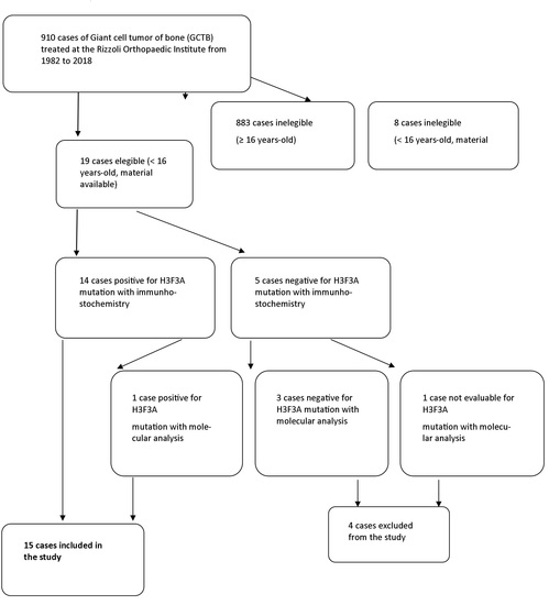
2. Materials and Methods

We retrieved all cases of GCTB in patients <16 years old from the archive of the Rizzoli Orthopedic Institute, treated between 1982 and 2018. Cases of brown tumor of hyperparathyroidism and non-ossifying fibroma were excluded due to the peculiar clinical–serological or radio-histological features, respectively.
Inclusion criteria were as follows:
  • Age <16 years old;
  • Sufficient clinical information available;
  • Histological slides and histological material suitable for immunohistochemistry or genetic analysis for H3F3A gene mutations available.
In all selected cases, all histological, radiological, and clinical features were reviewed.
Immunohistochemistry analysis for H3F3A was performed to confirm the diagnosis on undecalcified tissue. This was evaluated on paraffin-embedded tumor specimens with anti-histone H3.3; we used the primary antibodies against histone H3.3 G34W mutant protein (rabbit monoclonal, clone RM263, dilution 1:600; RevMAb Biosciences, South San Francisco, CA, USA), histone H3.3 G34R mutant protein (rabbit monoclonal, clone RM240, dilution 1:500; RevMAb Biosciences), and histone H3.3 G34V mutant protein (rabbit monoclonal, clone RM307, dilution 1:500; RevMAb Biosciences) [22]. Sections from the paraffin tumor blocks were cut with a microtome and mounted on microscope slides for immunohistochemical analysis. Unstained sections (tumor and control) were heat-treated at 60 °C for 20 min, deparaffinized, and immunostained on a Ventana BenchMark following the manufacturer’s guidelines (Ventana Medical Systems, Tucson, AZ, USA). Standard avidin–biotin complex peroxidase assays were performed to analyze the expression of H3F3A. Adult GCTB with molecular confirmation of H3F3A gene mutation was used as a positive external control. Based on the previously published study that evaluated the nuclear expression of H3F3A, it was considered positive with unequivocal strong crisp nuclear expression and negative with a not-detected expression [20,21,22,23].
In cases negative after immunohistochemistry, Sanger sequencing analysis for H3F3A gene variation was performed. For this analysis, >60 ng of DNA was amplified using AmpliTaq Gold 360 Master Mix (Applied Biosystems, Foster City, CA, USA) with 0.5 μM of primers (H3F3A: forward TGT TTG GTA GTT GCA TAT GGT GA; reverse H3F3A3 ACA AGA GAG ACT TTG TCC CAT T 239 bp [24]). The sequencing was performed by Bio-Research Fab (http://www.biofabresearch.it (accessed on 31 March 2021) Rome, Italy). Mutation analysis was conducted with Basic Local Alignment Search Tool (BLAST) in the NCBI database “National Center of Biotechnology Information Database” (http://www.ncbi.nlm.nih.gov/BLAST (accessed on 31 March 2021)). Electropherograms were exported to fast format and were aligned to the NCBI BLAST sequence of NM_002107.4 H3F3A mRNA. The samples negative to the sequencing analysis with the Sanger method were subsequently tested with allele specific locked nucleic acid quantitative PCR (ASLNAqPCR) as described in a previous paper [25]. A control group of ABC in patients <16 years old morphologically suspicious for the diagnosis of GCTB was used as a control group and analyzed immunohistochemically and genetically for H3F3A gene mutation.
Informed consent was collected from all patients by the standard procedure and with Rizzoli Institute ethics committee approval (CE AVEC 377/2019/Oss/IOR, 7 June 2019).

3. Results

From 1982 to 2018, a total of 910 cases of GCTB were retrieved from the archives of the Rizzoli Orthopedic Institute.
Nineteen cases met the inclusion criteria. Of note, eight excluded cases were originally diagnosed as GCTB in patients <16 years old but lacked histological material or sufficient documentation (counseling cases).
These 19 cases were tested immunohistochemically for H3.3 p.Gly34Trp (G34W, G34R, G34V). Fourteen cases were positive for G34W antibody and included in the study (Figure 1A). The five negative cases were subsequently molecularly analyzed for H3F3A gene mutation: three were negative, one was not evaluable, while the last one (case 11, Table 1) was positive (Figure 1B); only this last case was added to the series, reaching 15 cases. Considering that Sanger sequencing is the most used method with a limitation due to its limit of detection of about 20%, the three negative samples were subsequently confirmed also with ASLNAqPCR [26]. The other not-confirmed four cases were excluded from the study.
The control group consisted of 27 ABCs in patients <16 years old, morphologically suspicious for the diagnosis of GCTB. All these cases were negative both immunohistochemically and genetically for H3F3A gene mutation.
Clinical, radiological, histological, immunohistochemical, and molecular data of these selected 15 patients are summarized in Table 1.
Fourteen patients were female and only one was male. The mean and median age was 13 years old (range 8–15). In 5 cases (7, 8, 10, 11, and 14) initial imaging was not available and the location was only described in the radiological reports. In cases with available imaging, in long bones, two cases (cases 1 and 2) occurring in skeletally immature patients with an open growth plate were purely metaphyseal and centrally located (Figure 2A), while the seven cases occurring in skeletally mature patients with a closed growth plate were meta-epiphyseal and eccentric (case 15, Figure 2B).
Histologically, all cases showed typical histological features of GCTB (Figure 2C,D).
One patient was treated with denosumab and was alive with disease at the last follow-up. All the other patients were treated with curettage or resection, and two of these patients (cases 1 and 3) suffered from local recurrences treated with a new curettage; case 3 was lost at follow-up, while case 1 was alive without disease at the last follow-up.

4. Discussion

GCTB accounts for about 5% of bone tumors. Its common peak incidence is between the ages of 20 and 45 years. It has rarely been reported in pediatric patients, and a principal case series review of English literature is analyzed, compared to our experience, and shown in Table 2 [4,5,6,7,8,9]. The paper of Picci et al. [9] reported six cases treated at the Rizzoli Orthopedic Institute; however, these patients were treated before 1982, and for this reason, they are not included in the present series.
In the present study, we describe a series of 15 cases of GCTB in pediatric patients <16 years old, treated from 1982 to 2018 at the Rizzoli Orthopedic Institute.
As previously reported by other authors, GCTB in pediatric patients has a rare incidence (15/910, 1%) [4,5,6,7,8,9].
In our series, there is a pronounced female predominance (93%), while in the literature only a slight female predominance is reported [3], consistently noted in pediatric series [4,5,6,7,8,9]. Picci et al. [9] also reported a pronounced female predominance.
In our series, 87% of cases arose in long bones (tibia 8/15, 53%; femur 3/15, 20%; and radius 2/15, 13%), confirming previous reports [3,4,5,6,7], while one case each arose in sacrum and vertebra (7% each). No cases arose in the small bones of the hands and feet or the skull or showed a multifocal presentation.
In adult patients, the typical location of GCTB is in the meta-epiphyseal region of long bones, eccentrically. In our series, considering the cases occurring in long bones with available imaging (nine cases), in the two skeletally immature patients with an open growth plate (case 1 and 2), the lesions were purely metaphyseal and centrally located, while in the seven patients with a closed growth plate, the lesions were meta-epiphyseal and eccentric. This does not exactly correspond to the description of Campanacci [10] of a prevalent pure metaphyseal location in pediatric patients. However, if we consider the skeletal age of the patient and the status of the growth plate, the fact that the lesions showed a pure metaphyseal location in the two skeletally immature patients supports the hypothesis of a metaphyseal origin of GCTB. In the literature, it is difficult to check the exact correspondence between the status of the growth plate and the location of the lesions, since this is not described in each series. Picci et al. [9] described six cases of GCTB in skeletally immature patients with an open growth plate; with the limitation that these cases were not tested for the presence of H3F3A mutation, all of them showed a predominant metaphyseal location, with extension into the epiphysis in five patients, thus demonstrating that the presence of an open growth plate does not preclude the possibility of an epiphyseal extension, beyond the physis.
It is important to differentiate GCTB from its mimickers, particularly from ABC with “solid” features that can show similar clinical, radiological, and histological features and that is more frequent in pediatric patients.
Immunohistochemistry for H3.3 p.Gly34Trp (G34W, G34R, G34V) is a useful and predictable surrogate marker for molecular analysis in distinguishing GCTB from its mimickers.
Although clinically most of the cases of GCTB and other osteoclastic giant-cell-rich lesions behave in a similar benign fashion and have a similar treatment, patients with GCTB can rarely develop lung metastasis (3–7% of cases) or a secondary malignant transformation, mostly after radiation therapy [3,15,16,17,18,27], thus indicating the importance of differentiating between these entities for specific clinical management, especially in pediatric patients who could be affected by growth-related problems and angular deformities after surgery. This is the reason why in the last WHO classification [3], ABC is classified as a benign tumor, while GCTB is classified as locally aggressive, rarely metastasizing neoplasm.

5. Conclusions

In conclusion, we describe our series of GCTB in pediatric patients <16 years old. Although rare (1% of all GCTB), they can occur also in these age groups, with a pronounced female predominance (93%). Differently from the typical meta-epiphyseal location, in skeletally immature patients with an open growth plate, GCTB can be limited to the metaphysis or have a predominant metaphyseal location, with extension into the epiphysis. GCTB should be distinguished from other osteoclastic giant-cell-rich tumors, because of the differences in prognosis and treatment. Immunohistochemical or molecular detection of H3F3A gene mutation represents a specific, accessible, and reliable diagnostic tool in the differential diagnosis with other mimickers and pathologists should be aware to use these techniques in doubtful cases, also in young patients.

Author Contributions

Conceptualization, F.A. and M.G.; methodology, F.A., A.R., S.B. and M.G.; data curation, A.R., G.M., I.C., M.M., A.G. and T.F.; writing—original draft preparation, F.A.; writing—review and editing, A.R. and G.M. All authors have read and agreed to the published version of the manuscript.

Funding

This research received no external funding.

Institutional Review Board Statement

The study was conducted according to the guidelines of the Declaration of Helsinki and approved by the Ethics Committee of Area Vasta Emilia Centro della Regione Emilia-Romagna (CE AVEC 377/2019/Oss/IOR, 7 June 2019).

Informed Consent Statement

Informed consent was obtained from all subjects involved in the study.

Data Availability Statement

Data available on request due to restrictions (e.g., privacy or ethics). The data presented in this study are available on request from the corresponding author.

Acknowledgments

The authors are grateful to the Musculoskeletal Tumor Biobank (Biobanca dei Tumori Muscoloscheletrici; BIOTUM)—a member of the CRB-IOR—for providing us with the biological samples. This work was supported by Ministero della Salute (5 × 1000, Anno 2018 Redditi 2017, contributions to the IRCCS, Istituto Ortopedico Rizzoli).

Conflicts of Interest

The authors declare no conflict of interest. The funders had no role in the design of the study; in the collection, analyses, or interpretation of data; in the writing of the manuscript; or in the decision to publish the results.

References

  1. Forsyth, R.; Jundt, G. Giant cell lesion of the small bones. In WHO Classification of Tumours of Soft Tissue and Bone; Fletcher, C.D.M., Bridge, J.A., Hogendoorn, P.C.W., Mertens, F., Eds.; IARC Press: Lyon, France, 2013; 320p. [Google Scholar]
  2. Campanacci, M.; Baldini, N.; Boriani, S.; Sudanese, A. Giant-cell tumor of bone. J. Bone Joint Surg. 1987, 69, 106–114. [Google Scholar] [CrossRef] [PubMed]
  3. Flanagan, A.M.; Larousserie, F.; O’Donnel, P.G.; Yoshida, A. Giant cell tumor of bone. In WHO Classification of Soft Tissue and Bone Tumours, WHO Classification of Tumours, 5th ed.; IARC Press: Lyon, France, 2020; pp. 440–446. ISBN 9789283245025. [Google Scholar]
  4. Al-Ibraheemi, A.; Inwards, C.Y.; Zreik, R.T.; Wenger, D.E.; Jenkins, S.M.; Carter, J.M.; Boland, J.M.; Rose, P.S.; Jin, L.; Oliveira, A.M.; et al. Histologic spectrum of Giant Cell Tumor (GCT) of bone in patients 18 years of age and below. Am. J. Surg. Pathol. 2016, 40, 1702–1712. [Google Scholar] [CrossRef] [PubMed]
  5. Dahlin, D.C.; Cupps, R.E.; Johnson, E.W., Jr. Giant cell tumor: A study of 195 cases. Cancer 1970, 25, 1061–1070. [Google Scholar] [CrossRef]
  6. Strøm, T.M.; Skeie, A.T.; Lobmaier, I.K.; Zaikova, O. Giant cell tumor: A rare condition in the immature skeleton—A retrospective study of symptoms, treatment, and outcome in 16 children. Sarcoma 2016, 2016. [Google Scholar] [CrossRef] [PubMed]
  7. Puri, A.; Agarwal, M.G.; Shah, M.; Jambhekar, N.A.; Anchan, C.; Behle, S. Giant cell tumor of bone in children and adolescents. J. Pediatric Orthop. 2007, 27, 635–639. [Google Scholar] [CrossRef]
  8. Sydlik, C.; Dürr, H.R.; Bechtold-Dalla Pozza, S.; Weißenbacher, C.; Roeb, J.; Schmidt, H. Hypercalcaemia after treatment with denosumab in children: Bisphosphonates as an option for therapy and prevention? World J. Pediatr. 2020, 16, 520–527. [Google Scholar] [CrossRef]
  9. Picci, P.I.; Manfrini, M.A.; Zucchi, V.I.; Gherlinzoni, F.R.; Rock, M.I.; Bertoni, F.R.; Neff, J.R. Giant-cell tumor of bone in skeletally immature patients. J. Bone Joint Surg. 1983, 65, 486–490. [Google Scholar] [CrossRef] [PubMed]
  10. Campanacci, M.; Enneking, W.F. Bone and Soft Tissue Tumors: Clinical Features, Imaging, Pathology, and Treatment, 2nd ed.; Springer: Berlin, Germany, 1999; p. 118. [Google Scholar]
  11. Dumford, K.; Moore, T.; Walker, C.; Jaksha, J. Multifocal, metachronous, giant cell tumor of the lower limb. Skeletalradiology 2003, 32, 147–150. [Google Scholar] [CrossRef]
  12. Rendina, D.; Gennari, L.; de Filippo, G.; Merlotti, D.; de Campora, E.; Fazioli, F.; Scarano, G.; Nuti, R.; Strazzullo, P.; Mossetti, G. Evidence for increased clinical severity of familial and sporadic Paget’s disease of bone in Campania, Southern Italy. J. Bone Miner. Res. 2006, 21, 1828–1835. [Google Scholar] [CrossRef]
  13. Divisato, G.; Formicola, D.; Esposito, T.; Merlotti, D.; Pazzaglia, L.; del Fattore, A.; Siris, E.; Orcel, P.; Brown, J.P.; Nuti, R.; et al. ZNF687 mutations in severe Paget disease of bone associated with giant cell tumor. Am. J. Human Genet. 2016, 98, 275–286. [Google Scholar] [CrossRef] [Green Version]
  14. Salzer-Kuntschik, M. Differential diagnosis of giant cell tumor of bone. Verh. Dtsch. Ges. Pathol. 1998, 82, 154–159. [Google Scholar]
  15. Murphey, M.D.; Nomikos, G.C.; Flemming, D.J.; Gannon, F.H.; Temple, H.T.; Kransdorf, M.J. Imaging of giant cell tumor and giant cell reparative granuloma of bone: Radiologic-pathologic correlation. Radiographics 2001, 21, 1283–1309. [Google Scholar] [CrossRef] [Green Version]
  16. Gong, L.; Liu, W.; Sun, X.; Sajdik, C.; Tian, X.; Niu, X.; Huang, X. Histological and clinical characteristics of malignant giant cell tumor of bone. Virchows Arch. 2012, 460, 327–334. [Google Scholar] [CrossRef] [PubMed]
  17. Nascimento, A.G.; Huvos, A.G.; Marcove, R.C. Primary malignant giant cell tumor of bone: A study of eight cases and review of the literature. Cancer 1979, 44, 1393–1402. [Google Scholar] [CrossRef]
  18. Dominkus, M.; Ruggieri, P.; Bertoni, F.; Briccoli, A.; Picci, P.; Rocca, M.; Mercuri, M. Histologically verified lung metastases in benign giant cell tumours—14 cases from a single institution. Int. Orthop. 2006, 30, 499–504. [Google Scholar] [CrossRef] [PubMed] [Green Version]
  19. Behjati, S.; Tarpey, P.S.; Presneau, N.; Scheipl, S.; Pillay, N.; van Loo, P.; Wedge, D.C.; Cooke, S.L.; Gundem, G.; Davies, H.; et al. Distinct H3F3A and H3F3B driver mutations define chondroblastoma and giant cell tumor of bone. Nat. Genet. 2013, 45, 1479–1482. [Google Scholar] [CrossRef] [PubMed]
  20. Righi, A.; Mancini, I.; Gambarotti, M.; Picci, P.; Gamberi, G.; Marraccini, C.; dei Tos, A.P.; Simi, L.; Pinzani, P.; Franchi, A. Histone 3.3 mutations in giant cell tumor and giant cell-rich sarcomas of bone. Human Pathol. 2017, 68, 128–135. [Google Scholar] [CrossRef]
  21. Amary, F.; Berisha, F.; Ye, H.; Gupta, M.; Gutteridge, A.; Baumhoer, D.; Gibbons, R.; Tirabosco, R.; O’Donnell, P.; Flanagan, A.M. H3F3A (Histone 3.3) G34W immunohistochemistry: A reliable marker defining benign and malignant giant cell tumor of bone. Am. J. Surg. Pathol. 2017, 41, 1059. [Google Scholar] [CrossRef] [PubMed]
  22. Yamamoto, H.; Iwasaki, T.; Yamada, Y.; Matsumoto, Y.; Otsuka, H.; Yoshimoto, M.; Kohashi, K.; Taguchi, K.; Yokoyama, R.; Nakashima, Y.; et al. Diagnostic utility of histone H3.3 G34W, G34R, and G34V mutant-specific antibodies for giant cell tumors of bone. Hum. Pathol. 2018, 73, 41–50. [Google Scholar] [CrossRef] [PubMed]
  23. Mancini, I.; Righi, A.; Gambarotti, M.; Picci, P.; dei Tos, A.P.; Steven, D.; Billings, L.S.; Franchi, A. Phenotypic and molecular differences between giant-cell tumour of soft tissue and its bone counterpart. Histopathology 2017, 71, 453–460. [Google Scholar] [CrossRef]
  24. Girolami, I.; Mancini, I.; Simoni, A.; Baldi, G.G.; Simi, L.; Campanacci, D.; Beltrami, G.; Scoccianti, G.; D’Arienzo, A.; Capanna, R.; et al. Denosumab treated giant cell tumour of bone: A morphological, immunohistochemical and molecular analysis of a series. J. Clin. Pathol. 2016, 69, 240–247. [Google Scholar] [CrossRef]
  25. Gamberi, G.; Morandi, L.; Benini, S.; Resca, A.; Cocchi, S.; Magagnoli, G.; Donati, D.M.; Righi, A.; Gambarotti, M. Detection ofH3F3Ap.G35W and p.G35R in giant cell tumor of bone byAllele specific locked nucleic acid quantitative PCR (ASLNAqPCR). Pathol. Res. Pract. 2018, 214, 89–94. [Google Scholar] [CrossRef] [PubMed]
  26. Harrington, C.T.; Lin, E.I.; Olson, M.T.; Eshleman, J.R. Fundamentals of pyrosequencing. Arch. Pathol. Lab. Med. 2013, 137, 1296–1303. [Google Scholar] [CrossRef] [PubMed]
  27. Bertoni, F.; Bacchini, P.; Staals, E.L. Malignancy in giant cell tumor of bone. Cancer 2003, 97, 2520–2529. [Google Scholar] [CrossRef] [PubMed]
Figure 1. (A) Strong and diffuse immunohistochemical nuclear positivity for H3F3A in case 10 (immunohistochemistry for H3F3A, 200× magnification). (B) Molecular analysis of case 11 confirmed the presence of the mutation in heterozygosity.
Cancers 13 02585 g001
Figure 2. (A) Plane X-ray of the knee shows a purely lytic, centrally located lesion in the proximal metaphysis of the tibia with relatively well-defined margins underneath an immature growth plate; the cortex is thinned without interruptions (case 1). (B) Plane X-ray of the ankle shows osteolysis of the distal tibia located in the meta-epiphyseal region with aggressive radiological features: not well-defined margins, cortex expanded and interrupted, growth plate invaded and crossed (case 15). (C) Histological examples of giant cell tumor of the bone (case 14), with a highly vascular stroma, fibrosis, and reactive woven bone; acute hemorrhage, hemosiderin, xanthomatous histiocytes are admixed with neoplastic cells (hematoxylin and eosin, 50× magnification). (D) At higher magnification, the same tumor is composed of numerous osteoclast-like giant cells uniformly distributed throughout the tumor, which are larger than normal osteoclasts with numerous (>30) nuclei; mononuclear round to oval and spindle-shaped cells are dispersed together with the giant cells (hematoxylin and eosin, 200× magnification).
Cancers 13 02585 g002
Table 1. Clinical, radiological, histological, immunohistochemical, and molecular features of the 15 patients are collected in the table.
SexAgeSiteStatus of Growth PlateSize (Major Axis)IHC H3F3A G34WIHC H3F3A G34RIHC H3F3A G34VMolecular AnalysisSurgeryLRComplicationsLast FU (Months)Status
1F8Tibia, proximal central metaphysisopen5+--nacurettage and cement (+bone grafts)yes—8 monthsLeg length discrepancy48ned
2F10Femur, left central metaphysisopen6+--nachondrodiastasis and intercalarNOnone112ned
3F11Tibia, eccentric distal, meta-epiphysisclosed4+--nacurettage and cement (+bone grafts)NOnonelostna
4F12Tibia, eccentric distal, meta-epiphysisclosed4+--nacurettage and bone graftsNOnone123ned
5F13Femur, distal meta-epiphysisclosed9+--nacurettage and cementNOnone90ned
6F13Tibia, distal meta-epiphysis, closed5+--namassive graftingNOgraft obturation, revision with fibula42ned
7F13Radius, distal, meta-epiphysisnana+--naosteoarticular resection and graftingNOgraft failure, revision in arthrodesis204ned
8F13Tibia, central proximal metaphysisnana+--nanayes—12 monthsnone14ned
9F14Sacrumindeterminate8+--nanone, treated with denosumabnever operated on and in remissionna96awd
10F14Radius, distal epiphysisnana+--nacurettage and bone graftsNOnone120ned
11F15T12 vertebranana---+vertebrectomy and reconstructionNOinfection180ned
12F14Tibia, proximal meta-epiphysisclosed5+--nacurettage and cementNOnonelostna
13F13Tibia, proximal eccentric, meta-epiphysisclosed4+--nacurettage and cementNOnone12ned
14F14Distal femur, pathological fracturenana+--naresection and prosthesisNOnone144ned
15M15Tibia, distal, eccentric, meta-epiphysisclosed6+--nacurettage and cementNOnone6ned
Legend: M: male; F: female; IHC: immunohistochemistry; LR: local recurrences; FU: follow-up; na: not available; ned: no evidence of disease; awd: alive with disease.
Table 2. Summary of the principal case series review of English literature of GCTB in pediatric patients.
PublicationRange of Time CasesSex AgeBoneStatus of Growth PlateLocationTreatmentNoteMolecular Analysis
Ajay Puri et al., 2007 [7]January 2000 to December 2005
17 patients
14 F (82%) 3 M (18%) 10–18 yearslower end of the femur (n = 5, 29%)all open (17–100%)13 (76.5%) epiphysiometaphyseal in location14 IIC intralesional curettage2 local recurrences
the upper end of the tibia (n = 4, 24%)
the upper end of the fibula (n = 2)2 lower end-radius
distal end of radius (n = 2)2 not applicable3 wide excisions1 pulmonary nodule
patient each of the upper end of the humerus, metacarpal, clavicle, and cuboid (n = 1)
ThaleM. Asp Strøm et al., 2016 [6]1984 to 2015
16 patients
12 F (75%) 4 M (25%) 6–15 yearstibia (n = 4, 25%)all open (16–100%)4 (25%) epiphysiometaphyseal distal15 curettage
1 excision
2 local recurrences
fibula (n = 3, 18.75%)3 (18.75%) epiphysiometaphyseal proximal
clavicula (n = 3, 18.75%)1 (6.25%) proximal epiphysis
III metatarsal (n = 2, 12.5%)3 (18.75%) proximal (short bones)1 multicentric disease
sacrum (n = 2, 12.5%)
scapula (n = 1, 6.25%)2 (12.5%) distal (short bones)
radius (n = 1, 6.25%)
David C. Dahli et al., 1969 [5]1910 to 1969
21 patients (75%)
7 patients (25%)
21 F (75%)
15–20 years
uk no malignant transformation
7 M (75%)
12–14 years
Alyaa Al-Ibraheemi et al., 2016 * [4]all curettage and resection; all specimens of primary “GCT of bone” from patients 18 years old or younger43 F (68%)
20 M (32%)
8–18 years
tibia (n = 16, 25%)radiologic images (n = 15): 7 patients with open physes (47%); 8 patients with closed growth plates (53%)7 (21%) cases involved the metaphysis without extension into the epiphysiscurettage and resection1 multifocal4 patients G34W 1 patient G34L
femur (n = 14, 22%) 21 local recurrences (38%)
vertebral body (n = 13, 21%)23 (70%) cases involved the epiphysis and metaphysis2 (4%) pulmonary metastases 15 and 20 months after the diagnosis
radius (n = 4, 6%)
humerus (n = 4, 6%)
metacarp (n = 3, 5%)
fibula (n = 2, 3%)5-year progression-free survival was observed in 57% (95% confidence interval, 43–71%)
patella (n = 2, 3%)
calcaneus (n = 1, 2%)
navicular (n = 1, 2%)
phalanx (n = 1, 2%)3 (9%) cases were confined to the epiphysis
pelvis (n = 1, 2%)
ulna (n = 1, 2%)
Carmen Sydlik et al., 2020 [8]children underwent therapy with denosumab between September 2011 and December 2014 4 patients1 F (25%)
3 M (75%)
6–13 years
solid variant of ABC in the left os sacrum (n = 1, 25%)uk children with severe hypercalcemia after treatment with denosumab for unresectable giant cell tumors of bone and for aneurysmal bone cysts1 patient developed pulmonary metastasis
a giant cell tumor in lumbosacral spine (L5/S1) (n = 1, 25%)
left thigh and aneurismal
bone cyst with typical osteoclast-like giant cells and intense vascularization (n = 1, 25%)
a giant cell tumor localized in Th2
Picci Piero et al., 1983 [9]giant-cell tumor of bone in skeletally immature patients 6 patients5 F (90%)
1 M (10%)
10–14 years
proximal fibula (n = 1; 10%)6 patients with open physes (100%)epiphyseal plate involvement (n = 5/6; 83%)marginal resection (n = 1; 10%)
distal femur (n = 4; 80%)wide resection (n = 2; 40%)
proximal tibia (n = 1; 10%)curettage (n = 3; 60%)
* Recurrent and metastatic tumors.
Publisher’s Note: MDPI stays neutral with regard to jurisdictional claims in published maps and institutional affiliations.

