Molecular Features of Metaplastic Breast Carcinoma: An Infrequent Subtype of Triple Negative Breast Carcinoma

 ,
, 
Abstract
1. Introduction
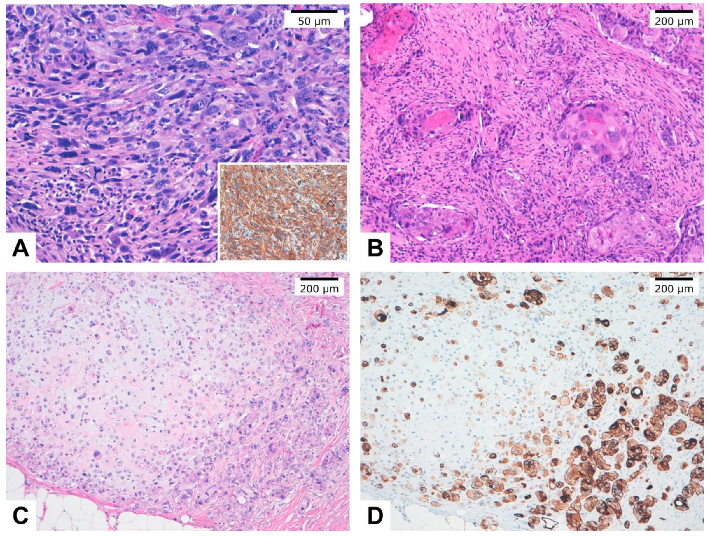
2. MBC Subtypes
3. Clinicopathological Features
4. Molecular Alterations
5. Epithelial to Mesenchymal Transition in MBC
6. Immune Microenvironment
7. MBC Prognosis
8. Targeted Therapy in MBC
9. Conclusions
Supplementary Materials
Author Contributions
Funding
Conflicts of Interest
References
- WHO Classification of Tumours Editorial Board; International Agency for Research on Cancer; World Health Organization. WHO classification of tumours. Breast Tumours; International Agency for Research on Cancer: Lyon, France, 2019; ISBN 978-92-832-4500-1. [Google Scholar]
- Barnes, P.J.; Boutilier, R.; Chiasson, D.; Rayson, D. Metaplastic breast carcinoma: Clinical–pathologic characteristics and HER2/neu expression. Breast Cancer Res. Treat. 2005, 91, 173–178. [Google Scholar] [CrossRef] [PubMed]
- Geyer, F.C.; Li, A.; Papanastasiou, A.D.; Smith, A.; Selenica, P.; Burke, K.A.; Edelweiss, M.; Wen, H.-C.; Piscuoglio, S.; Schultheis, A.M.; et al. Recurrent hotspot mutations in HRAS Q61 and PI3K-AKT pathway genes as drivers of breast adenomyoepitheliomas. Nat. Commun. 2018, 9, 1816. [Google Scholar] [CrossRef] [PubMed]
- Cheah, A.L.; Billings, S.D.; Rowe, J.J. Mesenchymal tumours of the breast and their mimics: A review with approach to diagnosis. Pathology 2016, 48, 406–424. [Google Scholar] [CrossRef] [PubMed]
- Piscuoglio, S.; Ng, C.K.Y.; Geyer, F.C.; Burke, K.A.; Cowell, C.F.; Martelotto, L.G.; Natrajan, R.; Popova, T.; Maher, C.A.; Lim, R.S.; et al. Genomic and transcriptomic heterogeneity in metaplastic carcinomas of the breast. NPJ Breast Cancer 2017, 3. [Google Scholar] [CrossRef]
- Vranic, S.; Stafford, P.; Palazzo, J.; Skenderi, F.; Swensen, J.; Xiu, J.; Spetzler, D.; Gatalica, Z. Molecular Profiling of the Metaplastic Spindle Cell Carcinoma of the Breast Reveals Potentially Targetable Biomarkers. Clin. Breast Cancer 2020. [Google Scholar] [CrossRef]
- McCart Reed, A.E.; Kalaw, E.; Nones, K.; Bettington, M.; Lim, M.; Bennett, J.; Johnstone, K.; Kutasovic, J.R.; Saunus, J.M.; Kazakoff, S.; et al. Phenotypic and molecular dissection of metaplastic breast cancer and the prognostic implications. J. Pathol. 2019, 247, 214–227. [Google Scholar] [CrossRef]
- Zhai, J.; Giannini, G.; Ewalt, M.D.; Zhang, E.Y.; Invernizzi, M.; Niland, J.; Lai, L.L. Molecular characterization of metaplastic breast carcinoma via next-generation sequencing. Hum. Pathol. 2019, 86, 85–92. [Google Scholar] [CrossRef]
- Tray, N.; Taff, J.; Singh, B.; Suh, J.; Ngo, N.; Kwa, M.; Troxel, A.B.; Chae, Y.K.; Kurzrock, R.; Patel, S.P.; et al. Metaplastic breast cancers: Genomic profiling, mutational burden and tumor-infiltrating lymphocytes. Breast 2019, 44, 29–32. [Google Scholar] [CrossRef]
- Afkhami, M.; Schmolze, D.; Yost, S.E.; Frankel, P.H.; Dagis, A.; Amanam, I.U.; Telatar, M.; Nguyen, K.; Yu, K.W.; Luu, T.; et al. Mutation and immune profiling of metaplastic breast cancer: Correlation with survival. PLoS ONE 2019, 14, 1–12. [Google Scholar] [CrossRef]
- Krings, G.; Chen, Y.Y. Genomic profiling of metaplastic breast carcinomas reveals genetic heterogeneity and relationship to ductal carcinoma. Mod. Pathol. 2018, 31, 1661–1674. [Google Scholar] [CrossRef]
- Ng, C.K.Y.; Piscuoglio, S.; Geyer, F.C.; Burke, K.A.; Pareja, F.; Eberle, C.A.; Lim, R.S.; Natrajan, R.; Riaz, N.; Mariani, O.; et al. The Landscape of Somatic Genetic Alterations in Metaplastic Breast Carcinomas. Clin. Cancer Res. 2017, 23, 3859–3870. [Google Scholar] [CrossRef] [PubMed]
- Joneja, U.; Vranic, S.; Swensen, J.; Feldman, R.; Chen, W.; Kimbrough, J.; Xiao, N.; Reddy, S.; Palazzo, J.; Gatalica, Z. Comprehensive profiling of metaplastic breast carcinomas reveals frequent overexpression of programmed death-ligand 1. J.of Clin. Pathol. 2017, 70, 255–259. [Google Scholar] [CrossRef] [PubMed]
- Edenfield, J.; Schammel, C.; Collins, J.; Schammel, D.; Edenfield, W.J. Metaplastic Breast Cancer: Molecular Typing and Identification of Potential Targeted Therapies at a Single Institution. Clin. Breast Cancer 2017, 17, e1–e10. [Google Scholar] [CrossRef] [PubMed]
- Ross, J.S.; Badve, S.; Wang, K.; Sheehan, C.E.; Boguniewicz, A.B.; Otto, G.A.; Yelensky, R.; Lipson, D.; Ali, S.; Morosini, D.; et al. Genomic profiling of advanced-stage, metaplastic breast carcinoma by next-generation sequencing reveals frequent, targetable genomic abnormalities and potential new treatment options. Arch. Pathol. Lab. Med. 2015, 139, 642–649. [Google Scholar] [CrossRef] [PubMed]
- Hayes, M.J.; Thomas, D.; Emmons, A.; Giordano, T.J.; Kleer, C.G. Genetic Changes of Wnt Pathway Genes Are Common Events in Metaplastic Carcinomas of the Breast. Clin. Cancer Res. 2008, 14, 4038–4044. [Google Scholar] [CrossRef] [PubMed]
- Pirot, F.; Chaltiel, D.; Ben Lakhdar, A.; Mathieu, M.C.; Rimareix, F.; Conversano, A. Squamous cell carcinoma of the breast, are there two entities with distinct prognosis? A series of 39 patients. Breast Cancer Res. Treat. 2020, 180, 87–95. [Google Scholar] [CrossRef]
- Takala, S.; Heikkilä, P.; Nevanlinna, H.; Blomqvist, C.; Mattson, J. Metaplastic carcinoma of the breast: Prognosis and response to systemic treatment in metastatic disease. Breast J. 2019, 25, 418–424. [Google Scholar] [CrossRef]
- Han, M.; Salamat, A.; Zhu, L.; Zhang, H.; Clark, B.Z.; Dabbs, D.J.; Carter, G.J.; Brufsky, A.M.; Jankowitz, R.C.; Puhalla, S.L.; et al. Metaplastic breast carcinoma: A clinical-pathologic study of 97 cases with subset analysis of response to neoadjuvant chemotherapy. Mod. Pathol. 2019, 32, 807–816. [Google Scholar] [CrossRef]
- Schroeder, M.C.; Rastogi, P.; Geyer, C.E.; Miller, L.D.; Thomas, A. Early and Locally Advanced Metaplastic Breast Cancer: Presentation and Survival by Receptor Status in Surveillance, Epidemiology, and End Results (SEER) 2010–2014. Oncologist 2018, 23, 481–488. [Google Scholar] [CrossRef]
- El Zein, D.; Hughes, M.; Kumar, S.; Peng, X.; Oyasiji, T.; Jabbour, H.; Khoury, T. Metaplastic Carcinoma of the Breast Is More Aggressive Than Triple-negative Breast Cancer: A Study From a Single Institution and Review of Literature. Clin. Breast Cancer 2017, 17, 382–391. [Google Scholar] [CrossRef]
- Zhang, Y.; Lv, F.; Yang, Y.; Qian, X.; Lang, R.; Fan, Y.; Liu, F.; Li, Y.; Li, S.; Shen, B.; et al. Clinicopathological features and prognosis of metaplastic breast carcinoma: Experience of a major Chinese cancer center. PLoS ONE 2015, 10, 1–13. [Google Scholar] [CrossRef] [PubMed]
- Lai, H.W.; Tseng, L.M.; Chang, T.W.; Kuo, Y.L.; Hsieh, C.M.; Chen, S.T.; Kuo, S.J.; Su, C.C.; Chen, D.R. The prognostic significance of metaplastic carcinoma of the breast (MCB) - A case controlled comparison study with infiltrating ductal carcinoma. Breast 2013, 22, 968–973. [Google Scholar] [CrossRef] [PubMed]
- Lee, H.; Jung, S.Y.; Ro, J.Y.; Kwon, Y.; Sohn, J.H.; Park, I.H.; Lee, K.S.; Lee, S.; Kim, S.W.; Kang, H.S.; et al. Metaplastic breast cancer: Clinicopathological features and its prognosis. J. Clin. Pathol. 2012, 65, 441–446. [Google Scholar] [CrossRef]
- Lester, T.R.; Hunt, K.K.; Nayeemuddin, K.M.; Bassett, R.L.; Gonzalez-Angulo, A.M.; Feig, B.W.; Huo, L.; Rourke, L.L.; Davis, W.G.; Valero, V.; et al. Metaplastic sarcomatoid carcinoma of the breast appears more aggressive than other triple receptor-negative breast cancers. Breast Cancer Res. Treat. 2012, 131, 41–48. [Google Scholar] [CrossRef] [PubMed]
- Jung, S.Y.; Kim, H.Y.; Nam, B.H.; Min, S.Y.; Lee, S.J.; Park, C.; Kwon, Y.; Kim, E.A.; Ko, K.L.; Shin, K.H.; et al. Worse prognosis of metaplastic breast cancer patients than other patients with triple-negative breast cancer. Breast Cancer Res. Treat. 2010, 120, 627–637. [Google Scholar] [CrossRef]
- Luini, A.; Aguilar, M.; Gatti, G.; Fasani, R.; Botteri, E.; Brito, J.A.D.; Maisonneuve, P.; Vento, A.R.; Viale, G. Metaplastic carcinoma of the breast, an unusual disease with worse prognosis: The experience of the European Institute of Oncology and review of the literature. Breast Cancer Res. Treat. 2007, 101, 349–353. [Google Scholar] [CrossRef]
- Beatty, J.D.; Atwood, M.; Tickman, R.; Reiner, M. Metaplastic breast cancer: Clinical significance. Am. J. Surg. 2006, 191, 657–664. [Google Scholar] [CrossRef]
- cBioPortal for Cancer Genomics. Available online: http://www.cbioportal.org/ (accessed on 20 May 2020).
- Reis-Filho, J.; Pinheiro, C.; Lambros, M.; Milanezi, F.; Carvalho, S.; Savage, K.; Simpson, P.; Jones, C.; Swift, S.; Mackay, A.; et al. EGFR amplification and lack of activating mutations in metaplastic breast carcinomas. J. Pathol. 2006, 209, 445–453. [Google Scholar] [CrossRef] [PubMed]
- Hennessy, B.T.; Stemke-hale, K.; Gilcrease, M.Z.; Krishnamurthy, S.; Lee, J.; Fridlyand, J.; Agarwal, R.; Joy, C.; Liu, W.; Stivers, D.; et al. Characterization of a Naturally Occurring Breast Cancer Subset Enriched in Epithelial-To-Mesenchymal Transition and Stem Cell Characteristics. Cancer Res. 2009, 69, 4116–4124. [Google Scholar] [CrossRef] [PubMed]
- Li, J.; Yang, L.; Gaur, S.; Zhang, K.; Wu, X.; Yuan, Y.-C.; Li, H.; Hu, S.; Weng, Y.; Yen, Y. Mutants TP53 p.R273H and p.R273C but not p.R273G Enhance Cancer Cell Malignancy. Hum. Mutat. 2014, 35, 575–584. [Google Scholar] [CrossRef]
- Lee, K.-H.; Hwang, H.-J.; Noh, H.J.; Shin, T.-J.; Cho, J.-Y. Somatic Mutation of PIK3CA (H1047R) Is a Common Driver Mutation Hotspot in Canine Mammary Tumors as Well as Human Breast Cancers. Cancers 2019, 11, 2006. [Google Scholar] [CrossRef]
- Mangone, F.; Bobrovnitchaia, I.; Salaorni, S.; Manuli, E.; Nagai, M. PIK3CA exon 20 mutations are associated with poor prognosis in breast cancer patients. Clinics 2012, 67, 1285–1290. [Google Scholar] [CrossRef]
- Baum, J.E.; Sung, K.-J.; Tran, H.; Song, W.; Ginter, P.S. Mammary Epithelial-Myoepithelial Carcinoma: Report of a Case With HRAS and PIK3CA Mutations by Next-Generation Sequencing. Int. J. Surg. Pathol. 2019, 27, 441–445. [Google Scholar] [CrossRef] [PubMed]
- Vivas, A.P.M.; Bomfin, L.E.; Pinto, C.A.L.; Nicolau, U.R.; Alves, F.A. Oral Metastasis of Metaplastic Breast Carcinoma in a Patient with Neurofibromatosis 1. Case Rep. Oncol. Med. 2014, 2014, 1–7. [Google Scholar] [CrossRef][Green Version]
- Suarez-Kelly, L.P.; Akagi, K.; Reeser, J.W.; Samorodnitsky, E.; Reeder, M.; Smith, A.; Roychowdhury, S.; Symer, D.E.; Carson, W.E. Metaplastic breast cancer in a patient with neurofibromatosis type 1 and somatic loss of heterozygosity. Cold Spring Harb. Mol. Case Stud. 2018, 4, 1–20. [Google Scholar] [CrossRef]
- Vellaisamy, G.; Mohanty, S.; Rout, P.; Manjunath, S. Metaplastic carcinoma of breast and neurofibromatosis 1: A rare association. Indian J. Med. Paediatr. Oncol. 2017, 38, 374. [Google Scholar] [CrossRef]
- Lee, H.S.; Jung, E.J.; Kim, J.Y.; Song, E.J.; Jeong, C.Y.; Ju, Y.T.; Lee, Y.J.; Hong, S.C.; Choi, B.H.; Lee, H.I. Metaplastic spindle cell carcinoma of the breast in a patient with neurofibromatosis type 1. Breast J. 2018, 24, 391–394. [Google Scholar] [CrossRef]
- Natsiopoulos, I.; Chatzichristou, A.; Stratis, I.; Skordalaki, A.; Makrantonakis, N. Metaplastic breast carcinoma in a patient with Von Recklinghausen’s disease. Clin. Breast Cancer 2007, 7, 573–575. [Google Scholar] [CrossRef] [PubMed]
- Chaudhry, U.S.; Yang, L.; Askeland, R.W.; Fajardo, L.L. Metaplastic Breast Cancer in a Patient with Neurofibromatosis. J. Clin. Imaging Sci. 2015, 5, 5–8. [Google Scholar] [CrossRef] [PubMed]
- Hafezi, F.; Perez Bercoff, D. The Solo Play of TERT Promoter Mutations. Cells 2020, 9, 749. [Google Scholar] [CrossRef] [PubMed]
- Weigelt, B.; Kreike, B.; Reis-Filho, J.S. Metaplastic breast carcinomas are basal-like breast cancers: A genomic profiling analysis. Breast Cancer Res. Treat. 2009, 117, 273–280. [Google Scholar] [CrossRef] [PubMed]
- Breuer, A.; Kandel, M.; Fisseler-Eckhoff, A.; Sutter, C.; Schwaab, E.; Lück, H.; Bois, A. BRCA1 germline mutation in a woman with metaplastic squamous cell breast cancer. Onkologie 2007, 30, 316–318. [Google Scholar] [CrossRef] [PubMed]
- Noël, J.-C.; Buxant, F.; Engohan-Aloghe, C. Low-grade adenosquamous carcinoma of the breast—A case report with a BRCA1 germline mutation. Pathol. Res. Pract. 2010, 206, 511–513. [Google Scholar] [CrossRef] [PubMed]
- Rashid, M.U.; Shah, M.A.; Azhar, R.; Syed, A.A.; Amin, A.; Hamann, U. A deleterious BRCA1 mutation in a young Pakistani woman with metaplastic breast carcinoma. Pathol. Res. Pract. 2011, 207, 583–586. [Google Scholar] [CrossRef] [PubMed]
- Ghilli, M.; Mariniello, D.M.; Cascione, F.; Cristaudo, A.; Cilotti, A.; Caligo, A.M.; Manca, G.; Colizzi, L.; Naccarato, G.A.; Roncella, M.S.C. Carcinosarcoma of the Breast: An Aggressive Subtype of Metaplastic Cancer. Report of a Rare Case in a Young BRCA-1 Mutated Woman. Clin. Breast Cancer 2017, 17, e31–e35. [Google Scholar] [CrossRef]
- Sarrió, D.; Rodriguez-Pinilla, S.M.; Hardisson, D.; Cano, A.; Moreno-Bueno, G.; Palacios, J. Epithelial-Mesenchymal Transition in Breast Cancer Relates to the Basal-like Phenotype. Cancer Res. 2008, 68, 989–997. [Google Scholar] [CrossRef]
- Thiery, J.P.; Acloque, H.; Huang, R.Y.J.; Nieto, M.A. Epithelial-mesenchymal transitions in development and disease. Cell 2009, 139, 871–890. [Google Scholar] [CrossRef]
- Oon, M.L.; Thike, A.A.; Tan, S.Y.; Tan, P.H. Cancer stem cell and epithelial–mesenchymal transition markers predict worse outcome in metaplastic carcinoma of the breast. Breast Cancer Res. Treat. 2015, 150, 31–41. [Google Scholar] [CrossRef]
- Lien, H.C.; Hsiao, Y.H.; Lin, Y.S.; Yao, Y.T.; Juan, H.F.; Kuo, W.H.; Hung, M.-C.; Chang, K.J.; Hsieh, F.J. Molecular signatures of metaplastic carcinoma of the breast by large-scale transcriptional profiling: Identification of genes potentially related to epithelial–mesenchymal transition. Oncogene 2007, 26, 7859–7871. [Google Scholar] [CrossRef]
- McQuerry, J.A.; Jenkins, D.F.; Yost, S.E.; Zhang, Y.; Schmolze, D.; Johnson, W.E.; Yuan, Y.; Bild, A.H. Pathway activity profiling of growth factor receptor network and stemness pathways differentiates metaplastic breast cancer histological subtypes. BMC Cancer 2019, 19, 1–14. [Google Scholar] [CrossRef] [PubMed]
- Díaz-Martín, J.; López-García, M.Á.; Romero-Pérez, L.; Atienza-Amores, M.R.; Pecero, M.L.; Castilla, M.Á.; Biscuola, M.; Santón, A.; Palacios, J. Nuclear TAZ expression associates with the triple-negative phenotype in breast cancer. Endocrine-Related Cancer 2015, 22, 443–454. [Google Scholar] [CrossRef] [PubMed]
- Zhang, Y.; Toy, K.A.; Kleer, C.G. Metaplastic breast carcinomas are enriched in markers of tumor-initiating cells and epithelial to mesenchymal transition. Mod. Pathol. 2012, 25, 178–184. [Google Scholar] [CrossRef] [PubMed]
- Castilla, M.Á.; Díaz-Martín, J.; Sarrió, D.; Romero-Pérez, L.; López-García, M.Á.; Vieites, B.; Biscuola, M.; Ramiro-Fuentes, S.; Isacke, C.M.; Palacios, J. MicroRNA-200 Family Modulation in Distinct Breast Cancer Phenotypes. PLoS ONE 2012, 7, e47709. [Google Scholar] [CrossRef] [PubMed]
- Taube, J.H.; Herschkowitz, J.I.; Komurov, K.; Zhou, A.Y.; Gupta, S.; Yang, J.; Hartwell, K.; Onder, T.T.; Gupta, P.B.; Evans, K.W.; et al. Core epithelial-to-mesenchymal transition interactome gene-expression signature is associated with claudin-low and metaplastic breast cancer subtypes. Proc. Natl. Acad. Sci. USA 2010, 107, 15449–15454. [Google Scholar] [CrossRef]
- Beca, F.; Sebastiao, A.P.M.; Pareja, F.; Dessources, K.; Lozada, J.R.; Geyer, F.; Selenica, P.; Zeizafoun, N.; Wen, H.Y.; Norton, L.; et al. Whole-Exome Analysis of Metaplastic Breast Carcinomas with Extensive Osseous Differentiation. Histopathology 2020. [CrossRef] [PubMed]
- De Beça, F.F.; Caetano, P.; Gerhard, R.; Alvarenga, C.A.; Gomes, M.; Paredes, J.; Schmitt, F. Cancer stem cells markers CD44, CD24 and ALDH1 in breast cancer special histological types. J. Clin. Pathol. 2013, 66, 187–191. [Google Scholar] [CrossRef]
- Dill, E.A.; Gru, A.A.; Atkins, K.A.; Friedman, L.A.; Moore, M.E.; Bullock, T.N.; Cross, J.V.; Dillon, P.M.; Mills, A.M. PD-L1 Expression and Intratumoral Heterogeneity Across Breast Cancer Subtypes and Stages: An Assessment of 245 Primary and 40 Metastatic Tumors. Am. J. Surg. Pathol. 2017, 41, 334–342. [Google Scholar] [CrossRef]
- Dongre, A.; Rashidian, M.; Reinhardt, F.; Bagnato, A.; Keckesova, Z.; Ploegh, H.L.; Weinberg, R.A. Epithelial-to-Mesenchymal Transition Contributes to Immunosuppression in Breast Carcinomas. Cancer Res. 2017, 77, 3982–3989. [Google Scholar] [CrossRef]
- Noman, M.Z.; Janji, B.; Abdou, A.; Hasmim, M.; Terry, S.; Tan, T.Z.; Mami-Chouaib, F.; Thiery, J.P.; Chouaib, S. The immune checkpoint ligand PD-L1 is upregulated in EMT-activated human breast cancer cells by a mechanism involving ZEB-1 and miR-200. OncoImmunology 2017, 6, e1263412. [Google Scholar] [CrossRef]
- Li, Y.; Zhang, N.; Zhang, H.; Yang, Q. Comparative prognostic analysis for triple-negative breast cancer with metaplastic and invasive ductal carcinoma. J. Clin. Pathol. 2019, 72, 418–424. [Google Scholar] [CrossRef]
- He, X.; Ji, J.; Dong, R.; Liu, H.; Dai, X.; Wang, C.; Esteva, F.J.; Yeung, S.C.J. Prognosis in different subtypes of metaplastic breast cancer: A population-based analysis. Breast Cancer Res. Treat. 2019, 173, 329–341. [Google Scholar] [CrossRef] [PubMed]
- Barquet-Muñoz, A.A.; Villarreal-Colin, P.P.; Herrera-Montalvo, A.A.; Soto-Reyes, E.; Pérez-Plasencia, C.; Coronel-Martínez, J.; Pérez-Montiel, D.; Vázquez-Romo, R.; Cantú De León, D. Metaplastic breast cancer: A comparison between the most common histologies with poor immunohistochemistry factors. BMC Cancer 2015, 15. [Google Scholar] [CrossRef] [PubMed]
- Mills, M.N.; Yang, G.Q.; Oliver, D.E.; Liveringhouse, C.L.; Ahmed, K.A.; Orman, A.G.; Laronga, C.; Hoover, S.J.; Khakpour, N.; Costa, R.L.B.; et al. Histologic heterogeneity of triple negative breast cancer: A National Cancer Centre Database analysis. Eur. J. Cancer 2018, 98, 48–58. [Google Scholar] [CrossRef] [PubMed]
- Ong, C.T.; Campbell, B.M.; Thomas, S.M.; Greenup, R.A.; Plichta, J.K.; Rosenberger, L.H.; Force, J.; Hall, A.; Hyslop, T.; Hwang, E.S.; et al. Metaplastic Breast Cancer Treatment and Outcomes in 2500 Patients: A Retrospective Analysis of a National Oncology Database. Ann. Surg Oncol. 2018, 25, 2249–2260. [Google Scholar] [CrossRef]
- Yu, J.I.; Choi, D.H.; Huh, S.J.; Ahn, S.J.; Lee, J.S.; Shin, K.H.; Kwon, Y.; Kim, Y.B.; Suh, C.O.; Kim, J.H.; et al. Unique characteristics and failure patterns of metaplastic breast cancer in contrast to invasive ductal carcinoma: A retrospective multicenter case-control study (KROG 13-07). Clin. Breast Cancer 2015, 15, e105–e115. [Google Scholar] [CrossRef]
- Song, Y.; Liu, X.; Zhang, G.; Song, H.; Ren, Y.; He, X.; Wang, Y.; Zhang, J.; Zhang, Y.; Sun, S.; et al. Unique clinicopathological features of metaplastic breast carcinoma compared with invasive ductal carcinoma and poor prognostic indicators. World J. Surg. Oncol. 2013, 11, 1–9. [Google Scholar] [CrossRef] [PubMed]
- Bae, S.Y.; Lee, S.K.; Koo, M.Y.; Hur, S.M.; Choi, M.Y.; Cho, D.H.; Kim, S.; Choe, J.H.; Lee, J.E.; Kim, J.H.; et al. The prognoses of metaplastic breast cancer patients compared to those of triple-negative breast cancer patients. Breast Cancer Res. Treat. 2011, 126, 471–478. [Google Scholar] [CrossRef]
- Rakha, E.A.; Tan, P.H.; Varga, Z.; Tse, G.M.; Shaaban, A.M.; Climent, F.; Van Deurzen, C.H.M.; Purnell, D.; Dodwell, D.; Chan, T.; et al. Prognostic factors in metaplastic carcinoma of the breast: A multi-institutional study. Br. J. Cancer 2015, 112, 283–289. [Google Scholar] [CrossRef]
- Aydiner, A.; Sen, F.; Tambas, M.; Ciftci, R.; Eralp, Y.; Saip, P.; Karanlik, H.; Fayda, M.; Kucucuk, S.; Onder, S.; et al. Metaplastic breast carcinoma versus triple-negative breast cancer: Survival and response to treatment. Medicine (United States) 2015, 94, 1–7. [Google Scholar] [CrossRef]
- Nagao, T.; Kinoshita, T.; Hojo, T.; Tsuda, H.; Tamura, K.; Fujiwara, Y. The differences in the histological types of breast cancer and the response to neoadjuvant chemotherapy: The relationship between the outcome and the clinicopathological characteristics. Breast 2012, 21, 289–295. [Google Scholar] [CrossRef]
- Chen, I.C.; Lin, C.H.; Huang, C.S.; Lien, H.C.; Hsu, C.; Kuo, W.H.; Lu, Y.S.; Cheng, A.L. Lack of efficacy to systemic chemotherapy for treatment of metaplastic carcinoma of the breast in the modern era. Breast Cancer Res. Treat. 2011, 130, 345–351. [Google Scholar] [CrossRef] [PubMed]
- Moulder, S.; Moroney, J.; Helgason, T.; Wheler, J.; Booser, D.; Albarracin, C.; Morrow, P.K.; Koenig, K.; Kurzrock, R. Responses to Liposomal Doxorubicin, Bevacizumab, and Temsirolimus in Metaplastic Carcinoma of the Breast: Biologic Rationale and Implications for Stem-Cell Research in Breast Cancer. J. Clin. Oncol. 2011, 29, e572–e575. [Google Scholar] [CrossRef] [PubMed]
- Al-Hilli, Z.; Choong, G.; Keeney, M.G.; Visscher, D.W.; Ingle, J.N.; Goetz, M.P.; Jakub, J.W. Metaplastic breast cancer has a poor response to neoadjuvant systemic therapy. Breast Cancer Res. Treat. 2019, 176, 709–716. [Google Scholar] [CrossRef]
- Cimino-Mathews, A.; Verma, S.; Figueroa-Magalhaes, M.C.; Jeter, S.C.; Zhang, Z.; Argani, P.; Stearns, V.; Connolly, R.M. A clinicopathologic analysis of 45 patients with metaplastic breast carcinoma. Am. J. Clin. Pathol. 2016, 145, 365–372. [Google Scholar] [CrossRef] [PubMed]
- Cortazar, P.; Zhang, L.; Untch, M.; Mehta, K.; Costantino, J.P.; Wolmark, N.; Bonnefoi, H.; Cameron, D.; Gianni, L.; Valagussa, P.; et al. Pathological complete response and long-term clinical benefit in breast cancer: The CTNeoBC pooled analysis. Lancet 2014, 384, 164–172. [Google Scholar] [CrossRef]
- Drekolias, D.; Mamounas, E.P. Metaplastic breast carcinoma: Current therapeutic approaches and novel targeted therapies. Breast J. 2019, 25, 1192–1197. [Google Scholar] [CrossRef] [PubMed]
- Moulder, S.; Helgason, T.; Janku, F.; Wheler, J.; Moroney, J.; Booser, D.; Albarracin, C.; Morrow, P.K.; Atkins, J.; Koenig, K.; et al. Inhibition of the phosphoinositide 3-kinase pathway for the treatment of patients with metastatic metaplastic breast cancer. Ann. Oncol. 2015, 26, 1346–1352. [Google Scholar] [CrossRef] [PubMed]
- Basho, R.K.; Yam, C.; Gilcrease, M.; Murthy, R.K.; Helgason, T.; Karp, D.D.; Meric-Bernstam, F.; Hess, K.R.; Valero, V.; Albarracin, C.; et al. Comparative Effectiveness of an mTOR-Based Systemic Therapy Regimen in Advanced, Metaplastic and Nonmetaplastic Triple-Negative Breast Cancer. Oncologist 2018, 23, 1300–1309. [Google Scholar] [CrossRef]
- Adams, S. Dramatic response of metaplastic breast cancer to chemo-immunotherapy. NPJ Breast Cancer 2017, 3, 8. [Google Scholar] [CrossRef]
- Al Sayed, A.D.; Elshenawy, M.A.; Tulbah, A.; Al-Tweigeri, T.; Ghebeh, H. Complete Response of Chemo-Refractory Metastatic Metaplastic Breast Cancer to Paclitaxel-Immunotherapy Combination. Am. J. Case Rep. 2019, 20, 1630–1635. [Google Scholar] [CrossRef]
- Robson, M.; Im, S.-A.; Senkus, E.; Xu, B.; Domchek, S.M.; Masuda, N.; Delaloge, S.; Li, W.; Tung, N.; Armstrong, A.; et al. Olaparib for Metastatic Breast Cancer in Patients with a Germline BRCA Mutation. N. Engl. J. Med. 2017, 377, 523–533. [Google Scholar] [CrossRef] [PubMed]
- Romero, D. EMBRACing a new PARP inhibitor? Nat. Rev. Clin. Oncol. 2018, 15, 655. [Google Scholar] [CrossRef] [PubMed]
- Litton, J.K.; Scoggins, M.E.; Hess, K.R.; Adrada, B.E.; Murthy, R.K.; Damodaran, S.; DeSnyder, S.M.; Brewster, A.M.; Barcenas, C.H.; Valero, V.; et al. Neoadjuvant Talazoparib for Patients With Operable Breast Cancer With a Germline BRCA Pathogenic Variant. J. Clin. Oncol. 2020, 38, 388–394. [Google Scholar] [CrossRef] [PubMed]


| Gene | MBC (series *) | TCGA (n= 153) | MSKCC (n = 46) |
|---|---|---|---|
| TP53 | 58.7% (13) | 88.9% | 87% |
| PIK3CA | 32.8% (13) | 6.5% | 13% |
| TERT | 29% (2) | - | - |
| KMT2D | 17% (3) | 2.6% | - |
| PIK3R1 | 11.2% (6) | 3.3% | 6.5% |
| PTEN | 12.7% (10) | 3.9% | 4.3% |
| RB1 | 9.5% (4) | 5.2% | 8.7% |
| NF1 | 9.8% (5) | 3.9% | 6.5% |
| HRAS | 8.5% (9) | 0.7% | - |
| ARID1A | 6% (3) | 2% | 4.3% |
| GNAS | 4.6% (3) | - | - |
| APC | 4.7% (4) | 3.3% | - |
| Gene | MBC (series *) | TCGA (n = 153) | MSKCC (n = 46) |
|---|---|---|---|
| MYC amplification | 17.3% (4) | 35.9% | 17.4% |
| CCNE1 amplification | 5.9% (4) | 12.4% | 13% |
| CCND1 amplification | 8.4% (5) | 2% | 4.3% |
| CDKN2A deletion | 19% (5) | 7.8% | 4.3% |
| PTEN deletion | 14.9% (4) | 17.6% | 6.5% |
| RB1 deletion | 6.5% (3) | 10.5% | 2.2% |
| Histological Subtype | TP53 n (%) | PIK3CA n (%) | TERT n (%) |
|---|---|---|---|
| Spindle cell carcinoma | 8/26 (30.7) | 10/21 (47.6) | 4/5 (80) |
| Squamous cell carcinoma | 23/27 (85) | 9/27 (33.3) | 1/5 (20) |
| Heterologous metaplastic carcinoma | 19/27 (70) | 4/27 (15) | 0/10 |
© 2020 by the authors. Licensee MDPI, Basel, Switzerland. This article is an open access article distributed under the terms and conditions of the Creative Commons Attribution (CC BY) license (http://creativecommons.org/licenses/by/4.0/).
Share and Cite
González-Martínez, S.; Pérez-Mies, B.; Carretero-Barrio, I.; Palacios-Berraquero, M.L.; Perez-García, J.; Cortés, J.; Palacios, J. Molecular Features of Metaplastic Breast Carcinoma: An Infrequent Subtype of Triple Negative Breast Carcinoma. Cancers 2020, 12, 1832. https://doi.org/10.3390/cancers12071832
González-Martínez S, Pérez-Mies B, Carretero-Barrio I, Palacios-Berraquero ML, Perez-García J, Cortés J, Palacios J. Molecular Features of Metaplastic Breast Carcinoma: An Infrequent Subtype of Triple Negative Breast Carcinoma. Cancers. 2020; 12(7):1832. https://doi.org/10.3390/cancers12071832
Chicago/Turabian StyleGonzález-Martínez, Silvia, Belén Pérez-Mies, Irene Carretero-Barrio, María Luisa Palacios-Berraquero, José Perez-García, Javier Cortés, and José Palacios. 2020. "Molecular Features of Metaplastic Breast Carcinoma: An Infrequent Subtype of Triple Negative Breast Carcinoma" Cancers 12, no. 7: 1832. https://doi.org/10.3390/cancers12071832
APA StyleGonzález-Martínez, S., Pérez-Mies, B., Carretero-Barrio, I., Palacios-Berraquero, M. L., Perez-García, J., Cortés, J., & Palacios, J. (2020). Molecular Features of Metaplastic Breast Carcinoma: An Infrequent Subtype of Triple Negative Breast Carcinoma. Cancers, 12(7), 1832. https://doi.org/10.3390/cancers12071832