

Recent Advances in Electrochemical Biosensors for Food Control
 ,
,  ,
,  ,
,  , and
, and 
Abstract
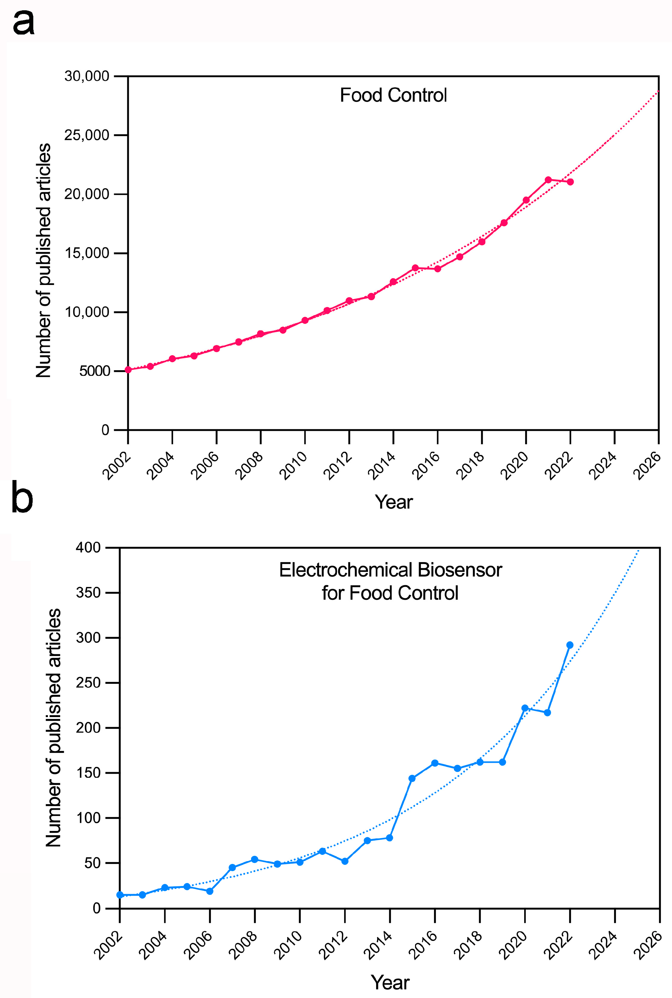
1. Introduction
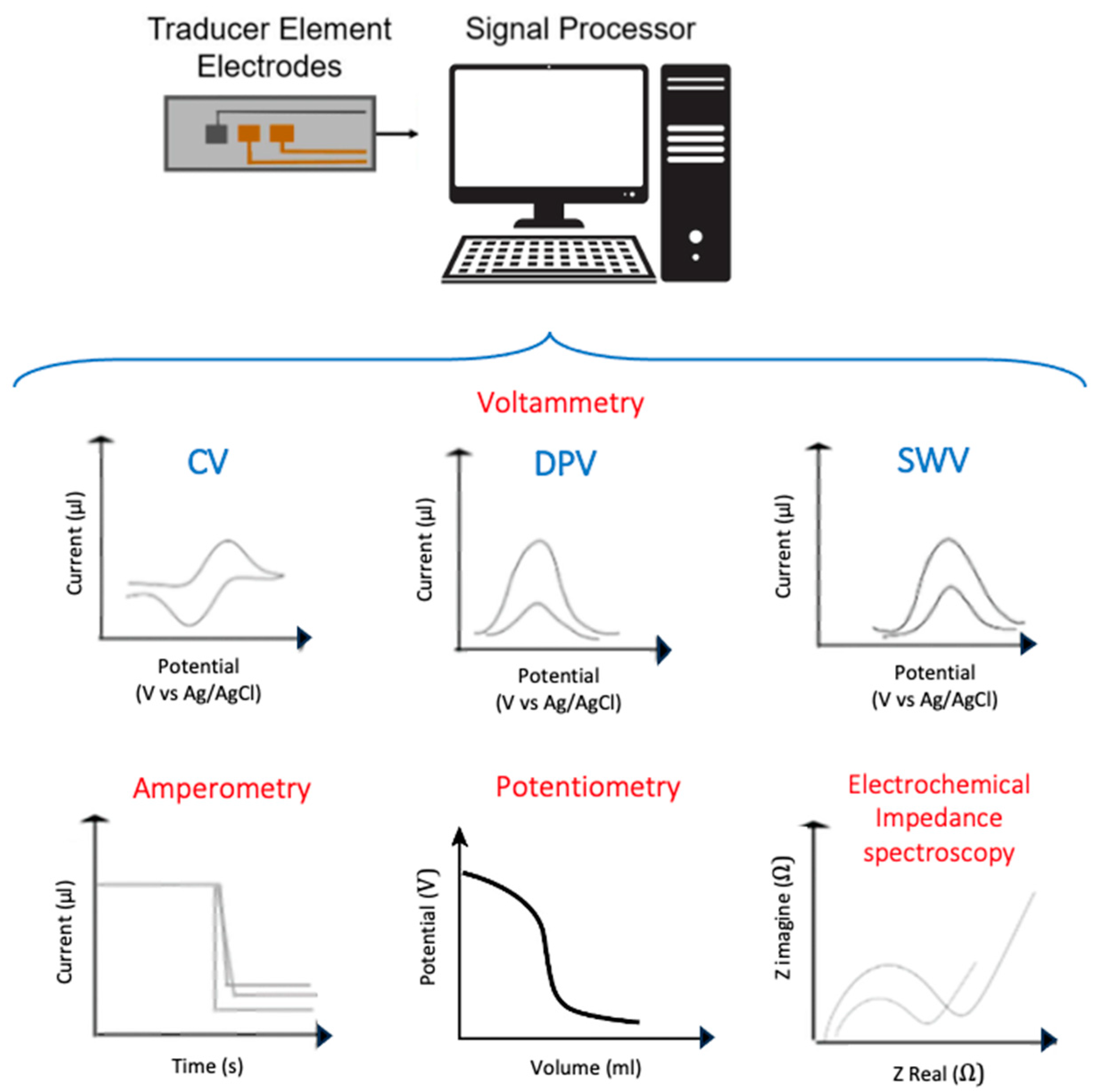
2. Electrochemical Biosensors
2.1. Electrochemical Genosensors
2.2. Electrochemical Immunosensors
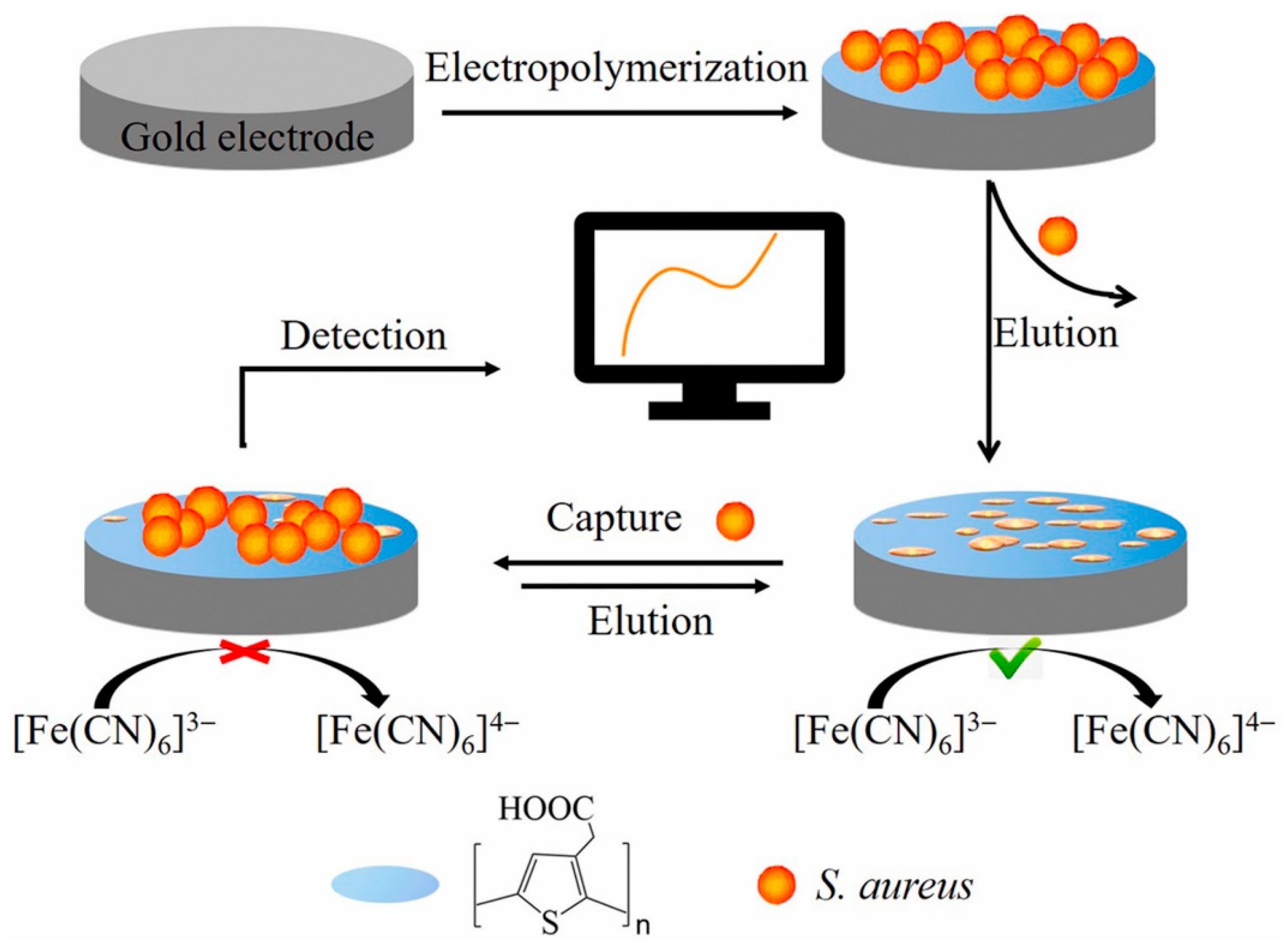
2.3. Electrochemical Sensors Based on Artificial Bioreceptors
2.4. Other Electrochemical Biosensors
3. Conclusions and Perspectives
Author Contributions
Funding
Conflicts of Interest
References
- WHO. 2022. Available online: https://cdn.who.int/media/docs/default-source/gho-documents/world-health-statistic-reports/worldhealthstatistics_2022.pdf (accessed on 1 March 2023).
- WHO. 2023. Available online: https://www.who.int/news-room/fact-sheets/detail/food-safety (accessed on 1 March 2023).
- Vidic, J.; Manzano, M.; Raj, V.S.; Pandey, R.P.; Chang, C.-M. Comparative meta-analysis of antimicrobial resistance from different food sources along with one health approach in Italy and Thailand. One Health 2023, 16, 100477. [Google Scholar]
- Morse, T.D.; Masuku, H.; Rippon, S.; Kubwalo, H. Achieving an integrated approach to food safety and hygiene—Meeting the sustainable development goals in sub-saharan Africa. Sustainability 2018, 10, 2394. [Google Scholar] [CrossRef]
- Omerović, N.; Djisalov, M.; Živojević, K.; Mladenović, M.; Vunduk, J.; Milenković, I.; Knežević, N.Ž.; Gadjanski, I.; Vidić, J. Antimicrobial nanoparticles and biodegradable polymer composites for active food packaging applications. Compr. Rev. Food Sci. Food Saf. 2021, 20, 2428–2454. [Google Scholar] [CrossRef] [PubMed]
- European Food Safety Authority and European Centre for Disease Prevention and Control. The European Union one health 2020 zoonoses report. EFSA J. 2021, 19, e06971. [Google Scholar]
- Vizzini, P.; Braidot, M.; Vidic, J.; Manzano, M. Electrochemical and optical biosensors for the detection of campylobacter and listeria: An update look. Micromachines 2019, 10, 500. [Google Scholar] [CrossRef] [PubMed]
- Zhao, X.; Lin, C.-W.; Wang, J.; Oh, D.H. Advances in rapid detection methods for foodborne pathogens. J. Microbiol. Biotechnol. 2014, 24, 297–312. [Google Scholar] [CrossRef]
- Vidic, J.; Vizzini, P.; Manzano, M.; Kavanaugh, D.; Ramarao, N.; Zivkovic, M.; Radonic, V.; Knezevic, N.; Giouroudi, I.; Gadjanski, I. Point-of-need DNA testing for detection of foodborne pathogenic bacteria. Sensors 2019, 19, 1100. [Google Scholar] [CrossRef]
- Vidic, J.; Auger, S.; Marin, M.; Rizzotto, F.; Haddad, N.; Guillou, S.; Guyard-Nicodème, M.; Vizzini, P.; Cossettini, A.; Manzano, M. Role of real-time DNA analyses, biomarkers, resistance measurement, and ecosystem management in Campylobacter risk analysis. In Present Knowledge in Food Safety; Elsevier: Amsterdam, The Netherlands, 2023; pp. 752–776. [Google Scholar]
- Cossettini, A.; Vidic, J.; Maifreni, M.; Marino, M.; Pinamonti, D.; Manzano, M. Rapid detection of Listeria monocytogenes, Salmonella, Campylobacter spp., and Escherichia coli in food using biosensors. Food Control 2022, 137, 108962. [Google Scholar] [CrossRef]
- Land, K.J.; Boeras, D.I.; Chen, X.-S.; Ramsay, A.R.; Peeling, R.W. REASSURED diagnostics to inform disease control strategies, strengthen health systems and improve patient outcomes. Nat. Microbiol. 2019, 4, 46–54. [Google Scholar] [CrossRef]
- Rotariu, L.; Lagarde, F.; Jaffrezic-Renault, N.; Bala, C. Electrochemical biosensors for fast detection of food contaminants–trends and perspective. TrAC Trends Anal. Chem. 2016, 79, 80–87. [Google Scholar] [CrossRef]
- Kaya, H.O.; Cetin, A.E.; Azimzadeh, M.; Topkaya, S.N. Pathogen detection with electrochemical biosensors: Advantages, challenges and future perspectives. J. Electroanal. Chem. 2021, 882, 114989. [Google Scholar] [CrossRef]
- Moro, G.; Bottari, F.; Van Loon, J.; Du Bois, E.; De Wael, K.; Moretto, L.M. Disposable electrodes from waste materials and renewable sources for (bio) electroanalytical applications. Biosens. Bioelectron. 2019, 146, 111758. [Google Scholar] [CrossRef]
- Podunavac, I.; Kukkar, M.; Léguillier, V.; Rizzotto, F.; Pavlovic, Z.; Janjušević, L.; Costache, V.; Radonic, V.; Vidic, J. Low-cost goldleaf electrode as a platform for Escherichia coli immunodetection. Talanta 2023, 259, 124557. [Google Scholar] [CrossRef]
- Bobrinetskiy, I.; Radovic, M.; Rizzotto, F.; Vizzini, P.; Jaric, S.; Pavlovic, Z.; Radonic, V.; Nikolic, M.V.; Vidic, J. Advances in nanomaterials-based electrochemical biosensors for foodborne pathogen detection. Nanomaterials 2021, 11, 2700. [Google Scholar] [CrossRef]
- Wang, X.; Dong, S.; Wei, H. Recent advances on nanozyme-based electrochemical biosensors. Electroanalysis 2023, 35, e202100684. [Google Scholar] [CrossRef]
- Sheng, K.; Jiang, H.; Fang, Y.; Wang, L.; Jiang, D. Emerging electrochemical biosensing approaches for detection of allergen in food samples: A review. Trends Food Sci. Technol. 2022, 121, 93–104. [Google Scholar] [CrossRef]
- Brett, C.M. Electrochemical impedance spectroscopy in the characterisation and application of modified electrodes for electrochemical sensors and biosensors. Molecules 2022, 27, 1497. [Google Scholar] [CrossRef]
- Singh, A.; Sharma, A.; Ahmed, A.; Sundramoorthy, A.K.; Furukawa, H.; Arya, S.; Khosla, A. Recent advances in electrochemical biosensors: Applications, challenges, and future scope. Biosensors 2021, 11, 336. [Google Scholar] [CrossRef] [PubMed]
- Ronkainen, N.J.; Halsall, H.B.; Heineman, W.R. Electrochemical biosensors. Chem. Soc. Rev. 2010, 39, 1747–1763. [Google Scholar] [CrossRef] [PubMed]
- Ciucci, F. Modeling electrochemical impedance spectroscopy. Curr. Opin. Electrochem. 2019, 13, 132–139. [Google Scholar] [CrossRef]
- Vidic, J.; Manzano, M. Electrochemical biosensors for rapid pathogen detection. Curr. Opin. Electrochem. 2021, 29, 100750. [Google Scholar] [CrossRef]
- Cho, I.-H.; Kim, D.H.; Park, S. Electrochemical biosensors: Perspective on functional nanomaterials for on-site analysis. Biomater. Res. 2020, 24, 6. [Google Scholar] [CrossRef]
- Lao, R.; Song, S.; Wu, H.; Wang, L.; Zhang, Z.; He, L.; Fan, C. Electrochemical interrogation of DNA monolayers on gold surfaces. Anal. Chem. 2005, 77, 6475–6480. [Google Scholar] [CrossRef]
- Cho, I.-H.; Lee, J.; Kim, J.; Kang, M.-S.; Paik, J.K.; Ku, S.; Cho, H.-M.; Irudayaraj, J.; Kim, D.-H. Current technologies of electrochemical immunosensors: Perspective on signal amplification. Sensors 2018, 18, 207. [Google Scholar] [CrossRef]
- Escobar, V.; Scaramozzino, N.; Vidic, J.; Buhot, A.; Mathey, R.; Chaix, C.; Hou, Y. Recent advances on peptide-based biosensors and electronic noses for foodborne pathogen detection. Biosensors 2023, 13, 258. [Google Scholar] [CrossRef] [PubMed]
- Capobianco, J.A.; Armstrong, C.M.; Lee, J.; Gehring, A.G. Detection of pathogenic bacteria in large volume food samples using an enzyme-linked immunoelectrochemical biosensor. Food Control 2021, 119, 107456. [Google Scholar] [CrossRef]
- Klass, S.H.; Sofen, L.E.; Hallberg, Z.F.; Fiala, T.A.; Ramsey, A.V.; Dolan, N.S.; Francis, M.B.; Furst, A.L. Covalent capture and electrochemical quantification of pathogenic E. coli. Chem. Commun. 2021, 57, 2507–2510. [Google Scholar] [CrossRef]
- El-Moghazy, A.Y.; Wisuthiphaet, N.; Yang, X.; Sun, G.; Nitin, N. Electrochemical biosensor based on genetically engineered bacteriophage T7 for rapid detection of Escherichia coli on fresh produce. Food Control. 2022, 135, 108811. [Google Scholar] [CrossRef]
- Razmi, N.; Hasanzadeh, M.; Willander, M.; Nur, O. Electrochemical genosensor based on gold nanostars for the detection of Escherichia coli O157: H7 DNA. Anal. Methods 2022, 14, 1562–1570. [Google Scholar] [CrossRef] [PubMed]
- Chen, J.; Jiang, Z.; Ackerman, J.D.; Yazdani, M.; Hou, S.; Nugen, S.R.; Rotello, V.M. Electrochemical nanoparticle–enzyme sensors for screening bacterial contamination in drinking water. Analyst 2015, 140, 4991–4996. [Google Scholar] [CrossRef]
- Oliveira, D.A.; Althawab, S.; McLamore, E.S.; Gomes, C.L. One-step fabrication of stimuli-responsive chitosan-platinum brushes for listeria monocytogenes detection. Biosensors 2021, 11, 511. [Google Scholar] [CrossRef]
- Feng, K.; Li, T.; Ye, C.; Gao, X.; Yang, T.; Liang, X.; Yue, X.; Ding, S.; Dong, Q.; Yang, M. A label-free electrochemical immunosensor for rapid detection of salmonella in milk by using CoFe-MOFs-graphene modified electrode. Food Control 2021, 130, 108357. [Google Scholar] [CrossRef]
- Xue, L.; Guo, R.; Huang, F.; Qi, W.; Liu, Y.; Cai, G.; Lin, J. An impedance biosensor based on magnetic nanobead net and MnO2 nanoflowers for rapid and sensitive detection of foodborne bacteria. Biosens. Bioelectron. 2021, 173, 112800. [Google Scholar] [CrossRef]
- Huang, F.; Xue, L.; Qi, W.; Cai, G.; Liu, Y.; Lin, J. An ultrasensitive impedance biosensor for Salmonella detection based on rotating high gradient magnetic separation and cascade reaction signal amplification. Biosens. Bioelectron. 2021, 176, 112921. [Google Scholar] [CrossRef]
- Sun, Q.; Liu, X.; Tang, H.; Qian, Y.; Gu, H.; He, H. A Sandwich-type Electrochemical Immunosensor for the Sensitive Determination of Salmonella Typhimurium in Food. Electroanalysis 2022, 34, 911–918. [Google Scholar] [CrossRef]
- Feng, K.; Li, T.; Ye, C.; Gao, X.; Yue, X.; Ding, S.; Dong, Q.; Yang, M.; Huang, G.; Zhang, J. A novel electrochemical immunosensor based on Fe3O4@ graphene nanocomposite modified glassy carbon electrode for rapid detection of Salmonella in milk. J. Dairy Sci. 2022, 105, 2108–2118. [Google Scholar] [CrossRef]
- Li, J.; Jiang, J.; Su, Y.; Liang, Y.; Zhang, C. A novel cloth-based supersandwich electrochemical aptasensor for direct, sensitive detection of pathogens. Anal. Chim. Acta 2021, 1188, 339176. [Google Scholar] [CrossRef]
- Cai, R.; Zhang, Z.; Chen, H.; Tian, Y.; Zhou, N. A versatile signal-on electrochemical biosensor for Staphylococcus aureus based on triple-helix molecular switch. Sens. Actuators B Chem. 2021, 326, 128842. [Google Scholar] [CrossRef]
- Wang, R.; Wang, L.; Yan, J.; Luan, D.; Wu, J.; Bian, X. Rapid, sensitive and label-free detection of pathogenic bacteria using a bacteria-imprinted conducting polymer film-based electrochemical sensor. Talanta 2021, 226, 122135. [Google Scholar] [CrossRef] [PubMed]
- Wu, W.; Yang, Y.; Wang, L.; Xu, T.; Wang, R. Electrochemical immunosensor based on mussel inspired coating for simultaneous detection and elimination of Staphylococcus aureus in drinks. RSC Adv. 2021, 11, 18252–18258. [Google Scholar] [CrossRef] [PubMed]
- Liu, S.; Li, Q.; Yang, H.; Wang, P.; Miao, X.; Feng, Q. An in situ quenching electrochemiluminescence biosensor amplified with aptamer recognition-induced multi-DNA release for sensitive detection of pathogenic bacteria. Biosens. Bioelectron. 2022, 196, 113744. [Google Scholar] [CrossRef]
- Cai, R.; Zhang, S.; Chen, L.; Li, M.; Zhang, Y.; Zhou, N. Self-assembled DNA nanoflowers triggered by a DNA walker for highly sensitive electrochemical detection of Staphylococcus aureus. ACS Appl. Mater. Interfaces 2021, 13, 4905–4914. [Google Scholar] [CrossRef]
- Wang, H.; Yang, L.; Tan, Y.; Deng, W.; Xie, Q. Ag2S quantum dots loaded dendritic mesoporous silica nanospheres as signal amplification labels for ultrasensitive electrochemical immuno-biosensor for Staphylococcus aureus. J. Electroanal. Chem. 2022, 919, 116496. [Google Scholar] [CrossRef]
- Somayeh, S.S.; Taghdisi, S.M.; Mortazavi, S.A.; Farideh, T.Y.; Abnous, K. A novel electrochemical biosensor for detection of micrococcal nuclease in milk based on a U-shaped DNA structure. Talanta 2023, 253, 123989. [Google Scholar] [CrossRef]
- Huang, L.; Yuan, N.; Guo, W.; Zhang, Y.; Zhang, W. An electrochemical biosensor for the highly sensitive detection of Staphylococcus aureus based on SRCA-CRISPR/Cas12a. Talanta 2023, 252, 123821. [Google Scholar] [CrossRef]
- Nguyen, T.T.-Q.; Gu, M.B. An ultrasensitive electrochemical aptasensor using Tyramide-assisted enzyme multiplication for the detection of Staphylococcus aureus. Biosens. Bioelectron. 2023, 228, 115199. [Google Scholar] [CrossRef]
- You, X.; Zhang, G.; Chen, Y.; Liu, D.; Ma, D.; Zhou, J.; Liu, Y.; Liu, H.; Qi, Y.; Liang, C. A novel electrochemical immunosensor for the sensitive detection of tiamulin based on staphylococcal protein A and silver nanoparticle-graphene oxide nanocomposites. Bioelectrochemistry 2021, 141, 107877. [Google Scholar] [CrossRef]
- Shan, X.; Kuang, D.; Feng, Q.; Wu, M.; Yang, J. A dual-mode ratiometric aptasensor for accurate detection of pathogenic bacteria based on recycling of DNAzyme activation. Food Chem. 2023, 423, 136287. [Google Scholar] [CrossRef]
- Ali, M.; Bacchu, M.; Das, S.; Akter, S.; Rahman, M.; Aly, M.A.S.; Khan, M. Label free flexible electrochemical DNA biosensor for selective detection of Shigella flexneri in real food samples. Talanta 2023, 253, 123909. [Google Scholar] [CrossRef]
- Yashini, M.; Auddy, I.; Shanmugasundaram, S.; Vidyalakshmi, R.; Sunil, C. Characterization of Antibody Immobilization on Chitosan/Gelatin-Modified Electrode and Its Application to Bacillus cereus Detection in Cereal-Based Food. Food Anal. Methods 2022, 15, 2382–2393. [Google Scholar] [CrossRef]
- Seo, Y.; Yoon, Y.; Lee, M.; Jang, M.; Kim, T.-H.; Kim, Y.; Yoo, H.Y.; Min, J.; Lee, T. Rapid electrochemical biosensor composed of DNA probe/iridium nanoparticle bilayer for Aphanizomenon flos-aquae detection in fresh water. Colloids Surf. B Biointerfaces 2023, 225, 113218. [Google Scholar] [CrossRef] [PubMed]
- Jiang, H.; Sun, Z.; Guo, Q.; Weng, X. Microfluidic thread-based electrochemical aptasensor for rapid detection of Vibrio parahaemolyticus. Biosens. Bioelectron. 2021, 182, 113191. [Google Scholar] [CrossRef]
- Jiang, H.; Sun, Z.; Zhang, C.; Weng, X. 3D-architectured aptasensor for ultrasensitive electrochemical detection of norovirus based on phosphorene-gold nanocomposites. Sens. Actuators B Chem. 2022, 354, 131232. [Google Scholar] [CrossRef]
- da Silva-Junio, A.G.; Frias, I.A.; Lima-Neto, R.G.; Migliolo, L.; e Silva, P.S.; Oliveira, M.D.; Andrade, C.A. Electrochemical biosensor based on Temporin-PTA peptide for detection of microorganisms. J. Pharm. Biomed. Anal. 2022, 216, 114788. [Google Scholar] [CrossRef]
- Dai, G.; Yao, H.; Yang, L.; Ding, Y.; Du, S.; Shen, H.; Mo, F. Rapid detection of foodborne pathogens in diverse foodstuffs by universal electrochemical aptasensor based on UiO-66 and methylene blue composites. Food Chem. 2023, 424, 136244. [Google Scholar] [CrossRef]
- Wu, C.; Wang, X.; Guo, L.; Huang, X.; Wu, L.; Huang, H. An electrochemical aptasensor based on exonuclease III-assisted signal amplification coupled with CRISPR-Cas12a for ochratoxin A detection. Food Control 2023, 109631. [Google Scholar] [CrossRef]
- Zhang, Q.; Zhang, M.; Guo, Z.; Li, J.; Zhu, Z.; Wang, Y.; Liu, S.; Huang, J.; Yu, J. DNA tetrahedron-besieged primer and DNAzyme-activated programmatic RCA for low-background electrochemical detection of ochratoxin A. Anal. Chim. Acta 2023, 1242, 340782. [Google Scholar] [CrossRef]
- Zhang, S.; Wang, Y.; Sheng, Q.; Yue, T. Electrochemical Aptasensor Based on ZnO-Au Nanocomposites for the Determination of Ochratoxin A in Wine and Beer. Processes 2023, 11, 864. [Google Scholar] [CrossRef]
- Liu, Y.; Guo, W.; Zhang, Y.; Lu, X.; Yang, Q.; Zhang, W. An accurate and ultrasensitive ratiometric electrochemical aptasensor for determination of Ochratoxin A based on catalytic hairpin assembly. Food Chem. 2023, 423, 136301. [Google Scholar] [CrossRef] [PubMed]
- Taghdisi, S.M.; Danesh, N.M.; Ramezani, M.; Alibolandi, M.; Nameghi, M.A.; Gerayelou, G.; Abnous, K. A novel electrochemical aptasensor for ochratoxin a sensing in spiked food using strand-displacement polymerase reaction. Talanta 2021, 223, 121705. [Google Scholar] [CrossRef] [PubMed]
- Hou, Y.; Long, N.; Xu, Q.; Li, Y.; Song, P.; Yang, M.; Wang, J.; Zhou, L.; Sheng, P.; Kong, W. Development of a Nafion-MWCNTs and in-situ generated Au nanopopcorns dual-amplification electrochemical aptasensor for ultrasensitive detection of OTA. Food Chem. 2023, 403, 134375. [Google Scholar] [CrossRef]
- Huang, T.; Wang, M.; Hong, N.; Cui, H.; Fan, Q.; Wei, G.; Qin, L.; Zhang, J.; Fan, H. An autonomous driven DNA walker-based electrochemical aptasensor for on-site detection of Ochratoxin A. Talanta 2023, 252, 123785. [Google Scholar] [CrossRef] [PubMed]
- Wu, C.; Wu, X.; Hou, F.; Wu, L.; Liu, G. An ultrasensitive electrochemical aptasensor based on Pd@ PCN-222 as a signal probe coupled with exonuclease III-assisted cycling amplification for the detection of ochratoxin A. Food Control 2022, 139, 109066. [Google Scholar] [CrossRef]
- Liang, X.; Zhao, F.; Xiao, C.; Yue, S.; Huang, Y.; Wei, M. A ratiometric electrochemical aptasensor for ochratoxin A detection. J. Chin. Chem. Soc. 2021, 68, 1271–1278. [Google Scholar] [CrossRef]
- Feng, B.; Suo, Z.; He, B.; Liu, Y.; Wei, M.; Jin, H. An innovative electrochemical aptasensor based on the dual signal amplification strategy of gold nanowires and bifunctional DNA nanoflowers. Sens. Actuators B Chem. 2023, 377, 132995. [Google Scholar] [CrossRef]
- Zhang, H.; Ye, S.; Huang, L.; Fan, S.; Mao, W.; Hu, Y.; Yu, Y.; Fu, F. An electrochemical biosensor for the detection of aflatoxin B1 based on the specific aptamer and HCR biological magnification. Anal. Methods 2023, 15, 99–108. [Google Scholar] [CrossRef] [PubMed]
- Zhong, T.; Li, S.; Li, X.; JiYe, Y.; Mo, Y.; Chen, L.; Zhang, Z.; Wu, H.; Li, M.; Luo, Q. A label-free electrochemical aptasensor based on AuNPs-loaded zeolitic imidazolate framework-8 for sensitive determination of aflatoxin B1. Food Chem. 2022, 384, 132495. [Google Scholar] [CrossRef]
- Asl, G.B.; Arvand, M.; Habibi, M.F. High affinity aptamers for ultra-sensitive detection of aflatoxin B1 in milk and groundnut samples with label-free photo-electrochemical aptasensor. Food Chem. 2022, 397, 133829. [Google Scholar] [CrossRef]
- Wang, N.; Liu, Q.; Hu, X.; Wang, F.; Hu, M.; Yu, Q.; Zhang, G. Electrochemical immunosensor based on AuNPs/Zn/Ni-ZIF-8–800@ graphene for rapid detection of aflatoxin B1 in peanut oil. Anal. Biochem. 2022, 650, 114710. [Google Scholar] [CrossRef]
- Wang, P.; Luo, B.; Liu, K.; Wang, C.; Dong, H.; Wang, X.; Hou, P.; Li, A. A novel COOH–GO–COOH–MWNT/pDA/AuNPs based electrochemical aptasensor for detection of AFB 1. RSC Adv. 2022, 12, 27940–27947. [Google Scholar] [CrossRef]
- Dos Santos, D.M.; Migliorini, F.L.; Soares, A.C.; Mattoso, L.H.; Oliveira, O.N., Jr.; Correa, D.S. Electrochemical Immunosensor Made with Zein-based Nanofibers for On-site Detection of Aflatoxin B1. Electroanalysis 2023, 35, e202100672. [Google Scholar] [CrossRef]
- Ou, G.; Zhao, A.; Liao, H.; Zhang, Z.; Xiao, F. Au nanopartics decorated urchin-like Bi2S3 on graphene wrapped carbon fiber microelectrode: Towards electrochemical immunosensor for sensitive determination of aflatoxin B1. J. Electroanal. Chem. 2023, 929, 117124. [Google Scholar] [CrossRef]
- Zhu, C.; Liu, D.; Li, Y.; Chen, T.; You, T. Label-free ratiometric homogeneous electrochemical aptasensor based on hybridization chain reaction for facile and rapid detection of aflatoxin B1 in cereal crops. Food Chem. 2022, 373, 131443. [Google Scholar] [CrossRef]
- Cui, H.; An, K.; Wang, C.; Chen, Y.; Jia, S.; Qian, J.; Hao, N.; Wei, J.; Wang, K. A disposable ratiometric electrochemical aptasensor with exonuclease I-powered target recycling amplification for highly sensitive detection of aflatoxin B1. Sens. Actuators B Chem. 2022, 355, 131238. [Google Scholar] [CrossRef]
- Huang, Q.; Lin, X.; Chen, D.; Tong, Q.-X. Carbon Dots/α-Fe2O3-Fe3O4 nanocomposite: Efficient synthesis and application as a novel electrochemical aptasensor for the ultrasensitive determination of aflatoxin B1. Food Chem. 2022, 373, 131415. [Google Scholar] [CrossRef] [PubMed]
- Liu, C.; Wu, T.; Zeng, W.; Liu, J.; Hu, B.; Wu, L. Dual-signal electrochemical aptasensor involving hybridization chain reaction amplification for aflatoxin B1 detection. Sens. Actuators B Chem. 2022, 371, 132494. [Google Scholar] [CrossRef]
- Ong, J.Y.; Phang, S.-W.; Goh, C.T.; Pike, A.; Tan, L.L. Impedimetric Polyaniline-Based Aptasensor for Aflatoxin B1 Determination in Agricultural Products. Foods 2023, 12, 1698. [Google Scholar] [CrossRef]
- Tang, X.; Catanante, G.; Huang, X.; Marty, J.-L.; Wang, H.; Zhang, Q.; Li, P. Screen-printed electrochemical immunosensor based on a novel nanobody for analyzing aflatoxin M1 in milk. Food Chem. 2022, 383, 132598. [Google Scholar] [CrossRef]
- Ahmadi, S.F.; Hojjatoleslamy, M.; Kiani, H.; Molavi, H. Monitoring of Aflatoxin M1 in milk using a novel electrochemical aptasensor based on reduced graphene oxide and gold nanoparticles. Food Chem. 2022, 373, 131321. [Google Scholar] [CrossRef]
- Hui, Y.; Peng, H.; Zhang, F.; Zhang, L.; Yufang, L.; Zhao, A.; Jia, R.; Wang, B.; Song, Y. A novel electrochemical aptasensor based on layer-by-layer assembly of DNA-Au@ Ag conjugates for rapid detection of aflatoxin M1 in milk samples. J. Dairy Sci. 2022, 105, 1966–1977. [Google Scholar] [CrossRef]
- Pérez-Fernández, B.; Maestroni, B.M.; Nakaya, S.; Bussalino, S.; Vlachou, C.; de la Escosura-Muñiz, A. Development, optimization and validation of an electrochemical immunosensor for determination of total aflatoxins in pistachio. Food Control 2023, 152, 109859. [Google Scholar] [CrossRef]
- Mao, Y.; Dang, M.; Zhang, J.; Huang, X.; Qiao, M.; Song, L.; Zhao, Q.; Ding, M.; Wang, Y.; Li, Z. Peptide amphiphile inspired self-assembled, ordered gold nanocomposites for improved sensitivity of electrochemical immunosensor: Applications in determining the total aflatoxin amount in food stuffs. Talanta 2022, 247, 123532. [Google Scholar] [CrossRef]
- Wen, X.; Huang, Q.; Nie, D.; Zhao, X.; Cao, H.; Wu, W.; Han, Z. A multifunctional n-doped cu–mofs (N–cu–mof) nanomaterial-driven electrochemical aptasensor for sensitive detection of deoxynivalenol. Molecules 2021, 26, 2243. [Google Scholar] [CrossRef]
- Wang, K.; He, B.; Xie, L.; Li, L.; Yang, J.; Liu, R.; Wei, M.; Jin, H.; Ren, W. Exonuclease III-assisted triple-amplified electrochemical aptasensor based on PtPd NPs/PEI-rGO for deoxynivalenol detection. Sens. Actuators B Chem. 2021, 349, 130767. [Google Scholar] [CrossRef]
- Wang, L.; Jin, H.; Wei, M.; Ren, W.; Zhang, Y.; Jiang, L.; Wei, T.; He, B. A DNAzyme-assisted triple-amplified electrochemical aptasensor for ultra-sensitive detection of T-2 toxin. Sens. Actuators B Chem. 2021, 328, 129063. [Google Scholar] [CrossRef]
- Zhang, Y.; He, B.; Zhao, R.; Bai, C.; Zhang, Y.; Jin, H.; Wei, M.; Ren, W.; Suo, Z.; Xu, Y. Electrochemical aptasensor based on the target-induced strand displacement strategy-driven for T-2 toxin detection. Sci. Total Environ. 2022, 849, 157769. [Google Scholar] [CrossRef]
- He, B.; Dong, X.N. BbvCI powered DNA walking machine-based Zr-MOFs-labeled electrochemical aptasensor using Pt@ AuNRs/Fe-MOFs/PEI-rGO as electrode modification material for patulin detection. Chem. Eng. J. 2021, 405, 126642. [Google Scholar] [CrossRef]
- Xu, J.; Liu, J.; Li, W.; Wei, Y.; Sheng, Q.; Shang, Y. A dual-signaling electrochemical aptasensor based on an in-plane gold nanoparticles–black phosphorus heterostructure for the sensitive detection of patulin. Foods 2023, 12, 846. [Google Scholar] [CrossRef] [PubMed]
- Lu, X.; He, B.; Liang, Y.; Wang, J.; Jiao, Q.; Liu, Y.; Guo, R.; Wei, M.; Jin, H.; Ren, W. An electrochemical aptasensor based on dual-enzymes-driven target recycling strategy for patulin detection in apple juice. Food Control 2022, 137, 108907. [Google Scholar] [CrossRef]
- Nodoushan, S.M.; Nasirizadeh, N.; Sedighian, H.; Kachuei, R.; Azimzadeh-Taft, M.; Fooladi, A.A.I. Detection of Staphylococcal Enterotoxin A (SEA) using a sensitive nanomaterial-based electrochemical aptasensor. Diam. Relat. Mater. 2022, 127, 109042. [Google Scholar] [CrossRef]
- Suo, Z.; Niu, X.; Liu, R.; Xin, L.; Liu, Y.; Wei, M. A methylene blue and Ag+ ratiometric electrochemical aptasensor based on Au@ Pt/Fe-NC signal amplification strategy for zearalenone detection. Sens. Actuators B Chem. 2022, 362, 131825. [Google Scholar] [CrossRef]
- Duan, F.; Rong, F.; Guo, C.; Chen, K.; Wang, M.; Zhang, Z.; Pettinari, R.; Zhou, L.; Du, M. Electrochemical aptasensing strategy based on a multivariate polymertitanium-metal-organic framework for zearalenone analysis. Food Chem. 2022, 385, 132654. [Google Scholar] [CrossRef] [PubMed]
- Lai, H.; Ming, P.; Wu, M.; Wang, S.; Sun, D.; Zhai, H. An electrochemical aptasensor based on P-Ce-MOF@ MWCNTs as signal amplification strategy for highly sensitive detection of zearalenone. Food Chem. 2023, 423, 136331. [Google Scholar] [CrossRef] [PubMed]
- Dong, N.; Liu, D.; Meng, S.; Liu, S.; You, T. Tetrahedral DNA nanostructure-enabled electrochemical aptasensor for ultrasensitive detection of fumonisin B1 with extended dynamic range. Sens. Actuators B Chem. 2022, 354, 130984. [Google Scholar] [CrossRef]
- Mu, Z.; Tian, J.; Wang, J.; Zhou, J.; Bai, L. A new electrochemical aptasensor for ultrasensitive detection of endotoxin using Fe-MOF and AgNPs decorated PN-CNTs as signal enhanced indicator. Appl. Surf. Sci. 2022, 573, 151601. [Google Scholar] [CrossRef]
- de Brito, A.R.; de Jesus, R.S.; de Carvalho Tavares, I.M.; Silva, F.N.; Santana, N.B.; Ferrao, S.P.B.; Bilal, M.; de Santana Santos, A.; Salay, L.C.; de Oliveira, J.R. Application of the electrochemical biosensor in the detection of lactose in skimmed milk. Surf. Interfaces 2021, 22, 100839. [Google Scholar] [CrossRef]
- Sundhoro, M.; Agnihotra, S.R.; Amberger, B.; Augustus, K.; Khan, N.D.; Barnes, A.; BelBruno, J.; Mendecki, L. An electrochemical molecularly imprinted polymer sensor for rapid and selective food allergen detection. Food Chem. 2021, 344, 128648. [Google Scholar] [CrossRef]
- Svigelj, R.; Zuliani, I.; Grazioli, C.; Dossi, N.; Toniolo, R. An effective label-free electrochemical aptasensor based on gold nanoparticles for gluten detection. Nanomaterials 2022, 12, 987. [Google Scholar] [CrossRef]
- Torre, R.; Freitas, M.; Costa-Rama, E.; Nouws, H.P.; Delerue-Matos, C. Food allergen control: Tropomyosin analysis through electrochemical immunosensing. Food Chem. 2022, 396, 133659. [Google Scholar] [CrossRef]
- Freitas, M.; Neves, M.M.; Nouws, H.P.; Delerue-Matos, C. Electrochemical Immunosensor for the Simultaneous Determination of Two Main Peanut Allergenic Proteins (Ara h 1 and Ara h 6) in Food Matrices. Foods 2021, 10, 1718. [Google Scholar] [CrossRef]
- Melinte, G.; Hosu, O.; Cristea, C.; Marrazza, G. Ara H1 peanut allergen detection using a labelled electrochemical aptasensor based on GO-COOH@ bimetallic composite platform. Food Chem. 2023, 400, 134074. [Google Scholar] [CrossRef]
- Jiang, H.; Guo, Q.; Zhang, C.; Sun, Z.; Weng, X. Microfluidic origami nano-aptasensor for peanut allergen Ara h1 detection. Food Chem. 2021, 365, 130511. [Google Scholar] [CrossRef]
- Guan, J.; He, K.; Gunasekaran, S. Selection of ssDNA aptamer using GO-SELEX and development of DNA nanostructure-based electrochemical aptasensor for penicillin. Biosens. Bioelectron. X 2022, 12, 100220. [Google Scholar] [CrossRef]
- Sullivan, C.; Lu, D.; Senecal, A.; Kurup, P. Voltammetric detection of arsenic (III) using gold nanoparticles modified carbon screen printed electrodes: Application for facile and rapid analysis in commercial apple juice. Food Chem. 2021, 352, 129327. [Google Scholar] [CrossRef] [PubMed]
- Raymundo-Pereira, P.A.; Gomes, N.O.; Shimizu, F.M.; Machado, S.A.; Oliveira, O.N., Jr. Selective and sensitive multiplexed detection of pesticides in food samples using wearable, flexible glove-embedded non-enzymatic sensors. Chem. Eng. J. 2021, 408, 127279. [Google Scholar] [CrossRef]
- Venegas, C.J.; Rodríguez, L.; Sierra-Rosales, P. Selective Label-Free Electrochemical Aptasensor Based on Carbon Nanotubes for Carbendazim Detection. Chemosensors 2023, 11, 117. [Google Scholar] [CrossRef]
- Brycht, M.; Łukawska, A.; Frühbauerová, M.; Pravcová, K.; Metelka, R.; Skrzypek, S.; Sýs, M. Rapid monitoring of fungicide fenhexamid residues in selected berries and wine grapes by square-wave voltammetry at carbon-based electrodes. Food Chem. 2021, 338, 127975. [Google Scholar] [CrossRef] [PubMed]
- Yan, Y.; Zhou, F.; Wang, Q.; Huang, Y. A sensitive electrochemical biosensor for quinolones detection based on Cu2+-modulated signal amplification. Microchem. J. 2023, 190, 108636. [Google Scholar] [CrossRef]
- Anchidin-Norocel, L.; Savage, W.K.; Gutt, G.; Amariei, S. Development, optimization, characterization, and application of electrochemical biosensors for detecting nickel ions in food. Biosensors 2021, 11, 519. [Google Scholar] [CrossRef]
- Liu, Y.; Kong, L.; Han, Z.; Yuan, R.; Chai, Y. An electrochemical biosensor based on a highly loaded 3D DNA nanostructure for ultrasensitive detection of Pb2+. Sens. Actuators B Chem. 2023, 382, 133486. [Google Scholar] [CrossRef]
- Wang, L.; Peng, X.; Fu, H. An electrochemical aptasensor for the sensitive detection of Pb2+ based on a chitosan/reduced graphene oxide/titanium dioxide. Microchem. J. 2022, 174, 106977. [Google Scholar] [CrossRef]
- Yuan, M.; Qian, S.; Cao, H.; Yu, J.; Ye, T.; Wu, X.; Chen, L.; Xu, F. An ultra-sensitive electrochemical aptasensor for simultaneous quantitative detection of Pb2+ and Cd2+ in fruit and vegetable. Food Chem. 2022, 382, 132173. [Google Scholar]
- Peng, Y.; Lu, B.; Deng, Y.; Yang, N.; Li, G. A dual-recognition-controlled electrochemical biosensor for accurate and sensitive detection of specific circulating tumor cells. Biosens. Bioelectron. 2022, 201, 113973. [Google Scholar] [CrossRef]
- Yadav, A.K.; Verma, D.; Lakshmi, G.; Eremin, S.; Solanki, P.R. Fabrication of label-free and ultrasensitive electrochemical immunosensor based on molybdenum disulfide nanoparticles modified disposable ITO: An analytical platform for antibiotic detection in food samples. Food Chem. 2021, 363, 130245. [Google Scholar] [CrossRef]
- Yin, W.-j.; Zhang, J.-x.; Wang, H.; Wang, Y.; Zeng, X.; Xu, Z.-L.; Yang, J.-Y.; Xiao, Z.-L.; Hammock, B.D.; Wen, P. A highly sensitive electrochemical immunosensor based on electrospun nanocomposite for the detection of parathion. Food Chem. 2023, 404, 134371. [Google Scholar] [CrossRef]
- Wang, B.; He, B.; Guo, R.; Jiao, Q.; Liang, Y.; Wang, J.; Liu, Y.; Ren, W.; Suo, Z. A competitive-type electrochemical immunosensor based on Ce-MOF@ Au and MB-Au@ Pt core–shell for nitrofuran metabolites residues detection. Bioelectrochemistry 2021, 142, 107934. [Google Scholar] [CrossRef]
- Wang, A.; You, X.; Liu, H.; Zhou, J.; Chen, Y.; Zhang, C.; Ma, K.; Liu, Y.; Ding, P.; Qi, Y. Development of a label free electrochemical sensor based on a sensitive monoclonal antibody for the detection of tiamulin. Food Chem. 2022, 366, 130573. [Google Scholar] [CrossRef]
- Zhu, L.; Dong, X.-X.; Gao, C.-B.; Gai, Z.; He, Y.-X.; Qian, Z.-J.; Liu, Y.; Lei, H.-T.; Sun, Y.-M.; Xu, Z.-L. Development of a highly sensitive and selective electrochemical immunosensor for controlling of rhodamine B abuse in food samples. Food Control 2022, 133, 108662. [Google Scholar] [CrossRef]
- Dorozhko, E.V.; Gashevskay, A.S.; Korotkova, E.I.; Barek, J.; Vyskocil, V.; Eremin, S.A.; Galunin, E.V.; Saqib, M. A copper nanoparticle-based electrochemical immunosensor for carbaryl detection. Talanta 2021, 228, 122174. [Google Scholar] [CrossRef] [PubMed]
- Aymard, C.; Kanso, H.; Serrano, M.J.; Pagán, R.; Noguer, T.; Istamboulie, G. Development of a new dual electrochemical immunosensor for a rapid and sensitive detection of enrofloxacin in meat samples. Food Chem. 2022, 370, 131016. [Google Scholar] [CrossRef] [PubMed]
- Wang, Y.; Gong, C.; Zhu, Y.; Wang, Q.; Geng, L. Signal-on electrochemical aptasensor for sensitive detection of sulfamethazine based on carbon quantum dots/tungsten disulfide nanocomposites. Electrochim. Acta 2021, 393, 139054. [Google Scholar] [CrossRef]
- Yu, C.; Li, L.; Ding, Y.; Liu, H.; Cui, H.; Zhang, F.; Lin, J.; Duan, Y. A sensitive molecularly imprinted electrochemical aptasensor for highly specific determination of melamine. Food Chem. 2021, 363, 130202. [Google Scholar] [CrossRef]
- Peng, H.; Hui, Y.; Zhang, L.; Zhang, F.; Liu, Y.; Zheng, J.; Jia, R.; Song, Y.; Wang, B. A novel electrochemical aptasensor based on Ti3C2-MOFs nanocomposites for rapid streptomycin detection in milk samples. Sens. Actuators B Chem. 2022, 368, 132119. [Google Scholar] [CrossRef]
- Hui, Y.; Yang, D.; Wang, W.; Liu, Y.; He, C.; Wang, B. A label-free electrochemical aptasensor based on a gold nanoparticle/carbon nanotube/metal–organic framework nanohybrid for ultrasensitive detection of streptomycin in milk samples. Food Chem. 2023, 402, 134150. [Google Scholar] [CrossRef] [PubMed]
- Hui, Y.; Peng, H.; Li, L.; Zhao, A.; Yang, D.; Wang, W.; Wang, B. A sensitive and selective electrochemical aptasensor based on gold nanoflower/polyethyleneimine (PEI)-functionalized metal organic framework nanocomposites for label-free determination of streptomycin in milk samples. Food Anal. Methods 2023, 16, 677–688. [Google Scholar] [CrossRef]
- Lu, M.; Cao, C.; Wang, F.; Liu, G. A polyethyleneimine reduced graphene oxide/gold nanocubes based electrochemical aptasensor for chloramphenicol detection using single-stranded DNA-binding protein. Mater. Des. 2021, 199, 109409. [Google Scholar] [CrossRef]
- Yang, J.; Zhong, W.; Yu, Q.; Zou, J.; Gao, Y.; Liu, S.; Zhang, S.; Wang, X.; Lu, L. MXene–AuNP-Based Electrochemical Aptasensor for Ultra-Sensitive Detection of Chloramphenicol in Honey. Molecules 2022, 27, 1871. [Google Scholar] [CrossRef] [PubMed]
- Nodehi, M.; Baghayeri, M.; Behazin, R.; Veisi, H. Electrochemical aptasensor of bisphenol A constructed based on 3D mesoporous structural SBA-15-Met with a thin layer of gold nanoparticles. Microchem. J. 2021, 162, 105825. [Google Scholar] [CrossRef]
- Song, J.; Lin, X.; Jiang, N.; Huang, M. Carbon-doped WO3 electrochemical aptasensor based on Box-Behnken strategy for highly-sensitive detection of tetracycline. Food Chem. 2022, 367, 130564. [Google Scholar] [CrossRef]
- Xie, B.; Peng, H.; Zhang, R.; Wang, C.; He, Y. Label-free electrochemical aptasensor based on stone-like gold nanoparticles for ultrasensitive detection of tetracycline. J. Phys. Chem. C 2021, 125, 5678–5683. [Google Scholar] [CrossRef]
- Naseri, M.; Niazi, A.; Bagherzadeh, K.; Konoz, E.; Samadikhah, H.R. Modified electrochemical aptasensor for ultrasensitive detection of tetracycline: In silico and in vitro studies. Food Chem. 2023, 421, 136195. [Google Scholar] [CrossRef] [PubMed]
- Chen, Q.; Du, M.; Xu, X. A label-free and selective electrochemical aptasensor for ultrasensitive detection of Di (2-ethylhexyl) phthalate based on self-assembled DNA nanostructure amplification. J. Electroanal. Chem. 2022, 914, 116300. [Google Scholar] [CrossRef]
- Wu, Q.; Tao, H.; Wu, Y.; Wang, X.; Shi, Q.; Xiang, D. A Label-Free Electrochemical Aptasensor Based on Zn/Fe Bimetallic MOF Derived Nanoporous Carbon for Ultra-Sensitive and Selective Determination of Paraquat in Vegetables. Foods 2022, 11, 2405. [Google Scholar] [CrossRef]
- Shi, Q.; Tao, H.; Wu, Y.; Chen, J.; Wang, X. An ultrasensitive label-free electrochemical aptasensing platform for thiamethoxam detection based on ZIF-67 derived Co-N doped porous carbon. Bioelectrochemistry 2023, 149, 108317. [Google Scholar] [CrossRef]
- Huang, Y.; Ye, D.; Yang, J.; Zhu, W.; Li, L.; Ding, Y. Dual recognition elements for selective determination of progesterone based on molecularly imprinted electrochemical aptasensor. Anal. Chim. Acta 2023, 1264, 341288. [Google Scholar] [CrossRef]
- Zhu, C.; Liu, X.; Li, Y.; Yu, D.; Gao, Q.; Chen, L. Dual-ratiometric electrochemical aptasensor based on carbon nanohorns/anthraquinone-2-carboxylic acid/Au nanoparticles for simultaneous detection of malathion and omethoate. Talanta 2023, 253, 123966. [Google Scholar] [CrossRef] [PubMed]
- Varmira, K.; Mohammadi, G.; Mahmoudi, M.; Khodarahmi, R.; Rashidi, K.; Hedayati, M.; Goicoechea, H.C.; Jalalvand, A.R. Fabrication of a novel enzymatic electrochemical biosensor for determination of tyrosine in some food samples. Talanta 2018, 183, 1–10. [Google Scholar] [CrossRef]
- Molinnus, D.; Muschallik, L.; Gonzalez, L.O.; Bongaerts, J.; Wagner, T.; Selmer, T.; Siegert, P.; Keusgen, M.; Schöning, M.J. Development and characterization of a field-effect biosensor for the detection of acetoin. Biosens. Bioelectron. 2018, 115, 1–6. [Google Scholar] [CrossRef]
- Dong, P.; Jiang, B.; Zheng, J. A novel acetylcholinesterase biosensor based on gold nanoparticles obtained by electroless plating on three-dimensional graphene for detecting organophosphorus pesticides in water and vegetable samples. Anal. Methods 2019, 11, 2428–2434. [Google Scholar] [CrossRef]
- Chauhan, N.; Narang, J.; Pundir, C. Immobilization of rat brain acetylcholinesterase on ZnS and poly (indole-5-carboxylic acid) modified Au electrode for detection of organophosphorus insecticides. Biosens. Bioelectron. 2011, 29, 82–88. [Google Scholar] [CrossRef]
- Butmee, P.; Tumcharern, G.; Songsiriritthigul, C.; Durand, M.J.; Thouand, G.; Kerr, M.; Kalcher, K.; Samphao, A. Enzymatic electrochemical biosensor for glyphosate detection based on acid phosphatase inhibition. Anal. Bioanal. Chem. 2021, 413, 5859–5869. [Google Scholar] [CrossRef] [PubMed]
- Zhang, P.; Sun, T.; Rong, S.; Zeng, D.; Yu, H.; Zhang, Z.; Chang, D.; Pan, H. A sensitive amperometric AChE-biosensor for organophosphate pesticides detection based on conjugated polymer and Ag-rGO-NH2 nanocomposite. Bioelectrochemistry 2019, 127, 163–170. [Google Scholar] [CrossRef]
- Zhang, J.; Wang, B.; Li, Y.; Shu, W.; Hu, H.; Yang, L. An acetylcholinesterase biosensor with high stability and sensitivity based on silver nanowire–graphene–TiO 2 for the detection of organophosphate pesticides. RSC Adv. 2019, 9, 25248–25256. [Google Scholar] [CrossRef] [PubMed]
- Chou, C.-C.; Lin, Y.-T.; Kuznetsova, I.; Wang, G.-J. Genetically Modified Soybean Detection Using a Biosensor Electrode with a Self-Assembled Monolayer of Gold Nanoparticles. Biosensors 2022, 12, 207. [Google Scholar] [CrossRef]
- Ge, H.; Wang, X.; Xu, J.; Lin, H.; Zhou, H.; Hao, T.; Wu, Y.; Guo, Z. A CRISPR/Cas12a-mediated dual-mode electrochemical biosensor for polymerase chain reaction-free detection of genetically modified soybean. Anal. Chem. 2021, 93, 14885–14891. [Google Scholar] [CrossRef] [PubMed]
- Chen, X.; Zhang, D.; Lin, H.; Wei, W.; Hao, T.; Hu, Y.; Wang, S.; Guo, Z. MXene catalyzed Faraday cage-type electrochemiluminescence immunosensor for the detection of genetically modified crops. Sens. Actuators B Chem. 2021, 346, 130549. [Google Scholar] [CrossRef]
- Flauzino, J.M.; Nguyen, E.P.; Yang, Q.; Rosati, G.; Panáček, D.; Brito-Madurro, A.G.; Madurro, J.M.; Bakandritsos, A.; Otyepka, M.; Merkoçi, A. Label-free and reagentless electrochemical genosensor based on graphene acid for meat adulteration detection. Biosens. Bioelectron. 2022, 195, 113628. [Google Scholar] [CrossRef]
- Ruiz-Valdepenas Montiel, V.; Gutiérrez, M.L.; Torrente-Rodríguez, R.M.; Povedano, E.; Vargas, E.; Reviejo, A.J.; Linacero, R.; Gallego, F.J.; Campuzano, S.; Pingarrón, J.M. Disposable amperometric polymerase chain reaction-free biosensor for direct detection of adulteration with horsemeat in raw lysates targeting mitochondrial DNA. Anal. Chem. 2017, 89, 9474–9482. [Google Scholar] [CrossRef]
- Manzanares-Palenzuela, C.L.; Martín-Fernández, B.; López, M.S.-P.; López-Ruiz, B. Electrochemical genosensors as innovative tools for detection of genetically modified organisms. TrAC Trends Anal. Chem. 2015, 66, 19–31. [Google Scholar] [CrossRef]
- Vizzini, P.; Manzano, M.; Farre, C.; Meylheuc, T.; Chaix, C.; Ramarao, N.; Vidic, J. Highly sensitive detection of Campylobacter spp. In chicken meat using a silica nanoparticle enhanced dot blot DNA biosensor. Biosens. Bioelectron. 2021, 171, 112689. [Google Scholar] [CrossRef]
- Martin, A.; Bouffier, L.; Grant, K.B.; Limoges, B.; Marchal, D. Real-time electrochemical LAMP: A rational comparative study of different DNA intercalating and non-intercalating redox probes. Analyst 2016, 141, 4196–4203. [Google Scholar] [CrossRef]
- Wong, E.L.; Erohkin, P.; Gooding, J.J. A comparison of cationic and anionic intercalators for the electrochemical transduction of DNA hybridization via long range electron transfer. Electrochem. Commun. 2004, 6, 648–654. [Google Scholar] [CrossRef]
- Manzano, M.; Viezzi, S.; Mazerat, S.; Marks, R.S.; Vidic, J. Rapid and label-free electrochemical DNA biosensor for detecting hepatitis A virus. Biosens. Bioelectron. 2018, 100, 89–95. [Google Scholar] [CrossRef]
- Fojta, M.; Hermanová, M.; Pivoňková, H.; Alduhaish, O.; Pumera, M. Genosensing on a 3D-printed nanocarbon electrode. Electrochem. Commun. 2023, 151, 107508. [Google Scholar]
- Habimana, J.d.D.; Ji, J.; Sun, X. Minireview: Trends in optical-based biosensors for point-of-care bacterial pathogen detection for food safety and clinical diagnostics. Anal. Lett. 2018, 51, 2933–2966. [Google Scholar] [CrossRef]
- Sun, C.; Liao, X.; Huang, P.; Shan, G.; Ma, X.; Fu, L.; Zhou, L.; Kong, W. A self-assembled electrochemical immunosensor for ultra-sensitive detection of ochratoxin A in medicinal and edible malt. Food Chem. 2020, 315, 126289. [Google Scholar] [CrossRef] [PubMed]
- Kim, J.; Park, M. Recent progress in electrochemical immunosensors. Biosensors 2021, 11, 360. [Google Scholar] [CrossRef]
- Stanković, V.; Đurđić, S.; Ognjanović, M.; Antić, B.; Kalcher, K.; Mutić, J.; Stanković, D.M. Anti-human albumin monoclonal antibody immobilized on EDC-NHS functionalized carboxylic graphene/AuNPs composite as promising electrochemical HSA immunosensor. J. Electroanal. Chem. 2020, 860, 113928. [Google Scholar] [CrossRef]
- Miodek, A.; Sauriat-Dorizon, H.; Chevalier, C.; Delmas, B.; Vidic, J.; Korri-Youssoufi, H. Direct electrochemical detection of PB1-F2 protein of influenza A virus in infected cells. Biosens. Bioelectron. 2014, 59, 6–13. [Google Scholar] [CrossRef]
- Miodek, A.; Vidic, J.; Sauriat-Dorizon, H.; Richard, C.-A.; Le Goffic, R.; Korri-Youssoufi, H.; Chevalier, C. Electrochemical detection of the oligomerization of PB1-F2 influenza A virus protein in infected cells. Anal. Chem. 2014, 86, 9098–9105. [Google Scholar] [CrossRef]
- Altintas, Z.; Akgun, M.; Kokturk, G.; Uludag, Y. A fully automated microfluidic-based electrochemical sensor for real-time bacteria detection. Biosens. Bioelectron. 2018, 100, 541–548. [Google Scholar] [CrossRef] [PubMed]
- Balbinot, S.; Srivastav, A.M.; Vidic, J.; Abdulhalim, I.; Manzano, M. Plasmonic biosensors for food control. Trends Food Sci. Technol. 2021, 111, 128–140. [Google Scholar] [CrossRef]
- Kotsiri, Z.; Vidic, J.; Vantarakis, A. Applications of biosensors for bacteria and virus detection in food and water—A systematic review. J. Environ. Sci. 2022, 111, 367–379. [Google Scholar] [CrossRef]
- Stoltenburg, R.; Reinemann, C.; Strehlitz, B. SELEX—A (r) evolutionary method to generate high-affinity nucleic acid ligands. Biomol. Eng. 2007, 24, 381–403. [Google Scholar] [CrossRef] [PubMed]
- Wei, H.; Bu, S.; Zhang, W.; Ma, L.; Liu, X.; Wang, Z.; Li, Z.; Hao, Z.; He, X.; Wan, J. An electrochemical biosensor for the detection of pathogenic bacteria based on dual signal amplification of Cu 3 (PO 4) 2-mediated click chemistry and DNAzymes. Analyst 2021, 146, 4841–4847. [Google Scholar] [CrossRef]
- Marin, M.; Nikolic, M.V.; Vidic, J. Rapid point-of-need detection of bacteria and their toxins in food using gold nanoparticles. Compr. Rev. Food Sci. Food Saf. 2021, 20, 5880–5900. [Google Scholar] [CrossRef]
- Marin, M.; Rizzotto, F.; Léguillier, V.; Péchoux, C.; Borezee-Durant, E.; Vidic, J. Naked-eye detection of Staphylococcus aureus in powdered milk and infant formula using gold nanoparticles. J. Microbiol. Methods 2022, 201, 106578. [Google Scholar] [CrossRef]
- Liu, Q.; Wang, J.; Boyd, B.J. Peptide-based biosensors. Talanta 2015, 136, 114–127. [Google Scholar] [CrossRef]
- Caratelli, V.; Fillo, S.; D’Amore, N.; Rossetto, O.; Pirazzini, M.; Moccia, M.; Avitabile, C.; Moscone, D.; Lista, F.; Arduini, F. based electrochemical peptide sensor for on-site detection of botulinum neurotoxin serotype A and C. Biosens. Bioelectron. 2021, 183, 113210. [Google Scholar] [CrossRef]
- Wang, P.; Sun, X.; Su, X.; Wang, T. Advancements of molecularly imprinted polymers in the food safety field. Analyst 2016, 141, 3540–3553. [Google Scholar] [CrossRef]
- Lorenzón, E.; Cespedes, G.; Vicente, E.; Nogueira, L.; Bauab, T.; Castro, M.; Cilli, E.M. Effects of dimerization on the structure and biological activity of antimicrobial peptide Ctx-Ha. Antimicrob. Agents Chemother. 2012, 56, 3004–3010. [Google Scholar] [CrossRef]
- Guan, Q.; Wang, G.-B.; Zhou, L.-L.; Li, W.-Y.; Dong, Y.-B. Nanoscale covalent organic frameworks as theranostic platforms for oncotherapy: Synthesis, functionalization, and applications. Nanoscale Adv. 2020, 2, 3656–3733. [Google Scholar] [CrossRef]
- Bartoloni, M.; Jin, X.; Marcaida, M.J.; Banha, J.; Dibonaventura, I.; Bongoni, S.; Bartho, K.; Gräbner, O.; Sefkow, M.; Darbre, T. Bridged bicyclic peptides as potential drug scaffolds: Synthesis, structure, protein binding and stability. Chem. Sci. 2015, 6, 5473–5490. [Google Scholar] [CrossRef]
- Wulff, G. The use of polymers with enzyme-analogous structures for the resolution of racemates. Angrew. Chem. Int. Ed. 1972, 11, 341. [Google Scholar]
- Haupt, K.; Mosbach, K. Molecularly imprinted polymers and their use in biomimetic sensors. Chem. Rev. 2000, 100, 2495–2504. [Google Scholar] [CrossRef]
- Salvo-Comino, C.; Rassas, I.; Minot, S.; Bessueille, F.; Arab, M.; Chevallier, V.; Rodriguez-Mendez, M.L.; Errachid, A.; Jaffrezic-Renault, N. Voltammetric sensor based on molecularly imprinted chitosan-carbon nanotubes decorated with gold nanoparticles nanocomposite deposited on boron-doped diamond electrodes for catechol detection. Materials 2020, 13, 688. [Google Scholar] [CrossRef]
- Golabi, M.; Kuralay, F.; Jager, E.W.; Beni, V.; Turner, A.P. Electrochemical bacterial detection using poly (3-aminophenylboronic acid)-based imprinted polymer. Biosens. Bioelectron. 2017, 93, 87–93. [Google Scholar] [CrossRef]
- Cao, Y.; Feng, T.; Xu, J.; Xue, C. Recent advances of molecularly imprinted polymer-based sensors in the detection of food safety hazard factors. Biosens. Bioelectron. 2019, 141, 111447. [Google Scholar] [CrossRef] [PubMed]
- Elfadil, D.; Lamaoui, A.; Della Pelle, F.; Amine, A.; Compagnone, D. Molecularly imprinted polymers combined with electrochemical sensors for food contaminants analysis. Molecules 2021, 26, 4607. [Google Scholar] [CrossRef] [PubMed]
- Rebelo, P.; Costa-Rama, E.; Seguro, I.; Pacheco, J.G.; Nouws, H.P.; Cordeiro, M.N.D.; Delerue-Matos, C. Molecularly imprinted polymer-based electrochemical sensors for environmental analysis. Biosens. Bioelectron. 2021, 172, 112719. [Google Scholar] [CrossRef] [PubMed]
- Shin, J.H.; Park, T.J.; Hyun, M.S.; Park, J.P. A phage virus-based electrochemical biosensor for highly sensitive detection of ovomucoid. Food Chem. 2022, 378, 132061. [Google Scholar] [CrossRef] [PubMed]
- Yoo, E.-H.; Lee, S.-Y. Glucose biosensors: An overview of use in clinical practice. Sensors 2010, 10, 4558–4576. [Google Scholar] [CrossRef] [PubMed]
- Sassolas, A.; Blum, L.J.; Leca-Bouvier, B.D. Immobilization strategies to develop enzymatic biosensors. Biotechnol. Adv. 2012, 30, 489–511. [Google Scholar] [CrossRef] [PubMed]
- Khalife, M.; Stankovic, D.; Stankovic, V.; Danicka, J.; Rizzotto, F.; Costanche, V.; Slama-Schwok, A.; Gaudu, P.; Vidic, J. Electrochemical biosensor based on NAD (P) H-dependent Quinone Reductase for rapid and efficient detection of vitamin K3. bioRxiv 2023. [Google Scholar] [CrossRef]


| Target | Technique | Bioreceptor | Electrode | Lod | Redox Indicator | Reference |
|---|---|---|---|---|---|---|
| E. coli | Osteryoung square wave voltammetry (OSWV) | Antibodies | Graphite felt electrode | 400 cells/mL | TMB | [29] |
| E. coli | Electrochemical impedance spectroscopy (EIS) | Bioorthogonal conjugation reaction | Gold electrode | 12 CFU/mL | Ferri/Ferrocyanide | [30] |
| E. coli | Differential pulse voltammetry (DPV) | Phages | Screen-printed carbon electrode | 1 CFU/mL | Ferri/Ferrocyanide | [31] |
| E. coli | Square wave voltammetry (SWV) | DNA | Gold coated electrode | 0.01 zM | Toluidine blue | [32] |
| E. coli, S. aureus | DPV | Enzyme | Gold coated electrode | 100 CFU/mL | / | [33] |
| Listeria | EIS | Aptamers | Platinum–iridium electrode | 3.3 CFU/mL | Hydroquinone, Hexaamineruthenium(III) chloride, Potassium ferrocyanide trihydrate | [34] |
| Salmonella | DPV | Antibodies | Glassy carbon electrode | 1.2 × 10² CFU/mL | Ferri/Ferrocyanide | [35] |
| S. typhimurium | EIS | Antibodies | Interdigitated microelectrode | 19 CFU/mL | MnO2/H2O2 | [36] |
| S. typhimurium | EIS | Antibodies | Interdigitated microelectrode | 101 CFU/mL | MnO2/H2O2 | [37] |
| S. typhimurium | EIS | Antibodies | Glassy carbon electrode | 35 CFU/mL | Ferri/Ferrocyanide | [38] |
| Salmonella | DPV | Antibodies | Glassy carbon electrode | 2.4 × 10² cfu/mL | Ferri/Ferrocyanide | [39] |
| S. typhimurium | DPV | Aptamers | Screen-printed carbon electrode | 16 CFU/mL | Methylene blue | [40] |
| S. aureus | DPV | Aptamer + dsDNA | Gold electrode | 8 CFU/mL | Ferri/Ferrocyanide | [41] |
| S. aureus | EIS | Antibodies | Gold electrode | 2 CFU/mL | Ferri/Ferrocyanide | [42] |
| S. aureus | DPV | Antibodies | Gold electrode | 28.55 CFU/mL | Potassium ferricyanide | [43] |
| S. aureus | Electrochemiluminescence (ECL) | Aptamers | Glassy carbon electrode | 3 CFU/mL | Tris (2,2-bipyridyl) dichlororuthenium (II) hexahydrate | [44] |
| S. aureus | DPV | Aptamers + ssDNA | Gold electrode | 9 CFU/mL | Methylene blue | [45] |
| S. aureus | DPV | Antibodies | Gold disk electrode | 2 CFU/mL | Ag2S/HNO3 | [46] |
| S. aureus | CV | DNA | Gold electrode | 2.37 × 10⁵ U/μL | Ferri/Ferrocyanide | [47] |
| S.aureus | SWV | SRCA-CRISPR (DNA) | Glassy carbon electrode | 3 CFU/mL | Methylene blue | [48] |
| S. aureus | Chronoamperometry | Aptamers | Gold electrode | 3 CFU/mL | Tyramide/Horseradish peroxidase/H2O2 | [49] |
| S. aureus | DPV | Antibodies | Gold electrode | 0.04 ng/mL | Silver nanoparticles | [50] |
| S. aureus | DPV | Aptamers | Glassy carbon electrode | 1 CFU/mL | Methylene blue | [51] |
| Shigella flexneri | DPV | SRCA-CRISPR (DNA) | Indium tin oxide electrode | 10 cells/ml | Ferri/Ferrocyanide | [52] |
| B. cereus | SWV | Antibodies | Glassy carbon electrode | 101 CFU/mL | Potassium ferricyanide | [53] |
| Aphanizomenon flos-aquae | CV, EIS | DNA | Gold electrode | 81.2 pg/mL 9.99 pg/mL | Ferri/Ferrocyanide | [54] |
| V. parahaemolyticus | DPV | Aptamers | Screen-printed carbon electrode | 5.74 CFU/mL | Ferri/Ferrocyanide | [55] |
| Norovirus | DPV | Aptamers | Screen-printed carbon electrode | 0.28 ng/mL | Ferri/Ferrocyanide | [56] |
| Multi bacteria | EIS | Peptides | Gold electrode | 101 CFU/mL | Ferri/Ferrocyanide | [57] |
| E. coli, S. aureus, and S. typhimurium | DPV | Aptamers | Screen-printed gold electrode | 3 CFU/mL | Methylene blue | [58] |
| Ochratoxin A | DPV | Enzymes + CRISPR-Cas12a | Glassy carbon electrode | 0.74 fg/mL | Ferrocene | [59] |
| Ochratoxin A | DPV | DNA tetrahedron | Gold electrode | 0.773 pg/mL | AgNCs/H2O2 | [60] |
| Ochratoxin A | DPV | Aptamers | Glassy carbon electrode | 0.05 pg/mL | Methylene blue | [61] |
| Ochratoxin A | SWV | Aptamers | Glassy carbon electrode | 81 fg/mL | Ferrocene, Methylene blue | [62] |
| Ochratoxin A | DPV | Aptamers | Gold electrode | 5 pM | Ferri/Ferrocyanide | [63] |
| Ochratoxin A | EIS | Aptamers | Screen-printed carbon electrode | 1 pg/mL | Potassium hexacyanoferrate (III) | [64] |
| Ochratoxin A | DPV | Aptamers | Gold electrode | 0.289 pg/mL | Tetraferrocene | [65] |
| Ochratoxin A | DPV | Aptamers | Gold electrode | 6.79 fg/mL | Horseradish peroxidase/H2O2 | [66] |
| Ochratoxin A | SWV | Aptamers | Gold electrode | 0.235 pg/mL | Ferrocene, Methylene blue | [67] |
| Ochratoxin A | DPV | Aptamers | Gold electrode | 1.12 fg/mL | Methylene blue | [68] |
| Aflatoxin B1 | DPV | Aptamers | Gold electrode | 2.84 fg/mL | Methylene blue | [69] |
| Aflatoxin B1 | DPV | Aptamers | Glassy carbon electrode | 1.82 pg/mL | Ferri/Ferrocyanide | [70] |
| Aflatoxin B1 | EIS | Aptamers | Titanium oxide nanotube arrays electrode | 1 pg/mL | Hexacyanoferrate | [71] |
| Aflatoxin B1 | DPV | Antibodies | Glassy carbon electrode | 0.18 ng/mL | Potassium ferricyanide | [72] |
| Aflatoxin B1 | DPV | Aptamers | Screen-printed carbon electrode | 15.16 ag/mL | Ferri/Ferrocyanide | [73] |
| Aflatoxin B1 | EIS | Antibodies | Screen-printed carbon/graphite electrode | 0.092 ng/mL | Ferri/Ferrocyanide | [74] |
| Aflatoxin B1 | DPV | Antibodies | Carbon fiber microelectrode | 8 pg/mL | Potassium ferricyanide | [75] |
| Aflatoxin B1 | Alternating Current Voltammetry (ACV) | Aptamers | Glassy carbon electrode | 38.8 pg/mL | Ferrocene, Methylene blue | [76] |
| Aflatoxin B1 | DPV | Aptamers | Indium tin oxide electrode | 0.032 pg/mL | Ferrocene, Methylene blue | [77] |
| Aflatoxin B1 | DPV | Aptamers | Glassy carbon electrode | 0.5 pM | Methylene blue | [78] |
| Aflatoxin B1 | SWV | Aptamers | Glassy carbon electrode | 0.12 pM | Ferrocene, Methylene blue | [79] |
| Aflatoxin B1 | EIS | Aptamers | Screen-printed carbon electrode | 0.01 nM | Potassium ferricyanide | [80] |
| Aflatoxin M1 | Chronoamperometry | Antibodies | Screen-printed carbon electrode | 0.09 ng/mL | Ferri/Ferrocyanide | [81] |
| Aflatoxin M1 | EIS | Aptamers | Pencil graphite electrode | 0.3 ng/L | Ferri/Ferrocyanide | [82] |
| Aflatoxin M1 | DPV | Aptamers | Gold electrode | 0.02 ng/mL | Methylene blue | [83] |
| Total Aflatoxins | Chronoamperometry | Antibodies | Screen-printed carbon electrode | 0.017 μg/L | TMB | [84] |
| Total Aflatoxins | DPV | Antibodies | Glassy carbon electrode | 0.05 pg mL | Potassium ferricyanide | [85] |
| Deoxynivalenol | DPV | Aptamers | Glassy carbon electrode | 0.008 ng mL | N-doped Cu-metallic organic framework | [86] |
| Deoxynivalenol | SWV | Aptamers | Gold electrode | 6.9 × 10⁻⁹ mg/mL | Methylene blue | [87] |
| T-2 | DPV | Aptamers | Gold electrode | 0.107 fg/mL | Methylene blue | [88] |
| T-2 | DPV | Aptamers | Gold electrode | 8.74 × 10⁻⁷ ng/mL | Methylene blue | [89] |
| Patulin | DPV | Aptamers | Gold electrode | 4.14 × 10⁻⁵ ng/mL | Methylene blue | [90] |
| Patulin | SWV | Aptamers | Glassy carbon electrode | 0.043 nM | Ferrocene monocarboxylic acid, Methylene blue | [91] |
| Patulin | DPV | Aptamers | Gold electrode | 0.217 fg/mL | Thionine | [92] |
| Staphylococcal Enterotoxin A | DPV | Aptamers | Screen-printed carbon electrode | 7.6 fM | Hematoxylin | [93] |
| Zearalenone | DPV | Aptamers | Gold electrode | 5 fg/mL | Methylene blue, Ag+ | [94] |
| Zearalenone | EIS | Aptamers | Gold electrode | 7 fg/mL | Ferri/Ferrocyanide | [95] |
| Zearalenone | DPV | Aptamers | Glassy carbon electrode | 1.0 × 10⁻⁵ ng/mL | Toluidine blue | [96] |
| Fumonisin B1 | DPV | Aptamers | Gold electrode | 0.306 fg/mL | Methylene blue | [97] |
| Endotoxins | DPV | Aptamers | Glassy carbon electrode | 0.55 fg/mL | Ferri/Ferrocyanide | [98] |
| Lactose | CV | Enzyme | Carbon electrode | 0.15 mmol/L | / | [99] |
| Soy | DPV | Molecularly imprinted polymers | Polymer-based screen-printed electrode | 100 ppb | Ferri/Ferrocyanide | [100] |
| Gluten | EIS | Aptamers | Screen-printed carbon electrode | 0.05 mg/L | Ferri/Ferrocyanide | [101] |
| Tropomyosin | Linear sweep voltammetry (LSV) | Antibodies | Screen-printed carbon electrode | 0.47 ng/mL | Indigo blue/Ag+ | [102] |
| Ara h 1/Ara h 6 | LSV | Antibodies | Screen-printed carbon electrode | 5.2 ng/mL and 0.017 ng/mL | Ag+ | [103] |
| Ara h 1 | DPV | Aptamers | Screen-printed carbon electrode | 1.66 nM | Ferrocene Dimethanol | [104] |
| Ara h 1 | DPV | Aptamers | Screen-printed carbon electrode | 21.6 ng/mL | Ferri/Ferrocyanide | [105] |
| Penicillin | DPV | Aptamers | Screen-printed carbon electrode | 0.05 nM | Ferri/Ferrocyanide | [106] |
| Arsenic | Square wave stripping voltammetry (SWSV) | NPs | Screen-printed carbon electrode | 16.73 μg/L | Potassium ferricyanide | [107] |
| Carbendazim, Diuron, Paraquat, Fenitrothion | DPV | NPs | Glove-embedded screen-printed carbon electrode | 4.7 × 10⁻⁸; 9.2 × 10⁻⁷; 2.4 × 10⁻⁸; 6.4 × 10⁻⁷ | Ferri/Ferrocyanide | [108] |
| Carbendazim | DPV | Aptamers | Screen-printed carbon electrode | 4.35 nM | Ferri/Ferrocyanide | [109] |
| Fenhexamid | SWV | NPs | Glassy carbon electrode | 1.32 μmol/L | BRB/methanol | [110] |
| Quinolones | EIS, CV | DNA | Gold electrode | 0.052 ng mL | Methylene blue | [111] |
| Nickel ions | LSV, CV | Synthetic receptors | Screen-printed carbon electrode | 0.005 mg/L | Nickel ions | [112] |
| Pb2+ | SWV | 3d DNA nanostructure | Glassy carbon electrode | 2.61 pM | Methylene blue | [113] |
| Pb2+ | DPV | Aptamers | Glassy carbon electrode | 0.33 ng/L | Ferri/Ferrocyanide | [114] |
| Pb2+ and Cd2+ | SWV | Aptamers | Gold electrode | 89.31 pmol/L; 16.44 pmol/L | Ferrocene, Methylene blue | [115] |
| Acrylamide | DPV | Aptamers | Glassy carbon electrode | 0.104 nM | Ferri/Ferrocyanide | [116] |
| Ampicillin | DPV | Antibodies | Indium tin oxide electrode | 0.028 μg/mL | Ferri/Ferrocyanide | [117] |
| Parathion | CV | Antibodies | Screen-printed carbon electrode | 2.26 pg/mL | HQ/HRP/H2O2 system | [118] |
| Nitrofurans | SWV | Antibodies | Gold electrode | 1.35 × 10⁻⁷ μg/L | Methylene blue | [119] |
| Tiamulin | DPV | Antibodies | Gold electrode | 0.003 ng/mL | Ferri/Ferrocyanide | [120] |
| Rhodamine B | CV | Antibodies | Screen-printed carbon electrode | 0.89 pg/mL | Cerium oxide | [121] |
| Carbaryl | Linear scan anodic stripping voltammetry (LSASV) | Antibodies | Gold-graphite electrode | 0.08 μg/kg | Copper conjugate label | [122] |
| Enrofloxacin | DPV | Antibodies | Screen-printed dual carbon electrode | 0.003 μg/mL | Aminoferrocene | [123] |
| Sulfamethazine | DPV | Aptamers | Glassy carbon electrode | 4.0 pM | Hexaamineruthenium(III) chloride | [124] |
| Melamine | DPV | Aptamers | Glassy carbon electrode | 6.7 × 10⁻¹³ M | Ferri/Ferrocyanide | [125] |
| Streptomycin | DPV | Aptamers | Gold electrode | 0.0033 nM | Methylene blue | [126] |
| Streptomycin | DPV | Aptamers | Glassy carbon electrode | 2.31 nM | Ferri/Ferrocyanide | [127] |
| Streptomycin | DPV | Aptamers | Gold electrode | 0.003 nM | Ferri/Ferrocyanide | [128] |
| Chloramphenicol | DPV | Aptamers | Gold electrode | 2.08 pmol/L | Ferri/Ferrocyanide | [129] |
| Chloramphenicol | DPV | Aptamers | Glassy carbon electrode | 0.03 pM | Ferri/Ferrocyanide | [130] |
| Bisphenol A | DPV | Aptamers | Gold electrode | 3.65 pM | Ferri/Ferrocyanide | [131] |
| Tetracycline | DPV | Aptamers | Glassy carbon electrode | 4.8 × 10⁻² nM | Ferri/Ferrocyanide | [132] |
| Tetracycline | DPV | Aptamers | Glassy carbon electrode | 3.2 × 10⁻¹⁶ M | Ferri/Ferrocyanide | [133] |
| Tetracycline | DPV | Aptamers | Glassy carbon electrode | 2.28 × 10⁻¹⁸ M | Ferri/Ferrocyanide | [134] |
| Di(2-ethylhexyl) phthalate | DPV | Aptamers | Gold electrode | 0.04 ng/mL | Methylene blue | [135] |
| Paraquat | DPV | Aptamers | Glassy carbon electrode | 0.34 g/L | Nickel hexacyanoferrate nanoparticles | [136] |
| Thiamethoxam | DPV | Aptamers | Glassy carbon electrode | 3.65 × 10⁻³ μg/L | Nickel hexacyanoferrate nanoparticles | [137] |
| Progesterone | DPV | Aptamers | Glassy carbon electrode | 1.73 × 10⁻¹⁵ M | Ferri/Ferrocyanide | [138] |
| Malathion and Omethoate | ACV | Aptamers | Glassy carbon electrode | 1.3 pg/mL; 2.8 pg/mL | Ferrocene, Methylene blue | [139] |
| Tyrosin | EIS | Enzyme | Glassy carbon electrode | 9 × 10⁻¹² mol/L | / | [140] |
| Acetoin | EIS | Enzyme | Capacitive electrolyte-insulator-semi-conductor | Not mention | / | [141] |
| Organophosphorus pesticides | EIS | Enzyme | Glassy carbon electrode | 2.78 × 10⁻¹¹ g/L | / | [142] |
| Organophosphorus pesticides | EIS | Enzyme | Gold electrode | 0.1 and 1.5 nM | / | [143] |
| Glyphosate | EIS | Enzyme | Screen-printed carbon electrode | 0.015 μg/mL, 0.045 μg/mL | / | [144] |
| Malathion, Trichlorfon | Amperometry | Enzyme | Glassy carbon electrode | 0.032 μg/L Malathion; 0.001 μg/L trichlorfon | / | [145] |
| Organophosphorus pesticides | EIS | Enzyme | Glassy carbon electrode | 7.4 nM | / | [146] |
| Soybean | EIS | DNA | Gold electrode | 1.792 ng/mL | Ferri/Ferrocyanide | [147] |
| Soybean | ECL, Fast Scan Voltammetry (FSV) | Cas12a (Cas protein family) | Magnetic glass carbon electrode | 0.3 fmol/L (ECL); 3 fmol/L (FSV) | Ferrocene, Bis(2,2′-bipyridine)-(5-aminophenanthroline)ruthenium bis(hexafluorophosphate) | [148] |
| GM crops | ECL | Antibodies | Magnetic glass carbon electrode | 0.001 ng/mL | PTCA and S2O82− | [149] |
Disclaimer/Publisher’s Note: The statements, opinions and data contained in all publications are solely those of the individual author(s) and contributor(s) and not of MDPI and/or the editor(s). MDPI and/or the editor(s) disclaim responsibility for any injury to people or property resulting from any ideas, methods, instructions or products referred to in the content. |
© 2023 by the authors. Licensee MDPI, Basel, Switzerland. This article is an open access article distributed under the terms and conditions of the Creative Commons Attribution (CC BY) license (https://creativecommons.org/licenses/by/4.0/).
Share and Cite
Rizzotto, F.; Khalife, M.; Hou, Y.; Chaix, C.; Lagarde, F.; Scaramozzino, N.; Vidic, J. Recent Advances in Electrochemical Biosensors for Food Control. Micromachines 2023, 14, 1412. https://doi.org/10.3390/mi14071412
Rizzotto F, Khalife M, Hou Y, Chaix C, Lagarde F, Scaramozzino N, Vidic J. Recent Advances in Electrochemical Biosensors for Food Control. Micromachines. 2023; 14(7):1412. https://doi.org/10.3390/mi14071412
Chicago/Turabian StyleRizzotto, Francesco, Majd Khalife, Yanxia Hou, Carole Chaix, Florence Lagarde, Natale Scaramozzino, and Jasmina Vidic. 2023. "Recent Advances in Electrochemical Biosensors for Food Control" Micromachines 14, no. 7: 1412. https://doi.org/10.3390/mi14071412
APA StyleRizzotto, F., Khalife, M., Hou, Y., Chaix, C., Lagarde, F., Scaramozzino, N., & Vidic, J. (2023). Recent Advances in Electrochemical Biosensors for Food Control. Micromachines, 14(7), 1412. https://doi.org/10.3390/mi14071412