
Biosensors for the Isolation and Detection of Circulating Tumor Cells (CTCs) in Point-of-Care Settings


Abstract
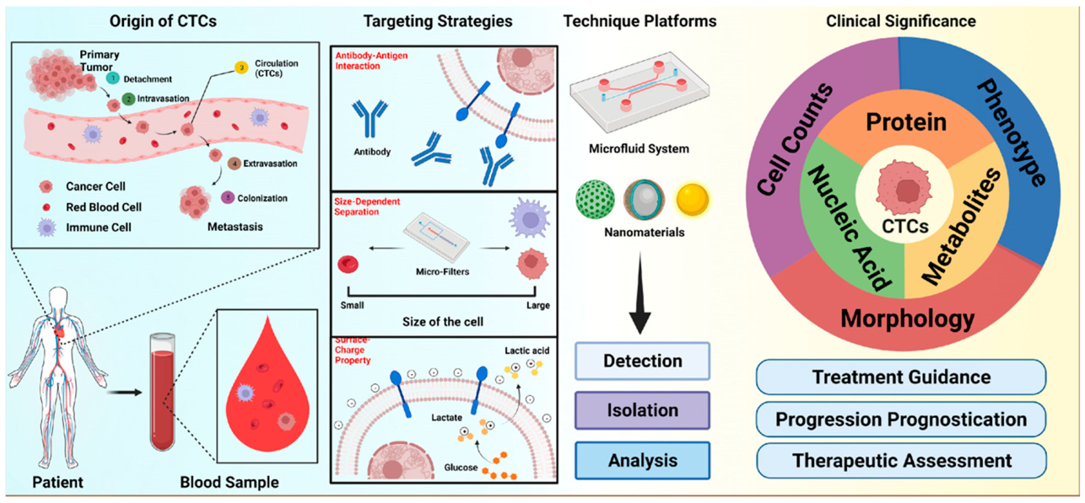
1. Introduction
2. CTC Isolation Techniques
2.1. Immunoaffinity
2.2. Purely Physical Cell Characteristics
2.3. Other Isolation Techniques
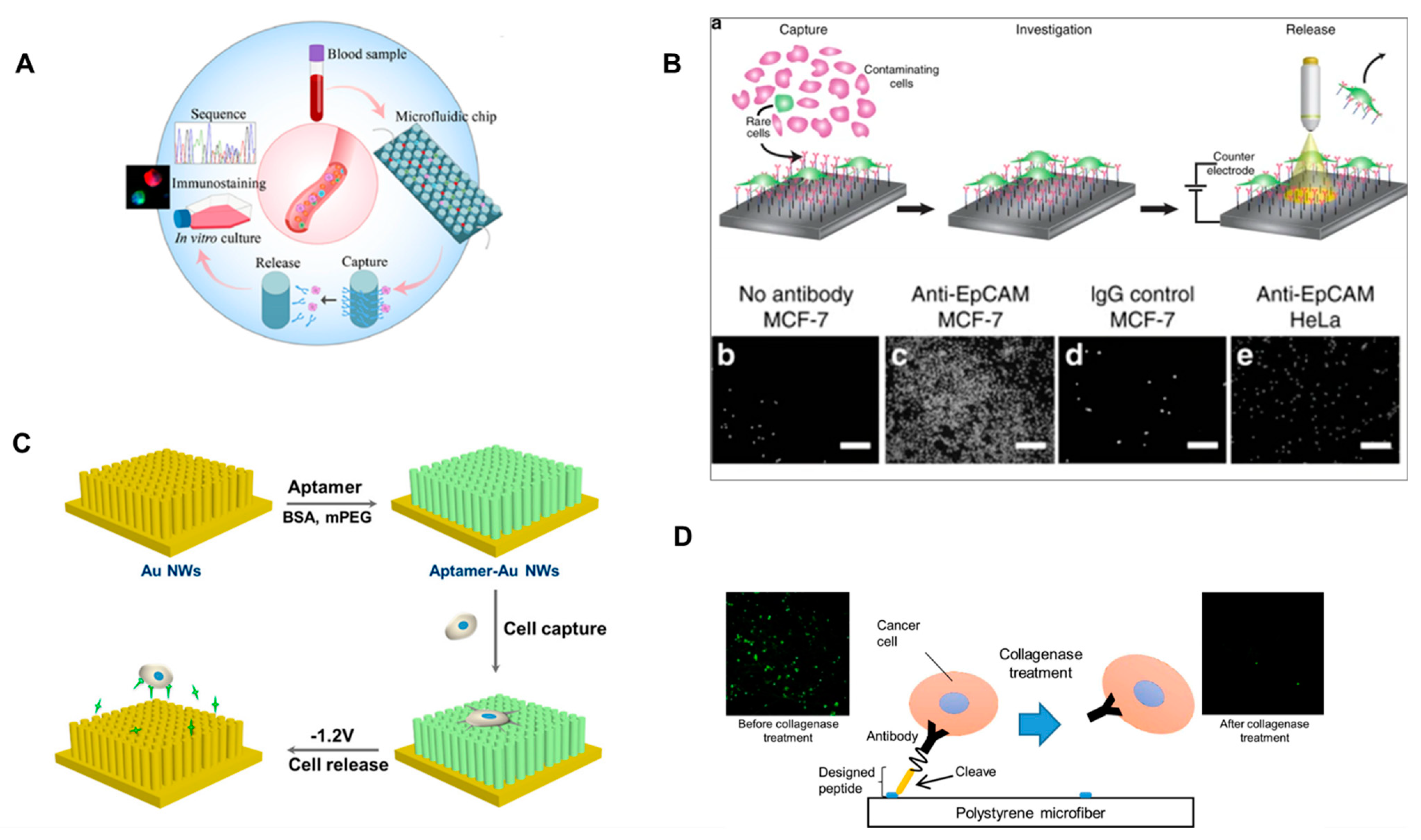
3. CTC Detachment/Release Techniques
3.1. Aptamers
3.2. Microdevices/Nanodevices
3.3. Light/Electrochemical

4. CTC Detection Techniques
4.1. Direct Detection (Pre-Enrichment)
4.2. Post-Enrichment Detection
5. Conclusions
Author Contributions
Funding
Data Availability Statement
Acknowledgments
Conflicts of Interest
References
- Ashworth, T. A case of cancer in which cells similar to those in the tumours were seen in the blood after death. Aust. Med. J. 1869, 14, 146. [Google Scholar]
- Nagrath, S.; Sequist, L.V.; Maheswaran, S.; Bell, D.W.; Irimia, D.; Ulkus, L.; Smith, M.R.; Kwak, E.L.; Digumarthy, S.; Muzikansky, A. Isolation of rare circulating tumour cells in cancer patients by microchip technology. Nature 2007, 450, 1235–1239. [Google Scholar] [CrossRef] [PubMed]
- Chen, J.; Oudeng, G.; Feng, H.; Liu, S.; Li, H.W.; Ho, Y.P.; Chen, Y.; Tan, Y.; Yang, M. 2D MOF Nanosensor-Integrated Digital Droplet Microfluidic Flow Cytometry for In Situ Detection of Multiple miRNAs in Single CTC Cells. Small 2022, 18, 2201779. [Google Scholar] [CrossRef] [PubMed]
- Zhang, Y.; Zhao, Y.; Cole, T.; Zheng, J.; Bayin, Q.; Guo, J.; Tang, S. Microfluidic flow cytometry for blood-based biomarker analysis. Analyst 2022, 147, 2895–2917. [Google Scholar] [CrossRef]
- Fan, Y.-J.; Hsiao, Y.-C.; Weng, Y.-L.; Chen, Y.-H.; Chiou, P.-Y.; Sheen, H.-J. Development of a parallel three-dimensional microfluidic device for high-throughput cytometry. Sens. Actuators B Chem. 2020, 320, 128255. [Google Scholar] [CrossRef]
- Ilyas, A.; Asghar, W.; Kim, Y.-T.; Iqbal, S.M. Parallel recognition of cancer cells using an addressable array of solid-state micropores. Biosens. Bioelectron. 2014, 62, 343–349. [Google Scholar] [CrossRef]
- Deng, Z.; Wu, S.; Wang, Y.; Shi, D. Circulating tumor cell isolation for cancer diagnosis and prognosis. EBioMedicine 2022, 83, 104237. [Google Scholar] [CrossRef]
- Sharma, S.; Zhuang, R.; Long, M.; Pavlovic, M.; Kang, Y.; Ilyas, A.; Asghar, W. Circulating tumor cell isolation, culture, and downstream molecular analysis. Biotechnol. Adv. 2018, 36, 1063–1078. [Google Scholar] [CrossRef]
- Kang, Y.-T.; Kim, Y.J.; Bu, J.; Chen, S.; Cho, Y.-H.; Lee, H.M.; Ryu, C.J.; Lim, Y.; Han, S.-W. Epithelial and mesenchymal circulating tumor cell isolation and discrimination using dual-immunopatterned device with newly-developed anti-63B6 and anti-EpCAM. Sens. Actuators B Chem. 2018, 260, 320–330. [Google Scholar] [CrossRef]
- Shi, W.; Wang, S.; Maarouf, A.; Uhl, C.G.; He, R.; Yunus, D.; Liu, Y. Magnetic particles assisted capture and release of rare circulating tumor cells using wavy-herringbone structured microfluidic devices. Lab Chip 2017, 17, 3291–3299. [Google Scholar] [CrossRef]
- Kwak, B.; Lee, J.; Lee, J.; Kim, H.S.; Kang, S.; Lee, Y. Spiral shape microfluidic channel for selective isolating of heterogenic circulating tumor cells. Biosens. Bioelectron. 2018, 101, 311–316. [Google Scholar] [CrossRef]
- Rao, L.; Meng, Q.F.; Huang, Q.; Wang, Z.; Yu, G.T.; Li, A.; Ma, W.; Zhang, N.; Guo, S.S.; Zhao, X.Z. Platelet–leukocyte hybrid membrane-coated immunomagnetic beads for highly efficient and highly specific isolation of circulating tumor cells. Adv. Funct. Mater. 2018, 28, 1803531. [Google Scholar] [CrossRef]
- Agerbæk, M.Ø.; Bang-Christensen, S.R.; Yang, M.-H.; Clausen, T.M.; Pereira, M.A.; Sharma, S.; Ditlev, S.B.; Nielsen, M.A.; Choudhary, S.; Gustavsson, T. The VAR2CSA malaria protein efficiently retrieves circulating tumor cells in an EpCAM-independent manner. Nat. Commun. 2018, 9, 3279. [Google Scholar] [CrossRef]
- Wan, S.; Kim, T.H.; Smith, K.J.; Delaney, R.; Park, G.-S.; Guo, H.; Lin, E.; Plegue, T.; Kuo, N.; Steffes, J. New labyrinth microfluidic device detects circulating tumor cells expressing cancer stem cell marker and circulating tumor microemboli in hepatocellular carcinoma. Sci. Rep. 2019, 9, 18575. [Google Scholar] [CrossRef]
- Renier, C.; Pao, E.; Che, J.; Liu, H.E.; Lemaire, C.A.; Matsumoto, M.; Triboulet, M.; Jeffrey, S.S.; Rettig, M.; Kulkarni, R.P. Label-free isolation of prostate circulating tumor cells using Vortex microfluidic technology. NPJ Precis. Oncol. 2017, 1, 15. [Google Scholar] [CrossRef]
- Au, S.H.; Edd, J.; Stoddard, A.E.; Wong, K.H.; Fachin, F.; Maheswaran, S.; Haber, D.A.; Stott, S.L.; Kapur, R.; Toner, M. Microfluidic isolation of circulating tumor cell clusters by size and asymmetry. Sci. Rep. 2017, 7, 2433. [Google Scholar] [CrossRef]
- Kang, Y.-T.; Doh, I.; Byun, J.; Chang, H.J.; Cho, Y.-H. Label-free rapid viable enrichment of circulating tumor cell by photosensitive polymer-based microfilter device. Theranostics 2017, 7, 3179. [Google Scholar] [CrossRef]
- Ribeiro-Samy, S.; Oliveira, M.I.; Pereira-Veiga, T.; Muinelo-Romay, L.; Carvalho, S.; Gaspar, J.; Freitas, P.P.; López-López, R.; Costa, C.; Diéguez, L. Fast and efficient microfluidic cell filter for isolation of circulating tumor cells from unprocessed whole blood of colorectal cancer patients. Sci. Rep. 2019, 9, 8032. [Google Scholar] [CrossRef]
- Fachin, F.; Spuhler, P.; Martel-Foley, J.M.; Edd, J.F.; Barber, T.A.; Walsh, J.; Karabacak, M.; Pai, V.; Yu, M.; Smith, K. Monolithic chip for high-throughput blood cell depletion to sort rare circulating tumor cells. Sci. Rep. 2017, 7, 10936. [Google Scholar] [CrossRef]
- Ahmed, M.G.; Abate, M.F.; Song, Y.; Zhu, Z.; Yan, F.; Xu, Y.; Wang, X.; Li, Q.; Yang, C. Isolation, Detection, and Antigen-Based Profiling of Circulating Tumor Cells Using a Size-Dictated Immunocapture Chip. Angew. Chem. Int. Ed. 2017, 56, 10681–10685. [Google Scholar] [CrossRef]
- Chen, K.; Dopico, P.; Varillas, J.; Zhang, J.; George, T.J.; Fan, Z.H. Integration of Lateral Filter Arrays with Immunoaffinity for Circulating-Tumor-Cell Isolation. Angew. Chem. 2019, 131, 7688–7692. [Google Scholar] [CrossRef]
- Ko, J.; Bhagwat, N.; Yee, S.S.; Black, T.; Redlinger, C.; Romeo, J.; O’Hara, M.; Raj, A.; Carpenter, E.L.; Stanger, B.Z. A magnetic micropore chip for rapid (<1 h) unbiased circulating tumor cell isolation and in situ RNA analysis. Lab Chip 2017, 17, 3086–3096. [Google Scholar] [PubMed]
- Waheed, W.; Alazzam, A.; Mathew, B.; Christoforou, N.; Abu-Nada, E. Lateral fluid flow fractionation using dielectrophoresis (LFFF-DEP) for size-independent, label-free isolation of circulating tumor cells. J. Chromatogr. B 2018, 1087, 133–137. [Google Scholar] [CrossRef] [PubMed]
- Hu, X.; Zhu, D.; Chen, M.; Chen, K.; Liu, H.; Liu, W.; Yang, Y. Precise and non-invasive circulating tumor cell isolation based on optical force using homologous erythrocyte binding. Lab Chip 2019, 19, 2549–2556. [Google Scholar] [CrossRef] [PubMed]
- Wu, M.; Huang, P.H.; Zhang, R.; Mao, Z.; Chen, C.; Kemeny, G.; Li, P.; Lee, A.V.; Gyanchandani, R.; Armstrong, A.J. Circulating tumor cell phenotyping via high-throughput acoustic separation. Small 2018, 14, 1801131. [Google Scholar] [CrossRef]
- Ilyas, A.; Asghar, W.; Allen, P.B.; Duhon, H.; Ellington, A.D.; Iqbal, S.M. Electrical detection of cancer biomarker using aptamers with nanogap break-junctions. Nanotechnology 2012, 23, 275502. [Google Scholar] [CrossRef]
- Bunka, D.H.; Stockley, P.G. Aptamers come of age–at last. Nat. Rev. Microbiol. 2006, 4, 588–596. [Google Scholar] [CrossRef]
- Ding, P.; Wang, Z.; Wu, Z.; Zhu, W.; Liu, L.; Sun, N.; Pei, R. Aptamer-based nanostructured interfaces for the detection and release of circulating tumor cells. J. Mater. Chem. B 2020, 8, 3408–3422. [Google Scholar] [CrossRef]
- Zhai, T.-T.; Ye, D.; Zhang, Q.-W.; Wu, Z.-Q.; Xia, X.-H. Highly efficient capture and electrochemical release of circulating tumor cells by using aptamers modified gold nanowire arrays. ACS Appl. Mater. Interfaces 2017, 9, 34706–34714. [Google Scholar] [CrossRef]
- Park, M.-H.; Reátegui, E.; Li, W.; Tessier, S.N.; Wong, K.H.; Jensen, A.E.; Thapar, V.; Ting, D.; Toner, M.; Stott, S.L. Enhanced isolation and release of circulating tumor cells using nanoparticle binding and ligand exchange in a microfluidic chip. J. Am. Chem. Soc. 2017, 139, 2741–2749. [Google Scholar] [CrossRef]
- Yoshihara, A.; Sekine, R.; Ueki, T.; Kondo, Y.; Sunaga, Y.; Nakaji-Hirabayashi, T.; Teramura, Y.; Takai, M. Rapid and highly efficient capture and release of cancer cells using polymeric microfibers immobilized with enzyme-cleavable peptides. Acta Biomater. 2018, 67, 32–41. [Google Scholar] [CrossRef]
- Li, W.; Reátegui, E.; Park, M.-H.; Castleberry, S.; Deng, J.Z.; Hsu, B.; Mayner, S.; Jensen, A.E.; Sequist, L.V.; Maheswaran, S. Biodegradable nano-films for capture and non-invasive release of circulating tumor cells. Biomaterials 2015, 65, 93–102. [Google Scholar] [CrossRef]
- Parker, S.G.; Yang, Y.; Ciampi, S.; Gupta, B.; Kimpton, K.; Mansfeld, F.M.; Kavallaris, M.; Gaus, K.; Gooding, J.J. A photoelectrochemical platform for the capture and release of rare single cells. Nat. Commun. 2018, 9, 2288. [Google Scholar] [CrossRef] [PubMed]
- Lv, S.-W.; Liu, Y.; Xie, M.; Wang, J.; Yan, X.-W.; Li, Z.; Dong, W.-G.; Huang, W.-H. Near-infrared light-responsive hydrogel for specific recognition and photothermal site-release of circulating tumor cells. ACS Nano 2016, 10, 6201–6210. [Google Scholar] [CrossRef]
- Kamińska, A.; Szymborski, T.; Witkowska, E.; Kijeńska-Gawrońska, E.; Świeszkowski, W.; Niciński, K.; Trzcińska-Danielewicz, J.; Girstun, A. Detection of circulating tumor cells using membrane-based sers platform: A new diagnostic approach for ‘liquid biopsy’. Nanomaterials 2019, 9, 366. [Google Scholar] [CrossRef]
- Wu, X.; Xia, Y.; Huang, Y.; Li, J.; Ruan, H.; Chen, T.; Luo, L.; Shen, Z.; Wu, A. Improved SERS-active nanoparticles with various shapes for CTC detection without enrichment process with supersensitivity and high specificity. ACS Appl. Mater. Interfaces 2016, 8, 19928–19938. [Google Scholar] [CrossRef]
- Xue, T.; Wang, S.; Ou, G.; Li, Y.; Ruan, H.; Li, Z.; Ma, Y.; Zou, R.; Qiu, J.; Shen, Z. Detection of circulating tumor cells based on improved SERS-active magnetic nanoparticles. Anal. Methods 2019, 11, 2918–2928. [Google Scholar] [CrossRef]
- Asghar, W.; Wan, Y.; Ilyas, A.; Bachoo, R.; Kim, Y.-T.; Iqbal, S.M. Electrical fingerprinting, 3D profiling and detection of tumor cells with solid-state micropores. Lab Chip 2012, 12, 2345–2352. [Google Scholar] [CrossRef]
- Ilyas, A.; Asghar, W.; Ahmed, S.; Lotan, Y.; Hsieh, J.-T.; Kim, Y.-T.; Iqbal, S.M. Electrophysiological analysis of biopsy samples using elasticity as an inherent cell marker for cancer detection. Anal. Methods 2014, 6, 7166–7174. [Google Scholar] [CrossRef]
- Galanzha, E.I.; Menyaev, Y.A.; Yadem, A.C.; Sarimollaoglu, M.; Juratli, M.A.; Nedosekin, D.A.; Foster, S.R.; Jamshidi-Parsian, A.; Siegel, E.R.; Makhoul, I. In vivo liquid biopsy using Cytophone platform for photoacoustic detection of circulating tumor cells in patients with melanoma. Sci. Transl. Med. 2019, 11, eaat5857. [Google Scholar] [CrossRef]
- Hu, Y.; Tang, W.; Cheng, P.; Zhou, Q.; Tian, X.; Wei, X.; He, H. Monitoring circulating tumor cells in vivo by a confocal microscopy system. Cytom. Part A 2019, 95, 657–663. [Google Scholar] [CrossRef] [PubMed]
- Soler, A.; Cayrefourcq, L.; Mazel, M.; Alix-Panabières, C. EpCAM-independent enrichment and detection of viable circulating tumor cells using the EPISPOT assay. Circ. Tumor Cells Methods Protoc. 2017, 1634, 263–276. [Google Scholar]
- Wu, L.-L.; Wen, C.-Y.; Hu, J.; Tang, M.; Qi, C.-B.; Li, N.; Liu, C.; Chen, L.; Pang, D.-W.; Zhang, Z.-L. Nanosphere-based one-step strategy for efficient and nondestructive detection of circulating tumor cells. Biosens. Bioelectron. 2017, 94, 219–226. [Google Scholar] [CrossRef]
- Chang, C.-L.; Huang, W.; Jalal, S.I.; Chan, B.-D.; Mahmood, A.; Shahda, S.; O’Neil, B.H.; Matei, D.E.; Savran, C.A. Circulating tumor cell detection using a parallel flow micro-aperture chip system. Lab Chip 2015, 15, 1677–1688. [Google Scholar] [CrossRef]
- Dizdar, L.; Fluegen, G.; van Dalum, G.; Honisch, E.; Neves, R.P.; Niederacher, D.; Neubauer, H.; Fehm, T.; Rehders, A.; Krieg, A. Detection of circulating tumor cells in colorectal cancer patients using the GILUPI CellCollector: Results from a prospective, single-center study. Mol. Oncol. 2019, 13, 1548–1558. [Google Scholar] [CrossRef]
- Chen, X.; Zhou, F.; Li, X.; Yang, G.; Zhang, L.; Ren, S.; Zhao, C.; Deng, Q.; Li, W.; Gao, G. Folate receptor–positive circulating tumor cell detected by LT-PCR–based method as a diagnostic biomarker for non–small-cell lung cancer. J. Thorac. Oncol. 2015, 10, 1163–1171. [Google Scholar] [CrossRef]


| Technology | Isolation Factors | Benefits | Drawbacks | Description | Reference |
|---|---|---|---|---|---|
| CROSS Chip (Microfluidic Cell Filter) | Size and deformability | 70% efficiency, high purity, cost-effective, easily applicable (low set-up), and higher sensitivity than CellSearch | To increase throughput, multiple screenings might be necessary; smaller CTCs are more difficult to obtain | A syringe pumps the blood sample into a microfluidic chip with filters that separates it into four sections for analysis | [18] |
| Labyrinth (Inertial Microfluidic Cell Filter) | Size (inertia) | High throughput, high yield, and high purity | Difficulty with focusing and separating smaller cells | The labyrinth design functions in separating CTCs from blood cells by filtering them through straight and curved channels | [14] |
| Optofluidic Cell Technology Chip | Size and the refractive index | High purity, high recovery, and no foreign material introduction | Time and cost unspecified | Uses molecules to bind CTCs to RBCs and then uses laser illumination to separate them on a chip, based on the refractive index | [24] |
| Dual-Immunopatterned microfluidic device | Surface antigen expression | High efficiency and captures mesenchymal cells | More time, possibly experiment-dependent, and less general applicability | Microfluidic device with two layers. Each layer is coated with a different antibody (anti-EpCAM or anti-63B6) to isolate and separate the types of CTCs | [9] |
| Acoustic Separation Device | Cell size/velocity (response to sound waves) | High throughput and cost-effective | Prior processes required for RBC removal and more time | The device uses the acoustic-wave field that is amplified by a PDMS barrier to send cells at different trajectories for separation | [25] |
| Wavy-Herringbone Microfluidic Device | Immunoaffinity (+EpCAM) and magnetic force | High efficiency, high purity, and high viability | Dependent on the dispersion of MPs (hard to control) and may be unable to capture non-EpCAM-expressing CTCs | A herringbone pattern was made on silicon wafers which were injected with bonded magnetic particles and anti-EpCAM. As the sample travels through, the CTCs are separated by the magnetic EpCAM | [10] |
| Vortex HT Chip | Size (movement) | Extremely high throughput, high viability, high purity, and no pre-processing steps | Certain CTCs not captured effectively, possible difficulties with cell recovery, and low capture efficiency | Laminar microvortices are created on this microdevice to separate CTCs from blood cells and other bodily fluids based on flow | [15] |
| Lateral filter array with immunoaffinity | Size and immunoaffinity (+EpCAM) | High efficiency, high purity, and high viability | May be unable to capture certain non-EpCAM-expressing CTCs | Embedded lateral filters in a serpentine channel on a microfluidic device. The blood sample flows through the main channel and the CTCs are caught in the filter. Immunoaffinity works in the lateral filters by testing for a bond force between certain antibodies and the cells as compared to the hydrodynamic force | [21] |
| Spiral Shape Microfluidic Channel | Magnetic force and immunoaffinity (+EpCAM) | High efficiency and high flow rate | May be unable to capture non-EpCAM-expressing CTCs; more tests with actual samples necessary | Magnetic nanoparticles bond with the EpCAM antibody. These are circulated through a spiral chamber with a decreasing radius and a permanent magnet. The magnetic force causes the bonded particles to be attracted and filter into specific regions for isolation | [11] |
| LFFF-DEP microfluidic device | Dielectrophoresis | No statistical results; however, based on the provided image, it seems to be efficient in separation | Further analysis necessary and time and throughput may be an issue | Oppositely charged electrodes are positioned on a glass wafer. Cancer cells are attracted to the positive DEP electrode, while normal blood cells are attracted to the negative DEP electrode | [23] |
| Cluster-isolating microfluidic device | Size and asymmetry | Able to isolate clusters, high recovery, and high viability | The flow rate has to be slowed to ensure viability (very low throughput) and less adept to isolating small clusters and single CTCs | Deterministic lateral displacement is used to isolate large clusters based on size (using micropillars with different sized gaps in between). During stage two, the clusters that were unable to be filtered by size are put through shaped micropillars that result in rotation if they are asymmetrical for separation | [16] |
| Magnetic Micropore CaTCh FISH Chip | Magnetic force (immunoaffinity of -CD45 on WBC) and size | Higher throughput compared to other magnetic chips, High recovery rate and the ability to conduct RNA analysis on the chip | WBC with low CD45 expression not filtered and cost unclear | Two-part system. The first section of the chip has magnetic traps at edges of the pores to attract WBCs labeled with MNPs. The RBCs and platelets are then filtered by size leaving the CTCs, which undergo RNA analysis on the chip | [22] |
| rVAR2 using IsoFlux system | Immunoaffinity (+ofCS) | High recovery, captures mesenchymal cells, and high sensitivity | May be unable to capture non-ofCS-expressing CTCs and may be expensive | Uses the IsoFlux system model (Dynabeads) but altered so the immunomagnetic capture is used with the rVAR2 protein to bond to the ofCS in the CTCs | [13] |
| PLT-WBC Immunomagnetic Beads | Immunoaffinity (+EpCAM) and magnetic force | High Efficiency, increased binding ability, and avoidance of WBC collection | May be unable to capture non-EpCAM-expressing CTCs and requires additional preparation process for magnetic beads | A hybrid membrane of platelets and WBCs is formed and coated onto magnetic particles. The particles are then treated with EpCAM-binding antibodies. The resulting magnetic beads target the CTCs while specifically avoiding homologus WBCs | [12] |
| Photosensitive Polymer-Based Microfilter | Size and deformability | High efficiency, relatively high viability, and simple set-up | Smaller CTCs are difficult to capture | The photosensitive polymer which is removed with UV exposure coats a microfilter with many densely dispersed slits. The slits have a larger inlet which decreases in diameter towards the outlet, trapping larger CTCs | [17] |
| SDI Chip | Immunoaffinity (+EpCAM) and size | High efficiency, high purity, and greater sensitivity as compared to CellSearch | Lower-expression EpCAM was more frequently not recovered and shear stress caused the dislodgement of many cells | Microchip on which triangular micropillars are coated with anti-EpCAM antibodies. The pillars are spaced and rotated to create a decreasing hydrodynamic gradient and gaps. Due to gradient cells migrating downstream and at certain locations due to their size and immunoaffinity, the CTCs become lodged at pillars | [20] |
| Monolithic CTC iChip | Immunoaffinity (-CD45, -CD16, and -CD66B) and size | High throughput and high recovery | Smaller cells had difficulty being caught, WBCs with low antigen expression levels and difficulty being caught, and average purity | The microfluidic chip first holds a size-based array with micropillars with a waste channel for RBCs and plasma. The next portion has magnets on either side. The magnetic-tagged WBCs are filtered to the sides, while the CTCs are caught in the middle (non-magnetic) portion. | [19] |
| Technology | Release Mechanism | Viability | Release Efficiency | Reference |
|---|---|---|---|---|
| Biodegradable Nano-Films | The enzyme solution degrades the polymeric film | ~90% | 95% | [32] |
| Aptamer Modified Gold Nanowires | Aptamer sgc8c conjugated with gold nanowires; electrochemical reduction desorption at −1.2 V for 30 s breaks the Au–S bonds | 90% | 96.2% | [29] |
| Gold Nanoparticles | NHS ligands bond the amine moiety to NeutrAvidin; glutathione is used to break bonds | 91.5% (average of two tests with MDA-MB-231 and PC3) | 82.5% (average of two tests with MDA-MB-231 and PC3) | [30] |
| Polymeric Microfibers | Peptides are bound to the polystyrene microfiber and anti-EpCAM antibody; collagenase type IV is used to cleave the peptide | 83% | >90% | [31] |
| Photoelectrochemical Single Cell Release | Light activates the electrons on a conduction band on a silicon surface; it increases the conductivity which prompts cleavage and single cell release | 90 ± 2.7% | 82 ± 5% | [33] |
| Light-Responsive Hydrogel | The near infrared-mediated photothermal activation of embedded gold nanorods causes temperature-responsive gelatin to dissolve rapidly at 37 °C | 95 ± 4% (overall thermal release rate) 92 ± 6% (release rate using specified light beams/photothermal selection) | 95 (overall thermal viability rate) 90% (viability rate using specified light beams/photothermal selection) | [34] |
| Technology | Labeling | Cost Description | Beneficial Qualities | Includes an Enrichment Step |
|---|---|---|---|---|
| CellSearch | Yes | USD 350 | FDA-approved | Yes |
| SERS active magnetic nanoparticles | No | Expensive equipment needed | High efficiency and high purity | No |
| Solid-state Micropores | No | Low cost | High sensitivity | No |
| Photoacoustic Cytophone | No | Low cost | High specificity | No |
| CaTCh FISH Chip | Yes | Costly | High sensitivity | Yes |
| EPISPOT Assay | Yes | USD 300–400 based on previous related assay costs | Simple and easily applicable | Yes |
| Nanosphere Detection | Yes | High efficiency and cell viability post enrichment | Yes | |
| GILUPI CellCollector | Yes | Described alternative methods as “costly” | Relatively high sensitivity | Yes |
| Parallel Flow Micro-Aperture Chip | Yes | High detection yield | Yes | |
| LT-PCR Based Method | Yes | Cost effective | Medium/high sensitivity and specificity | Yes |
| Confocal Microscopy | Yes | Very expensive (the product that served as the basis is USD 10k, but estimates range up to USD 100k+) | Extremely simple, no necessary adjustments | No |
Disclaimer/Publisher’s Note: The statements, opinions and data contained in all publications are solely those of the individual author(s) and contributor(s) and not of MDPI and/or the editor(s). MDPI and/or the editor(s) disclaim responsibility for any injury to people or property resulting from any ideas, methods, instructions or products referred to in the content. |
© 2023 by the authors. Licensee MDPI, Basel, Switzerland. This article is an open access article distributed under the terms and conditions of the Creative Commons Attribution (CC BY) license (https://creativecommons.org/licenses/by/4.0/).
Share and Cite
Goldstein, I.; Alyas, S.; Asghar, W.; Ilyas, A. Biosensors for the Isolation and Detection of Circulating Tumor Cells (CTCs) in Point-of-Care Settings. Micromachines 2023, 14, 1035. https://doi.org/10.3390/mi14051035
Goldstein I, Alyas S, Asghar W, Ilyas A. Biosensors for the Isolation and Detection of Circulating Tumor Cells (CTCs) in Point-of-Care Settings. Micromachines. 2023; 14(5):1035. https://doi.org/10.3390/mi14051035
Chicago/Turabian StyleGoldstein, Isaac, Sobia Alyas, Waseem Asghar, and Azhar Ilyas. 2023. "Biosensors for the Isolation and Detection of Circulating Tumor Cells (CTCs) in Point-of-Care Settings" Micromachines 14, no. 5: 1035. https://doi.org/10.3390/mi14051035
APA StyleGoldstein, I., Alyas, S., Asghar, W., & Ilyas, A. (2023). Biosensors for the Isolation and Detection of Circulating Tumor Cells (CTCs) in Point-of-Care Settings. Micromachines, 14(5), 1035. https://doi.org/10.3390/mi14051035