Review

Snake Venom Disintegrins and Cell Migration

Departamento de Ciências Fisiológicas, Universidade Federal de São Carlos, São Carlos, SP, 13565-905, Brazil
*
Author to whom correspondence should be addressed.
Toxins 2010, 2(11), 2606-2621; https://doi.org/10.3390/toxins2112606
Submission received: 24 August 2010 / Revised: 15 October 2010 / Accepted: 18 October 2010 / Published: 29 October 2010
(This article belongs to the Special Issue Disintegrins: Structure-Function and Translational Potential)

Abstract

:
Cell migration is a key process for the defense of pluricellular organisms against pathogens, and it involves a set of surface receptors acting in an ordered fashion to contribute directionality to the movement. Among these receptors are the integrins, which connect the cell cytoskeleton to the extracellular matrix components, thus playing a central role in cell migration. Integrin clustering at focal adhesions drives actin polymerization along the cell leading edge, resulting in polarity of cell movement. Therefore, small integrin-binding proteins such as the snake venom disintegrins that inhibit integrin-mediated cell adhesion are expected to inhibit cell migration. Here we review the current knowledge on disintegrin and disintegrin-like protein effects on cell migration and their potential use as pharmacological tools in anti-inflammatory therapy as well as in inhibition of metastatic invasion.

1. Introduction

Metastasis is one the major causes of death in patients with cancer. In order for a tumor to grow and metastasize, both tumor and endothelial cells must migrate and invade surrounding tissues [1,2]. Endothelial cell migration provides the blood supply that is essential to tumor cells. Once in the blood vessels, tumor cells must adhere to the endothelium and escape to a new site [3], two processes that depend on their invasive abilities. Therefore, the blockage of both tumor and endothelial cell migration and invasion is an interesting approach for the treatment of cancer patients. The key receptors involved in cell migration are the integrins, which connect the cells to the extracellular matrix of the tumor microenvironment. Integrin blocking usually results in inhibition of cell migration and tumor angiogenesis [4]. Here we review the effects of disintegrins, a group of integrin-binding proteins found in snake venoms, in the cell adhesion process and their application in anti-cancer and anti-metastatic therapy. The effects of disintegrin on neutrophil migration will also be briefly described for a better illustration of the role of these proteins in cell migration.

2. Integrins and Cell Migration

Integrins are cell surface receptors that play critical roles in cell adhesion and migration. These proteins are heterodimers of transmembrane α- and β-subunits that connect the extracellular matrix (ECM) to the cell cytoskeleton. Cell binding by integrins to their cognate extracellular ligands, such as collagen, laminin and fibronectin, triggers intracellular signaling pathways that control cytoskeleton organization, cell polarity and force generation [5]. Integrin clustering at focal complexes drives actin polymerization along the leading edge of migrating cells, contributing to changes in cell shape and polarity [6]. Integrins are also involved in the regulation of matrix-degrading proteases, a key step for the invasive phenotype. There are several excellent reviews on the role of integrins in cell migration and invasion [6,7,8], and therefore, this subject will not be addressed in the present review. Furthermore, several studies have described the correlation between a malignant phenotype and an altered integrin distribution on tumor cell surfaces [9]. The role of integrins in metastatic dissemination is also very well documented [10,11], and is beyond the scope of this review.

3. Angiogenesis

The term angiogenesis usually defines the growth of sprouts from capillary blood vessels that depends on a delicate balance between pro- and anti-angiogenic factors. In mammals, physiological angiogenesis is restricted to specific situations, including growth, tissue regeneration and reproduction. In contrast, some angiogenesis-dependent diseases, such as tumor growth, age-related macular degeneration and atherosclerosis, have been described [12].
Angiogenesis is primarily driven by tissue hypoxia that upregulates hypoxic factor-1 (HF-1) expression [7,13], which in turn induces vascular endothelial cell growth factor (VEGF) expression, a potent mitogen for endothelial cells [7,14]. Briefly, the angiogenic cascade includes degradation of basement membrane (BM) by matrix metalloproteases (MMPs) expressed by endothelial cells (EC), EC proliferation and tube formation, synthesis of a new BM, tube stabilization by pericytes and vascular smooth muscle cells and maturation into capillaries [15]. In addition, extracellular matrix degradation by MMPs plays a key role in the control of angiogenesis either by releasing VEGF from the ECM and facilitating EC migration, or by releasing angiogenesis inhibitors from larger extracellular matrix components such as endostatin from collagen XVIII [16,17], and angiostatin from plasminogen [15,18]. Angiogenesis is also influenced by integrins expressed on endothelial cells, vascular smooth muscle cells, fibroblasts, and platelets. These cells process signals from their microenvironment and respond by altering their cell-cell and cell-matrix adhesion, which allows migration and vascular remodeling over a period of days to weeks [19].
On the other hand, tumor angiogenesis is not particularly effective. Tumor vessels are structurally and biologically different with leaky characteristics that facilitate tumor cell dissemination by blood or lymphatic vessels [4,20]. Unlike in quiescent EC, the αvβ3 integrin is highly expressed by tumor EC, which helps the binding of new ECs to the provisional matrix components, including vitronectin and fibronectin that are deposited in the tumor microenvironment [4]. Accordingly, small integrin binding proteins have been tested for their ability to inhibit tumor angiogenesis. We will review the work that has been done with snake venom disintegrins in this field.

4. Disintegrins from Snake Venoms

Snake venom disintegrins are mostly derived from proteolytically processed precursor forms having a metalloprotease domain, named the snake venom metalloproteases (SVMPs). Members of this protein family have been classified according to their multi-domain structure into P-I, P-II and P-III classes [21]. Members of the P-I class are formed by a metalloprotease domain only while P-II proteins have a metalloproteinase and a disintegrin domain. P-III proteins have an additional cysteine-rich domain following the disintegrin region and in some cases, a lectin domain [22]. These last two classes (P-II and P-III) can be subdivided further according to the proteolytic processing of their domains and their ability to form dimeric structures [23]. Based on this classification, processed RGD-disintegrins are derived from the P-IIa class, while homo- and heterodimeric disintegrins are released in general from SVMPs belonging to the P-IId and P-IIe classes, respectively. P-III SVMPs originate the disintegrin-like proteins (DC, for Disintegrin, Cys-rich proteins) formed by covalently linked-disintegrin-like and Cys-rich domains [21]. By means of phylogenetic analysis, Calvete et al. [24] provided strong evidence that the diversity of disintegrins could be due to an accelerated evolution of surface-exposed residues with the inference that the RGD motif is the ancestral integrin-recognition motif from which other disintegrins have emerged via single-base substitutions to generate KGD-, MGD-, WGD-, and VGD-integrin binding motifs as well as others. The diversity of the integrin-binding loop, along with other conserved residues among disintegrin molecules, gives rise to different integrin specificities (Table 1). However, disintegrin structural complexity does not derive only from venom genomic structure and transcriptional regulation events but also from post-translational modifications that could be responsible for dimerization and disulfide bonding as previously suggested [21].
DC proteins are usually found in a processed form in snake venoms without the catalytic domain but they are not further separated into independent proteins due to a disulfide bond that connects both domains. The active RGD loop of P-II-derived disintegrins is modified into a cysteine-contained loop (D/ECD) in the DC proteins [25]. It has been recently suggested that distinct disulfide pairing strongly contributes to the conformation of the D domain, which could consequently influence the binding properties and specificities of DC proteins [26]. Depending on the disulfide pairing, the D domain may assume at least two types of conformation, a C-shaped or an I-shaped scaffold; the former was suggested to play a key role in substrate recognition by the catalytic domain [27]. The D domain also has adhesive properties to some proteins such as collagen I, as recently demonstrated.
Integrin binding ability is apparently more related to the Cys-rich domain [28,29] which was also shown to bind von Willebrand factor therefore helping substrate targeting for proteolysis by the metalloprotease domain [30]. In addition, the hyper-variable region (HVR), considered the major structurally distinct region among the P-III SVMPs and suggested to play a key role in target selection due to its protein-protein adhesive properties, is located in the Cys-rich domain [29]. The importance of HVR was recently evidenced for two elapidic SVMPs from Naja atra venom. Atragin, a SVMP with a C-shaped D domain, but not its homolog K-like, which has an I-shaped scaffold, inhibits the migration of both mouse fibroblasts and Chinese hamster ovary CHOK1 cells [26]. Synthetic peptides from the HVR of both atragin and K-like proteins showed similar results, since only the peptide from atragin HVR inhibited cell migration toward fibronectin [26].
Disintegrins and DC proteins are rich in Cys residues, which are mainly involved in disulfide bonds, resulting in proteolysis-resistant molecules. This is a crucial feature for a sustained half-life in the blood. DisBa-01, a recombinant disintegrin from Bothrops alternatus venom, could be detected up to 6 h hours after i.p. injection in mice [31]. Despite the high content of disulfide bonds, RGD-disintegrins can be produced in an active form in bacteria [32,33], thus allowing the production of large quantities as needed for in vivo tests. Recombinant DC proteins are more difficult to express in an active form in bacteria. However, the production of Cys-rich domains from P-III SVMPs in active form has been reported [29].

5. Effects of Disintegrins on Leukocyte Migration

Neutrophils can be recruited from blood circulation toward sites of inflammation due to signals from injured tissues. To reach these sites, neutrophils must express new receptors, including some integrins, on their cell surface that will allow their adhesion to the endothelium and subsequent migration into tissues [34]. In addition to several constitutively expressed integrins such as αMβ2, αvβ3 and α9β1, these cells express α2-, α3-, α4-, and α5β1 integrins upon activation [35,36], triggering integrin-signaling pathways that mediate actin polymerization, cytoskeletal organization, spreading and migration [37]. Accordingly, disintegrins were first tested as integrin inhibitors of neutrophil migration as potential anti-inflammatory candidates. However, the results showed that integrin-binding by disintegrins could also activate neutrophils and protect these cells from apoptosis [38,39], demonstrating the complexity of disintegrin effects.
Coelho et al. [38] first demonstrated the dual effect of the monomeric RGD-disintegrin jarastatin (JT), from Bothrops jararaca venom, on neutrophil chemotaxis. Jarastatin inhibited neutrophil migration toward fMLP, IL-8 and jarastatin itself in a concentration-dependent manner. However, JT also induced neutrophil chemotaxis when used as chemoattractant [38]. In addition, it was demonstrated that this disintegrin induced several downstream integrin-mediated signaling events such as actin polymerization, activation of focal adhesion kinase (FAK) and extracellular-regulated kinase-2 (erk-2) nuclear translocation, which resulted in a delay of spontaneous neutrophil apoptosis [40]. JT also inhibited neutrophil migration in vivo after intraperitoneal carrageenan injection [38]. Ocellatusin, an RGD-disintegrin from Echis ocellatus venom, also strongly induces neutrophil chemotaxis [41]. Later, the same group demonstrated that EC3, a heterodimeric MLD-disintegrin from Echis carinatus venom, also inhibited neutrophil chemotaxis and activated FAK and phosphoinositide 3-kinase (PI3K); however, in contrast to JT, EC3 inhibited Erk-2 translocation and had a pro-apoptotic effect [39]. Antibody competition assays showed that JT and EC3 effects are mediated by different neutrophil integrins. JT binds preferentially to αMβ2 while EC3 is a ligand for α9β1 in neutrophils [39,42]. In contrast, it has been recently shown that activation of α9β1 integrin by VLO5, a dimeric disintegrin from Vipera lebetina obtusa venom [43], inhibits neutrophil spontaneous apoptosis by up-regulating the expression of anti-apoptotic proteins Bcl-xL and by increasing the degradation of pro-apoptotic protein Bad [36]. The two dimeric disintegrins EC3 and VLO5 are an interesting example of how subtle the structural differences that lead to distinct biological effects can be. Both disintegrins are very similar and have the VGD motif on the A-subunit while the B-subunit has a single amino acid replacement N-terminally to the MLD adhesive motif. This MLD motif has been suggested to be responsible for the different binding specificities and biological effects. An alanine residue in EC3 (AMLD) is replaced in VOL5 by a threonine residue (TMLD), which could result in distinct affinities and effects, despite their binding to the same integrin receptor [36].
RGD-disintegrins were also demonstrated to activate human T lymphocytes via integrin signaling [44]. Flavoridin, from Trimeresurus flavoviridis, kistrin, from Agkistrodon rhodostoma, and echistatin, from Echis carinatus venoms, which all bind to α5β1 and αvβ3, induced T cell proliferation and CD69 expression in parallel with FAK and PI3K activation and NF-κB nuclear translocation [44].
Alternagin-C (ALT-C), an ECD-containing disintegrin-like protein from Bothrops alternatus venom induced neutrophil effects similar to those of JT [45]. It inhibited neutrophil migration in a concentration-dependent fashion despite being itself chemotactic to this cell type. ALT-C also induced significant cytoskeleton dynamic changes with polymerization of F-actin, FAK and PI3K activation, and erk-2 translocation as well. These effects were reproduced by an ECD-containing peptide derived from the ALT-C primary sequence [45,46]. Interestingly, ALT-C was demonstrated to be an inhibitor of α2β1 integrin-mediated cell adhesion to collagen I. Jararhagin-C, the released DC domain from the SVMP jararhagin isolated from Bothrops jararaca venom, also induced in vivo leukocyte rolling after topical application on murine cremaster muscle. These observations together confirm the key role for the α2β1 integrin in neutrophil migration [47].
The complexity of the DC proteins was further demonstrated by the work of Menezes and co-authors, showing that the recombinant Cys-rich domain of HF3 metalloprotease from Bothrops jararaca venom is able to activate leukocyte rolling in the microcirculation [29]. This protein was expressed in bacteria without the disintegrin-like domain thus providing convincing evidence of that DC proteins activity resides on the Cys-rich region. In addition, synthetic peptides derived from the HVR region of the Cys-rich domain of HF3 reproduced the same effect, which was also inhibited by anti-αMβ2 antibodies, confirming the integrin-mediated activity of the C domain in leukocytes.

6. Effects of Disintegrins on Endothelial Cell Migration and Angiogenesis

Triflavin from Trimeresurus flavoviridis venom was one of the first RGD-disintegrins shown to inhibit angiogenesis both in vitro and in vivo [48]. Triflavin (0.4 μM) strongly inhibited EC migration toward vitronectin and fibronectin nearly thirty orders of magnitude greater than the anti-αvβ3 monoclonal antibodies [48]. Triflavin was also more effective in inhibiting TNF-α-induced angiogenesis in the chicken chorioallantoic membrane (CAM) assay. Similar results were obtained with another RGD-disintegrin, rhodostomin, from Agkistrodon rhodostoma venom, which inhibits endothelial cell migration, invasion and tube formation evoked by bFGF in matrigel both in vitro and in vitro [49]. Rhodostomin effects were inhibited by anti-αvβ3 but not by anti-αvβ5 antibodies, thus supporting the hypothesis that the effects of RGD-disintegrins are mediated by blockade of the vitronectin receptor.
Native or recombinant contortrostatin (CN), a homodimeric RGD-disintegrin from Agkistrodon contortrix contortrix venom, is another example of anti-angiogenic disintegrin as demonstrated by several in vitro and in vivo models [50,51,52]. Liposomal delivery of CN has proven to be effective as an anti-angiogenic and anti-tumor agent in human ovarian and breast cancer animal models [52]. Similar results were obtained with vicrostatin, a quimeric recombinant CN variant, which was also demonstrated to induce apoptosis in tubulogenic HUVEC seeded between two matrigel layers [53]. A monomeric recombinant RGD-disintegrin from Bothrops alternatus venom, DisBa-01, produces similar effects in the matrigel plug model in nude mice [33]. In addition to inhibition of EC migration, saxatilin, an RGD disintegrin from Gloydius saxatilis also inhibited the migration of vitronectin-induced smooth muscle cells [54,55].
However, the complexity of disintegrin specificity and the process of migration do not allow for the conclusion that blocking the vitronectin receptor is sufficient for inhibiting cell migration. Echistatin, but not eristostatin (two short RGD-disintegrins), inhibits in vivo angiogenesis in the CAM assay [56]. There are at least three KTS-disintegrins that inhibit α1β1 integrin binding to its cognate ligands, collagen I and IV (Table 1) [57,58], and also inhibit EC migration toward these substrates [59]. Lebestatin is an example of a KTS-disintegrin isolated from Macrovipera lebetina that inhibits EC migration and VEGF-induced in vivo angiogenesis [59]. The presence of a WGD motif in CC8, a heterodimeric disintegrin from Echis carinatus, increases its inhibitory effect on αvβ3 and α5β1 integrins [60].
There are few reports regarding the effects of ECD-disintegrins on endothelial cell migration. Acurhagin-C, an ECD-disintegrin-like protein from Agkistrodon acutus venom, dose-dependently blocked HUVEC migration toward a vitronectin-coated membrane. Furthermore, acurhagin-C elicited endothelial anoikis via disruption of the αvβ3/FAK/PI3K survival cascade and subsequent initiation of the procaspase-3 apoptotic signaling pathway [61]. Similarly, ALT-C also inhibits EC migration both in vitro and in vivo [62,63].

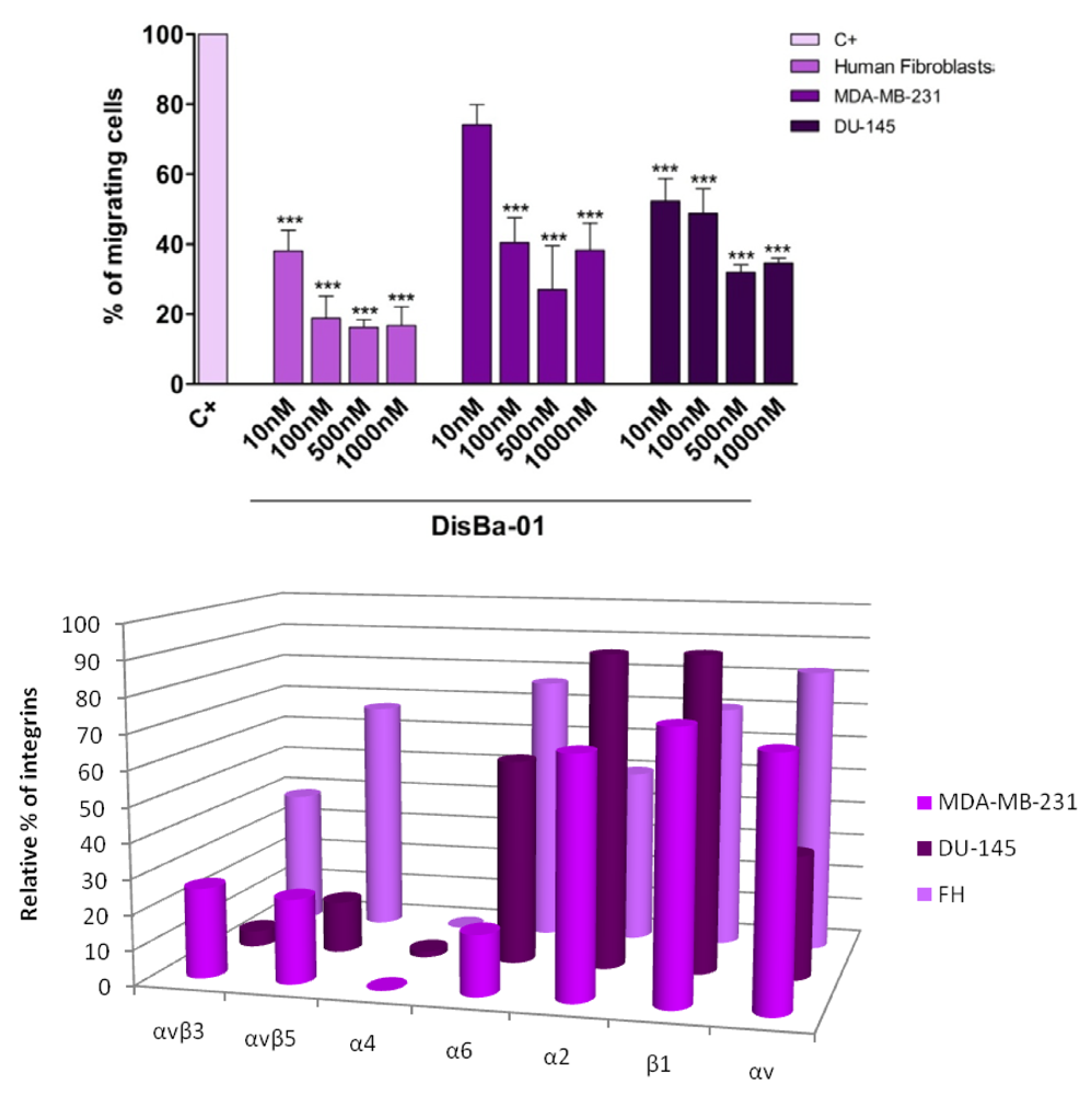
7. Effects of Disintegrins on Tumor Cell Migration

The ability of most snake venom disintegrins to inhibit cell adhesion has stimulated scientists to study these proteins as inhibitors of tumor cell dissemination. A volume of papers dealing with this subject can be found in the literature, most on RGD-disintegrin effects (Table 1). Morris et al. [64], were one of the first investigators to test eristostatin, an RGD-disintegrin from Eristocophis macmahoni, on individual metastasis steps such as cell arrest, extravasation and migration. Eristostatin treatment did not prevent tumor cell extravasation or migration [55]. However, it was shown later that eristostatin inhibited melanoma cell motility, an effect mediated by fibronectin-binding integrins [56]. Interestingly, this disintegrin, contrary to other RGD-disintegrins, did not inhibit angiogenesis, as stated before [56].
DisBa-01, an αvβ3 integrin-blocking RGD-disintegrin, inhibits not only endothelial cell in vivo migration [33] but also the in vitro migratory ability of fibroblasts and two tumor cell lines in a concentration-dependent fashion (Figure 1A). These three cell lines were compared in terms of integrin content by flow cytometry. Interestingly, the content of αvβ3 is relatively low in these three cell lines compared to the amount of α2β1. Among the tested cell lines, human fibroblasts are the most sensitive to DisBa-01 and, coincidently, also have the highest content of αvβ3 integrin (Figure 1B). However, a change in receptor density after disintegrin treatment is a possibility that cannot be excluded.
Figure 1. A (top) Effects of Disba-01 on the migration of three different cell lines. The cells were plated on the migration inserts in the presence of increasing concentrations of DisBa-01 for 22 h. Migration was expressed as a percentage of the control (100%). Cells were counted with a microscope (ten random fields per experiment). Results represents mean ± standard error of three individual experiments, *** p < 0.0001. B (bottom) Integrin profile of the cells tested in the migration assay using DisBa-01. Fibroblasts (FH) express αvβ3, αvβ5, α6, and β1. MDA-MB-231 breast tumor cells express αvβ3, αvβ5, α6, α2, and β1. DU-145 prostate tumor cells express more αvβ5, α6, α2, β1, and less αvβ3 and α4. The presence of integrin receptors on the cell surface was determined by flow cytometry with FITC-anti-αvβ3, αvβ5, α6, α4, α2, β1 and αv subunit integrin antibodies.
Toxins 02 02606 g001
Table 1 summarizes the effects of several disintegrins and DC proteins on tumor-related cell migration in different systems and the range of tested concentrations. The diversity of in vitro and in vivo assays makes a quantitative comparison difficult. Most tested proteins are effective in a micromolar range for in vivo assays while nanomolar concentrations are used in in vitro tests. From the data in the literature, it seems that disintegrins are general inhibitors of cell migration and invasion, despite being unspecific ligands, such as the RGD-disintegrins, or integrin-specific binding proteins, such as the KTS-disintegrins (Table 1). The disintegrin-like, Cys-rich proteins may also be included as integrin blockers of cell migration. Since integrin receptors are also quite indiscriminate as they support cell adhesion to several substrates, it seems highly reasonable that the general RGD-disintegrin scaffold of the integrin-binding motif could be employed as a prototype for drug design for new anti-metastatic therapies via blocking both tumor cell adhesion and tumor angiogenesis. In fact, at least two antagonists of platelet αIIbβ3 fibrinogen receptor that are currently being used in anticoagulant therapy (eptifibatide and aggrastat) were based on a snake venom disintegrin structure.
Table 1. Structural determinants, preferential integrin and the effects of disintegrins and disintegrin-like proteins on migration-dependent tumor cell activities.
DisintegrinStructureAdhesive motifPreferred integrinCognate ligandRelevant inhibitory activity (conc.*)Ref.
Legend: Vn, vitronectin; Fn, fibronectin; Fg, fibrinogen; TN, tenascin; ICAM-1, intercellular cell adhesion molecule-1; VCAM, vascular cell adhesion molecule. * Effective inhibitory concentration; ** in vivo assays.
salmosin 2monomeric mediumRGDαvβ3Vnangiogenesis ** (5 μg)[65]
saxatilinmonomeric mediumRGDαvβ3Vnangiogenesis (100 nM)[54]
jarastatinmonomeric mediumRGDαvβ3, α5β1, αMβ2Vn, Fn, ICAM-1melanoma lung metastasis ** (1 μM)[66]
flavoridinmonomeric mediumRGDα5β1Fnmelanoma lung metastasis ** (1 μM)[66]
kistrinmonomeric mediumRGDαvβ3Vnmelanoma lung metastasis **( 1 μM)[66]
colombistatinmonomeric mediumRGDndFntumor cell migration (IC50 = 1.8 μM)[67]
trigraminmonomeric mediumRGDαvβ3Vnbone metastasis **(100 μg/ml)[68]
DisBa-01monomeric mediumRGDαvβ3Vnmelanoma metastasis **(2 mg/Kg)[33]
eristostatinmonomeric shortRGDαIIbβ3Fgmelanoma metastasis (25 μg)[69]
echistatinmonomeric shortRGDαvβ3Vnosteoclast migration (10 nM)[70]
triflavinmonomeric shortRGDαvβ3Vnangiogenesis **(0.1–0.4 μM)[48]
contortrostatinHomodimericRGDα5β1, αvβ5Fntumor angiogenesis **(60 μg/day)[71]
alternagin-Cmonomeric D/CECDα2β1collagen Iangiogenesis **(1 μM)[63]
leberagin-Cmonomeric D/CECDαvβ3, α5β1, αvβ6Vn, Fnmelanoma cell adhesion (100 nM)[72]
acurhagin-Cmonomeric D/CECDαvβ3Vnangiogenesis **(0.4 μM)[61]
VLO5heterodimericVGD, MLDα9β1TN, VCAM glioblastoma growth (100 μg/ml)[73]
obtustatinmonomeric shortKTSα1β1collagen IVangiogenesis **(0.4 μg/μl)[74]
viperistatinmonomeric shortKTSα1β1collagen IVmelanoma cell transmigration (1–4 μM)[75]
lebestatinmonomeric shortKTSα1β1collagen IVangiogenesis **(0.1–0.5 μg/embryo)[59]
It has been suggested that the adhesive properties of the disintegrin-like, Cys-rich proteins could be primarily due to the Cys-rich domain, which was demonstrated to bind vWF [30,76]. Interestingly, half of integrin α subunits possess a von Willebrand factor A domain (also called I domain), which is in close proximity to the ligand-binding site [77]. Therefore, it is possible that the DC proteins could bind to the integrins presenting this domain, thus increasing specificity. However, this possibility remains to be demonstrated.

8. ADAMs and Cell Migration

Studies of the snake venom disintegrin effects on cell migration were conducted in parallel with those on mammalian ADAMs. The ADAM (A Disintegrin And Metallopeptidase) family of proteins comprises a snake venom-homolog group of multidomain proteins that play important roles in many biological processes, including cell migration, development and fertilization [78,79,80,81,82]. Structurally, the ADAM disintegrin and Cys-rich domains are more related to the DC proteins from SVMPs than the RGD-disintegrins. The disintegrin and Cys-rich domains of ADAM9 were shown to increase keratinocyte migration and MMP-9 activity in a wound healing in vitro model, an effect mediated by β1 integrin receptors [83]. More recently, it was reported that knockout animals for ADAM9 showed accelerated wound repair compared to controls, due apparently to the increased migration of keratinocytes [84]. Interestingly, ADAM15, the unique RGD-disintegrin among the ADAMs, suppressed CHO cell motility by inducing integrin α5β1 expression on the cell surface, thereby enhancing cell adhesion [85]. Additionally, it was demonstrated that the recombinant disintegrin domain of ADAM15 completely inhibited endothelial cell migration and tube formation in a three-dimensional fibrin gel [86]. More recently, the recombinant disintegrin domain of ADAM9 was demonstrated to strongly inhibit MDA-MB-231 breast tumor cell invasion on matrigel in vitro [87], thus suggesting a key role for this domain in the process of tumor cell invasion. Moreover, ADAM9 silencing completely inhibited breast tumor cell in vitro invasion on matrigel [88]. Recently, it was demonstrated that in vivo gene silencing of ADAM9 reduced tumor metastasis [89]. These studies and others in the field with different ADAMs [90,91,92], reinforce the importance of integrins as critical targets for drug design, as well as the potential of disintegrins as lead pharmaceutical compounds.

9. Concluding Remarks

Cell adhesion and migration are crucial steps for metastasis development, processes in which the integrins are strongly involved. Snake venom disintegrins have been shown to inhibit metastasis by effectively blocking integrin activities. Apparently, the lack of specificity of these molecules seems to be a positive factor in inhibition of cell adhesion. However, tumor cells have demonstrated their ability to overcome FAK-dependent anchorage and the subsequent anoikis by activating parallel routes such as those mediated by Src and p130CAS [4,93]. It would be interesting to determine if disintegrins can also block these pathways in order to inhibit tumor metastasis completely. A search for new molecules that impair other survival mechanisms of tumor cells may be necessary to achieve improved results in anti-metastatic therapy. Also fascinating is the possible ability of disintegrins to interfere with collective cell migration, which differs from the single cell process mainly by the fact that cells remain coupled by cell-cell junctions while moving. This process depends on simultaneous coordination of cell polarization and was demonstrated to be relevant for cancer invasion and metastasis [94,95,96]. The potential for disintegrins to inhibit collective tumor cell migration remains to be determined.

Acknowledgements

The authors would like to thank Marcia R. Cominetti and Oscar H. P. Ramos for critically reading the manuscript and the financial support of FAPESP (Fundação de Amparo à Pesquisa do Estado de São Paulo, SP, Brazil) and CNPq (Conselho Nacional de Desenvolvimento Científico e Tecnológico, Brazil).

References

  1. Barkan, D.; Green, J.E.; Chambers, A.F. Extracellular matrix: A gatekeeper in the transition from dormancy to metastatic growth. Eur. J. Canc. 2010, 46, 1181–1188. [Google Scholar]
  2. Fokas, E.; Engenhart-Cabillic, R.; Daniilidis, K.; Rose, F.; An, H.X. Metastasis: The seed and soil theory gains identity. Canc. Metastasis Rev. 2007, 26, 705–715. [Google Scholar]
  3. Joyce, J.A.; Pollard, J.W. Microenvironmental regulation of metastasis. Nat. Rev. Canc. 2009, 9, 239–252. [Google Scholar]
  4. Desgrosellier, J.S.; Cheresh, D.A. Integrins in cancer: Biological implications and therapeutic opportunities. Nat. Rev. 2010, 10, 9–22. [Google Scholar]
  5. Miranti, C.K.; Brugge, J.S. Sensing the environment: A historical perspective on integrin signal transduction. Nat. Cell Biol. 2002, 4, 83–90. [Google Scholar]
  6. Ulrich, F.; Heisenberg, C.P. Trafficking and cell migration. Traffic 2009, 10, 811–818. [Google Scholar]
  7. Geiger, T.R.; Peeper, D.S. Metastasis mechanisms. Biochim. Biophys. Acta 2009, 1796, 293–308. [Google Scholar]
  8. Hood, J.D.; Cheresh, D.A. Role of integrins in cell invasion and migration. Nat. Rev. Canc. 2002, 2, 91–100. [Google Scholar]
  9. Rathinam, R.; Alahari, S.K. Important role of integrins in the cancer biology. Canc. Metastasis Rev. 2010, 29, 223–237. [Google Scholar]
  10. Sahai, E. Mechanisms of cancer cell invasion. Curr. Opin. Genet. Dev. 2005, 15, 87–96. [Google Scholar]
  11. Brooks, S.A.; Lomax-Browne, H.J.; Carter, T.M.; Kinch, C.E.; Hall, D.M. Molecular interactions in cancer cell metastasis. Acta Histochem. 2010, 112, 3–25. [Google Scholar]
  12. Folkman, J. Angiogenesis: An organizing principle for drug discovery? Nat. Rev. Drug Discov. 2007, 6, 273–286. [Google Scholar] [CrossRef] [PubMed]
  13. Chen, W.T.; Huang, C.J.; Wu, M.T.; Yang, S.F.; Su, Y.C.; Chai, C.Y. Hypoxia-inducible factor-1alpha is associated with risk of aggressive behavior and tumor angiogenesis in gastrointestinal stromal tumor. Jpn. J. Clin. Oncol. 2005, 35, 207–213. [Google Scholar]
  14. Cross, M.J.; Claesson-Welsh, L. FGF and VEGF function in angiogenesis: Signalling pathways, biological responses and therapeutic inhibition. Trends Pharmacol. Sci. 2001, 22, 201–207. [Google Scholar]
  15. Chung, A.S.; Lee, J.; Ferrara, N. Targeting the tumour vasculature: Insights from physiological angiogenesis. Nat. Rev. Canc. 2010, 10, 505–514. [Google Scholar]
  16. O'Reilly, M.S.; Boehm, T.; Shing, Y.; Fukai, N.; Vasios, G.; Lane, W.S.; Flynn, E.; Birkhead, J.R.; Olsen, B.R.; Folkman, J. Endostatin: An endogenous inhibitor of angiogenesis and tumor growth. Cell 1997, 88, 277–285. [Google Scholar]
  17. Zetter, B.R. Angiogenesis and tumor metastasis. Annu. Rev. Med. 1998, 49, 407–424. [Google Scholar]
  18. Dong, Z.; Kumar, R.; Yang, X.; Fidler, I.J. Macrophage-derived metalloelastase is responsible for the generation of angiostatin in Lewis lung carcinoma. Cell 1997, 88, 801–810. [Google Scholar]
  19. Francavilla, C.; Maddaluno, L.; Cavallaro, U. The functional role of cell adhesion molecules in tumor angiogenesis. Semin. Cancer Biol. 2009, 19, 298–309. [Google Scholar]
  20. Turkbey, B.; Kobayashi, H.; Ogawa, M.; Bernardo, M.; Choyke, P.L. Imaging of tumor angiogenesis: Functional or targeted? Am. J. Roentgenol. 2009, 193, 304–313. [Google Scholar] [CrossRef]
  21. Fox, J.W.; Serrano, S.M. Insights into and speculations about snake venom metalloproteinase (SVMP) synthesis, folding and disulfide bond formation and their contribution to venom complexity. FEBS. J. 2008, 275, 3016–3030. [Google Scholar]
  22. Bjarnason, J.B.; Fox, J.W. Snake venom metalloendopeptidases: Reprolysins. Meth. Enzymol. 1995, 248, 345–368. [Google Scholar]
  23. Fox, J.W.; Serrano, S.M. Structural considerations of the snake venom metalloproteinases, key members of the M12 reprolysin family of metalloproteinases. Toxicon 2005, 45, 969–985. [Google Scholar]
  24. Calvete, J.J.; Domont, G.B.; Padron, G. Meeting report MPSA 2007. J. Proteomics 2008, 71, 4–10. [Google Scholar]
  25. Ramos, O.H.; Selistre-de-Araujo, H.S. Snake venom metalloproteases—Structure and function of catalytic and disintegrin domains. Comp. Biochem. Physiol. C. Toxicol. Pharmacol. 2006, 142, 328–346. [Google Scholar]
  26. Guan, H.H.; Goh, K.S.; Davamani, F.; Wu, P.L.; Huang, Y.W.; Jeyakanthan, J.; Wu, W.G.; Chen, C.J. Structures of two elapid snake venom metalloproteases with distinct activities highlight the disulfide patterns in the D domain of ADAMalysin family proteins. J. Struct. Biol. 2010, 169, 294–303. [Google Scholar]
  27. Takeda, S.; Igarashi, T.; Mori, H.; Araki, S. Crystal structures of VAP1 reveal ADAMs' MDC domain architecture and its unique C-shaped scaffold. Embo. J. 2006, 25, 2388–2396. [Google Scholar]
  28. Tanjoni, I.; Evangelista, K.; Della-Casa, M.S.; Butera, D.; Magalhaes, G.S.; Baldo, C.; Clissa, P.B.; Fernandes, I.; Eble, J.; Moura-da-Silva, A.M. Different regions of the class P-III snake venom metalloproteinase jararhagin are involved in binding to alpha2beta1 integrin and collagen. Toxicon 2010, 55, 1093–1099. [Google Scholar]
  29. Menezes, M.C.; Paes Leme, A.F.; Melo, R.L.; Silva, C.A.; Della Casa, M.; Bruni, F.M.; Lima, C.; Lopes-Ferreira, M.; Camargo, A.C.; Fox, J.W.; Serrano, S.M. Activation of leukocyte rolling by the cysteine-rich domain and the hyper-variable region of HF3, a snake venom hemorrhagic metalloproteinase. FEBS Lett. 2008, 582, 3915–3921. [Google Scholar]
  30. Serrano, S.M.; Kim, J.; Wang, D.; Dragulev, B.; Shannon, J.D.; Mann, H.H.; Veit, G.; Wagener, R.; Koch, M.; Fox, J.W. The cysteine-rich domain of snake venom metalloproteinases is a ligand for von Willebrand factor A domains: Role in substrate targeting. J. Biol. Chem. 2006, 281, 39746–39756. [Google Scholar]
  31. Normand, P. Personal Communication. 2010. Peter Mac Callum Cancer Center, Melbourne, VIC. [Google Scholar]
  32. Minea, R.; Swenson, S.; Costa, F.; Chen, T.C.; Markland, F.S. Development of a novel recombinant disintegrin, contortrostatin, as an effective anti-tumor and anti-angiogenic agent. Pathophysiol. Haemost. Thromb. 2005, 34, 177–183. [Google Scholar]
  33. Ramos, O.H.; Kauskot, A.; Cominetti, M.R.; Bechyne, I.; Salla Pontes, C.L.; Chareyre, F.; Manent, J.; Vassy, R.; Giovannini, M.; Legrand, C.; Selistre-de-Araujo, H.S.; Crepin, M.; Bonnefoy, A. A novel alpha(v)beta (3)-blocking disintegrin containing the RGD motive, DisBa-01, inhibits bFGF-induced angiogenesis and melanoma metastasis. Clin. Exp. Metastasis 2008, 25, 53–64. [Google Scholar]
  34. Ley, K.; Laudanna, C.; Cybulsky, M.I.; Nourshargh, S. Getting to the site of inflammation: The leukocyte adhesion cascade updated. Nat. Rev. Immunol. 2007, 7, 678–689. [Google Scholar]
  35. Lindbom, L.; Werr, J. Integrin-dependent neutrophil migration in extravascular tissue. Semin. Immunol. 2002, 14, 115–121. [Google Scholar]
  36. Saldanha-Gama, R.F.; Moraes, J.A.; Mariano-Oliveira, A.; Coelho, A.L.; Walsh, E.M.; Marcinkiewicz, C.; Barja-Fidalgo, C. alpha(9)beta(1) integrin engagement inhibits neutrophil spontaneous apoptosis: Involvement of Bcl-2 family members. Biochim. Biophys Acta 2010, 1803, 848–857. [Google Scholar]
  37. Williams, M.A.; Solomkin, J.S. Integrin-mediated signaling in human neutrophil functioning. J. Leukoc. Biol. 1999, 65, 725–736. [Google Scholar]
  38. Coelho, A.L.; de Freitas, M.S.; Oliveira-Carvalho, A.L.; Moura-Neto, V.; Zingali, R.B.; Barja-Fidalgo, C. Effects of jarastatin, a novel snake venom disintegrin, on neutrophil migration and actin cytoskeleton dynamics. Exp. Cell Res. 1999, 251, 379–387. [Google Scholar]
  39. Coelho, A.L.; de Freitas, M.S.; Mariano-Oliveira, A.; Rapozo, D.C.; Pinto, L.F.; Niewiarowski, S.; Zingali, R.B.; Marcinkiewicz, C.; Barja-Fidalgo, C. RGD- and MLD-disintegrins, jarastatin and EC3, activate integrin-mediated signaling modulating the human neutrophils chemotaxis, apoptosis and IL-8 gene expression. Exp. Cell Res. 2004, 292, 371–384. [Google Scholar]
  40. Coelho, A.L.; de Freitas, M.S.; Mariano-Oliveira, A.; Oliveira-Carvalho, A.L.; Zingali, R.B.; Barja-Fidalgo, C. Interaction of disintegrins with human neutrophils induces cytoskeleton reorganization, focal adhesion kinase activation, and extracellular-regulated kinase-2 nuclear translocation, interfering with the chemotactic function. FASEB. J. 2001, 15, 1643–1645. [Google Scholar]
  41. Smith, J.B.; Theakston, R.D.; Coelho, A.L.; Barja-Fidalgo, C.; Calvete, J.J.; Marcinkiewicz, C. Characterization of a monomeric disintegrin, ocellatusin, present in the venom of the Nigerian carpet viper, Echis ocellatus. FEBS Lett. 2002, 512, 111–115. [Google Scholar]
  42. Barja-Fidalgo, C.; Coelho, A.L.; Saldanha-Gama, R.; Helal-Neto, E.; Mariano-Oliveira, A.; Freitas, M.S. Disintegrins: Integrin selective ligands which activate integrin-coupled signaling and modulate leukocyte functions. Braz. J. Med. Biol. Res. 2005, 38, 1513–1520. [Google Scholar]
  43. Calvete, J.J.; Moreno-Murciano, M.P.; Theakston, R.D.; Kisiel, D.G.; Marcinkiewicz, C. Snake venom disintegrins: Novel dimeric disintegrins and structural diversification by disulphide bond engineering. Biochem. J. 2003, 372, 725–734. [Google Scholar]
  44. Neto, E.H.; Coelho, A.L.; Sampaio, A.L.; Henriques, M.G.; Marcinkiewicz, C.; de Freitas, M.S.; Barja-Fidalgo, C. Activation of human T lymphocytes via integrin signaling induced by RGD-disintegrins. Biochim. Biophys. Acta 2007, 1773, 176–184. [Google Scholar]
  45. Mariano-Oliveira, A.; Coelho, A.L.; Terruggi, C.H.; Selistre-de-Araujo, H.S.; Barja-Fidalgo, C.; de Freitas, M.S. Alternagin-C, a nonRGD-disintegrin, induces neutrophil migration via integrin signaling. Eur. J. Biochem. 2003, 270, 4799–4808. [Google Scholar]
  46. Souza, D.H.; Iemma, M.R.; Ferreira, L.L.; Faria, J.P.; Oliva, M.L.; Zingali, R.B.; Niewiarowski, S.; Selistre-de-Araujo, H.S. The disintegrin-like domain of the snake venom metalloprotease alternagin inhibits alpha2beta1 integrin-mediated cell adhesion. Arch. Biochem. Biophys. 2000, 384, 341–350. [Google Scholar]
  47. Clissa, P.B.; Lopes-Ferreira, M.; Della-Casa, M.S.; Farsky, S.H.; Moura-da-Silva, A.M. Importance of jararhagin disintegrin-like and cysteine-rich domains in the early events of local inflammatory response. Toxicon 2006, 47, 591–596. [Google Scholar]
  48. Sheu, J.R.; Yen, M.H.; Kan, Y.C.; Hung, W.C.; Chang, P.T.; Luk, H.N. Inhibition of angiogenesis in vitro and in vivo: comparison of the relative activities of triflavin, an Arg-Gly-Asp-containing peptide and anti-alpha(v)beta3 integrin monoclonal antibody. Biochim. Biophys. Acta 1997, 1336, 445–454. [Google Scholar]
  49. Yeh, C.H.; Peng, H.C.; Yang, R.S.; Huang, T.F. Rhodostomin, a snake venom disintegrin, inhibits angiogenesis elicited by basic fibroblast growth factor and suppresses tumor growth by a selective alpha(v)beta(3) blockade of endothelial cells. Mol. Pharmacol. 2001, 59, 1333–1342. [Google Scholar]
  50. Zhou, Q.; Nakada, M.T.; Arnold, C.; Shieh, K.Y.; Markland, F.S., Jr. Contortrostatin, a dimeric disintegrin from Agkistrodon contortrix contortrix, inhibits angiogenesis. Angiogenesis 1999, 3, 259–269. [Google Scholar]
  51. Markland, F.S.; Shieh, K.; Zhou, Q.; Golubkov, V.; Sherwin, R.P.; Richters, V.; Sposto, R.A. Novel snake venom disintegrin that inhibits human ovarian cancer dissemination and angiogenesis in an orthotopic nude mouse model. Haemostasis 2001, 31, 183–191. [Google Scholar]
  52. Swenson, S.; Costa, F.; Ernst, W.; Fujii, G.; Markland, F.S. Contortrostatin, a snake venom disintegrin with anti-angiogenic and anti-tumor activity. Pathophysiol. Haemost. Thromb 2005, 34, 169–176. [Google Scholar]
  53. Minea, R.O.; Helchowski, C.M.; Zidovetzki, S.J.; Costa, F.K.; Swenson, S.D.; Markland, F.S., Jr. Vicrostatin—An anti-invasive multi-integrin targeting chimeric disintegrin with tumor anti-angiogenic and pro-apoptotic activities. PLoS One 2010, 5, 10929. [Google Scholar]
  54. Jang, Y.J.; Kim, D.S.; Jeon, O.H.; Kim, D.S. Saxatilin suppresses tumor-induced angiogenesis by regulating VEGF expression in NCI-H460 human lung cancer cells. J. Biochem. Mol. Biol. 2007, 40, 439–443. [Google Scholar]
  55. Sohn, Y.D.; Cho, K.S.; Sun, S.A.; Sung, H.J.; Kwak, K.W.; Hong, S.Y.; Kim, D.S.; Chung, K.H. Suppressive effect and mechanism of saxatilin, a disintegrin from Korean snake (Gloydius saxatilis), in vascular smooth muscle cells. Toxicon 2008, 52, 474–480. [Google Scholar]
  56. Tian, J.; Paquette-Straub, C.; Sage, E.H.; Funk, S.E.; Patel, V.; Galileo, D.; McLane, M.A. Inhibition of melanoma cell motility by the snake venom disintegrin eristostatin. Toxicon 2007, 49, 899–908. [Google Scholar]
  57. Moreno-Murciano, M.P.; Monleon, D.; Calvete, J.J.; Celda, B.; Marcinkiewicz, C. Amino acid sequence and homology modeling of obtustatin, a novel non-RGD-containing short disintegrin isolated from the venom of Vipera lebetina obtusa. Protein Sci. 2003, 12, 366–371. [Google Scholar]
  58. Kisiel, D.G.; Calvete, J.J.; Katzhendler, J.; Fertala, A.; Lazarovici, P.; Marcinkiewicz, C. Structural determinants of the selectivity of KTS-disintegrins for the alpha1beta1 integrin. FEBS Lett. 2004, 577, 478–482. [Google Scholar]
  59. Olfa, K.Z.; Jose, L.; Salma, D.; Amine, B.; Najet, S.A.; Nicolas, A.; Maxime, L.; Raoudha, Z.; Kamel, M.; Jacques, M.; Jean-Marc, S.; Mohamed el, A.; Naziha, M. Lebestatin, a disintegrin from Macrovipera venom, inhibits integrin-mediated cell adhesion, migration and angiogenesis. Lab. Invest. 2005, 85, 1507–1516. [Google Scholar]
  60. Calvete, J.J.; Fox, J.W.; Agelan, A.; Niewiarowski, S.; Marcinkiewicz, C. The presence of the WGD motif in CC8 heterodimeric disintegrin increases its inhibitory effect on alphaII(b)beta3, alpha(v)beta3, and alpha5beta1 integrins. Biochemistry 2002, 41, 2014–2021. [Google Scholar]
  61. Wang, W.J. Acurhagin-C, an ECD disintegrin, inhibits integrin alphavbeta3-mediated human endothelial cell functions by inducing apoptosis via caspase-3 activation. Br. J. Pharmacol. 2010, 160, 1338–1351. [Google Scholar] [PubMed]
  62. Cominetti, M.R.; Terruggi, C.H.; Ramos, O.H.; Fox, J.W.; Mariano-Oliveira, A.; De Freitas, M.S.; Figueiredo, C.C.; Morandi, V.; Selistre-de-Araujo, H.S. Alternagin-C, a disintegrin-like protein, induces vascular endothelial cell growth factor (VEGF) expression and endothelial cell proliferation in vitro. J. Biol. Chem. 2004, 279, 18247–18255. [Google Scholar] [PubMed]
  63. Ramos, O.H.; Terruggi, C.H.; Ribeiro, J.U.; Cominetti, M.R.; Figueiredo, C.C.; Berard, M.; Crepin, M.; Morandi, V.; Selistre-de-Araujo, H.S. Modulation of in vitro and in vivo angiogenesis by alternagin-C, a disintegrin-like protein from Bothrops alternatus snake venom and by a peptide derived from its sequence. Arch. Biochem. Biophys. 2007, 461, 1–6. [Google Scholar]
  64. Morris, V.L.; Schmidt, E.E.; Koop, S.; MacDonald, I.C.; Grattan, M.; Khokha, R.; McLane, M.A.; Niewiarowski, S.; Chambers, A.F.; Groom, A.C. Effects of the disintegrin eristostatin on individual steps of hematogenous metastasis. Exp. Cell Res. 1995, 219, 571–578. [Google Scholar]
  65. Kang, I.C.; Chung, K.H.; Lee, S.J.; Yun, Y.; Moon, H.M.; Kim, D.S. Purification and molecular cloning of a platelet aggregation inhibitor from the snake (Agkistrodon halys brevicaudus) venom. Thromb. Res. 1998, 91, 65–73. [Google Scholar]
  66. Oliva, I.B.; Coelho, R.M.; Barcellos, G.G.; Saldanha-Gama, R.; Wermelinger, L.S.; Marcinkiewicz, C.; Benedeta Zingali, R.; Barja-Fidalgo, C. Effect of RGD-disintegrins on melanoma cell growth and metastasis: Involvement of the actin cytoskeleton, FAK and c-Fos. Toxicon 2007, 50, 1053–1063. [Google Scholar]
  67. Sanchez, E.E.; Rodriguez-Acosta, A.; Palomar, R.; Lucena, S.E.; Bashir, S.; Soto, J.G.; Perez, J.C. Colombistatin: A disintegrin isolated from the venom of the South American snake (Bothrops colombiensis) that effectively inhibits platelet aggregation and SK-Mel-28 cell adhesion. Arch. Toxicol. 2009, 83, 271–279. [Google Scholar]
  68. Yang, R.S.; Tang, C.H.; Chuang, W.J.; Huang, T.H.; Peng, H.C.; Huang, T.F.; Fu, W.M. Inhibition of tumor formation by snake venom disintegrin. Toxicon 2005, 45, 661–669. [Google Scholar]
  69. Danen, E.H.; Marcinkiewicz, C.; Cornelissen, I.M.; van Kraats, A.A.; Pachter, J.A.; Ruiter, D.J.; Niewiarowski, S.; van Muijen, G.N. The disintegrin eristostatin interferes with integrin alpha 4 beta 1 function and with experimental metastasis of human melanoma cells. Exp. Cell Res. 1998, 238, 188–196. [Google Scholar]
  70. Nakamura, I.; Tanaka, H.; Rodan, G.A.; Duong, L.T. Echistatin inhibits the migration of murine prefusion osteoclasts and the formation of multinucleated osteoclast-like cells. Endocrinology 1998, 139, 5182–5193. [Google Scholar]
  71. Lin, E.; Wang, Q.; Swenson, S.; Jadvar, H.; Groshen, S.; Ye, W.; Markland, F.S.; Pinski, J. The disintegrin contortrostatin in combination with docetaxel is a potent inhibitor of prostate cancer in vitro and in vivo. Prostate 2010, 70, 1359–1370. [Google Scholar]
  72. Limam, I.; Bazaa, A.; Srairi-Abid, N.; Taboubi, S.; Jebali, J.; Zouari-Kessentini, R.; Kallech-Ziri, O.; Mejdoub, H.; Hammami, A.; El Ayeb, M.; Luis, J.; Marrakchi, N. Leberagin-C, A disintegrin-like/cysteine-rich protein from Macrovipera lebetina transmediterranea venom, inhibits alphavbeta3 integrin-mediated cell adhesion. Matrix Biol. 2010, 29, 117–126. [Google Scholar]
  73. Brown, M.C.; Staniszewska, I.; Lazarovici, P.; Tuszynski, G.P.; Del Valle, L.; Marcinkiewicz, C. Regulatory effect of nerve growth factor in alpha9beta1 integrin-dependent progression of glioblastoma. Neuro. Oncol. 2008, 10, 968–980. [Google Scholar]
  74. Brown, M.C.; Staniszewska, I.; Del Valle, L.; Tuszynski, G.P.; Marcinkiewicz, C. Angiostatic activity of obtustatin as alpha1beta1 integrin inhibitor in experimental melanoma growth. Int. J. Canc. 2008, 123, 2195–2203. [Google Scholar]
  75. Staniszewska, I.; Walsh, E.M.; Rothman, V.L.; Gaathon, A.; Tuszynski, G.P.; Calvete, J.J.; Lazarovici, P.; Marcinkiewicz, C. Effect of VP12 and viperistatin on inhibition of collagen-receptor-dependent melanoma metastasis. Canc. Biol. Ther. 2009, 8, 1507–1516. [Google Scholar]
  76. Serrano, S.M.; Wang, D.; Shannon, J.D.; Pinto, A.F.; Polanowska-Grabowska, R.K.; Fox, J.W. Interaction of the cysteine-rich domain of snake venom metalloproteinases with the A1 domain of von Willebrand factor promotes site-specific proteolysis of von Willebrand factor and inhibition of von Willebrand factor-mediated platelet aggregation. FEBS. J. 2007, 274, 3611–3621. [Google Scholar]
  77. Moser, M.; Bauer, M.; Schmid, S.; Ruppert, R.; Schmidt, S.; Sixt, M.; Wang, H.V.; Sperandio, M.; Fassler, R. Kindlin-3 is required for beta2 integrin-mediated leukocyte adhesion to endothelial cells. Nat. Med. 2009, 15, 300–305. [Google Scholar]
  78. Mochizuki, S.; Okada, Y. ADAMs in cancer cell proliferation and progression. Canc. Sci. 2007, 98, 621–628. [Google Scholar]
  79. Arribas, J.; Bech-Serra, J.J.; Santiago-Josefat, B. ADAMs, cell migration and cancer. Canc. Metastasis Rev. 2006, 25, 57–68. [Google Scholar]
  80. Seals, D.F.; Courtneidge, S.A. The ADAMs family of metalloproteases: multidomain proteins with multiple functions. Genes Dev. 2003, 17, 7–30. [Google Scholar]
  81. Jia, L.G.; Shimokawa, K.; Bjarnason, J.B.; Fox, J.W. Snake venom metalloproteinases: structure, function and relationship to the ADAMs family of proteins. Toxicon 1996, 34, 1269–1276. [Google Scholar]
  82. Stone, A.L.; Kroeger, M.; Sang, Q.X. Structure-function analysis of the ADAM family of disintegrin-like and metalloproteinase-containing proteins (review). J. Protein Chem. 1999, 18, 447–465. [Google Scholar]
  83. Zigrino, P.; Steiger, J.; Fox, J.W.; Loffek, S.; Schild, A.; Nischt, R.; Mauch, C. Role of ADAM-9 disintegrin-cysteine-rich domains in human keratinocyte migration. J. Biol. Chem. 2007, 282, 30785–30793. [Google Scholar]
  84. Mauch, C.; Zamek, J.; Abety, A.N.; Grimberg, G.; Fox, J.W.; Zigrino, P. Accelerated wound repair in ADAM-9 knockout animals. J. Invest. Dermatol. 2010, 130, 2120–2130. [Google Scholar]
  85. Chen, Q.; Meng, L.H.; Zhu, C.H.; Lin, L.P.; Lu, H.; Ding, J. ADAM15 suppresses cell motility by driving integrin alpha5beta1 cell surface expression via Erk inactivation. Int. J. Biochem. Cell Biol. 2008, 40, 2164–2173. [Google Scholar]
  86. Trochon-Joseph, V.; Martel-Renoir, D.; Mir, L.M.; Thomaidis, A.; Opolon, P.; Connault, E.; Li, H.; Grenet, C.; Fauvel-Lafeve, F.; Soria, J.; Legrand, C.; Soria, C.; Perricaudet, M.; Lu, H. Evidence of antiangiogenic and antimetastatic activities of the recombinant disintegrin domain of metargidin. Canc. Res. 2004, 64, 2062–2069. [Google Scholar]
  87. Cominetti, M.R.; Martin, A.C.; Ribeiro, J.U.; Djaafri, I.; Fauvel-Lafeve, F.; Crepin, M.; Selistre-de-Araujo, H.S. Inhibition of platelets and tumor cell adhesion by the disintegrin domain of human ADAM9 to collagen I under dynamic flow conditions. Biochimie 2009, 91, 1045–1052. [Google Scholar]
  88. Selistre-de-Araujo, H.S. 2010; Universidade Federal de São Carlos, São Carlos, SP, Brazil. Unpublished work.
  89. Xu, Q.; Liu, X.; Cai, Y.; Yu, Y.; Chen, W. RNAi-mediated ADAM9 gene silencing inhibits metastasis of adenoid cystic carcinoma cells. Tumour Biol. 2010, 31, 217–224. [Google Scholar]
  90. Alfandari, D.; Cousin, H.; Gaultier, A.; Smith, K.; White, J.M.; Darribere, T.; DeSimone, D.W. Xenopus ADAM 13 is a metalloprotease required for cranial neural crest-cell migration. Curr. Biol. 2001, 11, 918–930. [Google Scholar]
  91. Maretzky, T.; Reiss, K.; Ludwig, A.; Buchholz, J.; Scholz, F.; Proksch, E.; de Strooper, B.; Hartmann, D.; Saftig, P. ADAM10 mediates E-cadherin shedding and regulates epithelial cell-cell adhesion, migration, and beta-catenin translocation. Proc. Natl. Acad. Sci. USA 2005, 102, 9182–9187. [Google Scholar]
  92. Wild-Bode, C.; Fellerer, K.; Kugler, J.; Haass, C.; Capell, A. A basolateral sorting signal directs ADAM10 to adherens junctions and is required for its function in cell migration. J. Biol. Chem. 2006, 281, 23824–23829. [Google Scholar]
  93. Guarino, M. Src signaling in cancer invasion. J. Cell Physiol. 2010, 223, 14–26. [Google Scholar]
  94. Ilina, O.; Friedl, P. Mechanisms of collective cell migration at a glance. J. Cell Sci. 2009, 122, 3203–3208. [Google Scholar]
  95. Friedl, P.; Noble, P.B.; Walton, P.A.; Laird, D.W.; Chauvin, P.J.; Tabah, R.J.; Black, M.; Zanker, K.S. Migration of coordinated cell clusters in mesenchymal and epithelial cancer explants in vitro. Canc. Res. 1995, 55, 4557–4560. [Google Scholar]
  96. Christiansen, J.J.; Rajasekaran, A.K. Reassessing epithelial to mesenchymal transition as a prerequisite for carcinoma invasion and metastasis. Canc. Res. 2006, 66, 8319–8126. [Google Scholar]

