Article

SIRT3, NF-κB/TNF-α and PI3K/Akt Pathways Mediate the Hepatoprotective Activity of Gossypin Against Concanavalin A-Induced Hepatic Fibrosis

Department of Pharmacology and Toxicology, Faculty of Pharmacy, Mansoura University, Mansoura 35516, Egypt
*
Author to whom correspondence should be addressed.
Toxins 2026, 18(2), 74; https://doi.org/10.3390/toxins18020074
Submission received: 19 December 2025 / Revised: 16 January 2026 / Accepted: 27 January 2026 / Published: 2 February 2026

Abstract

Chronic liver damage usually results in a pathological state of excessive deposition of extracellular matrix that is known as liver fibrosis. This study was designed to examine the potential preventive effect of the pentahydroxyglucosyl flavone, gossypin (GPN), against concanavalin A (Con A)-induced liver fibrosis in BALB/c albino mice. Methods: Liver fibrosis was induced by intravenous (IV) injection of Con A (10 mg/kg) once weekly for 4 weeks. GPN (10 and 20 mg/kg) was administered orally three times weekly for 4 weeks. At the end of the experiment, serum and liver tissue were obtained and used for different biochemical, histological, histochemical and molecular assessments. GPN (10 and 20 mg/kg) considerably ameliorated liver fibrosis induced by Con A. A marked decrease in serum levels of ALT, AST and LDH was observed upon GPN treatment, confirmed by histopathological analysis by H&E. GPN markedly reduced collagen deposition as confirmed by MT staining, reduced hepatic levels of Col-1 and TGF-β as well as inhibited α-SMA immunostaining. The hepatic oxidative stress biomarker, MDA, was markedly reduced, whereas hepatic antioxidant defense, TAC, was significantly enhanced. Furthermore, GPN effectively enhanced gene and protein expression of SIRT3. GPN downregulated hepatic proinflammatory biomarkers, NF-κB and TNF-α. Additionally, GPN caused a noticeable increase in the hepatic levels and expression of PI3K and Akt. GPN effectively attenuated Con A-induced liver fibrosis via reducing liver damage and collagen deposition majorly by lessening inflammation, oxidative stress and fibrosis. GPN modulated SIRT3, NF-κB/TNF-α and PI3K/Akt pathways.
Key Contribution: Gossypin is a novel antifibrotic agent against concanavalin A (Con A)-induced liver fibrosis in mice. GPN modulated SIRT3, NF-κB/TNF-α and PI3K/Akt pathways.

Graphical Abstract

1. Introduction

Chronic liver injury (CLI) is a sustained and progressive disorder marked by ongoing or recurring damage to liver tissue over time. It often arises from continuous exposure to hepatotoxic substances, metabolic irregularities, or immune-mediated processes, resulting in a progressive deterioration of liver function [1]. Prevalent etiologies include chronic infections (e.g., hepatitis B and C), alcohol misuse, non-alcoholic fatty liver disease, autoimmune hepatitis and exposure to hepatotoxic pharmaceuticals or chemicals [2]. The pathogenesis of chronic liver injury often entails persistent inflammation, hepatocyte destruction and disruption of tissue-healing processes. These mechanisms lead to the excessive accumulation of extracellular matrix proteins, resulting in liver fibrosis [3]. If left untreated, fibrosis may advance to cirrhosis, hepatic failure or hepatocellular cancer [4]. Liver fibrosis is characterized by the excessive formation of the extracellular matrix (ECM) [5]. The key event in its pathogenesis is the activation of hepatic stellate cells (HSCs), which transform into myofibroblast-like cells that proliferate, contract and produce large amounts of ECM, leading to liver tissue remodeling and fibrosis progression [6]. Several molecular signaling pathways regulate this process, with the transforming growth factor-beta (TGF-β) pathway being the most crucial [7]. TGF-β, particularly TGF-β1, activates HSCs promoting the synthesis of fibrotic components like collagen [8]. Other important signaling cascades include platelet-derived growth factor, which stimulates HSC proliferation; inflammatory cytokines such as interleukins (IL-6, IL-13, IL-17) and tumor necrosis factor-alpha (TNF-α), which modulate inflammation and HSC behavior; and pathways like phosphoinositide 3-kinase (PI3K)/protein kinase B (Akt), Wnt/β-catenin, nuclear factor kappa-B (NF-κB) and AMP-activated protein kinase that collectively contribute to HSC activation and fibrosis progression [9]. These interconnected pathways regulate the balance between fibrogenesis and fibrolysis, with ongoing research targeting these molecular mechanisms for potential therapeutic interventions to halt or reverse liver fibrosis.
Concanavalin A (Con A) is a widely utilized plant lectin that is extensively used as a model for investigating acute and chronic liver damage in murine subjects, as it may activate T cells and provoke immune-mediated liver damage [10]. Con A-induced liver damage closely resembles certain human liver disorders, including autoimmune hepatitis, acute liver failure and acute viral hepatitis, characterized by significant T-cell infiltration. Intravenous administration of Con A results in significant hepatic injury, marked by necrotic hepatocyte death and the subsequent release of liver enzymes into the circulatory system [11]. In addition, the Con A model offers a repeatable method to evaluate the effectiveness of chemicals in reducing liver damage and regulating the immune response, which opens the door to the assessment of possible therapeutic approaches. For this reason, it is a priceless resource for research into potential therapies for immune-mediated liver disorders [12].
Naturally occurring as a pentahydroxyglucosyl flavone, gossypin (GPN) is a kind of flavonoid. This compound was primarily extracted from plants like Hibiscus Vitifolius and Hibiscus furcatus, which are known for their medicinal qualities. From a pharmacological standpoint, GPN has shown great promise in several biological processes. It has potent antioxidant, anti-inflammatory, anticancer and neuroprotective properties [13,14]. Additionally, GPN exhibits significant pharmacological activities as an antioxidant, anti-inflammatory, neuroprotective, anti-cancer, anti-tumor and anti-diabetic agent [14,15]. Its pharmacological diversity makes it suitable for further study and therapeutic uses. To date, the hepatoprotective effect of GPN against experimentally induced fibrosis is still not fully studied. Hence, this study explored the hepatoprotective effect of GPN in an experimental model of Con A-induced fibrosis and shed light on the underlying mechanistic pathways.

2. Results

No significant difference was noticed between the normal group and the GPN group, so it was ignored for simplification.

2.1. GPN Opposed the Con A-Induced Elevation in Serum Biomarkers of Liver Function

As represented in Figure 1A–C, the levels of serum ALT, AST and LDH in the Con A group significantly increased by about 14.17-, 4.79- and 11.88-fold, respectively, compared to the control group. Meanwhile, oral administration of GPN (10 and 20 mg/kg) significantly reduced serum levels of ALT (by about 58.2% and 85.17%, respectively), AST (by about 52.5% and 68.5%, respectively) and LDH (by about 62.37% and 80.93%, respectively) in comparison to the Con A group.

2.2. GPN Attenuated Con A-Induced Hepatic Histopathological Lesions

As shown in Figure 2, H&E-stained liver sections of the Con A group revealed large areas of coagulative necrosis, vacuolar degeneration of hepatocytes, aggregation of inflammatory cells in portal areas, vascular dilation, and congestion. At the same time, normal hepatic architecture was observed in the control group. Treatment with GPN (10 and 20 mg/kg) significantly normalized these deteriorated effects (Figure 2E–H), respectively. In addition, semi-quantitative analysis was performed regarding inflammation, necrosis, vascular dilation and degeneration scores. Con A administration significantly increased these scores compared to the control group (Figure 2I–L), whereas GPN (10 and 20 mg/kg) reduced these scores. The higher drug dose showed a more significant reduction in these scores.
Histopathological examination using a Masson Trichrome (MT) stain of the liver sections separated from the Con A group revealed excessive bluish collagen deposition around blood vessels, in portal areas and invading hepatic parenchyma compared to the control group, which exhibited no collagen deposition in hepatic parenchyma. Treatment with GPN at both doses (10 and 20 mg/kg) significantly decreased this bluish collagen deposition. In addition, quantitative analysis was performed for the collagen-positive area and revealed that Con A injection significantly increased the collagen deposition by 8-fold compared to the control group. However, treatment with GPN (10 and 20 mg/kg) reduced this score by 61% and 87%, respectively, with a more profound reduction with the higher dose (Figure 3I).

2.3. GPN Ameliorated Hepatic Levels of TGF-β1 and Col-1 as Well as Hepatic Immunohistochemical Expression of α-SMA in Con A-Administered Mice

As demonstrated in Figure 4A,B, Con A caused a significant rise in the hepatic level of fibrotic markers, which was obviously controlled by GPN administration. This was reflected in the significant increase in TGF-β1 and Col-1 by about 1.91- and 4.55-fold, respectively, in the Con A group in comparison to the control group. Otherwise, this increased the levels of TGF-β1, and Col-1 was decreased considerably by GPN in a dose-dependent manner. The lower dose decreased TGF-β1 and Col-1 hepatic levels by 16.2% and 13.6%, respectively, while the higher dose decreased them by about 43.3% and 42.1%, respectively, in comparison with the Con A group.
Stellate cell proliferation and differentiation are closely associated with the development of hepatic fibrosis. These cells have been shown to express α-SMA [16,17]. Although α-SMA positivity in a few stellate cells of the liver is normal, α-SMA expression is significantly increased in chronic hepatitis due to stellate cell activation. In recent studies, stellate cell transcription has been claimed to be controlled by α-SMA [18,19]. The expression of α-SMA in hepatic sections of different groups indicated that the control group showed negatively immuno-stained hepatocytes. Contrarily, the Con-A group exhibited a strong immuno-stained specimen. The GPN10 + Con A group showed mild immunopositivity, and the GPN20 + Con A group showed mild immunopositivity in hepatocytes (Figure 4C(1–8). α-SMA hepatic expression in the Con A group was significantly increased by 20-fold in comparison to the control group. GPN 10 and 20 mg/kg significantly decreased α-SMA hepatic expression by 73% and 94%, respectively. The effect of GPN in reducing α-SMA hepatic expression was more evident with the higher dose (Figure 4(C9)).

2.4. GPN Influenced Hepatic Redox Status in Con A-Challenged Mice

Following repeated Con A administration, MDA hepatic level significantly elevated by about 3.32-fold (Figure 5A). Contrarily, the hepatic level of the antioxidant biomarker, TAC, was significantly reduced by about 73% related to the control group (Figure 5B). Clearly, oral administration of GPN (10 and 20 mg/kg) significantly decreased MDA hepatic levels by about 45.2% and 65.5%, respectively (Figure 5A), and increased TAC hepatic levels by about 0.48- and 1.36-fold, respectively (Figure 5B), in relation to the Con A group, favoring a dose-related effect.

2.5. GPN Enhanced Hepatic Protein and Gene Expression of SIRT3, in Con A-Administered Mice

Sirtuins refer to a family of histone deacetylases that depend on nicotinamide adenine dinucleotide (NAD+) [20] and contain seven members in mammals [21,22]. SIRT3 can suppress the activation of HSCs by targeting SOD2, reducing the accumulation of ROS, and leading to the reduction in TGF-β [23]. As demonstrated in Figure 6A, the hepatic levels of SIRT3 were significantly reduced by about 71.76% in the Con A group in comparison to the control group. GPN (10 and 20 mg/kg) markedly increased the SIRT3 hepatic levels by about 0.66- and 1.94-fold, respectively, compared to the Con A group, showing a dose-related effect. In comparison to control mice, Con A-injected mice had significantly lower gene expression levels of Sirt3 by 37.9%. Interestingly, both GPN doses (10 and 20 mg/kg) significantly increase SIRT3 hepatic gene expression by 1.54- and 1.9- fold, respectively, compared to the Con A group (Figure 6B).

2.6. GPN Alleviated NF-κB/TNF-α Pathway in Con A-Administered Mice

As demonstrated in Figure 7, the expression of NF-κB in hepatic sections of different groups indicated that the control group showed negatively immuno-stained hepatocytes, the Con-A group showed moderate to strong immunopositivity, the GPN10 + Con A group showed mild immunopositivity, and the GPN20 + Con A group showed weak immunopositivity in hepatocytes (Figure 7(A1–A8)). In comparison to the control group, the expression of NF-κB significantly increased by 57-fold in the Con A group. Treatment with low or high doses of GPN significantly decreased NF-κB expression by 63% and 91%, respectively, compared to the Con A group (Figure 7B). A significant increase in TNF-α hepatic levels was noticed after Con A injection by about 1.92-fold, in comparison to the control group. However, the hepatic levels of TNF-α were decreased considerably by GPN (10 and 20 mg/kg) administration in comparison with the Con A group by about 34.8% and 47.4%, respectively (Figure 7C).

2.7. GPN Enhanced Protein and Gene Expression of PI3K and Akt in the Hepatic Tissue of Con A-Challenged Mice
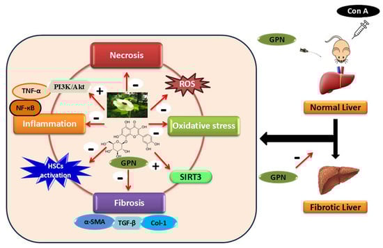
Western blotting results are represented in Figure 8A,B. Our data revealed that Con A repeated injection significantly decreased hepatic p-PI3K and p-Akt protein expression by about 66.47% and 67.43%, respectively, compared to the control group. Concurrently, hepatic p-PI3K protein expression levels were significantly increased upon oral administration of GPN (10 and 20 mg/kg) by about 1.4- and 2.4-fold, respectively, and p-Akt protein expression levels significantly increased upon oral administration of GPN (10 and 20 mg/kg) by about 1.78- and 2.4-fold, respectively. Similarly, Con A injections led to a significant decreased gene expression levels of PI3K and Akt by about 62.5% and 54.9%, respectively, compared to the control group. Interestingly, both GPN doses (10 and 20 mg/kg) significantly increased PI3K hepatic gene expression by 1.5- and 1.8- fold, respectively, while Akt hepatic gene expression was increased by 1.28- and 1.57- fold, respectively, compared to the Con A group (Figure 8D,E).

3. Discussion

Our study investigated the hepato-ameliorative effects of GPN in a mouse model of hepatic fibrosis triggered by Con A. As expected, our results showed that GPN markedly reduced biochemical, histopathological and molecular parameters of Con A-induced hepatic fibrosis. GPN effectively attenuated oxidant/antioxidant disruption and inflammatory response via modulation of NF-κB/TNF-α, PI3K/Akt and SIRT3 pathways.
Con A is widely used to establish a murine model of immune-mediated liver damage, both acute and chronic. Repeated administration of Con A creates a state of continuous cellular injury and immune response that resembles chronic hepatitis in humans, leading to the development of hepatic fibrosis [24]. In line with previous studies, our results showed that repeated injections of Con A led to a significant increase in serum levels of ALT, AST and LDH, indicating hepatocyte damage and liver dysfunction. The administration of GPN caused a significant decrease in these enzymes’ levels, suggesting it minimized hepatocyte death and maintained liver function. The ability of GPN to minimize liver injury is additionally confirmed by its ability to maintain the normal liver architecture demonstrated upon histopathological evaluation. Lesions with lower scores and thinner fibrous tissue were histological indicators of GPN’s ability to alleviate hepatic necrosis, inflammation and fibrosis. The hepatoprotective activity of GPN against acetaminophen-induced hepatotoxicity was mentioned in former research [25].
MT staining revealed a significant increase in collagen deposition upon Con A treatment, with a significant decrease in collagen deposition in case of GPN treatment at both doses. GPN antifibrotic activity was further confirmed by the assessment of hepatic levels of TGF-β and Col-1 and the hepatic immunostaining of α-SMA, a marker for the identification of activated hepatic stellate cells (HSCs)—the primary cell type responsible for ECM protein production in liver fibrosis—and myofibroblasts.
Herein, Con A led to a significant increase in the hepatic levels of TGF-β and Col-1, as well as α-SMA immunostaining, confirming significant fibrosis and collagen deposition that was previously mentioned by [26,27]. Novelly, GPN treatment significantly ameliorated the increase in hepatic TGF-β, Col-1, and α-SMA immunostaining in a dose-dependent manner, demonstrating a powerful effect on decreasing fibrosis and collagen deposition. To our knowledge, this is the first study to show the antifibrotic effect of GPN against hepatic fibrosis.
Several studies stated that Con A significantly increased the hepatic level of the lipid peroxidation biomarker, MDA, concomitant with a remarkable decrease in the hepatic level of antioxidative parameter, TAC [28,29,30]. Herein, the Con A injection led to a significant elevation in serum levels of MDA and a significant reduction in TAC. It turns out that administering GPN significantly decreased the hepatic MDA level and increased hepatic TAC compared to the Con A group, showing a dose-related effect. This finding is consistent with previous research, as GPN’s effect on restoring the oxidant/antioxidant status was also confirmed in case of lung inflammation and doxorubicin-induced cardiotoxicity [31,32].
SIRT3 shields hepatocytes from oxidative stress by scavenging reactive oxygen species (ROS) and preserving mitochondrial integrity [33]. Additionally, SIRT3 lessens macrophages’ infiltration and inflammatory response [34]. Of note, HSCs activation is inhibited by SIRT3 through the activation of TGF-β-Smad downstream signaling pathway in liver fibrosis [23]. Collectively, SIRT3 is thought to be a major target for the prevention and treatment of liver fibrosis and plays a crucial part in the pathophysiology of liver disease [35]. Our findings demonstrated that repeated injections of Con A led to a significant decrease in the hepatic level and gene expression of SIRT3, indicating hepatic dysfunction and deterioration. Both doses of GPN significantly elevated the SIRT3 hepatic level and gene expression, suggesting that the hepatoprotective effect of GPN may be mediated via enhancing SIRT3 and hence minimizing oxidative stress.
Inflammatory response and oxidative stress are closely linked and interconnected during the course of hepatic injury. Proinflammatory gene expression can be triggered by ROS through the activation of various transcription factors—most importantly, NF-κB— that further trigger the expression of multiple inflammatory mediators such as TNF-α [36]. Indeed, NF-κB promotes fibrogenesis through the regulation of fibrogenic responses. It stimulates HSCs to secrete chemokines, which makes them more sensitive to TGF-β and promotes fibrogenesis [37]. In our study, Con A led to a provoked inflammatory response, confirmed by the significant increase in NF-κB immunostaining and TNF-α hepatic level. The strong connection between GPN’s anti-inflammatory and antioxidant properties was demonstrated by its ability to regulate oxidative stress, which in turn prevented the production of TNF-α by inhibiting the NF-κB signaling pathway. Our data are in harmony with the previous study of Cinar, Yayla et al. (2024) [25] that showed the inhibitory effect of GPN on cytokine release in case of acetaminophen-induced hepatic injury.
Early on in fibrosis, the PI3K/Akt pathway is activated, which encourages hepatocyte survival and regeneration. However, this mechanism is hindered when fibrosis progresses, which results in the overproduction of ECM proteins and the activation of HSCs [38]. Furthermore, the PI3K/Akt pathway can regulate inflammatory responses and oxidative stress, two hallmarks of liver fibrosis. [39]. Additionally, the PI3K/Akt pathway is well-known to perform crucial roles in controlling inflammatory damage, cell division and apoptosis. [40,41]. A previous study showed that through the activation of PI3K/Akt signaling pathway, Ghrelin could attenuate Con A-induced hepatitis [42]. Therefore, we hypothesize that the activation of the PI3K/Akt pathway is associated with the anti-inflammatory and antifibrotic effects of GPN in case of Con A-induced fibrosis. In our study, there was a significant decrease in both hepatic p-PI3K and p-Akt proteins, as well as PI3K and Akt gene expression in the Con A group. Interestingly, dose-dependent treatment with GPN significantly increased hepatic phosphorylated protein and gene expression of PI3K and Akt. It may be acceptable to assume that GPN exerts its protective effect against hepatic fibrosis at least partly via activating the PI3K/Akt pathway. The link between SIRT3, NF-κB/TNF-α and PI3K/Akt pathways was demonstrated in previous studies. Zhao et al. confirmed that SIRT3 was involved in the regulation of nuclear expression of NF-κB p65, and silencing SIRT3 expression increased the nuclear expression of NF-κB p65 in HK-2 cells and proved that anti-inflammatory effects against Diosmin-induced renal fibrosis can be promoted by regulating SIRT3-mediated nuclear NF-κB p65 expression [43]. Zuo et al. found that PI3K/AKT signaling can regulate Sirt3 in activated microglia, but not reciprocally. And gastrodin exerts anti-inflammatory and antiapoptotic effects through the PI3K/AKT-Sirt3 signaling pathway in a model of hypoxic–ischemic brain damage [44].
This study has several limitations that should be considered for future research. First, the antifibrotic effects of gossypin were evaluated in a single immune-mediated liver fibrosis model, which may limit generalizability to other etiologies. Second, the study duration was relatively short. Third, a recognized antifibrotic reference compound was not included, preventing the direct comparison of gossypin’s efficacy with established agents. Finally, although the modulation of SIRT3, NF-κB/TNF-α and PI3K/Akt pathways was demonstrated, causal validation using selective pathway inhibitors or genetic knockdown/knockout approaches was not performed. The hierarchical relationship among SIRT3, NF-κB/TNF-α and PI3K/Akt signaling pathways could not be determined, as targeted mechanistic studies using selective inhibitors or genetic approaches were not feasible. Future studies addressing these limitations, including additional models, longer treatment periods, larger sample sizes, inclusion of reference compounds, and mechanistic validation, will strengthen the translational relevance of gossypin as an antifibrotic agent.
Collectively, our results revealed that GPN may possess a significant protective role against liver fibrosis by inhibiting oxidative stress, inflammation, and fibrosis. These outcomes favor the possible application of GPN as a treatment to avoid liver damage caused by CLI. Further clinical investigations are imperative to confirm that effect.

4. Materials and Methods

4.1. Chemicals

GPN powder was obtained from LEAP Chem Co. (Hong Kong, China) and suspended in carboxymethylcellulose. Sigma-Aldrich (Saint Louis, MO, USA) supplied Con A powder, and it was dissolved in water for injection. All other chemicals were of the finest grades.

4.2. Animals

Male mice (BALB/c albino) weighing between 25 and 35 g were used. A controlled environment with sustained temperature (25 °C) and light/dark cycles occurring every 12 h for the mice was adopted. Adequate water and standard rodent feed were freely supplied to mice. The code MU-ACUC (PHARM. PhD. 23.05.23) indicates Mansoura University’s acceptance of experimental methods and techniques, following the guidelines laid down by the Animal Care and Use Committee’s “The Principles of the Testing Facility Animal Care.”

4.3. Experimental Design

Randomly, mice were divided into five groups (each consisting of five mice): 1. Normal group: mice were administered the vehicle. 2. GPN group: mice received oral GPN (20 mg/kg) three times per week for 4 weeks. 3. Con A group, mice received a single IV injection of Con A (10 mg/kg) that was given weekly for 4 weeks. 4. GPN10 + Con A group: mice received GPN (10 mg/kg, oral) three times a week for 4 weeks and a single IV injection of Con A (10 mg/kg) once weekly for 4 weeks. 5. GPN20 + Con A group: mice received GPN (20 mg/kg, oral) three times a week for 4 weeks, and a single IV injection of Con A (10 mg/kg) once weekly for 4 weeks. Dose regimens of Con A and GPN were selected based on previous reports demonstrating the ability of Con A to induce liver fibrosis and the biological efficacy and safety of GPN within this dose range [25,45].
Animals were humanely terminated under anesthesia with ketamine (50 mg/kg, i.p.) twenty-four hours after the final injection of Con A, and every attempt was made to alleviate their discomfort. Blood samples were obtained from the retro-orbital plexus. After the blood was centrifuged, serum samples were taken and stored at −80 °C for further analysis. From all mice, the liver’s left lobe was prepared for histopathology and immunohistochemical (IHC) examination by fixing in neutral buffered formalin and embedded in paraffin. The right lobe of the liver was washed with phosphate-buffered saline (PBS, pH 7.4), homogenized and centrifuged at 4000 rpm for 15 min at 4 °C to extract the supernatants which were then kept at −80 °C for further analysis.

4.4. Assessment of Liver Functions

Commercially available kits from HUMAN Diagnostics (Wiesbaden, Germany) were used to measure the serum levels of selective liver enzymes of alanine aminotransferase (ALT), aspartate aminotransferase (AST) and lactate dehydrogenase (LDH), in line with the provided manufacturer’s instructions.

4.5. Histopathological Evaluation and Grading

For each animal, five random, non-overlapping fields per liver section were analyzed. Hematoxylin and eosin (H&E) staining was used to assess the extent of liver damage after liver tissue sections (5 μm) were prepared from the paraffin blocks. Scoring was performed on hepatic lesions in the form of vascular dilatation, inflammation, necrosis and degeneration, and was assessed in three fields from every slide. The grading of lesions was performed using a blind method. The scoring scale was as follows: 0 = normal, 1 = 25% or less, 2 = 26–50%, 3 = 51–75%, and 4 = 76–100% [46]. Additionally, Masson’s trichrome stain was used to assess the extent of fibrosis in the liver sections. The sections were examined using a light microscope. The Image J program, 1.51r (NIH, Bethesda, MD, USA) was used to measure the degree of fibrous tissue.

4.6. Assessment of Hepatic Oxidative Stress Biomarkers

Hepatic levels of total antioxidant capacity (TAC) and malondialdehyde (MDA) were measured using commercially available Bio-Diagnostic kits (Giza, Egypt, Cat No. TA 2513 and MD 2529, respectively) according to the manufacturer’s instructions.

4.7. Assessment of Hepatic Tissue Levels of Transforming Growth Factor Beta 1 (TGF-β1), Tumor Necrosis Factor-Alpha (TNF-α), Collagen Type 1 (Col-1), and Sirtuin 3 (SIRT3) by ELISA

To determine the levels of TGF-β1, SIRT3, TNF-α and Col-1, the following ELISA kits were utilized: TGF-β1 and TNF-α hepatic levels were determined using Abcam ELISA kit (Waltham, MA, USA, Cat no. ab119557, ab208348, respectively). For the Col-1 estimation, Invitrogen ELISA Kit, a biotechnology brand under ThermoFisher Scientific was used (Waltham, MA, USA, Cat no. EEL218). MyBioSource ELISA kit was used for the estimation of SIRT3 (San Diego, CA, USA, Cat no. MBS2023250), following the manufacturer’s protocol.

4.8. Assessment of Hepatic Tissue Levels of Phosphoinositide-3-Kinase (p-PI3K) and Protein Kinase B (p-Akt) by Western Blotting

Tissue samples (50–100 mg) were homogenized in 1 mL of TriFast reagent (Peqlab, VWR) using a glass–Teflon or power homogenizer. Following the addition of 0.2 mL chloroform, samples were vortexed, incubated and centrifuged at 12,000× g for 5 min at room temperature to separate RNA (aqueous phase), DNA (interphase) and proteins (organic phase). Proteins were precipitated using 1.5 mL isopropanol, washed in guanidinium hydrochloride/ethanol solution, then in 100% ethanol, and finally solubilized in 1% SDS. SDS-PAGE and Gel Preparation Proteins (30 µg per sample) were re-solved using 12% re-solving and 4% stacking polyacrylamide gels. Electrophoresis was carried out at 75 V through the stacking gel and 125 V through the re-solving gel for ~2 h. A broad-range protein ladder (Thermo Scientific™, Waltham, MA, USA, Cat# 26630) was used for molecular weight estimation. Gels were stained with 0.1% Coomassie Brilliant Blue R-250 and destained with a 1:3:6 mixture of glacial acetic acid, methanol and water. Western blot transfer and detection proteins were transferred to Hybond™ nylon membranes using a TE62 Transfer Tank (Hoefer Inc., Bridgewater, MA, USA) and blocked for 1 h in 2–5% non-fat milk in blotting buffer (25 mM Tris, 0.15 M NaCl, 0.1% Tween 20, pH 7.4). Membranes were incubated overnight at 4 °C with PI3K P85 and Akt primary antibodies from Cell signaling technology (Cat no. #4292 and #4060; 85 and 60 kDa, respectively), followed by HRP-conjugated secondary antibody for 1 h at room temperature. Washing was performed with multiple changes in blotting buffer for a total of 30–60 min after each antibody incubation. β-actin was used as a loading control. Detection and quantification were carried out using a GelDoc imaging system and Totallab software (v1.0.1) [47].

4.9. Assessment of Hepatic SIRT-3, PI3K, and Akt Gene Expression by RT-PCR

TRIzol™ Reagent (15596026, Life Technologies, Carlsbad, CA, USA) was used to extract total RNA from samples according to the manufacturer’s instructions. RNA purity and concentration were assessed spectrophotometrically (A260/A280 ratio). A 1 μg amount of total RNA was reverse-transcribed to cDNA using the QuantiTect Reverse Transcription Kit (Qiagen, Santa Clarita, CA, USA). The kit includes a genomic DNA elimination step using gDNA Wipeout Buffer, followed by reverse-transcription with Quantiscript Reverse Transcriptase and random hexamer primers. The reaction was incubated at 42 °C for 15 min, followed by enzyme inactivation at 95 °C for 3 min. The resulting cDNA was either used immediately or stored at –20 °C. Real-time PCR was performed using the Rotor-Gene Q System (Qiagen, USA) and Maxima SYBR Green/Fluorescein qPCR Master Mix (Thermo Scientific, USA). Each 25 μL reaction included 30 ng of cDNA, 300 nM gene-specific primers (listed in Table 1), and master mix components. Amplification conditions were as follows: Initial denaturation at 95 °C for 10 min.; 45 cycles of denaturation at 95 °C for 10 s, 60 °C for 15 s, and 72 °C for 15 s; and melting a melting curve from 72 °C to 95 °C, increasing by 1 °C/s. Each sample was analyzed in duplicate, and the average cycle threshold (Ct) value was used. Gene expression was quantified using the 2−ΔΔCt method [48]. CT values of target genes were normalized to the housekeeping gene β-actin. Expression fold changes were calculated relative to the control group (calibrator) as follows: ΔCt = Ct_target − Ct_reference, ΔΔCt = ΔCt_tested − ΔCt_control, and fold change = 2−ΔΔCt.

4.10. Immunohistochemical Analysis

The expression levels of the Alpha-smooth muscle actin (α-SMA), and the nuclear factor kappa B p65 (NF-κB) in the liver were estimated by immunostaining by the Avidin-Biotin Complex method, using an anti-SMA rabbit polyclonal antibody supplied from Servicebio (Wuhan, Hubei, China, Cat. No: GB111364, 1:1000), and an anti-NF-κB p65 rabbit polyclonal antibody supplied from ABclonal (Cambridge, MA, USA, Cat. No: A2547, 1:200).

4.11. Statistical Analysis

Mean ± SD are used to represent the data in each experimental group. Data normality was assessed using the Shapiro–Wilk test. Parametric measurements were performed using one-way ANOVA followed by Tukey’s multiple comparisons post hoc test. For non-parametric measurements, the Kruskal–Wallis test was used followed by Dunn’s post hoc test (data are expressed as median). A p-value < 0.05 was considered significant. Prism 9 from (GraphPad Software Inc., San Diego, CA, USA) was used to conduct statistical tests.

Author Contributions

Conceptualization, D.S.E.-A.; methodology, H.M.A. and M.E.; software, H.M.A.; validation, M.E. and M.S.S.; formal analysis, H.M.A.; investigation, H.M.A.; resources, H.M.A.; data curation, M.E. and M.S.S.; writing—original draft preparation, H.M.A. and M.S.S.; writing—review and editing, D.S.E.-A.; visualization, D.S.E.-A.; supervision, D.S.E.-A.; project administration, D.S.E.-A.; funding acquisition, H.M.A. All authors have read and agreed to the published version of the manuscript.

Funding

This research received no external funding.

Institutional Review Board Statement

The animal study protocol was approved by the Institutional Review Board of Mansoura University Animal Care and Use Committee (MU-ACUC) (PHARM. PhD. 23.05.23) on 23 May 2023.

Informed Consent Statement

Not applicable.

Data Availability Statement

The original contributions presented in this study are included in the article. Further inquiries can be directed to the corresponding author.

Acknowledgments

The authors acknowledge the support given by Walaa F. Awadin, Faculty of Veterinary Medicine, Mansoura University, for histopathological assessments.

Conflicts of Interest

The authors declare no conflicts of interest.

Abbreviations

The following abbreviations are used in this manuscript:
α-SMAAlpha-smooth muscle actin
AKTProtein Kinase B
ALTAlanine aminotransferase
ASTAspartate aminotransferase
CLIChronic liver injury
Col-1Collagen type 1
Con AConcanavalin A
ECMExtracellular matrix
GPNGossypin
HSCshepatic stellate cells
LDHLactate dehydrogenase
MDAMalondialdehyde
NF-κBNuclear factor kappa-B
PI3KPhosphoinositide 3-kinase
SIRT3Sirtuin 3
TACTotal antioxidant capacity
TGF-βTransforming growth factor-beta
TNF-αTumor necrosis factor-alpha

References

  1. Poynard, T.; Mathurin, P.; Lai, C.-L.; Guyader, D.; Poupon, R.; Tainturier, M.-H.; Myers, R.P.; Muntenau, M.; Ratziu, V.; Manns, M. A comparison of fibrosis progression in chronic liver diseases. J. Hepatol. 2003, 38, 257–265. [Google Scholar] [CrossRef]
  2. Fisher, K.; Vuppalanchi, R.; Saxena, R. Drug-induced liver injury. Arch. Pathol. Lab. Med. 2015, 139, 876–887. [Google Scholar] [CrossRef] [PubMed]
  3. Hora, S.; Wuestefeld, T. Liver injury and regeneration: Current understanding, new approaches, and future perspectives. Cells 2023, 12, 2129. [Google Scholar] [CrossRef] [PubMed]
  4. Tsuchida, T.; Lee, Y.A.; Fujiwara, N.; Ybanez, M.; Allen, B.; Martins, S.; Fiel, M.I.; Goossens, N.; Chou, H.-I.; Hoshida, Y. A simple diet-and chemical-induced murine NASH model with rapid progression of steatohepatitis, fibrosis and liver cancer. J. Hepatol. 2018, 69, 385–395. [Google Scholar] [CrossRef] [PubMed]
  5. Wang, F.-D.; Zhou, J.; Chen, E.-Q. Molecular mechanisms and potential new therapeutic drugs for liver fibrosis. Front. Pharmacol. 2022, 13, 787748. [Google Scholar] [CrossRef]
  6. Parola, M.; Pinzani, M. Liver fibrosis: Pathophysiology, pathogenetic targets and clinical issues. Mol. Aspects Med. 2019, 65, 37–55. [Google Scholar] [CrossRef]
  7. Brenner, D.A. Molecular pathogenesis of liver fibrosis. Trans. Am. Clin. Climatol. Assoc. 2009, 120, 361. [Google Scholar]
  8. Zhang, Y.E. Non-Smad pathways in TGF-β signaling. Cell Res. 2009, 19, 128–139. [Google Scholar] [CrossRef]
  9. Shamsan, E.; Almezgagi, M.; Gamah, M.; Khan, N.; Qasem, A.; Chuanchuan, L.; Haining, F. The role of PI3k/AKT signaling pathway in attenuating liver fibrosis: A comprehensive review. Front. Med. 2024, 11, 1389329. [Google Scholar] [CrossRef]
  10. Nabekura, T.; Matsuo, S.; Shibuya, A. Concanavalin-A-Induced Acute Liver Injury in Mice. Curr. Protoc. 2024, 4, e1117. [Google Scholar] [CrossRef]
  11. Li, S.-L.; Cao, R.; Hu, X.-F.; Xiong, P.; Zhao, G.-Y.; Xie, Y.-N.; Wang, Z.-M.; Li, Y.-K.; Yang, B.; Yang, J. Daidzein ameliorated concanavalin A-induced liver injury through the Akt/GSK-3β/Nrf2 pathway in mice. Ann. Transl. Med. 2021, 9, 1228. [Google Scholar] [CrossRef]
  12. Feng, Q.; Yao, J.; Zhou, G.; Xia, W.; Lyu, J.; Li, X.; Zhao, T.; Zhang, G.; Zhao, N.; Yang, J. Quantitative proteomic analysis reveals that arctigenin alleviates concanavalin A-induced hepatitis through suppressing immune system and regulating autophagy. Front. Immunol. 2018, 9, 1881. [Google Scholar] [CrossRef]
  13. Kunnumakkara, A.B.; Nair, A.S.; Ahn, K.S.; Pandey, M.K.; Yi, Z.; Liu, M.; Aggarwal, B.B. Gossypin, a pentahydroxy glucosyl flavone, inhibits the transforming growth factor beta-activated kinase-1-mediated NF-κB activation pathway, leading to potentiation of apoptosis, suppression of invasion, and abrogation of osteoclastogenesis. Blood J. Am. Soc. Hematol. 2007, 109, 5112–5121. [Google Scholar] [CrossRef] [PubMed]
  14. Song, B.; Shen, X.; Tong, C.; Zhang, S.; Chen, Q.; Li, Y.; Li, S. Gossypin: A flavonoid with diverse pharmacological effects. Chem. Biol. Drug Des. 2023, 101, 131–137. [Google Scholar] [CrossRef] [PubMed]
  15. Mohammed, M.A.; Amer, N.M.; Abdallah, H.M.; Saleh, M.S. A comprehensive tool in recycling plant-waste of Gossypium barbadense L agricultural and industrial waste extracts containing gossy pin and gossypol: Hepatoprotective, anti-inflammatory and antioxidant effects. Plant Methods 2024, 20, 54. [Google Scholar] [CrossRef] [PubMed]
  16. Kweon, Y.-O.; Goodman, Z.D.; Dienstag, J.L.; Schiff, E.R.; Brown, N.A.; Burkhardt, E.; Schoonhoven, R.; Brenner, D.A.; Fried, M.W. Decreasing fibrogenesis: An immunohistochemical study of paired liver biopsies following lamivudine therapy for chronic hepatitis B. J. Hepatol. 2001, 35, 749–755. [Google Scholar] [CrossRef]
  17. Guido, M.; Rugge, M.; Chemello, L.; Leandro, G.; Fattovich, G.; Giustina, G.; Cassaro, M.; Alberti, A. Liver stellate cells in chronic viral hepatitis: The effect of interferon therapy. J. Hepatol. 1996, 24, 301–307. [Google Scholar] [CrossRef]
  18. Nakatani, K.; Seki, S.; Kawada, N.; Kitada, T.; Yamada, T.; Sakaguchi, H.; Kadoya, H.; Ikeda, K.; Kaneda, K. Expression of SPARC by activated hepatic stellate cells and its correlation with the stages of fibrogenesis in human chronic hepatitis. Virchows Arch. 2002, 441, 466–474. [Google Scholar] [CrossRef]
  19. Mann, D.; Smart, D. Transcriptional regulation of hepatic stellate cell activation. Gut 2002, 50, 891–896. [Google Scholar] [CrossRef]
  20. Wu, Q.-J.; Zhang, T.-N.; Chen, H.-H.; Yu, X.-F.; Lv, J.-L.; Liu, Y.-Y.; Liu, Y.-S.; Zheng, G.; Zhao, J.-Q.; Wei, Y.-F. The sirtuin family in health and disease. Signal Transduct. Target. Ther. 2022, 7, 402. [Google Scholar] [CrossRef]
  21. Zhang, J.; Xiang, H.; Liu, J.; Chen, Y.; He, R.-R.; Liu, B. Mitochondrial Sirtuin 3: New emerging biological function and therapeutic target. Theranostics 2020, 10, 8315. [Google Scholar] [CrossRef] [PubMed]
  22. Zhao, X.; Zhang, M.; Wang, J.; Ji, K.; Wang, Y.; Sun, X.; Xu, C.; Wang, Q.; He, N.; Song, H. NMN ameliorated radiation induced damage in NRF2-deficient cell and mice via regulating SIRT6 and SIRT7. Free. Radic. Biol. Med. 2022, 193, 342–353. [Google Scholar] [CrossRef] [PubMed]
  23. Zhou, F.; Wang, A.; Li, D.; Wang, Y.; Lin, L. Pinocembrin from Penthorum chinense Pursh suppresses hepatic stellate cells activation through a unified SIRT3-TGF-β-Smad signaling pathway. Toxicol. Appl. Pharmacol. 2018, 341, 38–50. [Google Scholar] [CrossRef] [PubMed]
  24. Sharawy, M.H.; El-Kashef, D.H.; Shaaban, A.A.; El-Agamy, D.S. Anti-fibrotic activity of sitagliptin against concanavalin A-induced hepatic fibrosis. Role of Nrf2 activation/NF-κB inhibition. Int. Immunopharmacol. 2021, 100, 108088. [Google Scholar] [CrossRef]
  25. Cinar, I.; Yayla, M.; Toktay, E.; Binnetoğlu, D. Effects of gossypin on acetaminophen-induced hepatotoxicity in mice. Trak. Univ. J. Nat. Sci. 2024, 25. [Google Scholar] [CrossRef]
  26. Elfeky, M.G.; Mantawy, E.M.; Gad, A.M.; Fawzy, H.M.; El-Demerdash, E. Mechanistic aspects of antifibrotic effects of honokiol in Con A-induced liver fibrosis in rats: Emphasis on TGF-β/SMAD/MAPK signaling pathways. Life Sci. 2020, 240, 117096. [Google Scholar] [CrossRef]
  27. Ding, W.; Zhou, D.; Zhang, S.; Qian, J.; Yang, L.; Tang, L. Trimetazidine inhibits liver fibrosis and hepatic stellate cell proliferation and blocks transforming growth factor-β (TGFβ)/Smad signaling in vitro and in vivo. Bioengineered 2022, 13, 7147–7156. [Google Scholar] [CrossRef]
  28. Darwish, S.F.; El-Bakly, W.M.; El-Naga, R.N.; Awad, A.S.; El-Demerdash, E. Antifibrotic mechanism of deferoxamine in concanavalin A induced-liver fibrosis: Impact on interferon therapy. Biochem. Pharmacol. 2015, 98, 231–242. [Google Scholar] [CrossRef]
  29. Serrya, M.S.; Zaghloul, M.S. Mycophenolate mofetil attenuates concanavalin A-induced acute liver injury through modulation of TLR4/NF-κB and Nrf2/HO-1 pathways. Pharmacol. Rep. 2020, 72, 945–955. [Google Scholar] [CrossRef]
  30. Mohamed, G.A.; Ibrahim, S.R.; El-Agamy, D.S.; Elsaed, W.M.; Sirwi, A.; Asfour, H.Z.; Koshak, A.E.; Elhady, S.S. Cucurbitacin E glucoside alleviates concanavalin A-induced hepatitis through enhancing SIRT1/Nrf2/HO-1 and inhibiting NF-ĸB/NLRP3 signaling pathways. J. Ethnopharmacol. 2022, 292, 115223. [Google Scholar] [CrossRef]
  31. Yazğan, Y.; Yazğan, B. Gossypin regulated doxorubicin-induced oxidative stress and inflammation in H9c2 cardiomyocyte cells. Med. Rec. 2024, 6, 44–49. [Google Scholar] [CrossRef]
  32. Huang, H.; Wang, J.; Hussain, S.A.; Gangireddygari, V.S.R.; Fan, Y. Gossypin exert lipopolysaccharide induced lung inflammation via alteration of Nrf2/HO-1 and NF-κB signaling pathway. Environ. Toxicol. 2023, 38, 1786–1799. [Google Scholar] [CrossRef]
  33. Liu, J.; Li, D.; Zhang, T.; Tong, Q.; Ye, R.D.; Lin, L. SIRT3 protects hepatocytes from oxidative injury by enhancing ROS scavenging and mitochondrial integrity. Cell Death Dis. 2017, 8, e3158. [Google Scholar] [CrossRef]
  34. Zhang, T.; Fang, Z.; Linghu, K.G.; Liu, J.; Gan, L.; Lin, L. Small molecule-driven SIRT3-autophagy-mediated NLRP3 inflammasome inhibition ameliorates inflammatory crosstalk between macrophages and adipocytes. Br. J. Pharmacol. 2020, 177, 4645–4665. [Google Scholar] [CrossRef] [PubMed]
  35. Ning, Y.; Dou, X.; Wang, Z.; Shi, K.; Wang, Z.; Ding, C.; Sang, X.; Zhong, X.; Shao, M.; Han, X. SIRT3: A potential therapeutic target for liver fibrosis. Pharmacol. Ther. 2024, 257, 108639. [Google Scholar] [CrossRef] [PubMed]
  36. Biswas, S.K. Does the interdependence between oxidative stress and inflammation explain the antioxidant paradox? Oxid. Med. Cell. Longev. 2016, 2016, 5698931. [Google Scholar] [CrossRef] [PubMed]
  37. Seki, E.; Schwabe, R.F. Hepatic inflammation and fibrosis: Functional links and key pathways. Hepatology 2015, 61, 1066–1079. [Google Scholar] [CrossRef]
  38. Li, J.; Wang, T.; Liu, P.; Yang, F.; Wang, X.; Zheng, W.; Sun, W. Hesperetin ameliorates hepatic oxidative stress and inflammation via the PI3K/AKT-Nrf2-ARE pathway in oleic acid-induced HepG2 cells and a rat model of high-fat diet-induced NAFLD. Food Funct. 2021, 12, 3898–3918. [Google Scholar] [CrossRef]
  39. Abraldes, J.G.; Tarantino, I.; Turnes, J.; Garcia-Pagan, J.C.; Rodés, J.; Bosch, J. Hemodynamic response to pharmacological treatment of portal hypertension and long-term prognosis of cirrhosis. Hepatology 2003, 37, 902–908. [Google Scholar] [CrossRef]
  40. Maddika, S.; Ande, S.R.; Panigrahi, S.; Paranjothy, T.; Weglarczyk, K.; Zuse, A.; Eshraghi, M.; Manda, K.D.; Wiechec, E.; Los, M. Cell survival, cell death and cell cycle pathways are interconnected: Implications for cancer therapy. Drug Resist. Updates 2007, 10, 13–29. [Google Scholar] [CrossRef]
  41. Yang, F.-J.; He, Y.-H.; Zhou, J.-H. Fenofibrate pre-treatment suppressed inflammation by activating phosphoinositide 3 kinase/protein kinase B (PI3K/Akt) signaling in renal ischemia-reperfusion injury. J. Huazhong Univ. Sci. Technol. Med. Sci. 2015, 35, 58–63. [Google Scholar] [CrossRef]
  42. Mao, Y.; Wang, J.; Yu, F.; Cheng, J.; Li, H.; Guo, C.; Fan, X. Ghrelin reduces liver impairment in a model of concanavalin A-induced acute hepatitis in mice. Drug Des. Dev. Ther. 2015, 9, 5385–5396. [Google Scholar] [CrossRef] [PubMed]
  43. Zhao, W.-M.; Li, X.-L.; Zhu, Y.; Shi, R.; Wang, Z.-J.; Xiao, J.-P.; Wang, D.-G. Diosmin ameliorates renal fibrosis through inhibition of inflammation by regulating SIRT3-mediated NF-κB p65 nuclear translocation. BMC Complement. Med. Ther. 2024, 24, 29. [Google Scholar] [CrossRef] [PubMed]
  44. Zuo, H.-J.; Wang, P.-X.; Ren, X.-Q.; Shi, H.-L.; Shi, J.-S.; Guo, T.; Wan, C.; Li, J.-J. Gastrodin Regulates PI3K/AKT-Sirt3 Signaling Pathway and Proinflammatory Mediators in Activated Microglia. Mol. Neurobiol. 2024, 61, 2728–2744. [Google Scholar] [CrossRef] [PubMed]
  45. Salah, M.M.; Ashour, A.A.; Abdelghany, T.M.; Abdel-Aziz, A.-A.H.; Salama, S.A. Pirfenidone alleviates concanavalin A-induced liver fibrosis in mice. Life Sci. 2019, 239, 116982. [Google Scholar] [CrossRef]
  46. Elazab, S.T.; Hsu, W.H. Ferulic acid ameliorates concanavalin A-induced hepatic fibrosis in mice via suppressing TGF-β/smad signaling. Toxicol. Appl. Pharmacol. 2024, 492, 117099. [Google Scholar] [CrossRef]
  47. Shay, J.W. Using Antibodies: A Laboratory Manual. In Vitro Cell. Dev. Biol. 1999, 35, 425. [Google Scholar] [CrossRef]
  48. Livak, K.J.; Schmittgen, T.D. Analysis of relative gene expression data using real-time quantitative PCR and the 2(-Delta Delta C(T)) Method. Methods 2001, 25, 402–408. [Google Scholar] [CrossRef]
Figure 1. GPN opposed Con A-induced elevation in serum biomarkers of liver function. (A) Serum ALT, (B) Serum AST, and (C) Serum LDH, Data are expressed as mean ± SD (n = 5). p value < 0.05. * vs. Normal group, # vs. Con A group, $ vs. GPN10 + Con A group (one-way ANOVA). Con A: Concanavalin A, GPN: Gossypin, ALT: alanine aminotransferase, AST: aspartate aminotransferase, LDH: lactate dehydrogenase.
Toxins 18 00074 g001
Figure 2. GPN attenuated Con A-induced hepatic histopathological lesions using hematoxylin-eosin (H&E). (A,B) Hepatic sections of the control group showing normal radially organized hepatocytes around central veins with normal portal areas and sinusoids. (C,D) Hepatic sections from the Con A group showing large areas of coagulative necrosis (black arrow), vacuolar degeneration of hepatocytes (arrowhead), aggregation of inflammatory cells in portal areas (yellow arrows), and vascular dilation and congestion (blue arrows). (E,F) Hepatic sections of the GPN group showing normal radially organized hepatocytes around central veins with normal portal areas and sinusoids. (G,H) Hepatic sections of the GPN10 + Con A group showing smaller areas of coagulative necrosis (black arrow). (I,J) Hepatic sections of the GPN20 + Con A group showing normalized histology with no signs of necrosis or congestion or inflammation ((A,C,E,G,I) × 100, scale bar = 100 µm. (B,D,F,H,J) × 400, scale bar = 50 µm). (K) Inflammation score. (L) Necrosis score. (M) Vascular dilation score. (N) Degeneration score. Data are expressed as median (n = 5). p value < 0.05. * vs. Normal group, # vs. Con A group (Kruskal–Wallis followed by Dunn’s multiple comparison test). Con A: Concanavalin A, GPN: Gossypin.
Toxins 18 00074 g002
Figure 3. GPN counteracted collagen deposition induced by Con A administration using Masson Trichrome (MT) stain. (A,B) Hepatic sections of the control group showing no collagen deposition in hepatic parenchyma. (C,D) Hepatic sections from the Con A group showing excessive bluish collagen deposition (black arrows) around blood vessels. (E,F) Hepatic sections of the GPN group showing no collagen deposition in hepatic parenchyma. (G,H) Hepatic sections of the GPN10 + Con A group wing decreased bluish collagen deposition (black arrows) around blood vessels. (I,J) Hepatic sections of the GPN20 + Con A group showing no excess bluish collagen deposition around blood vessels ((A,C,E,G) ×100, bar= 100 µm; (B,D,F,H) ×400, scale bar = 50 µm). (K) Corresponding area percentage of the positive collagen deposition. Data are expressed as mean ± SD (n = 5). p value < 0.05 * vs. Normal group, # vs. Con A group (one-way ANOVA). Con A: Concanavalin A, GPN: Gossypin.
Toxins 18 00074 g003
Figure 4. GPN ameliorated hepatic levels of TGF-β1 and Col-1 as well as hepatic immunohistochemical expression of α-SMA in Con A-administered mice. (A) TGF-β1 hepatic level. (B) Col-1 hepatic level. (C) α-SMA hepatic immunohistochemical expression. (1,2) Hepatic sections of the control group showing negative staining for α-SMA in hepatocytes. (3,4) Hepatic sections from the Con A group showing positive brown staining for a-SMA that appears cytoplasmic and nuclear in many hepatocytes (black arrow). (5,6) Hepatic sections of the GPN10 + Con A group showing decreased positive brown staining for α-SMA that appears cytoplasmic and nuclear in some hepatocytes around central veins (black arrow). (7,8) Hepatic sections of the GPN20 + Con A group showing mild positive brown staining for α-SMA that appears cytoplasmic and nuclear in fewer hepatocytes around central veins (black arrows). (9) Corresponding area percentage of the positive α-SMA immunoreactivity. IHC counterstained with Mayer’s hematoxylin. Low magnification (×100; bar 100) and high magnification (×400; bar 50). Data were expressed as means ± SD (n = 5). p value < 0.05 * vs. Normal group, # vs. Con A group, $ vs. GPN10 + Con A group (one-way ANOVA). Con A: Concanavalin A, GPN: Gossypin, TGF-β1: Transforming growth factor-beta, Col-1: Collagen-1, α-SMA: Alpha-smooth muscle actin.
Toxins 18 00074 g004
Figure 5. GPN influenced hepatic redox status in Con A-challenged mice. (A) MDA hepatic level, (B) TAC hepatic level, Data were expressed as means ± SD (n = 5). p value < 0.05 * vs. Normal group, # vs. Con A group, $ vs. GPN10 + Con A group (one-way ANOVA). Con A: Concanavalin A, GPN: Gossypin, MDA: Malondialdehyde, TAC: Total antioxidant capacity.
Toxins 18 00074 g005
Figure 6. GPN enhanced hepatic protein and gene expression of SIRT3 in Con A-administered mice. (A): SIRT3 hepatic level, (B): SIRT3 hepatic mRNA expression. Data are expressed as means ± SD (n = 5 rats per group). p value <0.05 * vs. Normal group, # vs. Con A group, $ vs. GPN10 + Con A group (one-way ANOVA). Con A: Concanavalin A, GPN: Gossypin, Sirt3: Silent mating type information regulation 3 homolog.
Toxins 18 00074 g006
Figure 7. GPN alleviated NF-κB/TNF-α pathway in Con A-administered mice. (A). NF-κB hepatic immunohistochemical expression. (1,2). Hepatic sections of the control group showing negative staining for NF-κB in hepatocytes. (3,4). Hepatic sections from the Con A group showing positive brown staining for NF-κB that appears cytoplasmic and nuclear in many hepatocytes (black arrow). (5,6). Hepatic sections of the GPN10 + Con A group showing decreased positive brown staining for NF-κB that appears cytoplasmic and nuclear in some hepatocytes around central veins (black arrow). (7,8). Hepatic sections of the GPN20 + Con A group showing mild positive brown staining for NF-κB that appears cytoplasmic and nuclear in fewer hepatocytes around central veins (black arrows). (B). Corresponding area percentage of the positive NF-κB immunoreactivity. (C). TNF-α hepatic level. IHC counterstained with Mayer’s hematoxylin. Low magnification (×100; bar 100) and high magnification (×400; bar 50). Data are expressed as mean ± SD (n = 5). p value < 0.05 * vs. Normal group, # vs. Con A group, $ vs. GPN10 + Con A group (one-way ANOVA). Con A: Concanavalin A, GPN: Gossypin, NF-κB: Nuclear Factor Kappa B, TNF-α: Tumor necrosis factor-alpha.
Toxins 18 00074 g007
Figure 8. GPN enhanced hepatic protein and gene expression of PI3K and Akt in Con A-challenged mice. (A): Bands for Western blotting. (B): PI3K protein expression. (C): Akt protein expression. (D): PI3K gene expression. (E): Akt gene expression. Data were expressed as means ± SD (n = 5). p value < 0.05 * vs. Normal group, # vs. Con A group, $ vs. GPN10 + Con A group (one-way ANOVA). Con A: Concanavalin A, GPN: Gossypin, PI3k: Phosphoinositide 3-kinase, Akt: Protein kinase B.
Toxins 18 00074 g008
Table 1. Primer probe for genes under investigation.
GeneProbe
SIRT35-TGCACGGTCTGTCGAAGGTC-3
5-A TGTCAGGTTTCACAACGCCAGTA-3
PI3K5-AACACAGAAGACCAATACTC-3
5-TTCGCCATCTACCACTAC-3
Akt5-GTGGCAAGATGTGTATGAG-3
5-CTGGCTGAGTAGGAGAAC-3
b-actin5-CCT GGAGAAGAGCTATGAGC-3
5-A CAGGATTCCATACCCAGG-3
Disclaimer/Publisher’s Note: The statements, opinions and data contained in all publications are solely those of the individual author(s) and contributor(s) and not of MDPI and/or the editor(s). MDPI and/or the editor(s) disclaim responsibility for any injury to people or property resulting from any ideas, methods, instructions or products referred to in the content.

