Article

Antibacterial Activity of Jelleine-I, a Peptide Isolated from Royal Jelly of Apis mellifera, Against Colistin-Resistant Klebsiella pneumoniae

by
William Gustavo Lima
1,*,†,
Rayssa Maria Rodrigues Laia
1,†,
Julio Cesar Moreira Brito
2,
Daniel Augusto Guedes Reis Michel
3,
Rodrigo Moreira Verly
3,
Jarbas Magalhães Resende
4 and
Maria Elena de Lima
1,5
1
Faculdade Santa Casa de Belo Horizonte, Belo Horizonte 30110-005, Brazil
2
Fundação Ezequiel Dias (FUNED), Belo Horizonte 30510-010, Brazil
3
Departamento de Química, Faculdade de Ciências Exatas, Universidade Federal dos Vales do Jequitinhonha e Mucuri, Diamantina 39100-000, Brazil
4
Departamento de Química, Instituto de Ciências Exatas, Universidade Federal de Minas Gerais, Belo Horizonte 31270-901, Brazil
5
Programa de Pós-Graduação em Inovação Farmacêutica, Universidade Federal de Minas Gerais, Belo Horizonte 31270-901, Brazil
*
Author to whom correspondence should be addressed.
These authors contributed equally to the work.
Toxins 2025, 17(7), 325; https://doi.org/10.3390/toxins17070325
Submission received: 10 June 2025 / Revised: 19 June 2025 / Accepted: 23 June 2025 / Published: 25 June 2025
(This article belongs to the Section Animal Venoms)

Abstract

Klebsiella pneumoniae can acquire resistance mechanisms to colistin and present a pan-resistant phenotype. Therefore, new alternative agents are imperative to control this pathogen, and the peptide Jelleine-I stands out as a promising prototype. Here, the antibacterial activity of Jelleine-I against clinical isolates of colistin-resistant K. pneumoniae (CRKP) was investigated. Antimicrobial activity was assessed by determining the minimum inhibitory concentration (MIC), minimum bactericidal concentration (MBC) and time kill-curve assay. The release of 260 nm-absorbing materials (DNA/RNA) and the release of proteins were used in the lysis assay. Anti-biofilm activity was studied in microplates. In vivo activity was determined by the lethality assay using Tenebrio molitor larvae. The results show that the MIC of Jelleine-I ranged from 16 to 128 µM and the MBC was on average 128 µM. Jelleine-I at 200 µM killed all CRKP cells in suspension (106 colony-forming units (CFU)/mL) after 150 min of incubation. Jelleine-I acts on the CRKP cell membrane inducing lysis. Biomass and viability of CRKP-induced biofilms are reduced after treatment with Jelleine-I, and the use of this peptide in T. molitor larvae infected with CRKP reduces lethality and improves overall larval health. In conclusion, Jelleine-I is a potential prototype for the development of new antimicrobial agents.
Key Contribution: Jelleine-I has action against pan-resistant bacteria. The effect of Jelleine-I involves lysis by action on the membrane through a mechanism distinct from that known to polymyxins, so there is no cross-resistance between Jelleine-I and colistin or polymyxin B. The use of Jelleine-I (50 µg and 100 µg) reduces lethality in Tenebrio molitor larvae infected with a hypervirulent strain of colistin-resistant Klebsiella pneumoniae.

Graphical Abstract

1. Introduction

Klebsiella pneumoniae is a glucose-fermenting, gram-negative, lysine decarboxylase-positive, ornithine decarboxylase-negative, encapsulated, immotile, facultatively anaerobic bacterium [1,2]. This species was originally isolated by Edwin Klebs in 1875 from the airways of a patient dying of pneumonia. Later, K. pneumoniae was also described by Carl Friedländer in 1882, which led to it being called Friedlünder’s bacillus for a time [3]. Currently, K. pneumoniae is classified into the Enterobacteriales family and is recognized as one of the most opportunistic pathogens involved in healthcare-associated infections (HAI), causing pneumonia, meningitis, bloodstream infections, and urinary tract infections [4]. In fact, K. pneumoniae is responsible for approximately 11.8% of all hospital-acquired pneumonia and nearly 10% of all HAI cases worldwide [1,4,5]. In addition, this gram-negative bacillus is reported as the second most common cause of community-acquired urinary tract infections [6]. Patients with K. pneumoniae infections often have a poor prognosis, and even with proper treatment, mortality rates in these cases range from 30% up to 80% [3]. Of particular concern is also the high incidence of carbapenem resistance in this species, which necessitates the use of last-resort antimicrobials such as polymyxins (i.e., colistin and polymyxin B) [4]. However, due to the increasing prescription of polymyxins, the rates of resistance to this class of antimicrobials have increased considerably, and colistin-resistant K. pneumoniae (CRKP), a pan-drug resistance-related phenotype, have frequently been found in health centers around the world [7,8].
Consequently, new and unconventional antimicrobials against CRKP are urgently needed. Natural or modified antimicrobial peptides (AMPs) display interesting features as potential new antibacterial agents [9,10]. AMPs are recognized components of the innate immune response of virtually all living organisms, from prokaryotes to plants and animals [11]. However, AMPs are particularly prevalent in insects, as these animals lack a cell-mediated immune response, with AMPs playing a prominent role in their immune defense [12]. These antimicrobial agents have a broad spectrum of activity against gram-positive and gram-negative bacteria, exhibit a rapid bactericidal effect, are quickly degraded in the environment (not generating residues that could contaminate the environmental microbiota), are even effective against multi-resistant bacteria, can be easily synthesized in the laboratory, and have limited potential for induction of antimicrobial resistance [9].
In this context, Jelleine-I (H-PFKISIHL-NH2), an octapeptide isolated from the royal jelly of honeybee (Apis mellifera), has shown promising antibacterial activity, including against multi-resistant pathogens such as methicillin-resistant Staphylococcus aureus [13], extended spectrum beta-lactamase (ESBL)-producing Escherichia coli [13], beta-lactam-resistant Staphylococcus epidermidis [14], and piperacillin-resistant Pseudomonas aeruginosa [13]. Furthermore, a recent study has shown that Jelleine-I has high to moderate activity against pan-drug resistant clinical isolates of Acinetobacter baumanii (minimum inhibitory concentration (MIC) of 8 to 16 μM), highlighting its biological activity against colistin-resistant bacteria [15]. However, there is no evidence of action of Jelleine-I on CRKP. Furthermore, it is known that the main mechanism of polymyxin resistance involves structural alterations in the outer membrane, the primary target of Jelleine-I action [16,17]. In fact, the addition of positively charged motifs, such as phosphoethanolamine (PEA) and 4-Amino-4-deoxy-L-arabinose (L-Ara4N), in the lipid A of the outer membrane and the loss of lipopolysaccharide due to mutations in the lpxABC operon are the main known mechanisms of polymyxin resistance in K. pneumoniae [16]. Thus, it is important to characterize the mode of action of Jelleine-I on the membrane of polymyxin-resistant bacteria, such as CRKP. In light of this, the purpose of the present study was to investigate the effect of Jelleine-I against CRKP in vitro and in vivo and study its activity on the cell membrane of this microorganism.

2. Results and Discussion

Initially, the broth microdilution assay was utilized to study the antibacterial activity of Jelleine-I against 18 clinical isolates of CRKP. As shown in Table 1, the minimum inhibitory concentration (MIC) of Jelleine-I ranged from 16 to 128 µM. The MIC50 and MIC90, defined as the MICs that inhibited 50% and 90%, respectively, of the tested microorganisms, were also determined. Jelleine-I showed MIC50 and MIC90 values of 64 µM and 83.2 µM, respectively. The antibacterial activity of this peptide was predominantly bactericidal, with the minimum bactericidal concentration (MBC) ranging from 64 to 128 µM, while MBC50 (concentration that kills 50% of the tested microorganisms; 128 µM) and MBC90 (concentration that kills 90% of the tested microorganisms; 166.4 µM) showed similar values. Related results have been reported by Jia et al. [18], who found that Jelleine-I is active against an extended-spectrum beta-lactamase-producing K. pneumoniae reference line (ATCC 700603) with an MIC of 64 µM and MBC of 128 µM. In their turn, other studies have reported lower MIC values; for example, Kim et al. [19] showed that Jelleine-I at 8 µM is active against K. pneumoniae KCTC 2242, whereas Fontana et al. [20] described a 10 µM MIC against K. pneumoniae ATCC 13883. Such differences of antibacterial concentrations of Jelleine-I against K. pneumoniae can be explained by the resistance profile of the respective isolates. Indeed, multidrug-resistant specimens were used in the present study, as well as in that by Jia et al. [18]. On the other hand, the lineages employed by Kim et al. [19] and Fontana et al. [20] are sensitive to conventional antimicrobials, resulting in expected lower MIC values.
Next, a time-kill curve assay was performed to evaluate the activity of Jelleine-I against CRKP over time. According to Figure 1, Jelleine-I at a concentration of 200 μM was able to kill all microorganisms of a CRKP bacterial suspension, which contained 106 colony-forming units (CFU)/mL, after 150 min of incubation. Furthermore, a maximum microbicidal effect was also observed in suspensions treated with Jelleine-I at 100 μM after 180 min of incubation. A similar result was observed with polymyxin-resistant Acinetobacter baumannii, where incubation with Jelleine-I, at a concentration of 80 µM, induced maximum bactericidal effect after 180 min of incubation [15]. A rapid microbicidal effect was also observed with Jelleine-I on fungal cells; this peptide also exhibited maximum activity after 180 min of incubation against Candida albicans (256 µM), Nakaseomyces glabratus (formerly Candida glabrata) (128 µM), Pichia kudriavzevii (formerly Candida krusei) (128 µM), Candida parapsilosis (256 µM), and Candida tropicalis (64 µM) [23]. The fast elimination of the microbial agents by Jelleine-I is important, as it allows the infectious focus to be removed quickly, avoiding more severe consequences such as systemic infection, sepsis, prolonger antimicrobial therapy, increased hospitalization costs, superinfections, the need for invasive procedures, increased mortality, antimicrobial resistance, and disease recrudescence [24].
The action of Jelleine-I on the plasma membrane of CRKP was studied using a bacteriolytic capacity assay by measuring the release of DNA/RNA (260 nm-absorbing materials) and proteins from treated bacterial cells. Jelleine-I increased the release of 260 nm-absorbing intracellular material from CRKP cells at concentrations starting at 80 µM (Figure 2). Furthermore, the increase in protein concentration in the supernatant of a CRKP cell suspension treated with Jelleine-I revealed that this peptide indeed contributes to the extravasation of intracellular macromolecules, suggesting cell lysis. Melittin, a peptide known to lyse bacterial cells, was also able to induce release of DNA/RNA and proteins from bacterial cells, validating our experimental conditions. The increased release of cytoplasmic material is an important indication of an effect on the cell membrane, confirming numerous studies that have proposed Jelleine-I as a membranolytic peptide [15,18,25]. Castro et al. [15] showed that Jelleine-I-membrane interaction disturbs the organization of phospholipid bilayers. The authors has revealed that Jelleine-I alters the hydrodynamic diameter (Dh) and zeta potential (ζ-potential) in phospholipid vesicles synthetics that simulate bacterial membranes, beyond increasing the intracellular material release from polymyxin-resistant Acinetobacter baummannii [15]. Similarly, our study showed that the bacteriolytic effect of Jelleine-I is maintained even in strains with resistance to polymyxins, known for important alterations in the charge and structure of the cell membrane. These results show that resistance to polymyxins does not promote cross-resistance to Jelleine-I, suggesting the use of this AMP against Gram-negative pandrug-resistant bacteria.
Polymyxins induce disruption of the plasma membrane by displacing divalent cations (especially Ca2+ and Mg2+) bound to this cellular structure. In addition to being considered important cofactors in different microbial metabolic pathways, divalent cations play an important role in the biophysical stability of the cell membrane of microorganisms and in the osmotic resistance of the cell [26,27]. To evaluate whether the mechanism of action of Jelleine-I on the bacterial membrane is similar to that observed with polymyxins, we determined the MIC of the peptide in the presence of increasing concentrations of Ca2+ and Mg2+. Table 2 shows that the activity of Jelleine-I was not modified in the presence of divalent cations, suggesting that this compound does not act on the homeostasis of these ions. In turn, the activity of colistin is reduced in the presence of calcium and magnesium in a concentration-dependent manner (Table 2). Thus, even though the exact mode of action was not fully determined to Jelleine-I, comparative experiments with colistin showed a different mode of action, thus justifying the absence of cross-resistance between these two compounds.
Several AMPs are known to induce the formation of free radicals, especially reactive oxygen species (ROS) and reactive nitrogen species (RNS). These free radicals can disrupt microbial cell membranes, triggering oxidative stress that damages intracellular proteins and DNA, ultimately resulting in cell death [28]. To evaluate whether Jelleine-I induces oxidative damage in CRKP cells, the MIC of this peptide was determined in the presence of ascorbic acid, a potent antioxidant agent. The results, however, showed no change in MIC upon supplementation of the medium with the antioxidant, suggesting that Jelleine-I does not act by pro-oxidative pathways in CRKP. In contrast to our findings, Jia et al. [18] reported an increase in ROS concentration in S. aureus and E. coli cells treated with Jelleine-I. These differing results may be attributed to the methodology used, as Jia et al. [18] employed more sensitive fluorometric techniques for ROS measurement. However, it is important to note that an increase in ROS does not necessarily imply an antibacterial effect; verifying alterations in MIC values using pharmacological tools is crucial. Therefore, our study indicates that despite a potential increase in ROS generation, this effect likely has minimal influence on the overall antibacterial activity of Jelleine-I, as the inhibition of these agents did not alter the biological activity of the peptide.
One of the most important virulence factors of Gram-negative bacteria such as K. pneumoniae is their ability to form biofilms on biological tissue and inanimate surfaces. Biofilms are aggregates of microbial cells surrounded by self-produced exopolysaccharide matrices [29]. These structures exhibit enhance protection against antimicrobials, hospital sanitizers, host immune defenses, and adverse environmental conditions compared to free-living cells [30]. Therefore, the inability of conventional antimicrobial agents to target and destroy adherent bacterial biofilms is a major challenge for current antimicrobial therapies [29,30]. In this context, we investigated the effect of Jelleine-I on mature CRKP-formed biofilms in two aspects: biomass and viability of cells into biofilms. Jelleine-I was able to reduce the biomass of mature biofilm of a hypervirulent isolate of CRKP at concentrations of 80 µM (63.29 ± 12.96%; p-value 0.0076) and 160 µM (64.85 ± 9.06%; p-value 0.0108) in relation to control cells (100 ± 12.13%). Moreover, Jelleine-I also reduced the viability of CRKP cells within the biofilm at all concentrations tested (32 µM: 38.46 ± 10.33%, p-value = 0.0005; 80 µM: 13.72 ± 12.34%, p-value < 0.0001; and 160 µM: 14.91 ± 13.43%, p-value < 0.0001) compared to the untreated cells (100 ± 20.62%), suggesting a potent anti-biofilm effect (Figure 3). Similarly, other studies have proven the activity of Jelleine-I against biofilms of Staphylococcus aureus [31] and Listeria monocytogenes [32]. Indeed, antimicrobial peptides are known for their high activity against bacterial biofilms by inducing disruption or degradation of the membrane potential of cells embedded in the biofilm, inhibiting bacterial signaling systems by downregulating genes responsible for biofilm formation and transport of binding proteins, in addition to degrading the polysaccharide and biofilm matrix [33].
Based on the promising in vitro antibacterial activity, the Jelleine-I was investigated in a Tenebrio molitor larvae model of CRKP-infection. Initially, the toxicity of Jelleine-I was evaluated using a lethality assay following the administration of different doses of the peptide (1, 5, 10, 50, and 100 µg) to the larvae. As shown in Figure 4A, none of the doses studied induced significant mortality in T. molitor larvae during the studied period (72 h), suggesting low toxicity of Jelleine-I. In fact, previous studies have shown that Jelleine-I has low toxicity in vitro against renal cells (Vero), fibroblasts (NIH 3T3), macrophages (J774, THP1, and RAW264.7), cervical cells (HeLa), and murine erythrocytes, as well as in in vivo assays using Kunming mice (median lethal dose (LD50) > 1000 mg/kg) [20]. Next, we evaluated whether the use of Jelleine-I reduces the lethality of T. molitor larvae infected with a hypervirulent isolate of CRKP (108 CFU). At the highest doses tested (50 µg: 35% survival after 72 h; and 100 µg: 40% survival after 72 h), there was a reduction in the lethality of infected larvae when compared to the untreated group (0% survival after 24 h; p-value < 0.05) (Figure 4B). Furthermore, the general clinical score of infected larvae was higher in the groups treated with doses of 50 µg (3 (0–8)) and 100 µg (1 (0–7)) of Jelleine-I, when compared to larvae treated with saline (1 (0–4)) (Figure 4C). Similar results were observed by Jia et al. [31], who showed that neutropenic mice infected with E. coli treated with Jelleine-I (20 mg/Kg) had a survival rate of 25% after 10 days. Furthermore, Jia et al. [23] have shown that mice infected with C. albicans and treated with Jelleine-I (10 mg/Kg) daily for seven days have a 40% lethality after 14 days, which is significantly lower compared to untreated animals (100% lethality). In addition, in an invertebrate model, using Galleria mellonella larvae, Jelleine-I also caused significant protection against Listeria monocytogenes-induced lethality by increasing survival rates by 10%, 20%, and 30% at 10 mg/kg, 20 mg/kg, and 40 mg/kg, respectively [32].

3. Conclusions

The results show that Jelleine-I exhibits promising in vitro and in vivo activity against CRKP. Jelleine-I acts on planktonic cells and on biofilms of pandrug-resistant K. pneumoniae, exhibiting rapid bactericidal effect in the time-kill curve assay. Although the antimicrobial effect of Jelleine-I involves damage to the bacterial membrane, this mechanism of action differs from that observed with polymyxins, which act by displacing divalent cations bound to the plasma membrane. This explains the absence of cross-resistance between Jelleine-I and polymyxins in K. pneumoniae. Furthermore, this peptide proved to be safe and effective in in vivo assays using T. molitor larvae, being able to reduce the lethality of animals infected with a hypervirulent strain of CRKP at doses without detectable toxicity. These results also highlights the use of T. molitor larvae as a viable model to study the virulence and treatment of K. pneumoniae, which could be explored as a replacement for vertebrate animal models. In conclusion, this study highlights Jelleine-I as a promising prototype for the development of a novel antimicrobial against pandrug-resistant K. pneumoniae.

4. Materials and Methods

4.1. Reagents

Colistin, meropenem (InlabTM, São Paulo, SP, Brazil), crystal violet, glacial acetic acid, methanol, sodium chloride, magnesium chloride, calcium chloride, glucose (SynthTM, São Paulo, SP, Brazil), 3-(4,5-dimethylthiazol-2-yl)-2,5-diphenyltetrazolium bromide (MTT), Coomassie brilliant blue G 250, and bovine serum albumin (Sigma-AldrichTM, Frankfurt, HE, Germany) were purchased from commercial suppliers and used without further purification because the purity was greater than 99% in all cases. Mueller–Hinton broth (MHB), Muelle–-Hinton agar (MHA), and MacConkey agar were purchased from KasviTM (São José do Pinhais, PR, Brazil). Jelleine-I was obtained through solid-phase synthesis and subsequently purified and characterized in the Peptide Synthesis and Structure Laboratory (LASEP/UFMG), exactly as described previously [15]. After purification, a purity of 97.48% was obtained.

4.2. Microorganisms

In this study, were employed eighteen clinical isolates of colistin-resistant Klebsiella pneumoniae (CRKP) recovered from patients admitted in a tertiary hospital in Belo Horizonte (Minas Gerais, Brazil). All isolates were identified using biochemical-morphological tolls by an automated BD PhoenixTM system (BDTM, New York, NY, USA). Resistance to colistin was initially evaluated by the growth of isolates in MHB supplemented with colistin at a concentration of 4 µg/mL, as recommended by the Brazilian Committee on Antimicrobial Susceptibility Testing (BrCAST). Next, the resistance to colistin among the isolates that grew in the medium supplemented with this antimicrobial was confirmed by determination of minimum inhibitory concentration (MIC) second to the Clinical Laboratory and Standard Institute (CLSI) [34]. In addition, two reference lineages from the American Type Culture CollectionTM (ATCC) (K. pneumoniae ATCC 700603 and K. pneumoniae ATCC 43816) were kindly provided by the João XXIII hospital (Belo Horizonte, MG, Brazil) and included in this study as internal controls.

4.3. Antibacterial Activity

Inoculum preparation: The bacterial inoculum was standardized according to the CLSI rules [34]. Two to three isolated colonies, which were collected from a 24 h CRKP culture realized on MHA, were suspended in a sterile saline solution (0.85% NaCl; 10 mL) with the aid of a bacteriological loop. Next, the resulting suspension was adjusted to an optical density (OD) of 0.08–0.13 at 625 nm (Nova InstrumentsTM, Sao Paulo, SP, Brazil), which is equivalent to the McFarland 0.5 scale (108 colony-forming units (CFU)/mL). Finally, 50 μL of the bacterial suspension prepared in saline was transferred to MHB (10 mL) to produce a working inoculum at 106 CFU/mL.
Minimum inhibitory concentration (MIC): Bacteriostatic activity was assessed by determining the MIC according to the CLSI guidelines [34], with minor modifications [35]. One hundred microliters (100 μL) of a working inoculum of CRKP (106 CFU/mL) were added to microplates containing 100 μL of a twofold serial dilution (1–128 µM) of Jelleine-I or colistin in MHB. The concentration range chosen for the tests with Jelleine-I was determined based on previous studies by Castro et al. (2025), who evaluated the antibacterial effect of this peptide against pandrug resistant Acinetobacter baumannii [15]. The plates were then incubated at 35 ± 2 °C for 18 h, and the MIC was defined as the lowest concentration of the compounds that visibly inhibited bacterial growth. Each plate included wells with 200 µL of culture medium as a sterility control and wells with 100 µL of culture medium plus 100 µL of inoculum as a microorganism viability control.
Minimum bactericidal concentration (MBC): Bactericidal activity was assessed by transferring 100 µL from the wells showing no visible growth in the MIC assay to MHA plates, followed by incubation at 35 ± 2 °C for 24 h [35]. The MBC was defined as the lowest concentration of Jelleine-I that resulted in complete inhibition of bacterial growth.

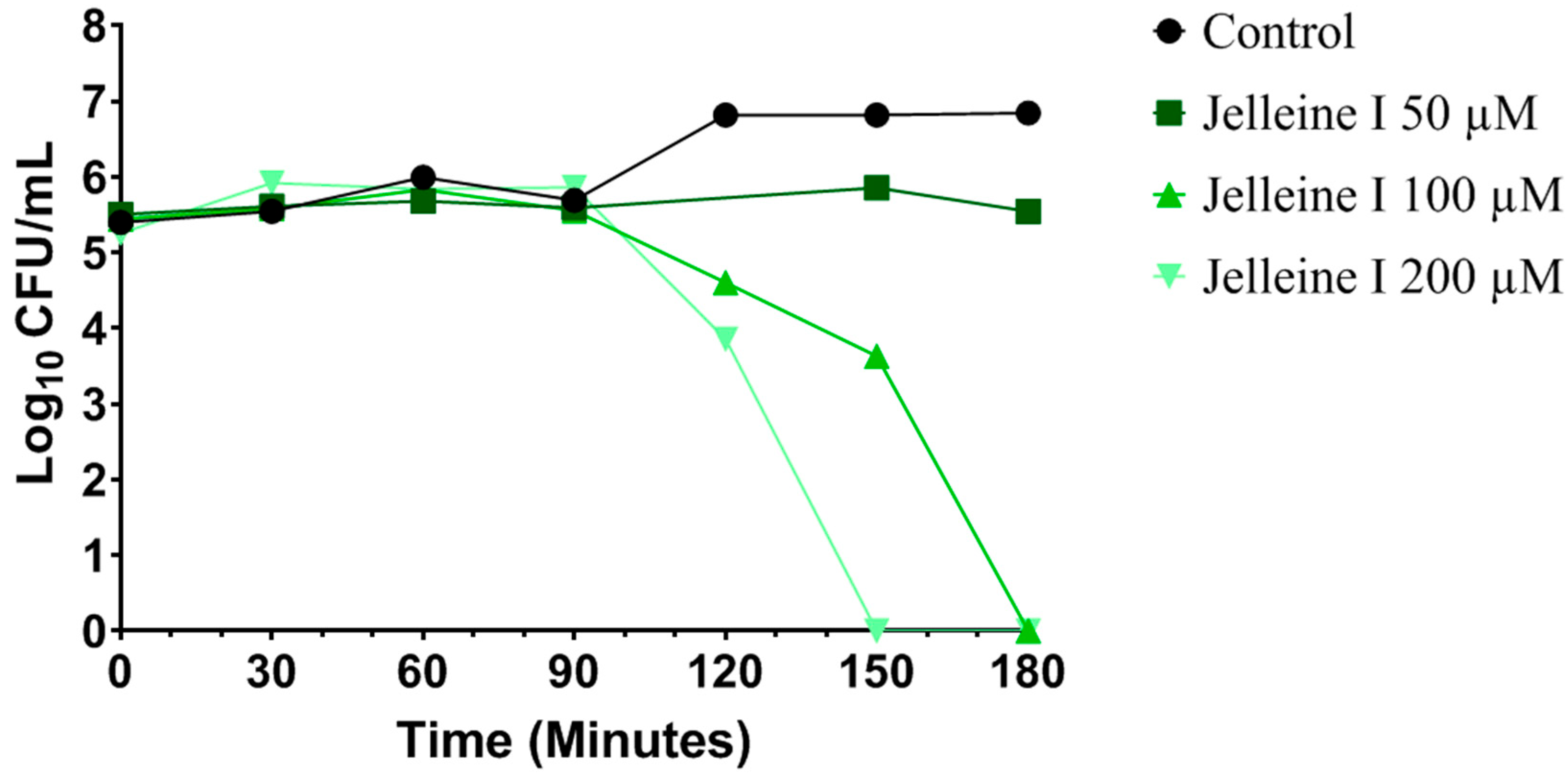
4.4. Time-Kill Curve

A pre-inoculum of CRKP (isolate 699) at a concentration of 108 CFU/mL was prepared in sterile saline (0.85% NaCl). Subsequently, 50 µL of pre-inoculum were added to test tubes containing MHB (10 mL) with Jelleine-I at different concentrations (50, 100, or 200 µM). The tubes were incubated at 35 ± 2 °C, and, in specific time points (0, 30, 60, 90, 120, 150, and 180 min), samples were serially diluted (10−1 to 10−6) in sterile saline (0.85% NaCl) and plated onto MacConkey agar. The plates were then incubated at 35 ± 2 °C for 24 h, and bacterial counts were determined and expressed as CFU/mL. Untreated cells served as the negative control [35].

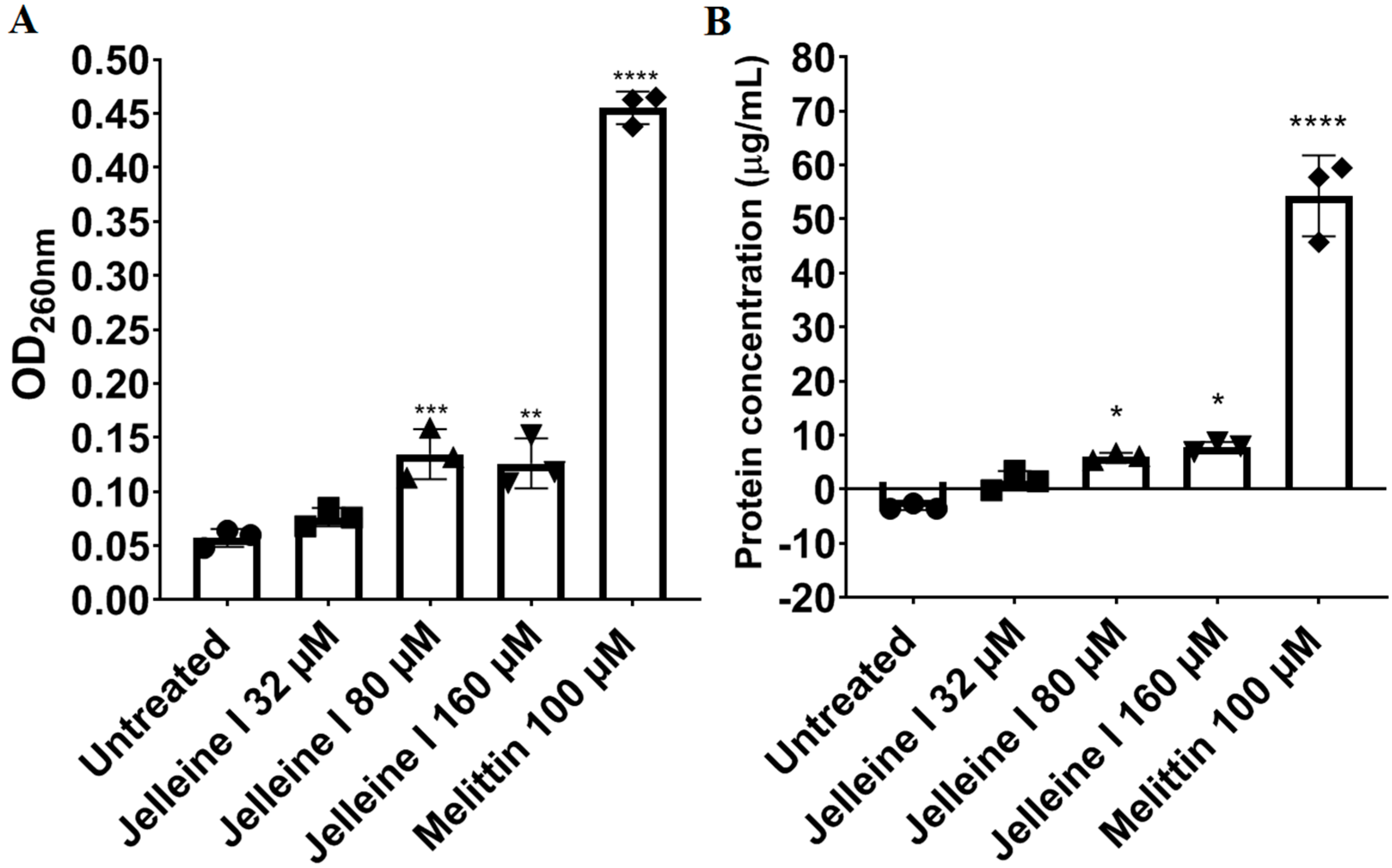
4.5. Release of Bacterial DNA/RNA

The release of intracellular components (DNA/RNA), indicated by absorbance at 260 nm, was quantified [35]. Bacterial suspensions of CRKP in 0.85% saline (108 CFU/mL) were treated with Jelleine-I at concentrations of 32, 80, or 160 µM. After incubation, 100 µL of each suspension were centrifuged (1000× g for 25 min at 4 °C), and the absorbance of the resulting supernatant was measured at the wavelength of 260 nm (Shimadzu™, Tokyo, OS, Japan). Finally, the results were plotted as OD620 versus time (hours). Melittin (100 µM) was used as a positive control.

4.6. Release of Protein

Protein release was studied by the Bradford method [36]. After exposure of a CRKP suspension at 106 CFU/mL prepared in saline (0.85% NaCl) to different concentrations of Jelleine-I (32, 80, or 160 µM) for 24 h, aliquots (100 µL) were collected and centrifuged (1000× g for 25 min at 4 °C). The supernatant was mixed with Coomassie brilliant blue G 250, and, after 2 min of reaction, the blue chromogen was measured spectrophotometrically at 595 nm (Nova InstrumentsTM, Sao Paulo, SP, Brazil). The protein concentration in the supernatant was determined with the aid of the straight-line equation obtained from a standard curve constructed with bovine serum albumin (2.5–100 µg/mL). Untreated cells were included as a negative control, and cells exposed to melittin, an agent known to lyse bacterial cells, were used as a positive control.

4.7. Binding to Divalent Cations

The ability of Jelleine-I to displace membrane-bound divalent cations (Ca2+ and Mg2+) from the membrane of CRKP was assessed by determining the MIC after supplementation of the medium with different concentrations of calcium chloride (20, 50, and 100 µM) and magnesium chloride (20, 50, and 100 µM) [37]. Increases in MIC from two dilutions in the supplemented media in relation to the control medium were considered suggestive of binding to divalent cations. Colistin, an antimicrobial from polymyxin class known to act by binding to cations present in the bacterial cell membrane, was used as a positive control.

4.8. Induction of Oxidative Stress

Oxidative damage induced by Jelleine-I was evaluated by determining the minimum inhibitory concentration (MIC) in MHB supplemented with ascorbic acid (100 µg/mL), a well-known antioxidant [38]. An increase in the MIC by at least twofold dilutions following the addition of ascorbic acid was considered indicative of oxidative damage.

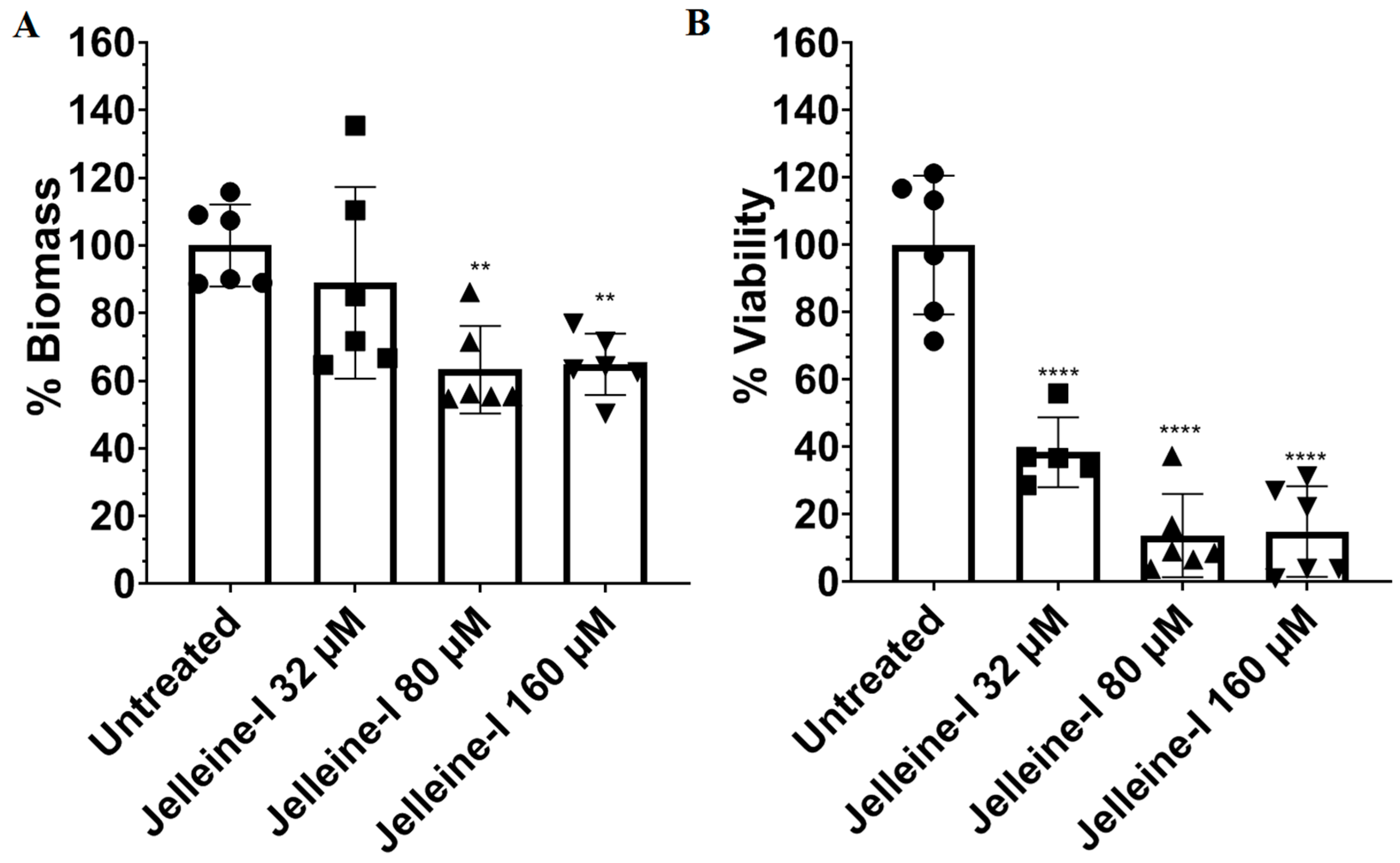
4.9. Anti-Biofilm Assay
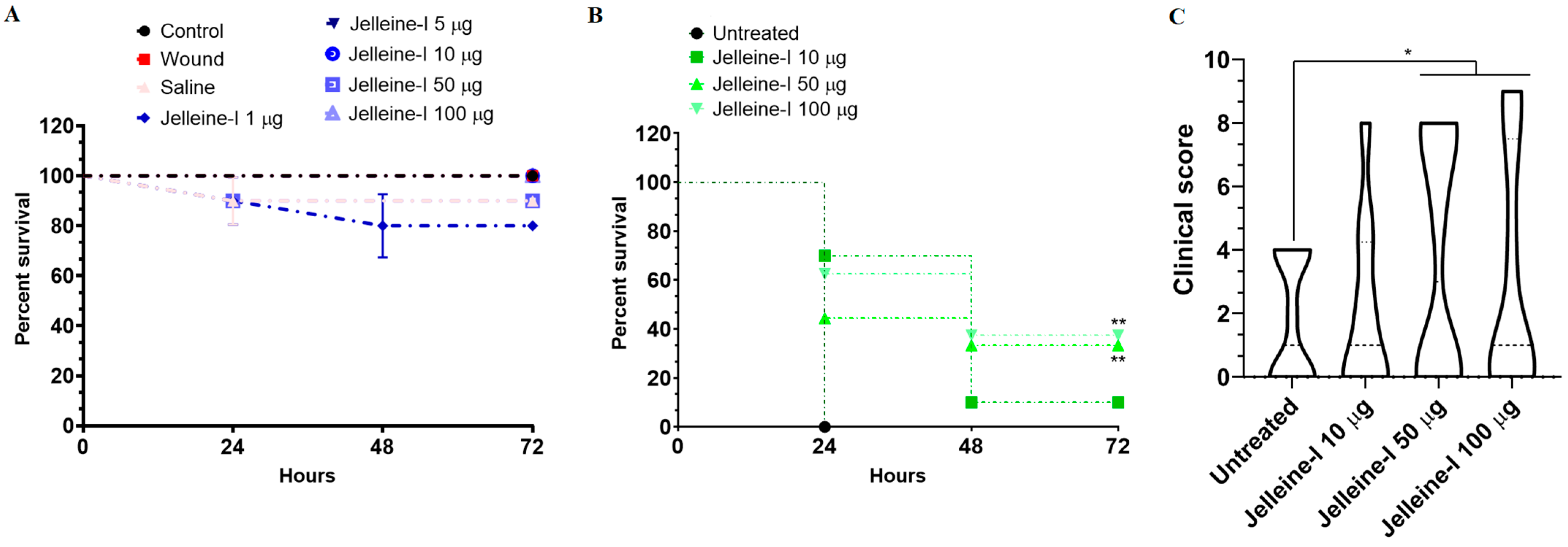
A CRKP inoculum (1 × 10⁶ CFU/mL; isolate 699) was incubated in MHB supplemented with 1 mM glucose at 35 ± 2 °C for 24 h to allow biofilm adhesion and formation. After incubation, the medium was discarded, and wells were washed with sterile saline (0.85% NaCl; three washes). Biofilms were then treated with Jelleine-I at concentrations of 32, 80, or 160 µM, followed by a second 24 h incubation at 35 ± 2 °C. Subsequently, the medium was removed, wells were washed again with sterile saline (three times), and the biofilms were fixed with methanol for 5 min at 37 °C. Fixed biofilms were stained with 0.1% (w/v) crystal violet for 30 min at room temperature, washed with saline (three times), air-dried, and solubilized with 30% glacial acetic acid. Absorbance was measured at 595 nm using a spectrophotometer (Bio-Tek Instruments™, Winooski, VT, USA), and results were expressed as the percentage of biofilm biomass relative to untreated control wells (set as 100%) [39]. In parallel, biofilm cell viability was evaluated using the MTT assay, as described previously [40].

4.10. In Vivo Assay

Invertebrates can provide a valuable alternative to traditional vertebrate animal models for studying the effect of antimicrobial compounds. In this sense, several studies have traditionally used Galleria mellonella larvae as a model to evaluate the antimicrobial properties of various compounds [41]. However, access and availability of G. mellonella larvae is limited in several regions, such as Brazil. To overcome this limitation, some studies have used Tenebrio molitor (Coleoptera: Tenebrionidae) larvae as an alternative, since the widespread use of this species as a nutritional factor for farm animals makes its access significantly easier [42,43,44]. Therefore, we chose to employ a T. molitor larval infection model to study the antibacterial activity of Jelleine-I against a hypervirulent strain of colistin-resistant Klebsiella pneumoniae (isolate 699).
Larvae of Tenebrio molitor (100–120 mg and 2 cm) were purchased from a regional commerce (Alvorada dos PássarosTM, Belo Horizonte, Mg, Brazil). All animals were maintained in plastic containers protected from light and filled with a commercial mixture of seeds and grains. Groups of ten larvae, randomly selected, were separated and placed in petri dishes. In the toxicity test, eight groups (n = 10) were prepared, and the larvae of each group were inoculated with a micro-syringe (HamiltonTM, Reno, NV, USA) containing 5 μL of solutions containing different doses of Jellein-I (1, 5, 10, 50, and 100 μg). A group treated with saline (NaCl 0.85%; 5 μL) and a control group of non-injected larvae were included as a negative control. In addition, a wound control group, in which the needle was inserted without the introduction of any vehicle, was used to evaluate the effect of trauma on larval survival. After the respective treatments, all groups were incubated at 37 °C for 72 h. Larval mortality was monitored daily, and individuals were considered dead if they were immobile, unresponsive to touch, or exhibited melanization, as described by Andrade-Oliveira et al. [42]. In the efficacy assay, each larva was injected with 5 μL of a hypervirulent colistin-resistant K. pneumoniae suspension (108 CFU) using a microsyringe. Two hours post-infection, larvae were treated with Jelleine-I at doses of 10, 50, or 100 μg, or with sterile 0.85% saline (control), then placed in Petri dishes and incubated at 37 °C for 72 h to support bacterial proliferation. Mortality and morbidity were assessed daily. Larval health was evaluated over the 72 h period based on three criteria, activity, melanization, and survival, using the health index scoring system anteriorly described [42].

4.11. Statistical Analysis

Data normality was assessed using the Shapiro–Wilk test. Data with normal distribution were represented as mean ± standard deviation, and data with non-Gaussian distribution were represented as median and interquartile range (P25–P75). One-way analysis of variance (ANOVA) followed by Tukey’s post-test were used to compare differences between data with normal distribution (release of 260 nm-absorbing material, release of proteins, biomass of biofilm, and viability of biofilm). Kruskall–Wallis followed by Dunn’s post-test was used to compare differences between data with non-normal distribution (clinical score). The lethality curves of T. molitor larvae were constructed using the Kaplan–Meier estimator, and the comparison between groups was performed by the log-rank test. All statistical analyses were assessed using GraphPad Prism 5.03 (GraphPad Software Inc. TM, LaJolla, CA, USA), and p values < 0.05 were considered statistically significant.

Author Contributions

Conceptualization, W.G.L. and R.M.R.L.; methodology, W.G.L., R.M.R.L., J.C.M.B. and D.A.G.R.M.; software, W.G.L. and R.M.R.L.; validation, R.M.R.L., W.G.L. and M.E.d.L.; formal analysis, R.M.R.L. and W.G.L.; investigation, R.M.R.L. and W.G.L.; resources, M.E.d.L.; data curation, R.M.R.L. and W.G.L.; writing—original draft preparation, R.M.R.L. and W.G.L.; writing—review and editing, W.G.L., J.C.M.B., R.M.V. and M.E.d.L.; visualization, R.M.R.L. and W.G.L.; supervision, M.E.d.L., J.M.R. and R.M.V.; project administration, W.G.L.; funding acquisition, M.E.d.L., J.M.R. and R.M.V. All authors have read and agreed to the published version of the manuscript.

Funding

Conselho Nacional de Desenvolvimento Científico e Tecnológico (CNPq) [Chamada CNPq 32/2023–Pós-Doutorado Júnior–PDJ 2023; 310638/2023-2–bolsa pesquisa M.E.d.L. Projects nos. 402653/2018-1 and 304337/2019-6]. Fundação de Amparo à Pesquisa do Estado de Minas Gerais (FAPEMIG)–[Projects APQ 01781-17, APQ 02238-17, RED-00185-23, APQ-01848-22 e APQ-00754-24]. Coordenação de Aperfeiçoamento de Pessoal de Nível Superior (CAPES) (CAPES/COFECUB—process 88887.879070/2023-00).

Institutional Review Board Statement

Not applicable.

Informed Consent Statement

Not applicable.

Data Availability Statement

The original contributions presented in this study are included in the article. Further inquiries can be directed to the corresponding author.

Acknowledgments

We also thank the CELAM and LAREMAR facility at UFMG and the João XXIII hospital of Fundação Hospitalar do Estado de Minas Gerais (FHEMIG).

Conflicts of Interest

The authors declare no conflicts of interest.

References

  1. Effah, C.Y.; Sun, T.; Liu, S.; Wu, Y. Klebsiella pneumoniae: An Increasing Threat to Public Health. Ann. Clin. Microbiol. Antimicrob. 2020, 19, 1. [Google Scholar] [CrossRef] [PubMed]
  2. Wyres, K.L.; Lam, M.M.C.; Holt, K.E. Population Genomics of Klebsiella pneumoniae. Nat. Rev. Microbiol. 2020, 18, 344–359. [Google Scholar] [CrossRef]
  3. Bengoechea, J.A.; Sa Pessoa, J. Klebsiella pneumoniae Infection Biology: Living to Counteract Host Defences. FEMS Microbiol. Rev. 2019, 43, 123–144. [Google Scholar] [CrossRef] [PubMed]
  4. Chang, D.; Sharma, L.; Dela Cruz, C.S.; Zhang, D. Clinical Epidemiology, Risk Factors, and Control Strategies of Klebsiella pneumoniae Infection. Front. Microbiol. 2021, 12, 750662. [Google Scholar] [CrossRef] [PubMed]
  5. Reyes, J.; Aguilar, A.C.; Caicedo, A. Carbapenem-Resistant Klebsiella pneumoniae: Microbiology Key Points for Clinical Practice. Int. J. Gen. Med. 2019, 12, 437–446. [Google Scholar] [CrossRef]
  6. Caneiras, C.; Lito, L.; Melo-Cristino, J.; Duarte, A. Community- and Hospital-Acquired Klebsiella pneumoniae Urinary Tract Infections in Portugal: Virulence and Antibiotic Resistance. Microorganisms 2019, 7, 138. [Google Scholar] [CrossRef]
  7. Balkhair, A.; Al-Muharrmi, Z.; Al’Adawi, B.; Al Busaidi, I.; Taher, H.B.; Al-Siyabi, T.; Al Amin, M.; Hassan, K.S. Prevalence and 30-Day All-Cause Mortality of Carbapenem-and Colistin-Resistant Bacteraemia Caused by Acinetobacter baumannii, Pseudomonas aeruginosa, and Klebsiella pneumoniae: Description of a Decade-Long Trend. Int. J. Infect. Dis. 2019, 85, 10–15. [Google Scholar] [CrossRef]
  8. Karami-Zarandi, M.; Rahdar, H.A.; Esmaeili, H.; Ranjbar, R. Klebsiella pneumoniae: An Update on Antibiotic Resistance Mechanisms. Future Microbiol. 2023, 18, 65–81. [Google Scholar] [CrossRef]
  9. Lima, W.G.; de Lima, M.E. Therapeutic Prospection of Animal Venoms-Derived Antimicrobial Peptides against Infections by Multidrug-Resistant Acinetobacter baumannii: A Systematic Review of Pre-Clinical Studies. Toxins 2023, 15, 268. [Google Scholar] [CrossRef]
  10. Bucataru, C.; Ciobanasu, C. Antimicrobial Peptides: Opportunities and Challenges in Overcoming Resistance. Microbiol. Res. 2024, 286, 127822. [Google Scholar] [CrossRef]
  11. Bechinger, B.; Gorr, S.-U. Antimicrobial Peptides: Mechanisms of Action and Resistance. J. Dent. Res. 2017, 96, 254–260. [Google Scholar] [CrossRef]
  12. Sahoo, A.; Swain, S.S.; Behera, A.; Sahoo, G.; Mahapatra, P.K.; Panda, S.K. Antimicrobial Peptides Derived From Insects Offer a Novel Therapeutic Option to Combat Biofilm: A Review. Front. Microbiol. 2021, 12, 661195. [Google Scholar] [CrossRef] [PubMed]
  13. Zhou, J.; Zhang, L.; He, Y.; Liu, K.; Zhang, F.; Zhang, H.; Lu, Y.; Yang, C.; Wang, Z.; Fareed, M.S.; et al. An Optimized Analog of Antimicrobial Peptide Jelleine-1 Shows Enhanced Antimicrobial Activity against Multidrug Resistant P. aeruginosa and Negligible Toxicity in Vitro and in Vivo. Eur. J. Med. Chem. 2021, 219, 113433. [Google Scholar] [CrossRef] [PubMed]
  14. Capparelli, R.; De Chiara, F.; Nocerino, N.; Montella, R.C.; Iannaccone, M.; Fulgione, A.; Romanelli, A.; Avitabile, C.; Blaiotta, G.; Capuano, F. New Perspectives for Natural Antimicrobial Peptides: Application as Antinflammatory Drugs in a Murine Model. BMC Immunol. 2012, 13, 61. [Google Scholar] [CrossRef] [PubMed]
  15. Castro, A.P.; Brito, J.C.M.; Candido, W.A.B.; Félix, A.S.; Verly, R.M.; Resende, J.M.; Lopes-de-Souza, L.; Chávez-Olórtegui, C.; Fernandes, S.O.A.; Cardoso, V.N. Jelleine-I Membrane Interaction-Related Biological Properties and Antimicrobial Activity against MDR, XDR, and PDR-Acinetobacter baumannii Clinical Isolates. ACS Omega 2025, 10, 10938–10948. [Google Scholar] [CrossRef]
  16. Nang, S.C.; Han, M.-L.; Yu, H.H.; Wang, J.; Torres, V.V.L.; Dai, C.; Velkov, T.; Harper, M.; Li, J. Polymyxin Resistance in Klebsiella pneumoniae: Multifaceted Mechanisms Utilized in the Presence and Absence of the Plasmid-Encoded Phosphoethanolamine Transferase Gene Mcr-1. J. Antimicrob. Chemother. 2019, 74, 3190–3198. [Google Scholar] [CrossRef]
  17. Lima, W.G.; Brito, J.C.M.; Verly, R.M.; de Lima, M.E. Jelleine, a Family of Peptides Isolated from the Royal Jelly of the Honey Bees (Apis mellifera), as a Promising Prototype for New Medicines: A Narrative Review. Toxins 2024, 16, 24. [Google Scholar] [CrossRef]
  18. Jia, F.; Wang, J.; Zhang, L.; Zhou, J.; He, Y.; Lu, Y.; Liu, K.; Yan, W.; Wang, K. Multiple Action Mechanism and in Vivo Antimicrobial Efficacy of Antimicrobial Peptide Jelleine-I. J. Pept. Sci. 2021, 27, e3294. [Google Scholar] [CrossRef]
  19. Kim, S.R.; Choi, K.-H.; Kim, K.-Y.; Kwon, H.-Y.; Park, S.-W. Development of a Novel Short Synthetic Antibacterial Peptide Derived from the Swallowtail Butterfly Papilio xuthus Larvae. J. Microbiol. Biotechnol. 2020, 30, 1305–1309. [Google Scholar] [CrossRef]
  20. Fontana, R.; Mendes, M.A.; de Souza, B.M.; Konno, K.; César, L.M.M.; Malaspina, O.; Palma, M.S. Jelleines: A Family of Antimicrobial Peptides from the Royal Jelly of Honeybees (Apis mellifera). Peptides 2004, 25, 919–928. [Google Scholar] [CrossRef]
  21. Brazilian Committee on Antimicrobial Susceptibility Testing (BrCAST). Tabelas de Pontos de Corte Para Interpretação de CIMs e Diâmetros de Halos. 2025. Available online: https://brcast.org.br/wp-content/uploads/2022/09/Tabela-pontos-de-corte-BrCAST-13-04-2024.pdf (accessed on 12 June 2025).
  22. Yu, V.L.; Hansen, D.S.; Ko, W.C.; Sagnimeni, A.; Klugman, K.P.; von Gottberg, A.; Goossens, H.; Wagener, M.M.; Benedi, V.J. International Klebseilla Study Group Virulence Characteristics of Klebsiella and Clinical Manifestations of K. pneumoniae Bloodstream Infections. Emerg. Infect. Dis. 2007, 13, 986–993. [Google Scholar] [CrossRef] [PubMed]
  23. Jia, F.; Wang, J.; Peng, J.; Zhao, P.; Kong, Z.; Wang, K.; Yan, W.; Wang, R. The in Vitro, in Vivo Antifungal Activity and the Action Mode of Jelleine-I against Candida Species. Amino Acids 2018, 50, 229–239. [Google Scholar] [CrossRef]
  24. Zhou, Y.; Zhang, J.; Chen, Y.; Wu, J.; Guo, B.; Wu, X.; Zhang, Y.; Wang, M.; Ya, R.; Huang, H. Combined PK/PD Index May Be a More Appropriate PK/PD Index for Cefoperazone/Sulbactam against Acinetobacter baumannii in Patients with Hospital-Acquired Pneumonia. Antibiotics 2022, 11, 703. [Google Scholar] [CrossRef]
  25. Cabrera, M.P.d.S.; Baldissera, G.; Silva-Gonçalves, L.d.C.; Souza, B.M.d.; Riske, K.A.; Palma, M.S.; Ruggiero, J.R.; Arcisio-Miranda, M. Combining Experimental Evidence and Molecular Dynamic Simulations to Understand the Mechanism of Action of the Antimicrobial Octapeptide Jelleine-I. Biochemistry 2014, 53, 4857–4868. [Google Scholar] [CrossRef]
  26. Yang, S.; Wang, H.; Zhao, D.; Zhang, S.; Hu, C. Polymyxins: Recent Advances and Challenges. Front. Pharmacol. 2024, 15, 1424765. [Google Scholar] [CrossRef] [PubMed]
  27. Lima, W.G.; Alves, M.C.; Cruz, W.S.; Paiva, M.C. Chromosomally Encoded and Plasmid-Mediated Polymyxins Resistance in Acinetobacter baumannii: A Huge Public Health Threat. Eur. J. Clin. Microbiol. Infect. Dis. 2018, 37, 1009–1019. [Google Scholar] [CrossRef] [PubMed]
  28. Liu, L.; Li, S.; Zhu, W.; Bao, Q.; Liang, Y.; Zhao, T.; Li, X.; Zhou, J. Study on the Mechanism of ROS-Induced Oxidative Stress Injury and the Broad-Spectrum Antimicrobial Performance of Nickel Ion-Doped V6O13 Powder. Sci. Rep. 2024, 14, 22374. [Google Scholar] [CrossRef] [PubMed]
  29. Donlan, R.M. Biofilms: Microbial Life on Surfaces. Emerg. Infect. Dis. 2002, 8, 881–890. [Google Scholar] [CrossRef]
  30. Mittal, S.; Sharma, M.; Chaudhary, U. Biofilm and Multidrug Resistance in Uropathogenic Escherichia coli. Pathog. Glob. Health 2015, 109, 26–29. [Google Scholar] [CrossRef]
  31. Jia, F.; Zhang, Y.; Wang, J.; Peng, J.; Zhao, P.; Zhang, L.; Yao, H.; Ni, J.; Wang, K. The Effect of Halogenation on the Antimicrobial Activity, Antibiofilm Activity, Cytotoxicity and Proteolytic Stability of the Antimicrobial Peptide Jelleine-I. Peptides 2019, 112, 56–66. [Google Scholar] [CrossRef]
  32. Shen, P.; Ding, K.; Wang, L.; Tian, J.; Huang, X.; Zhang, M.; Dang, X. In Vitro and in Vivo Antimicrobial Activity of Antimicrobial Peptide Jelleine-I against Foodborne Pathogen Listeria monocytogenes. Int. J. Food Microbiol. 2023, 387, 110050. [Google Scholar] [CrossRef]
  33. Yasir, M.; Willcox, M.D.P.; Dutta, D. Action of Antimicrobial Peptides against Bacterial Biofilms. Materials 2018, 11, 2468. [Google Scholar] [CrossRef] [PubMed]
  34. CLSI M07; Methods for Dilution Antimicrobial Susceptibility Tests for Bacteria That Grow Aerobically. Clinical and Laboratory Standards Institute: Wayne, PA, USA, 2018.
  35. Lima, W.G.; de Brito, J.C.M.; Cardoso, V.N.; Fernandes, S.O.A. In-Depth Characterization of Antibacterial Activity of Melittin against Staphylococcus aureus and Use in a Model of Non-Surgical MRSA-Infected Skin Wounds. Eur. J. Pharm. Sci. 2021, 156, 105592. [Google Scholar] [CrossRef]
  36. Kielkopf, C.L.; Bauer, W.; Urbatsch, I.L. Bradford Assay for Determining Protein Concentration. Cold Spring Harb. Protoc. 2020, 2020, 102269. [Google Scholar] [CrossRef] [PubMed]
  37. Zai, Y.; Ying, Y.; Ye, Z.; Zhou, M.; Ma, C.; Shi, Z.; Chen, X.; Xi, X.; Chen, T.; Wang, L. Broad-Spectrum Antimicrobial Activity and Improved Stability of a D-Amino Acid Enantiomer of DMPC-10A, the Designed Derivative of Dermaseptin Truncates. Antibiotics 2020, 9, 627. [Google Scholar] [CrossRef] [PubMed]
  38. Araújo-Neto, L.N.; de Lima, M.d.C.A.; de Oliveira, J.F.; de Souza, E.R.; Feitosa Machado, S.E.; de Souza Lima, G.M.; Silva Buonafina, M.D.; Brayner, F.A.; Alves, L.C.; Sandes, J.M.; et al. Thiophene-Thiosemicarbazone Derivative (L10) Exerts Antifungal Activity Mediated by Oxidative Stress and Apoptosis in C. albicans. Chem. Biol. Interact. 2020, 320, 109028. [Google Scholar] [CrossRef]
  39. Herrera, K.M.S.; Lopes, G.F.M.; Oliveira, M.E.; Sousa, J.F.; Lima, W.G.; Silva, F.K.; Brito, J.C.M.; Gomes, A.J.P.S.; Viana, G.H.R.; Soares, A.C.; et al. A 3-Alkylpyridine-Bearing Alkaloid Exhibits Potent Antimicrobial Activity against Methicillin-Resistant Staphylococcus aureus (MRSA) with No Detectable Resistance. Microbiol. Res. 2022, 261, 127073. [Google Scholar] [CrossRef]
  40. Trafny, E.A.; Lewandowski, R.; Zawistowska-Marciniak, I.; Stępińska, M. Use of MTT Assay for Determination of the Biofilm Formation Capacity of Microorganisms in Metalworking Fluids. World J. Microbiol. Biotechnol. 2013, 29, 1635–1643. [Google Scholar] [CrossRef]
  41. Barton, T.E.; Duignan, L.; Kadioglu, A.; Fothergill, J.L.; Neill, D.R. Galleria mellonella as an Antimicrobial Screening Model. J. Vis. Exp. 2024, 212, e67210. [Google Scholar] [CrossRef]
  42. Andrade-Oliveira, A.L.; Lacerda-Rodrigues, G.; Pereira, M.F.; Bahia, A.C.; Machado, E.d.A.; Rossi, C.C.; Giambiagi-deMarval, M. Tenebrio molitor as a Model System to Study Staphylococcus spp. Virulence and Horizontal Gene Transfer. Microb. Pathog. 2023, 183, 106304. [Google Scholar] [CrossRef]
  43. Canteri de Souza, P.; Custódio Caloni, C.; Wilson, D.; Sergio Almeida, R. An Invertebrate Host to Study Fungal Infections, Mycotoxins and Antifungal Drugs: Tenebrio molitor. J. Fungi 2018, 4, 125. [Google Scholar] [CrossRef] [PubMed]
  44. de Souza, P.C.; Morey, A.T.; Castanheira, G.M.; Bocate, K.P.; Panagio, L.A.; Ito, F.A.; Furlaneto, M.C.; Yamada-Ogatta, S.F.; Costa, I.N.; Mora-Montes, H.M.; et al. Tenebrio molitor (Coleoptera: Tenebrionidae) as an Alternative Host to Study Fungal Infections. J. Microbiol. Methods 2015, 118, 182–186. [Google Scholar] [CrossRef] [PubMed]
Figure 1. Time-kill curve with multiple concentrations of Jelleine-I (50, 100, and 200 µM) against a colistin-resistant Klebsiella pneumoniae lineage (isolate 699). The plot shows the number of logarithmic colony-forming units per milliliter (Log10 CFU/mL). Bacterial cells untreated were used as negative control (black circles).
Toxins 17 00325 g001
Figure 2. Determination of bacteriolysis through of assay of overflow of 260 nm-absorbing materials (DNA/RNA) (A) and through a test of release of proteins (B). The experiment was done in triplicate for statistical significance. One asterisk (*) indicates statistically different compared to the control with 0.05 < p-value < 0.01. Two asterisks (**) indicate statistically different compared to the control with 0.01 < p-value < 0.001. Three asterisks (***) indicate statistically different compared to the control with 0.001 < p-value < 0.0001. Four asterisks (****) indicate statistically different compared to the control with p-value < 0.0001. The results were analyzed by one-way variance analysis (ANOVA) with Dunnett post-hoc.
Toxins 17 00325 g002
Figure 3. Effect of different concentrations of Jelleine-I (32, 80, and 160 µM) on biomass (A) and viability (B) of biofilms formed by a hypervirulent colistin-resistant Klebsiella pneumoniae (isolate 699). The experiment was done in sextuplicate for statistical significance. Two asterisks (**) indicate statistically different compared to the control with 0.01 < p-value < 0.001. Four asterisks (****) indicate statistically different compared to the control with p-value < 0.0001. The results were analyzed by one-way variance analysis (ANOVA) with Dunnett post-hoc.
Toxins 17 00325 g003
Figure 4. Survival curves and clinical score of Tenebrio molitor larvae. (A) Survival of larvae after administration of different doses of Jelleine-I (1, 5, 10, 50, and 100 µg) to assess the in vivo toxicity of the peptide. (B) Survival curve of larvae infected with a hypervirulent clinical isolate of colistin-resistant Klebsiella pneumoniae (CRKP; isolate 699). Groups (n = 10) infected with CRKP and treated with Jelleine-I (10, 50, and 100 µg) after 2 h post-infection. Larvae (n = 10) infected and injected with sterile saline (0.9% NaCl) were used as the negative control. (C) Clinical score of larvae infected and treated with Jelleine-I (10, 50, and 100 µg) or saline. One asterisk (*) indicates statistically different compared to the control with 0.05 < p < 0.01. The lethality curves were constructed using the Kaplan–Meier estimator and statistically analyzed using the log-rank test. The clinical score results were analyzed using the Kruskal–Wallis test with Dunn’s post-test. The results are expressed as median and interquartile range (P25–P75).
Toxins 17 00325 g004
Table 1. Values of minimum inhibitory concentration (MIC) and minimum bactericidal concentration (MBC) of Jelleine-I peptide against clinical isolates of colistin-resistant Klebsiella pneumoniae.
MicroorganismsOriginCarbapenem Resistance aHypermucoviscity Phenotype bJelleine-I (µM)Colistin (µg/mL)
MICMBCMICProfile c
K. pneumoniae ATCC 700603UrineNoNo321280.25Sensible
K. pneumoniae ATCC 43816SecretionNoYes321280.5Sensible
K. pneumoniae 17SecretionYesYes646432Resistant
K. pneumoniae 27SecretionYesNo64>12864Resistant
K. pneumoniae 68SecretionYesYes16328Resistant
K. pneumoniae 76SecretionYesYes646432Resistant
K. pneumoniae 104UrineYesYes646464Resistant
K. pneumoniae 106UrineYesNo128128>128Resistant
K. pneumoniae 110UrineYesYes646432Resistant
K. pneumoniae 135BloodYesYes6412864Resistant
K. pneumoniae 139BloodYesYes6412864Resistant
K. pneumoniae 169UrineYesNo646432Resistant
K. pneumoniae 242SecretionYesNo12812832Resistant
K. pneumoniae 260SecretionYesNo646416Resistant
K. pneumoniae 289BloodYesNo6412832Resistant
K. pneumoniae 395BloodYesNo6412832Resistant
K. pneumoniae 627SecretionYesNo64>12832Resistant
K. pneumoniae 642SecretionYesNo646432Resistant
K. pneumoniae 689BloodYesYes6412832Resistant
K. pneumoniae 699BloodYesYes64128128Resistant
a Resistance to carbapenems was confirmed by the presence of visible growth (turbidity) in culture medium containing 8 µg/mL of meropenem [21]. b The method of the International Klebsiella pneumoniae Study Group was adopted [22]. c The resistance profile was judged according to the cutoff points established in the Brazilian Committee on Antimicrobial Susceptibility Testing (BrCAST), because the bacterial isolates included in this study were recovered from a tertiary hospital localized in southeastern Brazil. Thus, we consider colistin resistance to be assumed when the MIC is >2 µg/mL [21].
Table 2. Influence of divalent cations (Ca2+ and Mg2+) and ascorbic acid (100 µg/mL) on the values of minimum inhibitory concentration (MIC; µg/mL) of Jelleine-I or colistin against a hypervirulent clinical isolate of colistin-resistant Klebsiella pneumoniae (isolate 699).
ControlCa2+Mg2+Ascorbic Acid 100 µg/mL
20 µM50 µM100 µM20 µM50 µM100 µM
Jelleine-I1616161616161616
Colistin8816>64816>648
Disclaimer/Publisher’s Note: The statements, opinions and data contained in all publications are solely those of the individual author(s) and contributor(s) and not of MDPI and/or the editor(s). MDPI and/or the editor(s) disclaim responsibility for any injury to people or property resulting from any ideas, methods, instructions or products referred to in the content.

