Article

Lactobacillus fermentum Alleviates the Colorectal Inflammation Induced by Low-Dose Sub-Chronic Microcystin-LR Exposure

1
The Key Laboratory of Typical Environmental Pollution and Health Hazards of Hunan Province, Department of Epidemiology and Health Statistics, School of Public Health, Hengyang Medical School, University of South China, Hengyang 421001, China
2
Hunan Provincial Key Laboratory of Clinical Epidemiology, Xiangya School of Public Health, Central South University, Changsha 410017, China
3
Changsha Yuhua District Center for Disease Control and Prevention, Changsha 410014, China
4
Changsha Center for Disease Control and Prevention, Changsha 410004, China
5
Hengyang Medical School, The First Affiliated Hospital, University of South China, Hengyang 421001, China
*
Author to whom correspondence should be addressed.
Toxins 2023, 15(9), 579; https://doi.org/10.3390/toxins15090579
Submission received: 3 August 2023 / Revised: 30 August 2023 / Accepted: 13 September 2023 / Published: 19 September 2023

Abstract

:
Microcystin-LR (MC-LR) contamination is a worldwide environmental problem that poses a grave threat to the water ecosystem and public health. Exposure to MC-LR has been associated with the development of intestinal injury, but there are no effective treatments for MC-LR-induced intestinal disease. Probiotics are “live microorganisms that are beneficial to the health of the host when administered in sufficient quantities”. It has been demonstrated that probiotics can prevent or treat a variety of human diseases; however, their ability to mitigate MC-LR-induced intestinal harm has not yet been investigated. The objective of this study was to determine whether probiotics can mitigate MC-LR-induced intestinal toxicity and its underlying mechanisms. We first evaluated the pathological changes in colorectal tissues using an animal model with sub-chronic exposure to low-dose MC-LR, HE staining to assess colorectal histopathologic changes, qPCR to detect the expression levels of inflammatory factors in colorectal tissues, and WB to detect the alterations on CSF1R signaling pathway proteins in colorectal tissues. Microbial sequencing analysis and screening of fecal microorganisms differential to MC-LR treatment in mice. To investigate the role of microorganisms in MC-LR-induced colorectal injury, an in vitro model of MC-LR co-treatment with microorganisms was developed. Our findings demonstrated that MC-LR treatment induced an inflammatory response in mouse colorectal tissues, promoted the expression of inflammatory factors, activated the CSF1R signaling pathway, and significantly decreased the abundance of Lactobacillus. In a model of co-treatment with MC-LR and Lactobacillus fermentum (L. fermentum), it was discovered that L. fermentum substantially reduced the incidence of the colorectal inflammatory response induced by MC-LR and inhibited the protein expression of the CSF1R signaling pathway. This is the first study to suggest that L. fermentum inhibits the CSF1R signaling pathway to reduce the incidence of MC-LR-induced colorectal inflammation. This research may provide an excellent experimental foundation for the development of strategies for the prevention and treatment of intestinal diseases in MC-LR.
Key Contribution: Lactobacillus fermentans alleviates the colorectal injury caused by MC-LR by inhibiting CSF1R signaling pathway.

1. Introduction

In recent years, the eutrophication of water bodies with nitrogen and phosphorus and the intensification of the global warming trend have led to frequent outbreaks of cyanobacterial blooms in many regions of the world, and the release of a variety of highly active and toxic cyanobacterial toxins is one of the main dangers associated with blooms [1,2]. Cyanobacteria and cyanobacterial toxin pollution has been a significant environmental issue in China and the rest of the world for quite some time, with Microcystin-LR (MC-LR) being the cyanobacterial toxin with the widest distribution, highest production, and most severe toxicity [3,4,5]. In addition to being detected in natural water bodies such as rivers and lakes, MC-LR has also been detected in water factory water, terminal water, and bottled/barrel water. Furthermore, MC-LR can be enriched in a variety of agricultural and aquatic products, making the current pollution situation extremely grave [6,7]. Extremely hazardous MC-LR can be ingested through drinking water and aquatic products, causing injury to multiple organs, including the liver, kidneys, intestines, nerves, immune system, and reproductive organs [8,9]. MCs typically enter the human body via the oral route, such as drinking water and food, and the gastrointestinal tract is the first organ to ingest and be exposed to MCs [8]. Using a retrospective cohort study, Zhou et al. discovered that the level of exposure to MC-LR was positively correlated with the incidence of colorectal cancer [10]. Sedan et al. found that MC-LR significantly decreased intraepithelial lymphocytes in the intestine [11]. Low doses of MC-LR induced severe inflammatory responses and fibrogenesis in mouse intestines, according to our previous research [8].
There are 100 trillion microorganisms (including bacteria, viruses, fungi, and protists) in the human gut. These microorganisms can attach to the epithelium or mucus layer and produce biofilms that have a significant impact on host development [12]. Alterations in the intestinal microbiota are strongly linked to a wide range of diseases, particularly intestinal disorders. In a 96 h study conducted by Zhang et al., adult male crayfish were exposed to varying concentrations of MC-LR. After exposure to MC-LR, the abundance of bacteria such as Firmicutes and Planctomycetes at the phylum level and bacteria such as Dysgonomonas, Brevundimonas, and Anaerorhabdus at the genus level were significantly altered in crayfish intestines. In crayfish, the elimination of gut microorganisms can cause metabolic and genetic abnormalities related to gut microorganisms [13]. Li et al. discovered that exposure of African clawed toads to MC-LR reduced the abundance of Firmicutes and Bacteroidetes in their intestinal tracts, while Fusobacterium was significantly enriched [14]. Ding et al. discovered that MC-LR treatment decreased the relative abundance of zebrafish Vibrio bacteria while increasing the relative abundance of Methylobacterium and Pseudocardia bacteria [15]. MC-LR affects the diversity and abundance of intestinal flora, leading to intestinal microecological dysregulation, impaired nutrient absorption and metabolism, the inhibition of intestinal probiotic growth, the promotion of pathogenic bacterial colonization, and the induction of host intestinal diseases. No study has investigated the role of probiotics in MC-LR-induced intestinal injury.
Probiotics are “live microorganisms that are beneficial to the health of the host when administered in sufficient quantities” [16]. These probiotics are used to treat a variety of conditions, such as constipation, diarrhea, irritable bowel syndrome, allergies, obesity, diabetes, and cancer, among others [17]. Numerous studies have shown that probiotics play a crucial role in preventing or treating intestinal diseases. Several studies have demonstrated that probiotics can enhance the intestinal mucosal auto-preventive mechanism, antagonize the pathogenic effects of pathogenic bacteria, regulate the release of inflammatory factors, and quickly restore the intestinal flora [18]. Clostridium difficile spp. affects the accumulation of intraepithelial lymphocytes (IELs) and increases the number of regulatory T cells (Treg cells) in the colon, which downregulates the expression of inflammatory cytokines (TNF-, IL-12, IFN-, IL-1, and IL-6) and upregulates the expression of inhibitory cytokines (IL-10). Through inhibiting histone deacetylase (HDAC), butyrate produced by Clostridium butyricum can directly promote the differentiation of Tregs [19,20]. Recent evidence suggests that probiotics can influence immune regulation through modulating dendritic cell (DC) maturation and the production of tolerogenic DCs (tolDCs), which may then inhibit inflammatory responses. PSA from outer membrane vesicles (OMVs) derived from Bifidobacterium fragilis ameliorates DSS-induced colitis in rodents through affecting DCs [21]. MUC-2 levels are positively correlated with Akkermansia abundance in rodents with DSS-induced colitis, and Akkermansia supplementation can strengthen the colonic mucus barrier through increasing the thickness of the colonic mucus layer [22]. In conclusion, probiotics and their metabolites can modulate intestinal immune function and intestinal permeability as well as reduce intestinal inflammatory response via multiple mechanisms. Using an animal model, Zhao et al. demonstrated for the first time that a probiotic mixed flora (containing, among others, Lactobacillus rhamnosus and Bifidobacterium bifidum) ameliorated the hepatotoxicity of MC-LR and acted as an antidote [23]. To date, no research has examined whether probiotics have the same protective effect against MC-LR-induced intestinal toxicity.
CSF1R is a transmembrane tyrosine/serine kinase receptor expressed normally in mononuclear phagocytes. CSF1R is widely believed to be activated by autocrine or paracrine mechanisms and to play a central role in numerous diseases, including chronic inflammatory disorders, bone diseases, and tumors [24,25]. Long-term exposure to low-dose MC-LR activated the CSF1R/Rap1b signaling pathway through inducing oxidative stress and promoted the release of inflammatory and fibrosis-related factors, which led to the development of chronic inflammation and fibrosis in the colorectal tissue [8]. The CSF1R signaling pathway plays a crucial role in intestinal injury induced by MC-LR. Therefore, in this study, we also investigated the expression of proteins associated with the CSF1R signaling pathway to determine if the microorganisms we screened could influence MC-LR-induced colorectal injury via this crucial signaling pathway.
In this investigation, mice were exposed to low-dose MC-LR for six months in order to analyze and screen mouse feces for different microorganisms using microbial sequencing. And for the first time, the mitigating effect of microorganisms (particularly probiotics) on colorectal injury induced by low-dose sub-chronic MC-LR exposure was investigated using an in vitro model of microorganism and MC-LR co-treatment. This research may provide a solid theoretical and laboratory foundation for the development of preventative and therapeutic strategies for MC-LR-induced intestinal diseases.

2. Results

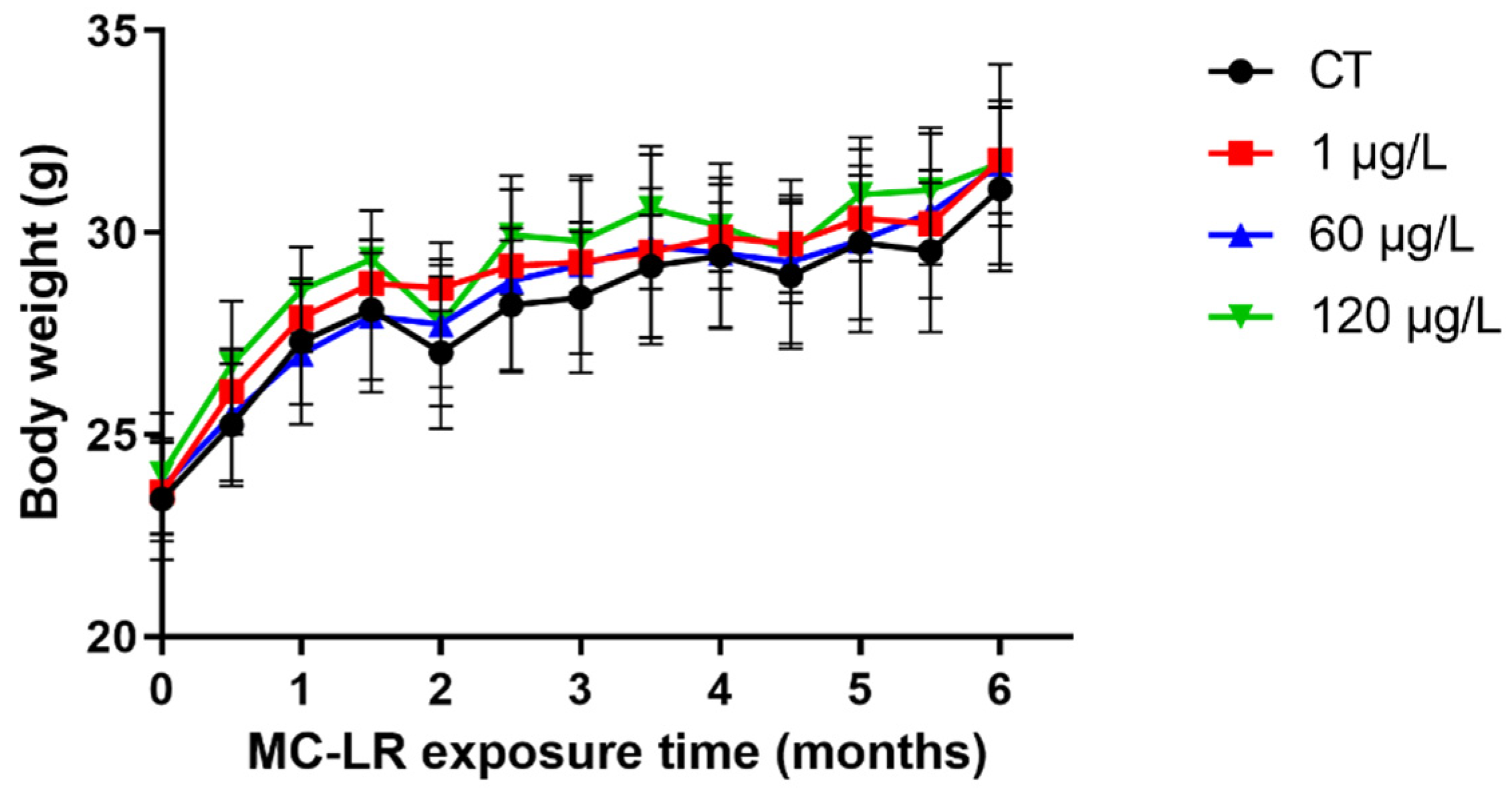
2.1. The Body Weight of MC-LR-Induced Mice

Throughout the duration of exposure, body weight was measured every two weeks. Neither mouse mortality nor abnormal behavior was observed during the 6-month investigation. As shown in Figure 1, the MC-LR group had no effect on the weight change of mice.

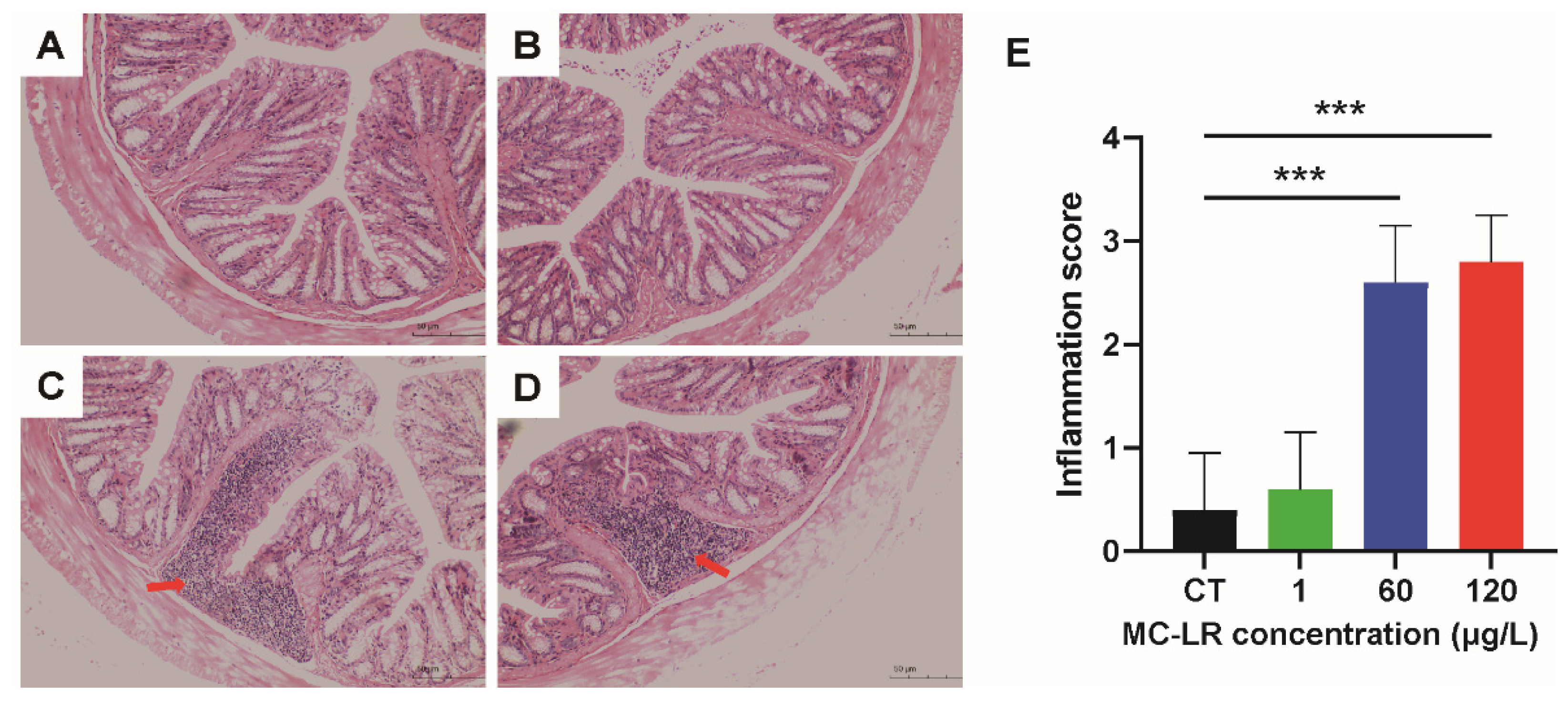
2.2. Histopathology Damage of MC-LR-Induced Mice

Each mouse’s colorectal tissue was stained with Hematoxylin-Eosin (HE), and Figure 2 depicts the pathological alterations of colorectal tissue. MC-LR can induce lymphocyte infiltration and inflammatory response in colorectal tissues relative to the control (CT) group.

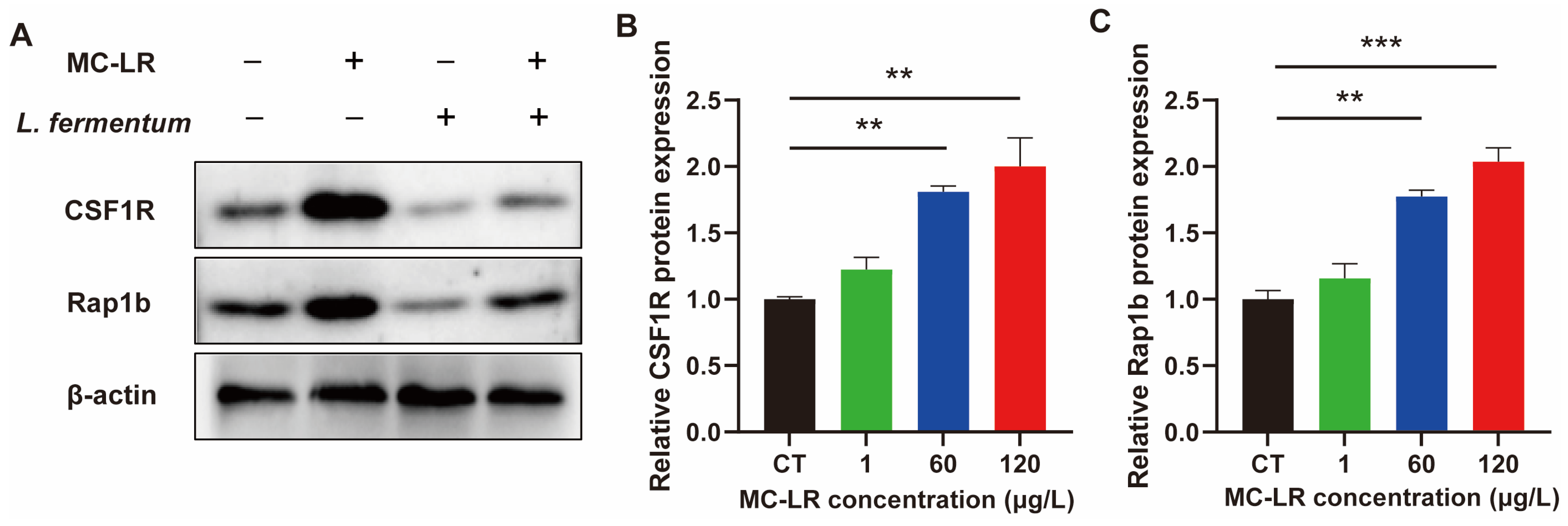
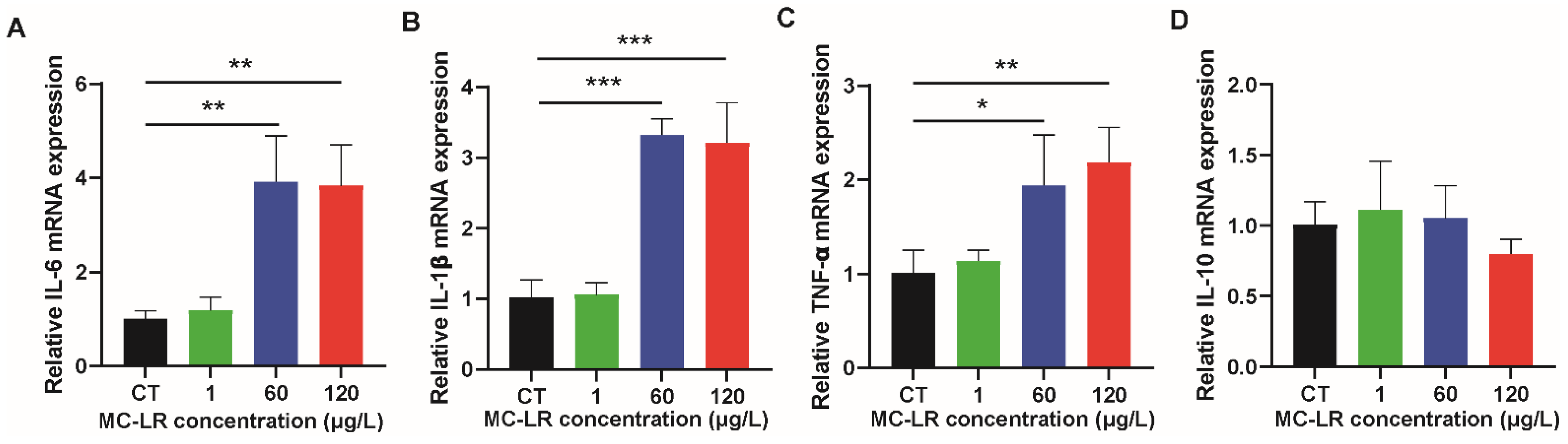
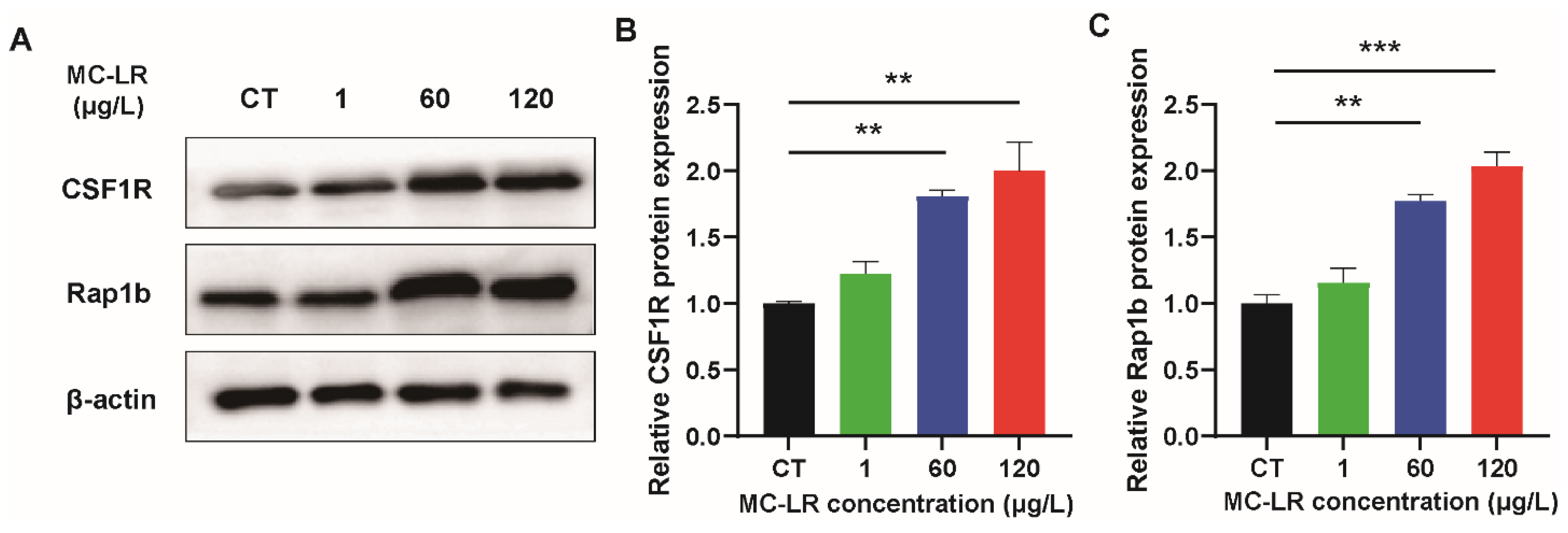
2.3. The Expression of Inflammatory Factors and CSF1R/Rap1b Signaling Pathway-Related Proteins in MC-LR-Induced Mice

In order to further verify the occurrence of colorectal tissue inflammation caused by MC-LR-treatment, we detected the expression level of inflammatory factors in colorectal tissue of mice via qPCR. The results are illustrated in Figure 3. In the MC-LR treatment groups, the expression of inflammatory factors (IL-6, TNF-α, and IL-1β) was increased relative to the CT group.
In our previous studies, it was found that MC-LR can induce chronic inflammation in colorectal through activating CSF1R/Rap1b signaling pathway [8]. Therefore, in this study, we verified the changes in the expression levels of CSF1R and Rap1b. Figure 4 demonstrates the outcomes. The expression of CSF1R and Rap1b were significantly up-regulated in the MC-LR + HFD treatment groups compared to the CT group.

2.4. Gut Microbiome Alterations of MC-LR-Induced Mice

Bacteroidetes and Firmicutes were the most represented groups in terms of phylum classification (Figure 5A). The twenty most common genera accounted for approximately 20% of all microorganisms (Figure 5B). To investigate the specific bacterial taxa associated with MC-LR exposure, the greatest taxonomic differences at the genus level were represented by the LDA score (Figure 5C). At the genus level, the Lactobacillus decreased in the MC-LR treatment groups (Figure 5D).

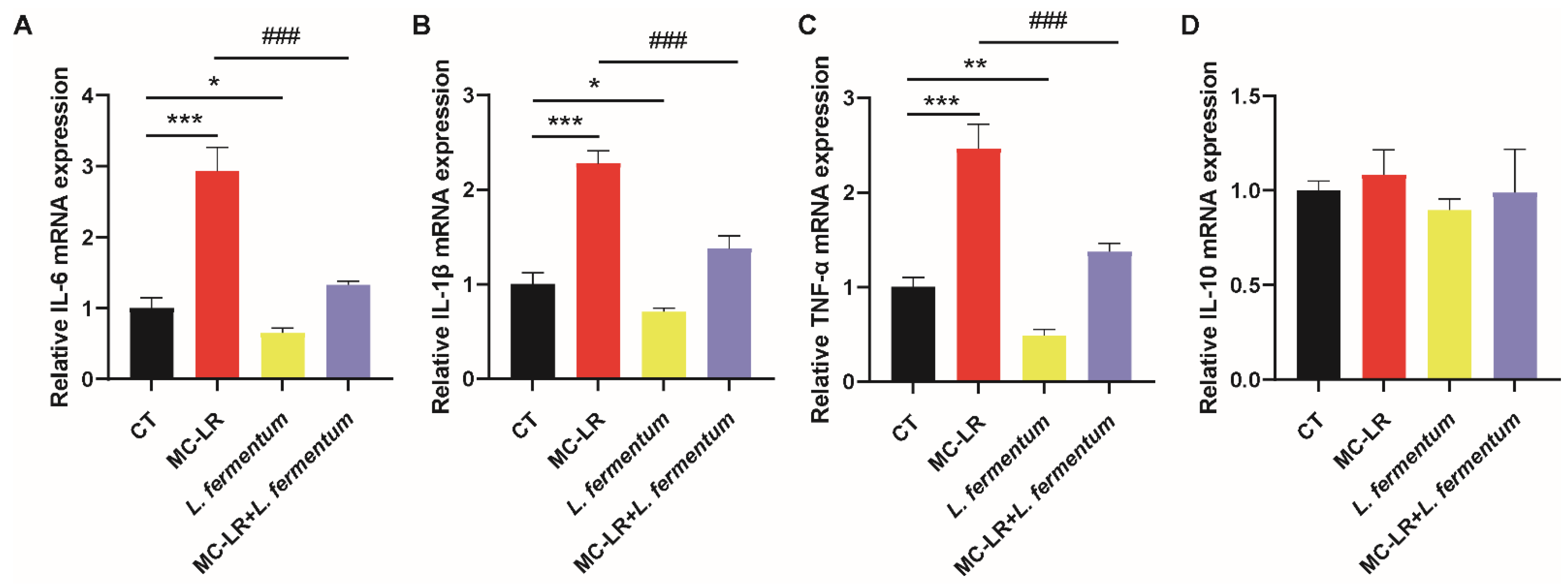
2.5. Effect of Lactobacillus fermentum (L. fermentum) Treatment on the Expression of CSF1R/Rap1b Signaling Pathway-Related Proteins and the Inflammatory Factors in MC-LR-Treated Cell Model

In addition, using normal colonic epithelial NCM460 cells, we conducted a preliminary test to determine whether L. fermentum can ameliorate the colorectal damage caused by MC-LR in an in vitro model. qPCR analysis revealed a significant upregulation of CSF1R/Rap1b and inflammatory factors (IL-6, TNF-α, and IL-1β) in the MC-LR-treated group (Figure 6). In contrast, after L. fermentum treatment, these expression level changes were substantially attenuated (Figure 7).

3. Discussion

In our dyeing experiments conducted on mice with sub-chronic exposure to low doses of MC-LR, we observed no significant difference in the trend of body weight gain between the MC-LR-treated group and the control group, indicating that sub-chronic exposure to low doses of MC-LR has no effect on mouse body weight. Consistent with our findings, Su et al. found that MC-LR gavage at 1000 g/kg for seven days had no effect on body weight changes in rodents compared to controls [26,27]. Similarly, Pan et al. found that mice exposed to varying concentrations of MC-LR for 90 days did not experience a significant decrease in body weight [28]. The failure of long-term exposure to low-dose MC-LR to cause weight loss in mice may be due to the fact that the dose was low and had not yet reached the threshold that could lead to changes in energy metabolism and other effects, or that low-dose stimulation and activation of the protective mechanism of the mice resulted in a tolerance. In addition, we evaluated the histopathological changes in the colorectum and found that mice with sub-chronic exposure to low-dose MC-LR had substantially increased colorectal inflammatory cell infiltration and significant changes in inflammatory response. He et al. demonstrated that MC-LR-exposed intestinal tissues exhibited a highly disorganized epithelial cell configuration, the absence of villi, and a significant number of eosinophils [29]. Zhang et al. also discovered that MC-LR-treated groups at 10 or 40 μg/L exhibited significant eosinophilic granulocytosis, lymphocytic infiltration, and epithelial vacuolization [30].
Sub-chronic exposure to modest doses of MC-LR increased the expression of inflammation-related genes in the colorectal tissues of mice, corroborating our previous report on the inflammatory-responsive effects of MC-LR on the small intestine [31]. The expression of pro-inflammatory factors such as IL-6, TNF-, and IL-1 was substantially higher in the MC-LR-treated group compared to the CT group. Similar to our findings, Li et al. found substantially elevated expression levels of TNF- and IL-1 in African Xenopus gut tissues treated with MC-LR [32]. According to the findings of Pan et al., mice exposed to varied concentrations of MC-LR for 90 days exhibited a dose-dependent increase in the expression of IL-6 and TNF- in tissues [29]. Ding et al. demonstrated that tadpoles exposed to MC-LR for 30 days had substantially elevated levels of IL-1 expression in their intestinal tissues, indicating the occurrence of significant inflammatory responses [15]. Similarly, Duan et al. found that the relative expression of the pro-inflammatory factors MyD88, Rel, and TNF-a in the intestinal tissues of crustaceans Penaeus vannamei exposed to MC-LR for 72 h was significantly increased and triggered the body’s immune response [33]. Our findings demonstrate for the first time that sub-chronic exposure to modest doses of MC-LR can result in tissue damage and inflammatory responses in mice with colorectal tissue.
Given the importance of gut microorganisms in all aspects of human health and the severe toxicity of environmental pollutants to the body, it is necessary to investigate the impact of changes in the composition of gut microorganisms caused by sub-chronic exposure to low-dose MC-LR [34,35]. Sub-chronic exposure to modest doses of MC-LR led to significant changes in the composition of the intestinal flora, according to the findings of this study. Lactobacillus levels in the feces of rodents treated with MC-LR were drastically reduced compared to the control group. Lactobacillus are rod-shaped, Gram-positive, partially anaerobic or mildly aerobic, lactic acid-producing bacteria [36]. Lactobacillus is a probiotic that contributes significantly to human health [37]. Lactobacillus can inhibit the development of harmful bacteria, maintain intestinal flora balance, and prevent intestinal infections and inflammation [38]. Lactobacillus degrades fiber and other indigestible food components, thereby facilitating digestion and nutrient absorption [39]. Lactobacillus reinforces the intestinal mucosal barrier, preventing harmful substances from accessing the bloodstream and enhancing immunity [36]. Lactobacillus is also capable of regulating immune system response and reducing allergic reaction symptoms [36]. Therefore, we hypothesized that Lactobacillus bacteria might protect against MC-LR-induced intestinal injury.
We constructed a cell model of MC-LR co-treated with Lactobacillus to determine whether Lactobacillus could reduce the intestinal inflammatory response caused by MC-LR. Our results showed that supplementation with L. fermentum significantly alleviated MC-LR induced colorectal inflammation. In line with our findings, numerous studies have demonstrated that L. fermentum can mitigate intestinal damage induced by various factors. Jian et al. demonstrated that fecal microbiota transplantation (FMT) of Lactobacillus plantarum to normal mice or elimination of Lactobacillus in the mouse gut using antibiotics significantly reduced irradiation-induced intestinal damage and averted mouse mortality [40]. According to Zhou et al., the administration of Lactobacillus plantarum substantially increased the number of intestinal stem cells (ISCs) and the expression of differentiation markers of cuprocytes, enterocytes, and enteroendocrine cells in the mouse colon. Lactobacillus plantarum stimulates the regeneration of colonic crypts in mice with colitis through regulating the proliferation and differentiation of intestinal stem cells (ISCs) and through modifying the gut microbiota profile [41]. Liu et al. found that Lactobacillus plantarum 23-1 significantly reduced body mass and adiposity index, improved serum and hepatic lipid levels, attenuated histopathological damage in the liver and small intestine, attenuated obesity-induced oxidative stress and inflammatory responses, and alleviated symptoms of obesity in HFD-fed mice [42]. Li et al. also discovered that Lactobacillus plantarum J26 could maintain the intestinal barrier, prevent LPS from crossing the intestinal barrier, correct the disorders of the intestinal-hepatic axis, inhibit the activation of the Toll-like receptor 4 (TLR4)-mediated MAPK signaling pathway, reduce hepatic inflammation, and restore liver function [43].
Our previous study showed that long-term exposure to low-dose MC-LR increased chronic colorectal inflammation, fibrosis, and barrier damage through activating the CSF1R/Rap1b signaling pathway [8]. Therefore, in this paper, we also verified whether L. fermentum inhibits CSF1R signaling pathway activation induced by MC-LR. The MC-LR+L. fermentum treatment group significantly inhibited the activation of the CSF1R signaling pathway induced by MC-LR treatment, indicating that L. fermentum can inhibit the CSF1R signaling pathway. Through inhibiting the CSF1R signaling pathway, our findings demonstrate for the first time that L. fermentum may exert anti-inflammatory effects to mitigate MC-LR-induced intestinal toxicity. Several extra studies have investigated the ameliorative effects of probiotics on injury-induced gut microbiota disorders. Wang et al. demonstrated that Lactobacillus plantarum DP189 remodeled the gut microbiota and reduced neurodegenerative lesions in rodents with Parkinson’s disease through reducing the number of pathogenic bacteria and increasing the abundance of probiotic bacteria. However, the present study has not yet investigated the mitigating effect of Lactobacillus on the MC-LR-induced disruption of intestinal microbes. Future in-depth studies may look into the role and mechanism of intestinal microbial disruption and remodeling in MC-LR-induced intestinal injury. Despite the fact that numerous studies have been conducted on the degradation of environmental pollutants by microorganisms [4], the present study has not yet determined whether the mitigating effect of L. fermentum on the enterotoxicity of MC-LR is due to its inhibitory effect on the release of inflammatory factors or its possible degrading effect on MC-LR. Therefore, the specific role and mechanism of L. fermentum or other probiotics in mitigating the damage caused by environmental pollutants can be further investigated, providing a solid foundation for the development of future disease treatment strategies and the search for specialized antidotes.

4. Conclusions

In this study, we discovered that sub-chronic exposure to low-dose MC-LR caused rodents to develop colorectal damage and an inflammatory response. It reveals for the first time the mitigating effect of L. fermentum on MC-LR-induced colorectal injury in mice, as well as the plausible mechanisms underlying this effect. The present study can establish the theoretical groundwork and provide new technical support for the development of safe and effective biocontrol technology to combat algal toxin pollution, as well as the creation of effective antidotes to mitigate the harmful effects of MC-LR.

5. Materials and Methods

5.1. Chemicals and Reagents

MC-LR with a purity ≥ 95% was purchased from Alexis Corporation (Lausen, Switzerland). L. fermentum was acquired from Henan BeNa Culture collection (BNCC). Normal colonic epithelial NCM460 cells were purchased from Otwo Biotech Inc., (Shenzhen, China). RPMI 1640 medium was purchased from Sigma (Livonia, MI, USA). RIPA buffer, bovine serum albumin (BSA), and the Pierce bicinchoninic acid (BCA) protein assay kit were purchased from Thermo Scientific Pierce (Rockford, IL, USA). The polyvinylidene fluoride (PVDF) membrane was purchased from Merck Millipore Ltd., (Billerica, MA, USA). Trizol Reagent was purchased from TaKaRa LA Taq (Shiga, Japan). The ChamQ Universal SYBR qRT-PCR Master Mix and HiScript® II Q RT SuperMix for qRT-PCR were purchased from Vazyme (Nanjing, China). The IL-6, TNF-α, IL-1β, and IL-10 ELISA kits were purchased from Neobioscience (Shenzhen, China). CSF1R antibody and Rap1b antibody were purchased from Abcam (Cambridge, UK). β-actin antibody and HRP-conjugated Affinipure Goat Anti-Rabbit IgG(H+L) were purchased from Proteintech (Wuhan, China).

5.2. Design of Animal Experiment

Six-to-eight-week-old, 20–22 g male C57BL/6J SPF mice were obtained from Hunan SJA Laboratory Animal Co., Ltd. and deposited at the Experimental Animal Centre of Central South University, China. The animals were fed standard rodent pellets and kept on a light–dark cycle of 12:12 h. Forty mice were randomly assigned to four groups: 1, 60, and 120 g/L MC-LR and a CT group. In this study, the MC-LR treatment concentration was selected according to WHO guidelines, peer literature, and our previous published papers [8,44,45,46]. According to the results of Fawell et al. and Zhao et al., concentrations of 1/2 NOAEL (120 µg/L) and 1/4 NOAEL (60 µg/L) of MC-LR were selected in this study as treatment doses for long-term exposure to low-dose MC-LR [45,46]. In order to investigate whether the maximum allowable concentration of MC-LR for humans would also cause injury to animals, we have added a 1 μg/L MC-LR treatment group according to WHO guidelines [44]. To assure statistical validity, our experimental design allocated 10 mice per treatment group to eliminate individual animal differences. And in accordance with the initiative of animal protection organizations and animal welfare, we followed the 4R principle to reduce the number of rodents utilized through sharing the grouping with other experimental designs. MC-LR was administered orally (via ingesting water) to mice in each group for six months at the concentrations indicated. Once every two weeks, body weight was determined. All experimental protocols were authorized by the Central South University Animal Care and Use Committee (permit number XYGW-2018-41). Serum and colorectal tissues were collected 24 h after the last day of the experiment.

5.3. Histological Analysis

Samples of colorectal tissue were immediately isolated and evaluated. The bloodstains were washed away with icy phosphate-buffered solution (PBS, pH = 7.2) and, afterwards, kept at room temperature overnight with 4% paraformaldehyde (PFA) within a sterile PBS buffer. The tissues had been encased in paraffin. After waxing, 4 micrometer-thick tissue splits were stained with HE. Using an optical microscope (Motic (Xiamen, China), BA210), the sections were then observed and photographed.

5.4. Metagenomic Sequencing

Wuhan Comtest Technology Co., Ltd. (Wuhan, China) was entrusted with the sequencing of mouse fecal microorganisms. Typically, the target sequences reflecting the composition and diversity of microbial colonies, such as microbial ribosomal RNA, are used as targets, and the corresponding primers are designed based on the conserved regions of the sequences, and sample-specific Barcode sequences are added, so as to amplify the variable regions of the rRNA genes or the specific gene fragments via PCR. PCR amplification is performed using the NEB Company’s Q5 high-fidelity DNA polymerase, and the amplification cycle is rigorously regulated. This endeavor utilized the Illumina MiSeq platform for the paired-end sequencing of community DNA fragments. The obtained sequences were classified into operational taxonomic units (OTUs), the diversity level of each sample was determined based on the abundance distribution of OTUs across samples, and the specific composition of each sample was analyzed at various taxonomic levels. The procedures were precisely as described by Yang et al. [8]. By means of LEfSe analysis, key differential microorganisms were screened for.

5.5. Design of Cell Experiment

We established an in vitro model of MC-LR with or without L. fermentum exposure. NCM460 normal colonic epithelial cells (Otwo Biotech Inc., Shenzhen, China) were maintained in RPMI 1640 medium supplemented with 10% FBS and tetracycline in a 5% CO2 cell incubator. Cells were separated into the following groups: CT; MC-LR; L. fermentum; MC-LR+L. fermentum. MC-LR were briefly rinsed twice with PBS and reconstituted in RPMI 1640. In six-well tissue culture plates, 1 × 10⁶ NCM460 cells were seeded and allowed to adhere overnight under incubation conditions. When the degree of cell confluence reached 60–70%, the cells were washed twice using PBS, and the serum-free medium was replaced to continue culturing the cells for 24 h to keep the cells in the same cell cycle. Then, in each well, either RPMI 1640 or 108 CFU/mL L. fermentum were added. In cellular experiments, we calculated the treatment dose of MC-LR for cellular experiments based on WHO guidelines and the MC-LR treatment dose from the acute research experiments of Fawell et al., in conjunction with the formula for converting the doses for cellular and animal experiments [44,45]. The final concentration of MC-LR was 200 μmol/L after dilution with RPMI 1640. After 1 h of incubation at 37 °C in 5% CO2, the supernatant was discarded [47,48]. After washing the cells with phosphate-buffered solution, total RNA and protein were extracted from each cohort.

5.6. Western Blotting (WB)

Proteins were isolated using sodium dodecyl sulphate-polyacrylamide gel electrophoresis (SDS-PAGE) and electroblotted onto a PVDF membrane. Protein Free Rapid Blocking Buffer was utilized to inhibit the membrane transfer. Antibodies were incubated with membranes overnight at 4 °C. The transplanted membrane was incubated for 60 m with goat anti-mouse IgG (H+L) HRP conjugate or goat anti-rabbit IgG (H+L) HRP conjugate. Using a Bio-Rad chemiluminescence imaging system (Bio-Rad, Hercules, CA, USA), protein bands were detected with Luminata Forte Western HRP substrate and quantified with a Bio-Rad chemiluminescence imaging system (Bio-Rad, Hercules, CA, USA). ImageJ 1.53 was used to measure the intensity of the band (Rawak Software, Inc., Stuttgart, Germany).

5.7. qRT-PCR

Trizol Reagent was used to extract RNA from colorectal tissues. RNA was reverse-transcribed with the HiScript® II Q RT Supermix for quantitative real-time PCR. qRT-PCR was conducted using ChamQ Universal SYBR qRT-PCR Master Mix (Analytikjen, Germany) on a qTOWER3 Real-Time PCR System. -actin was utilized for mRNA expression normalization. Using the 2−ΔΔCt method, the relative levels of mRNA were determined. Each experiment was repeated three times. Primer Premier 6.0 was utilized to design the primers shown in Table 1.

5.8. Statistical Analysis

Each experiment was performed a minimum of three times per modality. The data are presented as the mean and standard deviation, and statistical significance was determined when p < 0.05. A one-way ANOVA was conducted using SPSS version 22.0 (SPSS Inc., Chicago, IL, USA) to ascertain the statistical differences between the treatment groups.

Author Contributions

Y.Y.: Conceptualization, investigation, methodology, project administration, validation, visualization, writing—original draft, and writing—review and editing. C.W. and S.Z.: investigation and validation. F.S., Y.L., X.Y. and Y.T.: methodology, and writing—review and editing. X.F. and J.C.: conceptualization, methodology, and supervision. F.Y.: conceptualization, funding acquisition, project administration, resources, supervision, and writing—review and editing. All authors have read and agreed to the published version of the manuscript.

Funding

The work was supported by the Key Research and Development Projects in Hunan Province (2022SK2089) and the Huxiang Youth Talent Program (2021RC3107).

Institutional Review Board Statement

The animal study protocol was approved by the Xiangya School of Public Health of Central South University, China (Code: XYGW-2018–41, Date: 10 November 2018), and a proof/certificate of approval is available upon request.

Informed Consent Statement

Not applicable.

Data Availability Statement

Data will be made available on request.

Conflicts of Interest

The authors declare no conflict of interest.

References

  1. Huisman, J.; Codd, G.A.; Paerl, H.W.; Ibelings, B.W.; Verspagen, J.; Visser, P.M. Cyanobacterial blooms. Nat. Rev. Microbiol. 2018, 16, 471–483. [Google Scholar] [CrossRef] [PubMed]
  2. Feng, S.; Deng, S.; Tang, Y.; Liu, Y.; Yang, Y.; Xu, S.; Tang, P.; Lu, Y.; Duan, Y.; Wei, J.; et al. Microcystin-LR Combined with Cadmium Exposures and the Risk of Chronic Kidney Disease: A Case-Control Study in Central China. Environ. Sci. Technol. 2022, 56, 15818–15827. [Google Scholar] [CrossRef] [PubMed]
  3. Yang, F.; Massey, I.Y.; Guo, J.; Yang, S.; Pu, Y.; Zeng, W.; Tan, H. Microcystin-LR degradation utilizing a novel effective indigenous bacterial community YFMCD1 from Lake Taihu. J. Toxicol. Environ. Health A 2018, 81, 184–193. [Google Scholar] [CrossRef] [PubMed]
  4. Wei, J.; Pengji, Z.; Zhang, J.; Peng, T.; Luo, J.; Yang, F. Biodegradation of MC-LR and its key bioactive moiety Adda by Sphingopyxis sp. YF1: Comprehensive elucidation of the mechanisms and pathways. Water Res. 2023, 229, 119397. [Google Scholar] [CrossRef]
  5. Zheng, S.; Yang, Y.; Wen, C.; Liu, W.; Cao, L.; Feng, X.; Chen, J.; Wang, H.; Tang, Y.; Tian, L.; et al. Effects of environmental contaminants in water resources on nonalcoholic fatty liver disease. Environ. Int. 2021, 154, 106555. [Google Scholar] [CrossRef]
  6. Li, Y.; Chen, J.A.; Zhao, Q.; Pu, C.; Qiu, Z.; Zhang, R.; Shu, W. A cross-sectional investigation of chronic exposure to microcystin in relationship to childhood liver damage in the Three Gorges Reservoir Region, China. Environ. Health Perspect. 2011, 119, 1483–1488. [Google Scholar] [CrossRef]
  7. Lin, H.; Liu, W.; Zeng, H.; Pu, C.; Zhang, R.; Qiu, Z.; Chen, J.A.; Wang, L.; Tan, Y.; Zheng, C.; et al. Determination of Environmental Exposure to Microcystin and Aflatoxin as a Risk for Renal Function Based on 5493 Rural People in Southwest China. Environ. Sci. Technol. 2016, 50, 5346–5356. [Google Scholar] [CrossRef]
  8. Yang, Y.; Wang, H.; Wang, X.; Chen, L.; Liu, W.; Cai, D.; Deng, S.; Chu, H.; Liu, Y.; Feng, X.; et al. Long-term environmental levels of microcystin-LR exposure induces colorectal chronic inflammation, fibrosis and barrier disruption via CSF1R/Rap1b signaling pathway. J. Hazard. Mater. 2022, 440, 129793. [Google Scholar] [CrossRef]
  9. Du, C.; Zheng, S.; Yang, Y.; Feng, X.; Chen, J.; Tang, Y.; Wang, H.; Yang, F. Chronic exposure to low concentration of MC-LR caused hepatic lipid metabolism disorder. Ecotoxicol. Environ. Saf. 2022, 239, 113649. [Google Scholar] [CrossRef]
  10. Zhou, L.; Yu, H.; Chen, K. Relationship between microcystin in drinking water and colorectal cancer. Biomed. Environ. Sci. 2002, 15, 166–171. [Google Scholar]
  11. Sedan, D.; Laguens, M.; Copparoni, G.; Aranda, J.O.; Giannuzzi, L.; Marra, C.A.; Andrinolo, D. Hepatic and intestine alterations in mice after prolonged exposure to low oral doses of Microcystin-LR. Toxicon 2015, 104, 26–33. [Google Scholar] [CrossRef]
  12. Honda, K.; Littman, D.R. The microbiome in infectious disease and inflammation. Annu. Rev. Immunol. 2012, 30, 759–795. [Google Scholar] [CrossRef]
  13. Zhang, Y.; Li, Z.; Tian, X.; Xu, P.; Sun, K.; Ren, N. Acute toxic effects of microcystin-LR on crayfish (Procambarus clarkii): Insights from antioxidant system, histopathology and intestinal flora. Environ. Sci. Pollut. Res. Int. 2023, 30, 56608–56619. [Google Scholar] [CrossRef] [PubMed]
  14. Li, J.; Sun, H.; Wang, C.; Li, S.; Cai, Y. Subchronic Toxicity of Microcystin-LR on Young Frogs (Xenopus laevis) and Their Gut Microbiota. Front. Microbiol. 2022, 13, 895383. [Google Scholar] [CrossRef]
  15. Ding, W.; Shangguan, Y.; Zhu, Y.; Sultan, Y.; Feng, Y.; Zhang, B.; Liu, Y.; Ma, J.; Li, X. Negative impacts of microcystin-LR and glyphosate on zebrafish intestine: Linked with gut microbiota and microRNAs? Environ. Pollut. 2021, 286, 117685. [Google Scholar] [CrossRef]
  16. Salminen, S.; Collado, M.C.; Endo, A.; Hill, C.; Lebeer, S.; Quigley, E.; Sanders, M.E.; Shamir, R.; Swann, J.R.; Szajewska, H.; et al. The International Scientific Association of Probiotics and Prebiotics (ISAPP) consensus statement on the definition and scope of postbiotics. Nat. Rev. Gastroenterol. Hepatol. 2021, 18, 649–667. [Google Scholar] [CrossRef]
  17. Kim, S.K.; Guevarra, R.B.; Kim, Y.T.; Kwon, J.; Kim, H.; Cho, J.H.; Kim, H.B.; Lee, J.H. Role of Probiotics in Human Gut Microbiome-Associated Diseases. J. Microbiol. Biotechnol. 2019, 29, 1335–1340. [Google Scholar] [CrossRef]
  18. Abraham, B.P.; Quigley, E. Probiotics in Inflammatory Bowel Disease. Gastroenterol. Clin. N. Am. 2017, 46, 769–782. [Google Scholar] [CrossRef] [PubMed]
  19. Zhao, Q.; Yang, W.R.; Wang, X.H.; Li, G.Q.; Xu, L.Q.; Cui, X.; Liu, Y.; Zuo, X.L. Clostridium butyricum alleviates intestinal low-grade inflammation in TNBS-induced irritable bowel syndrome in mice by regulating functional status of lamina propria dendritic cells. World J. Gastroenterol. 2019, 25, 5469–5482. [Google Scholar] [CrossRef]
  20. Stoeva, M.K.; Garcia-So, J.; Justice, N.; Myers, J.; Tyagi, S.; Nemchek, M.; McMurdie, P.J.; Kolterman, O.; Eid, J. Butyrate-producing human gut symbiont, Clostridium butyricum, and its role in health and disease. Gut Microbes 2021, 13, 1–28. [Google Scholar] [CrossRef]
  21. Baradaran, G.S.; Asadzadeh, A.H.; Sorrentino, D.; Shahrokh, S.; Farmani, M.; Ashrafian, F.; Dore, M.P.; Keshavarz, A.R.S.; Mobin, K.S.; Zali, M.R. Probiotic-Induced Tolerogenic Dendritic Cells: A Novel Therapy for Inflammatory Bowel Disease? Int. J. Mol. Sci. 2021, 22, 8274. [Google Scholar] [CrossRef]
  22. Chen, T.; Wang, R.; Duan, Z.; Yuan, X.; Ding, Y.; Feng, Z.; Bu, F.; Liu, L.; Wang, Q.; Zhou, J.; et al. Akkermansia muciniphila Protects Against Psychological Disorder-Induced Gut Microbiota-Mediated Colonic Mucosal Barrier Damage and Aggravation of Colitis. Front. Cell. Infect. Microbiol. 2021, 11, 723856. [Google Scholar] [CrossRef] [PubMed]
  23. Zhao, J.; Tian, F.; Zhai, Q.; Yu, R.; Zhang, H.; Gu, Z.; Chen, W. Protective effects of a cocktail of lactic acid bacteria on microcystin-LR-induced hepatotoxicity and oxidative damage in BALB/c mice. RSC Adv. 2017, 7, 20480–20487. [Google Scholar] [CrossRef]
  24. El-Gamal, M.I.; Al-Ameen, S.K.; Al-Koumi, D.M.; Hamad, M.G.; Jalal, N.A.; Oh, C.H. Recent Advances of Colony-Stimulating Factor-1 Receptor (CSF-1R) Kinase and Its Inhibitors. J. Med. Chem. 2018, 61, 5450–5466. [Google Scholar] [CrossRef] [PubMed]
  25. Achkova, D.; Maher, J. Role of the colony-stimulating factor (CSF)/CSF-1 receptor axis in cancer. Biochem. Soc. Trans. 2016, 44, 333–341. [Google Scholar] [CrossRef]
  26. Su, R.C.; Blomquist, T.M.; Kleinhenz, A.L.; Khalaf, F.K.; Dube, P.; Lad, A.; Breidenbach, J.D.; Mohammed, C.J.; Zhang, S.; Baum, C.E.; et al. Exposure to the Harmful Algal Bloom (HAB) Toxin Microcystin-LR (MC-LR) Prolongs and Increases Severity of Dextran Sulfate Sodium (DSS)-Induced Colitis. Toxins 2019, 11, 371. [Google Scholar] [CrossRef] [PubMed]
  27. Su, R.C.; Warner, E.A.; Breidenbach, J.D.; Lad, A.; Blomquist, T.M.; Kleinhenz, A.L.; Modyanov, N.; Malhotra, D.; Kennedy, D.J.; Haller, S.T. CD40 Receptor Knockout Protects against Microcystin-LR (MC-LR) Prolongation and Exacerbation of Dextran Sulfate Sodium (DSS)-Induced Colitis. Biomedicines 2020, 8, 149. [Google Scholar] [CrossRef] [PubMed]
  28. Pan, C.; Chen, Y.; Xu, T.; Wang, J.; Li, D.; Han, X. Chronic exposure to microcystin-leucine-arginine promoted proliferation of prostate epithelial cells resulting in benign prostatic hyperplasia. Environ. Pollut. 2018, 242, 1535–1545. [Google Scholar] [CrossRef]
  29. He, J.; Shu, Y.; Dai, Y.; Gao, Y.; Liu, S.; Wang, W.; Jiang, H.; Zhang, H.; Hong, P.; Wu, H. Microcystin-leucine arginine exposure induced intestinal lipid accumulation and MC-LR efflux disorder in Lithobates catesbeianus tadpoles. Toxicology 2022, 465, 153058. [Google Scholar] [CrossRef]
  30. Kiu, R.; Hall, L.J. An update on the human and animal enteric pathogen Clostridium perfringens. Emerg. Microbes Infect. 2018, 7, 141. [Google Scholar] [CrossRef]
  31. Cao, L.; Huang, F.; Massey, I.Y.; Wen, C.; Zheng, S.; Xu, S.; Yang, F. Effects of Microcystin-LR on the Microstructure and Inflammation-Related Factors of Jejunum in Mice. Toxins 2019, 11, 482. [Google Scholar] [CrossRef] [PubMed]
  32. Cai, S.; Shu, Y.; Tian, C.; Wang, C.; Fang, T.; Xiao, B.; Wu, X. Effects of chronic exposure to microcystin-LR on life-history traits, intestinal microbiota and transcriptomic responses in Chironomus pallidivittatus. Sci. Total Environ. 2022, 823, 153624. [Google Scholar] [CrossRef] [PubMed]
  33. Duan, Y.; Xiong, D.; Wang, Y.; Dong, H.; Huang, J.; Zhang, J. Effects of Microcystis aeruginosa and microcystin-LR on intestinal histology, immune response, and microbial community in Litopenaeus vannamei. Environ. Pollut. 2020, 265, 114774. [Google Scholar] [CrossRef] [PubMed]
  34. Wang, G.; Pan, R.; Liang, X.; Wu, X.; Wu, Y.; Zhang, H.; Zhao, J.; Chen, W. Perfluorooctanoic acid-induced liver injury is potentially associated with gut microbiota dysbiosis. Chemosphere 2021, 266, 129004. [Google Scholar] [CrossRef]
  35. Ball, L.M.; Rafter, J.J.; Gustafsson, J.A.; Gustafsson, B.E.; Kohan, M.J.; Lewtas, J. Formation of mutagenic urinary metabolites from 1-nitropyrene in germ-free and conventional rats: Role of the gut flora. Carcinogenesis 1991, 12, 1–5. [Google Scholar] [CrossRef]
  36. O’Callaghan, J.; O’Toole, P.W. Lactobacillus: Host-microbe relationships. Curr. Top. Microbiol. Immunol. 2013, 358, 119–154. [Google Scholar] [CrossRef]
  37. Petrova, M.I.; Reid, G.; Vaneechoutte, M.; Lebeer, S. Lactobacillus iners: Friend or Foe? Trends Microbiol. 2017, 25, 182–191. [Google Scholar] [CrossRef]
  38. Heeney, D.D.; Gareau, M.G.; Marco, M.L. Intestinal Lactobacillus in health and disease, a driver or just along for the ride? Curr. Opin. Biotechnol. 2018, 49, 140–147. [Google Scholar] [CrossRef]
  39. Kong, Y.; Olejar, K.J.; On, S.; Chelikani, V. The Potential of Lactobacillus spp. for Modulating Oxidative Stress in the Gastrointestinal Tract. Antioxidants 2020, 9, 610. [Google Scholar] [CrossRef]
  40. Jian, Y.P.; Yang, G.; Zhang, L.H.; Liang, J.Y.; Zhou, H.L.; Wang, Y.S.; Xu, Z.X. Lactobacillus plantarum alleviates irradiation-induced intestinal injury by activation of FXR-FGF15 signaling in intestinal epithelia. J. Cell. Physiol. 2022, 237, 1845–1856. [Google Scholar] [CrossRef]
  41. Zhou, X.; Zhang, D.; Qi, W.; Hong, T.; Xiong, T.; Wu, T.; Geng, F.; Xie, M.; Nie, S. Exopolysaccharides from Lactobacillus plantarum NCU116 Facilitate Intestinal Homeostasis by Modulating Intestinal Epithelial Regeneration and Microbiota. J. Agric. Food Chem. 2021, 69, 7863–7873. [Google Scholar] [CrossRef]
  42. Liu, Z.; Zhao, J.; Sun, R.; Wang, M.; Wang, K.; Li, Y.; Shang, H.; Hou, J.; Jiang, Z. Lactobacillus plantarum 23-1 improves intestinal inflammation and barrier function through the TLR4/NF-kappaB signaling pathway in obese mice. Food Funct. 2022, 13, 5971–5986. [Google Scholar] [CrossRef] [PubMed]
  43. Li, H.; Cheng, S.; Huo, J.; Dong, K.; Ding, Y.; Man, C.; Zhang, Y.; Jiang, Y. Lactobacillus plantarum J26 Alleviating Alcohol-Induced Liver Inflammation by Maintaining the Intestinal Barrier and Regulating MAPK Signaling Pathways. Nutrients 2022, 15, 190. [Google Scholar] [CrossRef] [PubMed]
  44. World Health Organization. Health Organization. Health criteria and other sup-porting information. In Guidelines for Drinking-Water Quality, 2nd ed.; World Health Organization: Geneva, Switzerland, 2020; Volume 2. [Google Scholar]
  45. Fawell, J.K.; Mitchell, R.E.; Everett, D.J.; Hill, R.E. The toxicity of cyanobacterial toxins in the mouse: I microcystin-LR. Hum. Exp. Toxicol. 1999, 18, 162–167. [Google Scholar] [CrossRef] [PubMed]
  46. Zhao, Y.; Yan, Y.; Xie, L.; Wang, L.; He, Y.; Wan, X.; Xue, Q. Long-term environmental exposure to microcystins increases the risk of nonalcoholic fatty liver disease in humans: A combined fisher-based investigation and murine model study. Environ. Int. 2020, 138, 105648. [Google Scholar] [CrossRef]
  47. Li, X.; Sun, H.; Li, B.; Zhang, X.; Cui, J.; Yun, J.; Yang, Y.; Zhang, L.; Meng, Q.; Wu, S.; et al. Probiotics Ameliorate Colon Epithelial Injury Induced by Ambient Ultrafine Particles Exposure. Adv. Sci. 2019, 6, 1900972. [Google Scholar] [CrossRef] [PubMed]
  48. Zhao, L.; Xie, Q.; Etareri, E.S.; Liu, D.; Dong, J.; Ping, L.; Liu, F.; Li, B.; Huo, G. Bifidobacterium dentium N8 with potential probiotic characteristics prevents LPS-induced intestinal barrier injury by alleviating the inflammatory response and regulating the tight junction in Caco-2 cell monolayers. Food Funct. 2021, 12, 7171–7184. [Google Scholar] [CrossRef]
Figure 1. Effect of MC-LR on body weight. Data are presented as the mean ± SD.
Toxins 15 00579 g001
Figure 2. Effect of MC-LR on histopathological changes. (A) CT treatment group; (B) 1 µg/L MC-LR treatment group; (C) 60 µg/L MC-LR treatment group; (D) 120 µg/L MC-LR treatment group. (E) Inflammation score. The red arrow indicates lymphocyte infiltration. Bar = 50 μm means original magnification 50×. *** p < 0.001 compared with CT group.
Toxins 15 00579 g002
Figure 3. Effect of MC-LR on inflammatory factors. (A) The mRNA expression levels of IL-6; (B) the mRNA expression levels of IL-1β; (C) the mRNA expression levels of TNF-α; (D) the mRNA expression levels of IL-10. Data are presented as the mean ± SD; n = 5. * p < 0.05, ** p < 0.01, *** p < 0.001 compared with CT group.
Toxins 15 00579 g003
Figure 4. Effect of MC-LR on CSF1R/Rap1b signaling pathway. (A) WB analysis of proteins (CSF1R and Rap1b). (B,C) Relative quantitation of protein level normalized to β-actin. Data are presented as the mean ± SD; n = 5. ** p < 0.01, *** p < 0.001 compared with CT group.
Toxins 15 00579 g004
Figure 5. Effect of MC-LR on gut microbiome composition. (A) Representative phylum-level bacterial composition of the gut microbiome. (B) Representative genus-level bacterial composition of the gut microbiome. (C) The LDA score of the most differentially abundant taxa between CT and 120 µg/L MC-LR-treated groups as determined via LEfSe analysis. (D) Comparisons of the relative abundance of Lactobacillus between CT and MC-LR-treated groups. Data are presented as the mean ± SD; n = 5. ** p < 0.01 compared with CT group.
Toxins 15 00579 g005
Figure 6. Effects of L. fermentum on CSF1R/Rap1b signaling pathway in MC-LR treatment cell model. (A) The mRNA expression levels of IL-6; (B) the mRNA expression levels of IL-1β; (C) the mRNA expression levels of TNF-α; (D) the mRNA expression levels of IL-10. Data are presented as the mean ± SD; n = 3. * p < 0.05, ** p < 0.01, *** p < 0.001 compared with CT group; ### p < 0.001 compared with MC-LR treatment group.
Toxins 15 00579 g006
Figure 7. Effects of L. fermentum on inflammatory factors mRNA expression levels in MC-LR treatment cell model. (A) WB analysis of proteins (CSF1R and Rap1b). (B,C) Relative quantitation of protein level normalized to β-actin. Data are presented as the mean ± SD; n = 3. ** p < 0.01, *** p < 0.001 compared with CT group.
Toxins 15 00579 g007
Table 1. Primer sequences for qRT-PCR.
GenesForward Primer (5′–3′)Reverse Primer (5′–3′)
M-IL-6CCACGGCCTTCCCTACTTCTTGGGAGTGGTATCCTCTGTGA
M-TNF-αCCCACGTCGTAGCAAACCAACAAGGTACAACCCATCGGC
M-IL-1βGCACTACAGGCTCCGAGATGAAGTCGTTGCTTGGTTCTCCTTGT
M-IL-10AGAGCTGCGGACTGCCTTCAACCTGCTCCACTGCCTTGCT
M-β-ActinTCAAGATCATTGCTCCTCCTGAGACATCTGCTGGAAGGTGGACA
H-IL-6CCTTCGGTCCAGTTGCCTTCTCAGAGGTGAGTGGCTGTCTGTGT
H-TNF-αTGCTCCTCACCCACACCATCACCCAAAGTAGACCTGCCCAGAC
H-IL-1βTCTGTACCTGTCCTGCGTGTTGTCTGCTTGAGAGGTGCTGATGT
H-IL-10TGTTGCCTGGTCCTCCTGACTGCGCCTTGATGTCTGGGTCTTGG
H-β-ActinGCACTCTTCCAGCCTTCCTTCCCCGCCAGACAGCACTGTGTT
Disclaimer/Publisher’s Note: The statements, opinions and data contained in all publications are solely those of the individual author(s) and contributor(s) and not of MDPI and/or the editor(s). MDPI and/or the editor(s) disclaim responsibility for any injury to people or property resulting from any ideas, methods, instructions or products referred to in the content.

