Review

Modulation of Airway Expression of the Host Bactericidal Enzyme, sPLA2-IIA, by Bacterial Toxins

1
Unité de Biologie Cellulaire de l’Infection Microbionne, CNRS UMR3691, Institut Pasteur, Université de Paris Cité, 75015 Paris, France
2
Groupe de Recherche en Signalisation Cellulaire, Département de Biologie Médicale, Université du Québec à Trois-Rivières, Trois-Rivières, QC G8Z 4M3, Canada
3
Sorbonne Université, Inserm U938, Centre de Recherche Saint-Antoine (CRSA), 75012 Paris, France
4
Institut Pasteur, Université de Paris Cité, Mucoviscidose et Bronchopathies Chroniques, 75015 Paris, France
*
Authors to whom correspondence should be addressed.
Toxins 2023, 15(7), 440; https://doi.org/10.3390/toxins15070440
Submission received: 13 April 2023 / Revised: 23 June 2023 / Accepted: 29 June 2023 / Published: 3 July 2023

Abstract

:
Host molecules with antimicrobial properties belong to a large family of mediators including type-IIA secreted phospholipase A2 (sPLA2-IIA). The latter is a potent bactericidal agent with high selectivity against Gram-positive bacteria, but it may also play a role in modulating the host inflammatory response. However, several pathogen-associated molecular patterns (PAMPs) or toxins produced by pathogenic bacteria can modulate the levels of sPLA2-IIA by either inducing or inhibiting its expression in host cells. Thus, the final sPLA2-IIA concentration during the infection process is determined by the orchestration between the levels of toxins that stimulate and those that downregulate the expression of this enzyme. The stimulation of sPLA2-IIA expression is a process that participates in the clearance of invading bacteria, while inhibition of this expression highlights a mechanism by which certain bacteria can subvert the immune response and invade the host. Here, we will review the major functions of sPLA2-IIA in the airways and the role of bacterial toxins in modulating the expression of this enzyme. We will also summarize the major mechanisms involved in this modulation and the potential consequences for the pulmonary host response to bacterial infection.
Key Contribution: This review provides a description of the bacterial toxins that modulate the expression of sPLA2-IIA, the main mechanisms involved in this modulation and their pathophysiological consequences in the airways during infection of the host by pathogenic bacteria.

Graphical Abstract

1. Role of sPLA2-IIA in Infectious and Inflammatory Diseases

1.1. General Biological Functions of sPLA2-IIA

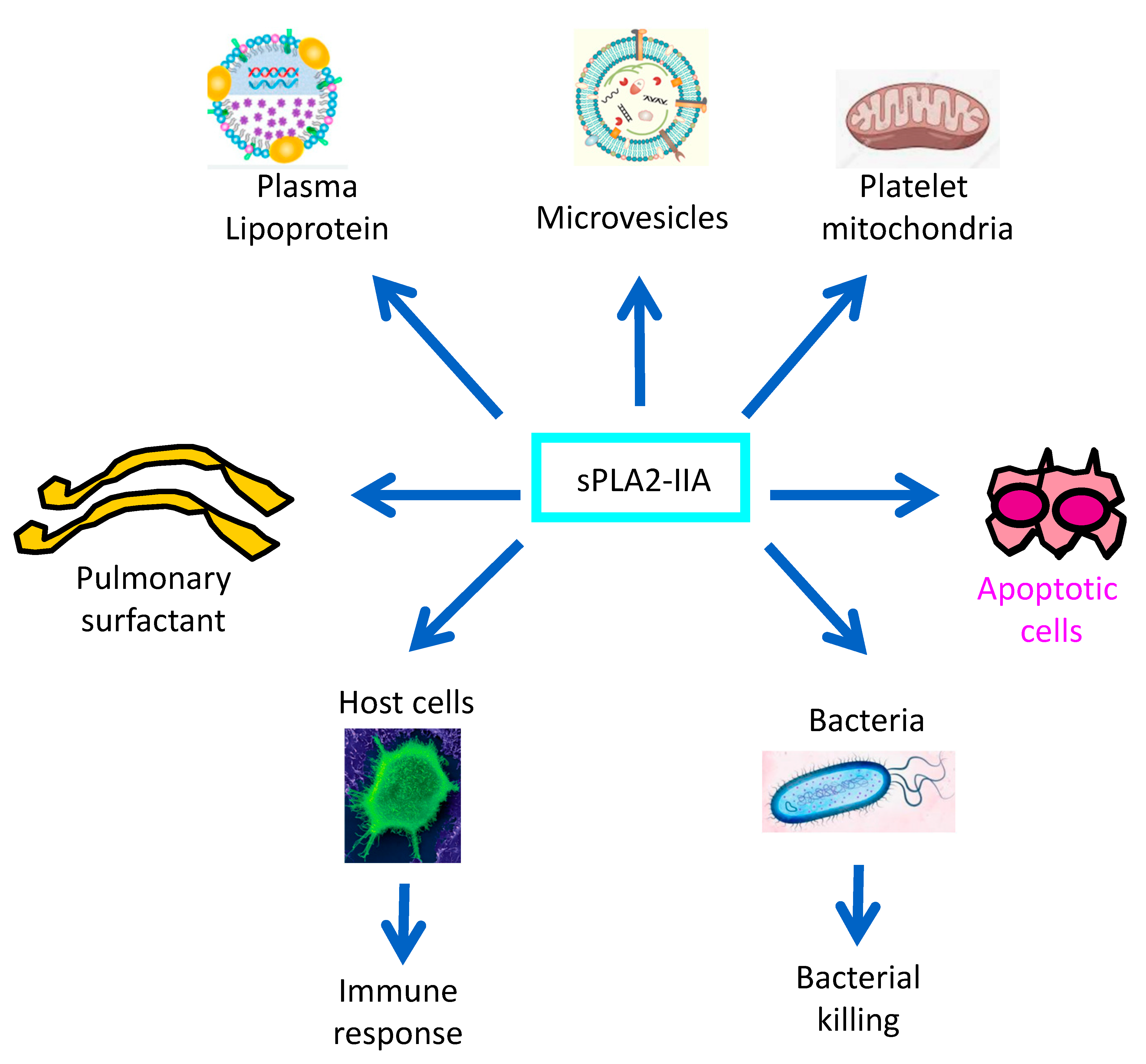
Phospholipase A2 (PLA2) enzymes hydrolyze the sn-2 position of phospholipids, resulting in the production of free fatty acids and lyso-phospholipids [1,2]. These enzymes are classified into two major families: the low molecular weight-secreted PLA2 (sPLA2) and the high molecular weight intracellular PLA2, such as the cytosolic PLA2 (cPLA2) [1,2]. Based on the number and position of their disulfide bridges, sPLA2 can be classified into several different types, one of which is sPLA2-IIA. PLA2 have been shown to release free arachidonic acid (AA), the precursor of proinflammatory eicosanoids, and to bind to specific receptors present on host surface membranes [3,4]. Initially, sPLA2-IIA was suggested to play a role in the development of various inflammatory diseases [5,6]. For example, this enzyme can hydrolyze pulmonary surfactant phospholipids involved in acute respiratory distress syndrome (ARDS). In addition, sPLA2-IIA has been shown to induce neuronal apoptosis in ischemic stroke [7]. Other studies have also shown the involvement of sPLA2-IIA in atherosclerotic lesions [8,9,10], the hydrolysis of mitochondrial membranes released by platelets [11] and plasma lipoproteins [12]. sPLA2-IIA has also been shown to generate lipid mediators from membrane vesicles of platelets and erythrocytes [13].
Thus, it is clear that sPLA2-IIA can be involved in various pathophysiological processes, but its high bactericidal property (especially against Gram-positive bacteria) is now accepted as its most established biological role [5,6,14]. Therefore, it is important to explore the mechanisms by which bacterial toxins can modulate the expression of this enzyme and the pathophysiological consequences of this modulation.

1.2. PAMPs, Toxins and Innate Immune Response to Bacterial Infections

Pathogen-associated molecular patterns (PAMPs) are microbial motifs that are highly conserved across a wide range of pathogens. They are essential for the survival of these pathogens and their detection by host cells [15]. They include several virulence factors, such as lipopeptides, lipoteichoic acid (LTA) or peptidoglycans (PGN) of Gram-positive bacteria and lipopolysaccharides (LPS), pili or flagellin of Gram-negative bacteria as well as the double-stranded RNA (dsRNA) of certain viruses [16]. Recognition of PAMPs by specialized host cells is the first step in the host immune response, leading to an inflammatory response and elimination of the invading pathogens. This process involves the interactions of PAMPs with cellular receptors called ‘pattern recognition receptors’, or PRRs.
On the other hand, bacteria also produce a variety of toxins in response to various environmental challenges. These include exotoxins that are actively expressed and secreted into the extracellular media or injected into host cells during the infection process [17,18]. The interaction of pathogens with host cells initiates signaling processes that lead to the production of anti-microbial peptides (AMPs) by these cells. AMPs represent a large family of peptides ranging from 10 to 150 amino acids. In particular, 153 AMPs have been found in humans with net positive charges on the surface of the molecules [19], including defensins, cathelicidin, the type IIA secreted phospholipase A2 (sPLA2-IIA), etc. These antimicrobial molecules interact with bacteria to inhibit the synthesis of bacterial membrane phospholipids, cleave polysaccharides of the bacterial cell wall or increase the permeability of the bacterial membrane [19], which ultimately results in the eradication of the pathogens or the reduction of their proliferation. In particular, sPLA2-IIA has a high net positive charge of +17 [20]. Table 1 shows the reported effects of some PAMPs and toxins on sPLA2-IIA expression by host cells (Table 1).

1.3. The sPLA2-IIA, an Endogenous Antibiotic-Like Protein of the Host

The bactericidal activity of sPLA2-IIA is related to its ability to efficiently penetrate the cell wall of Gram-positive bacteria. This particular property is due to the high net positive charge of sPLA2-IIA (+17) [20], whereas Gram-positive bacteria have high net anionic charges due to the presence of the D-alanyl moiety covalently linked to lipoteichoic acid (LTA) [38], which is a major membrane component of Gram-positive bacteria. Thus, the highly efficient and rapid binding of sPLA2-IIA to LTA by electrostatic interaction promotes the penetration of sPLA2-IIA into the peptidoglycan layer, another major wall component of Gram-positive bacteria. This leads to efficient hydrolysis of bacterial membrane lipids and subsequent bacterial killing [14]. One study has reported a classification of mouse and human sPLA2 based on their ability to kill the Gram-positive bacterium Staphylococcus aureus [39] and showed that sPLA2-IIA is the most bactericidal sPLA2 type. Indeed, the concentration of sPLA2-IIA in human tears of healthy subjects exceeds 30 µg/mL, and only 15–80 ng/mL of this protein is sufficient to kill S. aureus [40]. Additionally, concentrations of sPLA2-IIA rapidly increase in host biological fluids as a result of inflammation or bacterial infection [4,41,42], as discussed in Part 4 of this section. These concentrations are virtually sufficient to kill all Gram-positive bacteria that may invade the host. Thus, sPLA2-IIA can be considered a major player in the host’s innate immunity.

1.4. Role of the sPLA2-IIA in the Gut Microbiota–Lung Axis

Given its potent and selective antibacterial activity against Gram-positive bacteria, it is tempting to speculate that sPLA2-IIA might be involved in shaping the gut and pulmonary microbiota [43,44]. Indeed, two recent studies have investigated the influence of sPLA2-IIA on the gut microbiota [43,44]. Using gain- or loss-function assays, sPLA2-IIA was shown to play a central role in the composition of the gut microbiota by reducing the proportion of Gram-positive strains [43,44]. Most importantly, sPLA2-IIA-driven changes in the gut microbiota contributed to alterations in local and extra-intestinal immune responses, leading to increased susceptibility to cancer and arthritis [43,44]. This evidence suggests that intestinal sPLA2-IIA has profound effects on the host immune response through modulation of the gut microbiota and it may also have an impact on the pulmonary immune response by influencing the airway microbiota. Interestingly, there is a privileged relationship and communication between the lung and the gut (known as the gut–lung axis) that is mediated by the microbiota [45], and the gut microbiota has been associated with immunity to viral and bacterial infections [45,46,47,48]. Therefore, future studies are needed to better address the specific role of sPLA2-IIA in microbiota changes and the associated effects on the gut microbiota–lung axis.

1.5. sPLA2-IIA Levels in Biological Fluids of Infectious and Inflammatory Diseases

sPLA2-IIA was originally identified in synovial fluid from patients with rheumatoid arthritis [49], suggesting its involvement in excessive inflammatory conditions, such as autoimmunity. Subsequent studies have reported elevated levels of sPLA2-IIA in biological fluids from inflammatory diseases, including ARDS [50], pancreatitis, sepsis, cardiovascular disease [4,41,42] and in nasal fluids from patients with allergic rhinitis [51]. ARDS is defined as a life-threatening lung injury characterized by non-cardiogenic pulmonary edema and arterial hypoxemia [50]. The alteration of pulmonary surfactant is a hallmark of ARDS, accounting for increased surface tension at the air–liquid interface, resulting in impaired gas exchange and alveolar collapse. We have shown that sPLA2-IIA hydrolyzes surfactant phospholipids and its expression is inhibited by the surfactant protein A [4].
On the other hand, the pioneering studies of the Weiss group have reported the presence of sPLA2-IIA in rabbit peritoneal exudates [42] and baboon plasma [52] at levels sufficient to kill S. aureus. They showed that pre-treatments of these fluids with a specific sPLA2-IIA neutralizing antibody abolished the killing activity of these fluids [42,52]. We have also identified potent bactericidal activity against B. anthracis (the causative agent of anthrax) in human bronchoalveolar lavage fluids (BALFs) from patients with ARDS [53]. This bactericidal activity was positively correlated with the levels of sPLA2-IIA in the BALFs, and pretreatment of the BALFs with the sPLA2-IIA inhibitor LY311727 abolished the bactericidal activity [53]. We also showed that alveolar macrophages (AMs), the major source of sPLA2-IIA in a guinea pig model of ARDS [22], released sufficient amounts of sPLA2-IIA to kill B. anthracis [53]. This killing activity was inhibited by pretreatment of the AM medium with LY311727, suggesting that sPLA2-IIA is the major anthracidal factor released by AMs [53].

2. Bacterial Toxins That Upregulate sPLA2-IIA Expression

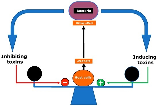
The toxins produced by invading bacteria can modulate sPLA2-IIA levels, either inducing or inhibiting its expression by host cells. Thus, the resulting sPLA2-IIA concentration during the infection process would depend on the balance between the levels of toxins that stimulate and those that downregulate the expression of this enzyme. The synthesis and/or secretion of bacterial toxins will be activated once the bacteria come into contact with host cells. The inhibition of sPLA2-IIA expression by bacterial toxins highlights a mechanism by which certain bacteria can subvert the host immune system. However, the same bacterium can either induce or inhibit sPLA2-IIA production depending on the expression kinetics of the bacterial toxins involved in sPLA2-IIA modulation. In the following sections, we will summarize the most important studies in the literature that have investigated the effects of bacterial toxins on sPLA2-IIA expression in the respiratory system and their pathophysiological consequences (Figure 1).

2.1. LPS Is the Major Bacterial Toxin Inducing sPLA2-IIA Expression by Host Cells

It is widely accepted that LPS is the major initiator of the inflammatory response that is caused by Gram-negative bacteria [15]. This response involves a range of enzymes and mediators produced by host cells in response to stimulation by LPS. sPLA2-IIA is produced by host cells during the inflammatory response. The stimulation of sPLA2-IIA synthesis in the context of infectious diseases has mostly been attributed to bacterial toxins, such as LPS. LPS is a potent inducer of sPLA2-IIA expression in various rodent models of inflammatory diseases [54]. LPS from E. Coli, P. aeruginosa and N. meningitidis has been shown to induce sPLA2-IIA production from a variety of cells, including AMs and bronchial epithelial cells (BECs) [5,31,55]. However, LPS-induced sPLA2-IIA expression in human BECs is much less potent than in gpAMs [22,55]. Other studies reported that AMs isolated from ARDS patients did not respond to LPS to produce PLA2 [56]. On the contrary, the production of sPLA2-IIA was induced in control patients only after LPS treatment. Thus, PLA2 isoforms may serve as markers of immune status in ARDS [56].
LPS-induced sPLA2-IIA expression has been shown to be cell-specific and species-dependent. We have reported that LPS was able to induce sPLA2-IIA expression in AMs, the major source of this enzyme in a guinea pig model of ARDS [4,57]. In this model, the induction of LPS-induced sPLA2-IIA expression occurs via an autocrine/paracrine process dependent on TNFα. In baboons, TNFα also plays a role as an intermediate in LPS-induced sPLA2-IIA production [52]. These findings contrast with those in rat astrocytes and human hepatoma cells, where LPS-induced sPLA2-IIA expression occurred via a TNFα-independent process [58,59].
sPLA2-IIA has clearly been shown to play a role in the hydrolysis of dipalmitoyl-phosphatidylcholine (DPPC) in an animal model of LPS-induced ARDS [4,57,60]. DPPC is one of the major phospholipids of pulmonary surfactants. Its hydrolysis is associated with the loss of surface tension of the surfactant complex, leading to subsequent alveolar collapse, a key clinical feature of ARDS [4]. Another study reported that pretreatment with the sPLA2-IIA inhibitor S-5920/LY315920Na attenuated lung injury induced by oleic acid [60]. This effect was accompanied by protection against lung surfactant degradation and the associated production of the inflammatory lipid mediators thromboxane A2 and leukotriene B4 [60].
Subsequent studies showed that Azithromycin exerted anti-inflammatory properties on lung epithelial cells by inhibiting LPS-induced sPLA2-IIA expression, but had no effect on AMs [61]. On the other hand, treatment of brain microvascular endothelial cells (BMVECs) with LPS resulted in increased release of sPLA2-IIA and nitrite into the culture medium [33]. This release was decreased by pretreatment of the cells with an NO donor, sodium nitroprusside, suggesting that sPLA2-IIA expression is under the control of the NO–JAK3–STAT1 pathway [62]. Furthermore, sPLA2-IIA stimulates neuronal cell death via apoptosis [7], which appears to be associated with the production of arachidonic acid (AA) metabolites, particularly prostaglandin D2 (PGD2). In addition, sPLA2-IIA is involved in neurodegeneration in the ischemic brain, highlighting the potential therapeutic use of sPLA2-IIA inhibitors in stroke therapy [7].

2.2. Role of Pili in the Induction of sPLA2-IIA Expression in the Airways

In our previous studies, we investigated whether N. meningitidis toxins or PAMPs, other than LPS, were able to stimulate sPLA2-IIA expression using gpAMs [31]. This Gram-negative bacterium is the causative agent of meningococcal disease and is exclusively adapted to humans, with the nasopharynx being its natural habitat [63]. The results showed that N. meningitidis stimulates sPLA2-IIA synthesis through gpAMs and that pili mediate this stimulation [31]. Indeed, an LPS-deficient mutant of this bacterium was still able to induce sPLA2-IIA synthesis and a pili-deficient mutant showed a significantly lower ability to stimulate sPLA2-IIA expression than the wild-type strain. Moreover, a pili preparation isolated from an LPS-deficient N. meningitidis strain stimulated sPLA2-IIA production via a process involving NF-κB activation [31]. These studies demonstrated that pili, in addition to LPS, can induce sPLA2-IIA expression by N. meningitidis. However, the receptor(s) and the signaling pathways involved in pili-induced sPLA2-IIA expression were not investigated in this study. However, it should be emphasized that N. meningitidis has been shown to exert toxic effects on human epithelial and endothelial cells due to a synergistic effect of LPS and pili [64].

2.3. The Type 3 Secretion System (T3SS) Toxin, Exotoxin S (ExoS), Plays a Key Role in sPLA2-IIA Expression in the Airways

We have also examined the expression and role of sPLA2-IIA in the airways of patients with cystic fibrosis (CF) and showed that the P. aeruginosa PAK strain, but not PAMPs, isolated from this bacterium, including LPS, HSL, CpG, flagellin and pili, stimulated sPLA2-IIA synthesis by BECs from these patients [23]. This observation suggests that LPS-induced sPLA2-IIA synthesis is cell type dependent, and also prompted us to identify the P. aeruginosa virulence factors involved in sPLA2-IIA production by BECs in the context of CF. Compared to its parental strain, the PAK mutant lacking T3SS induced sPLA2-IIA expression at much lower levels, whereas the T2SS-deficient strain induced it at similar levels [23], suggesting a specific ability of T3SS to induce sPLA2-IIA expression. Moreover, using a pharmacological approach, we showed that the signaling pathways (NF-κB, AP-1 and MAPK), known to induce sPLA2-IIA expression in various cell systems (see ref [5]), did not play a role in ExoS-induced sPLA2-IIA expression in BECs. We established that this expression was under the control of Krüppel-Like Factor 2 (KLF2), a particular transcription factor induced by bacterial toxins [65]. KLF2 belongs to the SP-1 zinc-finger transcription factor [66]. Although the signaling pathways by which ExoS induces KLF2 expression are still unknown, our studies showed that KLF2 induction by ExoS is independent of RhoA, one of the potential targets suggested by others [67]. ExoS is a bifunctional toxin with two enzymatic domains, GAP and ADPRT [17], but only the GAP domain is responsible for RhoA inactivation [68]. We also established that ADPRT, but not the GAP domain, plays a role in ExoS-induced sPLA2-IIA synthesis by BECs from CF patients [23].
These findings led us to investigate whether ExoS was able to induce sPLA2-IIA expression in cells other than epithelial cells. Using the T3SS-deficient PAK strains (ΔpscF mutant), we investigated the role of this toxin in gpAMs. Our results showed that this mutant induced sPLA2-IIA expression at similar levels compared to the parental stain. This suggests that, unlike human BECs, ExoS does not affect sPLA2-IIA expression in gpAMs. We also showed that the mutant lacking T2SS induced sPLA2-IIA expression in gpAMs at similar levels compared to the parental stain. These results clearly indicate that neither T2SS nor T3SS is involved in P. aeruginosa-induced sPLA2-IIA expression in gpAMs (Wu et al., manuscript in preparation).
However, the stimulatory effects of ExoS on sPLA2-IIA contrasted with its inhibitory effects on other mediators of the host immune response, such as cytokines and reactive oxygen species (ROS). Indeed, studies have reported that deletion of the exoS gene resulted in a significant increase in interleukin-8 (IL-8) production by Caco-2 cells. This finding suggested that P. aeruginosa produces a serine protease capable of degrading IL-8 in the culture medium of infected Caco-2 cells and that the expression of this protease is inhibited in cells infected with the ΔexoS mutant [69]. More recent studies have shown that P. aeruginosa can inhibit the ROS burst in neutrophils and that this inhibition occurs, at least in part, through an ExoS-mediated ADP-ribosylation of Ras in neutrophils [70]. However, the pathophysiological consequences of the opposing effects of ExoS on sPLA2-IIA and other mediators of the innate immune response remained to be elucidated.

2.4. Induction of sPLA2-IIA by Porphyromonas Gingivalis and Relevance to Periodontal Disease

P. gingivalis is a Gram-negative bacterium known to play a key role in the development of periodontal disease [71]. This bacterium has been shown to induce dramatic expression of sPLA2-IIA in human oral epithelial cells, suggesting that sPLA2-IIA might play a role in periodontal disease [72]. The induction of sPLA2-IIA expression by P. gingivalis occurs via a mechanism dependent on TLR activation or T3SS and was initiated by the production of arginine-gingipains (rgpA and rgpB). The latter belongs to the cysteine proteases group unique to this bacterium [72,73] that target a specific sequence of the Notch-1 extracellular domain. This process stimulates the translocation of the Notch-1 intracellular domain into the nucleus, leading to the transcriptional activation of the sPLA2-IIA gene [72].

2.5. PGN Is the Main Inducer of sPLA2-IIA Expression by Gram-Positive Bacteria

Our studies using gpAMs as a cell model showed that the Gram-positive bacterium B. anthracis can stimulate sPLA2-IIA expression. These studies demonstrated that the cell wall PGN purified from B. anthracis induced sPLA2-IIA expression through a process depending on NF-κB activation [26]. However, it is still unclear whether PGN stimulated sPLA2-IIA expression via two PGN recognition proteins, TLR2 or NOD-2 [74]. Recent reports showed that NOD may be involved in cell activation by spores of this bacterium [75]. However, we cannot rule out the fact that other bacterial components of the cell wall or secreted by B. anthracis are also involved in sPLA2-IIA expression in gpAMs.
In a subsequent study [28], we attempted to identify the S. aureus PAMP(s) and associated receptor(s) that induce sPLA2-IIA expression in gpAMs using PGN purified from S. aureus (ligand of TLR2 and NOD2) and LTA (ligand of TLR2). Our results revealed that PGN stimulated sPLA2-IIA expression in gpAMs, whereas LTA had only a limited effect on this expression. This suggested the possible involvement of the NOD2 receptor in PGN-induced sPLA2-IIA expression in gpAMPs [28]. To further verify the role of NOD2 in this process, gpAMs were incubated with the minimal active portions of the PGN, motif MDP (ligand of NOD2), MDPc (inactivated MDP), MALP-2 (ligand of TLR2/6), Pam3CSK4 (ligand of TLR2/1) or the cytosine guanosine dinucleotide (CpG, agonist of TLR9). Remarkably, only MDP was shown to induce sPLA2-IIA expression by gpAMs. These results indicate that NOD2 is probably the only receptor involved in PGN-induced sPLA2-IIA expression in gpAMs [28].

3. Bacterial Toxins That Downregulate sPLA2-IIA Expression

3.1. Inhibition of sPLA2-IIA Expression by Bacillus Anthracis Toxins

The exact mechanisms by which B. anthracis initiates the anthrax disease are not fully established. However, it is clear that this bacterium multiplicates rapidly in the blood stream, ultimately leading to the death of the host [76]. B. anthracis can subvert the host immune response [77,78] via a process involving the action of the B. anthracis edema toxin (ET) and lethal toxin (LT). This bacterium releases a binary A-B toxin composed of a single B transporter called a protective antigen (PA) and two alternative A components, lethal factor (LF) or edema factor (EF) [76]. LF and EF act in pairs, with PA leading to LT (=PA + LF) and ET (=PA + EF), respectively. Thus, PA serves as a transporter that delivers LF and EF into the host cell cytosol where they target specific molecular components [76].

3.1.1. Lethal Toxin Downregulates sPLA2-IIA Expression by AMs

Our previous studies showed that the expression of sPLA2-IIA was inhibited by LT during infection with gpAMPs by B. anthracis [53]. Next, we investigated the mechanisms by which LT alters sPLA2-IIA synthesis in this cell model. LT exhibits metallo-proteolytic activity toward the N-terminus of the MAPK-kinases. We have shown that this toxin inhibits the phosphorylation of MAPK p38 in gpAMs as well as sPLA2-IIA promoter activity in CHO cells [79]. The inhibition of sPLA2-IIA promoter activity was mimicked by co-transfection with the dominant negative construct of p38 (DN-p38) and reversed by the active form of p38-MAPK (AC-p38) [79]. Both LT and DN-p38 decreased NF-κB luciferase activity. However, neither LT nor a specific p-38 inhibitor interfered with LPS-induced IκBα degradation or NF-κB nuclear translocation in AMs [79]. Therefore, we concluded that sPLA2-IIA expression is induced via sequential MAPK-NF-κB activation and that LT alters this expression by interfering with NF-κB transactivation in the nucleus. This hypothesis is consistent with previous studies suggesting that the regulation of sPLA2-IIA in Jurkat cells involves epigenetic silencing by DNA hypermethylation [80].

3.1.2. Edema Toxin Impairs sPLA2-IIA Expression by AMs

In a subsequent study, we showed that ET inhibited sPLA2-IIA expression in gpAMs at the transcriptional level through a cAMP/protein kinase A-dependent process [26]. Moreover, the ET-deficient strains induced sPLA2-IIA expression, whereas the live B. anthracis strains expressing the functional ET inhibited this expression [26]. This suggests that B. anthracis can induce sPLA2-IIA expression probably via PGN and that ET injected into gpAMPs reduces this expression. This inhibition seems to occur via a cAMP/protein kinase A-dependent mechanism [26]. Our reports revealed that the cAMP increase was transient, reaching basal values within 24 h, in contrast to previous studies showing that cAMP accumulation increased within 48 h or more following ET addition to NIH/3T3 fibroblasts and RAW 267 macrophages [81].
Regardless of the kinetics of the cAMP increase during infection of gpAMPs by B. anthracis, the final concentrations of sPLA2-IIA should be considered as a balance between the inducing effect of PGN and the inhibitory effect of ET. However, it is worth highlighting that the effects of cAMP on sPLA2-IIA expression depend on the cell type considered. For example, in contrast to our results with gpAMs, other studies have shown that cAMP stimulates the transcriptional activity of the sPLA2-IIA gene in rat vascular smooth muscle cells. This stimulation depends on the interplay of the CCAAT/enhancer binding protein (C/EBP), NF-κB and Ets transcription factors [82].
Other studies have examined the effects of B. anthracis toxins on human dendritic cells and showed that both ET and LT inhibited the production of proinflammatory chemokines by these cells, with LT exhibiting the higher inhibitory effect. However, only LT impaired neutrophil recruitment in this model [83]. The authors suggested that ET and LT act in concert to suppress chemokine expression by human dendritic cells and that this action leads to an alteration in immune cell recruitment.
Taken together, these reports suggest that ET and LT may interfere with the immune system by altering sPLA2-IIA expression or by impairing chemokine expression and neutrophil recruitment, which may play a role in the innate host response to B. anthracis infection.

3.2. Inhibition of sPLA2-IIA Expression by a Bordetella Pertussis AC-Hly Toxin

The ability to inhibit sPLA2-IIA expression in gpAMs seems to be a general mechanism shared by several bacteria. Indeed, we have shown that a toxin secreted by Bordetella pertussis named adenylate cyclasehemolysin (AC-Hly) was also able to impair LPS-induced sPLA2-IIA expression by gpAMs. This inhibition is likely due to an increase in cAMP levels by AC-Hly, which is known to display calmodulin-dependent adenylate cyclase activity [84]. It should be noted, however, that the cyclic AMP-elevating capacity of AC-Hly is sufficient to ensure lung infection but not to ensure full virulence of B. pertussis [85]. Additionally, AC-Hly did not affect IL-8 production by gpAMs, suggesting that sPLA2-IIA suppression results from a selective effect of this toxin on sPLA2-IIA transcriptional activity. This finding is consistent with our previous studies showing that B. anthracis ET can downregulate sPLA2-IIA expression without affecting IL-8 secretion [84].

3.3. S. aureus Adenosine Inhibits sPLA2-IIA Expression and Associated Airway Killing

S. aureus produces several molecules, including adenosine, that dampen the host immunity [86]. Adenosine is produced by the highly conserved cell wall-anchored adenosine synthase A (AdsA) via the degradation of ATP, ADP and AMP [87]. We showed that an adenosine-deficient S. aureus mutant (∆adsA strain) enhanced the pulmonary expression of sPLA2-IIA in a guinea pig model of lung infection and was cleared more efficiently in the airways compared to the wild-type strain [28]. In addition, the ∆adsA strain induced sPLA2-IIA expression by gpAMs after the phagocytosis of S. aureus via a NOD2-NF-κB-dependent mechanism. The addition of exogenous adenosine to cultured gpAMs reduced S. aureus phagocytosis by these cells and impaired sPLA2-IIA synthesis. This occurred through a downregulation of p38 phosphorylation via adenosine receptors A2a-, A2b- and associated protein kinase A activation. In addition to its effects on phagocytosis, adenosine also acts as an anti-inflammatory mediator in macrophages via the cAMP/PKA axis [88]. This effect is mediated by altering NF-κB activity through PKA [89].
Taken together, these studies indicate that in the airway, S. aureus can escape sPLA2-IIA-mediated killing via adenosine-mediated alteration of phagocytosis and sPLA2-IIA synthesis. These processes also highlight the contribution of NOD2 in modulating sPLA2-IIA expression and suggest a mechanism by which S. aureus subverts host immunity based on the inhibition of the production of this antimicrobial enzyme.

4. Conclusions

As a mediator of the inflammatory response, sPLA2-IIA appears to act as a double-edged sword for the host (Figure 2). Indeed, in addition to the bactericidal functions and their regulation summarized in this review, sPLA2-IIA has been shown to play a role as a proinflammatory mediator involved in pathogenic processes. This includes alteration of pulmonary surfactant, hydrolysis of mitochondrial membrane released by platelets and interaction with plasma lipoproteins. sPLA2-IIA has also been shown to generate lipid mediators from membrane vesicles of platelets and erythrocytes and to play a role in neurotoxicity and cell apoptosis. The bactericidal activity of sPLA2-IIA has emerged as a central function of this enzyme, leading to the elimination of invading pathogenic bacteria and the modulation of microbiota composition. However, further work is required to identify the precise mechanisms involved in the crosstalk between sPLA2-IIA-mediated effects on gut and airway microbiota as well as on lung immune responses.
In general, Gram-negative bacteria, such as P. aeruginosa, are insensitive to the bactericidal effects of sPLA2-IIA but can induce its expression by host cells. This results in the selective elimination of Gram-positive bacteria, such as S. aureus, which are highly sensitive to sPLA2-IIA killing, a process that is advantageous for Gram-negative bacteria. However, the Gram-positive bacteria can also inhibit sPLA2-IIA expression, highlighting an evolutionary adaptive mechanism by which these bacteria evade the host’s innate immunity.
As shown in this review, several bacterial toxins can regulate sPLA2-IIA levels by either inducing or inhibiting their expression in host cells. These effects depend on the toxins and the host cell type considered. Thus, the final sPLA2-IIA concentration in host biological fluids can be considered as a balance between the stimulatory and inhibitory effects of these toxins. Our review was focused on bacterial toxins, but it is necessary to investigate whether other respiratory pathogens (viruses and fungi) can also modulate sPLA2-IIA expression in the airways and its potential role in the outcome of the infection. Nevertheless, the beneficial bactericidal functions of sPLA2-IIA highlight a potential therapeutic value of this enzyme in respiratory infectious diseases, especially in the context of the antibiotic resistance crisis.

Author Contributions

This manuscript was written by Y.W., E.P. and L.T. All authors have read and agreed to the published version of the manuscript.

Funding

This research received no external funding.

Institutional Review Board Statement

Not applicable.

Informed Consent Statement

Not applicable.

Data Availability Statement

Not applicable.

Acknowledgments

The authors warmly thank Nora Touqui for her precious and meticulous proofreading and English improvement of the present article.

Conflicts of Interest

The authors declare no conflict of interest.

Abbreviations

PAMPpathogen-associated molecular pattern
PRRspattern recognition receptors
PLA2phospholipase A2
sPLA2-IIAtype-IIA secreted phospholipase A2
cPLA2cytosolic PLA2
G+ bacteriumGram-positive bacterium
G- bacteriumGram-negative bacterium
LTAlipoteichoic acid
PGNpeptidoglycan
LPSlipopolysaccharides
dsDNAdouble-stranded DNA
ARDSacute respiratory distress syndrome
BALFbronchoalveolar lavage fluid
AMsalveolar macrophages
gpAMsguinea pig alveolar macrophages
BECsbronchial epithelial cells
DPPCdipalmitoyl-phosphatidylcholine
BMVECsbrain microvascular endothelial cells
AAarachidonic acid
PGD2prostaglandin D2
LOSlipooligosaccharides
T3SStype 3 secretion system
ExoSexotoxin S
CFcystic fibrosis
KLF2Krüppel-Like Factor 2
GAPGTPase activating protein
ADPRTADP ribosyltransferase
MDPmuramyl dipeptide
MALP-2macrophage-activating lipopeptide 2 kDa
CpGcytosine guanosine dinucleotide
ETedema toxin
LTlethal toxin
EFedema factor
PAprotective antigen
DN-p38dominant negative construct of p38
AC-Hlyadenylate cyclasehemolysin
C/EBPCCAAT/enhancer binding protein
cAMPcyclic adenosine monophosphate
AdsAadenosine synthase A

References

  1. Dennis, E.A. The growing phospholipase A2 superfamily of signal transduction enzymes. Trends Biochem. Sci. 1997, 22, 1–2. [Google Scholar] [CrossRef] [PubMed]
  2. Ghosh, M.; Tucker, D.E.; Burchett, S.A.; Leslie, C.C. Properties of the Group IV phospholipase A2 family. Prog. Lipid Res. 2006, 45, 487–510. [Google Scholar] [CrossRef] [PubMed]
  3. Murakami, M.; Lambeau, G. Emerging roles of secreted phospholipase A(2) enzymes: An update. Biochimie 2013, 95, 43–50. [Google Scholar] [CrossRef] [PubMed]
  4. Touqui, L.; Arbibe, L. A role for phospholipase A2 in ARDS pathogenesis. Mol. Med. Today 1999, 5, 244–249. [Google Scholar] [CrossRef]
  5. Touqui, L.; Alaoui-El-Azher, M. Mammalian secreted phospholipases A2 and their pathophysiological significance in inflammatory diseases. Curr. Mol. Med. 2001, 1, 739–754. [Google Scholar] [CrossRef] [PubMed]
  6. Nevalainen, T.J.; Graham, G.G.; Scott, K.F. Antibacterial actions of secreted phospholipases A2. Review. Biochim. Biophys. Acta 2008, 1781, 1–9. [Google Scholar] [CrossRef]
  7. Yagami, T.; Ueda, K.; Asakura, K.; Hata, S.; Kuroda, T.; Sakaeda, T.; Takasu, N.; Tanaka, K.; Gemba, T.; Hori, Y. Human group IIA secretory phospholipase A2 induces neuronal cell death via apoptosis. Mol. Pharm. 2002, 61, 114–126. [Google Scholar] [CrossRef]
  8. Kugiyama, K.; Ota, Y.; Takazoe, K.; Moriyama, Y.; Kawano, H.; Miyao, Y.; Sakamoto, T.; Soejima, H.; Ogawa, H.; Doi, H.; et al. Circulating levels of secretory type II phospholipase A(2) predict coronary events in patients with coronary artery disease. Circulation 1999, 100, 1280–1284. [Google Scholar] [CrossRef]
  9. Mattsson, N.; Magnussen, C.G.; Ronnemaa, T.; Mallat, Z.; Benessiano, J.; Jula, A.; Taittonen, L.; Kahonen, M.; Juonala, M.; Viikari, J.S.; et al. Metabolic syndrome and carotid intima-media thickness in young adults: Roles of apolipoprotein B, apolipoprotein A-I, C-reactive protein, and secretory phospholipase A2: The cardiovascular risk in young Finns study. Arter. Thromb. Vasc. Biol. 2010, 30, 1861–1866. [Google Scholar] [CrossRef]
  10. Rosenson, R.S.; Gelb, M.H. Secretory phospholipase A2: A multifaceted family of proatherogenic enzymes. Curr. Cardiol. Rep. 2009, 11, 445–451. [Google Scholar] [CrossRef]
  11. Boudreau, L.H.; Duchez, A.C.; Cloutier, N.; Soulet, D.; Martin, N.; Bollinger, J.; Pare, A.; Rousseau, M.; Naika, G.S.; Levesque, T.; et al. Platelets release mitochondria serving as substrate for bactericidal group IIA-secreted phospholipase A2 to promote inflammation. Blood 2014, 124, 2173–2183. [Google Scholar] [CrossRef]
  12. Pruzanski, W.; Lambeau, G.; Lazdunski, M.; Cho, W.; Kopilov, J.; Kuksis, A. Hydrolysis of minor glycerophospholipids of plasma lipoproteins by human group IIA, V and X secretory phospholipases A2. Biochim. Biophys. Acta 2007, 1771, 5–19. [Google Scholar] [CrossRef]
  13. Fourcade, O.; Simon, M.F.; Viode, C.; Rugani, N.; Leballe, F.; Ragab, A.; Fournie, B.; Sarda, L.; Chap, H. Secretory phospholipase A2 generates the novel lipid mediator lysophosphatidic acid in membrane microvesicles shed from activated cells. Cell 1995, 80, 919–927. [Google Scholar] [CrossRef]
  14. van Hensbergen, V.P.; Wu, Y.; van Sorge, N.M.; Touqui, L. Type IIA Secreted Phospholipase A2 in Host Defense against Bacterial Infections. Trends Immunol. 2020, 41, 313–326. [Google Scholar] [CrossRef]
  15. Janeway, C.A., Jr. Approaching the asymptote? Evolution and revolution in immunology. Cold. Spring Harb. Symp. Quant. Biol. 1989, 54 Pt 1, 1–13. [Google Scholar] [CrossRef]
  16. Kumar, V. Toll-like receptors in sepsis-associated cytokine storm and their endogenous negative regulators as future immunomodulatory targets. Int. Immunopharmacol. 2020, 89, 107087. [Google Scholar] [CrossRef] [PubMed]
  17. Hauser, A.R. The type III secretion system of Pseudomonas aeruginosa: Infection by injection. Nat. Rev. Microbiol. 2009, 7, 654–665. [Google Scholar] [CrossRef] [PubMed]
  18. Shenoy, A.R.; Furniss, R.C.D.; Goddard, P.J.; Clements, A. Modulation of Host Cell Processes by T3SS Effectors. Curr. Top Microbiol. Immunol. 2018, 416, 73–115. [Google Scholar] [CrossRef] [PubMed]
  19. Wang, G. Human antimicrobial peptides and proteins. Pharmaceuticals 2014, 7, 545–594. [Google Scholar] [CrossRef]
  20. Baker, S.F.; Othman, R.; Wilton, D.C. Tryptophan-containing mutant of human (group IIa) secreted phospholipase A2 has a dramatically increased ability to hydrolyze phosphatidylcholine vesicles and cell membranes. Biochemistry 1998, 37, 13203–13211. [Google Scholar] [CrossRef]
  21. Poltorak, A.; He, X.; Smirnova, I.; Liu, M.Y.; Van Huffel, C.; Du, X.; Birdwell, D.; Alejos, E.; Silva, M.; Galanos, C.; et al. Defective LPS signaling in C3H/HeJ and C57BL/10ScCr mice: Mutations in Tlr4 gene. Science 1998, 282, 2085–2088. [Google Scholar] [CrossRef]
  22. Arbibe, L.; Vial, D.; Rosinski-Chupin, I.; Havet, N.; Huerre, M.; Vargaftig, B.B.; Touqui, L. Endotoxin induces expression of type II phospholipase A2 in macrophages during acute lung injury in guinea pigs: Involvement of TNF-alpha in lipopolysaccharide-induced type II phospholipase A2 synthesis. J. Immunol. 1997, 159, 391–400. [Google Scholar] [CrossRef] [PubMed]
  23. Pernet, E.; Guillemot, L.; Burgel, P.R.; Martin, C.; Lambeau, G.; Sermet-Gaudelus, I.; Sands, D.; Leduc, D.; Morand, P.C.; Jeammet, L.; et al. Pseudomonas aeruginosa eradicates Staphylococcus aureus by manipulating the host immunity. Nat. Commun. 2014, 5, 5105. [Google Scholar] [CrossRef]
  24. Chaput, C.; Boneca, I.G. Peptidoglycan detection by mammals and flies. Microbes Infect. 2007, 9, 637–647. [Google Scholar] [CrossRef] [PubMed]
  25. Inohara, N.; Ogura, Y.; Fontalba, A.; Gutierrez, O.; Pons, F.; Crespo, J.; Fukase, K.; Inamura, S.; Kusumoto, S.; Hashimoto, M.; et al. Host recognition of bacterial muramyl dipeptide mediated through NOD2. Implications for Crohn’s disease. J. Biol. Chem. 2003, 278, 5509–5512. [Google Scholar] [CrossRef]
  26. Raymond, B.; Leduc, D.; Ravaux, L.; Le Goffic, R.; Candela, T.; Raymondjean, M.; Goossens, P.L.; Touqui, L. Edema toxin impairs anthracidal phospholipase A2 expression by alveolar macrophages. PLoS Pathog. 2007, 3, e187. [Google Scholar] [CrossRef] [PubMed]
  27. Schwandner, R.; Dziarski, R.; Wesche, H.; Rothe, M.; Kirschning, C.J. Peptidoglycan- and lipoteichoic acid-induced cell activation is mediated by toll-like receptor 2. J. Biol. Chem. 1999, 274, 17406–17409. [Google Scholar] [CrossRef] [PubMed]
  28. Pernet, E.; Brunet, J.; Guillemot, L.; Chignard, M.; Touqui, L.; Wu, Y. Staphylococcus aureus Adenosine Inhibits sPLA2-IIA-Mediated Host Killing in the Airways. J. Immunol. 2015, 194, 5312–5319. [Google Scholar] [CrossRef]
  29. Hayashi, F.; Smith, K.D.; Ozinsky, A.; Hawn, T.R.; Yi, E.C.; Goodlett, D.R.; Eng, J.K.; Akira, S.; Underhill, D.M.; Aderem, A. The innate immune response to bacterial flagellin is mediated by Toll-like receptor 5. Nature 2001, 410, 1099–1103. [Google Scholar] [CrossRef] [PubMed]
  30. Proft, T.; Baker, E.N. Pili in Gram-negative and Gram-positive bacteria—Structure, assembly and their role in disease. Cell Mol. Life Sci. 2009, 66, 613–635. [Google Scholar] [CrossRef]
  31. Touqui, L.; Paya, M.; Thouron, F.; Guiyoule, A.; Zarantonelli, M.L.; Leduc, D.; Wu, Y.; Taha, M.K.; Alonso, J.M. Neisseria meningitidis pili induce type-IIA phospholipase A2 expression in alveolar macrophages. FEBS Lett. 2005, 579, 4923–4927. [Google Scholar] [CrossRef]
  32. Vabulas, R.M.; Ahmad-Nejad, P.; da Costa, C.; Miethke, T.; Kirschning, C.J.; Hacker, H.; Wagner, H. Endocytosed HSP60s use toll-like receptor 2 (TLR2) and TLR4 to activate the toll/interleukin-1 receptor signaling pathway in innate immune cells. J. Biol. Chem. 2001, 276, 31332–31339. [Google Scholar] [CrossRef]
  33. Bauer, S.; Kirschning, C.J.; Hacker, H.; Redecke, V.; Hausmann, S.; Akira, S.; Wagner, H.; Lipford, G.B. Human TLR9 confers responsiveness to bacterial DNA via species-specific CpG motif recognition. Proc. Natl. Acad. Sci. USA 2001, 98, 9237–9242. [Google Scholar] [CrossRef] [PubMed]
  34. Hasko, G.; Linden, J.; Cronstein, B.; Pacher, P. Adenosine receptors: Therapeutic aspects for inflammatory and immune diseases. Nat. Rev. Drug. Discov. 2008, 7, 759–770. [Google Scholar] [CrossRef] [PubMed]
  35. Guermonprez, P.; Khelef, N.; Blouin, E.; Rieu, P.; Ricciardi-Castagnoli, P.; Guiso, N.; Ladant, D.; Leclerc, C. The adenylate cyclase toxin of Bordetella pertussis binds to target cells via the alpha(M)beta(2) integrin (CD11b/CD18). J. Exp. Med. 2001, 193, 1035–1044. [Google Scholar] [CrossRef] [PubMed]
  36. Bradley, K.A.; Mogridge, J.; Mourez, M.; Collier, R.J.; Young, J.A. Identification of the cellular receptor for anthrax toxin. Nature 2001, 414, 225–229. [Google Scholar] [CrossRef]
  37. Scobie, H.M.; Rainey, G.J.; Bradley, K.A.; Young, J.A. Human capillary morphogenesis protein 2 functions as an anthrax toxin receptor. Proc. Natl. Acad. Sci. USA 2003, 100, 5170–5174. [Google Scholar] [CrossRef]
  38. Neuhaus, F.C.; Baddiley, J. A continuum of anionic charge: Structures and functions of D-alanyl-teichoic acids in gram-positive bacteria. Microbiol. Mol. Biol. Rev. 2003, 67, 686–723. [Google Scholar] [CrossRef]
  39. Koduri, R.S.; Gronroos, J.O.; Laine, V.J.; Le Calvez, C.; Lambeau, G.; Nevalainen, T.J.; Gelb, M.H. Bactericidal properties of human and murine groups I, II, V, X, and XII secreted phospholipases A(2). J. Biol. Chem. 2002, 277, 5849–5857. [Google Scholar] [CrossRef]
  40. Qu, X.D.; Lehrer, R.I. Secretory phospholipase A2 is the principal bactericide for staphylococci and other gram-positive bacteria in human tears. Infect. Immun. 1998, 66, 2791–2797. [Google Scholar] [CrossRef]
  41. Murakami, M.; Nakatani, Y.; Atsumi, G.; Inoue, K.; Kudo, I. Regulatory functions of phospholipase A2. Crit. Rev. Immunol. 1997, 17, 225–283. [Google Scholar] [CrossRef]
  42. Weinrauch, Y.; Elsbach, P.; Madsen, L.M.; Foreman, A.; Weiss, J. The potent anti-Staphylococcus aureus activity of a sterile rabbit inflammatory fluid is due to a 14-kD phospholipase A2. J. Clin. Investig. 1996, 97, 250–257. [Google Scholar] [CrossRef]
  43. Miki, Y.; Taketomi, Y.; Kidoguchi, Y.; Yamamoto, K.; Muramatsu, K.; Nishito, Y.; Park, J.; Hosomi, K.; Mizuguchi, K.; Kunisawa, J.; et al. Group IIA secreted phospholipase A2 controls skin carcinogenesis and psoriasis by shaping the gut microbiota. JCI Insight 2022, 7, e152611. [Google Scholar] [CrossRef] [PubMed]
  44. Dore, E.; Joly-Beauparlant, C.; Morozumi, S.; Mathieu, A.; Levesque, T.; Allaeys, I.; Duchez, A.C.; Cloutier, N.; Leclercq, M.; Bodein, A.; et al. The interaction of secreted phospholipase A2-IIA with the microbiota alters its lipidome and promotes inflammation. JCI Insight 2022, 7, e152638. [Google Scholar] [CrossRef]
  45. Enaud, R.; Prevel, R.; Ciarlo, E.; Beaufils, F.; Wieers, G.; Guery, B.; Delhaes, L. The Gut-Lung Axis in Health and Respiratory Diseases: A Place for Inter-Organ and Inter-Kingdom Crosstalks. Front. Cell. Infect. Microbiol. 2020, 10, 9. [Google Scholar] [CrossRef] [PubMed]
  46. Ichinohe, T.; Pang, I.K.; Kumamoto, Y.; Peaper, D.R.; Ho, J.H.; Murray, T.S.; Iwasaki, A. Microbiota regulates immune defense against respiratory tract influenza A virus infection. Proc. Natl. Acad. Sci. USA 2011, 108, 5354–5359. [Google Scholar] [CrossRef]
  47. Khan, N.; Mendonca, L.; Dhariwal, A.; Fontes, G.; Menzies, D.; Xia, J.; Divangahi, M.; King, I.L. Intestinal dysbiosis compromises alveolar macrophage immunity to Mycobacterium tuberculosis. Mucosal Immunol. 2019, 12, 772–783. [Google Scholar] [CrossRef] [PubMed]
  48. Brown, R.L.; Sequeira, R.P.; Clarke, T.B. The microbiota protects against respiratory infection via GM-CSF signaling. Nat. Commun. 2017, 8, 1512. [Google Scholar] [CrossRef]
  49. Pruzanski, W.; Vadas, P. Secretory synovial fluid phospholipase A2 and its role in the pathogenesis of inflammation in arthritis. J. Rheumatol. 1988, 15, 1601–1603. [Google Scholar] [PubMed]
  50. Nakos, G.; Kitsiouli, E.; Hatzidaki, E.; Koulouras, V.; Touqui, L.; Lekka, M.E. Phospholipases A2 and platelet-activating-factor acetylhydrolase in patients with acute respiratory distress syndrome. Crit. Care Med. 2005, 33, 772–779. [Google Scholar] [CrossRef]
  51. Touqui, L.; Herpin-Richard, N.; Gene, R.M.; Jullian, E.; Aljabi, D.; Hamberger, C.; Vargaftig, B.B.; Dessange, J.F. Excretion of platelet activating factor-acetylhydrolase and phospholipase A2 into nasal fluids after allergenic challenge: Possible role in the regulation of platelet activating factor release. J. Allergy Clin. Immunol. 1994, 94, 109–119. [Google Scholar] [CrossRef]
  52. Weinrauch, Y.; Abad, C.; Liang, N.S.; Lowry, S.F.; Weiss, J. Mobilization of potent plasma bactericidal activity during systemic bacterial challenge. Role of group IIA phospholipase A2. J. Clin. Investig. 1998, 102, 633–638. [Google Scholar] [CrossRef]
  53. Gimenez, A.P.; Wu, Y.Z.; Paya, M.; Delclaux, C.; Touqui, L.; Goossens, P.L. High bactericidal efficiency of type iia phospholipase A2 against Bacillus anthracis and inhibition of its secretion by the lethal toxin. J. Immunol. 2004, 173, 521–530. [Google Scholar] [CrossRef]
  54. Hamaguchi, K.; Kuwata, H.; Yoshihara, K.; Masuda, S.; Shimbara, S.; Oh-ishi, S.; Murakami, M.; Kudo, I. Induction of distinct sets of secretory phospholipase A(2) in rodents during inflammation. Biochim. Biophys. Acta 2003, 1635, 37–47. [Google Scholar] [CrossRef]
  55. Medjane, S.; Raymond, B.; Wu, Y.; Touqui, L. Impact of CFTR DeltaF508 mutation on prostaglandin E2 production and type IIA phospholipase A2 expression by pulmonary epithelial cells. Am. J. Physiol. Lung Cell. Mol. Physiol. 2005, 289, L816–L824. [Google Scholar] [CrossRef] [PubMed]
  56. Hatzidaki, E.; Nakos, G.; Galiatsou, E.; Lekka, M.E. Impaired phospholipases A₂production by stimulated macrophages from patients with acute respiratory distress syndrome. Biochim. Biophys. Acta 2010, 1802, 986–994. [Google Scholar] [CrossRef] [PubMed]
  57. Arbibe, L.; Koumanov, K.; Vial, D.; Rougeot, C.; Faure, G.; Havet, N.; Longacre, S.; Vargaftig, B.B.; Bereziat, G.; Voelker, D.R.; et al. Generation of lyso-phospholipids from surfactant in acute lung injury is mediated by type-II phospholipase A2 and inhibited by a direct surfactant protein A-phospholipase A2 protein interaction. J. Clin. Investig. 1998, 102, 1152–1160. [Google Scholar] [CrossRef] [PubMed]
  58. Oka, S.; Arita, H. Inflammatory factors stimulate expression of group II phospholipase A2 in rat cultured astrocytes. Two distinct pathways of the gene expression. J. Biol. Chem. 1991, 266, 9956–9960. [Google Scholar] [CrossRef]
  59. Crowl, R.M.; Stoller, T.J.; Conroy, R.R.; Stoner, C.R. Induction of phospholipase A2 gene expression in human hepatoma cells by mediators of the acute phase response. J. Biol. Chem. 1991, 266, 2647–2651. [Google Scholar] [CrossRef]
  60. Furue, S.; Kuwabara, K.; Mikawa, K.; Nishina, K.; Shiga, M.; Maekawa, N.; Ueno, M.; Chikazawa, Y.; Ono, T.; Hori, Y.; et al. Crucial role of group IIA phospholipase A(2) in oleic acid-induced acute lung injury in rabbits. Am. J. Respir. Crit. Care Med. 1999, 160, 1292–1302. [Google Scholar] [CrossRef]
  61. Kitsiouli, E.; Antoniou, G.; Gotzou, H.; Karagiannopoulos, M.; Basagiannis, D.; Christoforidis, S.; Nakos, G.; Lekka, M.E. Effect of azithromycin on the LPS-induced production and secretion of phospholipase A2 in lung cells. Biochim. Biophys. Acta 2015, 1852, 1288–1297. [Google Scholar] [CrossRef] [PubMed]
  62. Wang, G.; Qian, P.; Xu, Z.; Zhang, J.; Wang, Y.; Cheng, S.; Cai, W.; Qian, G.; Wang, C.; Decoster, M.A. Regulatory effects of the JAK3/STAT1 pathway on the release of secreted phospholipase A₂-IIA in microvascular endothelial cells of the injured brain. J. Neuroinflamm. 2012, 9, 170. [Google Scholar] [CrossRef]
  63. Taha, M.K.; Alonso, J.M. Molecular epidemiology of infectious diseases: The example of meningococcal disease. Res. Microbiol. 2008, 159, 62–66. [Google Scholar] [CrossRef]
  64. Dunn, K.L.; Virji, M.; Moxon, E.R. Investigations into the molecular basis of meningococcal toxicity for human endothelial and epithelial cells: The synergistic effect of LPS and pili. Microb. Pathog. 1995, 18, 81–96. [Google Scholar] [CrossRef]
  65. O’Grady, E.P.; Mulcahy, H.; O’Callaghan, J.; Adams, C.; O’Gara, F. Pseudomonas aeruginosa infection of airway epithelial cells modulates expression of Kruppel-like factors 2 and 6 via RsmA-mediated regulation of type III exoenzymes S and Y. Infect. Immun. 2006, 74, 5893–5902. [Google Scholar] [CrossRef] [PubMed]
  66. McConnell, B.B.; Yang, V.W. Mammalian Kruppel-like factors in health and diseases. Physiol. Rev. 2010, 90, 1337–1381. [Google Scholar] [CrossRef] [PubMed]
  67. Dach, K.; Zovko, J.; Hogardt, M.; Koch, I.; van Erp, K.; Heesemann, J.; Hoffmann, R. Bacterial toxins induce sustained mRNA expression of the silencing transcription factor klf2 via inactivation of RhoA and Rhophilin 1. Infect. Immun. 2009, 77, 5583–5592. [Google Scholar] [CrossRef]
  68. Goehring, U.M.; Schmidt, G.; Pederson, K.J.; Aktories, K.; Barbieri, J.T. The N-terminal domain of Pseudomonas aeruginosa exoenzyme S is a GTPase-activating protein for Rho GTPases. J. Biol. Chem. 1999, 274, 36369–36372. [Google Scholar] [CrossRef]
  69. Okuda, J.; Hayashi, N.; Tanabe, S.; Minagawa, S.; Gotoh, N. Degradation of interleukin 8 by the serine protease MucD of Pseudomonas aeruginosa. J. Infect. Chemother. 2011, 17, 782–792. [Google Scholar] [CrossRef]
  70. Vareechon, C.; Zmina, S.E.; Karmakar, M.; Pearlman, E.; Rietsch, A. Pseudomonas aeruginosa Effector ExoS Inhibits ROS Production in Human Neutrophils. Cell Host Microbe 2017, 21, 611–618. [Google Scholar] [CrossRef]
  71. Hajishengallis, G.; Liang, S.; Payne, M.A.; Hashim, A.; Jotwani, R.; Eskan, M.A.; McIntosh, M.L.; Alsam, A.; Kirkwood, K.L.; Lambris, J.D.; et al. Low-abundance biofilm species orchestrates inflammatory periodontal disease through the commensal microbiota and complement. Cell Host Microbe 2011, 10, 497–506. [Google Scholar] [CrossRef] [PubMed]
  72. Al-Attar, A.; Alimova, Y.; Kirakodu, S.; Kozal, A.; Novak, M.J.; Stromberg, A.J.; Orraca, L.; Gonzalez-Martinez, J.; Martinez, M.; Ebersole, J.L.; et al. Activation of Notch-1 in oral epithelial cells by P. gingivalis triggers the expression of the antimicrobial protein PLA(2)-IIA. Mucosal. Immunol. 2018, 11, 1047–1059. [Google Scholar] [CrossRef]
  73. Potempa, J.; Sroka, A.; Imamura, T.; Travis, J. Gingipains, the major cysteine proteinases and virulence factors of Porphyromonas gingivalis: Structure, function and assembly of multidomain protein complexes. Curr. Protein Pept. Sci. 2003, 4, 397–407. [Google Scholar] [CrossRef] [PubMed]
  74. Dziarski, R.; Gupta, D. Peptidoglycan recognition in innate immunity. J. Endotoxin Res. 2005, 11, 304–310. [Google Scholar] [CrossRef]
  75. Glomski, I.J.; Fritz, J.H.; Keppler, S.J.; Balloy, V.; Chignard, M.; Mock, M.; Goossens, P.L. Murine splenocytes produce inflammatory cytokines in a MyD88-dependent response to Bacillus anthracis spores. Cell. Microbiol. 2007, 9, 502–513. [Google Scholar] [CrossRef]
  76. Mock, M.; Fouet, A. Anthrax. Annu. Rev. Microbiol. 2001, 55, 647–671. [Google Scholar] [CrossRef]
  77. Lincoln, R.E.; Hodges, D.R.; Klein, F.; Mahlandt, B.G.; Jones, W.I., Jr.; Haines, B.W.; Rhian, M.A.; Walker, J.S. Role of the lymphatics in the pathogenesis of anthrax. J. Infect. Dis. 1965, 115, 481–494. [Google Scholar] [CrossRef] [PubMed]
  78. Turnbull, P.C. Anthrax vaccines: Past, present and future. Vaccine 1991, 9, 533–539. [Google Scholar] [CrossRef]
  79. Raymond, B.; Ravaux, L.; Mémet, S.; Wu, Y.; Sturny-Leclère, A.; Leduc, D.; Denoyelle, C.; Goossens, P.L.; Payá, M.; Raymondjean, M.; et al. Anthrax lethal toxin down-regulates type-IIA secreted phospholipase A(2) expression through MAPK/NF-kappaB inactivation. Biochem. Pharm. 2010, 79, 1149–1155. [Google Scholar] [CrossRef]
  80. Menschikowski, M.; Hagelgans, A.; Kostka, H.; Eisenhofer, G.; Siegert, G. Involvement of epigenetic mechanisms in the regulation of secreted phospholipase A2 expressions in Jurkat leukemia cells. Neoplasia 2008, 10, 1195–1203. [Google Scholar] [CrossRef]
  81. Voth, D.E.; Hamm, E.E.; Nguyen, L.G.; Tucker, A.E.; Salles, I.I.; Ortiz-Leduc, W.; Ballard, J.D. Bacillus anthracis oedema toxin as a cause of tissue necrosis and cell type-specific cytotoxicity. Cell. Microbiol. 2005, 7, 1139–1149. [Google Scholar] [CrossRef] [PubMed]
  82. Antonio, V.; Brouillet, A.; Janvier, B.; Monne, C.; Bereziat, G.; Andreani, M.; Raymondjean, M. Transcriptional regulation of the rat type IIA phospholipase A2 gene by cAMP and interleukin-1beta in vascular smooth muscle cells: Interplay of the CCAAT/enhancer binding protein (C/EBP), nuclear factor-kappaB and Ets transcription factors. Biochem. J. 2002, 368, 415–424. [Google Scholar] [CrossRef] [PubMed]
  83. Cleret-Buhot, A.; Mathieu, J.; Tournier, J.N.; Quesnel-Hellmann, A. Both lethal and edema toxins of Bacillus anthracis disrupt the human dendritic cell chemokine network. PLoS ONE 2012, 7, e43266. [Google Scholar] [CrossRef] [PubMed]
  84. Wu, Y.; Raymond, B.; Goossens, P.L.; Njamkepo, E.; Guiso, N.; Paya, M.; Touqui, L. Type-IIA secreted phospholipase A2 is an endogenous antibiotic-like protein of the host. Biochimie 2010, 92, 583–587. [Google Scholar] [CrossRef]
  85. Skopova, K.; Tomalova, B.; Kanchev, I.; Rossmann, P.; Svedova, M.; Adkins, I.; Bibova, I.; Tomala, J.; Masin, J.; Guiso, N.; et al. Cyclic AMP-Elevating Capacity of Adenylate Cyclase Toxin-Hemolysin Is Sufficient for Lung Infection but Not for Full Virulence of Bordetella pertussis. Infect. Immun. 2017, 85, e00937-16. [Google Scholar] [CrossRef]
  86. Foster, T.J. Immune evasion by staphylococci. Nat Rev Microbiol 2005, 3, 948–958. [Google Scholar] [CrossRef]
  87. McCarthy, A.J.; Lindsay, J.A. Genetic variation in Staphylococcus aureus surface and immune evasion genes is lineage associated: Implications for vaccine design and host-pathogen interactions. BMC Microbiol. 2010, 10, 173. [Google Scholar] [CrossRef] [PubMed]
  88. Gonzales, J.N.; Gorshkov, B.; Varn, M.N.; Zemskova, M.A.; Zemskov, E.A.; Sridhar, S.; Lucas, R.; Verin, A.D. Protective effect of adenosine receptors against lipopolysaccharide-induced acute lung injury. Am. J. Physiol. Lung Cell. Mol. Physiol. 2014, 306, L497–L507. [Google Scholar] [CrossRef]
  89. Takahashi, N.; Tetsuka, T.; Uranishi, H.; Okamoto, T. Inhibition of the NF-kappaB transcriptional activity by protein kinase A. Eur. J. Biochem. 2002, 269, 4559–4565. [Google Scholar] [CrossRef]
Figure 1. Positive vs. negative modulation of sPLA2-IIA expression in host cells by the B. anthracis toxins. At the early step of host cell infection by B. anthracis, this bacterium induces sPLA2-IIA expression and secretion by these cells via the action of various PAMPs, including peptidoglycan (PG) or LTA. This expression occurs via a NOD-dependent pathway. The enzyme sPLA2-IIA, once secreted in the medium, interacts with bacteria, leading to their killing. In parallel, B. anthracis produces the toxins ET and LT that downregulate sPLA2-IIA expression. The final concentration of this enzyme in the cell medium is the balance between the inducing (by PG) and the inhibiting effects of PLA2-IIA expression (by ET and LT).
Toxins 15 00440 g001
Figure 2. Examples of established biological functions of sPLA2-IIA. sPLA2-IIA targets phospholipids of plasma lipoproteins, microvesicles or mitochondria released by platelets or erythrocytes, thus contributing to the inflammatory processes associated with diseases such as atherosclerosis and sepsis. This enzyme can also hydrolyze pulmonary surfactant phospholipids, an important feature of ARDS pathogenesis. sPLA2-IIA has been also shown to promote neurodegeneration through activation of neuronal cell apoptosis. But, the most studied functions of sPLA2-IIA are related to its bactericidal action and ability to release lipid mediators through the hydrolysis of host cell phospholipids.
Toxins 15 00440 g002
Table 1. Effects of bacterial PAMPs and toxins on sPLA2-IIA expression by host cells.
PAMPsBacteriumHost PRRsEffect on sPLA2-IIA ExpressionReferences
gpAMsBECs
LPSG−TLR4 aUpregulation bNo effect ca [21], b [22], c [23]
PeptidoglycanG+; G−NOD1 a, NOD2 aUpregulation b/a [24], [25], b [26]
Lipoteichoic acidG+TLR2 aNo effect b/a [27], b [28]
FlagellinG−TLR5 aNo effect bNo effect ca [29], b our unpublished data, c [28]
PiliG+; G−CD46, CD48, CD55, etc aUpregulation bNo effect ca [30], b [31], c [23]
HSP60G+; G−TLR2, TLR4 a//a [32]
CpG DNAG+, G−TLR9 a/No effect ba [33], b [23]
HSLG−//No effect aa [23]
ExoSG−//Upregulation aa [23]
AdenosineG+Adenosine receptor adownregulation b/a [34], b [28]
AC-HlyG−CD11b/CD18 integrin adownregulation b/a [35], b [28]
Edema toxinG+CMG2 a, TEM8 adownregulation b/a [36], [37], b [26]
Lethal toxinG+CMG2 a, TEM8 adownregulation b/a [36], [37], b [26]
/: not tested; gAMs: guinea pig alveolar macrophages; BECs: bronchial epithelial cells. a–c: indicate the corresponding references in the table.
Disclaimer/Publisher’s Note: The statements, opinions and data contained in all publications are solely those of the individual author(s) and contributor(s) and not of MDPI and/or the editor(s). MDPI and/or the editor(s) disclaim responsibility for any injury to people or property resulting from any ideas, methods, instructions or products referred to in the content.