Article

Exploring the Profile of Cell Populations and Soluble Immunological Mediators in Bothrops atrox Envenomations

by
Kerolaine Fonseca Coelho
1,2,†,
Juliana Costa Ferreira Neves
1,2,†,
Hiochelson Najibe Santos Ibiapina
1,2,
Fábio Magalhães-Gama
3,4,
Fabiane Bianca Albuquerque Barbosa
1,2,
Flavio Souza Silva
4,5,
Irmgardt Alicia María Wellmann
1,2,
Jacqueline Almeida Gonçalves Sachett
1,2,6,
Andréa Monteiro Tarragô
4,5,7,
Luiz Carlos Lima Ferreira
1,2,
Adriana Malheiro
1,4,5,7,
Wuelton Marcelo Monteiro
1,2 and
Allyson Guimarães Costa
1,2,4,5,7,8,*
1
Programa de Pós-Graduação em Medicina Tropical, Universidade do Estado do Amazonas (UEA), Manaus 69040-000, AM, Brazil
2
Instituto de Pesquisa Clínica Carlos Borborema, Fundação de Medicina Tropical Doutor Heitor Vieira Dourado (FMT-HVD), Manaus 69040-000, AM, Brazil
3
Programa de Pós-Graduação em Ciências da Saúde, Instituto René Rachou-Fundação Oswaldo Cruz (FIOCRUZ Minas), Belo Horizonte 30190-002, MG, Brazil
4
Diretoria de Ensino e Pesquisa, Fundação Hospitalar de Hematologia e Hemoterapia do Amazonas (HEMOAM), Manaus 69050-001, AM, Brazil
5
Programa de Pós-Graduação em Imunologia Básica e Aplicada, Universidade Federal do Amazonas (UFAM), Manaus 69067-005, AM, Brazil
6
Departamento de Ensino e Pesquisa, Fundação Alfredo da Matta (FUAM), Manaus 69065-130, AM, Brazil
7
Programa de Pós-Graduação em Ciências Aplicadas à Hematologia, Universidade do Estado do Amazonas (UEA), Manaus 69050-001, AM, Brazil
8
Escola de Enfermagem de Manaus, Universidade Federal do Amazonas (UFAM), Manaus 69057-070, AM, Brazil
*
Author to whom correspondence should be addressed.
These authors contributed equally to this work.
Toxins 2023, 15(3), 196; https://doi.org/10.3390/toxins15030196
Submission received: 27 January 2023 / Revised: 18 February 2023 / Accepted: 20 February 2023 / Published: 4 March 2023
(This article belongs to the Special Issue Venoms and Toxin-Mediated Local Manifestations)

Abstract

:
Bothrops atrox envenomations are common in the Brazilian Amazon. The venom of B. atrox is highly inflammatory, which results in severe local complications, including the formation of blisters. Moreover, there is little information on the immune mechanisms associated with this condition. Thus, a longitudinal study was carried out to characterize the profile of the cell populations and soluble immunological mediators in the peripheral blood and blisters in B. atrox patients s according to their clinical manifestations (mild and severe). A similar response in both B. atrox patient groups (MILD and SEV) was observed, with an increase in inflammatory monocytes, NKT, and T and B cells, as well as CCL2, CCL5, CXCL9, CXCL10, IL-1β and IL-10, when compared with the group of healthy blood donors. After the administration of antivenom, the participation of patrolling monocytes and IL-10 in the MILD group was observed. In the SEV group, the participation of B cells was observed, with high levels of CCL2 and IL-6. In the blister exudate, a hyperinflammatory profile was observed. In conclusion, we revealed the involvement of cell populations and soluble mediators in the immune response to B. atrox envenomation at the local and peripheral level, which is related to the onset and extent of the inflammation/clinical manifestation.
Key Contribution: Our results explore and demonstrate that cell populations and soluble mediators are important components of this inflammatory response; with a distinct profile between groups; which was differentiated according to the clinical evolution after Bothrops atrox snakebite.

1. Introduction

Snakebite envenomations represent a major public health problem and present high morbidity and mortality in tropical and subtropical countries [1,2,3,4]. In Brazil, most of these envenomations are caused by the genus Bothrops, with the species Bothrops atrox being responsible for 90% of reported cases in the Brazilian Amazon [5,6,7]. In 2021, 9902 snakebite envenomations were reported in this region, of which 8436 were attributed to this species [8]. The venom of B. atrox (BaV) consists of enzymes such as metalloproteases (MPs), serinoproteases (SPs) and phospholipases A2 (PLA2s), which promote local and systemic disorders as a result of their coagulotoxic, proteolytic and acute inflammatory activities [4]. The exacerbation of the inflammatory response, in conjunction with the tissue damage caused by BaV, results in the development of severe local complications [9,10].
MPs damage the basement membrane of capillaries and the epidermis, causing separation of the dermal–epidermal junction by degrading Type IV collagen, laminin and other components of the extracellular matrix and plasma membrane [11,12]. This degradation culminates in the structural destabilization of the tissue, causing the weakening and distention of microvessels [13,14,15]. In this context, PLA2s contribute to tissue damage through their myotoxic activity in muscle fibers [16,17]. Together, these events lead to extravasation and the formation of blisters, which is an aggravating factor that facilitates the entry of microorganisms and causes secondary infections, with a frequency of 40% [18,19]. In addition, studies carried out in the Brazilian Amazon observed that 6.6% of B. atrox patients evolve blister formation, of which 8.3% present with secondary infections [20].
There is also a release of biologically active peptides that induce the inflammatory process, activating endogenous signaling pathways, recruiting leukocytes and releasing numerous mediators [19,21,22].
For the maintenance of homeostasis after tissue injury, the coordinated activation of the inflammatory process is essential [23,24]. Several soluble immune mediators are associated with the inflammation promoted by snake venom, such as the cytokines TNF-α, IL-6, IL-12p70 and IL-10, and the chemokine CCL2 [25,26,27]. A study carried out in the Brazilian Amazon on patients bitten by B. atrox showed that, before antivenom therapy, there was an increase in the serum levels of CXCL-9, CXCL10, IL-6 and IL-10 [28]. Regarding the involvement of cell populations, studies have described an initial increase in polymorphonuclear cells, followed by an increase in mononuclear cells, with the action of monocytes/macrophages, dendritic cells (DC) and natural killer cells (NK), in addition to the participation of TCD4+ cells, TCD8+ cells and B cells, thus playing an important role in pathogenesis, with a complex and poorly explored response repertoire in humans [26,29,30]. Thus, this study sought to explore and describe the cellular profile and immunological mediators involved in B. atrox envenomation, which may help us to understand the pathogenesis and response to antivenom treatment in this clinical condition.

2. Results

2.1. Characterization of the Study Population and Laboratory Data

The main clinical and epidemiological characteristics of the study population are described in Table 1. In the MILD and SEV groups, there was a predominance of males, and the groups had a median age of 46 and 53, respectively, for MILD and SEV. The median age of the healthy blood donors (HD) group was 33 years. Most patients had had no previous snakebite envenomations, and most were mainly residents of rural areas. The lower limbs were the most affected, and the envenomations were mostly associated with work activities. Adverse reactions to the antivenom were greater in the SEV group, which was the same group that presented with a longer time between the moment of the bite and hospital admission. There was a significant difference in age, the affected anatomical site and the frequency of adverse reactions to the antivenom between the MILD and SEV groups (p = 0.0067, p = 0.010, p = 0.035, respectively).

2.2. Blood Count and Immunophenotyping Data in Bothrops atrox Snakebite Patients, Stratified into Mild and Severe Groups, before and after the Administration of Antivenom

To evaluate and compare the blood count and immunophenotyping values in B. atrox snakebite patients, which were stratified into the MILD and SEV groups, an intragroup and intergroup analysis of the hematological data was performed before and after the administration of antivenom, as described in Table 2. In general, the intragroup analysis showed a significant decrease in the values of the red and white series, and platelets at 48 h after serum therapy (T2) compared with the values before serum therapy (T0) and 24 h after serum therapy (T1) in both the MILD and SEV groups.

2.3. Snakebite Patients Are Marked by an Inflammatory Profile That Is Driven by a Substantial Increase in Immunological Mediators

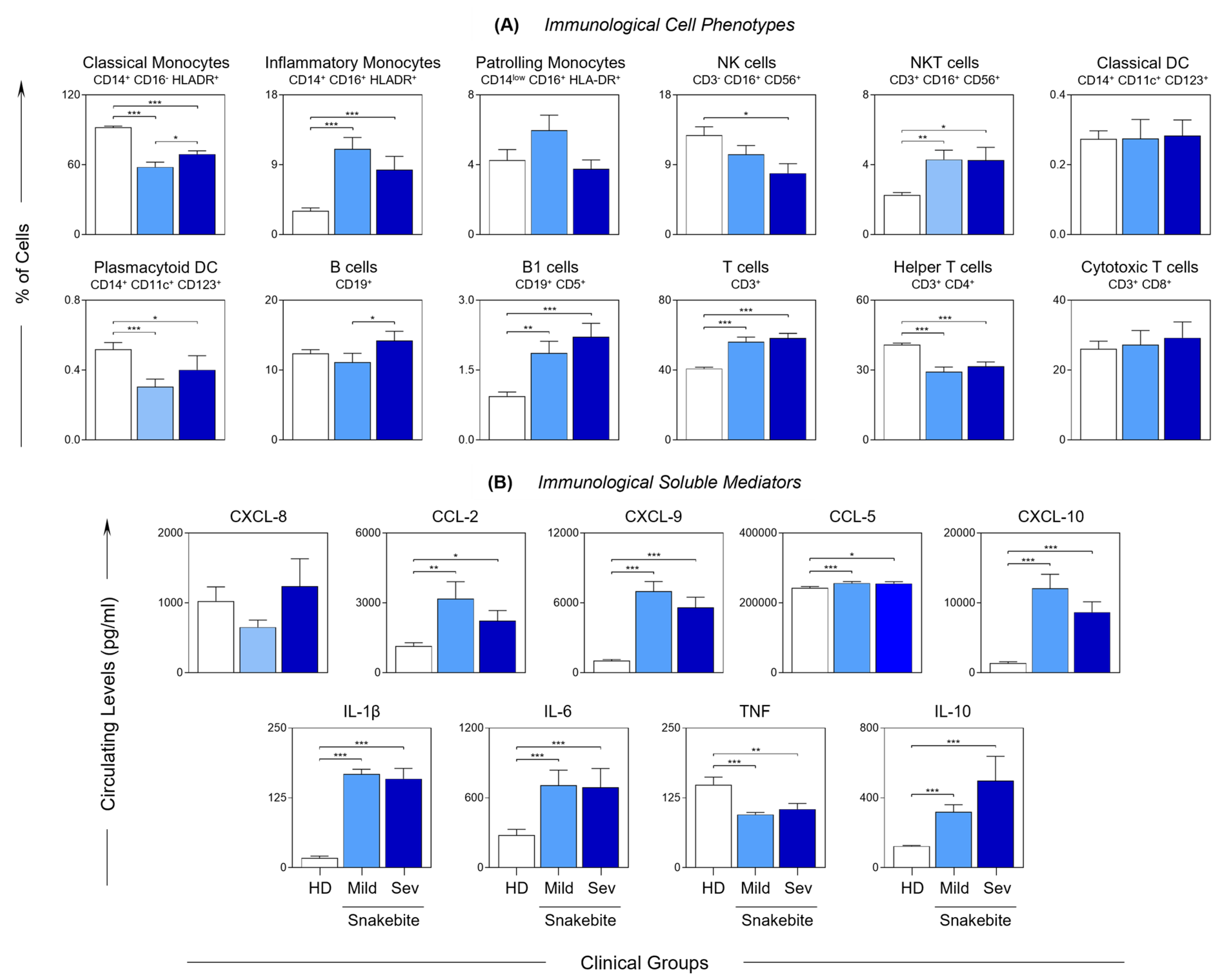
The immunophenotyping profile of B. atrox snakebite patients is presented in Figure 1A. Lower frequencies of classical monocytes, NK cells, plasmacytoid DC and helper T cells were observed in B. atrox snakebite patients (MILD and SEV) compared with the healthy donors (HD). On the other hand, inflammatory monocytes, NKT cells, and B1 and T cells were increased in B. atrox snakebite patients. In addition, the data demonstrated a decrease in classical monocyte and B cell populations in the MILD group compared with the SEV group. The profile of serum chemokines and cytokines observed in Bothrops snakebite patients are shown in Figure 1B. A general increase in the levels of the evaluated soluble immunological mediators, including CCL2, CXCL9, CCL5, CXCL10, IL-1β, IL-6 and IL-10, along with a unique decrease in TNF, was observed in B. atrox snakebite patients compared with the HD group.

2.4. Follow-Up of the Clinical Course of Mild and Severe Bothrops atrox Snakebite Patients Demonstrated Distinct Immunological Profiles at T2

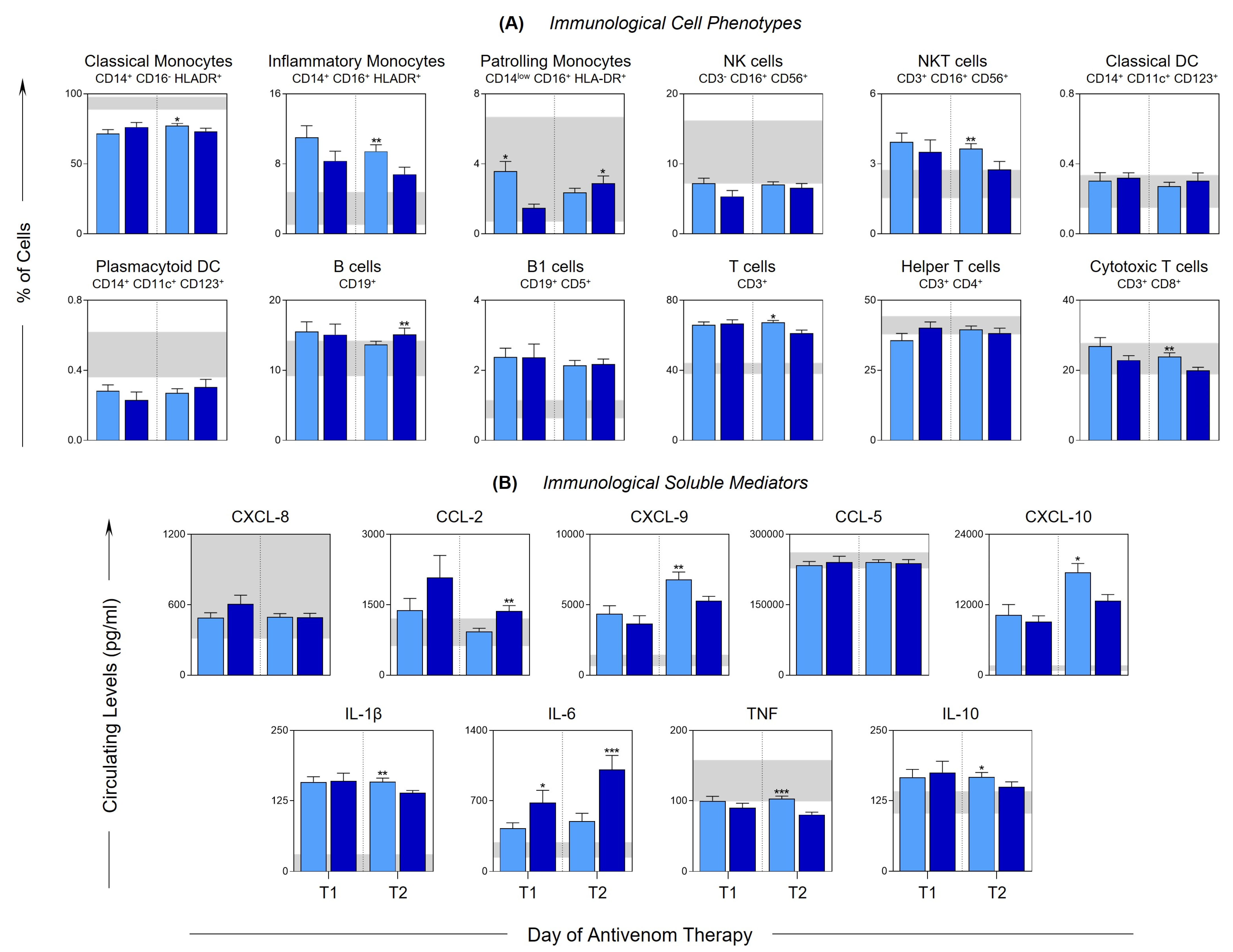
To assess the dynamics of the immune response after antivenom therapy, immunophenotypic characterization was performed and the levels of immunological mediators were evaluated at T1 (24 h after antivenom therapy) and T2 (48 h after antivenom therapy), in the MILD and SEV patients, and are presented in Figure 2A,B, respectively. At T1, there were few differences between the groups, which are represented by the increase in patrolling monocytes and a decrease in IL-6 in the MILD group. In contrast, a change in this behavior was observed at T2, since, compared with the SEV group, the MILD group showed a significant increase in the frequency of classical and inflammatory monocytes, NKT cells, and cytotoxic T and T cells, as well as in the levels of CXCL-9, CXCL-10, IL-1β, TNF and IL-10. In addition, a reduction in the frequency of patrolling monocytes and B cells was observed, along with the mediators CCL2 and IL-6.

2.5. Snakebite Patients Exhibit Complex Integrative Networks and Interactions between the Elements of Peripheral Blood and Blister Exudate

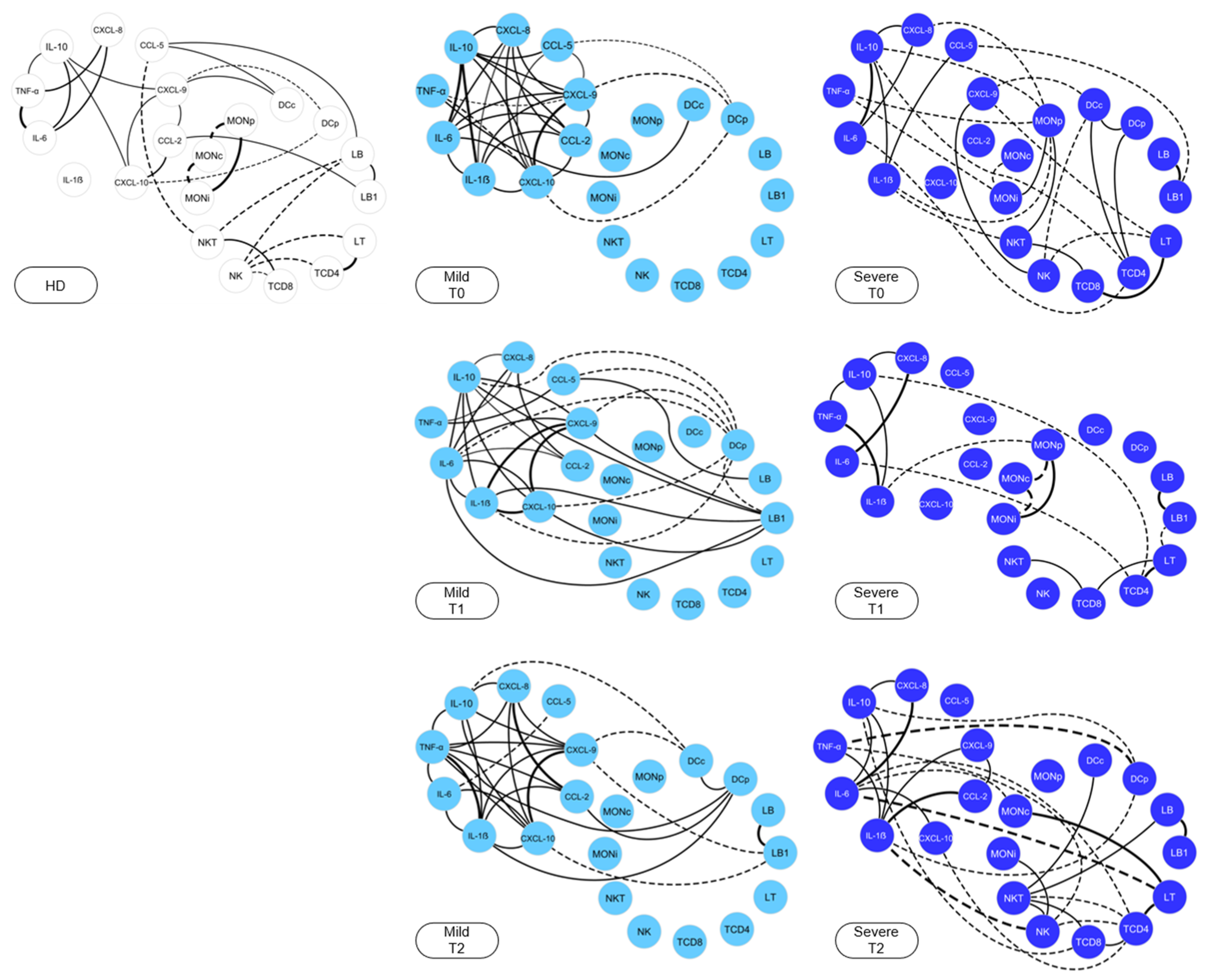
Integrative networks were constructed to demonstrate the interaction between the cell phenotypes and the immunological mediators at T0, T1 and T2, as shown in Figure 3. HDs were used as a reference group and demonstrated a restricted network of interactions between the immunological mediators, with a small number of neighborhood connections, together with cell populations marked by negative correlations. On the other hand, the MILD group presented a network rich in interactions at T0, with a high number of connections between chemokines and pro-inflammatory cytokines, together with the regulatory cytokine IL-10. The SEV group exhibited a distinct profile, with shading of the interactions between immunological mediators, and the presence of negative correlations.

2.6. Evaluation of Blister Exudate in Severe Bothrops atrox Snakebite Patients Revealed Cellular Infiltration and Inflammatory Status

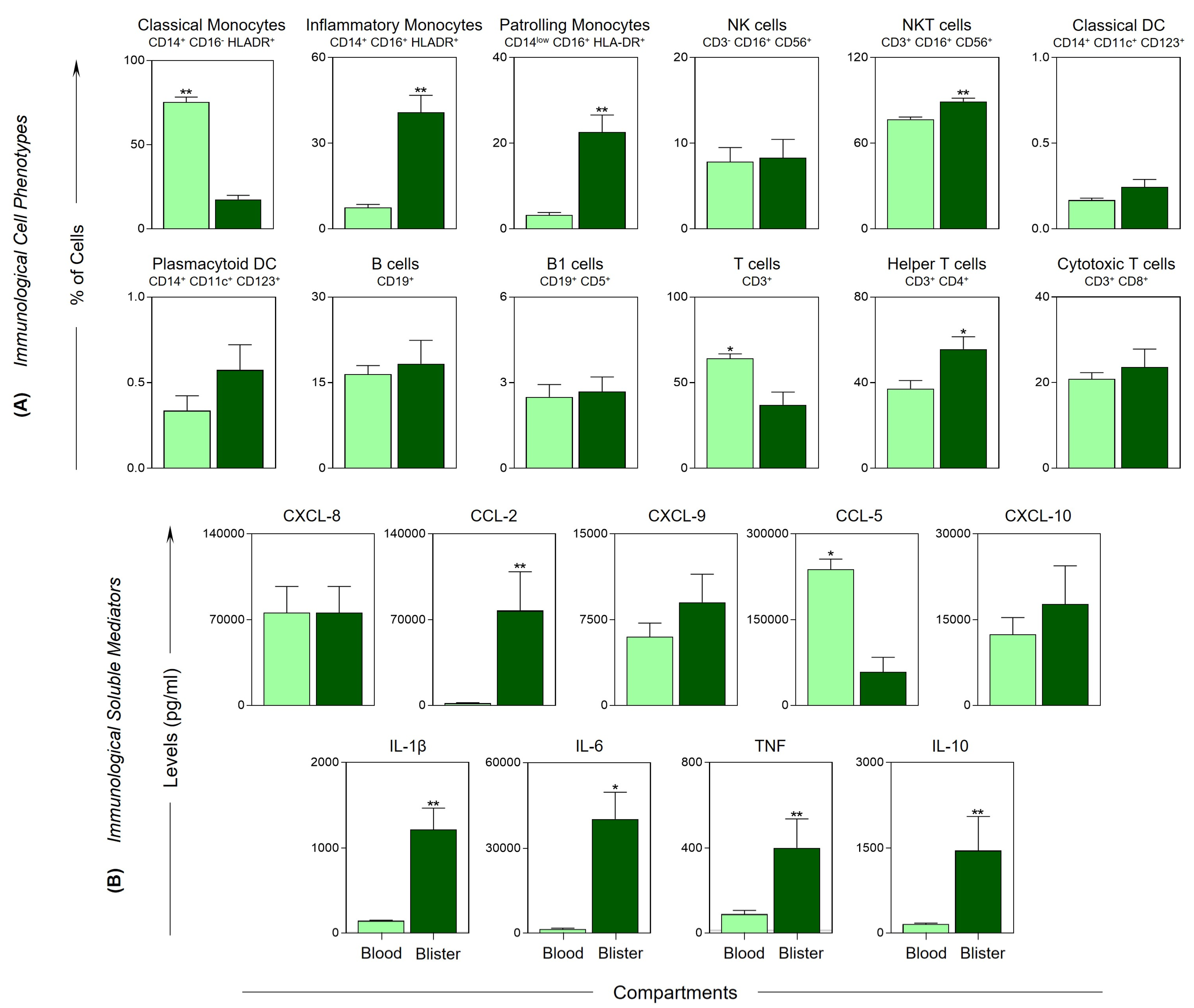
To obtain a broader view of the immune response, blister exudate from patients with severe tissue complications were evaluated at T2 and compared with peripheral blood, as shown in Figure 4. The data demonstrated infiltration of cell populations, with a significant increase in the frequency of inflammatory and patrolling monocytes, NKT cells and helper T cells (Figure 4A). In addition, the blister exudate also demonstrated higher levels of inflammatory mediators, with a significant increase in CCL2, IL-1β, IL-6, TNF and IL-10 (Figure 4B). On the other hand, classical monocytes, T cells and the chemokine receptor CCL5 were higher in the peripheral blood.

2.7. Snakebite Patients Exhibit Complex Integrative Networks and Interactions among the Elements of Peripheral Blood and Blister Exudate

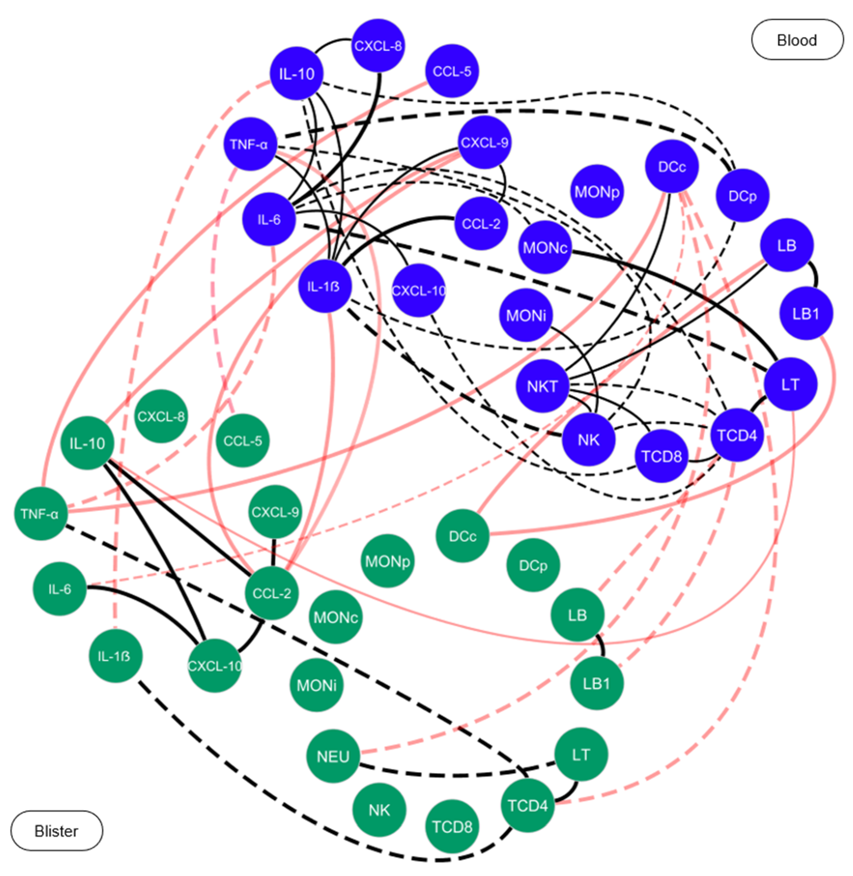
After antivenom therapy, at T1 and T2, it was possible to observe that the MILD group maintained a network profile similar to that of T0, with a slight decrease in the connections among pro-inflammatory mediators. Likewise, at T2, the SEV group exhibited a profile similar to that observed at T0, which was characterized by the presence of negative correlations among the cell populations and immunological mediators. Networks were also built for patients with severe tissue complications, with the aim of investigating the interactions among the elements of the blister exudate, and the communication with the elements of the peripheral blood, as presented in Figure 5. The results showed that, in general, the blister compartment was characterized by the absence of connections among the elements. However, it was possible to observe a clear interaction among the cell populations and the immunological mediators of the blister exudate and peripheral blood, thus indicating a process of recruitment to the inflammatory site.

3. Discussion

Tissue complications caused by Bothrops envenomation directly influence the prognosis of patients, and clinical and epidemiological factors are relevant in terms of aggravation of the outcome, with advanced age, and the absence of or delays in treatment being associated with the severity [33,34]. This corroborates our findings, as the SEV group showed a greater mean age and a longer time until arrival at the medical center. The high number of adverse reactions found in the SEV group may be related to the number of vials administered during treatment after classifying the accident [32,35] (Table 1). Changes in the hematological values were also observed in patients, with a hemorrhagic process leading to a decrease in erythrocytes, hemoglobin and platelets (Table 2). Clinically, the reductions in these parameters were correlated with the severity of the envenomation, and thrombocytopenia on admission was linked to the intense local and systemic hemorrhagic condition that is characteristic of victims of severe snakebite [36,37,38].
Neutrophilic leukocytosis with a leftward shift is also associated with the extent of tissue damage in patients, [25,39,40], and this was corroborated by the data observed in the hemogram, which showed an increase in the neutrophil count. Neutrophils are the first inflammatory cells to reach the damaged tissue, in addition to acting directly on infectious complications after the bite, and induce the synthesis of inflammatory mediators [41,42,43,44,45]. A significant decrease in neutrophils at T2 was also observed in both study groups (Table 2). They are also involved in adaptive immunity by regulating T cells’ functions [43]. Experimental studies have shown a rapid inversion in the ratio of neutrophils to lymphocytes, which shows that Bothrops venom promotes early immune changes, and the proportion of lymphocytes has been used as a predictor of severity in injured patients [11,46,47,48]. Similarly, it was observed that B. atrox snakebite patients presented lymphopenia compared with the control group, markedly in the SEV group, thus reinforcing its correlation with severity.
Some studies have reported that the mediators produced by neutrophils pave the way for the recruitment of inflammatory monocytes [49,50,51]. Monocytes are mononuclear phagocytes that, when recruited, differentiate into macrophages and secrete numerous pro-inflammatory cytokines, thereby intensifying leukocyte recruitment. Our results demonstrated an increase in the populations of inflammatory monocytes, NKT cells and B1 and T lymphocytes, along with increased production of chemokines and inflammatory cytokines (Figure 1A,B).
NKT cells correspond to populations of innate-like T cells that are rapidly activated and can potentiate the action of DCs, macrophages and B lymphocytes, or suppress the immune responses [52]. During patient follow-up, an increase in the frequency of NKT cells was observed in the MILD group (T1) (Figure 2A). A profile change was observed at T2, and compared with the SEV group, the MILD group showed a significant increase in the levels of CXCL9, CXCL10, IL-1β, TNF and IL-10 (Figure 2B).
The cytokine IL-10 is characterized as a key molecule in the regulation of the immune response, which is capable of suppressing the activation of neutrophils and monocytes, and, consequently, the production of chemokines and cytokines, thus regulating the inflammatory processes and cytotoxic pathways [53,54]. Our data from the integrative analysis of the immunological molecules demonstrated the effective participation of IL-10, with several interactions established before and after the administration of antivenom in the MILD group compared with the SEV group, which may be related to the lower severity in these patients (Figure 3). In additional, by comparing the cells and mediators present in the exudate and peripheral blood, we noticed an increase in the recruitment not only of NKT cells but also of inflammatory and patrolling monocytes, in addition to the CCL2, IL1-β, IL-6, TNF and IL-10 mediators, reflecting the local hyperinflammatory process (Figure 4A,B). This distinct response profile between the groups highlights the importance of IL-10 in controlling the immune response to venom toxins. Furthermore, we observed correlations among the molecules and cells present in the peripheral blood and in the blisters, which indicated the interactions among the components of the peripheral blood and the blister exudate (Figure 5).
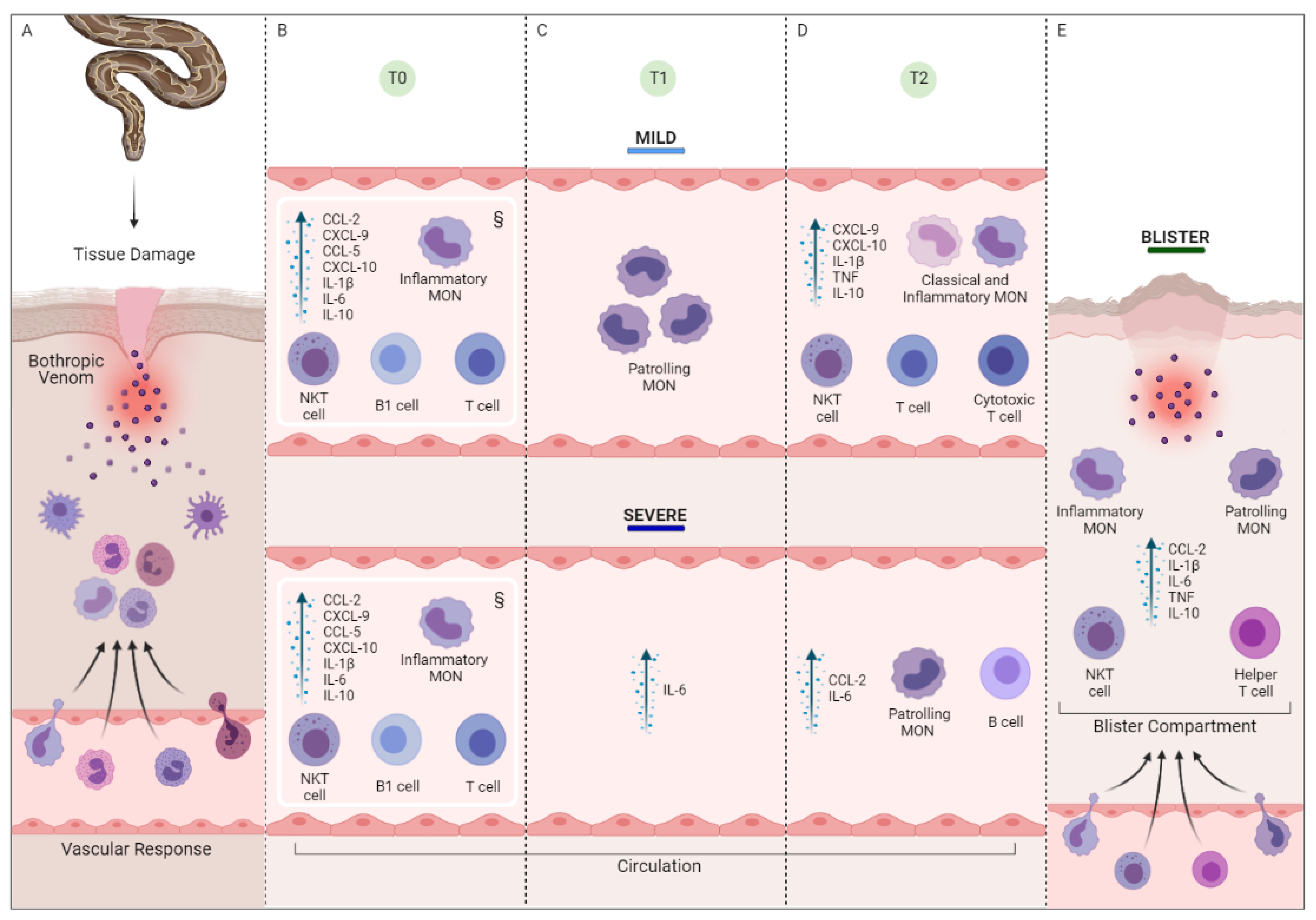
Based on our findings, a schematic model of the local and systemic inflammatory response was constructed (Figure 6). Before the administration of antivenom, we could see a similar response pattern between the groups, with an increase in the percentage of inflammatory monocytes, T lymphocytes, NKT and B cells, together with the chemokines CCL2, CXCL9, CCL5 and CXCL10, and the cytokines IL-1β and IL-10. After the administration of antivenom, we observed the participation of the patrolling monocytes and IL-10 in the MILD group. On the other hand, in the SEV group, the participation of the chemokine CCL2 was observed, which showed high concentrations, both in the circulation and in the exudate. CCL2 is one of the main chemoattractants of monocytes and has previously been described as a potential biomarker of severe tissue complications and systemic complications such as acute renal failure [55]. Finally, the blister exudate was characterized by hyperinflation, marked by the migration of classical and inflammatory monocytes with NKT and Th cells, together with elevated levels of IL-1β, IL-6, TNF and IL-10.
It is important to note that the study has certain limitations such as the short period of recruitment and the low frequency of blister development, reflecting the decrease in the number of samples. We emphasize that the short period of hospitalization (3 days on average, which reflects the efficiency of the health system) made a more extensive follow-up unfeasible. In addition, it was not possible to assess the presence of neutrophils in the peripheral blood and blister exudate via immunophenotyping. However, the results presented here may help us to understand the local and systemic immune response of these envenomations, which are of great clinical and epidemiological relevance. It is also necessary to highlight the difference in age between the control group (lower mean age) and the injured group (higher mean age), which may have an impact on the state of the immune system of these participants. Therefore, there is an evident need for additional studies that further evaluate the immune response of B. atrox snakebite victims, since most of the studies described in the literature have been performed in vitro or in an animal model.

4. Conclusions

The exacerbation of the inflammatory process after B. atrox snakebites has been studied in in vitro and in vivo experiments, and can be used to define the prognosis of the envenomations. Cells such as neutrophils and monocytes play a central role in the body’s defense; however, this same defensive role, if not properly regulated, can lead to the development of local and systemic complications. Our results explored and demonstrated that cell populations and soluble mediators are important components of this inflammatory response, with a distinct profile seen between the groups after the snakebites. The information present in this study clarifies the mechanism of the inflammatory response after Bothops snakebites. We suggest that further studies be carried out to identify the molecules or cells that may be predictive biomarkers in these patients.

5. Materials and Methods

5.1. Study Population and Design

The study population consisted of 48 individuals diagnosed with a B. atrox envenomation after seeking medical care at the Fundação de Medicina Tropical Dr. Heitor Vieira Dourado (FMT-HVD) between March 2019 and March 2021. The identification of B. atrox was performed by a zoologist from the research group.
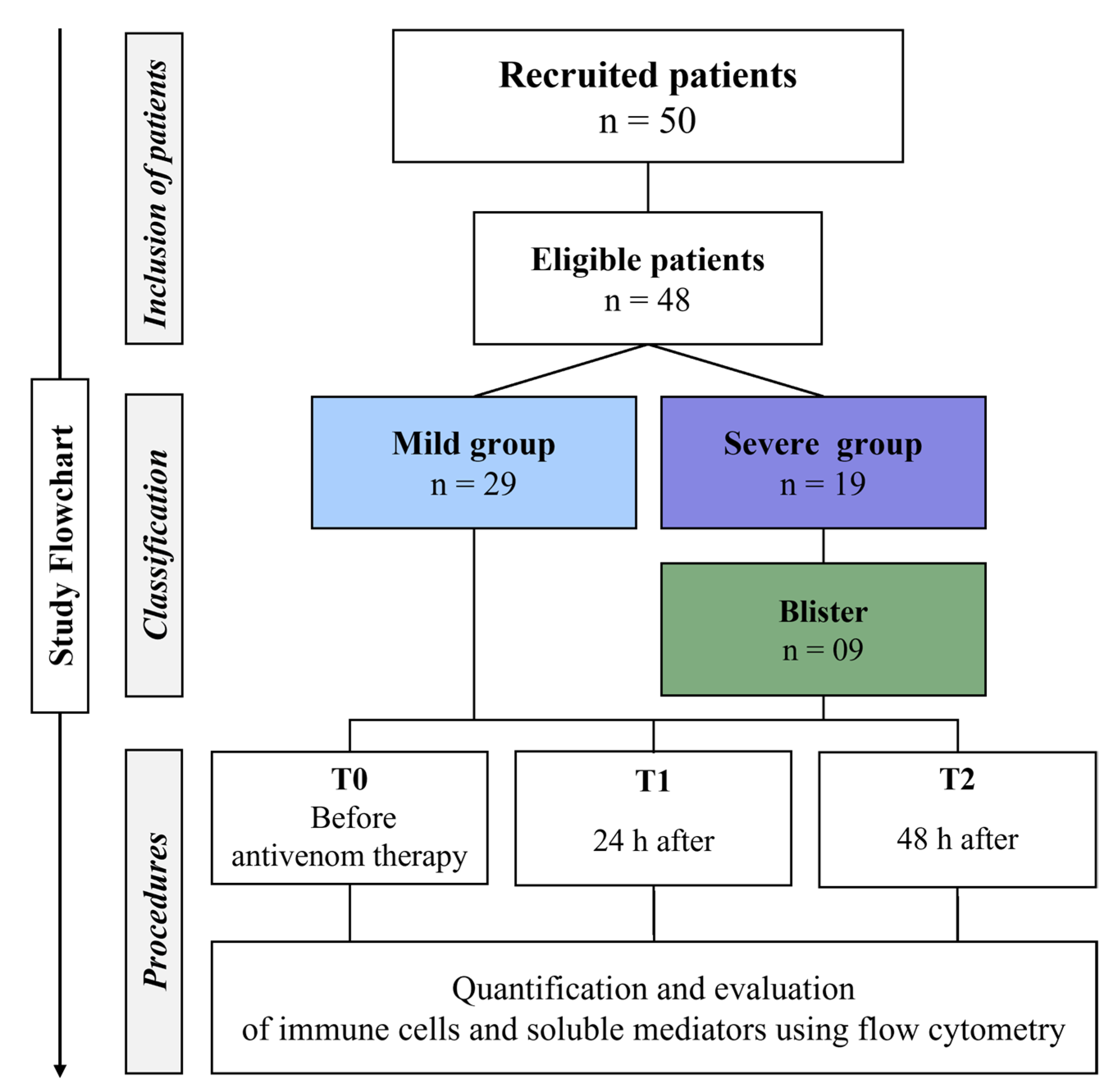
The patients included in the study were classified according to the Brazilian Ministry of Health’s guidelines [55] into two groups, referred to as mild (MILD) and severe (SEV). The MILD group was composed of patients who did not develop severe local and systemic manifestations in the first 48 h after hospitalization, and were treated with 2 to 8 vials of Bothrops antivenom. The severe group (SEV) included patients with severe local and systemic changes, with severe tissue complications and blistering, and were treated with 12 vials of Bothrops antivenom, as shown in Figure 7. The patients were aged over 18 years, had not received previous serum therapy in another health unit, and agreed to participate in the study by signing the informed consent form.
Pregnant women, individuals under the age of 18 or those who reported having a history of previous inflammatory diseases, such as diabetes, autoimmune diseases or immunodeficiency, individuals using platelet antiaggregants and/or anticoagulants, and those who arrived at the emergency clinic 24 h after the snakebite were not included. In addition, in partnership with the Fundação Hospitalar de Hematologia e Hemoterapia do Amazonas (HEMOAM), 48 healthy blood donors (HD) of both sexes with no history of snakebite were included as the control group.

5.2. Ethical Issues

This study was submitted to and approved by the Ethical Committee at FMT-HVD, with the protocol registration number #492,892. All the participants read and signed an informed consent form. The procedures performed are in accordance with the Declaration of Helsinki and Resolution 466/2012 of the Brazilian National Health Council for research involving human participants.

5.3. Collection of Biological Samples and Acquisition of Clinical and Laboratory Data

Patients were monitored and had their peripheral blood collected at 3 time points, referred to as T0 (before antivenom therapy), T1 (24 h after antivenom therapy) and T2 (48 h after antivenom therapy). In addition, blister exudate from patients with severe tissue injury was collected at T2. The peripheral blood and blister exudate were obtained via venipuncture and aspiration, respectively, with 4 mL of peripheral blood (BD Vacutainer EDTA K2) and 200–300 μL of exudate collected from each patient. Full blood counts were performed at T0, T1 and T2 in the hematology sector of the Fundação HEMOAM using the ADVIA 2120i Hematology System (Siemens Healthcare Diagnosis). The collected samples were stored at room temperature (20 °C to 25 °C) and processed within 4 h of collection. Examinations of the complementary and inflammation markers were not requested as part of the medical routine during the patients’ hospitalization period; therefore, our study did not collect these data.
There was no addition of anticoagulants. After blood collection and counting, samples were submitted to centrifugation at 900× g, for 15 min. The supernatant was collected and immediately stored at −80 °C until the evaluation of the soluble immunological mediators. Blister exudate was also submitted to centrifugation at 900× g for 15 min, and the supernatant was also collected and stored at −80 °C until the evaluation of the soluble immunological mediators. Subsequently, the volume of the supernatant of blood samples and blister exudate was made up with PBS and homogenized for cell immunophenotyping. Patients answered a questionnaire with sociodemographic and epidemiological variables to obtain their clinical and epidemiological data, and the clinical information present in the electronic medical records (iDoctor) of the FMT-HVD was also used.

5.4. Immunophenotypic Characterization and Analysis

The immunophenotypic characterization was performed using flow cytometry. The cells were obtained from an aliquot of 100 μL from the peripheral blood and were incubated at room temperature (20 °C to 25 °C) for 30 min in the presence of the following fluorescent-labeled specific anti-human cell surface monoclonal antibodies: anti-CD14-APC/CD16-FITC/HLA-DR-PE to identify the monocyte subtypes (classical, inflammatory and patrolling), anti-CD3-PECy7/CD16-FITC/CD56-PE to identify the natural killer cells (NK) and natural killer T cells (NKT), anti-CD14-APC/CD11c-PE/CD123-FITC to identify the classical and plasmacytoid dendritic cells (DC), anti-CD19-FITC/CD5-PE to identify the B and B1 cells, and anti-CD3-FITC/CD4-PECy7/CD8-PE to identify the helper T cells and cytotoxic T cells. Antibodies were purchased from BD Biosciences (San Diego, CA, USA), Beckman Coulter (Brea, CA, USA) and BioLegend (San Diego, CA, USA). It is worth noting that for labeling the cells of the blister exudate, the same labeling panel was used but with the implementation of the pan-leukocyte antibody (anti-CD45-PerCP) to verify whether the labeled cells corresponded to the leukocyte population of interest. After incubation, the cells were treated with 2 mL of an erythrocyte lysing solution (BD FACS Lysing Solution, BD Biosciences San Diego, CA, USA) for 10 min at room temperature. After two centrifugation steps and two washing steps with PBS, the cells were resuspended in 300 μL of PBS for acquisition in the cytometer.
Sample acquisition was performed on the FACSCanto II cytometer (BD Biosciences, San Jose, CA, USA) at HEMOAM. In total, 30,000 events were acquired for each sample to quantify the cell populations. Furthermore, for the morphometric and immunophenotypic identification of the cells, FlowJo Software (v10.8) was used, with the design of the gates set to select the target populations in graphs that combined the morphological characteristics (size and granulosity) with the immunophenotypic characteristics through the fluorescence of the monoclonal antibodies used to identify the target cells.

5.5. Quantification of the Soluble Immunological Mediators

The levels of the soluble immunological mediators, including chemokines (CXCL8, CCL2, CXCL9, CCL5 and CXCL10) and cytokines (IL-1β, IL-6, TNF and IL-10), were quantified using cytometric bead arrays (BD Human Chemokine and BD Human Inflammatory Cytokines kits, BD Biosciences, San Diego, CA, USA), according to the manufacturer’s instructions. Samples were acquired in the FACSCanto II cytometer (BD Biosciences, San Jose, CA, USA) at HEMOAM. FCAP-Array software v3 (BD Biosciences, San Jose, CA, USA) was used to calculate the chemokine and cytokine levels. Data were reported in picograms per milliliter (pg/mL) concentrations, according to the standard curves provided in the kits.

5.6. Conventional Statistical Analysis

The Shapiro–Wilk test was performed for each variable to verify the data’s normality and revealed a non-parametric distribution. The comparisons of the values between two independent groups were performed using the Mann–Whitney test, while comparisons between two dependent groups were performed using Wilcoxon’s matched pairs signed rank test. In addition, multiple comparisons among groups were performed using Kruskal–Wallis tests followed by Dunn’s test. In all cases, significance was considered at p < 0.05. GraphPad Prism v8.0.1 (GraphPad Software, San Diego, CA, USA) software was used for the statistical analyses. GraphPad Prism and PowerPoint software were used for graphical figures.

5.7. Correlation Network Analysis

The multiple correlations among the cell populations and the soluble immunological mediators in the patients of Bothrops snakebites were determined by using Spearman’s correlation coefficient in GraphPad Prism v8.0.1 (GraphPad Software, San Diego, CA, USA). Positive and negative correlations were considered significant when p < 0.05. Following this, the significant correlations were compiled using the open source Cytoscape software (v3.0.3, National Institute of General Medical Sciences, Bethesda, MD, USA). Immunological networks were constructed using circular layouts, in which each cell or mediator was represented by a globular node. The correlation index (r) was used to categorize the strength of a correlation as weak (r ≤ 0.35), moderate (r ≥ 0.36 to r ≤ 0.67) or strong (r ≥ 0.68), which was represented by connecting edges [31].

Author Contributions

K.F.C., J.C.F.N., F.M.-G., I.A.M.W., W.M.M. and A.G.C. designed and performed the experiments, analyzed the data and wrote the manuscript. K.F.C., J.C.F.N., H.N.S.I., F.M.-G. and A.G.C. analyzed the data and performed the experiments. J.C.F.N., F.M.-G., W.M.M. and A.G.C. revised the manuscript. J.C.F.N., F.B.A.B., F.S.S., I.A.M.W., J.A.G.S., A.M.T., L.C.L.F., A.M., W.M.M. and A.G.C. conceived and supervised the project, designed the experiments, interpreted the data, and wrote and revised the manuscript. All authors have read and agreed to the published version of the manuscript.

Funding

Financial support was provided in the form of grants from Fundação de Amparo à Pesquisa do Estado do Amazonas (FAPEAM) (Pró-Estado Program—#002/2008, #007/2018 and #005/2019, and POSGRAD Program #008/2021 and #005/2022), Conselho Nacional de Desenvolvimento Científico e Tecnológico (CNPq) and Coordenação de Aperfeiçoamento de Pessoal de Nível Superior (CAPES). K.F.C., J.C.F.N., H.N.S.I. and F.M.-G. have fellowships from FAPEAM, CAPES and CNPq (Masters and PHD students). A.M., W.M.M. and A.G.C. are Level 2 research fellows from CNPq. A.G.C. is a research fellow from FAPEAM (PRODOC Program #003/2022). The funders had no role in the study’s design, data collection and analysis, the decision to publish or the preparation of the manuscript.

Institutional Review Board Statement

The study was conducted in accordance with the Declaration of Helsinki, and approved by the FMT-HVD Research Ethics Committee (CEP/FMT-HVD) with the protocol code #2.831.006/2018, 20 August 2018.

Informed Consent Statement

Informed consent was obtained from all subjects involved in the study.

Data Availability Statement

The data used to support the findings of this study are included within the article.

Ethical Approvals and Consent to Participate

Patients were treated according to the recommendations of the Brazilian Ministry of Health.

Acknowledgments

We would like to thank all the researchers at FMT-HVD, HEMOAM and UFAM for their critical discussions and insightful and encouraging ideas, especially the Amazon Group for Interdisciplinary Health Research (Amazon InterScience) research group. We are also grateful for the support and thoughts that helped shape our intuition and the perspectives that are highlighted in this manuscript.

Conflicts of Interest

The authors declare that the research was conducted in the absence of any commercial or financial relationships that could be construed as a potential conflict of interest.

References

  1. World Health Organization. Snakebite Envenoming; World Health Organization: Geneva, Switzerland, 2019. [Google Scholar]
  2. Williams, D.J.; Faiz, M.A.; Abela-Ridder, B.; Ainsworth, S.; Bulfone, T.C.; Nickerson, A.D.; Habib, A.G.; Junghanss, T.; Fan, H.W.; Turner, M.; et al. Strategy for a globally coordinated response to a priority neglected tropical disease: Snakebite envenoming. PLoS Negl. Trop. Dis. 2019, 13, 12–14. [Google Scholar] [CrossRef] [PubMed] [Green Version]
  3. De Oliveira, S.S.; de Sampaio, V.; de Sachett, J.; Alves, E.C.; da Silva, V.C.; de Lima, J.A.A.; da Silva, I.M.; de Ferreira, L.C.; Fan, H.W.; Lacerda, M.V.G.; et al. Snakebites in the Brazilian Amazon: Current Knowledge and Perspectives. In Clinical Toxinology; Academia: San Francisco, CA, USA, 2017; p. 22. ISBN 9789400762886. [Google Scholar]
  4. Gutiérrez, J.M.; Calvete, J.J.; Habib, A.G.; Harrison, R.A.; Williams, D.J.; Warrell, D.A. Snakebite envenoming. Nat. Rev. 2017, 3, 17063. [Google Scholar] [CrossRef] [PubMed]
  5. Feitosa, E.S.; Sampaio, V.; Sachett, J.; Castro, D.B.; Noronha, M.D.; Lozano, J.L.; Muniz, E.; Ferreira, L.C.; Lacerda, M.V.; Monteiro, W.M. Snakebites as a largely neglected problem in the brazilian amazon: Highlights of the epidemiological trends in the state of amazonas. Rev. Soc. Bras. Med. Trop. 2015, 48, 34–41. [Google Scholar] [CrossRef] [PubMed]
  6. Brasil. Ministério da Saúde Acidentes de trabalho por animais peçonhentos entre trabalhadores do campo, floresta e águas, Brasil 2007 a 2017. Bol. Epidemiol. 2019, 50, 1–50. Available online: http://portalarquivos2.saude.gov.br/images/pdf/2019/marco/29/2018-059.pdf (accessed on 10 January 2023).
  7. Roriz, K.R.; Zaqueo, K.D.; Setubal, S.S.; Katsuragawa, T.H.; Silva, R.R.; Fernandes, C.F.; Cardoso, L.A.; Rodrigues, M.M.; Soares, A.M.; Stábeli, R.G.; et al. Epidemiological study of snakebite cases in Brazilian western Amazonia. Rev. Soc. Bras. Med. Trop. 2018, 51, 338–346. [Google Scholar] [CrossRef] [PubMed]
  8. SINAN. Sistema de Informação de Agravos de Notificação—Notificação de Acidentes Botrópicos no Brasil Segundo Região. Available online: https://portalsinan.saude.gov.br/ (accessed on 17 February 2023).
  9. Neiva, M.; Arraes, F.B.; de Souza, J.V.; Radis-Baptista, G.; da Silva, A.R.; Walter, M.E.; de Macedo Brigido, M.; Yamane, T.; Lopez-Lozano, J.L.; Astolfi-Filho, S. Transcriptome analysis of the Amazonian viper Bothrops atrox venom gland using expressed sequence tags (ESTs). Toxicon 2009, 53, 427–436. [Google Scholar] [CrossRef]
  10. Sousa, L.F.; Portes-Junior, J.A.; Nicolau, C.A.; Bernardoni, J.L.; Nishiyama-Jr, M.Y.; Amazonas, D.R.; Freitas-de-Sousa, L.A.; Mourão, R.H.; Chalkidis, H.M.; Valente, R.H.; et al. Functional proteomic analyses of Bothrops atrox venom reveals phenotypes associated with habitat variation in the Amazon. J. Proteomics 2017, 159, 32–46. [Google Scholar] [CrossRef]
  11. Rucavado, A.; Nicolau, C.A.; Escalante, T.; Kim, J.; Herrera, C.; Gutiérrez, J.M.; Fox, J.W. Viperid envenomationwound exudate contributes to increased vascular permeability via a DAMPs/TLR-4 mediated pathway. Toxins 2016, 8, 349. [Google Scholar] [CrossRef] [Green Version]
  12. Gutiérrez, J.M.; Escalante, T.; Rucavado, A.; Herrera, C.; Fox, J.W. A comprehensive view of the structural and functional alterations of extracellular matrix by snake venom metalloproteinases (SVMPs): Novel perspectives on the pathophysiology of envenoming. Toxins 2016, 8, 304. [Google Scholar] [CrossRef] [Green Version]
  13. Jiménez, N.; Escalante, T.; Gutiérrez, J.M.; Rucavado, A. Skin pathology induced by snake venom metalloproteinase: Acute damage, revascularization, and re-epithelization in a mouse ear model. J. Invest. Dermatol. 2008, 128, 2421–2428. [Google Scholar] [CrossRef]
  14. Gutiérrez, J.M.; Escalante, T.; Rucavado, A.; Herrera, C. Hemorrhage Caused by Snake Venom Metalloproteinases: A Journey of Discovery and Understanding. Toxins 2016, 8, 93. [Google Scholar] [CrossRef] [PubMed] [Green Version]
  15. Menaldo, D.L.; Bernardes, C.P.; Jacob-ferreira, A.L.; Nogueira-santos, C.G.; Casare-ogasawara, T.M.; Pereira-crott, L.S.; Sampaio, S.V. Effects of Bothrops atrox venom and two isolated toxins on the human complement system: Modulation of pathways and generation of anaphylatoxins. Mol. Immunol. 2016, 80, 91–100. [Google Scholar] [CrossRef] [PubMed]
  16. Rucavado, A.; Núñez, J.; Gutiérrez, J.M. Blister formation and skin damage induced by BaP1, a haemorrhagic metalloproteinase from the venom of the snake Bothrops asper. Int. J. Exp. Pathol. 1998, 79, 245–254. [Google Scholar]
  17. Herrera, C.; Escalante, T.; Voisin, M.B.; Rucavado, A.; Morazán, D.; Macêdo, J.K.A.; Calvete, J.J.; Sanz, L.; Nourshargh, S.; Gutiérrez, J.M.; et al. Tissue Localization and Extracellular Matrix Degradation by PI, PII and PIII Snake Venom Metalloproteinases: Clues on the Mechanisms of Venom-Induced Hemorrhage. PLoS Negl. Trop. Dis. 2015, 9, e0003731. [Google Scholar] [CrossRef]
  18. De Brito Sousa, J.D.; de Oliveira, S.S.; Sachett, J.; Fan, H.W.; Monteiro, W.M. Low accuracy of microscopic hematuria in detecting coagulopathy from Bothrops pit viper bites, Brazilian Amazon. Clin. Toxicol. 2019, 57, 816–818. [Google Scholar] [CrossRef]
  19. De Cássia Mothé Escocard, R.; Kanashiro, M.M.; Petretski, J.H.; Azevedo-Silva, J.; Queiroz de Carvalho, E.C.; Dias da Silva, W.; Kipnis, T.L. Neutrophils regulate the expression of cytokines, chemokines and nitric oxide synthase/nitric oxide in mice injected with Bothrops atrox venom. Immunobiology 2006, 211, 37–46. [Google Scholar] [CrossRef]
  20. Alves, E.C.; Sachett, J.D.; Sampaio, V.S.; Sousa, J.D.; Oliveira, S.S.; Nascimento, E.F.; Santos, A.D.; da Silva, I.M.; da Silva, A.M.; Wen, F.H.; et al. Predicting acute renal failure in Bothrops snakebite patients in a tertiary reference center, Western Brazilian Amazon. PLoS ONE 2018, 13, 33–37. [Google Scholar] [CrossRef] [PubMed]
  21. Baramova, E.N.; Shannon, J.D.; Bjarnason, J.B.; Fox, J.W. Degradation of extracellular matrix proteins by hemorrhagic metalloproteinases. Arch. Biochem. Biophys. 1989, 275, 63–71. [Google Scholar] [CrossRef] [PubMed]
  22. Gutiérrez, J.M.; Ownby, C.L. Skeletal muscle degeneration induced by venom phospholipases A 2: Insights into the mechanisms of local and systemic myotoxicity. Toxicon 2003, 42, 915–931. [Google Scholar] [CrossRef]
  23. Montecucco, C.; Gutiérrez, J.M.; Lomonte, B. Cellular pathology induced by snake venom phospholipase A2 myotoxins and neurotoxins: Common aspects of their mechanisms of action. Cell. Mol. Life Sci. 2008, 65, 2897–2912. [Google Scholar] [CrossRef]
  24. Wellmann, I.A.M.; Ibiapina, H.N.S.; Sachett, J.A.G.; Sartim, M.A.; Silva, I.M.; Oliveira, S.S.; Tarragô, A.M.; Moura-da-Silva, A.M.; Lacerda, M.V.G.; Ferreira, L.C.d.L.; et al. Correlating Fibrinogen Consumption and Profiles of Inflammatory Molecules in Human Envenomation’s by Bothrops atrox in the Brazilian Amazon. Front. Immunol. 2020, 11, 1874. [Google Scholar] [CrossRef] [PubMed]
  25. Moreira, V.; Dos-Santos, M.C.; Nascimento, N.G.; da Silva, H.B.; Fernandes, C.M.; D’Império Lima, M.R.; Teixeira, C. Local inflammatory events induced by Bothrops atrox snake venom and the release of distinct classes of inflammatory mediators. Toxicon 2012, 60, 12–20. [Google Scholar] [CrossRef] [PubMed]
  26. Leon, G.; Sanchez, L.; Hernandez, A.; Villalta, M.; Herrera, M.; Segura, A.; Estrada, R.; Maria Gutierrez, J. Immune Response Towards Snake Venoms. Inflamm. Allergy Drug Targets 2012, 10, 381–398. [Google Scholar] [CrossRef] [PubMed]
  27. Menaldo, D.L.; Bernardes, C.P.; Zoccal, K.F.; Jacob-ferreira, A.L.; Costa, T.R.; Del, M.P.F.M.; Naal, R.M.Z.G.; Frantz, F.G.; Faccioli, L.H.; Sampaio, S.V. Immune cells and mediators involved in the inflammatory responses induced by a P-I metalloprotease and a phospholipase A 2 from Bothrops atrox venom. Mol. Immunol. 2017, 85, 238–247. [Google Scholar] [CrossRef] [PubMed]
  28. Teixeira, C.; Cury, Y.; Moreira, V.; Picolo, G.; Chaves, F.; Picado, I.C.; Rica, U.D.C.; Jose, S.; Rica, C. Inflammation induced by Bothrops asper venom. Toxicon 2009, 54, 67–76. [Google Scholar] [CrossRef]
  29. Ibiapina, H.N.; Costa, A.G.; Sachett, J.A.; Silva, I.M.; Tarragô, A.M.; Neves, J.C.; Kerr, M.W.; Santana, M.F.; Martins-Filho, O.A.; Lacerda, M.V.; et al. An Immunological Stairway to Severe Tissue Complication Assembly in Bothrops atrox Snakebites. Front. Immunol. 2019, 10, 1–12. [Google Scholar] [CrossRef] [Green Version]
  30. Ministério da Saúde do Brasil. Guia de Vigilância em Saúde 3a Edição; Ministério da Saúde, Secretaria de Vigilância em Saúde, Coordenação-Geral de Desenvolvimento da Epidemiologia em Serviços: Brasilia, Brasil, 2019; p. 741.
  31. Da Silva, I.M.; Tavares, A.M. Comparative evaluation of adverse effects in the use of powder trivalent antivenom and liquid antivenoms in Bothrops snake bites. Rev. Soc. Bras. Med. Trop. 2012, 45, 523–525. [Google Scholar] [CrossRef] [Green Version]
  32. Zychar, B.; Dale, C.; Dermarchi, D.; Gonçalves, L. Contribution of metalloproteases, serine proteases and phospholipases A 2 to the inflammatory reaction induced by Bothrops jararaca crude venom in mice. Toxicon 2010, 55, 227–234. [Google Scholar] [CrossRef]
  33. Hamilton, K.K.; Hattori, R.; Esmon, C.T.; Sims, P.J. Complement proteins C5b-9 induce vesiculation of the endothelial plasma membrane and expose catalytic surface for assembly of the prothrombinase enzyme complex. J. Biol. Chem. 1990, 265, 3809–3814. [Google Scholar] [CrossRef]
  34. Bhaumik, S.; Beri, D.; Tyagi, J.; Clarke, M.; Sharma, S.K.; Williamson, P.R.; Jagnoor, J. Outcomes in intervention research on snakebite envenomation: A systematic review. F1000Research 2022, 11, 628. [Google Scholar] [CrossRef]
  35. Nicoleti, A.F.; de Medeiros, C.R.; Duarte, M.R.; França, F.O.d.S. Comparison of Bothropoides jararaca bites with and without envenoming treated at the vital brazil hospital of the Butantan institute, state of São Paulo, Brazil. Rev. Soc. Bras. Med. Trop. 2010, 43, 657–661. [Google Scholar] [CrossRef] [Green Version]
  36. Santoro, M.L.; Sano-Martins, I.S.; Fan, H.W.; Cardoso, J.L.C.; Theakston, R.D.G.; Warrell, D.A. Haematological evaluation of patients bitten by the jararaca, Bothrops jararaca, in Brazil. Toxicon 2008, 51, 1440–1448. [Google Scholar] [CrossRef] [PubMed]
  37. Cardoso, J.L.C.; Fan, H.W.; Franca, F.O.S.; Jorge, M.T.; Leite, R.P.; Nishioka, S.A.; Avila, A.; Tomy, S.C.; Santoro, M.L. Randomized comparative trial of three antivenoms in the treatment of envenoming by lance-headed vipers (Bothrops jararaca) in Sao Paulo, Brazil. Quartely J. Med. 1993, 86, 315–325. [Google Scholar]
  38. Cardoso, J.L.; Haddad, V., Jr.; França, F.S.; Wen, F.H. Animais Peçonhentos no Brasil—Biologia, Clínica e Terapêutica dos Acidentes, 2nd ed.; Sarvier: São Paulo, Brasil, 2009. [Google Scholar]
  39. Maria Fernandes, C.; Regina Zamuner, S.; Pavan Zuliani, J.; Rucavado, A.; Maria Gutiérrez, J.; de Fátima Pereira Teixeira, C. Inflammatory effects of BaP1 a metalloproteinase isolated from Bothrops asper snake venom: Leukocyte recruitment and release of cytokines. Toxicon 2006, 47, 549–559. [Google Scholar] [CrossRef] [PubMed]
  40. Suwansrinon, K.; Khow, O.; Mitmoonpitak, C.; Daviratanasilpa, S.; Chaiyabutr, N.; Sitprija, V. Effects of Russell’s viper venom fractions on systemic and renal hemodynamics. Toxicon 2007, 49, 82–88. [Google Scholar] [CrossRef]
  41. Ravindran, M.; Khan, M.A.; Palaniyar, N. Neutrophil extracellular trap formation: Physiology, pathology, and pharmacology. Biomolecules 2019, 9, 365. [Google Scholar] [CrossRef] [Green Version]
  42. Schofield, L.; Grau, G.E. Immunological processes in malaria pathogenesis. Nat. Rev. Immunol. 2005, 5, 722–735. [Google Scholar] [CrossRef]
  43. Qiu, Q.; Xiong, W.; Yang, C.; Dai, X.; Dan, X.; Yang, Z.; Jiao, Y.; Xiang, Y.; Liu, G.; Hardy, P. Lymphocyte-derived microparticles induce apoptosis of airway epithelial cells through activation of p38 MAPK and production of arachidonic acid. Apoptosis 2014, 19, 1113–1127. [Google Scholar] [CrossRef]
  44. Liew, P.X.; Kubes, P. The Neutrophil’s role during health and disease. Physiol. Rev. 2019, 99, 1223–1248. [Google Scholar] [CrossRef]
  45. Köse, A.; Akdeniz, A.; Babus, S.B.; Göçmen, M.; Temel, G.O. The Usefulness of Platelet Distribution Width and Platelet Distribution Width to Lymphocyte Ratio in Predicting Severity and Outcomes in Patients with Snakebite. Wilderness Environ. Med. 2021, 32, 284–292. [Google Scholar] [CrossRef]
  46. Lomonte, B. Tissue Damage and Inflamation Induced by Snake Venons; Tryckt & Bunden: Goteborg, Sweden, 1994. [Google Scholar]
  47. Carneiro, A.S.; Ribeiro, O.G.; Cabrera, W.H.K.; Vorraro, F.; De Franco, M.; Ibañez, O.M.; Starobinas, N. Bothrops jararaca venom (BjV) induces differential leukocyte accumulation in mice genetically selected for acute inflammatory reaction: The role of host genetic background on expression of adhesion molecules and release of endogenous mediators. Toxicon 2008, 52, 619–627. [Google Scholar] [CrossRef]
  48. D’Amélio, F.; Vigerelli, H.; Kerkis, I. Bothrops moojeni venom and its components–An overview. J. Venom Res. 2021, 11, 26–33. [Google Scholar]
  49. Soehnlein, O.; Zernecke, A.; Eriksson, E.E.; Rothfuchs, A.G.; Pham, C.T.; Herwald, H.; Bidzhekov, K.; Rottenberg, M.E.; Weber, C.; Lindbom, L. Neutrophil secretion products pave the way for inflammatory monocytes. Blood 2008, 112, 1461–1471. [Google Scholar] [CrossRef] [Green Version]
  50. Pires, W.L.; Kayano, A.M.; de Castro, O.B.; Paloschi, M.V.; Lopes, J.A.; Boeno, C.N.; Pereira, S.D.; Antunes, M.M.; Rodrigues, M.M.; Stábeli, R.G.; et al. Lectin isolated from Bothrops jararacussu venom induces IL-10 release by TCD4+ cells and TNF-α release by monocytes and natural killer cells. J. Leukoc. Biol. 2019, 106, 595–605. [Google Scholar] [CrossRef] [PubMed]
  51. Barraviera, B.; Lomonte, B.; Tarkowski, A.; Hanson, L.Å.; Meira, D.A. Acute-Phase Reactions, Including Cytokines, in Patients Bitten by Bothrops and Crotalus Snakes in Brazil. J. Venom. Anim. Toxins 1995, 1, 11–22. [Google Scholar] [CrossRef]
  52. De Vries, J.E. Immunosuppressive and anti-inflammatory properties of interleukin 10. Ann. Med. 1995, 27, 537–541. [Google Scholar] [CrossRef] [PubMed]
  53. Farsky, S.H.P.; Walber, J.; Costa-Cruz, M.; Curry, Y.; Teixeira, C.F.P. Leukocyte response induced by Bothrops jararaca crude venom: In vivo and in vitro studies. Toxicon 1997, 35, 185–193. [Google Scholar] [CrossRef] [PubMed]
  54. Neves, J.C.F.; Najibe, H.I.S.; Magalhães-gama, F.; Almeida, J.; Sachett, G.; Coelho, K.F.; Alves, E.C.; Tarragô, A.M.; Carlos, L.; Ferreira, D.L.; et al. CCL-2 and CXCL-8: Potential Prognostic Biomarkers of Acute Kidney Injury after a Bothrops atrox Snakebite. Hindawi Mediat. Inflamm. 2022, 2022, 14. [Google Scholar] [CrossRef]
  55. Ministerio da Saúde. Manual de Diagnóstico e Tratamento de Acidentes por Animais Peçonhentos, 2nd ed.; Funasa, Ed.; Ministerio da Saúde: Brasília, Brasil, 2001. [Google Scholar]
Figure 1. Immunological profile in Bothrops snakebite patients before the administration of antivenom. Cell phenotypes (A) and immunological soluble mediators (B) were evaluated before antivenom administration (T0) in the peripheral blood of Bothrops snakebite patients, stratified into mild (Toxins 15 00196 i001) and severe (Toxins 15 00196 i002) groups, and in healthy donors (Toxins 15 00196 i003). The frequency of the cell populations and the levels of the soluble immunological mediators were measured using flow cytometry and CBA, respectively. The results are presented using bar plots, with a linear scale. Statistical analyses were performed using the Mann–Whitney test. Significant differences are indicated by connecting lines and asterisks for p < 0.001 (***), p < 0.01 (**) or p < 0.05 (*).
Toxins 15 00196 g001
Figure 2. Dynamics of the immunological profile of Bothrops snakebite patients after the administration of antivenom. Cell phenotypes (A) and immunological soluble mediators (B) were evaluated at T1 (24 h after antivenom) and T2 (48 h after antivenom) in the peripheral blood of Bothrops snakebite patients stratified into mild (Toxins 15 00196 i004) and severe (Toxins 15 00196 i005) groups. The frequency of cell populations and the levels of soluble immunological mediators were measured using flow cytometry and CBA, respectively. The results are presented using bar plots, with a linear scale. The interquartile ranges (25–75°) for the results observed in the control group were used as a reference interval (gray background—Toxins 15 00196 i006). Statistical analyses were performed using the Mann–Whitney test. Significant differences are indicated by asterisks for p < 0.001 (***), p < 0.01 (**) and p < 0.05 (*).
Toxins 15 00196 g002
Figure 3. Integrative networks of Bothrops snakebite patients in the peripheral blood samples. Networks of Bothrops snakebite patients were constructed before (T0) and after (T1 and T2) the administration of antivenom, demonstrating the interactions between cell phenotypes and immunological soluble mediators. White nodes (Toxins 15 00196 i007) are used to identify healthy donors, light blue nodes (Toxins 15 00196 i008) are used to identify the MILD group, while dark blue (Toxins 15 00196 i009) is used to identify the SEVERE group. Solid lines between the elements indicate a positive correlation, while dashed lines indicate a negative correlation. The thickness of the lines indicates the strength of the correlation. The correlation index (r) was used to categorize the strength of the correlation as weak (r ≤ 0.35), moderate (r ≥ 0.36 to r ≤ 0.67) or strong (r ≥ 0.68).
Toxins 15 00196 g003
Figure 4. Comparison of blister exudate vs. peripheral blood in severe Bothrops snakebite patients. Cell phenotypes (A) and immunological soluble mediators (B) were evaluated at T2 (48 h after the administration of antivenom) in the blood (Toxins 15 00196 i010) and blister (Toxins 15 00196 i011) compartment of severe Bothrops snakebite patients. The frequency of cell populations and the levels of soluble immunological mediators were measured using flow cytometry and CBA, respectively. The results are presented using bar plots, with a linear scale. Statistical analyses were performed using Wilcoxon’s matched pairs signed rank test. Significant differences are indicated by asterisks for p < 0.01 (**) and p < 0.05 (*).
Toxins 15 00196 g004
Figure 5. Integrative networks of Bothrops snakebite patients in the peripheral blood and blister exudate. Networks of Bothrops snakebite patients were constructed before (T0) and after (T1 and T2) the administration of antivenom, demonstrating the interactions of cell phenotypes and immunological soluble mediators in blisters and peripheral blood. Green nodes (Toxins 15 00196 i012) represent the elements of blister exudate, and blue nodes (Toxins 15 00196 i013) are used to identify the elements of peripheral blood. Solid lines between the elements indicate a positive correlation, while dashed lines indicate a negative correlation. The red lines indicate the correlations between the blister exudate and peripheral blood. The thickness of lines indicates the strength of the correlation. The correlation index (r) was used to categorize the strength of the correlation as weak (r ≤ 0.35), moderate (r ≥ 0.36 to r ≤ 0.67) or strong (r ≥ 0.68).
Toxins 15 00196 g005
Figure 6. Schematic summary of the local and systemic inflammatory response to B. atrox envenomation with and without severe tissue complications. Quadrant (A) represents the bite and subcutaneous/intramuscular injection of BaV, inducing tissue damage and promoting the activation of the resident cells, the vascular response and the recruitment of circulating cells to the affected site. (B) Pre-serum therapy response of patients stratified into mild and severe groups, compared with the control group, represented by the symbol (§). The case groups show a similar pattern of response at T0, with an increase in the frequency of inflammatory monocytes, T lymphocytes, NKT and B cells. (C) Changes in response profile 24 h after the administration of serum therapy. The possible action of patrolling monocytes was seen in the mild group and the inflammatory cytokine IL-6 in the severe group. (D) The percentage of cells increased 48 h post-treatment in the MILD group for classic and inflammatory monocytes and TCD8+ lymphocytes, in addition to CXCL9, CXCL10, IL1β, TNF and IL-10. In the SEV group, the response profile demonstrated the possible action of patrolling monocytes, B lymphocytes and CCL2 and IL-6 molecules. Quadrant (E) represents the response at the local level, with the action of subpopulations of the inflammatory and patrolling monocytes, as well as NKT and TCD4+ lymphocytes, and the CCL2, IL1 β, IL-6 and IL-10 mediators.
Toxins 15 00196 g006
Figure 7. Study flowchart. In total, 48 patients were considered eligible, which were divided into two groups according to their clinical evolution and were classified as either mild or severe. The mild group was composed of 29 patients and the severe group comprised 19 patients. In addition, of the patients with a severe snakebite, 9 had tissue complications and blister formation, and the transudate was collected 48 h after administration of the antivenom. Subsequently, the cellular profile and soluble immunological mediators were evaluated via immunophenotyping and CBA, respectively, before the administration of antivenom and 24 and 48 h after administration.
Toxins 15 00196 g007
Table 1. Clinical and demographic data of Bothrops snakebite patients, stratified into mild and severe groups.
Demographic and Clinical CharacteristicsHD (n = 48)B. atrox Patientsp-Value
Mild (n = 29)Severe (n = 19)
Gender (n, male/female)34/1426/315/40.248
Age (median and (IQR))33(23–44)46 (28–60)53 (30–63)0.0067 #
Previous snakebite (n, yes/no)-3/265/140.146
Zone of occurrence (n, rural/urban)-26/315/40.304
Anatomical site of the snakebite (n, upper/lower) limb)-4/259/100.010 #
Anatomical site of the snakebite (n, right/left)-11/1812/70.087
Work-related accident (n, yes/no)-10/197/120.867
Adverse reaction to the antivenom (n, yes/no) *-8/2111/80.035 #
Time between snakebite and administration of the antivenom (hours, median (IQR))-3 (2–6)6 (3–9)0.129
HD, healthy blood donor; IQR, interquartile range. Significant differences at p < 0.05 for comparisons between the mild and severe groups are represented in bold with the superscript symbol #. * Adverse reactions to antivenom, found in the mild and severe groups, were classified as anaphylactic clinical reactions, and reactions described in the literature as mild or rare events, such as pruritus and skin reactions, hyperemia and nausea [31,32].
Table 2. Blood count and immunophenotyping data of Bothrops snakebite patients, stratified into mild and severe groups, before and after serum therapy.
ParametersMild (n = 29)Severe (n = 19)HD (n = 48)
T0T1T2T0T1T2T0
Red blood cells5.2 (4.8–5.3)4.6 (4.4–4.9) a3.2 (2.5–4.5) a.b5.2 (4.9–5.4)4.8 (4.6–5.1) a3.7 (0.7–4.7) a.b5.0 (4.6–5.4)
Hemoglobin15.0 (14.3–15.4)13.6 (12.1–14.2) a8.8 (6.8–13.0) a.b15.2 (14.5–15.6)13.9 (13.2–15.4) a11.4 (1.9–13.5) a.b14.9 (13.6–16.0)
Platelets221 (167–259) §191 (182–232)117 (97–171) a.b.#185 (149–231) §202 (151–243)61 (31–193) a.b243 (211–282)
Total leukocytes10.9 (8.4–14.6) §11.6 (8.7–15.0)4.8 (3.8–7.6) a.b13.3 (9.7–15.6) §12.3 (9.1–15.0)8.3 (1.3–11.5) a.b14.9 (13.6–16.0)
Neutrophils77.0 (67.7–88.9) §67.8 (62.9–77.8) a47.4 (36.8–62.3) a.b80.3 (69.0–89.0) §71.0 (64.4–82.9) 60.2 (10.0–77.7) a.b56.1 (50.0–61.5)
Lymphocytes13.0 (5.0–21.3) §16.9 (13.3–25.5) a13.6 (11.3–20.5) #12.1 (5.3–20.3) §15.5 (10.0–22.0) a8.5 (1.3–10.7) b29.7 (26.4–34.6)
Monocytes4.0 (3.5–5.7) §5.6 (4.5–6.7) a4.1 (3.3–5.7) b4.1 (3.1–5.1) §5.4 (4.0–6.6) 4.4 (0.7–5.3) b6.0 (5.1–6.6)
Eosinophils2.0 (0.9–4.7) §1.8 (1.0–2.6) 2.8 (2.2–3.2) b1.8 (0.4–5.8) §2.3 (1.2–3.5) 1.8 (1.0–4.9)3.3 (2.3–6.1)
Basophils0.4 (0.2–0.8)0.3 (0.2–0.4) a0.3 (0.2–0.3) #0.3 (0.2–0.4) §0.3 (0.2–0.4) 0.1 (0.0–0.3) a.b0.5 (0.3–0.9)
T0, before serum therapy; T1, 24 h after serum therapy; T2, 48 h after serum therapy; HD, healthy donor; IQR, interquartile range. Reference values: red blood cells, 4.5–6.0 × 10⁶/µL); hemoglobin, 12–18 g/dL; platelets, 130–400 × 10³/µL; total leukocytes, 5.2–12.4 × 10³/µL; neutrophils, 1.9–8.0 × 10³/µL; lymphocytes, 0.9–5.2 × 10³/µL; monocytes, 0.16–1 × 10³/µL; eosinophils, 0.1–5.0 × 10³/µL; basophils, 0.0–2.0 × 10³/µL. Significant differences at p < 0.05 for intragroup analysis are represented in bold with the superscript letters “a” and “b” refer to comparisons with T0 and T1, respectively. Significant differences at p < 0.05 for intergroup analysis are represented in bold with the superscript symbols # and §, which refer to comparisons with the severe group and the healthy donor group, respectively.
Disclaimer/Publisher’s Note: The statements, opinions and data contained in all publications are solely those of the individual author(s) and contributor(s) and not of MDPI and/or the editor(s). MDPI and/or the editor(s) disclaim responsibility for any injury to people or property resulting from any ideas, methods, instructions or products referred to in the content.