Article

The Effect of Mushroom Culture Filtrates on the Inhibition of Mycotoxins Produced by Aspergillus flavus and Aspergillus carbonarius

1
Department of Ecology, Aquaculture, and Agriculture, University of Zadar, 23000 Zadar, Croatia
2
Department of Environmental Biology, Sapienza University of Rome, 00185 Rome, Italy
3
Department of Life Sciences, University of Trieste, 34127 Trieste, Italy
*
Author to whom correspondence should be addressed.
Toxins 2023, 15(3), 177; https://doi.org/10.3390/toxins15030177
Submission received: 30 December 2022 / Revised: 16 February 2023 / Accepted: 22 February 2023 / Published: 25 February 2023
(This article belongs to the Special Issue Advances in Rapid Detection and Reduction of Aflatoxins)

Abstract

:
Two of the mycotoxins of greatest agroeconomic significance are aflatoxin B1 (AFB1), and ochratoxin A (OTA). It has been reported that extracts from some wood-decaying mushrooms, such as Lentinula edodes and Trametes versicolor showed the ability to inhibit AFB1 or OTA biosynthesis. Therefore, in our study, a wide screening of 42 isolates of different ligninolytic mushrooms was assayed for their ability to inhibit the synthesis of OTA in Aspergillus carbonarius and AFB1 in Aspergillus flavus, in order to find a metabolite that can simultaneously inhibit both mycotoxins. The results showed that four isolates produce metabolites able to inhibit the synthesis of OTA, and 11 isolates produced metabolites that inhibited AFB1 by >50%. Two strains, the Trametes versicolor strain TV117 and the Schizophyllum commune strain S.C. Ailanto, produced metabolites able to significantly inhibit (>90%) the synthesis of both mycotoxins. Preliminary results suggest that the mechanism of efficacy of the S. commune rough and semipurified polysaccharides could be analogous to that found previously for Tramesan®, by enhancing the antioxidant response in the target fungal cells. The overall results indicate that S. commune’s polysaccharide(s) could be a potential agent(s) in biological control and/or a useful component of the integrated strategies able to control mycotoxin synthesis.
Key Contribution: The effect of mushroom culture filtrates on the inhibition of mycotoxins produced by Aspergillus flavus and Aspergillus carbonarius demonstrate that mushrooms are an invaluable source of bioactive compounds. Since these organisms can grow on almost every substrate (even wastes) becomes determinant their use in a view of circular economy to limit the amount of hazardous compounds into feed and foodstuff.

1. Introduction

Mycotoxins are low-molecular-weight secondary metabolites synthesized by some filamentous fungi that are toxic to vertebrates [1,2]. The effects of mycotoxins on humans and animals vary according to their chemical structure and concentration and can cause acute and/or chronic effects. Cytotoxicity is the main cause of acute mycotoxicosis, while genotoxicity, which in some cases can have carcinogenic effects, is connected to chronic mycotoxicosis [2,3,4]. Often, more than one mycotoxin is found on contaminated food and feed commodities. The research on possible synergic effects of co-contamination with different mycotoxins is still ongoing [2,5,6], although, the synergistic effects of mycotoxins have not been sufficiently studied to date. Aflatoxins (AF) B1, B2, G1, and G2, as well as ochratoxin A (OTA), are among the most important and studied mycotoxins. Aflatoxin B1 (AFB1) is classified as carcinogenic (group 1A) by the International Agency for Research on Cancer [7] while OTA is classified as a possible carcinogen for humans, group 2B [8]. Aflatoxin B1 and ochratoxin A are important in a range of economically important commodities. Both mycotoxins are synthesized by species of the genus Aspergillus which are xerophilic and able to grow under water stress conditions, much wider than those allowing plant growth. They can thus colonize and contaminate a range of food commodities both in the pre-and postharvest period [9]. Moreover, both AFB1 and OTA are stable molecules and relatively heat resistant during food and feed processing [10]. Furthermore, both can be transmitted along the food chain and stored in some tissues of animals fed with contaminated feed [11]. Due to the severity of the toxin, the presence of approximately a dozen of mycotoxins, including AFB1 and OTA, are regulated by EC regulation [12].
Different strategies to control the presence of mycotoxins in food and feed have been applied for many years, however, none of them have been completely successful. Thus, there is still a need for minimization and prevention control strategies for these mycotoxins. Many strategies have been predominantly based on the use of chemical compounds (pesticides and fungicides). The number of chemicals that were applied in the prevention of fungal growth and/or mycotoxin control has been banned by authorities [13,14,15]. This, together with rising environmental awareness of the population, led to an increase in the research for new more environmentally friendly strategies in mycotoxin control. Detoxification methods can be applied to contaminated seeds, and several physical, chemical, and biological methods are under research. Moreover, some interesting results were obtained by using curcumin and purified enzymes, including laccases, manganese peroxidase, and the recently identified bacillus aflatoxin-degrading enzyme [16,17,18,19,20,21].
In the last 20 years, different research confirmed that mushroom polysaccharides could represent a valid tool in mycotoxin control [22,23,24,25,26]. The most studied inhibiting effects of mushroom metabolites are those made with Lentinula edodes [22] and Trametes versicolor [15,24,25] using lyophilized culture filtrates. Moreover, Scarpari and collaborators [25] were able to identify the active principles produced by T. versicolor, which was an exopolysaccharide released in its culture filtrate, called tramesan. tramesan can act as a pro-antioxidant in different organisms and has biological activity in blocking AFB1 and OTA production probably by acting on gene expression and consequent stimulation of antioxidant activity in the fungal cell [22,23,24]. Furthermore, Loncar and collaborators [15] have demonstrated that tramesan fragments larger than seven units inhibited up to 90% AFB1 synthesis by A. flavus and up to 81% of the biosynthesis of OTA by A. carbonarius. Even if tramesan is a very promising tool in the challenge against the presence of mycotoxins in food and feed, the production costs could be too high for large-scale use. Moreover, tramesan is produced by a living organism and some mutation(s) could alter its structure and efficacy. Given the similar mechanism of action of the mushroom polysaccharides studied so far, the present study screened 42 isolates of different ligninolytic mushrooms present in the collection of the Laboratory of Plant Pathology and Mycology of the Sapienza University of Rome for their capacity to produce metabolites able to control simultaneously the biosynthesis of both AFB1 and OTA. The goal was to identify the tool that could be effectively used for the significant control of AFB1 and OTA production and use in food and feed applications. Furthermore, the aim was to clarify the structure of the mushroom polysaccharides capable of inhibiting the synthesis of mycotoxins, so that it could be established whether there is a common structure that can be used in designing synthetic glucans for mycotoxin control.

2. Results

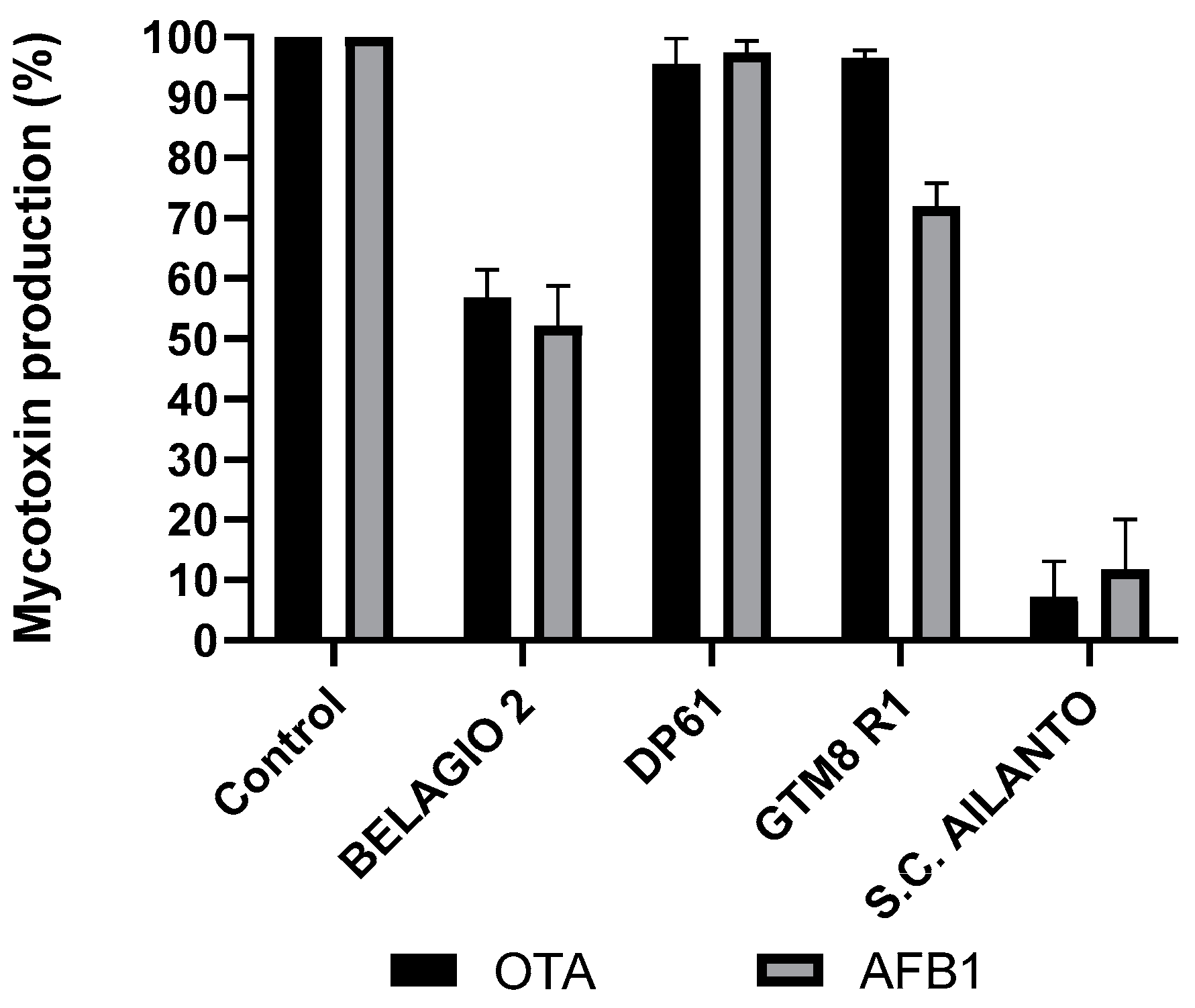
2.1. Wide Screening Mushrooms Assay

This research aimed to identify mushroom polysaccharides able to inhibit the synthesis of both AFB1 and OTA. The first step was a wide screening assay of lyophilized liquid culture filtrates (LFC) of the different isolates of different mushroom species. The inhibition of OTA and AFB1 biosynthesis in the presence of 2% w/v of LCF of different isolates of ligninolytic mushrooms expressed as a percentage compared to nontreated samples (control) are shown in Table 1. For the purpose of this study, only inhibition rates higher than 50% were considered significant.
The results indicate that the inhibition of both mycotoxins was more isolate-dependent than species-dependent. This is particularly visible in the case of different isolates of G. adspersum. One isolate of G adspersum, COLLETTA 12, showed significant inhibition (p < 0.001) of both mycotoxins, however, the inhibition was lower than 50%, and therefore considered of no significance for the scope of this research. P COLLEGNO 2A inhibited more OTA biosynthesis than AFB1. One, Tiglio, inhibited the biosynthesis of both mycotoxins for about 30%, and the LCF of four isolates showed inhibition of AFB1 synthesis over 50% while the inhibition of OTA was under 50% (Figure 1). A similar situation can be observed for S. commune isolates. Two isolates, DP 61 and GTM8 R1 showed low inhibition rates for both toxins. Isolate BELLAGIO 2 showed inhibition rates between 40 and 50% for both toxins while lyophilized filtrate of isolate S. C. Alianto inhibited the biosynthesis of both mycotoxins for over 90% (Figure 2). The yield of LCFs doesn’t seem to be directly correlated with the inhibition of AFB1 and OTA. Different isolates of some species, like Fomes fomentarius, show minor variations in LCF yield, while for the others (i.e., Ganoderma adspermum and G. resinaceum), the variation in the yield was higher (Table 1). The aim of this research was to isolate a mushroom polysaccharide able to provide a high inhibition of both mycotoxins and compare it with tramesan. Therefore the LCF of Schizophyllum commune SC Alianto was chosen for further research.

2.2. Influence of the LCF of S. commune on AFB1 and OTA Present in the Media

To verify that LCF of S. commune inhibits mycotoxin synthesis and does not interfere with mycotoxins present in the media (degradation, adsorption, or others), the known concentration of standards of AFB1 and OTA in potato dextrose broth (PDB) were incubated for three days in the presence and absence (control) of S. commune’s LCF 2% w/v. The results (Table 2) show that LCF does not interfere with the toxins present in the media and that the lower concentration of mycotoxins observed in the culture with the presence of LCF is due to the inhibition of the toxin synthesis rather than due to the interference with the mycotoxins present in the culture media.

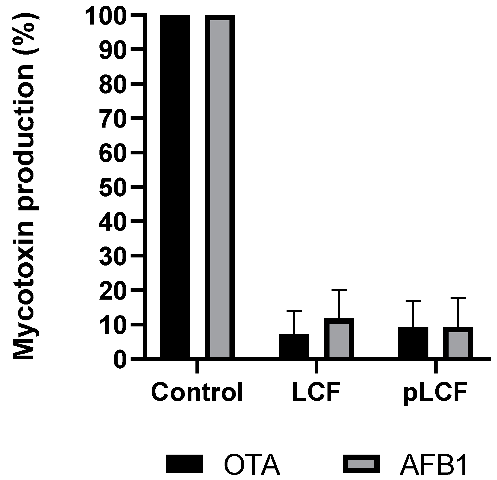
2.3. Assay of the Influence of LCF and Partially Purified pLCF of S. commune S. C. Alianto

LCFs of S. commune S. C. Ailanto were partially purified by pure ethanol precipitation and subsequent dialysis (see the Section 5 for details). Obtained partially purified LCF contained mostly polysaccharides and glyco material, and the protein part was further reduced by treatment with proteinase K followed by centrifugation which gave two fractions, namely a supernatant, sLCF, and a precipitate, pLCF. UV spectroscopy of the two fractions showed the absence of absorbance at about 280 nm, thus suggesting that proteins were successfully removed (data not shown). Both fractions were lyophilized and assayed at a concentration of 2% w/v on AFB1 and OTA production. The sLCF fraction showed no inhibition of mycotoxin synthesis (data not shown) while the effect of pLCF on the synthesis of AFB1 and OTA was not significantly different (p > 0.001) from that obtained with LCF (Figure 3). The results obtained with pLCF suggested that a polysaccharide (or polysaccharides) might be involved in the inhibition.

2.4. Composition Analysis of pLCF and sLCF Fractions

The monosaccharides present in the two fractions were determined by gas–liquid chromatography after methanolysis and derivatization to trimethylsilyl methyl glycosides. The fraction sLCF was found to contain mannose, galactose, and glucose in relative molar ratios of 8:2:1, while the sample pLCF was to be mainly constituted of glucose, with only traces of mannose.

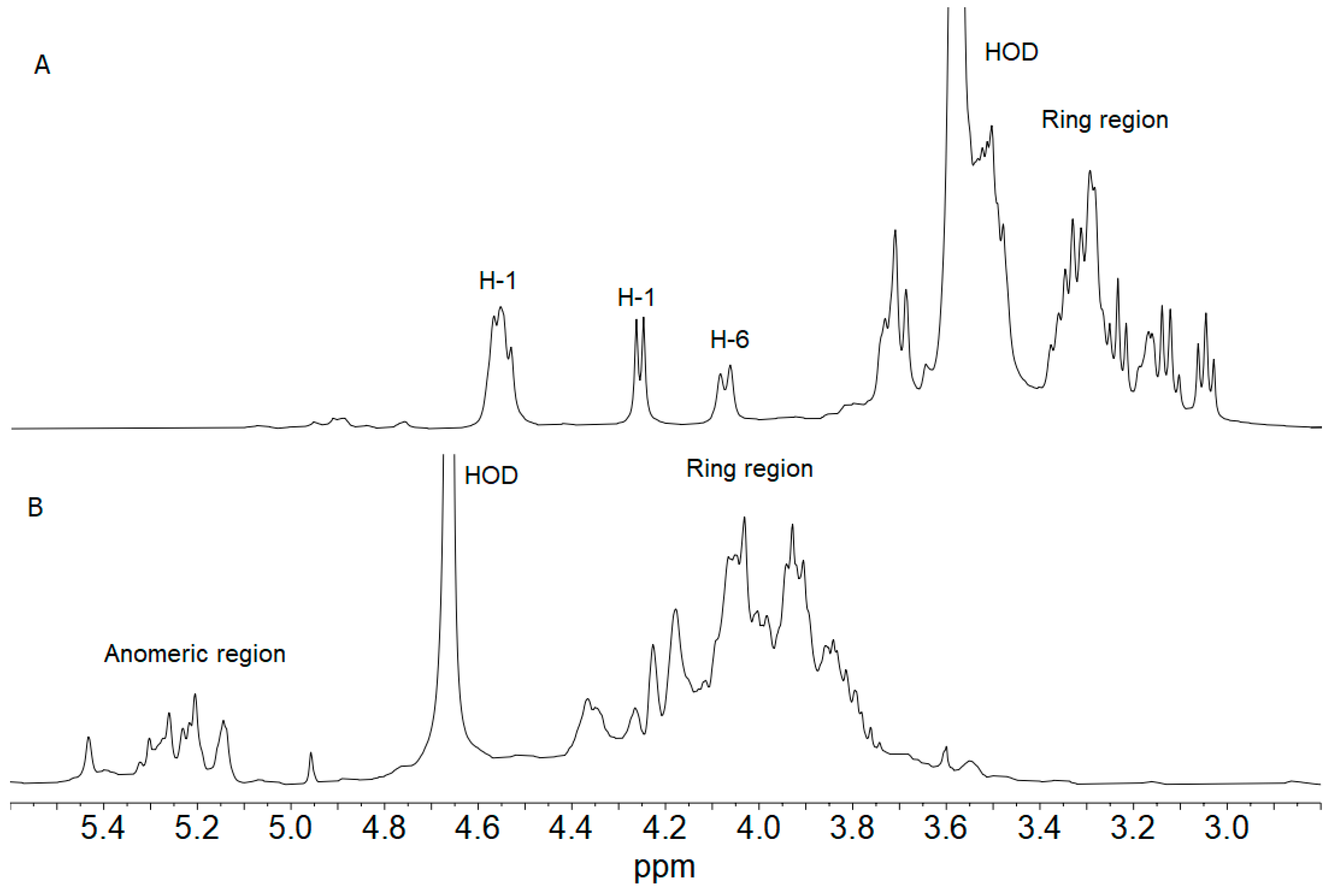
2.5. 1H NMR Spectroscopy of pLCF and sLCF Fractions

The 1H NMR spectrum of pLCF was recorded at 70 °C in deuterated DMSO: D2O in a 6:1 ratio in order to overcome the high viscosity of its water solutions and to eliminate the resonances arising from exchangeable hydroxyl protons. The 1H NMR spectrum is shown in Figure 4A. Apart from two intense signals at 2.53 (not shown) and 3.57 ppm attributed to DMSO and residual HOD, respectively, it contains signals typical of polysaccharides. Comparison of the pLCF spectrum with that of the β-glucan Schizophyllan recorded in the same experimental conditions [27] showed that the two samples are extremely similar, if not identical. Resorting to the data in reference [27], the signal at 4.24 ppm, together with the three overlapping resonances between 4.60 and 4.50 ppm, were assigned to the anomeric protons of β-glucose residues, while that at 4.08 was attributed to the H-6 of the branched β-glucose residue. The ring proton resonances included in the range 3.80–3.00 ppm are also in very good agreement with those attributed to Schizophyllan.
The 1H NMR spectrum of sLCF, recorded at 50 °C in D2O (Figure 4B), showed signals typical of anomeric and ring protons of polysaccharides. In particular, the anomeric region contains many resonances, some of them also overlapped, in agreement with sugars in the α-anomeric configuration. The abundance of signals might be attributed to a mixture of polysaccharides or the presence of a rather complicated and not-so-regular repeating unit, as often found in fungi polysaccharides.

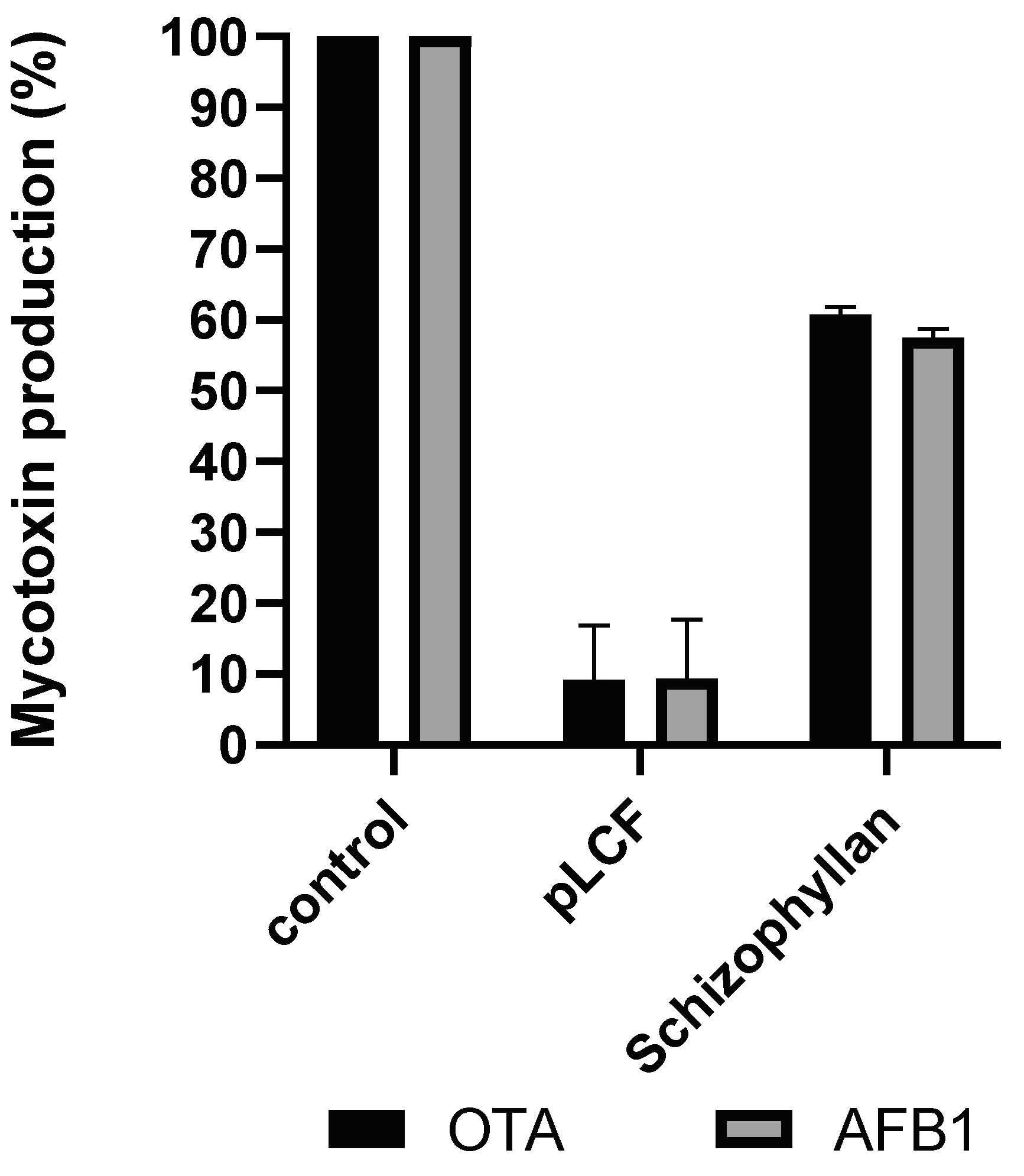
2.6. Assay of the Influence of Partially Purified pLCF of S. commune S. C. Alianto and Commercial Schizophylan (SCH) on AFB1 and OTA Biosynthesis

To verify its eventual involvement in mycotoxin inhibition, SCH was assayed for the inhibition of AFB1 and OTA. The results show that SCH has some inhibiting effects (about 40%) on both AFB1 and OTA, however, this inhibition is significantly (p < 0.001) lower than the one observed in pLCF and LCF (Figure 5).

2.7. Mechanism of Action of pLCF

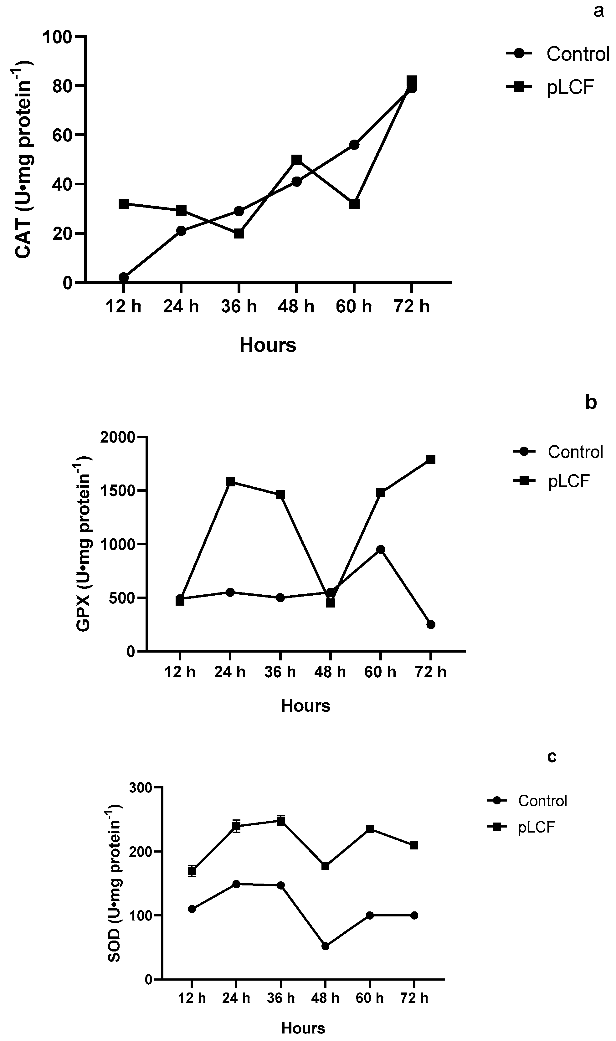
2.7.1. The Activity of Antioxidant Enzymes CAT (a), GPX (b), and SOD (c), during the Time Course in the Mycelium of A. flavus

As depicted in Figure 6c, SOD activity was significantly stimulated (p < 0.001) by the addition of pLCF in the substrate, in comparison to the control, and a peak was evident at 36 h of incubation. Even if a decrease appeared after this time, the activity of the SOD enzymes remained higher than that in control up to 72 h of incubation. CAT (Figure 6a) activity in the presence of pLCF of S. commune was not significantly different (p > 0.001) compared to those detected in the control. GPX (Figure 6b) was significantly stimulated (p < 0.001) by the presence of pLCF with the highest peak during 72 h of incubation

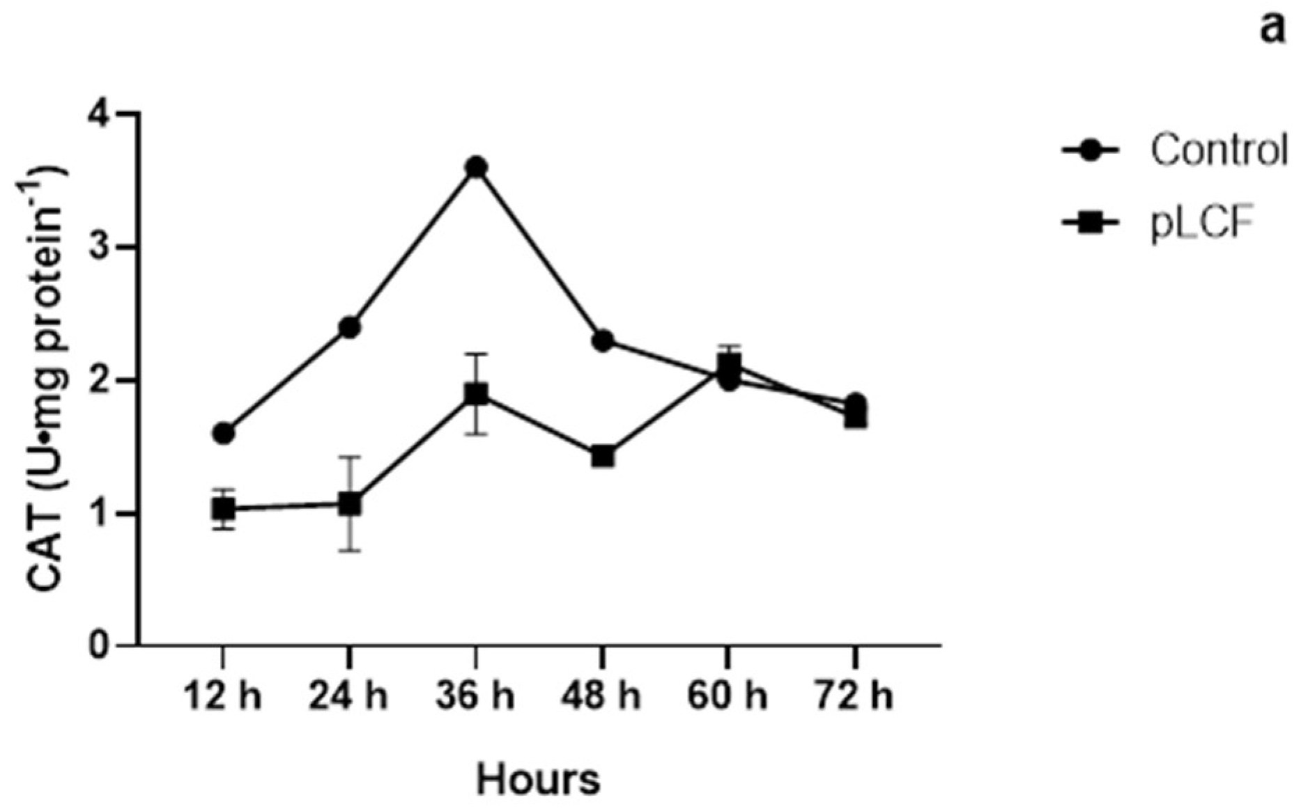
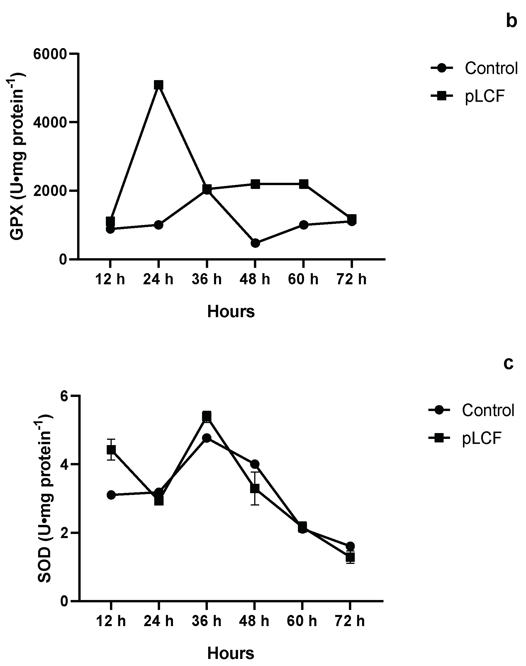
2.7.2. The Activity of Antioxidant Enzymes CAT (a), GPX (b), and SOD (c), during the Time Course in the Mycelium of A. carbonarius

CAT activity was significantly stimulated (p < 0.001) by the addition of the pLCF filtrates of S. commune in the substrate in comparison to the control (Figure 7a) with the highest peak at 36 h of incubation. After 36 h, the CAT enzyme decreased, and at 72 h of incubation reached the control level. SOD activity was not significantly stimulated (p > 0.001) by the addition of the pLCF of S. commune in the substrate in comparison to the control (Figure 7c). GPX was significantly stimulated (p < 0.001) by the presence of pLCF with the highest peak at 24 h of incubation (Figure 7b).

3. Discussion

Mushroom ꞵ-glucans are widely reported as biologically active compounds and are considered biological response modifiers, or BRM [28,29]. Moreover, fungal β-glucans could be among the factors responsible for the inhibiting effect of aflatoxins [22,23,24], both by acting as free radical scavengers [30] or by stimulating antioxidant enzymes in fungal cells [22,23,24,25]. The wide screening of 42 mushroom isolates showed that different mushroom isolates were able to inhibit, to some extent, the biosynthesis of AFB1 and/or OTA, though only two LCFs, T. versicolor isolate TV 117 and S. commune isolate S.C Ailanto, were able to inhibit the biosynthesis of both mycotoxins, AFB1 and OTA, for more than 50%. Therefore it was decided to continue the research only with this strain. Moreover, the results showed the high variability of the inhibiting effects on the mycotoxin synthesis of metabolites produced by different isolates of the same mushroom species, which is consistent with the results reported by Reverberi and collaborators [22,23] and Zjalic and collaborators [24]. In this research, different isolates of L. edodes and T. versicolor were assayed on aflatoxin inhibition. The observed variability was in any case lower than the one observed in the case of the actual research. The variability in inhibition rates among the different isolates of the same species could depend on the genetic structure of the strain and its capacity to produce inhibiting compound(s). Also, the variability could be a consequence of different environmental adaptations of different strains, and active compounds produced under different environmental conditions such as temperature, pH, and levels of oxygenation [22,24]. Moreover, different types of molecules, some with antioxidant activity per se and others as indirect antioxidants, have been reported as active in AFB1 and/or OTA control [17,20,31]. Other compounds have been reported to stimulate the biosynthesis of AFB1 [32]. The composition of LCF depends on the isolate’s metabolism and it is a mixture of different compounds, some of which could inhibit mycotoxin synthesis while others could enhance it [22,26,33]. Differences in LCF yield could also be explained by the variations in the metabolism of different strains.
Results reported in Table 2 confirmed that a putative polysaccharide produced by S. commune interferes with toxin synthesis and that it is not involved in the degradation or some kind of masking, of the two toxins. Partial purification of LCF to pLCF highly enhanced the concentration of polysaccharides. The fact that this purification did not reflect on mycotoxin inhibition indicates that polysaccharide(s) is involved in the inhibition. Furthermore, pLCF contains polysaccharides with a higher MM, while sLFC, in which smaller saccharides were present, had no effects on AFB1 and OTA inhibition (data not shown), which is in line with the general consideration that high molecular mass glucans appear to be more effective than those of low molecular mass [15,34,35].
S. commune is known as a producer of bioactive β-glucan, schizophyllan (SCH). SCH is produced during mushroom growth, and it is released in the media in liquid fermentation [36]. Antitumoral and other BRM activities of SCH have been described [29,35]. Its structure is well-known. It is a nonionic homoglucan, with a β-(1-3)-linked backbone and single β-(1-6)-linked glucose side chains at approximately every third residue, generally with an MM of several hundred kDa. It has also been reported that its biological activity depends on its MM as well as its triple helix conformation [35,37]. Composition analysis of pLCF revealed that it is a homoglucan, while 1H NMR spectroscopy showed a very high similarity with the spectrum of SCH reported in reference [27]. In order to undoubtedly establish that pLCF has the same primary structure as SCH, further 2D NMR experiments have to be performed.
It is known that oxidative stress in fungal cells is a trigger for the synthesis of AFB1 and OTA [22,31]. Mushroom polysaccharides obtained from L. edodes and T. versicolor inhibit AFB1 and OTA synthesis by activating the fungal antioxidant system and thus preventing oxidative disbalance in fungal cells [15,22,23,24,25,31]. It could be suggested that the S.C. Ailanto pLCF influences the cascade of signals that allows mycotoxin biosynthesis, even if we cannot indicate at which step of the pathway this event occurs. Nevertheless, it could be hypothesized that β-glucans, and other compounds contained in the pLCF filtrate of S.C. Ailanto, could be able to inhibit mycotoxin biosynthesis in A. flavus and A. carbonarius by enhancing the internal antioxidant system. In particular, pLCF stimulated the activity of SOD and GPX (Figure 6 and Figure 7), which is consistent with the results obtained by Reverberi and collaborators [31] and Zjalic and collaborators [24], in which the activity of these two enzymes was more important for AFB1 inhibition than the activity of CAT. The stimulation of enzyme activity involves Apyap1. This protein is activated by the presence of mushroom polysaccharides and enhances the transcription of antioxidant genes in the fungal cell [31,38]. The receptor(s) for mushroom polysaccharides that initiates the cascade is still unknown. Similar mechanisms of action of pLCF and tramesan could suggest that they bind to the same type of receptor and reveling the structure of other S.C Ailanto polysaccharides active in mycotoxin inhibition, which could contribute to a better understanding of this mechanism.

4. Conclusions

In conclusion, the S. commune isolate studied in this research could be regarded as a potential agent in biological control or as a useful component among the integrated strategies against mycotoxin-producing fungi in food and feed products. Nevertheless, future research is required to confirm that pLCF is indeed SCH and further determine which specific saccharidic sequence is active in the inhibition of AFB1 and OTA so that it can be synthetically produced and used for large-scale applications in agriculture. In particular, the discovery of the very active part of the polysaccharide would be especially relevant, from an economic point of view, as its synthesis could be more cost effective. Moreover, fungal and plant extracts could represent a promising tool in mycotoxin control, compared to chemicals [39,40,41,42,43], due to their lower toxicity when released into the environment.

5. Materials and Methods

5.1. Fungal Strains and Mushrooms Used in this Study

A. flavus (Speare) (NRRL 3357), a producer of aflatoxin B1, was cultured on Potato dextrose agar (PDA, Himedia, Mumbai, India), at 30 °C for 7 days in dark conditions, and a suspension of 1 × 10⁶ conidia per mL in sterilized distilled water was used as an inoculum in all experiments.
A. carbonarius, a producer of ochratoxin A, was isolated by the Laboratory of Plant Pathology and Mycology of the Sapienza University of Rome, Italy. It was grown on PDA at 30 °C for 7 days in dark conditions, and a suspension of 1 × 10⁶ conidia per mL in sterilized distilled water was used as an inoculum in all experiments. All the mushroom isolates (Table 3) were provided by the fungal germplasm bank of the Department of Agricultural, Forest and Food Sciences, University of Turin, Italy, Prof. Paolo Gonthier, and his group.

5.2. Production of Mushroom Unrefined Lyophilized Filtrates (LCFs)

The 42 isolates of different mushroom cultures detailed above were kept on a potato dextrose agar (PDA, Himedia, Mumbai, India) medium at 4 °C, and the cultures were subcultured onto fresh medium every 30 days. Three plugs of 1 cm diameter of each isolate were inoculated in sterile conditions in 100 mL of potato dextrose broth (PDB, Himedia, Mumbai, India) and incubated for 15 days at 25 °C under shaken conditions (100 rpm). Then, the liquid cultures were homogenized, in sterile conditions. After homogenization, an aliquot (5% v/v) of the fungal cultures was inoculated in 500 mL of PDB in 1L-Erlenmeyer flasks and incubated for 21 days at 25 °C under rotary shaken conditions (100 rpm). After incubation, the mycelia were separated from the culture filtrates by subsequent filtrations through 0.45 μm filters (Sartorius, Goettingen, Germany), to eliminate all the mycelia. Mycelia-purified culture filtrates were concentrated in rota-vapor (Rotavapor® R-300, Buchi, Essen City, Germany), and up to a concentration of 100 mL as described by Parroni and collaborators [42]. Concentrated culture filtrates were lyophilized and utilized for subsequent analyses as reported in [24]. Finally, concentrations of 2% w/v of each mushroom exopolysaccharide unrefined substrate were utilized for the experiment.

5.3. Yield

To calculate the yield of LCF, one L of culture filtrates was reduced in volume 10 times in rotavapor at 45 °C, and 50 mL of the reduced LCF were placed in glass backers previously weighed (after being dried for 48 h at 80 °C in thermostat) and lyophilized. After the lyophilization, the backers were weighed again and the DW of LCF was calculated.

5.4. In Vitro Screening of Mushroom Polysaccharides on Inhibition of Mycotoxin Synthesis by Aspergillus flavus and Aspergillus carbonarius

For the purpose of wide screening of mushroom polysaccharides, the microtiter-based bioassay using 96-well microplates was performed. This assay allowed the testing of all the fractions using small quantities of the compounds with effective replication (n-10) over short periods of time, as previously described by Loncar and collaborators [15].
Screening assays were carried out with 100 conidia of each strain (A. flavus or A. carbonarius), independently, in 10 μL of sterilized distilled water. This was inoculated together with 190 μL of PDB, in the presence or absence of lyophilized mushroom filtrates in each of the wells. A nutrient medium (PDB) inoculated with the fungus A. flavus or A. carbonarius was used as a control. The microtiter plates were incubated at 30 °C for 3 days in dark conditions. Different cultures were independently filtered with Millipore filters (0.22 μm, Sartorius, Goettingen, Germany). The filtrate was subsequently used for mycotoxin extraction.

5.5. Mycotoxins Extraction and Quantification

5.5.1. Ochratoxin A

The different cultures were independently filtered through Millipore filters (0.22 μm, Whatman, Merck, Darmstadt, Germany). The extraction of OTA was performed, for each condition (control and treated with 2% w/v of different mushroom polysaccharides), using the extraction solution of Acetonitrile: Water: Acetic acid (79:20:1 v/v), with the addition of 5 µL of Quercetin (≥95% Sigma-Aldrich, Merck, Darmstadt, Germany) (100 µM) as an internal standard, as previously reported in Fanelli and collaborators [44]. The mixture was vortexed for 1 min, centrifuged, and then the lower phase was drawn off. The extraction was repeated three times and the samples were concentrated under a N2 stream and redissolved in 50 μL of methanol. The concentration of OTA was determined by HPLC/MS (Agilent, Waldbronn, Germany) and expressed in ppb as described by Amezqueta and collaborators [45].

5.5.2. Aflatoxin B1

The different cultures were independently filtered through Millipore filters (0.22 μm, Whatman, Merck, Darmstadt, Germany). The extraction of AFB1 was performed for each condition (control and treated with 2% w/v of different mushroom polysaccharides), using the extraction solution of Chloroform (VWR, Randor, PA, USA): Methanol (VWR, Randor, PA, USA) (2:1 v/v) with the addition of 5 µL of Quercetin (≥95% Sigma-Aldrich, Merck, Darmstadt, Germany) (100 µM) as an internal standard. The mixture was vortexed for 1 min, centrifuged, and then the lower phases were drawn off. The extraction was repeated three times and the samples were concentrated under a N2 stream, then redissolved in 50 μL of methanol. The concentration of AFB1 was quantified by HPLC/MS (Agilent, Waldbronn, Germany) and expressed in ppb, as described by Fanelli and collaborators [44].

5.6. Influence of LCF on Mycotoxins Already Present in the Culture

To verify whether LCF has any influence on mycotoxins already present in the culture media, a solution of PDB containing 10 μg L−1 of AFB1 or OTA was prepared and 2% (w/v) of LCF was added to 25 mL of solution. The control was a PDB solution containing AFB1 or OTA. The samples were incubated for 3 days in a thermostat at 25 °C. The samplings were done after inoculation of LCF (T0) and after 3 days of incubation (T3). The test was done in 3 replicates.

5.7. Mycotoxin Inhibition Assay with S. commune Unrefined Extract (LCF)

The unrefined extract of the exopolysaccharide S. commune (LCF) at a concentration of 1% w/v (data not shown) and 2% w/v was added to PDB (0.5 mL) and used as a nutrient medium for the cultivation of A. flavus and A. carbonarius. The nutrient medium with or without exopolysaccharides (control), was inoculated with a conidial suspension of A. flavus or A. carbonarius in a concentration of 106 conidia in 0.2 mL of sterilized distilled water. The samples were incubated for up to 6 days at 25 °C in dark conditions as described by Parroni and collaborators [42].

5.8. Analysis of Monosaccharides in pLCF and sLCF Fractions

Trimethylsilyl methyl glycosides were obtained by derivatization with the reagent Sylon™ HTP (Merck Life Science S.r.l., Milano, Italy) after methanolysis of the polysaccharide with 3 M HCl in methanol at 85 °C for 16 h [43]. Analytical gas–liquid chromatography was performed on an Agilent Technologies 6850 gas chromatograph equipped with a flame ionization detector and using He as carrier gas. An HP-1 capillary column (Agilent Technologies Italia S.p.A., Milano, Italy, 30 m) was used to separate trimethylsilylated methyl glycosides (temperature program: 150–280 °C at 3 °C/min).

5.9. Purification of S. commune Unrefined Polysaccharide (LCF)

Unrefined lyophilized liquid culture filtrate (LCF) was precipitated with ethanol (VWR, Randor, PA, USA) (99%), on ice, to remove the lipids. After the centrifugation (45 min/13,000 rpm at 4 °C, Z326K HERMLE, Gosheim, Germany), the pellet was resuspended with 1 mL of 10 mM potassium phosphate buffer (VWR, Randor, PA, USA) (pH = 7.5). After resuspension, proteinase K (Thermo Fisher Scientific, Waltham, MA, USA) was added to the sample and left overnight in a shaking water bath at 37 °C. During the night the sample turned to gel form. The sample was centrifuged (40 min/4000 rpm at 4 °C), however, most of the sample remained in gel form, while only a small part of the fraction remained in the supernatant. Therefore, the precipitate (gel form) (pLCF) was separated from the supernatant (sLCF) and separately subjected to dialysis (10 kDa, Sigma-Aldrich, St. Louis, MO, USA) against the water. After 2 days of dialysis, the samples were lyophilized and utilized for subsequent analysis.

5.10. Assays of the Different Variants of S. commune Polysaccharide (pLCF and sLCF) on Mycotoxin Inhibition

5.10.1. Assay of the Semi purified Lyophilized (pLCF) Polysaccharide Fraction from S. commune on Aflatoxin B1 and Ochratoxin A Biosynthesis

The pLCF (375 mg) was rehydrated in 5 mL of H2O. To disrupt the triple helix of the schizophyllan structure, the gel was heated for 4h at 140 °C, as described by Zhang and collaborators [29]. After the heating treatment, the gel took the form of a viscous solution.
Semi purified exopolysaccharide (pLCF) at a concentration of 1% w/v and 2% w/v was added to PDB (0.5 mL) and used as a nutrient medium for the cultivation of A. Flavus or A. carbonarius. The nutrient medium was inoculated with a suspension of fungal conidia (A. flavus or A. carbonarius) in a concentration of 106 conidia in 0.2 mL of sterilized distilled water. PDB inoculated with A. flavus or A. carbonarius was used as a control. The samples were incubated for 6 days at 25 °C in dark conditions.

5.10.2. Assay of the Fraction of the Polysaccharide from S. commune (sLCF) on Ochratoxin A and Aflatoxin B1 Biosynthesis

The lyophilized supernatant sLCF was extracted from the polysaccharide S. commune at a concentration of 1% w/v (data not shown), and 2% w/v was added to PDB (0.5 mL) and used as a nutrient medium for the cultivation of A. flavus or A. carbonarius. The nutrient medium, with a dissolved fraction of sLCF, was inoculated with a conidia suspension of the two test fungi using the same procedure as for pLCF.

5.11. Assay of the Comparison between Commercial Schizophyllan and Semi-Purified Extract (pLCF) of S. Commune on Inhibition of Mycotoxin Synthesis

The inhibition of mycotoxin production in A. Flavus and A. Carbonarius was compared between the pLCF extract of S. Commune and commercial β-glucan Schizophyllan (Invivogen, Toulouse, France), using the microtiter plate assay described by Loncar and collaborators [15]. The mycotoxin analyses were performed, as described by Fanelli and collaborators [44] and Amezqueta and collaborators [45].

5.12. Ultraviolet Visible Spectrophotometry

The possible presence of proteins in both samples was checked by UV analysis (Ultraviolet Visible Spectrophotometer, Thermo Fisher Scientific, Waltham, MA, USA). The pLCF and sLCF were dissolved separately in 2.4 mL of H2O (sLCF was completely soluble in water) and both samples were diluted 100 times before analysis.

5.13. 1H NMR Spectroscopy

Five mg of the pLCF sample were exchanged three times with 99.9% D2O by lyophilization (Lyophilizer FD-10, Labfreez, Changshan, Hunan, China) and then dissolved in 0.6 mL of DMSO-d6 and 99.96% D2O in a 6:1 ratio. Two mg of the sLCF sample were exchanged twice with 99.9% D2O by lyophilization and then dissolved in 0.6 mL of 99.96% D2O. Spectra were recorded on a 500 MHz VARIAN spectrometer (Varian Cary*50 UV-Vis, Agilent, Santa Clara, CA, USA) operating at 70 °C for pLCF and 50 °C for sLCF. Chemical shifts are given in parts per million, using the internal Me2SO signal (1H = 2.53 ppm) for the pLCF 1H spectrum and using the residual HOD signal (1H = 4.50 ppm at 50 °C) for the sLCF 1H spectrum. The 1H NMR spectra were processed using MestreNova 9.2 software (Mestrelab Research, Santiago de Compostela, Spain).

5.14. Analysis of Antioxidant Enzyme Activities in A. flavus and A. carbonarius Treated and Nontreated with Lyophilized Semipurified Filtrates (pLCF) of S. commune

The activities of superoxide dismutase (SOD), catalase (CAT), and glutathione peroxidase (GPX) were analyzed in (50 mg) mycelia of A. flavus and A. carbonarius treated and nontreated with 2% w/v of pLCF of S. commune, as previously described at Reverberi and collaborators [28]. The antioxidant activities were reported as unit mg protein−1.

5.15. Quantification of the Proteins

The quantification of the proteins in LCF and pLCF was performed according to the Bradford method [46].

5.16. Statistics

The arithmetic mean was used as the mean value, while the standard deviation was used as an indicator of the dispersion around the arithmetic mean. The test of the difference in relation to the reference value was carried out using the t-test for one independent measurement where 100 (control) was used as the reference value. Examination of the difference between fungi was carried out using the t-test for independent measurements, and the differences were considered significant when the p-value was <0.001. The analysis was done in the statistical software STATISTICA 12, Tibco, Palo Alto, CA, USA.

Author Contributions

Conceptualization: P.C., S.Z., J.L. and M.R.; experimental work: J.L., B.B., A.M. and M.B.; data analysis and interpretation: P.C., B.B., M.R., S.Z. and J.L.; writing—draft version: S.Z., M.R., J.L. and A.M.; reviewing and editing: S.Z., P.C., M.R., J.L., B.B. and A.M. All authors have read and agreed to the published version of the manuscript.

Funding

This research received no external funding.

Data Availability Statement

Not applicable.

Acknowledgments

We are grateful to Paolo Gonthier (University of Torino, Italy) and his staff for providing us all the strains of wood rot fungi used in this study. I would like to express my gratitude to my primary supervisor, Slaven Zjalic, who guided me throughout this project. I wish to acknowledge the help provided by the technical and support staff at Sapienza University, Roma, Italy. I would also like to thank Alice Motola who supported me and who helped me finalize my project.

Conflicts of Interest

The authors declare no conflict of interest.

References

  1. Ciegler, A.; Bennett, J.W. Mycotoxins and Mycotoxicoses. BioScience 1980, 30, 512–515. [Google Scholar] [CrossRef]
  2. Bennett, J.W.; Klich, M. Mycotoxins. Clin. Microbiol. Rev. 2003, 16, 497–516. [Google Scholar] [CrossRef] [PubMed] [Green Version]
  3. Sharma, V.; Patial, V. Food mycotoxins: Dietary interventions implicated in the prevention of mycotoxicosis. ACS Food Sci. Technol. 2021, 1, 1717–1739. [Google Scholar] [CrossRef]
  4. Pereira, C.S.; Cunha, S.C.; Fernandes, J.O. Prevalent mycotoxins in animal feed: Occurrence and analytical methods. Toxins 2019, 11, 290. [Google Scholar] [CrossRef] [PubMed] [Green Version]
  5. Hussein, H.S.; Brasel, J.M. Toxicity, metabolism, and impact of mycotoxins on humans and animals. Toxicology 2001, 167, 101–134. [Google Scholar] [CrossRef] [PubMed]
  6. Speijers, G.J.A.; Speijers, M.H.M. Combined toxic effects of mycotoxins. Toxicol. Lett. 2004, 153, 91–98. [Google Scholar] [CrossRef] [PubMed]
  7. IARC. Monographs on the Evaluation of Carcinogenic Risks to Humans: Chemical Agents and Related Occupations. A Review of Human Carcinogens; IARC: Lyon, France, 2012; pp. 224–248. [Google Scholar]
  8. Tao, Y.; Xie, S.; Xu, F.; Liu, A.; Wang, Y.; Chen, D.; Pan, Y.; Huang, L.; Peng, D.; Wang, X.; et al. Ochratoxin A: Toxicity, oxidative stress and metabolism. Food Chem. Toxicol. 2018, 112, 320–331. [Google Scholar] [CrossRef] [PubMed]
  9. Khatoon, A.; ul Abidin, Z. Mycotoxicosis–diagnosis, prevention and control: Past practices and future perspectives. Toxin Rev. 2018, 39, 1–16. [Google Scholar] [CrossRef]
  10. Navale, V.; Vamkudoth, K.R.; Ajmera, S.; Dhuri, V. Aspergillus derived mycotoxins in food and the environment: Prevalence, detection, and toxicity. Toxicol. Rep. 2021, 8, 1008–1030. [Google Scholar] [CrossRef] [PubMed]
  11. Bryden, W.L. Mycotoxins in the food chain: Human health implications. Asia Pac. J. Clin. Nutr. 2007, 16, 95–101. [Google Scholar] [CrossRef] [PubMed]
  12. European Union. COMMISSION REGULATION (EC) No 1881/2006 of 19 December 2006 setting maximum levels for certain contaminants in foodstuffs. Off. J. Eur. Union 2006, L364, 5–24. [Google Scholar]
  13. Van Egmond, H.P.; Schothorst, R.C.; Jonker, M.A. Regulations relating to mycotoxins in food. Anal. Bioanal. Chem. Res. 2007, 389, 147–157. [Google Scholar] [CrossRef] [PubMed] [Green Version]
  14. Binder, E.M. Managing the risk of mycotoxins in modern feed production. J. Anim. Feed Sci. Technol. 2007, 133, 149–166. [Google Scholar] [CrossRef]
  15. Loncar, J.; Bellich, B.; Parroni, A.; Reverberi, M.; Rizzo, R.; Zjalić, S.; Cescutti, P. Oligosaccharides derived from Tramesan: Their structure and activity on mycotoxin inhibition in Aspergillus flavus and Aspergillus carbonarius. Biomolecules 2021, 11, 243. [Google Scholar] [CrossRef] [PubMed]
  16. Loi, M.; Fanelli, F.; Zucca, P.; Liuzzi, V.; Quintieri, L.; Cimmarusti, M.; Monaci, L.; Haidukowski, M.; Logrieco, A.; Sanjust, E.; et al. Aflatoxin B1 and M1 Degradation by Lac2 from Pleurotus pulmonarius and Redox Mediators. Toxins 2016, 8, 245. [Google Scholar] [CrossRef] [PubMed] [Green Version]
  17. Wang, Y.; Liu, F.; Zhou, X.; Liu, M.; Zang, H.; Liu, X.; Shan, A.; Feng, X. Alleviation of Oral Exposure to Aflatoxin B1-Induced Renal Dysfunction, Oxidative Stress, and Cell Apoptosis in Mice Kidney by Curcumin. Antioxidants 2022, 11, 1082. [Google Scholar] [CrossRef] [PubMed]
  18. Xu, L.; Ahmed, M.F.E.; Sangare, L.; Zhao, Y.; Selvaraj, J.N.; Xing, F.; Wang, Y.; Yang, H.; Liu, Y. Novel aflatoxin-degrading enzyme from Bacillus shackletonii L7. Toxins 2017, 9, 36. [Google Scholar] [CrossRef] [PubMed]
  19. Yehia, R.S. Aflatoxin detoxification by manganese peroxidase purified from Pleurotus ostreatus. Braz. J. Microbiol. 2014, 45, 127–133. [Google Scholar] [CrossRef] [PubMed] [Green Version]
  20. Zhou, Z.; Li, R.; Ng, T.B.; Lai, Y.; Yang, J.; Ye, X. A new laccase of Lac 2 from the white rot fungus Cerrena unicolor 6884 and Lac 2-mediated degradation of aflatoxin B1. Toxins 2020, 12, 476. [Google Scholar] [CrossRef] [PubMed]
  21. Zaccaria, M.; Dawson, W.; Russel Kish, D.; Reverberi, M.; Bonaccorsi di Patti, M.C.; Domin, M.; Cristiglio, V.; Chan, B.; Dellafiora, L.; Gabel, F.; et al. Experimental–theoretical study of laccase as a detoxifier of aflatoxins. Sci. Rep. 2023, 13, 860. [Google Scholar] [CrossRef] [PubMed]
  22. Reverberi, M.; Fabbri, A.A.; Zjalic, S.; Ricelli, A.; Punelli, F.; Fanelli, C. Antioxidant enzymes stimulation in Aspergillus parasiticus by Lentinula edodes inhibits aflatoxin production. Appl. Microbiol. Biotechnol. 2005, 69, 207–215. [Google Scholar] [CrossRef] [PubMed]
  23. Reverberi, M.; Ricelli, A.; Zjalic, S.; Fabbri, A.A.; Fanelli, C. Natural functions of mycotoxins and control of their biosynthesis in fungi. Appl. Microbiol. Biotechnol. 2010, 87, 899–911. [Google Scholar] [CrossRef] [PubMed]
  24. Zjalic, S.; Reverberi, M.; Ricelli, A.; Granito, M.V.; Fanelli, C.; Fabbri, A.A. Trametes versicolor: A possible tool for aflatoxin control. Int. J. Food Microbiol. 2006, 107, 243–249. [Google Scholar] [CrossRef] [PubMed]
  25. Scarpari, M.; Parroni, A.; Zaccaria, M.; Fattorini, L.; Bello, C.; Fabbri, A.A.; Bianchi, G.; Scala, V.; Zjalic, S.; Fanelli, C. Aflatoxin control in maize by Trametes versicolor. Toxins 2014, 6, 3426–3437. [Google Scholar] [CrossRef] [PubMed] [Green Version]
  26. Ricelli, A.; Fabbri, A.A.; Trionfetti-Nisini, P.; Reverberi, M.; Zjalic, S.; Fanelli, C. Inhibiting effect of different edible and medicinal mushrooms on the growth of two ochratoxigenic microfungi. Int. J. Med. Mushrooms 2002, 4, 8. [Google Scholar] [CrossRef]
  27. Tada, R.; Harada, T.; Nagi-Miura, N.; Adachi, Y.; Nakajima, M.; Yadomae, T.; Ohno, N. NMR characterization of the structure of a β-(1→3)-d-glucan isolate from cultured fruit bodies of Sparassis crispa. Carbohydr. Res. 2007, 342, 2611–2618. [Google Scholar] [CrossRef] [PubMed]
  28. Rau, U. Glucans secreted by fungi. Turk. Electron. J. Biotechnol. 2004, 2, 30–36. [Google Scholar]
  29. Zhang, G.Q.; Chen, Q.J.; Sun, J.; Wang, H.X.; Han, C.H. Purification and characterization of a novel acid phosphatase from the split gill mushroom Schizophyllum commune. J. Basic Microbiol. 2013, 53, 868–875. [Google Scholar] [CrossRef] [PubMed]
  30. Slameňová, D.; Lábaj, J.; Križková, L.; Kogan, G.; Šandula, J.; Bresgen, N.; Eckl, P. Protective effects of fungal (1→3)-β-D-glucan derivatives against oxidative DNA lesions in V79 hamster lung cells. Cancer Lett. 2003, 198, 153–160. [Google Scholar] [CrossRef] [PubMed]
  31. Reverberi, M.; Zjalic, S.; Ricelli, A.; Punelli, F.; Camera, E.; Fabbri, C.; Picardo, M.; Fanelli, C.; Fabbri, A.A. Modulation of antioxidant defense in Aspergillus parasiticus is involved in aflatoxin biosynthesis: A role for the Ap yapA gene. Eukaryot. Cell 2008, 7, 988–1000. [Google Scholar] [CrossRef] [PubMed] [Green Version]
  32. Yu, J.; Mohawed, S.M.; Bhatnagar, D.; Cleveland, T.E. Substrate induced lipase gene expression and aflatoxin production in Aspergillus parasiticus and Aspergillus flavus. J. Appl. Microbiol. 2003, 95, 1334–1342. [Google Scholar] [CrossRef] [PubMed] [Green Version]
  33. Barberis, C.; Astoreca, A.; Fernandez-Juri, G.; Chulze, S.; Dalcero, A.; Magnoli, C. Use of propyl paraben to control growth and ochratoxin A production by Aspergillus section Nigri species on peanut meal extract agar. Int. J. Food Microbiol. 2009, 136, 133–136. [Google Scholar] [CrossRef] [PubMed]
  34. Franz, G. Polysaccharides in pharmacy: Current applications and future concepts. Planta Med. 1989, 55, 493–497. [Google Scholar] [CrossRef] [PubMed]
  35. Kojima, T.; Tabata, K.; Itoh, W.; Yanaki, T. Molecular weight dependence of the antitumor activity of schizophyllan. J. Agric. Food Chem. 1986, 50, 231–232. [Google Scholar] [CrossRef]
  36. Kikumoto, S. Polysaccharide produced by Schizophyllum commune, I. Formation and some properties of an extracellular polysaccharide. Nippon. Nogeikagaku Kaishi 1970, 44, 337–342. [Google Scholar] [CrossRef] [Green Version]
  37. Kidd, P.M. The use of mushroom glucans and proteoglycans in cancer treatment. Altern. Med. Rev. 2000, 5, 4–27. [Google Scholar] [PubMed]
  38. Reverberi, M.; Zjalic, S.; Punelli, F.; Ricelli, A.; Fabbri, A.A.; Fanelli, C. Apyap1 affects aflatoxin biosynthesis during Aspergillus parasiticus growth in maize seeds. Food Addit. Contam. 2007, 24, 1070–1075. [Google Scholar] [CrossRef] [PubMed] [Green Version]
  39. Mahoney, N.; Molyneux, R.J. Phytochemical inhibition of aflatoxigenicity in Aspergillus flavus by constituents of walnut (Juglans regia). J. Agric. Food Chem. 2004, 52, 1882–1889. [Google Scholar] [CrossRef] [PubMed]
  40. Sánchez, E.; Heredia, N.; García, S. Inhibition of growth and mycotoxin production of Aspergillus flavus and Aspergillus parasiticus by extracts of Agave species. Int. J. Food Microbiol. 2005, 98, 271–279. [Google Scholar] [CrossRef] [PubMed]
  41. Abuajah, C.I.; Ogbonna, A.C.; Osuji, C.M. Functional components and medicinal properties of food: A review. J. Food Sci. Technol. 2015, 52, 2522–2529. [Google Scholar] [CrossRef] [PubMed] [Green Version]
  42. Parroni, A.; Bellabarba, A.; Beccaccioli, M.; Scarpari, M.; Reverberi, M.; Infantino, A. Use of the secreted proteome of Trametes versicolor for controlling the cereal pathogen Fusarium langsethiae. Int. J. Mol. Sci. 2019, 20, 4167. [Google Scholar] [CrossRef] [PubMed] [Green Version]
  43. Kakehi, K.; Honda, S. Silyl ethers of carbohydrates. In Analysis of Carbohydrates by GLC and MS, 1st ed.; Biermann, C.J., McGinnis, G.D., Eds.; CRC Press: Boca Raton, FL, USA, 2021; Volume 1, pp. 43–85. [Google Scholar]
  44. Fanelli, C.; Tasca, V.; Ricelli, A.; Reverberi, M.; Zjalic, S.; Finotti, E.; Fabbri, A.A. Inhibiting effect of medicinal mushroom Lentinus edodes (Berk.) Sing. (Agaricomycetideae) on aflatoxin production by Aspergillus parasiticus Speare. Int. J. Med. Mushrooms 2000, 2, 4. [Google Scholar] [CrossRef]
  45. Amezqueta, S.; Schorr-Galindo, S.; Murillo-Arbizu, M.; Gonzalez-Peñas, E.; De Cerain, A.L.; Guiraud, J.P. OTA-producing fungi in foodstuffs: A review. Food Control 2012, 26, 259–268. [Google Scholar] [CrossRef] [Green Version]
  46. Kruger, N.J. The Bradford Method for protein quantitation. In The Protein Protocols Handbook; Walker, J.M., Ed.; Springer Protocols Handbooks; Humana Press: Totowa, NJ, USA, 2019. [Google Scholar] [CrossRef]
Figure 1. The effect of different isolates of mushroom Ganoderma adspersum lyophilized rough filtrates on ochratoxin A production in A. carbonarius and aflatoxin B1 production in A. flavus after 3 days incubation at 30 °C. Results are expressed as a percentage of mycotoxins produced relative to the control. The data are the mean ± SD of three determinations of five separate experiments.
Toxins 15 00177 g001
Figure 2. The effect of different isolates of the mushroom S. commune lyophilized filtrates on ochratoxin A production by A. carbonarius and aflatoxin B1 production by A. flavus after 3 days incubation at 30 °C. Results are expressed as a percentage of mycotoxin relative to the control. The data are the mean ± SD of three determinations of five separate experiments.
Toxins 15 00177 g002
Figure 3. Comparison of the inhibition of biosynthesis of AFB1 and OTA in treatments with LCF and semipurified (pLCF) metabolite of S.C. Ailanto, compared with the control. Results are expressed as a percentage of mycotoxins produced relative to the control. The data are the mean ± SD of three different determinations of five separate experiments.
Toxins 15 00177 g003
Figure 4. 1H NMR spectra of fractions pLCF (A) recorded at 70 °C in deuterated DMSO:D2O in a 6:1 ratio and sLCF (B) recorded at 50 °C in D2O. Some assignments are indicated.
Toxins 15 00177 g004
Figure 5. Comparison of the inhibition of biosynthesis of AFB1 and OTA in treatments with semi-purified pLCF (2% w/v) S. C. Ailanto metabolite and commercial polysaccharide Schizophyllan (2% w/v) compared to control. Results are expressed as a percentage of mycotoxins produced relative to the control. The data are the mean ± SD of three different determinations of five separate experiments.
Toxins 15 00177 g005
Figure 6. The activity of antioxidant enzymes CAT (a), GPX (b), and SOD (c), during the time course in the mycelium of A. Flavus, treated and not treated with semipurified filtrates pLCF of S. commune.
Toxins 15 00177 g006
Figure 7. The activity of antioxidant enzymes CAT (a), GPX (b), and SOD (c), during the time course in the mycelium of A. carbonarius, treated and not treated with semipurified filtrates (pLCF) of S. commune.
Toxins 15 00177 g007aToxins 15 00177 g007b
Table 1. Inhibition rate (%) of LCF obtained from different fungal isolates on aflatoxin B1 and ochratoxin A production. Inhibitions < 50% were not considered significant for the purpose of this research and are reported as not significant (NS). Yield (DW) of LCFs is expressed in g • L−1. All data are the mean ± SD of determinations of ten separate experiments.
CodeGeneraSpeciesInhibition
of OTA(%) ± SD
T-Value (p)Inhibition
of AF (%) ± SD
T-Value (p)Yield
g • L−1
CF 16Agrocybeaegerita6.64 ± 1.55134.41 (<0.001)70.18 ± 7.618.76 (0.001)7.21 ± 0.50
CF 249Agrocybeaegerita31.48 ± 3.7540.82 (<0.001)61.73 ± 7.3811.60 (<0.001)6.82 ± 0.52
CF 250Agrocybeaegerita23.17 ± 3.3351.66 (<0.001)58.48 ± 8.2411.27 (<0.001)7.22 ± 0.48
CF 278Armillariamellea4.35 ± 2.6082.24 (<0.001)25.60 ± 2.5864.56 (<0.001)8.12 ± 0.46
CF 253Fomesfomentarius37.06 ± 5.5525.34 (<0.001)19.91 ± 3.0458.88 (<0.001)9.32 ± 0.53
CF 255Fomesfomentarius35.68 ± 4.6131.17 (<0.001)57.00 ± 7.9212.14 (<0.001)9.29 ± 0.50
CF 262Fomesfomentarius7.24 ± 5.4538.09 (<0.001)45.70 ± 5.2123.33 (<0.001)9.45 ± 0.49
P.COLLEGNO 2AGanodermaadspersum25.30 ± 3.3350.21 (<0.001)3.45 ± 0.95227.61 (<0.001)9.11 ± 0.52
COLETTA 12Ganodermaadspersum4.97 ± 3.6957.62 (<0.001)5.78 ± 3.5759.06 (<0.001)9.51 ± 0.60
ROBUR (69.2)Ganodermaadspersum8.20 ± 6.2033.11 (<0.001)69.94 ± 7.598.86 (0.001)9.59 ± 049
Meisino 9Ganodermaadspersum8.00 ± 4.9541.57 (<0.001)53.01 ± 5.4119.42 (<0.001)7.62 ± 0.51
TiglioGanodermaadspersum25.36 ± 3.1253.56 (<0.001)26.28 ± 3.4847.33 (<0.001)9.54 ± 0.54
Cep 10Ganodermaadspersum4.14 ± 1.61133.24 (<0.001)68.32 ± 9.207.70 (0.002)8.18 ± 0.57
Diano 2Ganodermaadspersum27.10 ± 3.5446.11 (<0.001)54.20 ± 7.2914.05 (<0.001)9.89 ± 0.54
CF 17Ganodermalucidum4.60 ± 2.6979.20 (<0.001)5.11 ± 4.8543.79 (<0.001)9.56± 0.57
CF 223Ganodermalucidum7.31 ± 4.3747.48 (<0.001)20.02 ± 2.2180.94 (<0.001)9.12 ± 0.49
CF 264Ganodermalucidum28.82 ± 2.9354.24 (<0.001)54.70 ± 5.4018.75 (<0.001)9.53 ± 0.51
DP 1Ganodermaresinaceum20.23 ± 2.6467.55 (<0.001)21.58 ± 2.9459.58 (<0.001)9.68 ± 0.52
PLATANO ROMAGanodermaresinaceum33.80 ± 3.7639.33 (<0.001)5.18 ± 4.7944.23 (<0.001)9.72 ± 0.48
P.VERDI 920Ganodermaresinaceum41.27 ± 5.8022.65 (<0.001)25.28 ± 3.5447.20 (<0.001)7.88 ± 0.43
780.2Ganodermaresinaceum42.06 ± 5.7722.45 (<0.001)25.10 ± 2.7960.08 (<0.001)9.82 ± 0.51
DP 63Ganodermaresinaceum43.34 ± 5.1424.67 (<0.001)36.70 ± 5.2826.81 (<0.001)9.79 ± 0.46
ASPROMONTE 52Ganodermaresinaceum53.36 ± 8.0712.93 (<0.001)31.29 ± 3.1848.38 (<0.001)8.89 ± 0.41
DP 23Ganodermaresinaceum6.32 ± 4.9242.62 (<0.001)4.87 ± 5.8036.65 (<0.001)9.68 ± 0.50
C.TRENTO 43 BGanodermaresinaceum30.53 ± 4.2136.86 (<0.001)2.45 ± 1.10198.08 (<0.001)10.14 ± 0.47
P.VERDI 4AGanodermaresinaceum5.18 ± 3.7456.64 (<0.001)3.60 ± 1.56138.07 (<0.001)9.99 ± 0.52
G.R. 853Ganodermaresinaceum6.43 ± 3.2464.65 (<0.001)28.20 ± 3.0053.53 (<0.001)9.53 ± 0.49
CF 258Ganodermaresinaceum4.17 ± 1.09196.13 (<0.001)42.71 ± 6.3820.09 (<0.001)7.78 ± 0.51
CF 6Grifolafrondosa10.09 ± 3.2861.24 (<0.001)6.25 ± 4.1450.67 (<0.001)10.25 ± 0.41
CF 19Grifolafrondosa7.47 ± 3.4859.44 (<0.001)24.95 ± 3.1353.68 (<0.001)9.94 ± 0.51
CF 225Heterobasidionannosum5.84 ± 3.0968.06 (<0.001)23.00 ± 3.0057.33 (<0.001)7.34 ± 0.49
CF 224Phellinuspfefferi5.76 ± 4.2849.27 (<0.001)5.91 ± 4.5346.40 (<0.001)6.85 ± 0.59
CF 25Pleurotuseryngii26.56 ± 3.2350.85 (<0.001)6.27 ± 4.1650.40 (<0.001)6.83 ± 0.50
CF 38Pleurotuseryngii69.92 ± 9.666.96 (0.002)23.38 ± 3.2153.33 (<0.001)7.48 ± 0.55
CF 28Polyporussulphureus24.92 ± 2.5067.26 (<0.001)2.53 ± 2.18100.12 (<0.001)9.58 ± 0.72
CF 280Trameteshirsuta7.22 ± 5.6536.69 (<0.001)20.16 ± 2.0886.00 (<0.001)7.92 ± 0.54
T.V. 117Trametesversicolor85.75 ± 11.762.71 (0.054)89.00 ± 9.002.73 (0.052)6.68 ± 0.50
BELAGIO 2Schizophyllumcommune43.20 ± 5.2324.31 (<0.001)47.91 ± 6.7217.34 (<0.001)9.81 ± 0.48
DP61Schizophyllumcommune4.56 ± 4.8643.87 (<0.001)2.59 ± 1.96111.31 (<0.001)7.92 ± 0.51
GTM8 R1Schizophyllumcommune3.46 ± 1.53141.44 (<0.001)28.12 ± 3.9540.68 (<0.001)8.76 ± 0.56
CF 18Strophariaeugosoannulata2.76 ± 0.44490.75 (<0.001)35.34 ± 4.9029.51 (<0.001)9.92 ± 0.49
S.C. AILANTOSchizophyllumcommune92.76 ± 6.612.45 (0.070)88.20 ± 8.213.22 (0.032)8.59 ± 0.50
Table 2. Effect of LCF of S. commune. on AFB1 and OTA already present in the media after 3 days of incubation at 25 °C. Results are expressed as a percent of the initial concentration of mycotoxins, which was 10 g • L−1 The data are the mean ± SD of three different determinations of LCF that showed the presence of mannose, galactose, and glucose in molar ratio 8:2:1.
AFB1OTA
Time (Days)0303
CONT10097.03 ± 2.8710096.14 ± 3.75
LCF10095.94 ± 3.8510096.48 ± 3.01
Table 3. Different mushroom isolates used for the experiment.
CodeGenusSpecies
CF 16AgrocybeAegerita
CF 249AgrocybeAegerita
CF 250AgrocybeAegerita
CF 278ArmillariaMellea
CF 253Fomesfomentarius
CF 255Fomesfomentarius
CF 262Fomesfomentarius
P.COLLEGNO 2AGanodermaadspersum
COLLETTA 12Ganodermaadspersum
ROBUR (69.2)Ganodermaadspersum
Meisino 9Ganodermaadspersum
TiglioGanodermaadspersum
Cep.10Ganodermaadspersum
Diano 2Ganodermaadspersum
CF 17Ganodermalucidum
CF 223Ganodermalucidum
CF 264Ganodermalucidum
DP1Ganodermaresinaceum
PLATANO ROMAGanodermaresinaceum
P.VERDI 920Ganodermaresinaceum
780.2Ganodermaresinaceum
DP63Ganodermaresinaceum
ASPROMONTE 52Ganodermaresinaceum
DP23Ganodermaresinaceum
C.TRENTO 34 BGanodermaresinaceum
P. VERDI 4AGanodermaresinaceum
G.R. 853Ganodermaresinaceum
CF 258Ganodermaresinaceum
CF 6Grifolafrondosa
CF 19Grifolafrondosa
CF 225Heterobasidionannosum
CF 224Phellinuspfefferi
CF 25Pleurotuseryngii
CF 38Pleurotuseryngii
CF 28Polyporussulphureus
BELLAGIO 2Schizophyllumcommune
DP61Schizophyllumcommune
GTM8 R1Schizophyllumcommune
S.C. AILANTOSchizophyllumcommune
CF 18Strophariarugosoannulata
CF 280Trameteshirsuta
T.V. 117Trametesversicolor
Disclaimer/Publisher’s Note: The statements, opinions and data contained in all publications are solely those of the individual author(s) and contributor(s) and not of MDPI and/or the editor(s). MDPI and/or the editor(s) disclaim responsibility for any injury to people or property resulting from any ideas, methods, instructions or products referred to in the content.

