Article

Clinical and Evolutionary Implications of Dynamic Coagulotoxicity Divergences in Bothrops (Lancehead Pit Viper) Venoms

by
Lachlan Allan Bourke
1,*,
Christina N. Zdenek
1,
Anita Mitico Tanaka-Azevedo
2,
Giovanni Perez Machado Silveira
2,
Sávio Stefanini Sant’Anna
2,
Kathleen Fernandes Grego
2,
Caroline Fabri Bittencourt Rodrigues
2 and
Bryan Grieg Fry
1,*
1
Venom Evolution Lab, School of Biological Sciences, University of Queensland, St. Lucia, QLD 4072, Australia
2
Laboratrio de Herpetologia, Instituto Butantan, Sao Paulo 05503-900, Brazil
*
Authors to whom correspondence should be addressed.
Toxins 2022, 14(5), 297; https://doi.org/10.3390/toxins14050297
Submission received: 16 March 2022 / Revised: 19 April 2022 / Accepted: 20 April 2022 / Published: 22 April 2022
(This article belongs to the Special Issue Insights into the Action and Application of Animal Toxins)

Abstract

:
Despite coagulotoxicity being a primary weapon for prey capture by Bothrops species (lancehead pit vipers) and coagulopathy being a major lethal clinical effect, a genus-wide comparison has not been undertaken. To fill this knowledge gap, we used thromboelastography to compare 37 venoms, from across the full range of geography, taxonomy, and ecology, for their action upon whole plasma and isolated fibrinogen. Potent procoagulant toxicity was shown to be the main venom effect of most of the species tested. However, the most basal species (B. pictus) was strongly anticoagulant; this is consistent with procoagulant toxicity being a novel trait that evolved within Bothrops subsequent to their split from anticoagulant American pit vipers. Intriguingly, two of the arboreal species studied (B. bilineatus and B. taeniatus) lacked procoagulant venom, suggesting differential evolutionary selection pressures. Notably, some terrestrial species have secondarily lost the procoagulant venom trait: the Mogi Mirim, Brazil locality of B. alternatus; San Andres, Mexico locality of B. asper; B. diporus; and the São Roque of B. jararaca. Direct action on fibrinogen was extremely variable; this is consistent with previous hypotheses regarding it being evolutionary decoupled due to procoagulant toxicity being the primary prey-capture weapon. However, human patients live long enough for fibrinogen depletion to be clinically significant. The extreme variability may be reflective of antivenom variability, with these results thereby providing a foundation for such future work of clinical relevance. Similarly, the venom diversification trends relative to ecological niche will also be useful for integration with natural history data, to reconstruct the evolutionary pressures shaping the venoms of these fascinating snakes.
Key Contribution: While procoagulant toxicity mediated by the activation of clotting factors to produce strong, well-ordered fibrin clots is a defining feature of Bothrops venoms, some derived species are shown to lack this trait. In addition, the background direct cleavage of fibrinogen, which is of clinical relevance, is highly variable, which may impact upon clinical effects and antivenom effectiveness.

1. Introduction

Snake venoms exert potent pathophysiological effects upon any physiological pathway reachable by the bloodstream, and with blood coagulation itself potently affected by many venomous snake species [1,2,3,4,5,6]. Venoms may be procoagulant via the activation of blood clotting factors (e.g., factor X activators and prothrombin activators) [1,2,4,5,6] or anticoagulant via the inhibition of factors (e.g., factor IX and X inhibitors), the hydrolysis of phospholipids, or the cleaving of factors in a destructive, non-clotting manner [3,6,7,8]. Fibrinogen is a common zymogen that is targeted by venoms. It may be destructively cleaved by metalloproteases or serine proteases in a non-clotting manner, thereby contributing to anticoagulation through the depletion of fibrinogen levels [8]. Another direct venom action upon fibrinogen is pseudo-procoagulation (a form of anticoagulation), whereby kallikrein-type serine proteases (aka ‘thrombin-like’) cleave fibrinogen imprecisely, leading to a disordered-lattice network that culminates in weak clots that quickly break down, thereby also contributing to the depletion of fibrinogen levels [1,3,4,5,6,9].
Despite their medical significance, most groups of venomous snakes have not been investigated in a systematic manner. The wide-ranging genus Bothrops (lancehead pit vipers) is no exception. Bothrops is a highly speciose genus of pit viper, with 45 species currently recognised in the genus [10], distributed across Latin-America (Mexico, Central America, and South America). The speciose nature and wide range of Bothrops across Latin America is a result of its evolutionary history. The common ancestor of the Bothrops genus was the first viper to invade South America approximately 23–10 million years ago [11] and it rapidly diversified into the numerous vacant niches, devoid of vipers, across the landscape. Today, Bothrops species are found across many habitats and diverse niches [11,12,13,14], allowing for both inter- and intra-specific venom variation. In fact, Bothrops are known to exhibit geographical venom variation, with the same species having different venom action based on their geography/habitat [15,16,17]. For example, Segura et al. [17] found venom variation between Mexican and Costa Rican B. asper venom samples, with Mexican B. asper venom having higher lethality to mice and higher in vivo defibrinogenating activity, but lower in vivo haemorrhagic activity and lower in vitro coagulant activity compared to Costa Rican B. asper venom samples. Sousa et al. [16] investigated, at a finer scale, B. atrox in a section of the Brazilian Amazon (western region of the state of Pará), and found differences in venom composition and in the function between venom samples collected from different habitats. Consequently, due to the large and intricate landmass of Latin America and the many habitat types one species may inhabit across its range, more studies on intra-specific, geography-based venom variation are needed in this genus.
Each year in Latin America, there are approximately 137,000–150,000 cases of envenomation from snakes and 3400–5000 deaths [18]. Bothrops accounts for 70–96.6% of enevenomations in South America [19,20,21,22]. Bites are commonly from B. atrox and B. asper [22,23,24], although other species are also involved in the snakebite burden on both the mainland (e.g., B. jararaca [25], B. jararacussu [26], B. pictus [27]) and on the Caribbean islands (e.g., Bothrops lanceolatus on Martinique and B. caribbaeus on Saint Lucia [28,29,30,31]). In addition to bites by the aforementioned terrestrial species, bites also occur from the highly derived arboreal species B. bilineatus and B. taeniatus [32]. In central America 50–80% of bites are attributed to Bothrops (specifically B. asper) [24,33]. Mexican epidemiological data are scarce, with little information on the snakes responsible for envenomations, although B. asper possibly causes a large number of envenomations [34]. It is estimated there are approximately 28,000 envenomations per year in Mexico [35].
Bothrops species are well known for their coagulotoxic venom effects, with procoagulant venom action on human plasma often found in venom effect studies [15,36,37] Amongst other actions, prey capture by Bothrops species is facilitated by potent activation of factor X and prothrombin by metalloprotease enzymes; this causes an overdose scenario in which massive amounts of endogenous thrombin are produced, leading to rampant generation of well-ordered fibrin clots [36,38,39,40,41,42,43]. This ultimately leads to prey subjugation through stroke induction [41,44,45]. Pseudo-procoagulant activity—whereby thrombin is cleaved imprecisely, producing weak, transient clots that are easily broken down—has also been observed in Bothrops venoms when tested on human fibrinogen [43,46]. In fact, kallikrein-type serine protease enzymes responsible for pseudo-procoagulant activity are common among Bothrops venoms [47,48,49,50,51]. As this action is slower than the procoagulant toxicity, it has been proposed as being an evolutionary relic left over from the anticoagulant snakes from which Bothrops diverged prior to uniquely evolving procoagulant toxicity [36,43]. However, while the prey capture role is unresolved, the direct action upon fibrinogen is of relevance to human envenomations, as bite victims survive long enough for fibrinogen depletion to become a significant variable, and thereby contribute to consumptive coagulopathy [24,52]. Other activities of clinical significance have been documented in Bothrops venoms, including direct actions upon platelets, and vessel damage [53].
Prior studies have demonstrated that potent procoagulant toxicity through the activation of both factor X and prothrombin is a defining trait of Bothrops venoms [42,43,46]. This distinguishes them from other members of the American pit viper clade, which are typically anticoagulant (including pseudo-procoagulant) in their action upon the blood [37,54,55,56]. A seminal study on this subject revealed that of the 26 Bothrops species studied, only B. taeniatus (formerly B. castelnaudi) was anticoagulant [46]. Notably, this species is also highly derived in that it is one of the few species that is fully arboreal. Inhibition of coagulation factors was noted as the anticoagulant action [46,57]. Furthermore, in Nahas et al. [46] the lack of a direct pseudo-procoagulant action upon fibrinogen was noted for B. erythromelas which, instead, destructively cleaved fibrinogen in a non-clotting manner. In a recent study it was shown that B. erythromelas inability to clot fibrinogen is due to the deletion of SVTLE genes, which are responsible for fibrinogen clotting [58].
A common haemostatic assay used in clinical settings to manage and treat patients and predict patient outcomes is thromboelastography (TEG) [59,60,61,62]. TEG is extremely useful for measuring blood coagulation parameters as a myriad of aspects are ascertained simultaneously, including amplitude (A) (strength of the clot) and reaction time (R) (time until clot formation), thus aiding in the diagnosis and treatment of patients admitted to hospital with coagulopathy. In fact, it has proven useful in guiding the management of coagulopathy in snakebite patients [63]. This technology has also gained traction in the scientific research world, in which researchers use it to test the coagulotoxic action of diverse snake venoms on fibrinogen and plasma, including Bothrops species [9,36,37,64,65].
Despite their evolutionary novelty and extreme medical importance, the relative effects of Bothrops venoms upon coagulation are data-deficient. While an extensive amount of research on Bothrops species and their venom (specifically B. atrox and B. asper) has been undertaken, many species’ coagulotoxic venom action remains relatively under-studied, including the arboreal B. bilineatus and B. taeniatus, and island dwelling B. caribbaeus. To date, a single paper examines the in vitro effects of B. caribbaeus venom on human plasma [66]. Only a few studies on the in vitro coagulotoxic effects of B. bilineatus venoms have also been performed [46,67,68]. More studies examining in vitro coagulotoxic venom effects have been performed on B. taeniatus [46,57,69,70], although the species’ venom remains relatively under-studied.
In this study, we investigated the coagulotoxic venom action of 37 Bothrops venom samples (comprising 19 different species and numerous geographical localities) on human plasma and fibrinogen using an in vitro assay: thromboelastography. By using numerous geographical localities and testing many under-studied species, this study aims to provide a detailed description of the clotting action of these evolutionarily novel and clinically important venomous snakes. We hope our results can guide the future research and management of Bothrops bites, particularly from under-studied species (such as the arboreal and island-dwelling Bothrops) and/or snakes from under-studied regions.

2. Results

2.1. Venom Action on Human Plasma

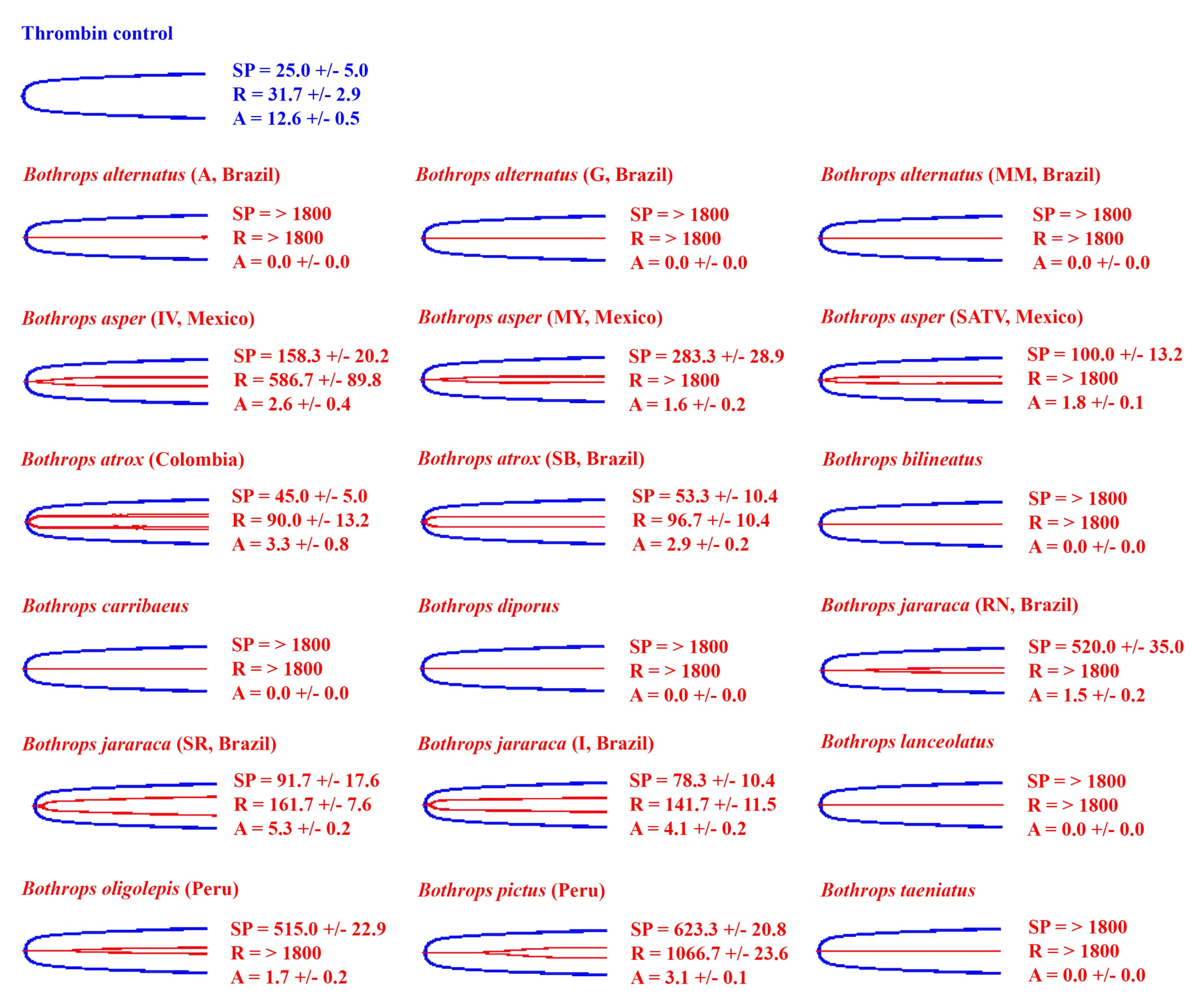
The spontaneous clotting control of human plasma (negative control) had a reaction time (R) of 861.7 ± 33.3 and an amplitude (A) of 26.0 ± 1.8 mm (Figure 1 and Figure 2). The positive controls R was 28.3 ± 2.9 s (thrombin control) and 31.7 ± 2.9 s (FXa control), while A was 28.6 ± 1.0 mm (n = 3) (thrombin control) and 27.7 ± 0.7 mm (FXa control) (Figure 1).
Ancestral state reconstructions (Figure 2) highlight the clotting activity of Bothrops across the phylogeny. A diverse range of clotting activity on human plasma was seen in the Bothrops samples tested, with R ranging from 10 to 1800 s (machine maximum reading), and A ranging from 0 to 25.2 mm. Consistent with procoagulant toxicity being a defining feature of Bothrops venoms, most venoms quickly formed strong clots, such as those of the positive controls (Figure 1 and Figure 2). Although the clots were strong, no venom reached the clotting strengths of the controls. Despite most species forming quick and strong clots, there was variability observed between them (Figure 1 and Figure 2). In particular, significant variation in the relative procoagulant potency was observed between B. alternatus, B. asper, B. atrox, and B. jararaca, and the localities. Conspicuously slower, but still procoagulant, was B. lanceolatus (Figure 1 and Figure 2).
Not all species venom formed fast, strong clots (Figure 1 and Figure 2). Consistent with Bothrops evolving from anticoagulant snakes, the most basal species, B. pictus, prevented plasma from clotting before the machine maximum time was reached, thereby displaying extremely potent anticoagulant toxicity. Intriguingly, while the other two studied localities of B. alternatus were potently procoagulant, the Mogi Mirim, Brazil locality was anticoagulant, thus representing a secondary loss of the derived procoagulant trait. The venom did clot plasma, albeit weakly (A = 1.7 mm), but only started clotting plasma after an extended period of time (SP = 1010 ±/ 65.6 s, shown on Figure 1); thus, it was deemed anticoagulant. Note that R was not recorded for the Mogi Mirim, Brazil locality (R > 1800) as A was less than 2 mm. While one arboreal species (B. oligolepis) acted in a procoagulant manner, rapidly producing a strong clot, the two others studied (B. bilineatus and B. taeniatus) displayed unique patterns in the plasma thromboelastography studies (Figure 1 and Figure 2). The arboreal species B. taeniatus marginally extended clotting time past that of the negative (spontaneous clotting) control, but conspicuously, clot strength was greatly reduced. Despite a clot being registered before the negative control (spontaneous clotting time) for another arboreal species, B. bilineatus, the clot strength was, like that of B. taeniatus, greatly reduced. Similarly, despite clotting being registered at time values shorter than those of the negative (spontaneous clotting) control, only weak clots were formed for the San Andres, Mexico locality of B. asper, B. diporus, and the São Roque locality of B. jararaca (Figure 1 and Figure 2).

2.2. Venom Action on Human Fibrinogen

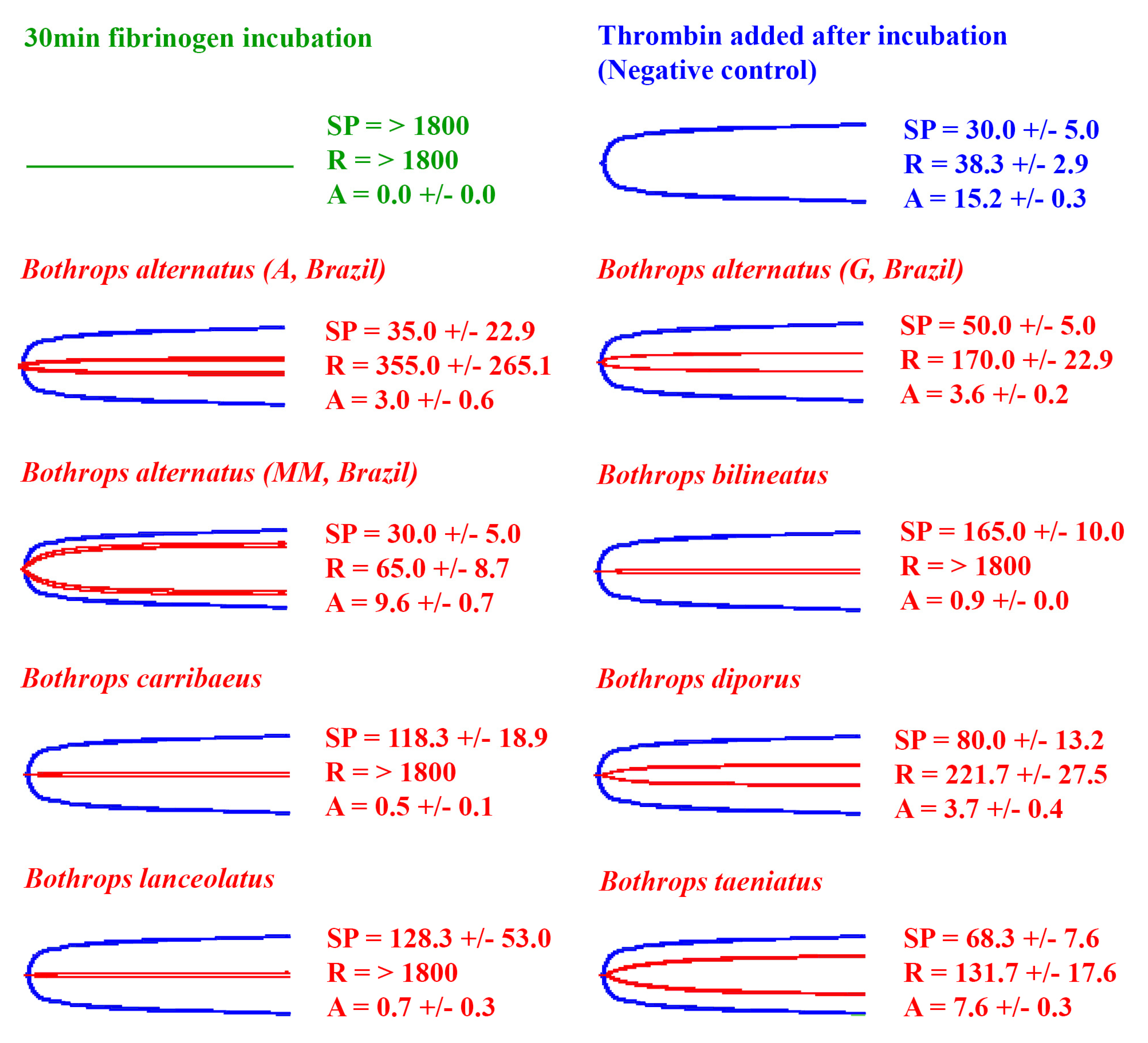
In contrast to recalcified plasma, fibrinogen does not spontaneously clot. Thus, all the results were compared against the thrombin control (positive control). The thrombin control had a reaction time (R) of 31.7 ± 2.9 s and an amplitude (A) of 12.6 ± 0.5 mm (Figure 3 and Figure 4).
In order to ascertain whether the species noted above that lack true procoagulant toxicity (San Andres, Mexico locality of B. asper, B. diporus, São Roque locality of B. jararaca, and the arboreal species B. bilineatus and B. taeniatus) clot fibrinogen, and to ascertain if this direct action on fibrinogen was a background activity for other species, thromboelastography studies were repeated using purified fibrinogen in place of plasma. Ancestral state reconstructions (Figure 4) were used to highlight the clotting activity of Bothrops across the phylogeny.
Consistent with the plasma patterns, the San Andres, Mexico locality of B. asper and the São Roque locality of B. jararaca directly clotted fibrinogen in a pseudo-procoagulant manner, forming weak fibrin clots relative to the thrombin control (Figure 3 and Figure 4). Therefore, these species lack the true procoagulant trait of factor activation (representing a secondary loss of this trait), and instead, only produce the pseudo-procoagulant form of clotting.
Consistent with pseudo-procoagulant action on fibrinogen being an evolutionary relic for most species, there was no clear organismal phylogenetic pattern observed for fibrinogen clotting for the other species that displayed procoagulant venom on human plasma; venoms that possess pseudo-procoagulant action are apparently randomly distributed across the phylogeny, and many species lack this activity (Figure 3). Additionally, and also consistent with pseudo-procoagulant action on fibrinogen being an evolutionary relic, is that most venoms acting in this manner are slower than that of anticoagulant vipers, which use action upon fibrinogen as part of their predatory arsenal [9,55,74,75].

2.3. Fibrinogen Destruction Venom Activity

The eight venoms that did not induce a measurable clot on fibrinogen (A = 0, R = > 1800, flatline on TEG trace) (Figure 3 and Figure 4) were further tested for their ability to cleave fibrinogen in a non-clotting, destructive manner (Figure 5). This was undertaken using the Claussian method [76], whereby after the incubation of venom with fibrinogen, a vast excess of thrombin was added. Any increase in clotting time or decrease in clot strength observed is due to a depletion of fibrinogen levels.
All eight venoms that did not clot fibrinogen (B. diporus, B. alternatus (Araraquara, Guararena, and Mogi Mirim, Brazil), B. lanceolatus, B. caribbaeus, B. bilineatus, and B. taeniatus) exhibited fibrinogen destruction to varying degrees (Figure 5). The island dwellers (B. lanceolatus and B. caribbaeus) and the arboreal B. bilineatus were the most potent fibrinogen destroyers (A < 1.0 mm), while B. alternatus (Mogi Mirim, Brazil) was the weakest (A = 9.6 ± 0.7 mm). Like the pseudo-procoagulant fibrinogen clotting, no clear organismal phylogenetic pattern was observed for fibrinogen destruction, with venoms that possess this action apparently randomly distributed across the phylogeny (Figure 3).

3. Discussion

By undertaking the most comprehensive study to date of Bothrops coagulotoxic effects (37 venoms spanning 19 species and covering the full taxonomical and ecological diversity, including arboreal and island species), this study produced data with tangible, real-world applications by providing information regarding potential clinical effects, while also contributing to the theoretical knowledge of their evolutionary biology. Procoagulant toxicity was shown to be a defining trait of the genus (Figure 1 and Figure 2). Although studies on specific coagulation factor activation were not performed, the strong procoagulant toxicity is likely achieved through coagulation factor activation (e.g., prothrombin and FX activation) [36,38,39,40,41,42,43]. Consistent with previous studies using more limited datasets [15,16,17,43] interspecific and intraspecific variations were noted.
A conspicuous result was the documentation of B. caribbaeus and B. lanceolatus as being procoagulant in this study, when prior work had suggested that these species lacked this trait [66,77]. However, in both prior studies, recalcification was not undertaken for the citrated plasma, as they followed a 1983 protocol that does not include calcium and, consequently, does not reproduce physiological conditions [78]. This is a flaw in the study design, as it has been shown that some Bothrops venoms themselves are calcium-dependent in their action [42,43]. Similarly, in neither study was the phospholipid cofactor added, which has also been shown to be an important co-factor for many procoagulant venoms [5,79]. Thus, in the absence of calcium and phospholipid, little, if any, venom action would be evident; this was clearly the case in the prior B. caribbaeus and B. lanceolatus studies as both venoms were procoagulant upon recalcified plasma in this study, and produced strong, stable clots (Figure 1 and Figure 2). Furthermore, Bourke et al. [15] tested BothroFav (a monospecific B. lanceolatus antivenom) efficacy on procoagulant B. atrox and B. asper and showed that the antivenom was able to neutralise the procoagulant venom effects, specifically those of B. atrox. This suggests that procoagulant toxins are present in B. lanceolatus, as horses immunised with B. lanceolatus venom produce an antivenom (BothroFav) that can neutralise such toxins. Overall, the procoagulant activity of the island-dwelling Bothrops in this study is interesting and deserves further attention in future studies.
The most basal species, B. pictus [71,80], was like most non-Bothrops American pit vipers [37,55,56,81] in that it lacked procoagulant venom; it was anticoagulant on human plasma (Figure 1 and Figure 2). This result might be indicative of the common ancestor of all Bothrops having a distinct anticoagulant venom action compared to the often procoagulant action seen in the more derived Bothrops. Indeed, the limited studies that have been performed on the sister genus Bothrocophias indicate that their venom has low coagulant activity [82,83]. Interestingly, despite being anticoagulant on human plasma, when tested on isolated fibrinogen, B. pictus venom induced a weak clot (A = 3.1 ± 0.1 mm) after an extended period (R = 1066.7 ± 23.6) (Figure 3). This indicates that pseudo-procoagulant (aka ‘thrombin-like’) enzymes are present in the venom, which confirms previous studies [84]. When injected into prey items, the pseudo-procoagulant enzymes likely act synergistically with other anticoagulant toxins, consuming clotting factors to produce an anticoagulant pathology.
Furthermore, underscoring the dynamic nature of venom diversification, the characteristic Bothrops procoagulant venom trait was secondarily lost in several species. This includes the derived arboreal species B. bilineatus and B. taeniatus (Figure 1 and Figure 2). This is consistent with previous reports [46,57,67,70]. Note that previous literature on B. bilineatus subspecies found that B. b. bilineatus clotted human plasma [46,67], while B. b. smaragdinus did not [67]. The subspecies of the present studies venom sample is unknown. Some terrestrial species have also secondarily lost the procoagulant venom trait: the Mogi Mirim, Brazil locality of B. alternatus; the San Andres, Mexico locality of B. asper; B. diporus; and the São Roque of B. jararaca. The divergence within B. alternatus is consistent with previous reports on other localities that found extreme variation between venoms in Brazil [70], which is also consistent with this wide-ranging species, in fact, being a species complex.
Unlike procoagulant toxicity, which is well defined as being a potent predatory weapon through stroke induction, it has been suggested that the pseudo-procoagulant direct action upon fibrinogen to produce weak, short-lived fibrin clots is an evolutionary relic and does not play a role in prey capture, as it is much slower in action than procoagulant factor activation [36,43]. However, as this theory was advanced through datasets containing limited venom diversity, the role has remained enigmatic in the absence of a comprehensive study such as this one. The results demonstrate that there was a lack of phylogenetic signal for this trait in that the variations were random between species and locality (Figure 3 and Figure 4). This, indeed, lends supports to the hypothesis that this trait is an ancestral relic and, assuming no ecological difference among the Bothrops species examined, not subjected to purifying selection pressure. However, as has been noted in clinical cases [24,52], human bite victims survive long enough for the additional depletion of fibrinogen to contribute to the consumptive coagulopathy; therefore, the documentation of the species with the greatest relative potency in this regard will contribute to the body of knowledge, which is useful for designing evidence-based clinical management strategies. The species with the most relatively potent action (small R and small A values) in this regard were the terrestrial species B. jararacussu (all tested), B. asper (Costa Rica and Ecuador only amongst those tested in this study), B. atrox (all tested), B. leucurus, B. moojeni (all tested), B. neuwiedi (Curitiba, Brazil only amongst those tested in this study), B. pauloensis, and B. jararaca (all tested except Rio Negrino). This suggests that in clinical cases, these species may produce additional fibrinogen depletion on top of that consumed by the procoagulant mode of action.
In addition, some other species showed the ability to destructively (non-clotting) cleave fibrinogen, which would also contribute to fibrinogen depletion (Figure 5). Bothrops diporus, B. alternatus (Araraquara, Guararena, and Mogi Mirim, Brazil), B. lanceolatus, B. caribbaeus, B. bilineatus, and B. taeniatus all displayed the ability to destroy fibrinogen. The most potent were the island species B. lanceolatus and B. caribbaeus and the arboreal species B. bilineatus. Interestingly, despite destroying fibrinogen (anticoagulant effect) B. lanceolatus and B. caribbaeus were also potently procoagulant on human plasma (Figure 1, Figure 2 and Figure 5). This is counter-intuitive: procoagulant toxins work to produce a clot while fibrinogen-destroying toxins work to stop the production of a clot. The observed clot on human plasma is likely reflective of the procoagulant toxins taking over and producing fibrin before fibrinogen destruction can occur. Other contrasting venom actions were seen for the Guararena and Araraquara locality B. alternatus, which produced strong clots in human plasma, but also destroyed fibrinogen (Figure 1, Figure 2 and Figure 5).
Interestingly, extensive variation was shown within B. alternatus, with the Mogi Mirim locality being much weaker than the other two localities in the fibrinogen destruction trait (Figure 5), despite lacking the procoagulant venom trait as shown in human plasma tests (Figure 1 and Figure 2). This suggests that toxins that inhibit upstream coagulation factors, rather than directly affect fibrinogen, are involved in producing the anticoagulant effects on human plasma observed for the B. alternatus Mogi Mirim locality venom. For the other species that lacked a procoagulant trait, the terrestrial species B. diporus and the arboreal species B. bilineatus and B. taeniatus, have upregulated the ability to directly cleave fibrinogen in a destructive (non-clotting) manner. This suggests that, at least in these venoms, the action may have a role in prey capture, with these snakes having reverted back to the ancestral fibrinogen-depleting anticoagulant condition. In addition, relative to procoagulant species, the arboreal species may have greater reliance upon the neurotoxic activity documented for those venoms [85,86,87] to subjugate prey. A lack of procoagulant activity might be beneficial in allowing neurotoxins to spread throughout the body, as localised clots might “trap” neurotoxins, preventing them from spreading. This theory of toxin synergism and clots “trapping” toxins is discussed in Jackson et al. [88]. Furthermore, B. bilineatus and B. taeniatus feed on a variety of vertebrates [14,89,90]; thus, rather than neurotoxicity evolving to target a particular prey type, neurotoxins may have evolved in the venom to incapacitate prey items quickly, decreasing the chance of prey dropping from the trees and escaping. This hypothesis, however, needs to be tested by ascertaining relative neurotoxic effects on prey-lineage targets.
The secondary change from procoagulant to anticoagulant cleavage of fibrinogen, whether in a destructive manner or a pseudo-procoagulant manner, is almost certainly due to differential selection pressure for the species that display these traits. The arboreal species are under clear divergent selection pressure for prey-escape potential, thus providing a hypothesis regarding the change in venom phenotype to the destructive fibrinogenolytic form of anticoagulation. However, there is an enigmatic question of what selection pressures (e.g., changes in prey preference or changes in prey-escape potential) led to the divergence—relative to other terrestrial Bothrops—for B. diporus to convergently evolve with these arboreal species the destructive fibrinogenolytic form of anticoagulation. What is also enigmatic is the question of why the San Andres, Mexico locality of B. asper and the São Roque locality of B. jararaca convergently evolved relative to each other to lose the procoagulant trait, and instead, have the pseudo-procoagulant fibrinogenolytic venom phenotype. These results, therefore, provide a starting point for natural history observations to reconstruct the selection pressures leading from these species, which are nested deep within the procoagulant Bothrops clade, to diverge to a fibrinogenolytic venom phenotype.
This documentation of extreme coagulotoxicity diversity across the Bothrops genus is a tangible benefit, as clinical observation of envenomated patients corroborate these results, with patients experiencing the haemostatic disorders of unclottable blood and systemic bleeding [23,24,25,91,92].
Understanding the caveats of a study are important so that appropriate conclusions can be made. An important caveat in this study is that all tests are in vitro, without flow of blood and without multiple systems interacting; hence, the conclusion of the results needs to be interpreted with this in mind. In vitro studies are effective for investigating specific venom actions upon the coagulation cascade, such as in the present study. As in vivo systems are complex and dynamic, in vitro results may not line up with in vivo results. An important note, however, is that this study was undertaken using human plasma and; thus, while in vitro, it may produce results more suggestive of potential human envenomation effects than in vivo animal-model studies, if the venoms have a differential effect between humans and animal-models. This is, indeed, recognised as a critical issue and a limitation of the applicability of animal-based research for human medicine [93]. Regardless, future in vivo work should be conducted before making any therapeutic recommendations regarding clinical care. Lastly, this study only tested venom on human plasma and fibrinogen, yet some Bothrops venoms have been shown to have different action when tested on the plasma of different prey types [43]. Although it is beyond the scope of this study, future studies should test a wide range of Bothrops venom on non-human animal plasmas to test for differences in venom action among prey types, in order to provide information about evolutionary selection pressures leading to changes in the venom phenotype.
Overall, this study vastly improves our fundamental knowledge base on the coagulotoxic action of Bothrops venoms. We hope this wealth of new knowledge for toxinologists will fuel many future studies, as well as provide a solid platform for evolutionary biologists to use alongside natural history observations of these fascinating snakes.

4. Materials and Methods

4.1. Venom Sample Preparation

Venom work was undertaken under University of Queensland Animal Ethics Approval 2021/AE000075. All venoms used in the study were in lyophilised form and obtained from the University of Queensland’s Venom Evolution Lab’s long-term venom collection, venom suppliers, and collaborators. Thirty-seven venoms were used in the study, including nineteen different species and numerous geographical variants (Table 1).
Venoms were prepared to a 1 mg/mL stock solution (50:50 double deionised water (ddH20):glycerol) using a Thermo Fisher Scientific™ NanoDrop 2000 (Waltham, MA, USA) at 280 nm wavelength, and stored at −80 °C until experimentation, during which they were stored at −20 °C. This storage limited enzyme degradation, as did the use of glycerol in venom stocks, which function to stabilise enzymes.

4.2. Plasma and Fibrinogen Preparation

Human plasma work was performed under University of Queensland Biosafety Approval #IBC134BSBS2015 and Human Ethics Approval #2016000256. The Australian Red Cross (44 Musk Street, Kelvin Grove, QLD 4059, Australia) supplied human platelet-poor plasma (3.2% citrated, O positive, Label # A540021261153) under research approval #16-04QLD-10. Once obtained, plasma was stored at −80 °C until aliquoted. Plasma was aliquoted into 1.5 mL tubes by defrosting in a water bath at 37 °C and aliquoting into 1.5 mL tubes in a biosafety cabinet, to avoid contaminating the plasma. Plasma was then stored at −80 °C until experimentation.
Human fibrinogen (Sigma Aldrich, St. Louis, MO, USA) was prepared to 4 mg/mL by diluting 100 mg of fibrinogen with Owren Koller (OK) buffer to a volume of 25 mL. This solution was then vortexed until solubilised; it was aliquoted, then the tubes were flash-frozen with liquid nitrogen and immediately stored at −80 °C.

4.3. Thromboelastography Experiments

Two TEG® 5000 Thrombelastograph® Haemostasis Analyser systems were used concurrently, containing two reaction stations each. Natural cups and pins were placed into each channel and the heating plate set to 37 °C. All reagents were then pipetted into the cup as per our previously validated protocol [43]: 72 μL CaCl (25 mM stock solution Stago Cat# 00367); 72 μL phospholipid (Stago Cat# 00597), solubilised in Owren Koller (OK) buffer (Stago Cat #00360); 20 μL OK buffer; and 7 μL venom sample or control sample (negative control: 7 μL 50:50 ddH20: glycerol, thrombin control: 7 μL thrombin, or FXa control: 7 μL FXa). A total of 189 μL plasma or fibrinogen, which had been thawed in a water bath at 37 °C for 5 min, was taken out of the water bath and then pipetted into each cup, after which the 360 μL sample was pipette-mixed and the machine was run for 30 min. For each cup, the time from pipetting the plasma into the cup until the start of each test (lifting the cup into the test machine and pressing start) was 10 s. Each venom test was performed in triplicate (n = 3).
Venoms that did not clot fibrinogen (the TEG trace was a flatline) were immediately tested for fibrinogen destruction via a Claussian method [76], using our previously validated protocol [94]. Fibrinogen destruction tests were conducted because fibrinogen does not spontaneously clot, like human plasma; thus, a flatline could indicate that the venom either has no action on fibrinogen or that the venom destroys fibrinogen. In this test, 7 μL of thrombin was added to the cups, which already had a 360 μL sample in them from the prior test. Then, the whole solution was pipette mixed and run again for 30 min. The results from the venom runs were compared to the negative control, where a blank (7 μL ddH20/glycerol) replaced the venom, and thrombin induced a clot upon fibrinogen in the absence of venom. Note that some venoms that produced a flatline on the initial fibrinogen destruction test qualitatively produced micro ‘clots’ (small strands). These were noted for four venoms (B. alternatus (Mogi Mirim, Brazil), B. alternatus (Guararena, Brazil), B. alternatus (Araraquara, Brazil), and B. lanceolatus (Martinique)) and were ignored in the study; this is because they were not recorded on the machine and, therefore, were unlikely to be clinically significant.
The parameters produced by thromboelastography include split point (SP), reaction time (R), amplitude (A) and maximum amplitude (MA). SP is the time until the tracing splits, representing the first formation of fibrin strands. R is the time until the first detectable clot appears (defined by the machine as enough resistance to produce an amplitude >2 mm). R is the most often-used variable to indicate clot initiation. R is where classic coagulation assays, such as prothrombin time (PT) and partial thromboplastin time (PTT) assays are completed; thus, TEG provides a more detailed picture of coagulation than these assays. Both A and MA represent clot strength. Clot strength is measured as the width of the tracing in mm (the greater the width, the greater the clot strength). A is the strength of the clot at the latest time point, while MA is the maximum strength of the clot reached during the run. MA is only recorded after A > 2 mm (A is equal to MA until MA is determined). MA is calculated using the small deviation method (time = 3 min), in which MA is only calculated if itdoes not deviate more than 1 mm for at least 3 min, thus explaining why A is sometimes >MA.
For a detailed description of how TEG works, see Supplementary File S1.

4.4. Data Analysis

Phylogenetic trees were initially produced using TimeTree (TimeTree.org) and exported to Mesquite (version 3.61), where additional branches for localities and certain species were made manually. TimeTree collates data from published studies to produce a phylogeny from the specified genus which, in this study, is Bothrops. The resulting tree was missing two of the species analysed in the study: Bothrops barnetti and B. oligolepis. The missing species were manually added to the tree and their branch lengths estimated, based on phylogenies from [71,72,73]. The Mesquite tree file (.phy file) is available in the Supplementary Materials (Supplementary File S2). The tree was then imported into the statistical software R (version 3.6.1) using the APE package [95]. The contMAP function of the phytools package [96] was used to estimate ancestral states, using maximum likelihood, and to visually represent the presented trait over the tree. Four trees were produced: a tree produced from human plasma reaction time data, a tree produced from human plasma amplitude data, a tree produced from fibrinogen reaction time data, and a tree produced from fibrinogen amplitude data. Each data file used to produce the trees can be found in the Supplementary Materials (Supplementary File S3). The R code is also available in the Supplementary Materials (Supplementary File S4). The trees were then exported from R and edited with Adobe Photoshop (version 21.2.11) and Adobe Acrobat Pro DC (version 2021.011.20039) to produce Figure 2 and Figure 4. Editing consisted of positioning the reaction time and amplitude trees opposite each other, adding details of species names, and adding the mean ± standard deviation of R and A values for each respective branch.
Thromboelastography traces were exported from the TEG 5000, and Figure 1, Figure 3 and Figure 5 produced in Adobe Photoshop. All thromboelastography raw data for each venom can be found in the Supplementary Materials (Supplementary File S5), with the following parameters supplied: SP, R, Angle, A, and MA. Note that in the raw data, A is never exactly 0 mm as the sample always induces some resistance. In this study we classed all A values as 0 mm if the trace was a flatline. Additionally, note that for consistency, Amplitude (A) is used in Figure 1, Figure 2, Figure 3, Figure 4 and Figure 5 instead of Maximum Amplitude (MA), since for some venoms, MA could not be calculated (MA is only calculated if A > 2.0 mm). Furthermore, R was used instead of SP in phylogenetic trees (Figure 2 and Figure 4) as R represents the time until detectable clot formation rather than just the first deposition of fibrin strands (SP). R was deemed a more meaningful parameter to plot.

Supplementary Materials

The following supporting information can be downloaded at: https://www.mdpi.com/article/10.3390/toxins14050297/s1, Supplementary File S1: Thromboelastography approach; Supplementary File S2: .phy tree file; Supplementary File S3: Data files for trees; Supplementary File S4: R code used in analyses; Supplementary File S5: Thromboelastography raw data.

Author Contributions

Conceptualisation, L.A.B. and B.G.F.; data curation, L.A.B.; formal analysis, L.A.B. and B.G.F.; investigation, L.A.B.; methodology, L.A.B., C.N.Z. and B.G.F.; visualisation, L.A.B. and B.G.F.; resources, A.M.T.-A., G.P.M.S., S.S.S., K.F.G., C.F.B.R., B.G.F.; writing—original draft, L.A.B.; writing—review and editing, L.A.B., B.G.F., C.N.Z., A.M.T.-A., G.P.M.S., S.S.S., K.F.G., C.F.B.R.; supervision, B.G.F. and C.N.Z.; project administration and funding acquisition, B.G.F. All authors have read and agreed to the published version of the manuscript.

Funding

This work was funded by the Australian Research Council Discovery Project DP190100304 to B.G.F. A PhD Scholarship from the University of Queensland was awarded to L.A.B.

Institutional Review Board Statement

Work was undertaken with the following University of Queensland institutional review board approvals: the Human Ethics Committee #2016000256, the Biosafety Committee #IBC134BSBS2015, and the Animal Ethics Committee 2021/AE000075 (15 March 2021).

Informed Consent Statement

Not applicable.

Data Availability Statement

Raw data are available in Supplementary File S5.

Acknowledgments

The authors also thank the Government of St. Lucia, and the St. Lucia Department of Forestry, for allowing specimens of Bothrops caribbaeus to be maintained at Kentucky Reptile Zoo for the purposes of an assurance colony and beneficial research, and also thank the Kentucky Reptile Zoo for donating venom to us from this precious, and fascinating species.

Conflicts of Interest

The authors declare no conflict of interest.

References

  1. Kini, R.M. Serine proteases affecting blood coagulation and fibrinolysis from snake venoms. Pathophysiol. Haemost. Thromb. 2005, 34, 200–204. [Google Scholar] [CrossRef] [PubMed]
  2. Kini, R.M. The intriguing world of prothrombin activators from snake venom. Toxicon 2005, 45, 1133–1145. [Google Scholar] [CrossRef]
  3. Kini, R.M. Anticoagulant proteins from snake venoms: Structure, function and mechanism. Biochem. J. 2006, 397, 377. [Google Scholar] [CrossRef] [PubMed] [Green Version]
  4. Kini, R.M.; Koh, C.Y. Metalloproteases affecting blood coagulation, fibrinolysis and platelet aggregation from snake venoms: Definition and nomenclature of interaction sites. Toxins 2016, 8, 284. [Google Scholar] [CrossRef] [PubMed] [Green Version]
  5. Kini, R.M.; Rao, V.S.; Joseph, J.S. Procoagulant proteins from snake venoms. Pathophysiol. Haemost. Thromb. 2001, 31, 218–224. [Google Scholar] [CrossRef]
  6. Markland, F.S. Snake venoms and the hemostatic system. Toxicon 1998, 36, 1749–1800. [Google Scholar] [CrossRef]
  7. Kini, R.M.; Evans, H.J. A model to explain the pharmacological effects of snake venom phospholipases A2. Toxicon 1989, 27, 613–635. [Google Scholar] [CrossRef]
  8. Swenson, S.; Markland, F.S. Snake venom fibrin(ogen)olytic enzymes. Toxicon 2005, 45, 1021–1039. [Google Scholar] [CrossRef]
  9. Bourke, L.A.; Youngman, N.J.; Zdenek, C.N.; op den Brouw, B.; Violette, A.; Fourmy, R.; Fry, B.G. Trimeresurus albolabris snakebite treatment implications arising from ontogenetic venom comparisons of anticoagulant function, and antivenom efficacy. Toxicol. Lett. 2020, 327, 2–8. [Google Scholar] [CrossRef]
  10. Alencar, L.; Martins, M.; Greene, H. Evolutionary history of vipers. eLS 2018, 1–10. [Google Scholar] [CrossRef]
  11. Wüster, W.; da Graca Salomão, M.; Quijada-Mascareñas, J.A.; Thorpe, R.S.; BBBSP. Origins and evolution of the South American pitviper fauna: Evidence from mitochondrial DNA sequence analysis. In Biology of the Vipers; Eagle Mountain Publishing: Eagle Mountain, UT, USA, 2002; pp. 111–128. [Google Scholar]
  12. Fiorillo, B.F.; Tozetti, A.M.; Martins, M. Habitat use by five species of sympatric pitvipers (Bothrops, Crotalus) in a Brazilian savannah. Herpetol. Notes 2020, 13, 951–960. [Google Scholar]
  13. Martins, M.; Araujo, M.S.; Sawaya, R.J.; Nunes, R. Diversity and evolution of macrohabitat use, body size and morphology in a monophyletic group of neotropical pitvipers (Bothrops). J. Zool. 2001, 254, 529–538. [Google Scholar] [CrossRef] [Green Version]
  14. Martins, M.; Marques, O.A.; Sazima, I. Ecological and phylogenetic correlates of feeding habits in neotropical pitvipers of the genus Bothrops. In Biology of the Vipers; Schuett, G.W., Höggren, M., Douglas, M.E., HW, G., Eds.; Eagle Mountain Publishing: Eagle Mountain, UT, USA, 2002; pp. 307–328. [Google Scholar]
  15. Bourke, L.A.; Zdenek, C.N.; Neri-Castro, E.; Bénard-Valle, M.; Alagón, A.; Gutiérrez, J.M.; Sanchez, E.F.; Aldridge, M.; Fry, B.G. Pan-American lancehead pit-vipers: Coagulotoxic venom effects and antivenom neutralisation of Bothrops asper and B. atrox geographical variants. Toxins 2021, 13, 78. [Google Scholar] [CrossRef] [PubMed]
  16. Sousa, L.F.; Holding, M.L.; Del-Rei, T.H.M.; Rocha, M.M.T.; Mourão, R.H.V.; Chalkidis, H.M.; Prezoto, B.; Gibbs, H.L.; Moura-da-Silva, A.M. Individual variability in Bothrops atrox snakes collected from different habitats in the Brazilian Amazon: New findings on venom composition and functionality. Toxins 2021, 13, 814. [Google Scholar] [CrossRef] [PubMed]
  17. Segura, Á.; Herrera, M.; Villalta, M.; Vargas, M.; Uscanga-Reynell, A.; de León-Rosales, S.P.; Jiménez-Corona, M.E.; Reta-Mares, J.F.; Gutiérrez, J.M.; León, G. Venom of Bothrops asper from Mexico and Costa Rica: Intraspecific variation and cross-neutralization by antivenoms. Toxicon 2012, 59, 158–162. [Google Scholar] [CrossRef] [PubMed]
  18. Gutiérrez, J.M.; Calvete, J.J.; Habib, A.G.; Harrison, R.A.; Williams, D.J.; Warrell, D.A. Snakebite envenoming. Nat. Rev. Dis. Primers. 2017, 3, 17063. [Google Scholar] [CrossRef] [PubMed]
  19. Chippaux, J.-P. Epidemiology of envenomations by terrestrial venomous animals in Brazil based on case reporting: From obvious facts to contingencies. J. Venom Anim. Toxins Incl. Trop. Dis. 2015, 21, 1–17. [Google Scholar] [CrossRef] [Green Version]
  20. da Costa, M.K.B.; da Fonseca, C.S.; Navoni, J.A.; Freire, E.M.X. Snakebite accidents in Rio Grande do Norte state, Brazil: Epidemiology, health management and influence of the environmental scenario. Trop. Med. Int. Health 2019, 24, 432–441. [Google Scholar] [CrossRef]
  21. Dolab, J.A.; de Roodt, A.R.; de Titto, E.H.; García, S.I.; Funes, R.; Salomón, O.D.; Chippaux, J.-P. Epidemiology of snakebite and use of antivenom in Argentina. Trans R Soc. Trop. Med. Hyg. 2014, 108, 269–276. [Google Scholar] [CrossRef]
  22. Mota-da-Silva, A.; Colombini, M.; Moura-da-Silva, A.M.; Souza, R.M.; Monteiro, W.M.; Bernarde, P.S. Epidemiological and clinical aspects of snakebites in the upper Juruá River region, western Brazilian Amazonia. Acta Amazonica 2019, 50. [Google Scholar] [CrossRef]
  23. Monteiro, W.M.; Contreras-Bernal, J.C.; Bisneto, P.F.; Sachett, J.; Mendonça da Silva, I.; Lacerda, M.; Guimarães da Costa, A.; Val, F.; Brasileiro, L.; Sartim, M.A.; et al. Bothrops atrox, the most important snake involved in human envenomings in the amazon: How venomics contributes to the knowledge of snake biology and clinical toxinology. Toxicon X 2020, 6, 100037. [Google Scholar] [CrossRef]
  24. Otero-Patiño, R. Epidemiological, clinical and therapeutic aspects of Bothrops asper bites. Toxicon 2009, 54, 998–1011. [Google Scholar] [CrossRef] [PubMed]
  25. Santoro, M.L.; Sano-Martins, I.S.; Fan, H.W.; Cardoso, J.L.C.; Theakston, R.D.G.; Warrell, D.A. Haematological evaluation of patients bitten by the jararaca, Bothrops jararaca, in Brazil. Toxicon 2008, 51, 1440–1448. [Google Scholar] [CrossRef] [PubMed]
  26. Milani Júnior, R.; Jorge, M.T.; de Campos, F.P.; Martins, F.P.; Bousso, A.; Cardoso, J.L.; Ribeiro, L.A.; Fan, H.W.; França, F.O.; Sano-Martins, I.S.; et al. Snake bites by the jararacuçu (Bothrops jararacussu): Clinicopathological studies of 29 proven cases in São Paulo State, Brazil. QJM 1997, 90, 323–334. [Google Scholar] [CrossRef] [PubMed] [Green Version]
  27. Maguiña Vargas, C.; Henríquez, C.; Ilquimiche, L.; Mostorino, R.; Gotuzzo Herencia, E.; Legua, P.; Echevarría Zárate, J.; Seas Ramos, C. Ofidismo por Bothrops pictus en el Hospital Nacional Cayetano Heredia: Estudio prospectivo de 23 casos. Folia Dermatol. Peru 1998, 9, 41–48. [Google Scholar]
  28. Resiere, D.; Mégarbane, B.; Valentino, R.; Mehdaoui, H.; Thomas, L. Bothrops lanceolatus bites: Guidelines for severity assessment and emergent management. Toxins 2010, 2, 163–173. [Google Scholar] [CrossRef] [PubMed] [Green Version]
  29. Thomas, L.; Chausson, N.; Uzan, J.; Kaidomar, S.; Vignes, R.; Plumelle, Y.; Bucher, B.; Smadja, D. Thrombotic stroke following snake bites by the “Fer-de-Lance” Bothrops lanceolatus in Martinique despite antivenom treatment: A report of three recent cases. Toxicon 2006, 48, 23–28. [Google Scholar] [CrossRef]
  30. Thomas, L.; Tyburn, B.; Bucher, B.; Pecout, F.; Ketterle, J.; Rieux, D.; Smadja, D.; Garnier, D.; Plumelle, Y. Prevention of thromboses in human patients with Bothrops Lanceolatus envenoming in Martinique: Failure of anticoagulants and efficacy of a monospecific antivenom. Am. J. Trop. Med. Hyg. 1995, 52, 419–426. [Google Scholar] [CrossRef]
  31. Numeric, P.; Moravie, V.; Didier, M.; Chatot-Henry, D.; Cirille, S.; Bucher, B.; Thomas, L. Multiple cerebral infarctions following a snakebite by Bothrops caribbaeus. Am. J. Trop. Med. Hyg. 2002, 67, 287–288. [Google Scholar] [CrossRef] [Green Version]
  32. Smalligan, R.; Cole, J.; Brito, N.; Laing, G.D.; Mertz, B.L.; Manock, S.; Maudlin, J.; Quist, B.; Holland, G.; Nelson, S.; et al. Crotaline snake bite in the Ecuadorian Amazon: Randomised double blind comparative trial of three South American polyspecific antivenoms. BMJ 2004, 329, 1129. [Google Scholar] [CrossRef] [Green Version]
  33. Gutiérrez, J.M. Current challenges for confronting the public health problem of snakebite envenoming in Central America. J. Venom. Anim. Toxins Incl. Trop. Dis. 2014, 20, 7. [Google Scholar] [CrossRef] [PubMed] [Green Version]
  34. Neri-Castro, E.; Bénard-Valle, M.; de León, J.L.; Boyer, L.; Alagón, A. Envenomations by reptiles in Mexico. In Handbook of Venoms and Toxins of Reptiles; CRC Press: Boca Raton, FL, USA, 2021; pp. 529–542. [Google Scholar]
  35. Kasturiratne, A.; Wickremasinghe, A.R.; de Silva, N.; Gunawardena, N.K.; Pathmeswaran, A.; Premaratna, R.; Savioli, L.; Lalloo, D.G.; de Silva, H.J. The global burden of snakebite: A literature analysis and modelling based on regional estimates of envenoming and deaths. PLoS Med. 2008, 5, e218. [Google Scholar] [CrossRef] [PubMed] [Green Version]
  36. Rodrigues, C.F.B.; Zdenek, C.N.; Bourke, L.A.; Seneci, L.; Chowdhury, A.; Freitas-de-Sousa, L.A.; de Alcantara Menezes, F.; Moura-da-Silva, A.M.; Tanaka-Azevedo, A.M.; Fry, B.G. Clinical implications of ontogenetic differences in the coagulotoxic activity of Bothrops jararacussu venoms. Toxicol. Lett. 2021, 348, 59–72. [Google Scholar] [CrossRef] [PubMed]
  37. Nielsen, V.G.; Frank, N.; Afshar, S. De novo assessment and review of Pan-American pit viper anticoagulant and procoagulant venom activities via kinetomic analyses. Toxins 2019, 11, 94. [Google Scholar] [CrossRef] [Green Version]
  38. Hofmann, H.; Bon, C. Blood coagulation induced by the venom of Bothrops atrox. 1. Identification, purification, and properties of a prothrombin activator. Biochemistry 1987, 26, 772–780. [Google Scholar] [CrossRef]
  39. Hofmann, H.; Bon, C. Blood coagulation induced by the venom of Bothrops atrox. 2. Identification, purification, and properties of two factor X activators. Biochemistry 1987, 26, 780–787. [Google Scholar] [CrossRef]
  40. Hofmann, H.; Dumarey, C.; Bon, C. Blood coagulation induced by Bothrops atrox venom: Idendification and properties of a factor X activator. Biochimie 1983, 65, 201–210. [Google Scholar] [CrossRef]
  41. Loría, G.D.; Rucavado, A.; Kamiguti, A.S.; Theakston, R.D.G.; Fox, J.W.; Alape, A.; Gutiérrez, J.M.A. Characterization of ‘basparin A,’ a prothrombin-activating metalloproteinase, from the venom of the snake Bothrops asper that inhibits platelet aggregation and induces defibrination and thrombosis. Arch. Biochem. Biophys. 2003, 418, 13–24. [Google Scholar] [CrossRef]
  42. Sousa, L.F.; Bernardoni, J.L.; Zdenek, C.N.; Dobson, J.; Coimbra, F.; Gillett, A.; Lopes-Ferreira, M.; Moura-da-Silva, A.M.; Fry, B.G. Differential coagulotoxicity of metalloprotease isoforms from Bothrops neuwiedi snake venom and consequent variations in antivenom efficacy. Toxicol. Lett. 2020, 333, 211–221. [Google Scholar] [CrossRef]
  43. Sousa, L.F.; Zdenek, C.N.; Dobson, J.S.; Op den Brouw, B.; Coimbra, F.C.P.; Gillett, A.; Del-Rei, T.H.M.; Chalkidis, H.D.M.; Sant’Anna, S.; Teixeira-da-Rocha, M.M.; et al. Coagulotoxicity of Bothrops (lancehead pit-vipers) venoms from Brazil: Differential biochemistry and antivenom efficacy resulting from prey-driven venom variation. Toxins 2018, 10, 411. [Google Scholar] [CrossRef] [Green Version]
  44. Herrera, M.; Fernández, J.; Vargas, M.; Villalta, M.; Segura, Á.; León, G.; Angulo, Y.; Paiva, O.; Matainaho, T.; Jensen, S.D.; et al. Comparative proteomic analysis of the venom of the taipan snake, Oxyuranus scutellatus, from Papua New Guinea and Australia: Role of neurotoxic and procoagulant effects in venom toxicity. J. Proteom. 2012, 75, 2128–2140. [Google Scholar] [CrossRef] [PubMed]
  45. Martin, C.J. On some effects upon the blood produced by the injection of the venom of the Australian black snake (Pseudechis Porphyriacus). J. Physiol. 1893, 15, 380–400. [Google Scholar] [CrossRef] [PubMed] [Green Version]
  46. Nahas, L.; Kamiguti, A.S.; Barros, M.A.R. Thrombin-like and factor X-activator components of Bothrops snake venoms. Thromb. Haemost. 1979, 41, 314–328. [Google Scholar] [CrossRef]
  47. Sant’Ana, C.D.; Ticli, F.K.; Oliveira, L.L.; Giglio, J.R.; Rechia, C.G.V.; Fuly, A.L.; Selistre de Araújo, H.S.; Franco, J.J.; Stabeli, R.G.; Soares, A.M.; et al. BjussuSP-I: A new thrombin-like enzyme isolated from Bothrops jararacussu snake venom. Comp. Biochem. Physiol. Part A Mol. Integr. Physiol. 2008, 151, 443–454. [Google Scholar] [CrossRef]
  48. Magalhães, A.; Magalhães, H.P.B.; Richardson, M.; Gontijo, S.; Ferreira, R.N.; Almeida, A.P.; Sanchez, E.F. Purification and properties of a coagulant thrombin-like enzyme from the venom of Bothrops leucurus. Comp. Biochem. Physiol. Part A Mol. Integr. Physiol. 2007, 146, 565–575. [Google Scholar] [CrossRef] [PubMed]
  49. Costa, F.L.S.; Rodrigues, R.S.; Izidoro, L.F.M.; Menaldo, D.L.; Hamaguchi, A.; Homsi-Brandeburgo, M.I.; Fuly, A.L.; Soares, S.G.; Selistre-de-Araújo, H.S.; Barraviera, B.; et al. Biochemical and functional properties of a thrombin-like enzyme isolated from Bothrops pauloensis snake venom. Toxicon 2009, 54, 725–735. [Google Scholar] [CrossRef] [PubMed]
  50. Pérez, A.V.; Rucavado, A.; Sanz, L.; Calvete, J.J.; Gutiérrez, J.M. Isolation and characterization of a serine proteinase with thrombin-like activity from the venom of the snake Bothrops Asper. Braz. J. Med. Biol. Res. 2008, 41, 12–17. [Google Scholar] [CrossRef] [Green Version]
  51. Petretski, J.; Kanashiro, M.; Silva, C.; Alves, E.; Kipnis, T. Two related thrombin-like enzymes present in Bothrops atrox venom. Braz. J. Med. Biol. Res. 2000, 33, 1293–1300. [Google Scholar] [CrossRef] [Green Version]
  52. Wellmann, I.A.M.; Ibiapina, H.N.S.; Sachett, J.A.G.; Sartim, M.A.; Silva, I.M.; Oliveira, S.S.; Tarragô, A.M.; Moura-da-Silva, A.M.; Lacerda, M.V.G.; Ferreira, L.C.d.L.; et al. Correlating fibrinogen consumption and profiles of inflammatory molecules in human envenomation’s by Bothrops atrox in the Brazilian Amazon. Front. Immunol. 2020, 11, 1874. [Google Scholar] [CrossRef]
  53. Larréché, S.; Chippaux, J.-P.; Chevillard, L.; Mathé, S.; Résière, D.; Siguret, V.; Mégarbane, B. Bleeding and thrombosis: Insights into pathophysiology of Bothrops venom-related hemostasis disorders. Int. J. Mol. Sci. 2021, 22, 9643. [Google Scholar] [CrossRef]
  54. Seneci, L.; Zdenek, C.N.; Chowdhury, A.; Rodrigues, C.F.B.; Neri-Castro, E.; Bénard-Valle, M.; Alagón, A.; Fry, B.G. A clot twist: Extreme variation in coagulotoxicity mechanisms in Mexican neotropical rattlesnake venoms. Front. Immunol. 2021, 12, 552. [Google Scholar] [CrossRef] [PubMed]
  55. Seneci, L.; Zdenek, C.N.; Bourke, L.A.; Cochran, C.; Sánchez, E.E.; Neri-Castro, E.; Bénard-Valle, M.; Alagón, A.; Frank, N.; Fry, B.G. A symphony of destruction: Dynamic differential fibrinogenolytic toxicity by rattlesnake (Crotalus and Sistrurus) venoms. Comp. Biochem. Physiol. C Toxicol. Pharmacol. 2021, 245, 109034. [Google Scholar] [CrossRef]
  56. Youngman, N.J.; Peng, Y.-H.; Harris, R.J.; Jones, L.; Llinas, J.; Haworth, M.; Gillett, A.; Fry, B.G. Differential coagulotoxic and neurotoxic venom activity from species of the arboreal viperid snake genus Bothriechis (palm-pitvipers). Comp. Biochem. Physiol. C Toxicol. Pharmacol. 2022, 256, 109326. [Google Scholar] [CrossRef]
  57. Kamiguti, A.S.; Sousa E Silva, M.C.C.; Morena, P.; Nahas, L. The anticoagulant effect of Bothrops castelnaudi snake venom (Castelnaud’s pit viper). Toxicon 1985, 23, 383–391. [Google Scholar] [CrossRef]
  58. Lotto, N.P.; de Albuquerque Modesto, J.C.; Sant’Anna, S.S.; Grego, K.F.; Guarnieri, M.C.; Lira-da-Silva, R.M.; Santoro, M.L.; Oguiura, N. The absence of thrombin-like activity in Bothrops erythromelas venom is due to the deletion of the snake venom thrombin-like enzyme gene. PLoS ONE 2021, 16, e0248901. [Google Scholar] [CrossRef]
  59. Kaaber, A.B.; Jans, Ø.; Dziegiel, M.H.; Stensballe, J.; Johansson, P.I. Managing patients on direct factor Xa inhibitors with rapid thrombelastography. Scand. J. Clin. Lab. Investig. 2021, 81, 661–669. [Google Scholar] [CrossRef] [PubMed]
  60. Da Luz, L.T.; Nascimento, B.; Shankarakutty, A.K.; Rizoli, S.; Adhikari, N.K.J. Effect of thromboelastography (TEG®) and rotational thromboelastometry (ROTEM®) on diagnosis of coagulopathy, transfusion guidance and mortality in trauma: Descriptive systematic review. Crit. Care 2014, 18, 518. [Google Scholar] [CrossRef] [Green Version]
  61. Leeper, C.M.; Gaines, B.A. Viscoelastic hemostatic assays in the management of the pediatric trauma patient. Semin. Pediatric Surg. 2017, 26, 8–13. [Google Scholar] [CrossRef] [PubMed]
  62. Wiśniewski, A.; Karczmarska-Wódzka, A.; Sikora, J.; Sobczak, P.; Lemanowicz, A.; Filipska, K.; Ślusarz, R. Hypercoagulability as measured by thrombelastography may be associated with the size of acute ischemic infarct—A pilot study. Diagnostics 2021, 11, 712. [Google Scholar] [CrossRef]
  63. Drevets, P.D.; Tien, L.; LaCoursiere, R.J.; Burgbacher, T.E.; Fox, E.D. Rattlesnake Envenomation in a Venom-Naive Man with Significant Coagulopathy and Severe Oropharyngeal Edema Requiring Emergent Surgical Airway. J. Curr. Surg. 2021, 11, 15–20. [Google Scholar] [CrossRef]
  64. Zdenek, C.N.; Chowdhury, A.; Haw, G.Y.H.; Violette, A.; Fourmy, R.; Christ, T.; Vonk, F.J.; Fry, B.G. Taxon-selective venom variation in adult and neonate Daboia russelii (Russell’s Viper), and antivenom efficacy. Toxicon 2022, 205, 11–19. [Google Scholar] [CrossRef] [PubMed]
  65. Nielsen, V.G.; Wagner, M.T.; Frank, N. Mechanisms responsible for the anticoagulant properties of neurotoxic Dendroaspis venoms: A viscoelastic analysis. Int. J. Mol. Sci. 2020, 21, 2082. [Google Scholar] [CrossRef] [Green Version]
  66. Gutiérrez, J.M.; Sanz, L.; Escolano, J.; Fernández, J.; Lomonte, B.; Angulo, Y.; Rucavado, A.; Warrell, D.A.; Calvete, J.J. Snake venomics of the Lesser Antillean pit vipers Bothrops caribbaeus and Bothrops lanceolatus: Correlation with toxicological activities and immunoreactivity of a heterologous antivenom. J. Proteome Res. 2008, 7, 4396–4408. [Google Scholar] [CrossRef]
  67. Sanz, L.; Quesada-Bernat, S.; Pérez, A.; De Morais-Zani, K.; Sant’Anna, S.S.; Hatakeyama, D.M.; Tasima, L.J.; De Souza, M.B.; Kayano, A.M.; Zavaleta, A.; et al. Danger in the canopy. Comparative proteomics and bioactivities of the venoms of the South American palm pit viper Bothrops bilineatus subspecies bilineatus and smaragdinus and Antivenomics of B. b. bilineatus (Rondônia) venom against the Brazilian pentabothropic antivenom. J. Proteome Res. 2020, 19, 3518–3532. [Google Scholar] [CrossRef] [PubMed]
  68. Rodrigues, C.R.; Teixeira-Ferreira, A.; Vargas, F.F.R.; Guerra-Duarte, C.; Costal-Oliveira, F.; Stransky, S.; Lopes-de-Souza, L.; Dutra, A.A.A.; Yarlequé, A.; Bonilla, C.; et al. Proteomic profile, biological activities and antigenic analysis of the venom from Bothriopsis bilineata smaragdina (“loro machaco”), a pitviper snake from Peru. J. Proteom. 2018, 187, 171–181. [Google Scholar] [CrossRef] [PubMed]
  69. Kuch, U.; Mebs, D.; Gutiérrez, J.M.; Freire, A. Biochemical and biological characterization of Ecuadorian pitviper venoms (genera Bothriechis, Bothriopsis, Bothrops and Lachesis). Toxicon 1996, 34, 714–717. [Google Scholar] [CrossRef]
  70. Sanchez, E.F.; Freitas, T.V.; Ferreira-Alves, D.L.; Velarde, D.T.; Diniz, M.R.; Cordeiro, M.N.; Agostini-Cotta, G.; Diniz, C.R. Biological activities of venoms from South American snakes. Toxicon 1992, 30, 95–103. [Google Scholar] [CrossRef]
  71. Alencar, L.R.V.; Quental, T.B.; Grazziotin, F.G.; Alfaro, M.L.; Martins, M.; Venzon, M.; Zaher, H. Diversification in vipers: Phylogenetic relationships, time of divergence and shifts in speciation rates. Mol. Phylogenet. Evol. 2016, 105, 50–62. [Google Scholar] [CrossRef]
  72. Carrasco, P.A.; Mattoni, C.I.; Leynaud, G.C.; Scrocchi, G.J. Morphology, phylogeny and taxonomy of South American bothropoid pitvipers (Serpentes, Viperidae). Zool. Scr. 2012, 41, 109–124. [Google Scholar] [CrossRef]
  73. Fenwick, A.M.; Gutberlet, R.L.; Evans, J.A.; Parkinson, C.L. Morphological and molecular evidence for phylogeny and classification of South American pitvipers, genera Bothrops, Bothriopsis, and Bothrocophias (Serpentes: Viperidae). Zool. J. Linn. Soc. 2009, 156, 617–640. [Google Scholar] [CrossRef] [Green Version]
  74. Debono, J.; Bos, M.H.A.; Frank, N.; Fry, B. Clinical implications of differential antivenom efficacy in neutralising coagulotoxicity produced by venoms from species within the arboreal viperid snake genus Trimeresurus. Toxicol. Lett. 2019, 316, 35–48. [Google Scholar] [CrossRef]
  75. Debono, J.; Bos, M.H.A.; Coimbra, F.; Ge, L.; Frank, N.; Kwok, H.F.; Fry, B.G. Basal but divergent: Clinical implications of differential coagulotoxicity in a clade of Asian vipers. Toxicol. In Vitro 2019, 58, 195–206. [Google Scholar] [CrossRef]
  76. Clauss, A. Gerinnungsphysiologische Schnellmethode zur Bestimmung des Fibrinogens. Acta Haematol. 1957, 17, 237–246. [Google Scholar] [CrossRef]
  77. Bogarín, G.; Romero, M.; Rojas, G.; Lutsch, C.; Casadamont, M.; Lang, J.; Otero, R.; Gutiérrez, J.M.A. Neutralization, by a monospecific Bothrops lanceolatus antivenom, of toxic activities induced by homologous and heterologous Bothírops snake venoms. Toxicon 1999, 37, 551–557. [Google Scholar] [CrossRef]
  78. Theakston, R.D.; Reid, H.A. Development of simple standard assay procedures for the characterization of snake venom. Bull。 World Health Organ. 1983, 61, 949–956. [Google Scholar]
  79. Lister, C.; Arbuckle, K.; Jackson, T.N.W.; Debono, J.; Zdenek, C.N.; Dashevsky, D.; Dunstan, N.; Allen, L.; Hay, C.; Bush, B.; et al. Catch a tiger snake by its tail: Differential toxicity, co-factor dependence and antivenom efficacy in a procoagulant clade of Australian venomous snakes. Comp. Biochem. Physiol. C Toxicol. Pharmacol. 2017, 202, 39–54. [Google Scholar] [CrossRef] [PubMed] [Green Version]
  80. Hamdan, B.; Guedes, T.B.; Carrasco, P.A.; Melville, J. A complex biogeographic history of diversification in Neotropical lancehead pitvipers (Serpentes, Viperidae). Zool. Scr. 2020, 49, 145–158. [Google Scholar] [CrossRef]
  81. Youngman, N.J.; Carlsson, D.J.; Jones, L.; Neri-Castro, E.; Alagón, A.; Fry, B.G. Cloud serpent coagulotoxicity: The biochemical mechanisms underpinning the anticoagulant actions of Mixcoatlus and Ophryacus venoms. Toxicon 2022, 211, 44–49. [Google Scholar] [CrossRef] [PubMed]
  82. Salazar-Valenzuela, D.; Mora-Obando, D.; Fernández, M.L.; Loaiza-Lange, A.; Gibbs, H.L.; Lomonte, B. Proteomic and toxicological profiling of the venom of Bothrocophias campbelli, a pitviper species from Ecuador and Colombia. Toxicon 2014, 90, 15–25. [Google Scholar] [CrossRef]
  83. Pereañez, J.A.; Preciado, L.M.; Fernández, J.; Camacho, E.; Lomonte, B.; Castro, F.; Cañas, C.A.; Galvis, C.; Castaño, S. Snake venomics, experimental toxic activities and clinical characteristics of human envenomation by Bothrocophias myersi (Serpentes: Viperidae) from Colombia. J. Proteom. 2020, 220, 103758. [Google Scholar] [CrossRef]
  84. Vivas-Ruiz, D.E.; Sandoval, G.A.; Gonzalez-Kozlova, E.; Zarria-Romero, J.; Lazo, F.; Rodríguez, E.; Magalhães, H.P.B.; Chávez-Olortegui, C.; Oliveira, L.S.; Alvarenga, V.G.; et al. Fibrinogen-clotting enzyme, pictobin, from Bothrops pictus snake venom. Structural and functional characterization. Int. J. Biol. Macromol. 2020, 153, 779–795. [Google Scholar] [CrossRef]
  85. Romero-Vargas, F.; Rocha, T.; Cruz-Höfling, M.; Rodrigues-Simioni, L.; Ponce-Soto, L. Biochemical characterization of a PLA2 Btae TX-I isolated from Bothriopsis taeniata snake venom: A pharmacological and morphological study. J. Clin. Toxicol. 2014, 4. [Google Scholar] [CrossRef] [Green Version]
  86. Floriano, R.S.; Carregari, V.C.; de Abreu, V.A.; Kenzo-Kagawa, B.; Ponce-Soto, L.A.; da Cruz-Höfling, M.A.; Hyslop, S.; Marangoni, S.; Rodrigues-Simioni, L. Pharmacological study of a new Asp49 phospholipase A2 (Bbil-TX) isolated from Bothriopsis bilineata smargadina (forest viper) venom in vertebrate neuromuscular preparations. Toxicon 2013, 69, 191–199. [Google Scholar] [CrossRef]
  87. Rodrigues-Simioni, L.; Floriano, R.S.; Rostelato-Ferreira, S.; Sousa, N.C.; Marangoni, S.; Ponce-Soto, L.A.; Carregari, V.C.; Hyslop, S. Presynaptic action of Bothriopsis bilineata smargadina (forest viper) venom in vitro. Toxicon 2011, 58, 140–145. [Google Scholar] [CrossRef]
  88. Jackson, T.N.W.; Koludarov, I.; Ali, S.A.; Dobson, J.; Zdenek, C.N.; Dashevsky, D.; Op den Brouw, B.; Masci, P.P.; Nouwens, A.; Josh, P.; et al. Rapid radiations and the race to redundancy: An investigation of the evolution of Australian elapid snake venoms. Toxins 2016, 8, 309. [Google Scholar] [CrossRef]
  89. da Fonseca, W.L.; de Souza Oliveira, A.; Correa, R.R.; Bernarde, P.S. Caudal luring in the Neotropical two-striped forest pitviper Bothrops bilineatus smaragdinus Hoge, 1966 in the western Amazon. Herpetol. Notes 2019, 12, 365–374. [Google Scholar]
  90. Venegas, P.J.; Chávez Arribasplata, J.C.; Almora, E.; Grilli, P.G.; Duran, V. New observations on diet of the South American two-striped forest-pitviper Bothrops bilineatus smaragdinus (Hoge, 1966). Cuad Herpetol. 2019. [Google Scholar] [CrossRef]
  91. Silva de Oliveira, S.; Campos Alves, E.; dos Santos Santos, A.; Freitas Nascimento, E.; Tavares Pereira, J.P.; Mendonça da Silva, I.; Sachett, J.; dos Santos Ibiapina, H.N.; Santos Sarraf, L.K.; Contreras Bernal, J.C.; et al. Bothrops snakebites in the Amazon: Recovery from hemostatic disorders after Brazilian antivenom therapy. Clin. Toxicol. 2020, 58, 266–274. [Google Scholar] [CrossRef] [PubMed]
  92. Silva de Oliveira, S.; C Alves, E.; S Santos, A.; F Nascimento, E.; T Pereira, J.P.; M Silva, I.; A G Sachett, J.; S Sarraff, L.K.; Freitas-de-Sousa, L.A.; Colombini, M.; et al. Bleeding disorders in Bothrops atrox envenomations in the Brazilian Amazon: Participation of hemostatic factors and the impact of tissue factor. Toxins 2020, 12, 554. [Google Scholar] [CrossRef]
  93. Silva, A.; Hodgson, W.C.; Tasoulis, T.; Isbister, G.K. Rodent lethality models are problematic for evaluating antivenoms for human envenoming. Front. Pharmacol. 2022, 13, 384. [Google Scholar] [CrossRef] [PubMed]
  94. Debono, J.; Bos, M.H.A.; Nouwens, A.; Ge, L.; Frank, N.; Kwok, H.F.; Fry, B.G. Habu coagulotoxicity: Clinical implications of the functional diversification of Protobothrops snake venoms upon blood clotting factors. Toxicol. In Vitro 2019, 55, 62–74. [Google Scholar] [CrossRef] [PubMed] [Green Version]
  95. Paradis, E.; Claude, J.; Strimmer, K. APE: Analyses of phylogenetics and evolution in R language. Bioinformatics 2004, 20, 289–290. [Google Scholar] [CrossRef] [Green Version]
  96. Revell, L.J. Phytools: An R package for phylogenetic comparative biology (and other things). Methods Ecol. Evol. 2012, 3, 217–223. [Google Scholar] [CrossRef]
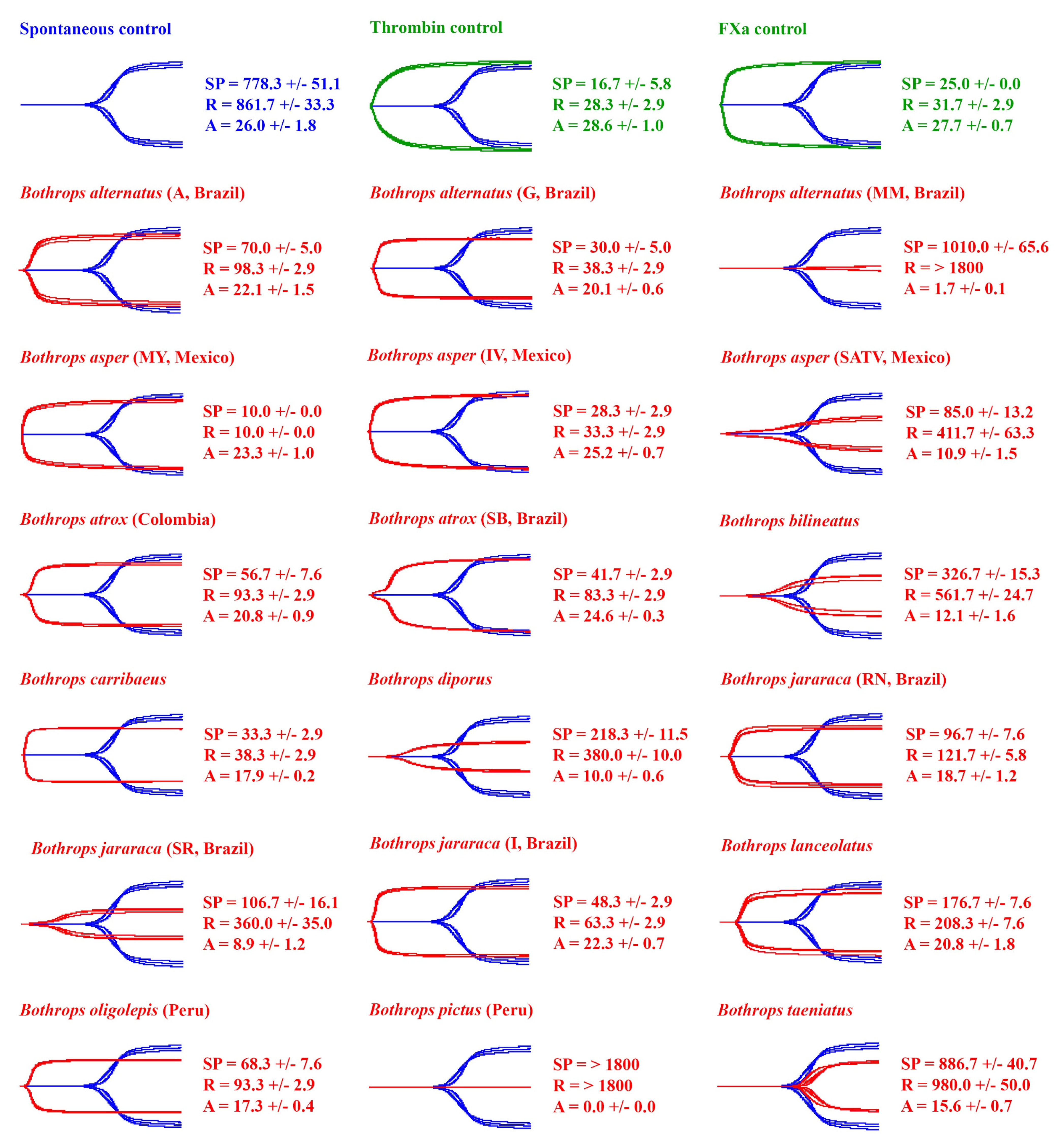
Figure 1. Thromboelastography traces showing the action of Bothrops venoms (red traces) on human plasma. A total of 18 representatives (red traces) of the 37 venoms examined (Table 1) are overlaid on top of the spontaneous control (blue traces) and ordered in the figure alphabetically. Three controls were performed: a negative control (spontaneous clotting time of plasma, blue traces), and two positive controls (thrombin control and FXa control, green traces). Three clotting parameters are shown: SP (split point—time until tracing splits, representing start of clotting), R (reaction time—time until amplitude = 2 mm, representing time until detectable clot), and A (Amplitude—width of tracing at latest time point, representing clot strength at latest time point). All values are mean ± standard deviation (n = 3). Each test lasted 1800 s, so > 1800 s indicates that the parameter was not recorded in this time. The spontaneous clotting control of human plasma (negative control) had an SP of 778.3 ± 51.1 s, R of 861.7 ± 33.3 s, and A of 26.0 ± 1.8 mm. Locality details for locality abbreviations used in this figure can be found in Table 1.
Toxins 14 00297 g001
Figure 2. Ancestral state reconstructions of Bothrops venom clotting parameters from thromboelastography on human plasma. The parameters R (reaction time—time until amplitude = 2 mm, representing time until detectable clot in seconds) (left) and A (amplitude—width of tracing at latest time point, representing clot strength at latest time point in mm) (right) are shown. All values are mean ± standard deviation (n = 3) and spontaneous clotting control values are shown below each phylogeny. The colour gradient ranges from violet to black, with violet representing faster clotting times (left hand side) and stronger clots (right hand side). Note: due to the high dynamicity of venom evolution, the node bar ranges quickly become broad as one moves down the tree. The phylogeny was produced using timetree.org and updated with information from Alencar et al. [71], Carrasco et al. [72], and Fenwick et al. [73]. Each test lasted 1800 s, so R >1800 s indicates that R was not recorded in this time. Note: although B. alternatus (MM, Brazil) has an R > 1800 s, a weak clot was still observed (A = 1.7 ± 0.1) (R is only recorded if A > 2 mm). The spontaneous clotting control of human plasma (negative control) had a reaction time (R) of 861.7 ± 33.3 and amplitude (A) of 26.0 ± 1.8 mm. Locality details for locality abbreviations used in this figure can be found in Table 1.
Toxins 14 00297 g002
Figure 3. Thromboelastography traces showing the action of Bothrops venoms (red traces) on human fibrinogen. A total of 18 representatives (red traces) of the 37 venoms examined (Table 1) are overlaid on top of the thrombin control (blue traces) and ordered in the figure alphabetically. Three clotting parameters are shown: SP (split point—time until tracing splits, representing start of clotting), R (reaction time—time until amplitude = 2 mm, representing time until detectable clot), and A (amplitude—width of tracing at latest time point, representing clot strength at latest time point). All values are mean ± standard deviation (n = 3). Each test lasted 1800 s, so >1800 s indicates that the parameter was not recorded in this time. The thrombin control had an SP of 25.0 ± 5.0 s, R of 31.7 ± 2.9 s, and A of 12.6 ± 0.5 mm. Locality details for locality abbreviations used in this figure can be found in Table 1.
Toxins 14 00297 g003
Figure 4. Ancestral state reconstructions of Bothrops venom clotting parameters from thromboelastography on human fibrinogen. The parameters R (reaction time—time until amplitude = 2 mm, representing time until detectable clot in seconds; left) and A (amplitude—width of tracing at latest time point, representing clot strength at latest time point in mm; right) are shown. All values are mean +/− standard deviation (n = 3) and thrombin control values are shown below each phylogeny. The colour gradient ranges from violet to black, with violet representing faster clotting times (left hand side) and stronger clots (right hand side). Note: due to the high dynamicity of venom evolution, the node bar ranges quickly become broad as one moves down the tree. Each test lasted 1800 s, so R > 1800 s indicates that R was not recorded in this time. Bold species names indicate no R parameter was recorded in the test time; thus, R = >1800, and the A parameter = 0 mm (no clot observed, TEG trace flatlined). Note: although Bothrops barnetti (Peru) and B. oligolepis (MM, Brazil) have an R > 1800 s, a weak clot was still observed (A = 1.6 ± 0.1 and 1.7 ± 0.2, respectively; R is only record if A > 2 mm). The phylogeny was produced using timetree.org and updated with information from Alencar et al. [71], Carrasco et al. [72], and Fenwick et al. [73]. The thrombin control had an R of 31.7 ± 2.9 s and A of 12.6 ± 0.5 mm. Locality details for locality abbreviations used in this figure can be found in Table 1.
Toxins 14 00297 g004
Figure 5. Thromboelastography traces showing the action of Bothrops venoms that did not clot fibrinogen (8/37 venoms) in a fibrinogen destruction assay. Venoms are ordered alphabetically in the figure. For each test, fibrinogen was incubated with venom for 30 min before the addition of thrombin in a Claussian protocol. The green trace represents the trace of each sample after 30 min incubation (no clot formed). The positive control (thrombin control) is shown in blue and venom samples (red traces) are overlaid on top of the thrombin control (blue traces). Three clotting parameters are shown: SP (split point—time until tracing splits, representing start of clotting), R (reaction time—time until amplitude = 2 mm, representing time until detectable clot), and A (amplitude—width of tracing at latest time point, representing clot strength at latest time point). All values are mean ± standard deviation (n = 3). Each test lasted 1800 s, so >1800 s indicates that the parameter was not recorded in this time. The thrombin control had an SP of 30.0 ± 5.0 s, R of 38.3 ± 2.9 s, and A of 15.2 ± 0.3 mm. Locality details for locality abbreviations used in this figure can be found in Table 1.
Toxins 14 00297 g005
Table 1. Bothrops venoms used in the study with additional details supplied: whether venoms are from single individuals or pooled from multiple individuals (n values are supplied if known), locality, sex, age, and source (captive-born or wild-caught snakes). Note: captive-born snakes originate from snakes from the specified locality. Locality abbreviations are the abbreviation for each locality used in the study (“–” = no abbreviation given).
SpeciesPooled (n) or IndividualLocalityLocality Abbreviation in Figures (If Used)SexApprox Age (yrs)Source, and Wild-Caught or Captive Bred Stock
B. alternatusIndividualMogi Mirim–SP, BrazilMM, BrazilF9Instituto Butantan, wild-caught in 2009
B. alternatusIndividualGuararena–SP, BrazilG, BrazilF7Instituto Butantan, captive-born in 2011, 17a specimen from litter
B. alternatusIndividualAraraquara–SP, BrazilA, BrazilF5Instituto Butantan, wild-caught in 2013
B. atroxIndividualSão Bento–MA, BrazilSB, BrazilF12Instituto Butantan, wild-caught in 2009
B. atroxPooled (2)Balbira–AM, Brazil x São Bento–MA, BrazilBxSB, BrazilF8Institute Butantan, captive-born in 2010, l1a specimen from litter
Balbira–AM, Brazil x São Bento–MA, BrazilF8Institute Butantan, captive-born in 2010, 7a specimen from litter
B. atroxPooled (66)French Guiana-M + FAdultsLatoxan, captive-born and wild-caught snakes
B. atroxPooled (n values not supplied)Alto Marañon, Peru (Amazon rainforest)PeruUNKNAdultsEFS, wild-caught
B. atroxUNKNSuriname-UNKNAdultsKentucky reptile zoo, unknown
B. atroxUNKNColombia-UNKNAdultsKentucky reptile zoo, unknown
B. asperPooled (40)Costa Rica (Pacific region)Costa RicaUNKNAdultsJMG, wild-caught
B. asperPooled (2)Ecuador-MAdultsLatoxan, captive-born
B. asperIndividualMérida, Yucatán, MexicoMY, MexicoUNKNAdultUNAM, wild-caught
B. asperIndividualSan Andres, Tuxtla, Veracruz, MexicoSATV, MexicoUNKNAdultUNAM, wild-caught
B. asperIndividualIxtaczoquitlan, Veracruz, MexicoIV, MexicoUNKNYoung adultUNAM, wild-caught
B. barnettiPooled (n values not supplied)Talara, Department of Tubmes, PeruPeruUNKNAdultsUNMSM, wild-caught
B. bilineatusPooled (2)UNKN-F + FAdultsM-toxins, imported
B. caribbaeusUNKNSt. Lucia, West Indies-UNKNAdultsKentucky reptile zoo, wild-caught
B. diporusUNKNUNKN-UNKNAdultsVEL, imported
B. jararacussuPooled (2)Juquitiba–SPJ, BrazilF3Instituto Butantan, wild-caught in 2015
Juquitiba–SPF3Instituto Butantan, wild-caught in 2015
B. jararacussuIndividualCubatão–SPC, BrazilF3Instituto Butantan, wild-caught in 2015
B. jararacaIndividualRio Negrino–SCRN, BrazilF1Instituto Butantan, wild-caught in 2017
B. jararacaIndividualSão Roque–SPSR, BrazilF3Instituto Butantan, wild-caught in 2015
B. jararacaIndividualIbiúna–SPI, BrazilF3Instituto Butantan, wild-caught in 2015
B. lanceolatusUNKNMartinique-UNKNAdultsLatoxan, imported
B. leucurusPooled (3)Porto Seguro–BAPS, BrazilF3Instituto Butantan, captive-born in 2016, 5a specimen from litter
Porto Seguro–BAF3Instituto Butantan, captive-born in 2016, 13a specimen from litter
Porto Seguro–BAM3Instituto Butantan, captive-born in 2016, 14a specimen from litter
B. mattogrossensisPooled (3)Porto Murtinho–MSPM, BrazilF6Instituto Butantan, captive-born in 2012, 4a specimen from litter
Porto Murtinho–MSF9Instituto Butantan, wild-caught in 2009
B. mattogrossensisUNKNBolivia-UNKNAdultsVEL, imported
B. moojeniIndividualPalmas–TOP, BrazilF13Instituto Butantan, wild-caught in 2005
B. moojeniPooled (2)Gaúcha do Norte–MTGdN, BrazilF8Instituto Butantan, captive-born in 2010, 13a specimen from litter
Gaúcha do Norte–MTF8Instituto Butantan, captive-born in 2010, 5a specimen from litter
B. neuwiediIndividualSalto Pirapora–SPSP, BrazilM6Instituto Butantan, captive-born in 2013, 3a specimen from litter
B. neuwiediIndividualCuritiba–PRC, BrazilF2Instituto Butantan, wild-caught in 2017
B. neuwiediIndividualMunhoz–MGM, BrazilF4Instituto Butantan, wild-caught in 2015
B. oligolepisPooled (3)La Merced, Chanchamayo, Peru (central rainforest región)PeruUNKNAdultsUNMSM, wild-caught
B. pauloensisPooled (3)São Simão–SPSS, BrazilF6Instituto Butantan, captive-born in 2012, 6a specimen from litter
São Simão–SPF7Instituto Butantan, captive-born in 2011, 2a specimen from litter
São Simão–SPF7Instituto Butantan, captive-born in 2011, 6a specimen from litter
B. pictusPooled (7)Districts of Carabayllo and Comas, PeruPeruUNKNAdultsUNMSM, wild-caught
B. pubescensUNKNUruguay-UNKNAdultsVEL, imported
B. taeniatusUNKNUNKN-UNKNAdultsVEL, imported
EFS = Laboratory of Biochemistry of Proteins from Animal Venoms, Research and Development Center, Ezequiel Dias Foundation, Belo Horizonte, MG 30510-010, Brazil; JMG = José María Gutiérrez, Instituto Clodomiro Picado, Facultad de Microbiología, Universidad de Costa Rica, San José 11501, Costa Rica; UNAM = Alejandro Alagon, Departamento de Medicina Molecular y Bioprocesos, Instituto de Biotecnologa, Universidad Nacional Autónoma de México, Av. Universidad 2001, Cuernavaca, Morelos 62210, Mexico; UNNSM = Armando Yarleque, Universidad Nacional Mayor de San Marcos, Lima, Peru; VEL = Venom Evolution Lab, UQ, Australia.
Publisher’s Note: MDPI stays neutral with regard to jurisdictional claims in published maps and institutional affiliations.