Article

Extreme Procoagulant Potency in Human Plasma of Venoms from the African Viperid Genera Atheris, Cerastes, and Proatheris and the Relative Efficacy of Antivenoms and Synthetic Enzyme-Inhibitors

1
Venom Evolution Lab, School of Biological Science, University of Queensland, St. Lucia, QLD 4072, Australia
2
Department of Biochemistry & Microbiology, North South University, Dhaka 1229, Bangladesh
3
Ophirex, Inc., Corte Madera, CA 94925, USA
4
California Academy of Sciences, San Francisco, CA 94118, USA
5
Inosan Biopharma, 28108 Madrid, Spain
6
MicroPharm Limited, Newcastle Emlyn SA38 9BY, UK
*
Authors to whom correspondence should be addressed.
Toxins 2022, 14(12), 836; https://doi.org/10.3390/toxins14120836
Submission received: 1 November 2022 / Revised: 16 November 2022 / Accepted: 25 November 2022 / Published: 1 December 2022

Abstract

:
The African viperid snake genera Atheris, Cerastes, and Proatheris are closely related, similar in size, but occupy extremely divergent ecological niches (arboreal in tropical rainforests, fossorial in deserts, and swamp-dwelling, respectively). Their venoms have not previously been subjected to comparative analyses for their action upon the coagulation of blood, most notably with significant data deficiencies from Atheris and Proatheris. In contrast, the closely related genus Echis is well-documented as capable of producing potent procoagulant effects. In light of this, we set out to compare the coagulotoxic actions of Atheris ceratophora, A. chlorechis, A. desaixi, A. nitschei, A. squamigera, C. cerastes, C. cerastes gasperettii, C. vipera, and Proatheris superciliaris and explore potential pharmacological interventions to reestablish normal blood coagulation. All venoms displayed extremely potent procoagulant effects, over twice as fast as the most potent Echis reported to date. Although Cerastes is used in the immunising mixture of two different regionally available antivenoms (Inoserp-MENA with C. cerastes, C. cerastes gasperettii, C. vipera and Saudi Arabian polyvalent with C. cerastes), none of the other species in this study are included in the immunising mixture of any antivenom. Notably, all the Cerastes species were only neutralised by the Inoserp-MENA antivenom. C. cerastes venom was not neutralised well by the Saudi Arabian antivenom, with the low levels of recognition for any of the Cerastes venoms suggesting a strong regional variation in the venom of this species, as the C. cerastes venom tested was of African (Tunisian) origin versus Saudi locality used in that antivenom’s production. The other antivenoms (Micropharm EchiTAbG, ICP EchiTAb-Plus-ICP, Inosan Inoserp Pan-Africa, Premium Serums PANAF Sub-Sahara Africa, South African Vaccine Producers Echis, South African Vaccine Producers Polyvalent) all displayed trivial-to-no ability to neutralise the procoagulant toxicity of any of the Atheris, Cerastes, or Proatheris venoms. Comparative testing of the enzyme inhibitors DMPS, marimastat, and prinomastat, revealed a very potent neutralising capacity of marimastat, with prinomastat showing lower but still significant potency at the same molar concentration, while a 5× molar concentration of DMPS had no apparent effect on procoagulant venom effects normalized by the other inhibitors. These results and methods contribute to the body of knowledge of potential clinical effects and data necessary for evidence-based advancement of clinical management strategies.
Key Contribution: This work revealed that venoms of Atheris, Cerastes, and Proatheris species are amongst the most potently procoagulant viper venoms tested to-date, with Atheris particularly potent (on par with extremely toxic elapid snakes such as taipans). Eight regionally available antivenoms failed to neutralise this pathophysiological action, with the exception of Inosan antivenom which neutralised Cerastes venoms. The metalloprotease inhibitors marimastat and prinomastat were effective, suggestive of potential clinical utility.

1. Introduction

Despite snakebite being a well-documented source of significant human morbidity and mortality [1,2], data are lacking for both pathophysiological effects and treatment options for the venoms of many species [3]. Snake venoms exert multi-dimensional attacks upon any part of the body reachable by the bloodstream, and blood coagulation itself is a particular target for many venoms [4]. Coagulotoxic pathophysiological actions not only aid in prey capture, but contribute significantly to the tremendous human snakebite burden.
Within Africa, a diversity of snakes have potent effects upon the blood coagulation cascade. Within the Colubridae family, the sister genera Dispholidus (“Boomslang”) and Thelatornis are lethally procoagulant through the activation of prothrombin. Treatment options are limited, as only Dispholidus is effectively neutralised by Boomslang antivenom [5]. Similarly, within the Elapidae family, spitting cobras in particular have strong anticoagulant effects through the inhibition of the activated clotting factors FXa and thrombin, and neither action is neutralised by South African polyvalent antivenom [6]. However, recent studies have shown that the enzyme inhibitor varespladib is effective against this action [6,7,8]. Within the Lamprophidae family, Atractaspis venoms are potently procoagulant through the activation of Factor X, with no antivenom able to effectively neutralise this effect [9].
Significantly, the greatest human impact of coagulotoxic snakebite envenomings in Africa is due to species within the Viperidae family. Of these, the most intensively studied are the venoms of the Echis genus, which display extreme procoagulant effects and treatment efficacy suffers from regional variations in antivenom efficacy [10]. Similarly, the coagulotoxic effects of Bitis venoms have also been well-studied, with most displaying anticoagulant effects through myriad of mechanisms, except for B. worthingtoni which is uniquely procoagulant through the activation of Factor X [11,12,13]. As well, Causus venoms have been shown to be anticoagulant through the destruction of fibrinogen, with extreme variation in antivenom efficacy [14]. Within the group of venoms in this study, others have shown that Cerastes and Proatheris are procoagulant and Cerastes, through the activation of Factor X [15]. Though known to cause coagulopathy, hemolysis, thrombocytopenia and renal failure, neither Cerastes nor Proatheris have been systematically evaluated for relative neutralisation by regionally available antivenoms [16,17,18,19,20,21,22,23,24,25,26]. Few studies have considered the closely related genus Atheris [27], though the few data available suggest a strong procoagulant trait with relative in vitro antivenom efficacy remaining unknown prior to the present study [28].
It should be noted that other studies have reported a lack of procoagulant toxicity for Atheris, Cerastes, and Proatheris venoms [29,30]. However, these studies relied upon a laboratory protocol that it did not add the clotting cofactors calcium and phospholipid to the citrated plasmas as part of the methods [29,30,31]. This is an important methodological deficiency as citrate is typically added to plasma to prevent clotting by chelating calcium. Further, platelet poor plasma contains low or no phospholipid as this is contributed in whole blood by the platelets [3]. Not only do many venoms themselves require calcium for activity, but if the venoms activate Factor X even in the absence of calcium, this effect would not be discernible as FX itself requires calcium for its endogenous action of prothrombin activation [5,6,10,32,33,34,35,36,37,38,39,40,41,42,43,44,45]. Thus, while strong procoagulant activity has been reported in a single study of Atheris venoms in recalcified plasma [28] and in one physiologically relevant protocol of Proatheris [42], the studies which reported a lack of procoagulant toxicity for these two genera (and Cerastes) [29,30] must be regarded as potentially erroneous due to likely methodological deficiencies. Similarly a study which reported anticoagulant activity for Cerastes cerastes [28] may be due to any number of factors ranging from a regional or ontogenetic variation of the venom to specimen misidentification to methodological issues, with further investigation needed to unravel this issue. These discrepancies [28,29,30] underscore the need for comparative testing in a standardised protocols most closely replicating physiological conditions.
To confirm our hypotheses and ensure methodological consistency with recent standards, we set out to fill some of these knowledge gaps regarding Atheris, Cerastes, and Proatheris venoms using validated protocols for ascertaining coagulotoxicity. In addition, we also evaluated the relative effectiveness of antivenoms and small-molecule enzyme-inhibitors against the coagulotoxic effects of these venoms [5,6,10,32,33,34,35,36,37,38,39,40,41,42,43,44,45]. We tested the venoms of Atheris ceratophora, A. chlorechis, A. desaixi, A. nitschei, A. squamigera, C. cerastes, C. cerastes gasperettii, C. vipera, and Proatheris superciliaris against regionally available antivenoms (ICP EchiTAb-Plus-ICP, Micropharm EchiTAbG, Inosan Inoserp-MENA, Inosan Inoserp Pan-Africa, Premium Serums PANAF Sub-Sahara Africa, Saudi Arabian polyvalent, South African Vaccine Producers Echis, and South African Vaccine Producers Polyvalent) and against several promising enzyme inhibitors 2,3-Dimercapto-1-propanesulfonic acid (DMPS), marimastat, and prinomastat. As all genera have been reported as capable of producing severe clinical effects [16,17,18,19,20,21,22,23,24,25,26], this work will contribute data essential for the evidence-based design of clinical management strategies. This work is also of evolutionary interest as it examines the effect of extremely divergent ecological niche occupation relative to venom effects in related genera such as Atheris that are arboreal specialists in tropical rainforests, Cerastes species that are semi-fossorial specialists in sandy deserts, and Proatheris inhabiting low-lying marshes and flood plains [46,47,48].

2. Results

All raw values can be found in the supplementary material in File S1. Supplementary Sheet African viperid genera Atheris, Cerastes, and Proatheris.xlsx. Plasma clotting conditions were first ascertained by determining the negative control values (spontaneous clotting time subsequent to the addition of calcium), which was 386.5 +/− 13.7 s. Consistent with potent procoagulant toxicity, all venoms tested significantly accelerated the clotting time relative to the negative control (Figure 1). Indeed, the venoms were amongst the fastest viperid venoms to-date tested under the same standardised conditions (only the Indian population of Daboia russelii achieved a similar potency (10.4 s) [49]. Notably, at 9.97 +/− 0.6, A. desaxii venom is the most potently procoagulant viperid venom tested to-date, a level of potency on par with extremely fast acting Australian elapid venoms in the Oxyuranus and Pseudonaja genera [44].
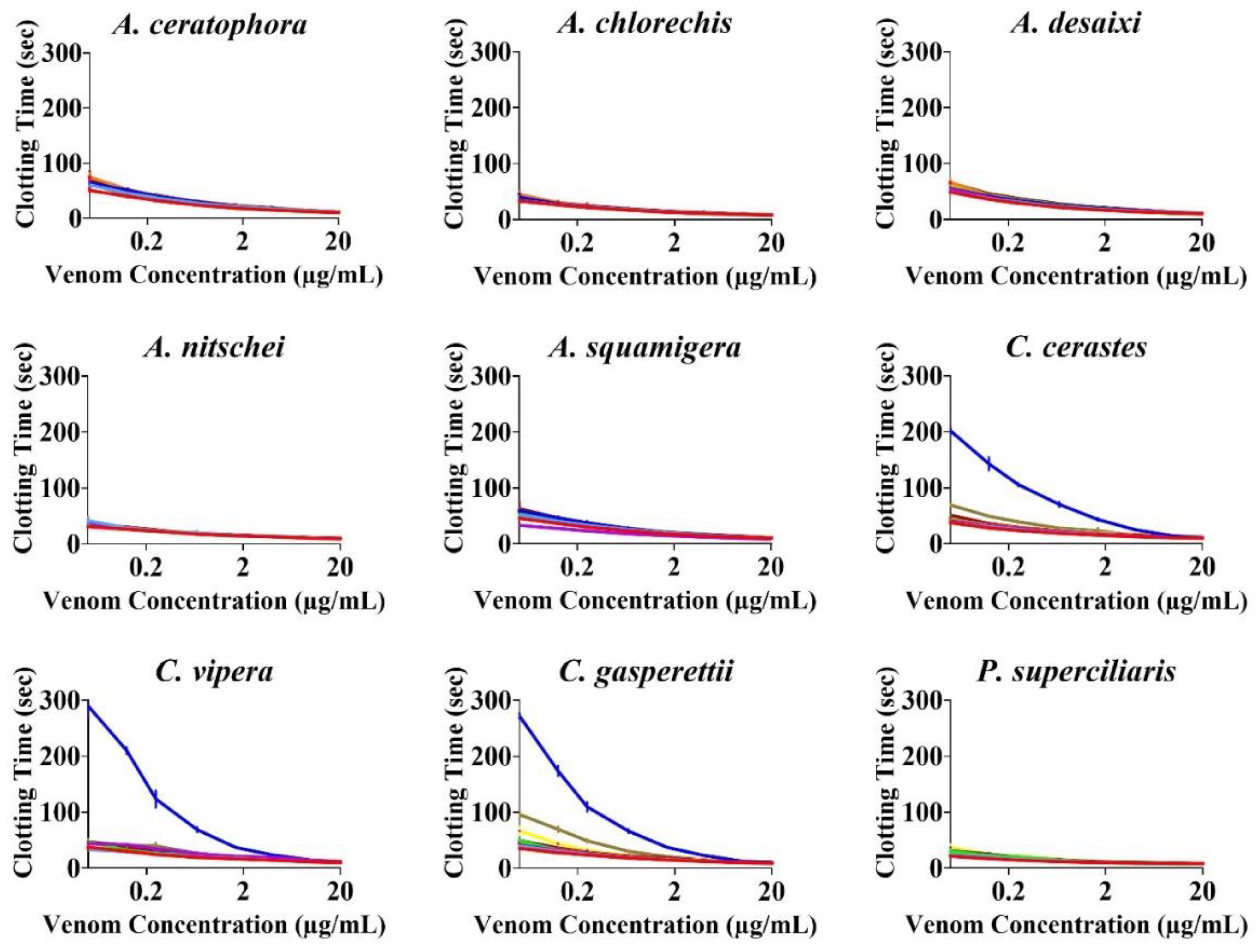
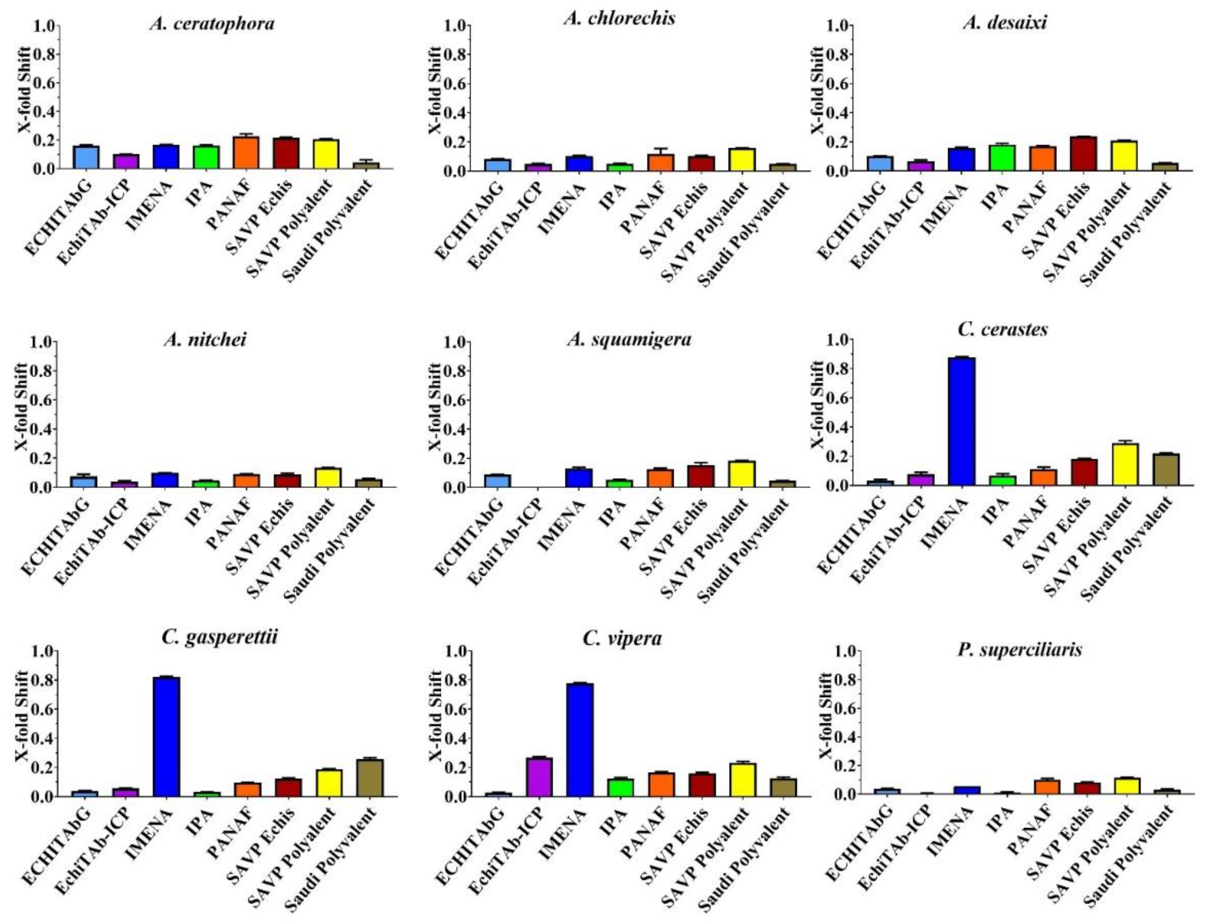
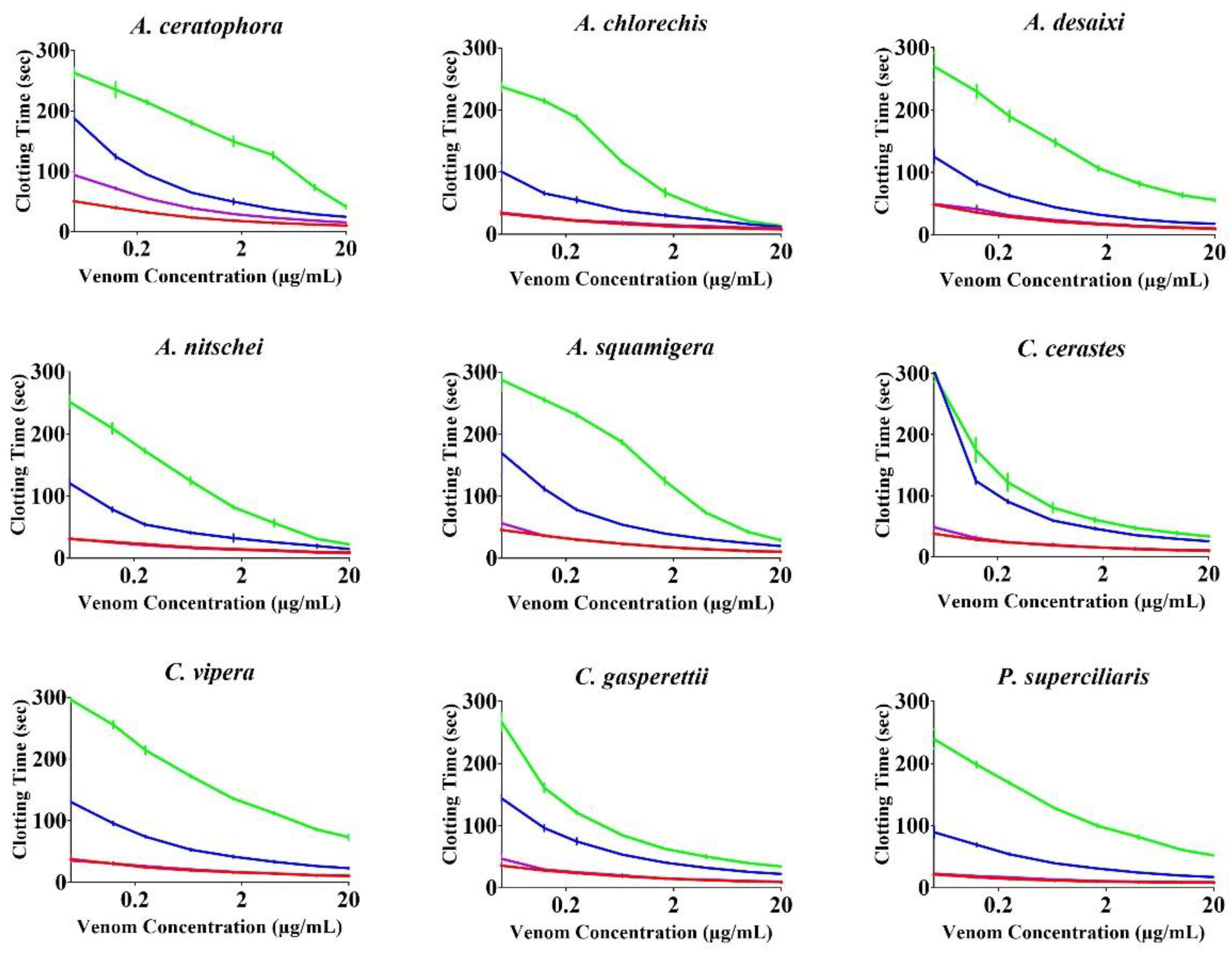
None of the antivenoms evaluated, with the exception of Inoserp-MENA, which displayed efficacy against all three Cerastes venoms, were able to effectively neutralise the other venoms (Figure 2 and Figure 3). The enzyme-inhibitors displayed variation in efficacy. Marimastat consistently outperformed both prinomastat and DMPS (Figure 4 and Figure 5). While the relative efficacy of the three small molecule inhibitors were similar to the results for the procoagulant toxicity of venom from the colubrid snake Rhabdophis subminiatus, they are in contrast to results for other viperid snake venoms, where prinomastat showed greater in vitro efficacy than marimastat [32,33,41,42,50]. Conspicuously, even at a 5× concentration, DMPS failed to neutralise any of the venoms, consistent with apparently lesser efficacy seen in vitro against the procoagulant toxicity of other viperid venoms [32,33,41,42,50,51].
As marimastat is a specific inhibitor of metalloproteases, these results suggest that the procoagulant toxicity is due to the presence of snake venom metalloproteases (SVMP) in the venoms. This provides insights into the evolutionary history of their venoms. Specifically, these species are closely related to Echis, which use metalloprotease toxins in venom to trigger procoagulant effects in prey, as do the basal Bitis species B. worthingtoni [52]. This indicates that SVMP-driven procoagulant toxicity is an early arising trait within viperid snakes and that anticoagulant species within this clade (such as [non-B. worthingtoni] Bitis, Eristicophis, Montivipera, and Pseudocerastes other than P. urarachnoides) represent derived states. However, future work examining relative effect in activating Factor X versus prothrombin is required to ascertain whether both are basal activities, or if one of these activation strategies is a derived example. A testable hypothesis for such future work is that Factor X activation has been shown to be a widely distributed trait, characterised for B. worthingtoni, Cerastes, Echis, as a basal trait within the palearctic viper clade Daboia, Macrovipera, and Vipera, and Pseudocerastes urarachnoides [40,52]. In contrast, amongst true vipers prothrombin activation is a much rarer, known only from Echis species and P. urarachnoides. Thus, it is hypothesised that Factor X activation was present in the last common ancestor of extant true vipers, and that both the prothrombin activation procoagulant trait and the anticoagulant traits are derived activities.
It is important to interpret this work with the appropriate caveats. First is that this work only examined coagulopathy, the antivenom/inhibitor patterns may vary for other pathophysiological effects (such as myotoxicity and neurotoxicty). Secondly, as the work was undertaken under the idealised conditions of preincubating the venoms with the antivenom or inhibitor, future In Vivo testing is required as the next preclinical step before clinical trials can be undertaken to confirm the efficacy of Inoserp-MENA against Cerastes venoms or the utility of marimastat against Atheris, Cerastes, and Proatheris venoms. Reciprocally, the failure of an antivenom or inhibitor to neutralise the venom under idealised circumstances used in this study, strongly suggests that in vivo efficacy would be unlikely in the more dynamic physiological system of a living organism. Thus, except for Inoserp-MENA against Cerastes, it is hypothesized that the other antivenoms, and DMPS from within the inhibitors in this study would be unable to neutralise the procoagulant component of the venom induced pathology In Vivo (regardless of any efficacy against other pathophysiological actions such as myotoxicity). Additional studies are thus needed to continue to advance the development of effective treatments to these dangerous venoms.

3. Materials and Methods

3.1. Stock Preparation

3.1.1. Venoms

All venom work was conducted under the University of Queensland Animal Ethics Approval 2021/AE000075 and UQ Biosafety Committee Approval # IBC/134B/SBS/2015 (Brisbane, Australia for both). Pooled lyophilized venoms purchased from the licensed bioproduct company Latoxan (Valence, France) were: Atheris ceratophora (Tanzania), A. chlorechis (Ghana), A. desaixi (Kenya), A. nitschei (Burundi), A. squamigera (Sub-Saharan Africa), Cerastes cerastes (Tunisia), C. gasperettii (Saudi Arabia), C. vipera (Egypt), and Proatheris superciliaris (Mozambique). Venom stocks were prepared by reconstituting dry venom with 50% glycerol and deionized water to produce 1 mg/mL concentrated stock; stored at −20 °C until further use. Thermo Fisher Scientific™ NanoDrop 2000 UV–Vis Spectrophotometer (Thermofisher, Sydney, Australia) was used to measure the concentration in triplicate at 280 nm wavelength.

3.1.2. Plasma

Frozen human platelet-poor plasma (3.2% citrated) was supplied by the Australian Red Cross (44 Musk Street, Kelvin Grove, QLD 4059, Australia) under research approval #16-04QLD-10. Human plasma work was performed under University of Queensland Biosafety Approval #IBC/134B/SBS/2015 and Human Ethics Approval #2016000256. The plasma was thawed and aliquoted at 1.2 mL quantities, followed by flash-freezing in liquid nitrogen. The aliquots were stored at −80 °C until required for testing. During experiments, these aliquots were defrosted at 37 °C in a Thermo Haake ARCTIC water bath.

3.1.3. Antivenom and Enzyme Inhibitors

Eight regional antivenoms for treating the above viper envenomings were selected (Table 1). Each vial was in 10 mL volume. All the venoms (lyophilized venoms were reconstituted using OK buffer) were centrifuged at 14,000 RCF on Allegra™ X-22R Centrifuge (Beckman Coulter, Brea, CA, USA) at 4 °C for 10 min; the supernatant collected and stored at 4 °C. The antivenoms’ working concentration was 5% (v/v) using the OK buffer. Code names were given for easier reference.
Small molecule metalloprotease inhibitors tested in this study were, 2,3-Dimercaptopropanesulfonic acid sodium salt monohydrate (DMPS) (catalogue # D8016, Sigma Aldrich) (St. Louis, MO, USA), marimastat ((2S,3R)-N4-[(1S)-2,2-Dimethyl-1-[(methylamino)carbonyl] propyl]-N1,2-dihydroxy-3-(2 methylpropyl)butanediamide) (catalogue # M2699, Sigma Aldrich) (St. Louis, MO, USA), and prinomastat hydrochloride ((S)-2,2-Dimethyl-4-((p-(4-pyridyloxy) phenyl) sulfonyl) -3- thiomorpholinecarbohydroxamic acid hydrocholride (catalogue# PZ0198, Sigma Aldrich) (St. Louis, MO, USA). The powder of these inhibitors were first dissolved in 10% dimethyl sulfoxide (DMSO) and further diluted using deionized water to form 10 mM (prinomastat and marimastat) and 20 mM (DMPS) stock solutions, respectively, and stored at −80 °C. During tests, the working stock of the inhibitors was prepared by further diluting the stock to 2 mM for prinomastat and marimastat and 10 mM for DMPS.

3.2. Experimental Conditions

3.2.1. Coagulotoxicity Effects on Plasma

Venom effect on coagulation of plasma was tested by utilising STA-R Max® (Stago, Asnières sur Seine, France) coagulation analyser. From 1 mg/mL venom stock, working stock of 100 μg/mL was prepared by diluting the main stock with OK Buffer (Stago catalogue #00360). Concentration curves (8-points) with serial dilutions of: 1, 1/2, 1/5, 1/12.5, 1/30, 1/80, 1/160, and 1/400 were run by loading the working stock onto the analyser. Inside the analyser, 100 µg/mL working stock was serially diluted to form final reaction concentrations of: 20, 10, 4, 1.6, 0.67, 0.25, 0.125, and 0.05 µg/mL, respectively. Venom stock was added in a cuvette (according to dilution factor) followed by the addition of 50 µL of 0.025 M calcium chloride (Stago catalogue # 00367), 25 µL of OK buffer, and 50 µL of phospholipid (Stago catalogue #00597). The mixture was then incubated for 2 min at 37 °C, and 75 μL of plasma was added immediately right after incubation, and the clotting time was recorded. The whole process is carried out automatically by programmed robotic operation. For positive control, coagulation activator kaolin (Stago C·K Prest standard kit, Stago catalogue #00597) was used, and for the negative control, 50% glycerol/deionized water was replaced with venom.

3.2.2. Antivenom and Enzyme-Inhibitor Efficacy

To investigate the efficacy of antivenoms and enzyme inhibitors in neutralizing the toxic effects of venom upon plasma clotting, the above-mentioned 8-point concentration curves were repeated. In this case, the 25 µL of OK buffer (added to the cuvette before incubation) was replaced with 25 µL of antivenom (0.5% final concentration) or inhibitors (0.2 mM final concentration for prinomastat and marimastat and 1.0 mM final concentration for DMPS). All assays were run in triplicates. Both the experimental conditions were based on validated protocols carried out previously [32,33,41,42,50,51].

3.2.3. Statistical Analyses

All data plotting and statistical analyses were done by using GraphPad PRISM 8.1.1 (GraphPad Prism Inc., La Jolla, CA, USA). The area under the curve (AUC) for both venom and antivenom/inhibitors was analysed using the software. X-fold shift was generated using the formulae [(AUC of venom incubated with inhibitors/AUC of venom)—1] utilising Excel. This X-fold shift was scrutinized in a manner where a value of “0” indicated no neutralization (no shift in clotting curve). In contrast, values >0 indicated venom neutralization (change in clotting time curve). Thus, evaluating the activity of the antivenom/inhibitors against venom coagulotoxicity on plasma.

Supplementary Materials

The following supporting information can be downloaded at: https://www.mdpi.com/article/10.3390/toxins14120836/s1, File S1. Supplementary Sheet African viperid genera Atheris, Cerastes, and Proatheris.xlsx.

Author Contributions

Conceptualization, B.G.F.; methodology, B.G.F., A.C.; validation, B.G.F., A.C.; formal analysis, B.G.F., A.C.; investigation, B.G.F., A.C.; resources, R.S., M.A., B.G.F.; data curation, B.G.F., A.C..; writing—A.C., B.G.F.; writing—review and editing, A.C., B.G.F., M.R.L., R.C., R.S., M.A.; visualization, B.G.F.; supervision, B.G.F.; project administration, B.G.F.; funding acquisition, B.G.F. All authors have read and agreed to the published version of the manuscript.

Funding

This research was funded by Australian Research Council Discovery Project DP190100304. M.R.L. and R.W.C. have shares and salary support from Ophirex, Inc., a Public Benefit Corporation.

Institutional Review Board Statement

All venom work was conducted under the University of Queensland Animal Ethics Approval 2021/AE000075 (15 March 2001) and UQ Biosafety Committee Approval # IBC/134B/SBS/2015. All human plasma work was performed under University of Queensland Biosafety Approval #IBC/134B/SBS/2015 and Human Ethics Approval #2016000256.

Informed Consent Statement

Not applicable.

Data Availability Statement

Data can be found in the supplementary material provided with the paper.

Conflicts of Interest

The authors declare no conflict of interest.

References

  1. Kasturiratne, A.; Wickremasinghe, A.R.; De Silva, N.; Gunawardena, N.K.; Pathmeswaran, A.; Premaratna, R.; Savioli, L.; Lalloo, D.G.; De Silva, H.J. The Global Burden of Snakebite: A Literature Analysis and Modelling Based on Regional Estimates of Envenoming and Deaths. PLoS Med. 2008, 5, 1591–1604. [Google Scholar] [CrossRef] [Green Version]
  2. Harrison, R.A.; Hargreaves, A.; Wagstaff, S.C.; Faragher, B.; Lalloo, D.G. Snake Envenoming: A Disease of Poverty. PLoS Negl. Trop. Dis. 2009, 3, e569. [Google Scholar] [CrossRef] [Green Version]
  3. Fry, B.G. Snakebite: When the Human Touch Becomes a Bad Touch. Toxins 2018, 10, 170. [Google Scholar] [CrossRef] [Green Version]
  4. Fry, B.G.; Roelants, K.; Champagne, D.E.; Scheib, H.; Tyndall, J.D.A.; King, G.F.; Nevalainen, T.J.; Norman, J.A.; Lewis, R.J.; Norton, R.S.; et al. The Toxicogenomic Multiverse: Convergent Recruitment of Proteins into Animal Venoms. Annu. Rev. Genom. Hum. Genet. 2009, 10, 483–511. [Google Scholar] [CrossRef] [Green Version]
  5. Debono, J.; Dobson, J.; Casewell, N.R.; Romilio, A.; Li, B.; Kurniawan, N.; Mardon, K.; Weisbecker, V.; Nouwens, A.; Kwok, H.F.; et al. Coagulating Colubrids: Evolutionary, Pathophysiological and Biodiscovery Implications of Venom Variations between Boomslang (Dispholidus Typus) and Twig Snake (Thelotornis Mossambicanus). Toxins 2017, 9, 171. [Google Scholar] [CrossRef] [Green Version]
  6. Bittenbinder, M.A.; Zdenek, C.N.; den Brouw, B.O.; Youngman, N.J.; Dobson, J.S.; Naude, A.; Vonk, F.J.; Fry, B.G. Coagulotoxic Cobras: Clinical Implications of Strong Anticoagulant Actions of African Spitting Naja Venoms That Are Not Neutralised by Antivenom but Are by LY315920 (Varespladib). Toxins 2018, 10, 516. [Google Scholar] [CrossRef] [Green Version]
  7. Chowdhury, A.; Lewin, M.R.; Zdenek, C.; Carter, R.; Fry, B.G. The Relative Efficacy of Chemically Diverse Small-Molecule Enzyme-Inhibitors against Anticoagulant Activities of African Spitting Cobra (Naja Species) Venoms. Front. Immunol. 2021, 12, 4215. [Google Scholar] [CrossRef]
  8. Bittenbinder, M.A.; Dobson, J.S.; Zdenek, C.N.; den Brouw, B.O.; Naude, A.; Vonk, F.J.; Fry, B.G. Differential Destructive (Non-Clotting) Fibrinogenolytic Activity in Afro-Asian Elapid Snake Venoms and the Links to Defensive Hooding Behavior. Toxicol. Vitr. 2019, 60, 330–335. [Google Scholar] [CrossRef]
  9. Oulion, B.; Dobson, J.S.; Zdenek, C.N.; Arbuckle, K.; Lister, C.; Coimbra, F.C.P.; den Brouw, B.O.; Debono, J.; Rogalski, A.; Violette, A.; et al. Factor X Activating Atractaspis Snake Venoms and the Relative Coagulotoxicity Neutralising Efficacy of African Antivenoms. Toxicol. Lett. 2018, 288, 119–128. [Google Scholar] [CrossRef] [Green Version]
  10. Rogalski, A.; Soerensen, C.; den Brouw, B.O.; Lister, C.; Dashvesky, D.; Arbuckle, K.; Gloria, A.; Zdenek, C.N.; Casewell, N.R.; Gutiérrez, J.M.; et al. Differential Procoagulant Effects of Saw-Scaled Viper (Serpentes: Viperidae: Echis) Snake Venoms on Human Plasma and the Narrow Taxonomic Ranges of Antivenom Efficacies. Toxicol. Lett. 2017, 280, 159–170. [Google Scholar] [CrossRef]
  11. Youngman, N.J.; Lewin, M.R.; Carter, R.; Naude, A.; Fry, B.G. Efficacy and Limitations of Chemically Diverse Small-Molecule Enzyme-Inhibitors against the Synergistic Coagulotoxic Activities of Bitis Viper Venoms. Molecules 2022, 27, 1733. [Google Scholar] [CrossRef]
  12. Youngman, N.J.; Harris, R.J.; Huynh, T.M.; Coster, K.; Sundman, E.; Braun, R.; Naude, A.; Hodgson, W.C.; Fry, B.G. Widespread and Differential Neurotoxicity in Venoms from the Bitis Genus of Viperid Snakes. Neurotox. Res. 2021, 1, 697–704. [Google Scholar] [CrossRef]
  13. Youngman, N.J.; Walker, A.; Naude, A.; Coster, K.; Sundman, E.; Fry, B.G. Varespladib (LY315920) Neutralises Phospholipase A 2 Mediated Prothrombinase-Inhibition Induced by Bitis Snake Venoms. Comp. Biochem. Physiol. Part C 2020, 236, 108818. [Google Scholar] [CrossRef]
  14. Coimbra, F.C.P.; Dobson, J.; Zdenek, C.N.; Op Den Brouw, B.; Hamilton, B.; Debono, J.; Masci, P.; Frank, N.; Ge, L.; Kwok, H.F.; et al. Does Size Matter? Venom Proteomic and Functional Comparison between Night Adder Species (Viperidae: Causus) with Short and Long Venom Glands. Comp. Biochem. Physiol. Part C 2018, 211, 7–14. [Google Scholar] [CrossRef]
  15. Yamada, D.; Sekiya, F.; Morita, T. Prothrombin and Factor X Activator Activities in the Venoms of Viperidae Snakes. Toxicon 1997, 35, 1581–1589. [Google Scholar] [CrossRef]
  16. Razok, A.; Shams, A.; Yousaf, Z. Cerastes Cerastes Snakebite Complicated by Coagulopathy and Cardiotoxicity with Electrocardiographic Changes. Toxicon 2020, 188, 1–4. [Google Scholar] [CrossRef]
  17. Labib, R.S.; Azab, M.H.; Farag, N.W. Effects of Cerastes Cerastes (Egyptian Sand Viper) and Cerastes Vipera (Sahara Sand Viper) Snake Venoms on Blood Coagulation: Separation of Coagulant and Anticoagulant Factors and Their Correlation with Arginine Esterase and Protease Activit. Toxicon 1981, 19, 85–94. [Google Scholar] [CrossRef]
  18. Mebs, D.; Holada, K.; Kornalík, F.; Simák, J.; Vanková, H.; Müller, D.; Schoenemann, H.; Lange, H.; Herrmann, H.W. Severe Coagulopathy after a Bite of a Green Bush Viper (Atheris Squamiger): Case Report and Biochemical Analysis of the Venom. Toxicon 1998, 36, 1333–1340. [Google Scholar] [CrossRef]
  19. Stach, V.J.; Vagenknechtová, Z.; Hoskovec, E.; Valenta, J.; Stach, Z.; Vagenknechtová, E.; Hoskovec, D. Splenic Rupture and Massive Hemoperitoneum Due to Coagulopathy after Atheris Viper Snakebite. Prague Med. Rep. 2021, 122, 216–221. [Google Scholar] [CrossRef]
  20. Valenta, J.; Hlavackova, A.; Stach, Z.; Stikarova, J.; Havlicek, M.; Michalek, P. Fibrinogenolysis in Venom-Induced Consumption Coagulopathy after Viperidae Snakebites: A Pilot Study. Toxins 2022, 14, 538. [Google Scholar] [CrossRef]
  21. Valenta, J.; Stach, Z.; Fricova, D.; Zak, J.; Balik, M. Envenoming by the Viperid Snake Proatheris Superciliaris: A Case Report. Toxicon 2008, 52, 392–394. [Google Scholar] [CrossRef]
  22. Elkabbaj, D.; Hassani, K.; Jaoudi, R. El Acute Renal Failure Following the Saharan Horned Viper (Cerastes Cerastes) Bite. Arab J. Nephrol. Transplant. 2012, 5, 159–161. [Google Scholar] [CrossRef]
  23. Keyler, D.E. Envenomation by the Lowland Viper (Proatheris Superciliaris): Severe Case Profile Documentation. Toxicon 2008, 52, 836–841. [Google Scholar] [CrossRef]
  24. Hatten, B.W.; Bueso, A.; French, L.K.; Hendrickson, R.G.; Horowitz, B.Z. Envenomation by the Great Lakes Bush Viper (Atheris Nitschei). Clin. Toxicol. 2013, 51, 114–116. [Google Scholar] [CrossRef]
  25. Warrell, D.A. Proatheris Superciliaris: The Deadly Venom of a Rare and Elusive Snake Revealed. Toxicon 2008, 52, 833–835. [Google Scholar] [CrossRef]
  26. Imperato, N.S.; Amaducci, A.M.; Abo, B.N.; Koons, A.L.; Fikse, D.J.; Katz, K.D. African Bush Viper Envenomation: A Case Report. Cureus 2022, 14, e28040. [Google Scholar] [CrossRef]
  27. Alencar, L.R.V.; Quental, T.B.; Grazziotin, F.G.; Alfaro, M.L.; Martins, M.; Venzon, M.; Zaher, H. Diversification in Vipers: Phylogenetic Relationships, Time of Divergence and Shifts in Speciation Rates. Mol. Phylogenet. Evol. 2016, 105, 50–62. [Google Scholar] [CrossRef]
  28. Nielsen, V.G.; Frank, N. Role of Heme Modulation in Inhibition of Atheris, Atractaspis, Causus, Cerastes, Echis, and Macrovipera Hemotoxic Venom Activity. Hum. Exp. Toxicol. 2019, 38, 216–226. [Google Scholar] [CrossRef]
  29. Ainsworth, S.; Slagboom, J.; Alomran, N.; Pla, D.; Alhamdi, Y.; King, S.I.; Bolton, F.M.S.; Gutiérrez, J.M.; Vonk, F.J.; Toh, C.H.; et al. The Paraspecific Neutralisation of Snake Venom Induced Coagulopathy by Antivenoms. Commun. Biol. 2018, 1, 34. [Google Scholar] [CrossRef] [Green Version]
  30. Laing, G.D.; Compton, S.J.; Ramachandran, R.; Fuller, G.L.J.; Wilkinson, M.C.; Wagstaff, S.C.; Watson, S.P.; Kamiguti, A.S.; Theakston, R.D.G.; Senis, Y.A. Characterization of a Novel Protein from Proatheris Superciliaris Venom: Proatherocytin, a 34-KDa Platelet Receptor PAR1 Agonist. Toxicon 2005, 46, 490–499. [Google Scholar] [CrossRef]
  31. Theakston, R.D.G.; Reid, H.A. Development of Simple Standard Assay Procedures for the Characterization of Snake Venoms. Bull. World Health Organ. 1983, 61, 949–956. [Google Scholar]
  32. Chowdhury, A.; Zdenek, C.N.; Dobson, J.S.; Bourke, L.A.; Soria, R.; Fry, B.G. Clinical Implications of Differential Procoagulant Toxicity of the Palearctic Viperid Genus Macrovipera, and the Relative Neutralization Efficacy of Antivenoms and Enzyme Inhibitors. Toxicol. Lett. 2021, 340, 77–88. [Google Scholar] [CrossRef]
  33. Chowdhury, A.; Zdenek, C.N.; Lewin, M.R.; Carter, R.; Jagar, T.; Ostanek, E.; Harjen, H.; Aldridge, M.; Soria, R.; Haw, G.; et al. Venom-Induced Blood Disturbances by Palearctic Viperid Snakes, and Their Relative Neutralization by Antivenoms and Enzyme-Inhibitors. Front. Immunol. 2021, 12, 2251. [Google Scholar] [CrossRef]
  34. Debono, J.; Bos, M.H.A.; Coimbra, F.; Ge, L.; Frank, N.; Kwok, H.F.; Fry, B.G. Basal but Divergent: Clinical Implications of Differential Coagulotoxicity in a Clade of Asian Vipers. Toxicol. Vitr. 2019, 58, 195–206. [Google Scholar] [CrossRef]
  35. Debono, J.; Bos, M.H.A.; Do, M.S.; Fry, B.G. Clinical Implications of Coagulotoxic Variations in Mamushi (Viperidae: Gloydius) Snake Venoms. Comp. Biochem. Physiol. Part C Toxicol. Pharmacol. 2019, 225, 108567. [Google Scholar] [CrossRef]
  36. Debono, J.; Bos, M.H.A.; Frank, N.; Fry, B. Clinical Implications of Differential Antivenom Efficacy in Neutralising Coagulotoxicity Produced by Venoms from Species within the Arboreal Viperid Snake Genus Trimeresurus. Toxicol. Lett. 2019, 316, 35–48. [Google Scholar] [CrossRef]
  37. Debono, J.; Bos, M.H.A.; Nouwens, A.; Ge, L.; Frank, N.; Kwok, H.F.; Fry, B.G. Habu Coagulotoxicity: Clinical Implications of the Functional Diversification of Protobothrops Snake Venoms upon Blood Clotting Factors. Toxicol. Vitr. 2019, 55, 62–74. [Google Scholar] [CrossRef] [Green Version]
  38. Dobson, J.; Yang, D.C.; den Brouw, B.O.; Cochran, C.; Huynh, T.; Kurrupu, S.; Sánchez, E.E.; Massey, D.J.; Baumann, K.; Jackson, T.N.W.; et al. Rattling the Border Wall: Pathophysiological Implications of Functional and Proteomic Venom Variation between Mexican and US Subspecies of the Desert Rattlesnake Crotalus Scutulatus. Comp. Biochem. Physiol. Part C Toxicol. Pharmacol. 2018, 205, 62–69. [Google Scholar] [CrossRef] [Green Version]
  39. Lister, C.; Arbuckle, K.; Jackson, T.N.W.; Debono, J.; Zdenek, C.N.; Dashevsky, D.; Dunstan, N.; Allen, L.; Hay, C.; Bush, B.; et al. Catch a Tiger Snake by Its Tail: Differential Toxicity, Co-Factor Dependence and Antivenom Efficacy in a Procoagulant Clade of Australian Venomous Snakes. Comp. Biochem. Physiol. Part C Toxicol. Pharmacol. 2017, 202, 39–54. [Google Scholar] [CrossRef] [Green Version]
  40. Op Den Brouw, B.; Coimbra, F.C.P.; Bourke, L.A.; Huynh, T.M.; Vlecken, D.H.W.; Ghezellou, P.; Visser, J.C.; Dobson, J.S.; Fernandez-Rojo, M.A.; Ikonomopoulou, M.P.; et al. Extensive Variation in the Activities of Pseudocerastes and Eristicophis Viper Venoms Suggests Divergent Envenoming Strategies Are Used for Prey Capture. Toxins 2021, 13, 112. [Google Scholar] [CrossRef]
  41. Seneci, L.; Zdenek, C.N.; Chowdhury, A.; Rodrigues, C.F.B.; Neri-Castro, E.; Bénard-Valle, M.; Alagón, A.; Fry, B.G. A Clot Twist: Extreme Variation in Coagulotoxicity Mechanisms in Mexican Neotropical Rattlesnake Venoms. Front. Immunol. 2021, 12, 552. [Google Scholar] [CrossRef] [PubMed]
  42. Youngman, N.J.; Chowdhury, A.; Zdenek, C.N.; Coster, K.; Sundman, E.; Braun, R.; Fry, B.G. Utilising Venom Activity to Infer Dietary Composition of the Kenyan Horned Viper (Bitis Worthingtoni). Comp. Biochem. Physiol. Part C Toxicol. Pharmacol. 2021, 240, 108921. [Google Scholar] [CrossRef] [PubMed]
  43. Zdenek, C.N.; den Brouw, B.O.; Dashevsky, D.; Gloria, A.; Youngman, N.J.; Watson, E.; Green, P.; Hay, C.; Dunstan, N.; Allen, L.; et al. Clinical Implications of Convergent Procoagulant Toxicity and Differential Antivenom Efficacy in Australian Elapid Snake Venoms. Toxicol. Lett. 2019, 316, 171–182. [Google Scholar] [CrossRef] [PubMed]
  44. Zdenek, C.N.; Hay, C.; Arbuckle, K.; Jackson, T.N.W.; Bos, M.H.A.; den Brouw, B.O.; Debono, J.; Allen, L.; Dunstan, N.; Morley, T.; et al. Coagulotoxic Effects by Brown Snake (Pseudonaja) and Taipan (Oxyuranus) Venoms, and the Efficacy of a New Antivenom. Toxicol. Vitr. 2019, 58, 97–109. [Google Scholar] [CrossRef] [PubMed] [Green Version]
  45. Sousa, L.F.; Zdenek, C.N.; Dobson, J.S.; den Brouw, B.O.; Coimbra, F.C.P.; Gillett, A.; Del-Rei, T.H.M.; de Chalkidis, H.M.; Sant’Anna, S.; Teixeira-Da-Rocha, M.M.; et al. Coagulotoxicity of Bothrops (Lancehead Pit-Vipers) Venoms from Brazil: Differential Biochemistry and Antivenom Efficacy Resulting from Prey-Driven Venom Variation. Toxins 2018, 10, 411. [Google Scholar] [CrossRef] [Green Version]
  46. Mallow, D.; David, L.; Nilson, G. True Vipers: Natural History and Toxinology of Old World Vipers; Krieger Publishing Company: Malabar, FL, USA, 2003. [Google Scholar]
  47. Dobiey, M.; Gernot, V. Venomous Snakes of Africa; Edition Chimaira: Frankfurt, Germany, 2007; ISBN 9783899733655. [Google Scholar]
  48. Phelps, T. Old World Vipers: A Natural History of the Azemiopinae and Viperinae; Edition Chimaira: Frankfurt, Germany, 2010; ISBN 389973470X. [Google Scholar]
  49. den Brouw, B.O.; Coimbra, F.C.P.; Casewell, N.R.; Ali, S.A.; Vonk, F.J.; Fry, B.G. A Genus-Wide Bioactivity Analysis of Daboia (Viperinae: Viperidae) Viper Venoms Reveals Widespread Variation in Haemotoxic Properties. Int. J. Mol. Sci. 2021, 22, 13486. [Google Scholar] [CrossRef]
  50. Rodrigues, C.F.B.; Zdenek, C.N.; Bourke, L.A.; Seneci, L.; Chowdhury, A.; Freitas-de-Sousa, L.A.; de Alcantara Menezes, F.; Moura-da-Silva, A.M.; Tanaka-Azevedo, A.M.; Fry, B.G. Clinical Implications of Ontogenetic Differences in the Coagulotoxic Activity of Bothrops Jararacussu Venoms. Toxicol. Lett. 2021, 348, 59–72. [Google Scholar] [CrossRef]
  51. Chowdhury, A.; Lewin, M.R.; Carter, R.W.; Casewell, N.R.; Fry, B.G. Keel Venom: Rhabdophis Subminiatus (Red-Necked Keelback) Venom Pathophysiologically Affects Diverse Blood Clotting Pathways. Toxicon 2022, 218, 19–24. [Google Scholar] [CrossRef]
  52. Xie, B.; Dashevsky, D.; Rokyta, D.; Ghezellou, P.; Fathinia, B.; Shi, Q.; Richardson, M.K.; Fry, B.G. Dynamic Genetic Differentiation Drives the Widespread Structural and Functional Convergent Evolution of Snake Venom Proteinaceous Toxins. BMC Biol. 2022, 20, 4. [Google Scholar] [CrossRef]
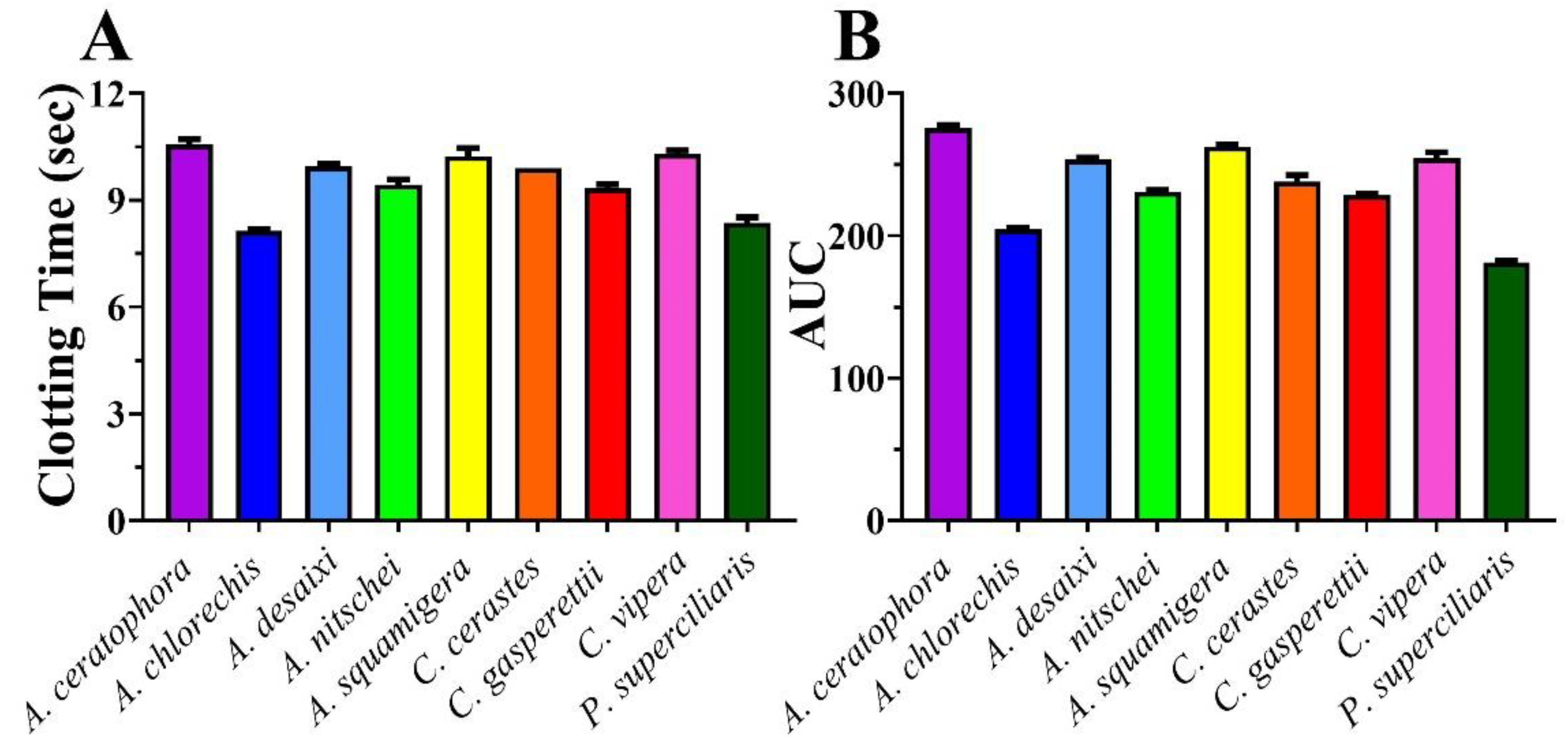
Figure 1. (A) 20 µg/mL venom concentration clotting times in human plasma. (B) Area Under Curve (AUC) generated from 8-point concentration curves in human plasma; more potent venoms have lower AUCs. Values are mean +/− SD of n = 3.
Toxins 14 00836 g001
Figure 2. 8-point logarithm concentration curves of antivenom against venom. x-axis showing concentrations of venom in µg/mL, and y-axis showing clotting times in seconds of human plasma. Values are mean ± SD of n = 3. Only Inosan’s Inoserp-MENA (dark blue) had any significant effect, and only against Cerastes species. The Saudi Polyvalent (brown)_ displayed very low levels recognition of Cerastes venoms. Colours are as for Figure 3.
Toxins 14 00836 g002
Figure 3. Bar graphs of X-fold magnitude of shift of plasma clotting time due to incubation of antivenoms, calculated by the formula [(AUC of antivenom + venom/AUC of venom) − 1]. A value of 0 indicates no shift (no neutralization by antivenom), while a value above 0 indicates neutralization by antivenom. Values are mean ± SD of n = 3. Only Inosan’s Inoserp-MENA (dark blue) had any significant effect, and only against Cerastes species.
Toxins 14 00836 g003
Figure 4. 8-point logarithm concentration curves of SMIs against venom. x-axis showing concentrations of venom in µg/mL and y-axis showing clotting times in seconds of human plasma. Colour legend: Red = Venom, Purple = DMPS (final concentration 1.0 mM %), Indigo = Prinomastat (final concentration 0.2 mM %), Green = Marimastat (final concentration 0.2 mM %). Values are mean ± SD of n = 3.
Toxins 14 00836 g004
Figure 5. Bar graphs of X-fold magnitude of shift of plasma clotting time due to incubation of SMIs, calculated by the formula [(AUC of SMI + venom/AUC of venom)-1]. A value of 0 indicates no shift (no neutralization by SMIs), while a value above 0 indicates neutralization by SMIs. Values are mean ± SD of n = 3. x-axis showing SMIs while y-axis showing X-fold magnitude of shift.
Toxins 14 00836 g005
Table 1. Antivenom details.
Product NameCompanyImmunising SpeciesCodes Used in This Study
EchiTAbGMicroPharm Ltd., Newcastle Emlyn, UKEchis ocellatusECHITAbG
EchiTAb-Plus-ICPInstituto Clodomiro picado Univerdad de Costa RicaBitis arietans, E. ocellatus, Naja nigricollisEchiTAb-ICP
INOSERP MENA
(Middle East and North Africa)
INOSAN Biopharma, Madrid, SpainB. arietans, C. cerastes, C. gasperettii, C. vipera, Daboia deserti, D. mauritanica, D. palaestinae, E. carinatus sochureki, E. coloratus, E.s khosatzkii, E. leucogaster, E. megalocephalus, E. omanensis, E. pyramidum, Macrovipera lebetina obtusa, M. l. transmediterranea, M. l. turanica, Montivipera bornmuelleri, Montivipera raddei kurdistanica, Pseuocerastes fieldi, P. persicus, Vipera latastei, Naja haje, N. nubiae, N. pallida, Walterinnesia aegyptiaIMENA
INOSERP PAN-AFRICAINOSAN Biopharma, Madrid, SpainB. arietans, B. gabonica, B. rhinoceros, Dendroaspis angusticeps, Dendroaspis jamesoni, D. polylepis, D. viridis, E. leucogaster, E. ocellatus, E. pyramidum, Naja haje, N. katiensis, N. melanoleuca, N. nigricollis, N. nivea, N. pallidaIPA
PANAF
PREMIUM (Sub-Saharan Africa)
Premium Serums, Maharashtra, IndiaB arietans, B. gabonica, B. nasicornis, B. rhinoceros, Dendroaspis angusticeps, D. jamesoni, D. polylepis, D. viridis, Echis carinatus, E. leucogaster, E. ocellatus, Naja nigricollis, N. haje, N. melanoleucaPANAF
SAVP EchisSouth African Vaccine ProducersE. carinatus, E. ocellatus, E. coloratus, Cerastes spp.SAVP Echis
SAVP
Polyvalent
South African Vaccine ProducersB. arietans, B. gabonica, D. angusticeps, D. jamesoni, D. polylepis, Hemachatus haemachatus, N. annulifera, N. melanoleuca, N.mossambica, N. niveaSAVP Polyvalent
Polyvalent Snake
Antivenom
National Antivenom and Vaccine Production Centre, Saudi ArabiaB. arietans, C. cerastes, E. carinatus, E. coloratus, N. Haje, W. AegyptiaSaudi Polyvalent
Publisher’s Note: MDPI stays neutral with regard to jurisdictional claims in published maps and institutional affiliations.