Article

Susceptibility of Oocytes from Gilts and Sows to Beauvericin and Deoxynivalenol and Its Relationship with Oxidative Stress

by
Eric J. Schoevers
1,†,
Regiane R. Santos
2,* and
Bernard A. J. Roelen
3
1
Farm Animal Health, Department Population Health Sciences, Faculty of Veterinary Medicine, Utrecht University, Yalelaan 104, 3584 CM Utrecht, The Netherlands
2
Schothorst Feed Research, P.O. Box 533, 8200 AM Lelystad, The Netherlands
3
Embryology, Anatomy and Physiology, Department Clinical Sciences, Faculty of Veterinary Medicine, Utrecht University, Yalelaan 1, 3584 CT Utrecht, The Netherlands
*
Author to whom correspondence should be addressed.
Eric J. Schoevers passed away before publication of this work was completed.
Toxins 2021, 13(4), 260; https://doi.org/10.3390/toxins13040260
Submission received: 18 March 2021 / Revised: 1 April 2021 / Accepted: 3 April 2021 / Published: 6 April 2021
(This article belongs to the Section Mycotoxins)

Abstract

:
Beauvericin (BEA) and deoxynivalenol are toxins produced by Fusarium species that can contaminate food and feed. The aim of this study was to assess the effects of these mycotoxins on the maturation of oocytes from gilts and sows. Furthermore, the antioxidant profiles in the oocytes’ environment were assessed. Cumulus-oocyte-complexes (COCs) from gilts and sows were exposed to beauvericin (BEA) or deoxynivalenol (DON) and matured in vitro. As an extra control, these COCs were also exposed to reactive oxygen species (ROS). The maturation was mostly impaired when oocytes from gilts were exposed to 0.02 μmol/L DON. Oocytes from sows were able to mature even in the presence of 5 μmol/L BEA. However, the maturation rate of gilt oocytes was already impaired by 0.5 μmol/L BEA. It was observed that superoxide dismutase (SOD) and glutathione (GSH) levels in the follicular fluid (FF) of gilt oocytes was higher than that from sows. However, the expression of SOD1 and glutathione synthetase (GSS) was higher in the oocytes from sows than in those from gilts. Although DON and BEA impair cell development by diverse mechanisms, this redox imbalance may partially explain the vulnerability of gilt oocytes to these mycotoxins.
Key Contribution: This study showed a higher sensitivity of oocytes from gilts to the mycotoxins deoxynivalenol (DON) or beauvericin (BEA), when compared with those from sows. An analysis of antioxidant levels in the oocyte environment, i.e., oocyte, surrounding cumulus cells, and follicular fluid, suggests that the gilt oocytes are more susceptible to oxidative stress due to an imbalance in SOD and GSH levels.

1. Introduction

Mycotoxins are food and feed contaminants produced by several fungal species. Depending on the dosage and toxin type, feed contamination may impair animal health, production performance, or reproductive function. The negative effects of several mycotoxins on female porcine reproduction has previously been demonstrated in in vitro [1,2,3,4,5,6] and in vivo studies [7,8]. From the studied mycotoxins, zearalenone (ZEN) and alternariol (AOH) simulate estrogenic activity, competing with estrogen receptors and, hence, interfering with oocyte growth [7], maturation [6], embryo development [1,6], and hormonal function of the progeny [8]. Other mycotoxins like deoxynivalenol (DON) and beauvericin (BEA) also negatively affect oocyte development [2,6]. Deoxynivalenol is a Fusarium mycotoxin able to bind to ribosomes, inducing ribotoxic stress responses [9], which inhibit translation and activate mitogen-activated protein (MAP) kinases [10], resulting in cell apoptosis, increased release of reactive oxygen species (ROS), and the production of superoxide by mitochondria [11]. Beauvericin is another Fusarium mycotoxin commonly present in grains contaminated with DON. BEA can exhibit antibacterial, antiviral, antifungal, antiparasitic, insecticidal, and anticarcinogenic activities [12,13]. These effects may be related to the ability of BEA to induce oxidative stress [14].
Healthy cells are constantly maintaining a balance between ROS production and antioxidant activity to guarantee the redox balance. The same mechanism is observed in gametes. Reactive oxygen species serve as signal molecules playing a regulatory role in oocyte maturation, but excess of ROS needs to be neutralized by antioxidants to protect the oocyte and the subsequently produced embryo [15]. Antioxidants produced by cells (enzymatic) or acquired via the diet (non-enzymatic) will scavenge ROS by their conversion into water and alcohol molecules. The responsible enzymatic antioxidants are superoxide dismutase (SOD), catalase (CAT), glutathione peroxidase (GPx), and glutathione reductase (GSR), whereas non-enzymatic antioxidants include vitamins and glutathione (GSH), among others [16]. In addition, when cells are challenged with oxidative stress, various chaperones can be activated, like the heat shock protein 70 (HSP70) to assure protein folding and normal cell function [17], whereas an overproduction of ROS will increase the expression of the hypoxia inducible factor (HIF1A) [18]. The relationship of these two markers with mycotoxin exposure was previously demonstrated [19,20].
In vitro maturation is less efficient with oocytes from gilts compared with oocytes from sows [21]. The reduced capacity of gilt oocytes to properly mature in vitro is related to their limited exposure to hormones [22,23], but also because they are more susceptible to oxidative stress [24]. In pre-pubertal gilts, the endocrine signals to initiate puberty allow the growth of follicles up to ~6 mm, with a subsequent high atresia rate, and the first estrus of gilts is classified as a heat-no-serve, not being considered for insemination [25]. Yuan et al. [24] demonstrated that prepubertal oocytes cannot cope with the redox balance as efficiently as oocytes from adult sows, particularly because of decreased expression of glutaredoxin 2 (GLRX2), protein disulfide isomerase 4 (PDIA4), and thioredoxin reductase (TXNRD1), and increased glutathione (GSH) content in gilt oocytes. However, the surrounding cumulus cells and follicular fluid (FF) are also important. The FF is initially produced by granulosa cells surrounding the immature oocyte, and this fluid will fill the antral cavity and contains hormones, growth factors, nutrients, reactive oxygen species (ROS), and antioxidants [26]. In antral follicles, the communication between oocyte and cumulus cells in the cumulus-oocyte complexes (COCs) allows the cytoplasmic and nuclear maturation and fertilization of the oocyte [27]. Both of these compartments count on a redox system to maintain oocyte quality, via enzymatic and non-enzymatic antioxidant defense [28].
The metabolism and transcription in oocytes from prepubertal gilts are not similar to those in cycling sows, affecting the development potential [29], and this might also be related to oxidative stress. Based on this, we hypothesize that oocytes from gilts and sows will respond differently to DON and BEA exposure during in vitro maturation, and that the follicular redox system plays an important role in these differences. In the present study, we evaluated the effect of BEA, DON, and the ROS hydrogen peroxide (H2O2) at different concentrations on in vitro maturation of oocytes from gilts and sows. Furthermore, antioxidant levels in the FF, oocytes, and cumulus cells were established, as well as the relative mRNA expression of redox markers.

2. Results

2.1. Oocytes from Gilts Are More Sensitive to H2O2, BEA, and DON during Maturation Than Those from Sows

Oocytes from gilts and sows were exposed to a reactive oxygen species (H2O2) and two Fusarium mycotoxins (BEA and DON) during in vitro maturation. A significant decrease in the rate of nuclear maturation was observed when oocytes from gilts were in vitro matured in the presence of 50 or 100 μmol/L H2O2, whereas oocytes from sows were unaffected. Moreover, the degeneration rate of gilt oocytes significantly increased after maturation in the presence of 50 or 100 μmol/L H2O2, and of sow oocytes in the presence of 100 μmol/L H2O2. For both maturation and degeneration rates, there was a significant linear effect with the increase of the H2O2 concentration in the maturation medium (Table 1).
A significant decrease in the rate of nuclear maturation was observed with gilt oocytes already in the presence of 0.5 μmol/L BEA, and this negative impact was even more prominent at concentrations of 2.5 or 5.0 μmol/L. Differently, BEA did not affect the maturation of sow oocytes at the tested concentrations when compared to control. Likewise, the degeneration rate of gilt oocytes was significantly increased in the presence of BEA regardless of the tested concentration, while BEA did not significantly alter the degeneration of sow oocytes. For both maturation and degeneration rates, there was a significant linear effect with an increase of the BEA concentration in the maturation medium (Table 2).
Regardless of the age (gilts or sows), a significant decrease in the rate of nuclear maturation was observed when oocytes were in vitro matured in the presence of 0.02 or 0.2 μmol/L DON, and this negative impact was even more prominent at a concentration of 2.0 μmol/L. Also, gilt oocytes were more sensitive to DON exposure than sow oocytes. Regarding the degeneration rate, no effect of DON exposure was observed. A significant linear effect with an increase of the DON concentration in the maturation medium was observed only when evaluating maturation rate (Table 3).

2.2. Oxidative Stress in Oocytes and Cumulus Cells Depends on the Maturation Stage and Donor Age, i.e., Gilts or Sows

To understand the higher sensitivity to stressors of oocytes from gilts than from sows, markers of oxidative stress were evaluated before and after in vitro maturation.
The total equivalent antioxidant capacity (TEAC) in the follicular fluid was determined, but no differences were observed (data not shown). Glutathione (GSH) and superoxide dismutase (SOD) activity were significantly higher in the FF from gilts than from sows. When evaluating oocytes before (GV stage) or after maturation (M2 stage), no differences were observed when comparing gilts with sows. It was remarkable that GSH levels significantly increased in mature oocytes, while the SOD activity had significantly decreased in their cumulus cells. Furthermore, the SOD activity had significantly decreased in the cumulus cells from mature sow oocytes, whereas this was not observed in mature oocytes from gilts (Figure 1).
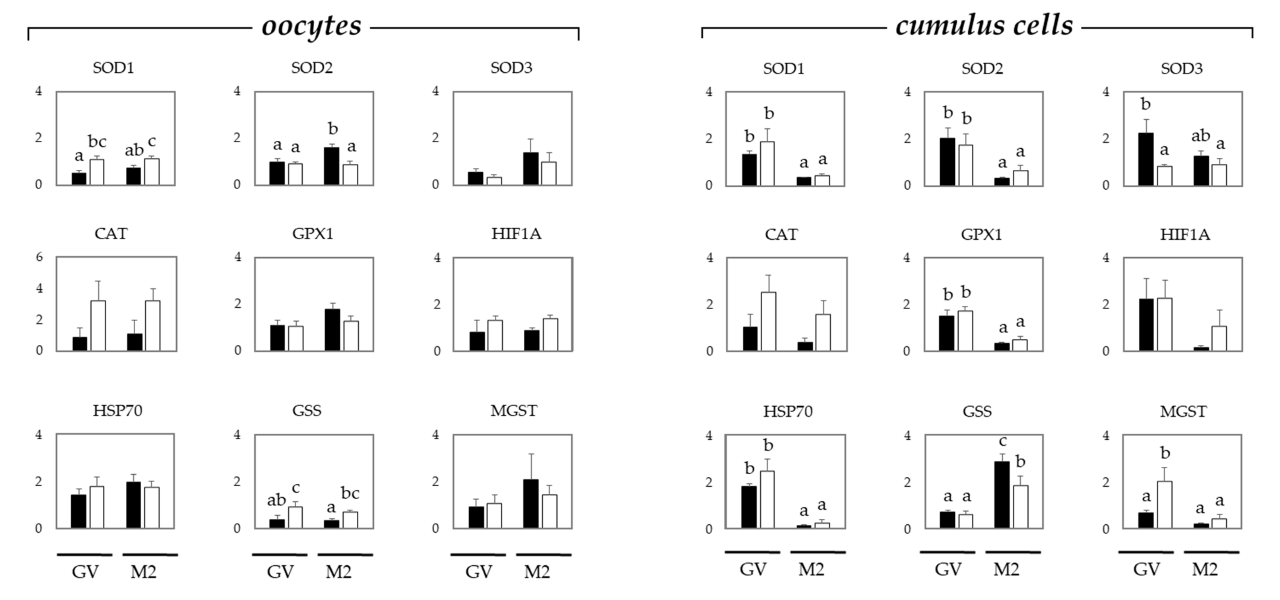
To study the oxidative stress in oocytes and cumulus cells, gene expression levels were examined in COCs by quantitative reverse transcription-polymerase chain reaction (qRT-PCR) (Figure 2).
In oocytes, the relative expression of SOD1 was not affected by the maturation stage, but it was significantly higher in sows than in gilts. The relative expression of SOD2 was significantly increased only in mature oocytes from gilts, whereas no changes were observed in the expression of SOD3. The relative expression of glutathione synthetase (GSS) was significantly higher in the oocytes from sows than in those from gilts, regardless of the maturation stage. The relative expression of catalase (CAT), glutathione peroxidase 1 (GPX1), hypoxia-inducible factor 1 alpha (HIF1A), heat shock protein 70 (HSP70), and microsomal glutathione S-transferase (MGST) was similar in the oocytes from gilts and sows, regardless of the maturation stage (GV or M2).
The expression of SOD1, SOD2, GPX1, and HSP70 in cumulus cells was significantly decreased after maturation regardless of the donor age, i.e., gilts or sows. The relative expression of SOD3 and MGST was significantly the highest in the cumulus cells from immature COCs from gilts and sows, respectively. The relative expression of GSS was significantly higher in the cumulus cells from mature oocytes from gilts than in those from sows. The relative expression of CAT and HIF1A was similar in the cumulus cells from gilts and sows, regardless of the maturation stage (GV or M2).

3. Discussion

In the present study, we demonstrated that oocytes from gilts are more sensitive than those from sows when exposed to DON or BEA during in vitro maturation, and this effect is influenced by the redox balance in these gametes. Previous studies showed that the maturation of oocytes from sows is impaired when performed in the presence of 0.2 μmol/L DON [4].
Herein, a decrease in the maturation rate was already observed after exposure of oocytes to 0.02 μmol/L DON, and this decrease was more evident in the oocytes from gilts. Han et al. [3] also demonstrated the negative impact of DON on the maturation of gilt oocytes at relatively high concentrations of this mycotoxin: the maturation rate decreased to 55% after exposure to 2.0 μmol/L DON and to less than 10% when exposed to 3.0 μmol/L DON. Disruption of oocyte maturation was most likely caused by an increased rate of apoptosis and epigenetic modification after exposure to 3.0 μmol/L DON [3]. In another study, malformation of meiotic spindles were observed after exposure to 0.2 and 2.0 μmol/L DON, with a decrease in the maturation rate of maturation rate to 25% when the oocytes were exposed to 2.0 μmol/L DON [4]. The sensitivity of oocytes to oxidative stress during maturation was also demonstrated after exposure to H2O2 when oocytes from gilts were the most affected.
DON does not affect the glutathione levels in oocytes [4], most likely since the cumulus cells protect oocytes by synthesizing and transporting GSH to oocytes [30,31]. In the present study, GSH levels in the oocytes and cumulus cells were not affected by the donor age (gilts or sows), but by the maturation stage in oocytes. Likewise, Pawlak et al. [32] observed an increase in the GSH content in matured porcine oocytes, regardless of the donor (prepubertal or cycling gilt). In the present study, the GSH content was significantly the highest in gilt FF. FF is a plasma-like fluid containing metabolites, proteins, ROS, and antioxidants from COCs, but also plasma products including antioxidants [33], where the GSH and SOD levels in FF are usually similar to those in the serum [34]. The GSH content in FF is not related to follicular size, but its presence will protect the oocytes against oxidation during growth and maturation [35], although there is no correlation between GSH and steroids in FF [36]. A decrease in the SOD activity in FF has been correlated with follicular growth in pigs [37] and high fertility rates in human [38]. It seems, however, that GSH and SOD levels in the FF do not predict resistance to toxins. Furthermore, the antioxidant capacity of granulosa cells exposed to AOH was increased when these cells were cultured in the presence of FF from sows when compared with FF from gilts [39]. Also, regardless of the FF source, no protection was observed when granulosa cells were exposed to BEA, showing that the FF alone cannot cope with the stress caused by this mycotoxin [39]. The ability of SOD to inhibit ROS activity in the cumulus cells was significantly decreased after maturation of oocytes from sows, but this effect was not observed in the cumulus cells from gilts. Increased SOD activity can be an indicator of oxidative stress, which could explain why gilt oocytes were more sensitive to the stress conditions during in vitro maturation. Such effects were even more robust when gilt oocytes were matured in the presence of BEA, and an interaction with dose and animal age was observed. There was no effect of BEA on the nuclear maturation when oocytes were cultured in the presence of BEA up to 5.0 μmol/L. However, a ten-fold lower concentration was sufficient to impair the maturation of gilt oocytes, and all oocytes degenerated after exposure to 5.0 μmol/L BEA. In a previous study, oocytes from gilts presented a significant decrease in maturation only when exposed to 2.5 μmol/L BEA [5]. However, the oocytes were cultured in a medium containing sow FF and not gilt FF as in the present study.
Interestingly, exposure to ROS (H2O2) also resulted in an impaired maturation rate in gilt oocytes, indicating that the intrinsic redox balance from gilt oocytes is different from that of sow oocytes. To understand the differences between gametes from gilts and sows, the mRNA expression of markers for oxidative stress was evaluated in oocytes and cumulus cells before and after in vitro maturation.
The SOD enzymes metabolize the ROS O2 into H2O2 and O2, and different enzymes can be categorized: SOD1, Cu/ZnSOD is cytoplsmaic, SOD2, MnSOD is mitochondrial, and SOD3 with a structure homologous to SOD1 [40] is secreted by cells. The expression of SOD1 was significantly higher in sows’ oocytes regardless of the maturation stage. The gene expression increase of this cytosolic SOD indicates that oocytes from sows are more active in neutralizing superoxide anions in the cytoplasm, whereas the increase of SOD2 in mature gilts’ oocytes is expected because oocytes derived from small follicles of slaughterhouse ovaries also express high levels of SOD2 [41]. However, this may also reflect an incorrect degradation of maternal mRNA prior to the activation of the embryonic genome [42]. The SOD3 expression in oocytes and cumulus cells from gilts and sows was similar within each maturation stage, except for the cumulus cells from immature oocytes that presented a SOD3 upregulation. This SOD is responsible for scavenging O2 in extracellular spaces and may explain why the FF from gilts had a significantly higher activity of this antioxidant. A proper functioning of the antioxidant enzymes SOD and CAT is crucial when cells are exposed to BEA [43]. Deoxynivalenol disrupts the normal function of mitochondria, affecting the normal function of SOD2 and increasing the accumulation of ROS in the cells [44]. In the present study, CAT relative expression was not affected by the COCs’ donor age and the imbalance in SOD expression probably explains the decreased maturation rate of gilt oocytes exposed to BEA and DON.
The relative expression of GPX1 and HSP70 was related to oocyte maturation and observed only in cumulus cells. Microsomal glutathione S-transferase was upregulated in immature oocytes from sows. The expression of MGST is increased in steroidogenically active cells [45], explaining the present results. Oocytes from sows had a higher expression of GSS than gilts, regardless of the maturation stage, whereas this effect was not observed in cumulus cells, which had a high expression of GSS when harvested from in vitro matured gilt oocytes. It has been demonstrated that GSS expression is higher in oocytes from sheep better able to develop to blastocysts [46]. Also, oocytes from prepubertal mice have a decreased capacity to synthesize GSH, increasing the chances of oocyte damage via oxidative stress [47]. Although oocytes and cumulus cells are connected, a completely different picture was observed in cumulus cells, where maturation was characterized by an upregulation of GSS, especially in those from matured gilt oocytes that present a lower glucose metabolism than sows, resulting in a decrease in GSH-S-transferase [48]. Cumulus cells synthesize GSH during oocyte maturation to guarantee fertilization and embryo development [49]. Possibly, in gilts, this was a compensatory mechanism due to the low GSS expression in mature oocytes. Both DON [50] and BEA [51] induce oxidative stress and also decrease the capacity to synthesize GSH, partly explaining the high susceptibility of gilt COCs to these mycotoxins during maturation.
When studying the potential effects of mycotoxins on female reproduction, it is crucial to consider that exposure via dietary contamination will give a more accurate model to predict risks [52]. Herewith, we show that the age of the donors will also affect the toxicity results, indicating that prepubertal females are more sensitive to mycotoxin exposure. Pigs are appropriate models to determine the effects of toxic compounds in humans due to the physiological similarities [53]. Therefore, chronic dietary exposure of children and prepubertal girls to DON and BEA may affect their fertility in adult life, and these mycotoxins are commonly found in infant food [54]. The tested exposure levels of DON in the present studies are similar to those detected in porcine and human plasma. The plasma level in pigs chronically exposed to DON reached 0.09 μmol/L [55], whereas DON was detected in the urine from healthy consumers at levels up to 1.25 μmol/L [56]. The detection of BEA in biological samples is a complex process because this mycotoxin is present in the diet at far lower concentrations than DON and is rapidly metabolized to a range of yet uncharacterized derivatives [57].

4. Conclusions

In conclusion, oocytes from gilts are more susceptible to the toxic effects of DON and BEA when compared with oocytes from sows. This effect is observed when immature oocytes start their maturation and the redox balance in sows is better controlled that in gilt oocytes and are mostly related to the imbalance of SOD and GSH in oocytes. Both DON and BEA do not affect cell viability solely via the stimulation of oxidative stress, but their negative impact will be potentiated in cells that cannot properly scavenge metabolic products like ROS.

5. Materials and Methods

5.1. Chemicals

Chemicals were purchased from Sigma (St. Louis, MO, USA) unless stated otherwise. BEA (purity > 97%) was dissolved in dimethyl sulfoxide (DMSO) to obtain a stock solution of 20 mM. Aliquots of these stock solutions were stored at −20 °C. On the day of experiment, the stock solution was diluted in culture medium and sonicated for 15 min to reach the concentration of 50 μM. DON (purity > 97%) was dissolved in DMSO to obtain a stock solution of 20 mM. Aliquots of these stock solutions were stored at −20 °C. On the day of experiment, the DON stock solution was diluted in culture medium and sonicated for 15 min to reach the concentration of 20 μM. The final concentration of DMSO in the treatment medium was 0.02% v/v. Therefore, medium used as control also had 0.02% DMSO. Culture media were equilibrated at 5% CO2 and 38.5 °C for at least two hours before use.

5.2. Selection and Culture of Cumulus Oocyte Complexes

Collection and selection of porcine cumulus oocyte complexes (COCs) were performed as described [58]. Briefly, ovaries from prepubertal gilts and sows collected from a local slaughterhouse were transported to the laboratory and maintained at 30 °C. Antral follicles (2–6 mm in diameter) were aspirated and COCs with a compact cumulus mass were selected, transferred to 4-(2-hydroxyethyl)-1-piperazineethanesulfonic acid (HEPES)-buffered M199 (Gibco BRL, Paisley, UK) supplemented with penicillin and streptomycin, and washed 3 times in oocyte maturation medium (OMM) consisting of M199 supplemented with 2.2 mg/mL NaHCO3, 200 μM cysteamine, with or without 10% (v/v) sow follicular fluid. Follicular fluid was collected from sow ovarian follicles measuring 5–8 mm in diameter. COCs were cultured for 20–22 h at 38.5 °C in a humidified atmosphere of 5% CO2 in air in groups of 35–50 in 500 μL of OMM with 0.05 international units (IU)/mL recombinant human follicle stimulating hormone (FSH) (Organon, Oss, The Netherlands), followed by a 20–22 h culture period in OMM without FSH.

5.3. Assessment of Cumulus Oocyte Complex Viability and Oocyte Nuclear Maturation

At 44 h of culture, COCs were incubated with 2 μM Ethidium Homodimer-1 (EtHD-1; Invitrogen-Molecular Probes, Eugene, OR, USA) in OMM and cultured for another 4 h. COCs were washed 3 times in phosphate buffered saline (PBS) and fixed in 2% (w/v) formaldehyde. Subsequently, COCs were incubated for 5 min in 0.1 μg/mL 4,6-diamino-2-phenyl-indole (DAPI; Invitrogen-Molecular Probes, Landsmeer, the Netherlands), mounted under a coverslip with anti-fade mounting medium (Vectashield; Vectorlab, Burlingame, CA, USA), and imaged by confocal laser scanning microscopy (CLSM; Leica SPE-II, Heidelberg, Germany). Imaging was performed using a 368 and 543 nm laser to excite DAPI and EthD-1, respectively. Nuclei of cumulus cells were considered degenerated when stained for both DAPI and EthD-1, or when nuclei had a condensed or fragmented appearance. Viable oocytes containing a metaphase spindle together with a polar body were considered at the Metaphase II (M2) stage.

5.4. Markers of Antioxidant Capacity and Oxidative Stress

Total equivalent antioxidant capacity (TEAC) in the FF (mM) was measured by using an assay kit from Sigma (Saint Louis, MO, USA; no. CS0790). Glutathione (GSH) levels in the FF (nmol/mL) and oocytes (relative fluorescence units (RFU)/min) was measured by using a kit from BioVision (Milpitas, CA, USA; no. K740-100). Hydrogen peroxide (H2O2) in FF (nmol/mL) and oocytes (pmol/oocyte) was measured by using a kit from BioVision (no. K265-200). Superoxide dismutase (SOD) activity (% of inhibition) in the FF, oocytes, and cumulus cells was measured by using an assay kit from BioVision (no. K335-100).

5.5. Quantitative Reverse Transcription-PCR

Total RNA was extracted with an RNAeasy Mini Kit (Qiagen, Valencia CA, USA) as per manufacturer’s instructions. Total RNA (10 μL) was kept at 70 °C for 5 min and then chilled on ice. Ten μL of a mastermix containing 4 μL 5 × 1st strand buffer, 0.4 μL random primers (0.09 IU/mL), 0.2 μL RNAsin (40 IU/mL), 0.75 μL Superscript®III (200 IU/mL) (Invitrogen, Groningen, The Netherlands), 2 μL dithiothreitol (0.1 M), 1 μL deoxyribose nucleoside triphosphate (dNTP) mix (10 mM), and 1.65 μL H2O were added and the mixture was incubated at 50 °C for 1 h. As a negative control, reverse transcriptase was replaced by H2O (minus reverse transcriptase (-RT) blanks). Samples were subsequently kept at 70 °C for 15 min and stored at −20 °C. The PCR mixture contained 0.1 μL cDNA, 0.1 μL forward and reverse primers (100 μM each) (Isogen, Maarssen, The Netherlands), 10 μL iQ SYBR Green supermix (Bio-Rad Laboratories, Hercules, CA, USA), and 9.8 μL H2O. The oligonucleotide primers were designed using Primer Select software (DNAstar, Madison WI, USA), and amplification profiles are listed in Table 4. After an initial denaturation step at 95 °C for 3 min, 40 cycles were carried out consisting of 95 °C for 5 s, the primer-specific annealing temperature (Table 4) for 5 s, and 72 °C for 20 s. Quantitative RT-PCR was performed in duplicate on two replicates of cDNA and singular on the -RT blanks. For each target gene, all the samples were quantified simultaneously in one run in a 96-well plate using a real-time PCR detection system (MyiQ Single-color, Real-Time Detection System; Bio-Rad). Melting curves were plotted to verify single product amplification. Standard curves made on cDNA dilutions were used to calculate the relative starting quantities. RNA expression was calculated as the ratio of RNA starting quantity of the target gene/geometric mean of two reference genes (BACT and B2M, cumulus; GAPDH and PGK1, oocytes).

5.6. Statistical Analysis

The COCs from gilts or sows were randomly assigned to treatment conditions prior to each experiment. Statistical analysis was conducted with the GenStat statistical software (GenStat for Windows 20th Edition, VSN International, Hemel, Hempstead, UK; https://www.vsni.co.uk/downloads/genstat/, accessed on 5 April 2021). Data were compared with analysis of variance (ANOVA). The null hypothesis was that there was no treatment effect on the response parameter. Treatment means were compared with Tukey’s post-hoc test, where the sow or gilt was the experimental unit. Linear and quadratic regression were applied to assess the effects of H2O2, BEA, and DON concentrations on oocyte maturation and degenerations. Data are presented as mean ± standard error of the mean (SEM). The p-value of the statistical model is given per response parameter. Significance was declared at p ≤ 0.05.

Author Contributions

Conceptualization, E.J.S. and B.A.J.R.; methodology, E.J.S.; validation, E.J.S.; formal analysis, E.J.S. and R.R.S.; investigation, E.J.S.; resources, B.A.J.R.; data curation, E.J.S. and R.R.S.; writing—original draft preparation, E.J.S. and R.R.S.; writing—review and editing, R.R.S. and B.A.J.R.; supervision, B.A.J.R.; project administration, E.J.S.; funding acquisition, B.A.J.R. All authors have read and agreed to the published version of the manuscript. E.J.S. passed away before publication of this work was completed.

Funding

This research received no external funding.

Institutional Review Board Statement

Ethical review and approval were waived for this study, due to the use of slaughterhouse material.

Informed Consent Statement

Not applicable.

Data Availability Statement

Data sharing not applicable.

Acknowledgments

We would like to acknowledge all members of the Farm Animal Health division, for assisting in COC collection and critical feedback.

Conflicts of Interest

The authors declare no conflict of interest.

Abbreviations

ANOVAAnalysis of variance
AOHAlternariol
B2Mβ-2-microglobulin
BACTβ-Actin
BEABeauvericin
CATCatalase
cDNAComplement deoxyribonucleic acid
COCsCumulus oocyte complexes
Cu/ZnSODCopper/zink superoxide dismutase
DAPI4′,6-diamidino-2-phenylindole
DMSODimethyl sulfoxide
dNTPDeoxyribonucleotide triphosphate
DON Deoxynivalenol
EthD-1Ethidium homodimer-1
FFFollicular fluid
FSHFollicle-stimulating hormone
GAPDHGlyceraldehyde 3-phosphate dehydrogenase
GLRX2Glutaredoxin 2
GPxGlutathione peroxidase
GPX1Glutathione peroxidase 1
GSHGlutathione
GSRGlutathione reductase
GSSGlutathione synthetase
GVGerminal vesicle
HEPES4-(2-hydroxyethyl)-1-piperazineethanesulfonic acid
HIF1AHypoxia-inducible factor 1
HSP70Heat shock protein 70
IUInternational units
M2Metaphase II
MGSTMicrosomal glutathione S-transferase
MnSODManganese superoxide dismutase
OMMOocyte maturation medium
PBSPhosphate buffer saline
PCRPolymerase chain reaction
PDIA4Protein disulfide isomerase 4
PGK1Phosphoglycerate kinase 1
qRT-PCRquantitative reverse transcription-polymerase chain reaction
RFURelative fluorescence units
RNARibonucleic acid
ROSReactive oxygen species
-RTMinus reverse transcriptase
SEMStandard error of the mean
SODSuperoxide dismutase
SOD1Superoxide dismutase 1
SOD2Superoxide dismutase 2
SOD3Superoxide dismutase 3
TEACTotal equivalent antioxidant activity
TXNRD1Thioredoxin reductase
ZENZearalenone

References

  1. Alm, H.; Greising, T.; Brussow, K.P.; Torner, H.; Tiemann, U. The Influence of the Mycotoxins Deoxynivalenol and Zearalenol on in vitro Maturation of Pig Oocytes and in vitro Culture of Pig Zygotes . Toxicol. In Vitro 2002, 16, 643–648. [Google Scholar] [CrossRef]
  2. Malekinejad, H.; Schoevers, E.J.; Daemen, I.J.; Zijlstra, C.; Colenbrander, B.; Fink-Gremmels, J. Exposure of Oocytes to the Fusarium Toxins Zearalenone and Deoxynivalenol Causes Aneuploidy and Abnormal Embryo Development in Pigs. Biol. Reprod. 2007, 77, 840–847. [Google Scholar] [CrossRef] [Green Version]
  3. Han, J.; Wang, Q.C.; Zhu, C.C.; Liu, J.; Zhang, Y.; Cui, X.S.; Kim, N.H.; Sun, S.C. Deoxynivalenol Exposure Induces Autophagy/Apoptosis and Epigenetic Modification Changes during Porcine Oocyte Maturation. Toxicol. Appl. Pharmacol. 2016, 300, 70–76. [Google Scholar] [CrossRef]
  4. Schoevers, E.J.; Fink-Gremmels, J.; Colenbrander, B.; Roelen, B.A. Porcine Oocytes are most Vulnerable to the Mycotoxin Deoxynivalenol during Formation of the Meiotic Spindle. Theriogenology 2010, 74, 968–978. [Google Scholar] [CrossRef]
  5. Schoevers, E.J.; Santos, R.R.; Fink-Gremmels, J.; Roelen, B.A. Toxicity of Beauvericin on Porcine Oocyte Maturation and Preimplantation Embryo Development. Reprod. Toxicol. 2016, 65, 159–169. [Google Scholar] [CrossRef] [PubMed] [Green Version]
  6. Schoevers, E.J.; Santos, R.R.; Roelen, B.A.J. Alternariol Disturbs Oocyte Maturation and Preimplantation Development. Mycotoxin. Res. 2020, 36, 93–101. [Google Scholar] [CrossRef] [PubMed]
  7. Schoevers, E.J.; Santos, R.R.; Colenbrander, B.; Fink-Gremmels, J.; Roelen, B.A. Transgenerational Toxicity of Zearalenone in Pigs. Reprod. Toxicol. 2012, 34, 110–119. [Google Scholar] [CrossRef] [PubMed]
  8. Benthem de Grave, X.; Saltzmann, J.; Laurain, J.; Rodriguez, M.A.; Molist, F.; Dänicke, S.; Santos, R.R. Transmission of Zearalenone, Deoxynivalenol, and Their Derivatives from Sows to Piglets during Lactation. Toxins 2021, 13, 37. [Google Scholar] [CrossRef]
  9. Pestka, J.J. Deoxynivalenol: Mechanisms of Action, Human Exposure, and Toxicological Relevance. Arch. Toxicol. 2010, 84, 663–679. [Google Scholar] [CrossRef] [PubMed]
  10. Zhou, H.R.; Lau, A.S.; Pestka, J.J. Role of Double-stranded RNA-activated Protein Kinase R (PKR) in Deoxynivalenol-induced Ribotoxic Stress Response. Toxicol. Sci. 2003, 74, 335–344. [Google Scholar] [CrossRef] [Green Version]
  11. Payros, D.; Alassane-Kpembi, I.; Pierron, A.; Loiseau, N.; Pinton, P.; Oswald, I.P. Toxicology of Deoxynivalenol and Its Acetylated and Modified Forms. Arch. Toxicol. 2016, 90, 2931–2957. [Google Scholar] [CrossRef]
  12. Caloni, F.; Fossati, P.; Anadon, A.; Bertero, A. Beauvericin: The Beauty and the Beast. Environ. Toxicol. Pharmacol. 2020, 75, 103349. [Google Scholar] [CrossRef] [PubMed]
  13. Olleik, H.; Nicoletti, C.; Lafond, M.; Courvoisier-Dezord, E.; Xue, P.; Hijazi, A.; Baydoun, E.; Perrier, J.; Maresca, M. Comparative Structure-activity Analysis of the Antimicrobial Activity, Cytotoxicity, and Mechanism of Action of the Fungal Cyclohexadepsipeptides Enniatins and Beauvericin. Toxins 2019, 11, 514. [Google Scholar] [CrossRef] [PubMed] [Green Version]
  14. Mallebrera, B.; Prosperini, A.; Font, G.; Ruiz, M.J. In vitro Mechanisms of Beauvericin Toxicity: A Review. Food Chem. Toxicol. 2018, 111, 537–545. [Google Scholar] [CrossRef] [PubMed]
  15. Agarwal, A.; Gupta, S.; Sharma, R.K. Role of Oxidative Stress in Female Reproduction. Reprod. Biol. Endocrinol. 2005, 3, 28. [Google Scholar] [CrossRef] [Green Version]
  16. Pisoschi, A.M.; Pop, A. The Role of Antioxidants in the Chemistry of Oxidative Stress: A Review. Eur. J. Med. Chem. 2015, 97, 55–74. [Google Scholar] [CrossRef] [PubMed]
  17. El Golli-Bennour, E.; Bacha, H. Hsp70 Expression as Biomarkers of Oxidative Stress: Mycotoxins’ Exploration. Toxicology 2011, 287, 1–7. [Google Scholar] [CrossRef] [PubMed]
  18. Yalu, R.; Oyesiji, A.E.; Eisenberg, I.; Imbar, T.; Meidan, R. HIF1A-dependent Increase in Endothelin 2 Levels in Granulosa Cells: Role of Hypoxia, LH/cAMP, and Reactive Oxygen Species. Reproduction 2015, 149, 11–20. [Google Scholar] [CrossRef] [Green Version]
  19. Osselaere, A.; Santos, R.; Hautekiet, V.; De Backer, P.; Chiers, K.; Ducatelle, R.; Croubels, S. Deoxynivalenol Impairs Hepatic and Intestinal Gene Expression of Selected Oxidative Stress, Tight Junction and Inflammation Proteins in Broiler Chickens, but Addition of an Adsorbing Agent Shifts the Effects to the Distal Parts of the Small Intestine. PLoS ONE 2013, 8, e69014. [Google Scholar] [CrossRef] [PubMed] [Green Version]
  20. Xie, S.; Zheng, L.; Wan, M.; Niu, J.; Liu, Y.; Tian, L. Effect of Deoxynivalenol on Growth Performance, Histological Morphology, Anti-oxidative Ability and Immune Response of Juvenile Pacific White Shrimp, Litopenaeus vannamei. Fish Shellfish Immunol. 2018, 82, 442–452. [Google Scholar] [CrossRef]
  21. Marchal, R.; Feugang, J.M.; Perreau, C.; Venturi, E.; Terqui, M.; Mermillod, P. Meiotic and Developmental Competence of Prepubertal and Adult Swine Oocytes. Theriogenology 2001, 56, 17–29. [Google Scholar] [CrossRef]
  22. Pinkert, C.A.; Kooyman, D.L.; Baumgartner, A.; Keisler, D.H. In vitro Development of Zygotes from Superovulated Prepubertal and Mature Gilts. J. Reprod. Fert. 1989, 87, 63–66. [Google Scholar] [CrossRef] [Green Version]
  23. Koenig, J.L.F.; Stormshak, F. Cytogenetic Evaluation of Ova from Pubertal and Third-estrous Gilts. Biol. Reprod. 1993, 49, 1158–1162. [Google Scholar] [CrossRef] [PubMed] [Green Version]
  24. Yuan, Y.; Wheeler, M.B.; Krisher, R.L. Disrupted Redox Homeostasis and Aberrant Redox Gene Expression in Porcine Oocytes Contribute to Decreased Developmental Competence. Biol. Reprod. 2012, 87, 78. [Google Scholar] [CrossRef] [PubMed]
  25. Knox, R.V. Physiology and Endocrinology Symposium: Factors Influencing Follicle Development in Gilts and Sows and Management Strategies Used to Regulate Growth for Control of Estrus and Ovulation. J. Anim. Sci. 2019, 97, 1433–1445. [Google Scholar] [CrossRef] [PubMed]
  26. Ambekar, A.S.; Nirujogi, R.S.; Srikanth, S.M.; Chavan, S.; Kelkar, D.S.; Hinduja, I.; Zaveri, K.; Keshava Prasad, T.S.; Harsha, H.C.; Pandey, A.; et al. Proteomic Analysis of Human Follicular Fluid: A New Perspective Towards Understanding Folliculogenesis. J. Proteom. 2013, 87, 68–77. [Google Scholar] [CrossRef]
  27. Albertini, D.F.; Combelles, C.M.; Benecchi, E.; Carabatsos, M.J. Cellular Basis for Paracrine Regulation of Ovarian Follicle Development. Reproduction 2001, 121, 647–653. [Google Scholar] [CrossRef]
  28. Da Broi, M.G.; Giorgi, V.S.; Wang, F.; Keefe, D.L.; Albertini, D.; Navarro, P.A. Influence of Follicular Fluid and Cumulus Cells on Oocyte Quality Clinical Implications. J. Assist. Reprod. Genet. 2018, 35, 735–751. [Google Scholar] [CrossRef]
  29. Paczkowski, M.; Yuan, Y.; Fleming-Waddell, J.; Bidwell, C.A.; Spurlock, D.; Krisher, R.L. Alterations in the Transcriptome of Porcine Oocytes Derived from Prepubertal and Cyclic Females is Associated with Developmental Potential. J. Anim. Sci. 2011, 89, 3561–3571. [Google Scholar] [CrossRef]
  30. Furnus, C.C.; de Matos, D.G.; Moses, D.F. Cumulus Expansion during in vitro Maturation of Bovine Oocytes: Relationship with Intracellular Glutathione Level and Its Role on Subsequent Embryo Development. Mol. Reprod. Dev. 1998, 51, 76–83. [Google Scholar] [CrossRef]
  31. Schoevers, E.J.; Colenbrander, B.; Roelen, B.A. Developmental Stage of the Oocyte during Antral Follicle Growth and Cumulus Investment Determines in vitro Embryo Development of Sow Oocytes. Theriogenology 2007, 67, 1108–1122. [Google Scholar] [CrossRef] [PubMed]
  32. Pawlak, P.; Warzych, E.; Hryciuk, M.; Lechniak, D. Transcript Abundance, Glutathione and Apoptosis Levels Differ Between Porcine Oocytes Collected from Prepubertal and Cyclic Gilts. Theriogenology 2015, 84, 86–93. [Google Scholar] [CrossRef] [PubMed]
  33. Freitas, C.; Neto, A.C.; Matos, L.; Silva, E.; Ribeiro, A.; Silva Carvalho, J.L.; Almeida, H. Follicular Fluid Redox Involvement for Ovarian Follicle Growth. J. Ovarian Res. 2017, 10, 1–10. [Google Scholar] [CrossRef] [PubMed]
  34. Da Broi, M.G.; Albuquerque, F.O.; Andrade, A.Z.; Cardoso, R.L.; Jordao Junior, A.A.; Navarro, P.A. Increased Concentration of 8-hydroxy-2′-deoxyguanosine in Follicular Fluid of Infertile Women with Endometriosis. Cell Tissue Res. 2016, 366, 231–242. [Google Scholar] [CrossRef]
  35. Liu, R.H.; Li, Y.H.; Jiao, L.H.; Wang, X.N.; Wang, H.; Wang, W.H. Extracellular and Intracellular Factors Affecting Nuclear and Cytoplasmic Maturation of Porcine Oocytes Collected from Different Sizes of Follicles. Zygote 2002, 10, 253–260. [Google Scholar] [CrossRef]
  36. Bisseling, J.G.; Knapen, M.F.; Goverde, H.J.; Mulder, T.P.; Peters, W.H.; Willemsen, W.N.; Thomas, C.M.; Steegers, E.A. Glutathione S-transferases in Human Ovarian Follicular Fluid. Fertil. Steril. 1997, 68, 907–911. [Google Scholar] [CrossRef] [Green Version]
  37. Basini, G.; Simona, B.; Santini, S.E.; Grasselli, F. Reactive Oxygen Species and Anti-oxidant Defences in Swine Follicular Fluids. Reprod. Fertil. Dev. 2008, 20, 269–274. [Google Scholar] [CrossRef]
  38. Sabatini, L.; Wilson, C.; Lower, A.; Al-Shawaf, T.; Grudzinkas, J.G. Superoxide Dismutase Activity in Human Follicular Fluid after Controlled Ovarian Hyperstimulation in Women Undergoing in vitro Fertilization. Fertil. Steril. 1999, 72, 1027–1034. [Google Scholar] [CrossRef]
  39. Santos, R.R.; Schoevers, E.J.; Wu, X.; Roelen, B.A.J.; Fink-Gremmels, J. The Protective Effect of Follicular Fluid against the Emerging Mycotoxins Alternariol and Beauvericin. World Mycotoxin J. 2015, 8, 445–450. [Google Scholar] [CrossRef]
  40. Von Mengden, L.; Klamt, F.; Smitz, J. Redox Biology of Human Cumulus Cells: Basic Concepts, Impact on Oocyte Quality, and Potential Clinical Use. Antioxid. Redox Signal. 2020, 10, 522–535. [Google Scholar] [CrossRef] [Green Version]
  41. Lonergan, P.; Gutierrez-Adan, A.; Rizos, D.; Pintado, B.; de la Fuente, J.; Boland, M.P. Relative Messenger RNA Abundance in Bovine Oocytes Collected in vitro or in vivo before and 20 hr after the Preovulatory Luteinizing Hormone Surge. Mol. Reprod. Dev. 2003, 66, 297–305. [Google Scholar] [CrossRef]
  42. Combelles, C.M.; Gupta, S.; Agarwal, A. Could Oxidative Stress Influence the in vitro Maturation of Oocytes? Reprod. Biomed. Online 2009, 18, 868–880. [Google Scholar] [CrossRef]
  43. Mallebrera, B.; Juan-Garcia, A.; Font, G.; Ruiz, M.J. Mechanisms of Beauvericin Toxicity and Antioxidant Cellular Defense. Toxicol. Lett. 2016, 246, 28–34. [Google Scholar] [CrossRef]
  44. Wu, Q.-H.; Wang, X.; Yang, W.; Nussler, A.K.; Xiong, L.-Y.; Kuca, K.; Dohnal, V.; Zhang, X.-J.; Yuan, Z.-H. Oxidative stress-mediated Cytotoxicity and Metabolism of T-2 Toxin and Deoxynivalenol in Animals and Humans: An Update. Arch. Toxicol. 2014, 88, 1309–1326. [Google Scholar] [CrossRef]
  45. Keira, M.; Nishihira, J.; Ishibashi, T.; Tanaka, T.; Fujimoto, S. Identification of a Molecular Species in Porcine Ovarian Luteal Glutathione S-transferase and Its Hormonal Regulation by Pituitary Gonadotropins. Arch. Biochem. Biophys. 1994, 308, 126–132. [Google Scholar] [CrossRef] [PubMed]
  46. Hidaka, T.; Fukumoto, Y.; Yamamoto, S.; Ogata, Y.; Horiuchi, T. Variations in Bovine Embryo Production between Individual Donors for OPU-IVF are Closely Related to Glutathione Concentrations in Oocytes during in vitro Maturation. Theriogenology 2018. [Google Scholar] [CrossRef] [PubMed]
  47. Jiao, G.Z.; Cao, X.Y.; Cui, W.; Lian, H.Y.; Miao, Y.L.; Wu, X.F.; Han, D.; Tan, J.H. Developmental Potential of Prepubertal Mouse Oocytes is Compromised Due Mainly to Their Impaired Synthesis of Glutathione. PLoS ONE 2013, 8, e58018. [Google Scholar] [CrossRef] [Green Version]
  48. Paczkowski, M.; Krisher, R. Aberrant Protein Expression is Associated with Decreased Developmental Potential in Porcine Cumulus-oocyte Complexes. Mol. Reprod. Dev. 2010, 77, 51–58. [Google Scholar] [CrossRef]
  49. Maedomari, N.; Kikuchi, K.; Noguchi, J.; Kaneko, H.; Ohnuma, K.; Nakai, M.; Shino, M.; Nagai, T.; Kashiwazaki, N. Cytoplasmic Glutathione Regulated by Cumulus Cells during Porcine Oocyte Maturation Affects Fertilization and Embryonic Development in vitro. Theriogenology 2007, 67, 983–993. [Google Scholar] [CrossRef] [PubMed]
  50. Wang, J.; Jin, Y.; Wu, S.; Yu, H.; Zhao, Y.; Fang, H.; Shen, J.; Zhou, C.; Fu, Y.; Li, R.; et al. Deoxynivalenol Induces Oxidative Stress, Inflammatory Response and Apoptosis in Bovine Mammary Epithelial Cells. J. Anim. Physiol. Anim. Nutr. 2019, 103, 1663–1674. [Google Scholar] [CrossRef]
  51. Juan-Garcia, A.; Carbone, S.; Ben-Mahmoud, M.; Sagratini, G.; Manes, J. Beauvericin and Ochratoxin A Mycotoxins Individually and Combined in HepG2 Cells Alter Lipid Peroxidation, Levels of Reactive Oxygen Species and Glutathione. Food Chem. Toxicol. 2020, 139, 111247. [Google Scholar] [CrossRef]
  52. Santos, R.R.; Schoevers, E.J.; Roelen, B.A.J.; Fink-Gremmels, J. Mycotoxins and Female Reproduction: In vitro Approaches. World Mycotoxin J. 2013, 6, 245–253. [Google Scholar] [CrossRef]
  53. Santos, R.R.; Schoevers, E.J.; Roelen, B.A.J. Usefulness of Bovine and Porcine IVM/IVF Models for Reproductive Toxicology. Reprod. Biol. Endocrinol. 2014, 12, 117. [Google Scholar] [CrossRef] [PubMed] [Green Version]
  54. Habschied, K.; Kanižai Šarić, G.; Krstanović, V.; Mastanjević, K. Mycotoxins—Biomonitoring and Human Exposure. Toxins 2021, 13, 113. [Google Scholar] [CrossRef]
  55. Goyarts, T.; Danicke, S. Bioavailability of the Fusarium Toxin Deoxynivalenol (DON) from Naturally Contaminated Wheat for the Pig. Toxicol. Lett. 2006, 163, 171–182. [Google Scholar] [CrossRef]
  56. Meky, F.A.; Turner, P.C.; Ashcroft, A.E.; Miller, J.D.; Qiao, Y.L.; Roth, M.J.; Wild, C.P. Development of a Urinary Biomarker of Human Exposure to Deoxynivalenol. Food. Chem. Toxicol. 2003, 41, 265–273. [Google Scholar] [CrossRef]
  57. Serrano, A.B.; Capriotti, A.L.; Cavaliere, C.; Piovesana, S.; Samperi, R.; Ventura, S.; Lagana, A. Development of a Rapid LC-MS/MS Method for the Determination of Emerging Fusarium Mycotoxins Enniatins and Beauvericin in Human Biological Fluids. Toxins 2015, 7, 3554–3571. [Google Scholar] [CrossRef] [PubMed] [Green Version]
  58. Schoevers, E.J.; Kidson, A.; Verheijden, J.H.; Bevers, M.M. Effect of Follicle-stimulating Hormone on Nuclear and Cytoplasmic Maturation of Sow Oocytes in vitro. Theriogenology 2003, 59, 2017–2028. [Google Scholar] [CrossRef]
  59. Wang, X.; Zhu, X.; Liang, X.; Xu, H.; Liao, Y.; Lu, K.; Lu, S. Effects of Resveratrol on in vitro Maturation of Porcine Oocytes and Subsequent Early Embryonic Development Following Somatic Cell Nuclear Transfer. Reprod. Domest. Anim. 2019, 54, 1195–1205. [Google Scholar] [CrossRef] [PubMed]
  60. Pereira, B.A.; Zangeronimo, M.G.; Castillo-Martin, M.; Gadani, B.; Chaves, B.R.; Rodriguez-Gil, J.E.; Bonet, S.; Yeste, M. Supplementing Maturation Medium with Insulin Growth Factor I and Vitrification-Warming Solutions With Reduced Glutathione Enhances Survival Rates and Development Ability of in vitro Matured Vitrified-Warmed Pig Oocytes. Front. Physiol. 2019, 9, 1894. [Google Scholar] [CrossRef] [Green Version]
  61. Kwiatkowska, J.; Wasowska, B.; Gilun, P. Expression of Hypoxia Inducible Factor 1α and Antioxidant Enzymes: Superoxide Dismutases-1 and -2 in Ischemic Porcine Endometrium. Reprod. Biol. 2017, 17, 289–293. [Google Scholar] [CrossRef] [PubMed]
  62. Chmielewska, M.; Tosiewicz, K.; Podlasz, P.; Wasowicz, K. The Expression of Mitochondrial, Cytoplasmic and Extracellular Superoxide Dismutase in the Colonic Wall of Pigs Suffering from Swine Dysenteria. Pol. J. Vet. Sci. 2013, 16, 463–467. [Google Scholar] [CrossRef] [PubMed] [Green Version]
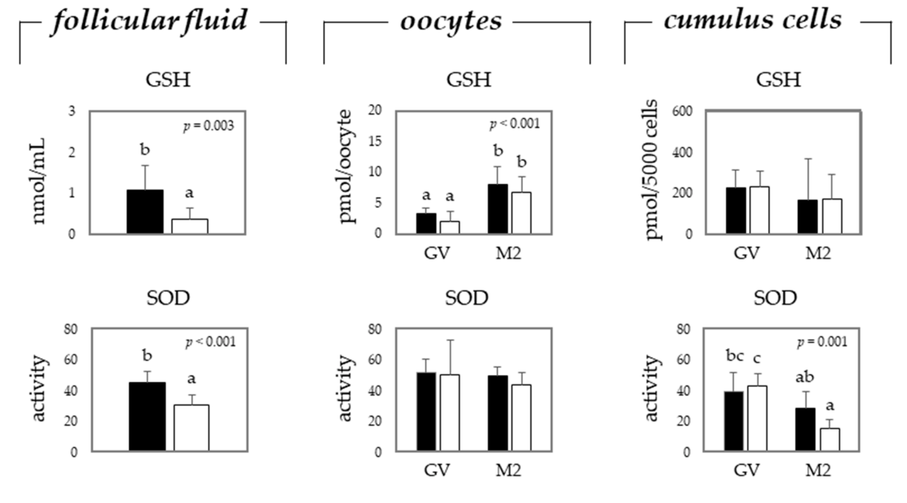
Figure 1. Mean (±SEM) levels of glutathione (GSH) and superoxide dismutase (SOD) in the follicular fluid, oocytes, and cumulus cells from gilts (black bars) and sows (white bars). Oocytes and cumulus cells were analyzed before (germinal vesicle (GV) stage) and after (metaphase 2 (M2) stage) in vitro maturation. Each mean represents values from 5 replicates per gilt or sow (pooled follicular fluid from antral follicles measuring 2–8 mm, and 50 COCs/replicate/gilt or sow), using the animal as the experimental unit. a–c: different letters indicate significant difference among treatments, i.e., gilts vs. sows for follicular fluid, and gilts vs. sows and GV vs. M2 for oocytes and cumulus cells (p ≤ 0.05).
Toxins 13 00260 g001
Figure 2. Mean (±SEM) mRNA relative expression of markers for oxidative stress in oocytes, and cumulus cells from gilts (black bars) and sows (white bars). Gene abbreviations are above each graph, full names of the different genes are presented in the main text. Oocytes and cumulus cells were analyzed before (GV stage) and after (M2 stage) in vitro maturation. Each mean represents values from 5 replicates per gilt or sow (50 COCs/replicate/gilt or sow), using the animal as the experimental unit. a–c: different letters indicate significant difference among treatments, gilts vs. sows and GV vs. M2 (p ≤ 0.05).
Toxins 13 00260 g002
Table 1. Mean (±standard error of the mean (SEM)) rates of nuclear maturation and degeneration of porcine in vitro matured in the absence (control) or in the presence of H2O2 at different concentrations.
TreatmentAgeMaturation RateDegeneration Rate
ControlGilt82.9 c4.8 a
ControlSow86.9 c2.4 a
10 μmol/LGilt65.7 c9.1 a
10 μmol/LSow86.8 c2.5 a
50 μmol/LGilt35.7 b26.8 b
50 μmol/LSow71.9 c9.7 a
100 μmol/LGilt7.7 a88.1 c
100 μmol/LSow65.5 c30.4 b
Control 84.9 3.6
10 μmol/L 76.2 5.8
50 μmol/L 53.8 18.2
100 μmol/L 36.6 59.2
Gilt48.0 32.2
Sow77.8 11.2
Effect of: p-valueSEMp-valueSEM
Treatment × Age 0.0177.60<0.0013.34
Age <0.0013.80<0.0011.67
Treat <0.0015.38<0.0012.36
Treatment Linear 0.006 <0.001
Treatment Quadratic 0.494 0.072
Each mean represents values from 5 replicates per gilt or sow (50 cumulus oocyte complexes (COCs)/replicate/gilt or sow), using the animal as the experimental unit. a–c: different letters indicate significant difference among treatments (p ≤ 0.05).
Table 2. Mean (± SEM) rates of nuclear maturation and degeneration of porcine in vitro matured in the absence (control) or in the presence of BEA at different concentrations (0.5, 2.5, or 5.0 μmol/L).
TreatmentAgeMaturation RateDegeneration Rate
ControlGilt74.0 cd11.7 a
ControlSow77.4 cd1.5 a
0.5 μmol/LGilt46.8 b28.1 b
0.5 μmol/LSow82.0 d2.7 a
2.5 μmol/LGilt4.8 a91.6 c
2.5 μmol/LSow72.3 cd9.7 a
5.0 μmol/LGilt0.0 a100.0 c
5.0 μmol/LSow64.8 c11.1 a
Control 75.7 6.6
0.5 μmol/L 64.4 15.5
2.5 μmol/L 38.6 50.7
5.0 μmol/L 32.4 55.5
Gilt31.4 57.9
Sow74.1 6.2
Effect of: p-valueSEMp-valueSEM
Treatment × Age <0.0015.08<0.0013.59
Age <0.0012.54<0.0011.80
Treat <0.0013.59<0.0012.54
Treatment Linear 0.016 0.010
Treatment Quadratic 0.857 0.319
Each mean represents values from 5 replicates per gilt or sow (50 COCs/replicate/gilt or sow), using the animal as the experimental unit. a–d: different letters indicate significant difference among treatments (p ≤ 0.05).
Table 3. Mean (± SEM) rates of nuclear maturation and degeneration of porcine in vitro matured in the absence (control) or in the presence of DON at different concentrations (0.02, 0.2, or 2.0 μmol/L).
TreatmentAgeMaturation RateDegeneration Rate
ControlGilt67.5 13.3
ControlSow73.2 9.1
0.02 μmol/LGilt37.1 12.7
0.02 μmol/LSow63.5 7.5
0.2 μmol/LGilt22.9 29.7
0.2 μmol/LSow59.7 8.8
2.0 μmol/LGilt8.7 35.4
2.0 μmol/LSow42.5 7.5
Control 70.4 c11.2
0.02 μmol/L 50.3 b10.1
0.2 μmol/L 41.3 b19.2
2.0 μmol/L 25.6 a21.5
Gilt34.0 a22.8 b
Sow59.7 b8.2 a
Effect of: p-valueSEMp-valueSEM
Treatment × Age 0.1787.170.0895.04
Age <0.0013.580.0012.52
Treat <0.0015.070.1013.57
Treatment Linear 0.026 0.798
Treatment Quadratic 0.863 0.885
Each mean represents values from 5 replicates per gilt or sow (50 COCs/replicate/gilt or sow), using the animal as the experimental unit. a–c: different letters indicate significant difference among treatments (p ≤ 0.05).
Table 4. Primer pairs sequence used in polymerase chain reaction (PCR) to detect the primary partial cDNA products in porcine oocytes and cumulus cells.
GeneProteinSequenceProduct Size (bp)Annealing
Temperature (°C)
Gene Bank Accession Number
Reference
B2Mβ-2-microglobulinF: 5′-TTCACACCGCTCCAGTAG-3′
R: 5′-CCAGATACATAGCAGTTCAGG-3′
16660.0NM_213978
BACTβ-ActinF: 5′-CATCACCATCGGCAACGAGC-3′
R: 5′-TAGAGGTCCTTGCGGATGTC-3′
14156.0AY550069
PGK1Phosphoglycerate kinase 1F: 5′-AGATAACGAACAACCAGAGG-3′
R: 5′-TGTCAGGCATAGGGATACC-3′
12656.0AY677198
GAPDHGlyceraldehyde 3-phosphate
dehydrogenase
F: 5′ -TCGGAGTGAACGGATTTG-3′
R: 5′-CCTGGAAGATGGTGATGG-3′
21961.0AF017079
Target
CAT [59]CatalaseF: 5′-ATGTGCAGGCTGGATCTCAC-3′
R: 5′-GCACAGGAGAATCTTGCATCC-3′
15555.0XM_021081498.1
GPX1 [60]Glutathione peroxidase 1F: 5′-CAAGAATGGGGAGATCCTGA-3′
R: 5′-GTCA TTGCGACACACTGGAG-3′
21764.5NM_2142201.1
GSSGlutathione synthetaseF: 5′-TGGTTTACTTCCGGGATGGC-3′
R: 5′-CGCCTACTCTGCTTAGCTCC-3′
15964.5NM_001244625.1
HIF1A [61]Hypoxia inducible factor 1 alphaF: 5′-TCAGCTATTTGCGTGTGAGG-3′
R: 5′-TTCACAAATCAGACCAAGC-3′
47961.0NM_001123124.1
HSP70 [60]Heat shock protein 70F: 5′-ATGTCCGCTGCAAGAGAAGT-3′
R: 5′-GGCGTCAAACACGGTATTCT-3′
21664.5NM_001123127.1
MGSTMicrosomal glutathione S-transferase 1F: 5′-CGAGGAATTAAGATAGAGAAAGCCT-3′
R: 5′-TGGGCCCATTTGGAAAATACTGA-3′
24764.5NM_214300.2
SOD1 [59]Superoxide dismutase 1F: 5′-CGAGCTGAAGGGAGAGAAGA-3′
R: 5′-ACATTGCCCAGGTCTCCAAC-3′
19964.5NM_001190422.1
SOD2 [61]Superoxide dismutase 2F: 5′-CCCTGGAGCCGCACATC-3′
R: 5′-TTTTTCAGCGCCTCCTG-3′
11564.5NM_214127.2
SOD3 [62]Superoxide dismutase 3R: 5′-ACTCCTGCCATGCTGACG-3′
F: 5′-TGCCAGATCTCCGTCACTTT-3′
14064.5DQ915492.1
Publisher’s Note: MDPI stays neutral with regard to jurisdictional claims in published maps and institutional affiliations.