Article

Molecular Identification and Toxin Analysis of Alexandrium spp. in the Beibu Gulf: First Report of Toxic A. tamiyavanichii in Chinese Coastal Waters

1
Key Laboratory of Environment Change and Resources Use in Beibu Gulf, Ministry of Education, Nanning Normal University, Nanning 530001, China
2
Guangxi Laboratory on the Study of Coral Reefs in the South China Sea, Guangxi University, Nanning 530004, China
3
Guangxi Key Laboratory of Marine Environmental Science, Beibu Gulf Marine Research Center, Guangxi Academy of Sciences, Nanning 530007, China
4
Third Institute of Oceanography, Ministry of Natural Resources, Xiamen 361005, China
5
Biology Department, Woods Hole Oceanographic Institution, Woods Hole, MA 02543, USA
*
Author to whom correspondence should be addressed.
Toxins 2021, 13(2), 161; https://doi.org/10.3390/toxins13020161
Submission received: 25 January 2021 / Revised: 14 February 2021 / Accepted: 16 February 2021 / Published: 18 February 2021
(This article belongs to the Special Issue Phycotoxins: Chemistry and Biochemistry)

Abstract

:
The frequency of harmful algal blooms (HABs) has increased in China in recent years. Information about harmful dinoflagellates and paralytic shellfish toxins (PSTs) is still limited in China, especially in the Beibu Gulf, where PSTs in shellfish have exceeded food safety guidelines on multiple occasions. To explore the nature of the threat from PSTs in the region, eight Alexandrium strains were isolated from waters of the Beibu Gulf and examined using phylogenetic analyses of large subunit (LSU) rDNA, small subunit (SSU) rDNA, and internal transcribed spacer (ITS) sequences. Their toxin composition profiles were also determined using liquid chromatography-tandem mass spectrometry (LC-MS/MS). All eight strains clustered in the phylogenetic tree with A. pseudogonyaulax, A. affine, and A. tamiyavanichii from other locations, forming three well-resolved groups. The intraspecific genetic distances of the three Alexandrium species were significantly smaller than interspecific genetic distances for Alexandrium species. Beibu Gulf isolates were therefore classified as A. pseudogonyaulax, A. affine, and A. tamiyavanichii. No PSTs were identified in A. pseudogonyaulax, but low levels of gonyautoxins (GTXs) 1 to 5, and saxitoxin (STX) were detected in A. tamiyavanichii (a total of 4.60 fmol/cell). The extremely low level of toxicity is inconsistent with PST detection above regulatory levels on multiple occasions within the Beibu Gulf, suggesting that higher toxicity strains may occur in those waters, but were unsampled. Other explanations including biotransformation of PSTs in shellfish and the presence of other PST-producing algae are also suggested. Understanding the toxicity and phylogeny of Alexandrium species provides foundational data for the protection of public health in the Beibu Gulf region and the mitigation of HAB events.
Key Contribution: The first record of Alexandrium affine, A. pseudogonyaulax and A. tamiyavanichii from the seawaters of Beibu Gulf, and the first report of toxicity and phylogeny of A. tamiyavanichii from Chinese coastal waters.

1. Introduction

The genus Alexandrium is an important toxic and harmful algal blooms (HABs) dinoflagellate distributed in coastal waters worldwide [1]. This genus was first described by Halim [2] and it has since become one of the most studied dinoflagellate genera. Among the over 30 species of Alexandrium reported to date, at least half can cause paralytic shellfish poisoning (PSP) [3,4]. Paralytic shellfish toxins (PSTs) accumulate in shellfish, fish, and other organisms through the food web. Humans who consume shellfish containing PSTs can become poisoned; PSTs thus represents a major health hazard [1,5]. Alexandrium is a common species in the coastal waters of China. Over the past 15 years, 24 HABs due to Alexandrium spp. have occurred in the Bohai, Yellow, and South China Seas [5], with disastrous consequences for local marine fisheries, ecosystems, and coastal landscapes.
The morphological identification of Alexandrium is based primarily on the fine structure of the apical pore complex, the first apical and sixth precingular plates, the presence/absence of a ventral pore and its location, the shape and size of sulcal plates, and whether they form chains [6,7,8]. However, morphological differences between the many species of Alexandrium are small. Some characteristics are plastic, such as the presence or absence of a ventral pore and the position of the anterior connecting pore. The identification of Alexandrium is thus inadequate when only morphological standards are applied. One newer method of identifying marine microalgae is DNA molecular identification. Applying this method to Alexandrium classification along with phylogenetic studies can help to identify species in this genus [8,9]. Studies on the molecular identification of Alexandrium have mostly focused on its large (LSU rDNA) and small (SSU rDNA) subunit, and non-coding internal transcribed spacer (ITS) ribosomal DNA sequences. For example, the Alexandrium tamarense species complex can be classified into A. catenella (Group Ⅰ, conspecific as A. fundyense), A. mediterraneum (Group Ⅱ), A. tamarense (Group Ⅲ), A. pacificum (Group Ⅳ), and A. australiense (Group Ⅴ) based on rDNA sequences [6,10,11,12]. New species A. diversaporum, A. fragae, and A. pohangense have recently been identified based on their cell morphology and 18S rDNA, 28S rDNA, and ITS ribosomal gene sequences [8,13,14]. Gu et al. [15,16] also isolated and germinated multiple strains of Alexandrium from cysts in sediments along the coast of China, and discovered the newly recorded species, A. andersonii, A. fraterculum, A. leei, A. ostenfeldii, A. pseudogonyaulax, A. tamutum through LSU rDNA and ITS sequence analysis.
Beibu Gulf is a natural, semi-enclosed, shallow gulf located in the northwestern continental shelf of the South China Sea. It is bordered on the west, north, and east by Vietnam, Guangxi, and Hainan Island, respectively. Beibu Gulf has long been regarded as the last clean sea zone in Chinese coastal waters. However, the number, scale, and areas of algal blooms in the region have significantly increased recently, and the number of species causing HABs has also increased [17,18]. Accurate identification of causative species is the biological basis for HAB prevention and monitoring. For Alexandrium spp. in the Beibu Gulf, a detailed investigation is needed. Alexandrium cysts in sediments from the Beibu Gulf near Fangchenggang have been germinated and then identified by optical microscopy, scanning electron microscopy, and LSU rDNA and ITS sequence analyses as A. affine, A. andersonii, A. leei, A. pseudogonyaulax, and A. pacificum (as Atama complex, Group IV) [6,10,16]. Cysts in samples from Fangchenggang and Beihai have been identified by optical microscopy as A. minutum and A. pacificum (as A. tamarense) [6,10,19,20]. Alexandrium pacificum (as A. tamarense and A. catenella) [6,10,11,21,22] is so far the only species that have been recorded from the waters in Beibu Gulf. Although Alexandrium cysts serve as seed banks in algal blooms, the species composition and toxicity of Alexandrium in the waters are inconsistent with these cysts [23,24]. Therefore, the species composition of the natural waters of Beibu Gulf needs further study.
Alexandrium catenella, A. minutum, A. ostenfeldii, and A. pacificum (as A. tamarense) species complex from the Bohai, Yellow, East China Sea, and South China Seas contain PSTs [6,10,16,25,26]. Wang et al. [27] also detected C1, C2, gonyautoxins (GTX)3, and 5, and NEO in the ATGX02 strain of A. pacificum (as A. tamarense) germinated near Weizhou Island in the Beibu Gulf, among which, C2 and GTX5 accounted for the highest proportion (>90%). The toxin content was 30.67–40.97 fmol/cell, but the PSP toxin was undetectable in the AAGX01 strain of A. affine. Zou et al. [26] tested the same ATGX02 strain of A. pacificum and found a toxin that included GTX1, 2, 3, and 4, dcSTX, and STX, which differed from that found by Wang et al. [27]. The toxin content was 4.03 fmol/cell, and the toxicity was 0.69 pg STXeq/cell [26], classifying it as a low-toxicity algal strain. In the study of Gu et al. [16], a total of 18 strains of A. affine, A. andersonii, A. leei, A. pseudogonyaulax, and A. pacificum (as Atama complex, Group IV) were germinated from cysts in the Beibu Gulf near Fangchenggang, but these strains’ PSTs were not analyzed. Currently, data about toxin production by Alexandrium strains in the natural waters of the Beibu Gulf remain scant, although PST contamination of shellfish was reported in this area [28,29,30], and occasionally exceeds food safety thresholds [28,29]. The source of those PSTs remains unknown.
Here we classify the species and define the toxicity of Alexandrium isolates from Beibu Gulf waters and characterize the toxin-producing algae that can cause PST contamination of shellfish. We isolated eight Alexandrium strains from Weizhou Island in the Beibu Gulf, an area with a high incidence of HABs [17] and analyzed their characteristic LSU rDNA, SSU rDNA, and ITS sequences to achieve their species identification. We also applied liquid chromatography-tandem mass spectrometry (LC-MS/MS) to analyze toxin composition and content. Here, we describe the molecular phylogeny of three Alexandrium species in the Beibu Gulf, their relationships with Alexandrium in other marine areas, and differences in toxin production.

2. Results

2.1. Sequence Analysis

The average lengths of the LSU rDNA, SSU rDNA, and ITS sequences obtained from the eight strains of Alexandrium from the Beibu Gulf were 1382, 1712, and 586 bp, respectively. The average base composition of the LSU rDNA sequences was 27.0% A, 30.8% T, 25.5% G, and 16.7% C, respectively, and the A+T content was greater than the G+C content (57.8% vs. 42.2%). The average base composition of the SSU rDNA sequences was 27.6% A, 29.5% T, 25.1% G, and 17.8% C, and the A+T content was greater than G+C content (57.1% vs. 42.9%). The average base composition of the ITS sequences was 24.4% A, 33.4% T, 23.9% G, and 18.3% C, and the A+T content was greater than G+C content (57.8% vs. 42.2%). These sequence composition characteristics were similar to those of the Alexandrium sequences that were used to construct the phylogenetic tree (Table 1). Among the three sequences, the SSU rDNA sequence had the highest and lowest proportions of conserved (82.6%) and variable (15.5%) sites, respectively, confirming that the SSU rDNA sequence is more conserved than the LSU rDNA and the ITS sequences (Table 1).

2.2. Phylogenetic Tree Analysis

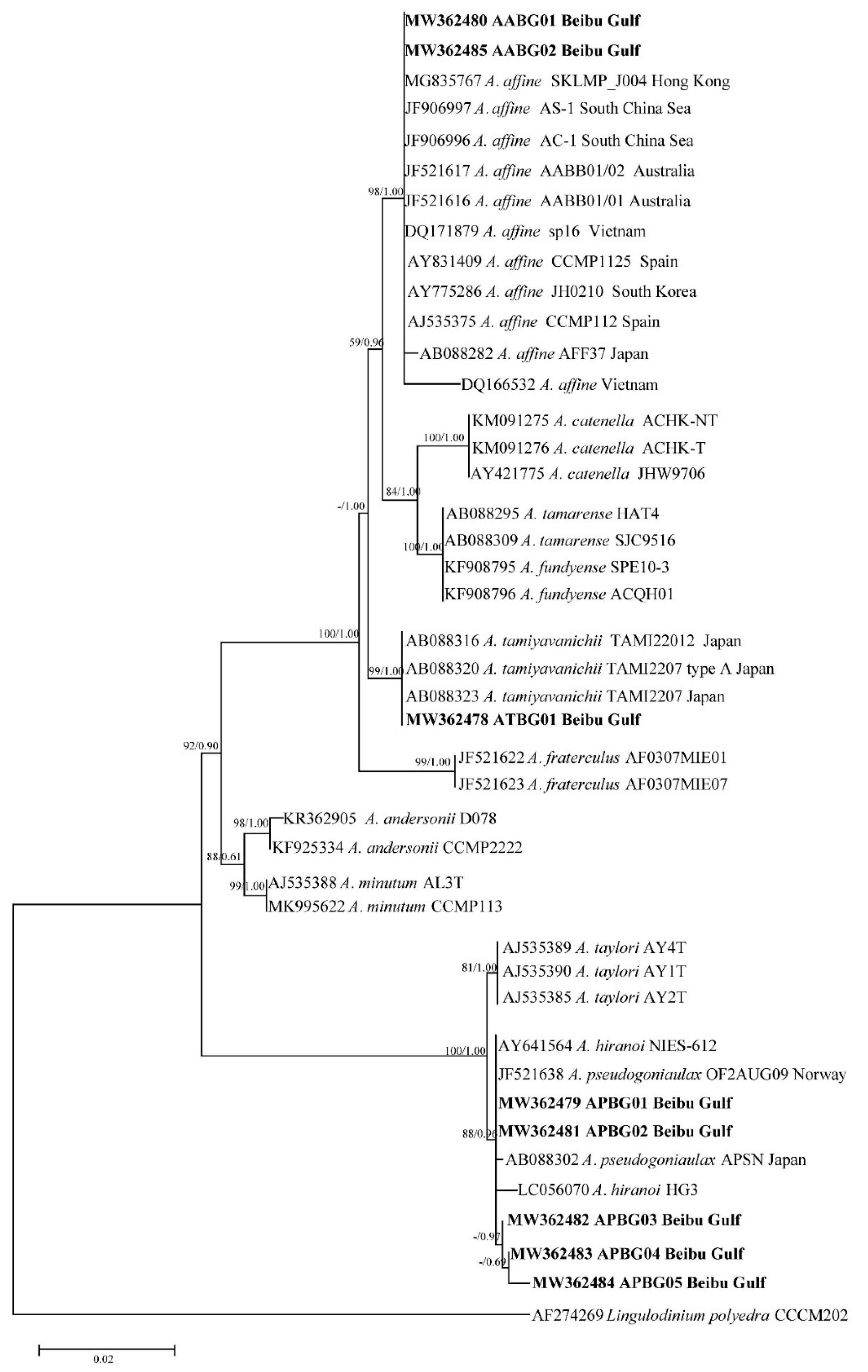
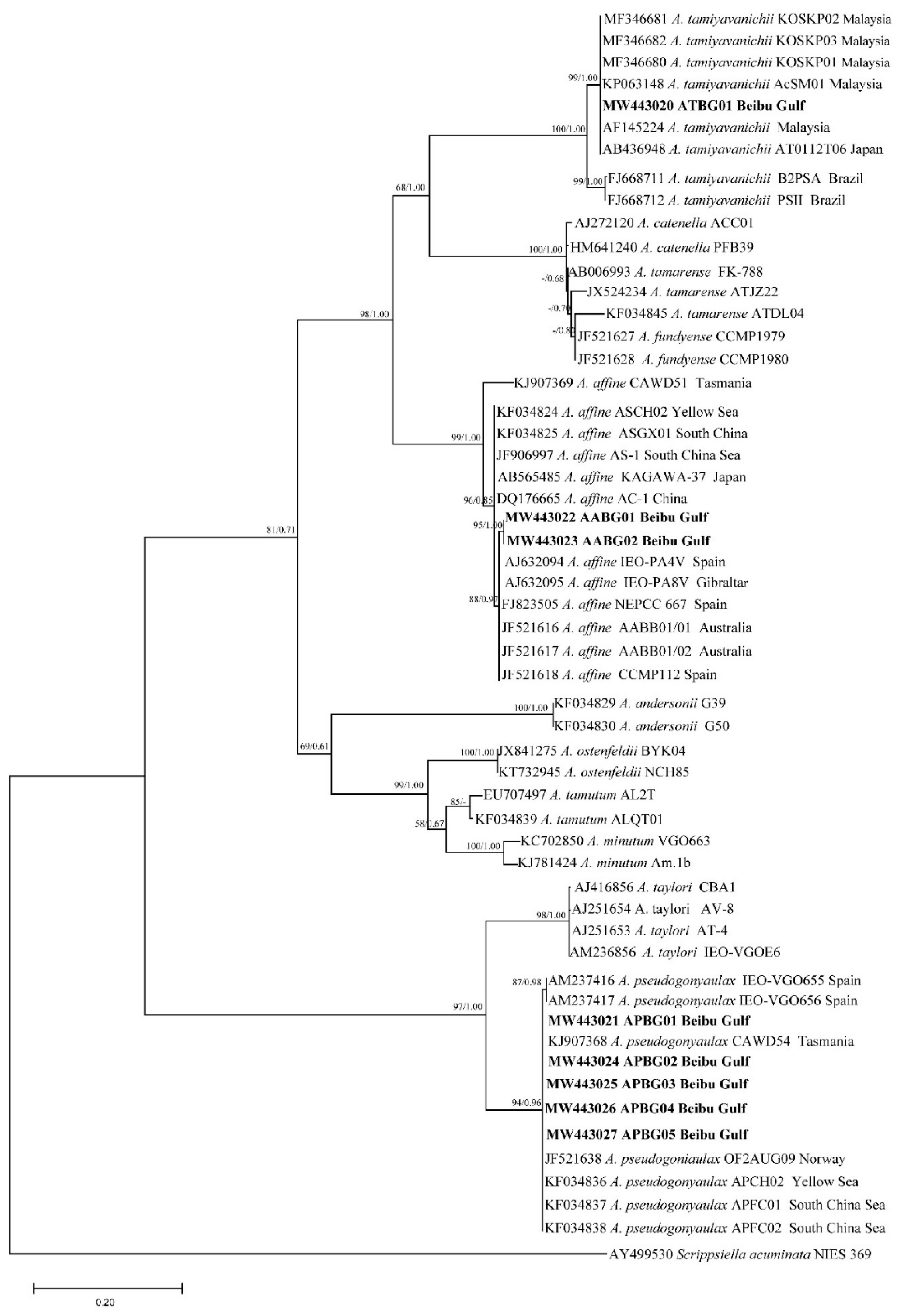
Phylogenetic trees of the LSU rDNA, SSU rDNA, and ITS sequences were constructed using Goniodoma polyedricum, Lingulodinium polyedra, and Scrippsiella acuminata as outgroups (Figure 1, Figure 2 and Figure 3). The topological structures of the trees using the maximum likelihood and Bayesian inference were generally consistent; thus, only the maximum likelihood trees are shown. The three phylogenetic trees all showed that five of the Alexandrium strains from the Beibu Gulf (APBG01, APBG02, APBG03, APBG04, APBG05) and A. pseudogonyaulax from various marine regions worldwide clustered into a large branch, with bootstrap values/Bayesian posterior probabilities of 83/0.58, 88/0.96, and 94/0.96, respectively. The Beibu Gulf ATBG01 strain clustered with A. tamiyavanichii from other regions, with bootstrap values/Bayesian posterior probabilities of 99/1.00, 99/1.00, and 100/1.00, respectively. Two other Alexandrium strains (AABG01, AABG02) from Beibu Gulf clustered with A. affine from other marine regions, with bootstrap values/Bayesian posterior probabilities of 99/1.00, 98/1.00, and 99/1.00, respectively (Figure 1, Figure 2 and Figure 3). A. tamarense, A. fundyense, and A. catenella clustered together in a large branch, with bootstrap values/Bayesian posterior probabilities of 99/1.00, 84/1.00, and 100/1.00, respectively, and the remaining Alexandrium species also formed clear branches with strong bootstrap values and posterior probabilities (Figure 1, Figure 2 and Figure 3).
The Jukes–Cantor genetic distance matrix calculated based on LSU rDNA, SSU rDNA, and ITS sequences showed genetic distances among the A. pseudogonyaulax APBG01, APBG02, APBG03, APBG04, APBG05 strains of 0.000–0.014, 0.000–0.004, and 0.002–0.028, respectively. The A. pseudogonyaulax strains with the highest homology were the APFC01 and APFC02 strains from the South China Sea (LSU), the OF2AUG09 strain from Norway (SSU), the APFC01 and APFC02 strains from the South China Sea, the APCH01 strain from the Yellow Sea, and the CAWD54 strain from New Zealand (ITS), with genetic distances of 0.000–0.002, 0.000–0.004, and 0.000, respectively, whereas the genetic distances of LSU rDNA, SSU rDNA, and ITS from other A. pseudogonyaulax strains worldwide were 0.000–0.021, 0.000–0.009, and 0.000–0.037, respectively. The intraspecific genetic distances of the three sequences of A. pseudogonyaulax were 0.000–0.021, 0.000–0.009, and 0.000–0.037, respectively, and generally smaller than the interspecific genetic distances of 0.009–0.881, 0.000–0.111, and 0.206–1.368 for Alexandrium species.
The genetic distances of the LSU rDNA, SSU rDNA, and ITS sequences of the two A. affine AABG01, AABG02 strains from Beibu Gulf were 0.000, 0.000, and 0.005, respectively. They were most closely related to the Korean LMBE_V129 (LSU), Vietnamese sp16 (SSU), and Spanish IEO-PA4V (ITS) strains of A. affine, with genetic distances of 0.001, 0.000, and 0.006, respectively, whereas the genetic distances of LSU rDNA, SSU rDNA, and ITS with the other worldwide A. affine strains were 0.001–0.015, 0.000–0.008, and 0.006–0.072, respectively. The intraspecific genetic distances of the three A. affine sequences were 0.000–0.020, 0.000–0.009, and 0.000–0.072, respectively, and also significantly smaller than the interspecific genetic distances of 0.100–0.576, 0.011–0.089, and 0.319–1.219 from other Alexandrium species.
The ATBG01 strain of A. tamiyavanichii from Beibu Gulf was most closely related to the Japanese AT0112T06 (LSU) and TAMI22012 (SSU) strains and the Malaysian AcSM01 (ITS) strain, with genetic distances of 0.000, 0.001, and 0.000, respectively, whereas the genetic distances of LSU rDNA, SSU rDNA, and ITS with other A. tamiyavanichii strains worldwide were 0.000–0.013, 0.001–0.002, and 0.000–0.060, respectively. The intraspecific genetic distances of the three A. tamiyavanichii sequences were 0.000–0.013, 0.000–0.002, and 0.000–0.060, which were also significantly smaller than the interspecific genetic distances of 0.138–0.881, 0.011–0.097, and 0.357–1.368 from other Alexandrium species.

2.3. Toxin Analysis

Table 2 shows the toxins analyzed in the Beibu Gulf APBG04 strain of A. pseudogonyaulax and the ATBG01 strains of A. tamiyavanichii, as well as the PST toxin composition and content. No PSTs were detectable in A. pseudogonyaulax APBG04, whereas multiple PST congeners were detected in A. tamiyavanichii ATBG01, including GTX1,4, GTX2,3, GTX5, and STX, at 0.88, 1.2, 0.32, and 2.20 fmol/cell, respectively (mole percent: 19.1%, 26.1%, 7.0%, and 47.8%, respectively); the total PST content was 4.60 fmol/cell.

3. Discussion

Alexandrium species are distributed worldwide, and many of them are toxic [1]. Determining the species composition, toxicity, and biogeographic distribution of Alexandrium spp. in a marine area is a prerequisite for HAB control and PSP mitigation. Alexandrium margalefii and A. tamiyavanichii are recently discovered species in China using optical and fluorescence microscopy [31,32]. Ten species of Alexandrium, A. affine, A. andersonii, A. catenella, A. fraterculum, A. leei, A. minutum, A. ostenfeldii, A. pseudogonyaulax, A. pacificum (as A. tamarense), and A. tamutum, have been identified in the East China, South China, Yellow, and Bohai Seas using optical, fluorescence and scanning electron microscopy, and rDNA molecular identification techniques [6,10,16,33]. While A. acatenella, A. catenella (as A. fundyense), A. hiranoi, A. minutum, A. ostenfeldii, A. pseudogonyaulax, A. pacificum, and A. tamiyavanichii were identified in Hong Kong waters morphologically [6,34], but molecular sequences are not available to confirm their identities. Considering the identification of Alexandrium is inadequate when only morphological standards are used, the real number of Alexandrium species in Chinese waters still remains to be determined.
Eight Beibu Gulf Alexandrium strains were identified based on LSU rDNA, SSU rDNA, and ITS sequences as A. affine, A. pseudogonyaulax, and A. tamiyavanichii. These results are unsurprising, as A. pseudogonyaulax and A. affine are globally distributed and have been reported in the sediments of Beibu Gulf previously [16], whereas A. tamiyavanichii is a warm-water species distributed in temperate to tropical regions such as Japan, Thailand, Malaysia, Brazil, and Mexico [35,36,37,38,39]. Both A. affine and A. pseudogonyaulax have been identified in the East China, South China, and Yellow Seas [16], while A. tamiyavanichii has been found only in the East China Sea [31] and in waters near Hong Kong [34] based on morphology. This is the first report of A. tamiyavanichii from the Beibu Gulf confirmed with molecular sequences. This is also the first report on the toxicity of A. tamiyavanichii from Chinese coastal waters.
Except for the A. tamarense species complex, the three phylogenetic trees generally formed clear branches of Alexandrium species with good bootstrap values. The positions of the three Beibu Gulf species on the trees were consistent with those of other published studies [13,14], indicating that Alexandrium species can be distinguished using LSU rDNA, SSU rDNA, and ITS sequences. However, the phylogenetic tree constructed using SSU rDNA has occasional exceptions, because A. hiranoi, A. pseudogonyaulax, and strains APBG01–05 clustered together (Figure 2). Alexandrium hiranoi and A. pseudogonyaulax are difficult to identify using SSU rDNA gene sequences, which have been noted previously [13,40].
Generally, algal strains of the same species from the same or similar geographic locations have small base differences and are closely related; the present findings on A. tamiyavanichii are consistent with this. A. tamiyavanichii from the Beibu Gulf is most closely related to the Japanese strains AT0112T06 and TAMI22012, and the Malaysian strain AcSM01, and relatively distantly related to the Brazilian PSII strain. A relationship between the different strains of A. affine and A. pseudogonyaulax in terms of genetic distance and geographical was not obvious, however. For example, in the ITS tree, Beibu Gulf A. affine had close homology with strains of this species from the geographically distant waters of Spain, Australia, and Gibraltar, and less homology with algal strains from the geographically close waters of China and Japan. The LSU rDNA and ITS trees of A. pseudogonyaulax also showed that strains with different geographical origins clustered on the same large branch with no obvious small-branch topology. Others have also noted similar phenomena [14,16], which might be associated with the physiological and ecological features of Alexandrium species [1,4,41].
Previous tests of A. pseudogonyaulax did not reveal any PSTs, but did find the toxin goniodomin A (GDA) [40,42,43]. We did not detect PSTs in five strains of Beibu Gulf A. pseudogonyaulax either; whether they can produce GDA awaits further investigation. Two strains of Beibu Gulf A. affine were lost during culture maintenance, so PSTs could not be analyzed. Current literature suggests that A. affine is largely nontoxic [16,44] or only potentially toxic [45]. However, A. affine strains with low PST toxin content (<1.0–2.28 fmol/cell), including NeoSTX, STX, and GTX1-4, have been isolated from Ha Long Bay, Vietnam, near the Beibu Gulf [46].
A. tamiyavanichii strains from various marine areas worldwide, including the Beibu Gulf strain described herein, can produce PST toxins, but toxin composition and content differ significantly (Table 3).
We detected only GTX1-5 and STX toxins in the Beibu Gulf strain, which is similar to the PSTs composition of the Chula 6 strains from Thailand and the KOSKP01, KOSKP02, and KOSKP03 strains from Malaysia [38,47,57], but different from other A. tamiyavanichii strains from these countries as well as strains from Japan, Brazil, and South Africa (Table 3). The range in PST content of A. tamiyavanichii reported in the literature is 1.17 fmol/cell–3.07 × 106 fmol/cell. The PST content of one A. tamiyavanichii strain isolated from the recent cases of PSP outbreak at Kuantan Port, Malaysia [38] reached an extreme maximum of 3.07 × 10⁶ fmol/cell, which is 100,000 times higher than strain ATBG01 in this study. The Beibu Gulf A. tamiyavanichii strain is a low-toxicity algal strain, but more strains need to be tested in the future, and measurements from bloom populations are needed as well.
Based on this and previous studies, Beibu Gulf Alexandrium species appear to be either non-toxic or weakly toxic [16,26,27]. This is, however, inconsistent with the high level of PST detection rate in Beibu Gulf shellfish, which have exceeded food safety standards on multiple occasions (Table 4).
Possible explanations for this inconsistency include the following:
(1)
Low-toxicity algal strains are widely distributed in the Beibu Gulf and survive for long periods. PSTs can thus accumulate in shellfish through time to levels sufficient to exceed regulatory standards. Anderson et al. [62] noted a similar phenomenon in Daya Bay, China where A. pacificum has very low toxicity of 7.2–12.7 fmol/cell, yet PSP poisoning still occasionally occurs there.
(2)
Based on reports of A. tamiyavanichii toxin content varying by more than 5 orders of magnitude among isolates from the same locations in Malaysia and by factors of 857 in Thailand and 651 in Japan (Table 3), it seems very likely that other highly toxic A. tamiyavanichii strains occur in the Beibu Gulf that were not isolated. Low-abundance Alexandrium spp. in seawater can be missed due to limitations of small numbers of culture isolations, and thus highly toxic strains could easily be overlooked. Furthermore, because isolates were only established from Weizhou Island in the Beibu Gulf, and not from other highly productive areas of the Gulf, such as Qinzhou Bay and Tieshan Harbor, the presence of highly toxic species in these regions cannot be ruled out. In this regard, it is of note that the PST composition of shellfish from the Beibu Gulf differs significantly from the A. tamiyavanichii isolates analyzed here (Table 3 and Table 4). This could reflect biotransformation (see below) and/or different toxic Alexandrium species or strains from those analyzed here.
(3)
Alexandrium in field populations can grow faster and produce more toxins than laboratory cultures, as has been observed by others through variations in temperature, salinity, light, and nutrients [63,64,65,66]. Brosnahan et al. [67] recently reported in situ growth rates for A. catenella that were more than twice those observed in laboratory cultures under similar growth conditions.
(4)
Metabolism and bioconversion processes in different shellfish greatly affect the accumulation and potency of PSTs [68,69]. For example, the PSP toxicity of Paphia undulata is over 1000 times higher than that of Meretrix lusoria in the Beibu Gulf [29]. Thus, further study of biotransformation of PSTs in different shellfish from the Beibu Gulf is needed.
(5)
Although Alexandrium spp.are the principal causative organisms of PSP in many regions, the presence of other PST-producing microalgae, such as Gymnodinium catenatum and Pyrodinium bahamense [70,71], and atypical toxin-producing organisms such as brackish cyanobacteria, as well as calcareous red macroalgae [72], cannot be ruled out.

4. Conclusions

The Beibu Gulf region is experiencing rapid economic growth, frequent HABs, and the increasingly serious problem of PST contamination of shellfish. To explore this issue, potential PST toxin-producing algae in the Alexandrium genus were isolated from Weizhou Island, a HAB hotspot in the Beibu Gulf and characterized phylogenetically and toxicologically. For the first time, A. affine, A. pseudogonyaulax, and A. tamiyavanichii were definitively identified in plankton samples from the Beibu Gulf, among which, A. tamiyavanichii is new to this body of water. This species showed small base differences from Asian strains of the same species from nearby locations. There was no observed relationship between genetic distance and the geographical distribution of various A. affine and A. pseudogonyaulax strains. A. pseudogonyaulax strains did not produce PSTs, whereas A. tamiyavanichii produced 4.6 fmol/cell of toxin, indicating that it is a low-toxicity strain in comparison to global isolates. This finding of low toxicity is inconsistent with the detection of relatively high levels of PSTs in shellfish from the Beibu Gulf, suggesting that higher toxicity strains may occur in those waters, but were unsampled. Other explanations including biotransformation of PSTs in shellfish and the presence of other PST-producing algae are also suggested. This is the first report of toxic A. tamiyavanichii in the coastal waters in China. In future studies, potential PST-producing microalgae and toxic shellfish should be collected from the major localities in the Beibu Gulf to further elucidate species diversity, biogeographical distribution, toxin-producing characteristics, and relationships with PST vectors in the region. This would provide much-needed insights into PST sources and aid in the protection of public health in the Beibu Gulf.

5. Materials and Methods

5.1. Algal Source and Culture Conditions

Water samples containing phytoplankton collected from the waters of Weizhou Island in the Beibu Gulf four times between the summer 2018 and summer 2019 (Figure 4), were transported and stored as described in the “Specification for Marine Monitoring” (GB/T17378-2007). The samples were visually assessed using a TS100 inverted microscope (Nikon Corp., Tokyo, Japan). Algal cells of interest were isolated from water samples using capillary tubes, washed 3 or 4 times with sterilized seawater, then cultured, resulting in eight Alexandrium strains (Table 5). The culture conditions were f/2 medium, 22 °C, light intensity 150 μE/m2/s, and a 12:12 h light-dark cycle. The algal strains ATBG01 and APBG0 were cultured to the exponential growth phase, then toxin was extracted from 6 × 10⁶ algal cells.

5.2. Extraction of DNA, PCR Amplification, and Sequencing

Fresh algae (30 mL) in the exponential growth phase were collected and sedimented by centrifugation at 7000× g for 1 min, then the supernatants were discarded. Total DNA was obtained using BioFastSpin Plant Genomic DNA Extraction Kits (Bioer, Hangzhou, China), and characteristic LSU rDNA, SSU rDNA, and ITS fragments were amplified by PCR. The amplification primers (5′→3′) were listed in Table 6. The PCR reaction volume was 40 µL, and included 20 µL of 2 × Es Taq Master Mix (CoWin Biosciences, Cambridge, MA, USA), 1 µL of DNA template, 17 µL of ddH2O, and 1 µL each of forward and reverse primer. The PCR program comprised pre-denaturation at 94 °C for 5 min, 35 cycles of 94°C for 30 s, 56 °C for 30 s, and 72 °C for 30 s, and elongation at 72 °C for 5 min. The PCR products were sequenced at a commercial laboratory (TsingKe Biological Technology Company (Beijing, China).

5.3. Sequence Analyses

We searched the LSU rDNA, SSU rDNA, and ITS sequences of genes from the eight Alexandrium strains in the NCBI database using BLAST, and downloaded 43 LSU rDNA, 34 SSU rDNA, and 44 ITS sequences with high species similarity and coverage. We used the online BioEdit v7.2.5 software (https://bioedit.software.informer.com/, accessed on 8 January 2021), for multiple sequence alignment of the obtained sequences. We calculated and analyzed genetic distances within and between species and calculated base compositions, conserved, variable, parsimony informative, and singleton sites, and transition/transversion ratios using online MEGA-X v10.1.5 software (https://www.megasoftware.net/, accessed on 10 January 2021). We confirmed that GIR+I+G was the best alternative model using the AIC function in downloadable Modeltest3.7 software (https://sourceforge.net/projects/modeltest/, accessed on 10 January 2021). We constructed a maximum likelihood phylogenetic tree for the sequences obtained using MEGA-X v10.1.5 software, selected GIR+I+G as the best alternative model, and tested the confidence of the branches by running 1000 bootstrap replications. We constructed a Bayesian phylogenetic tree using Bayesian inference (BI) in MrBayes v3.1.2 software (http://nbisweden.github.io/MrBayes/, accessed on 13 January 2021), again with GIR+I+G as the best replacement model, and 4 Markov chain operations were run for 1 × 10⁶ generations for Bayesian analysis, with sampling every 500 generations. The phylogenetic tree was viewed and downloaded using FigTree v1.4.0 software, and edited using Adobe Acrobat DC v2020.013.20074.

5.4. Toxin Extraction

Algal suspensions (6 L) in the exponential growth phase were centrifuged at 4000 rpm for 8 min (15 °C), the supernatant was decanted, and the sedimented microalgal cells were stored at −80 °C. After three freeze-thaw cycles, stored algal cells were suspended in 10 mL of 0.1 mol/L acetic acid, sonicated on ice for 20 min, and centrifuged at 8000 rpm for 10 min at 4 °C. The supernatant was collected, the pellet was extracted again, the supernatants were combined and adjusted to a volume of 20 mL with 0.1 mol/L acetic acid, and 1.0 mL was passed through 0.22 µm mobile phase filter membranes (Shanghai ANPEL Laboratory Technologies Inc., Shanghai, China) for LC-MS/MS analysis.

5.5. Liquid Chromatography-Mass Spectrometry

We analyzed the PSP toxins, GTX1, GTX2, GTX3, GTX4, GTX5, dcGTX2, dcGTX3, STX, dcSTX, neoSTX, dcNEO, C1, and C2 using liquid chromatography-tandem mass spectrometry (LC-MS/MS). Chromatographic conditions: These comprised a Welch Ultimate Amide chromatographic column (4.6 × 150 mm × 3.5 μm); mobile phase A, 2 mmol/L ammonium formate (containing 50 mM formic acid); B, acetonitrile; injection volume, 10 μL; column temperature, 40 °C; flow rate, 0.6 mL/min. The gradient elution program was 0–1 min, 90% A; 1–3 min, linear gradient from 90% A to 10% A; 3–10 min, 10% A; 10–11 min, linear gradient from 10% A to 90%; 11–13 min, 90% A.
Mass spectrometry conditions: ESI ion source, positive/negative polarity switching scan mode, capillary voltage, 4000 V; nozzle voltage, 500 V; sheath gas temperature, 350 °C; sheath gas flow, 11 L/min; drying gas temperature, 320 °C; drying gas flow, 5 L/min; nebulizer pressure, 45 psi. Specific multiple reaction monitoring parameters and conditions are shown in Table 7.

Author Contributions

Conceptualization, Y.X.; methodology, Y.X., X.H. and T.Z.; software, X.H. and H.L.; validation, Y.X., H.G. and D.M.A.; formal analysis, Y.X., X.H. and H.L.; investigation, T.Z. and F.L.; resources, T.Z. and F.L.; data curation, Y.X.; writing—original draft preparation, Y.X. and X.H.; writing—review and editing, Y.X., H.G. and D.M.A.; visualization, X.H.; supervision, Y.X.; project administration, Y.X.; funding acquisition, Y.X. and D.M.A. All authors have read and agreed to the published version of the manuscript.

Funding

This research was funded by the National Natural Science Foundation of China (41976155, 41506137), the Natural Science Foundation of Guangxi Province (2020GXNSFDA297001, 2016GXNSFBA380037), the Woods Hole Center for Oceans and Human Health (National Science Foundation grant OCE-1840381 and National Institutes of Health grants NIEHS-1P01-ES028938-01), the Opening Project of Guangxi Laboratory on the Study of Coral Reefs in the South China Sea (GXLSCRSCS2019002), the Opening Foundation of Key Laboratory of Environment Change and Resources Use in Beibu Gulf Ministry of Education (Nanning Normal University), and the Guangxi Key Laboratory of Earth Surface Processes and Intelligent Simulation (Nanning Normal University) (GTEU-KLOP-K1803).

Institutional Review Board Statement

Not applicable.

Informed Consent Statement

Not applicable.

Data Availability Statement

Data are available upon request, please contact the correspondence author Yixiao Xu.

Conflicts of Interest

The authors declare no conflict of interest.

References

  1. Anderson, D.M.; Alpermann, T.J.; Cembella, A.D.; Collos, Y.; Masseret, E.; Montresor, M. The globally distributed genus Alexandrium: Multifaceted roles in marine ecosystems and impacts on human health. Harmful Algae 2012, 14, 10–35. [Google Scholar] [CrossRef] [Green Version]
  2. Halim, Y. Alexandrium minutum nov. g. nov. sp. Dinoflagellé provocant des “eaux rouges”. Vie Milieu 1960, 11, 102–105. [Google Scholar]
  3. Mackenzie, L.; Salas, M.D.; Adamson, J.; Beuzenberg, V. The dinoflagellate genus Alexandrium (Halim) in New Zealand coastal waters: Comparative morphology, toxicity and molecular genetics. Harmful Algae 2004, 3, 71–92. [Google Scholar] [CrossRef]
  4. Bill, B.D.; Moore, S.K.; Hay, L.R.; Anderson, D.M.; Trainer, V.L. Effects of temperature and salinity on the growth of Alexandrium (Dinophyceae) isolates from the Salish Sea. J. Phycol. 2016, 52, 230–238. [Google Scholar] [CrossRef] [Green Version]
  5. Liang, Y.; Li, D.; Yao, J.; Jin, W.; Gong, C.; Liu, R. Progresses in investigation and research on phycotoxins and toxic microalgaes in the coastal waters of China. Oceanol. Et Limnol. Sin. 2019, 50, 511–524. [Google Scholar]
  6. Litaker, R.W.; Fraga, S.; Montresor, M.; Brosnahan, M.; Anderson, D.M.; Hoppenrath, M.; Murray, S.; Wolny, J.; John, U.; Sampedro, N.; et al. A practical guide to new nomenclature for species within the “Alexandrium tamarense species complex”. Harmful Algae News 2018, 61, 13–15. [Google Scholar]
  7. Balech, E. The Genus Alexandrium Halim (Dinoflagellata); Sherkin Island Marine Station: Cork, Ireland, 1995; p. 452. [Google Scholar]
  8. Murray, S.A.; Hoppenrath, M.; Orr, R.J.S.; Bolch, C.; John, U.; Diwan, R.; Yauwenas, R.; Harwood, T.; Salas, M.D.; Neilan, B.; et al. Alexandrium diversaporum sp. nov., a new non-saxitoxin producing species: Phylogeny, morphology and sxtA genes. Harmful Algae 2014, 31, 54–65. [Google Scholar] [CrossRef]
  9. Mertens, K.N.; Adachi, M.; Anderson, D.M.; Band-Schmidt, C.J.; Bravo, I.; Brosnahan, M.L.; Bolch, C.J.S.; Calado, A.J.; Carbonell-Moore, M.C.; Chomérat, N.; et al. Morphological and phylogenetic data do not support the split of Alexandrium into four genera. Harmful Algae 2020, 98, 101902. [Google Scholar] [CrossRef]
  10. John, U.; Litaker, R.W.; Montresor, M.; Murray, S.; Brosnahan, M.L.; Anderson, D.M. Formal revision of the Alexandrium tamarense species complex (Dinophyceae) taxonomy: The introduction of five species with emphasis on molecular-based (rDNA) classification. Protist 2014, 165, 779–804. [Google Scholar] [CrossRef] [Green Version]
  11. Fraga, S.; Sampedro, N.; Larsen, J.; Moestrup, Ø.; Calado, A.J. Arguments against the proposal 2302 by John & al. to reject the name Gonyaulax catenella (Alexandrium catenella). Taxon 2015, 64, 634–635. [Google Scholar]
  12. Prud’homme van Reine, W.F. Report of the nomenclature committee for algae: 15. Taxon 2017, 66, 191–192. [Google Scholar] [CrossRef] [Green Version]
  13. Lim, A.S.; Jeong, H.J.; Kim, J.H.; Lee, S.Y. Description of the new phototrophic dinoflagellate Alexandrium pohangense sp. nov. from Korean coastal waters. Harmful Algae 2015, 46, 49–61. [Google Scholar] [CrossRef]
  14. Branco, S.; Oliveira, M.M.M.; Salgueiro, F.; Vilar, M.C.P.; Azevedo, S.M.F.O.; Menezes, M. Morphology and molecular phylogeny of a new PST-producing dinoflagellate species: Alexandrium fragae sp. nov. (Gonyaulacales, Dinophyceae). Harmful Algae 2020, 95, 101793. [Google Scholar] [CrossRef] [PubMed]
  15. Gu, H. Morphology, phylogenetic position, and ecophysiology of Alexandrium ostenfeldii (Dinophyceae) from the Bohai Sea, China. J. Syst. Evol. 2011, 49, 606–616. [Google Scholar] [CrossRef]
  16. Gu, H.; Zeng, N.; Liu, T.; Yang, W.; Mueller, A.; Krock, B. Morphology, toxicity, and phylogeny of Alexandrium (Dinophyceae) species along the coast of China. Harmful Algae 2013, 27, 68–81. [Google Scholar] [CrossRef]
  17. Xu, Y.; Zhang, T.; Zhou, J. Historical occurrence of algal blooms in the northern Beibu Gulf of China and implications for future trends. Front. Microbiol. 2019, 10, 451. [Google Scholar] [CrossRef] [Green Version]
  18. Kang, Z.; Yang, B.; Lai, J.; Ning, Y.; Zhong, Q.; Lu, D.; Liao, R.; Wang, P.; Solomon, F.D.; She, Z.; et al. Phaeocystis globosa bloom monitoring: Based on P. globosa induced seawater viscosity modification adjacent to a nuclear power plant in Qinzhou Bay, China. J. Ocean Univ. China 2020, 19, 1207–1220. [Google Scholar] [CrossRef]
  19. Zou, C. Genetic Structure, Phylogeography Relationship and PSP Toxin Profile of Alexandrium tamarense Species Complex. Master’s Thesis, Jinan University, Guangzhou, China, 2015. [Google Scholar]
  20. Lu, X. Biogeological Distribution of Dinoflagellate Cysts in Surface Sediments from Southern Chinese Coast and Their Roles as a Signal of Environmental Changes. Ph.D. Thesis, Jinan University, Guangzhou, China, 2016. [Google Scholar]
  21. Jiang, F.; Chen, B.; He, B.; Xu, M.; Zhuang, J.; Zhang, R.; Lei, F. Phytoplankton community in coastal waters of Qinzhou Bay in Guangxi. Guangxi Sci. 2012, 19, 268–275. [Google Scholar]
  22. Wang, Z.; Ma, C.; Xu, Z. Phylogenetic relationship of four Alexandrium tamarense/catenella isolates from Southeast Chinese costal waters using rDNA sequences. Mar. Environ. Sci. 2015, 34, 1–5. [Google Scholar]
  23. Anderson, D.M.; Stock, C.A.; Keafer, B.A.; Nelson, A.B.; Thompson, B.; Mcgillicuddy, D.J.; Keller, M.; Matrai, P.A.; Martin, J. Alexandrium fundyense cyst dynamics in the Gulf of Maine. Deep Sea Res. Part II Top. Stud. Oceanogr. 2005, 52, 2522–2542. [Google Scholar] [CrossRef]
  24. Martin, J.L.; Legresley, M.M.; Hanke, A.R. Thirty years—Alexandrium fundyense cyst, bloom dynamics and shellfish toxicity in the Bay of Fundy, eastern Canada. Deep Sea Res. Part II Top. Stud. Oceanogr. 2014, 103, 27–39. [Google Scholar] [CrossRef]
  25. Wang, D.; Jiang, T.; Hsieh, D.P.H. Toxin composition variations in cultures of Alexandrium species isolated from the coastal waters of southern China. Harmful Algae 2005, 4, 109–121. [Google Scholar] [CrossRef]
  26. Zou, C.; Ye, R.; Zheng, J.; Luo, Z.; Gu, H.; Yang, W.; Li, H.; Liu, J. Molecular phylogeny and PSP toxin profile of the Alexandrium tamarense species complex along the coast of China. Mar. Pollut. Bull. 2014, 89, 209–219. [Google Scholar] [CrossRef]
  27. Wang, D.; Zhang, S.; Gu, H.; Chan, L.; Hong, H. Paralytic shellfish toxin profiles and toxin variability of the genus Alexandrium (Dinophyceae) isolated from the Southeast China Sea. Toxicon 2006, 48, 138–151. [Google Scholar] [CrossRef]
  28. Zhong, X.; Liu, D.; Liang, H. Risk assessment of paralytic shellfish poison in exported shellfish from Beibu Gulf. Enterp. Sci. Technol. Dev. 2010, 22, 314–315. [Google Scholar]
  29. Zhao, P.; Qin, X.; Hu, K.; Li, Z.; Jiang, N.; Xu, Y. Paralytic shellfish poisoning of bivalves in seafood market, Beibu Gulf of Guangxi. Ecol. Sci. 2016, 35, 73–77. [Google Scholar]
  30. Dai, Z.; Guo, W.; Zhang, C.; Wang, P.; Huang, M.; Li, C. Detection and safety evaluation of paralytic shellfish toxins in 7 species of Qinzhou Bay economically shellfish. Food Res. Dev. 2018, 39, 120–125. [Google Scholar]
  31. Guo, L.; Lu, S. A new record of Alexandrium tamiyavanichii Balech from the coast of China. In Proceedings of the 8th Member Conference of the Chinese Phycological Society and 16th Academic Symposium, Shanghai, China, 11 November 2011; p. 191. [Google Scholar]
  32. Guo, L. Morphological Taxonomy and Ecological Distribution on Alexandrium in Chinese Coastal Waters. Master’s Thesis, South China Normal University, Guangzhou, China, 2012. [Google Scholar]
  33. Li, D.; Lu, D.; Dai, X.; He, P.; Xia, P.; Wang, H. Morphological-phylogenetic analysis and population dynamics of Alexandrium tamarense in the bloom area of the East China Sea. Oceanol. Et Limnol. Sin. 2014, 45, 1241–1250. [Google Scholar]
  34. Law, S.P.C.; Lee, F.Y.K. Harmful Marine Microalgae in Hong Kong, 2nd ed.; Agriculture, Fisheries and Conservation Department: Hong Kong, China, 2013; pp. 1–47.
  35. Hashimoto, T.; Matsuoka, S.; Yoshimatsu, S.A.; Miki, K.; Nishibori, N.; Nishio, S.; Noguchi, T. First paralytic shellfish poison (PSP) infestation of bivalves due to toxic dinoflagellate Alexandrium tamiyavanichii, in the southeast coasts of the Seto Inland Sea, Japan. J. Food Hyg. Soc. Jpn. 2002, 43, 1–5. [Google Scholar] [CrossRef] [Green Version]
  36. Menezes, M.; Varela, D.; Proença, L.A.D.O.; Tamanaha, M.D.S.; Paredes, J. Identification of the toxic alga Alexandrium tamiyavanichi (Dinophyceae) from northeastern brazil: A combined morphological and rDNA sequence (partial LSU and ITS) approach. J. Phycol. 2010, 46, 1239–1251. [Google Scholar] [CrossRef]
  37. Sagara, T.; Taniyama, S.; Yoshimatsu, S.; Takatani, T.; Hashimoto, T.; Nishibori, N.; Nishio, S.; Arakawa, O. Toxicity and toxin profile of the dinoflagellate Alexandrium tamiyavanichii and toxic mussels in Harima-Nada of Seto Inland Sea, Japan. Shokuhinseigaku Zasshi 2010, 51, 170–177. [Google Scholar] [CrossRef] [PubMed] [Green Version]
  38. Hamdan, N.A.; Hassan, M.S.A.; Hamid, S.D.; Mohammad-Noor, N.; Bunnori, N.M. Toxicity profiles between strains of Alexandrium tamiyavanichii. Int. J. Adv. Sci. Eng. Technol. 2018, 6, 2321–9009. [Google Scholar]
  39. Hernandez-Becerril, D.U.; Lau, W.L.; Hii, K.S.; Leaw, C.P.; Varona-Cordero, F.; Lim, P.T. Abundance and distribution of the potentially toxic thecate dinoflagellate Alexandrium tamiyavanichii (Dinophyceae) in the central Mexican pacific, using the quantitative PCR method. Front. Mar. Sci. 2018, 5, 366. [Google Scholar] [CrossRef]
  40. Triki, H.Z.; Laabir, M.; Moeller, P.; Chomérat, N.; Daly-Yahia, O.K. First report of goniodomin A production by the dinoflagellate Alexandrium pseudogonyaulax developing in southern Mediterranean (Bizerte Lagoon, Tunisia). Toxicon 2016, 111, 91–99. [Google Scholar] [CrossRef] [Green Version]
  41. Yang, I.; John, U.; Beszteri, S.; Glöckner, G.; Krock, B.; Goesmann, A.; Cembella, A.D. Comparative gene expression in toxic versus non-toxic strains of the marine dinoflagellate Alexandrium minutum. BMC Genom. 2010, 11, 248. [Google Scholar] [CrossRef] [Green Version]
  42. Krock, B.; Tillmann, U.; Wen, Y.; Hansen, P.J.; Larsen, T.O.; Andersen, A.J.C. Development of a LC-MS/MS method for the quantification of goniodomins A and B and its application to Alexandrium pseudogonyaulax strains and plankton field samples of Danish coastal waters. Toxicon 2018, 155, 51–60. [Google Scholar] [CrossRef] [Green Version]
  43. Harris, C.M.; Reece, K.S.; Stec, D.F.; Scott, G.P.; Jones, W.M.; Hobbs, P.L.M.; Harris, T.M. The toxin goniodomin, produced by Alexandrium spp., is identical to goniodomin A. Harmful Algae 2020, 92, 101707. [Google Scholar] [CrossRef]
  44. Draredja, M.A.; Frihi, H.; Boualleg, C.; Abadie, E.; Laabir, M. Distribution of dinoflagellate cyst assemblages in recent sediments from a southern Mediterranean lagoon (Mellah, Algeria) with emphasis on toxic species. Environ. Sci. Pollut. Res. Int. 2020, 27, 25173–25185. [Google Scholar] [CrossRef] [PubMed]
  45. Lee, K.H.; Jeong, H.J.; Kang, H.C.; Ok, J.H.; You, J.H.; Park, S.A. Growth rates and nitrate uptake of co-occurring red-tide dinoflagellates Alexandrium affine and A. fraterculus as a function of nitrate concentration under light-dark and continuous light conditions. Algae 2019, 34, 237–251. [Google Scholar] [CrossRef] [Green Version]
  46. Nguyen-Ngoc, L. An autecological study of the potentially toxic dinoflagellate Alexandrium affine isolated from Vietnamese waters. Harmful Algae 2004, 3, 117–129. [Google Scholar] [CrossRef]
  47. Kodama, M.; Ogata, T.; Fukuyo, Y.; Ishimaru, T.; Wisessang, S.; Sartanu, K.; Panichyakarn, V.; Piyakarncana, T. Protogonyaulax cohorticula, a toxic dinoflagellate found in the Gulf of Thailand. Toxicon 1988, 26, 707–712. [Google Scholar] [CrossRef]
  48. Ogata, T.; Pholpunthin, P.; Fukuyo, Y.; Kodama, M. Occurrence of Alexandrium cohorticula in Japanese coastal water. J. Appl. Phycol. 1990, 2, 351–356. [Google Scholar] [CrossRef]
  49. Usup, G.; Pin, L.C.; Ahmad, A.; Teen, L.P. Alexandrium (Dinophyceae) species in Malaysian waters. Harmful Algae 2002, 1, 265–275. [Google Scholar] [CrossRef]
  50. Ruiz Sebastián, C.; Etheridge, S.M.; Cook, P.A.; O’Ryan, C.; Pitcher, G.C. Phylogenetic analysis of toxic Alexandrium (Dinophyceae) isolates from South Africa: Implications for the global phylogeography of the Alexandrium tamarense species complex. Phycologia 2005, 44, 49–60. [Google Scholar] [CrossRef]
  51. Nagai, S.; Suzuki, M.; Matsuyama, Y.; Hakura, S.; Go, J. Molecular and toxicity analysis of toxic dinoflagellate A. tamiyavanichii isolated from Seto Inland Sea. In Proceedings of the Annual Meeting of the Japanese Society of Fisheries Science Conference Abstracts, Tokyo, Japan; 2005; p. 259. [Google Scholar]
  52. Lim, P.T.; Ogata, T. Salinity effect on growth and toxin production of four tropical Alexandrium species (Dinophyceae). Toxicon 2005, 45, 699–710. [Google Scholar] [CrossRef]
  53. Lim, P.T.; Leaw, C.P.; Usup, G.; Kobiyama, A.; Koike, K.; Ogata, T. Effects of light and temperature on growth, nitrate uptake, and toxin production of two tropical dinoflagellates: Alexandrium tamiyavanichii and Alexandrium minutum (Dinophyceae). J. Phycol. 2006, 42, 786–799. [Google Scholar] [CrossRef]
  54. Usup, G.; Cheah, M.Y.; Ng, B.K.; Leaw, C.P.; Ahmad, A. Toxin profile and relative toxicity of three paralytic shellfish poisoning toxin-producing dinoflagellates from Malaysia. Malays. Appl. Biol. 2006, 35, 41–45. [Google Scholar]
  55. Beppu, R.; Nojima, K.; Tsuruda, S.; Gomez-Delan, G.; Barte-Quilantang, M.; Taniyama, S.; Sagara, T.; Nishio, S.; Takayama, H.; Miyazawa, K.; et al. Occurrence of PSP-producing dinoflagellate Alexandrium tamiyavanichii in Bingo-Nada, the central coastal water of the Seto Inland Sea, Hiroshima Prefecture, Japan. Mar. Pollut. Bull. 2008, 56, 758–763. [Google Scholar] [CrossRef]
  56. Oh, S.J.; Matsuyama, Y.; Nagai, S.; Itakura, S.; Yoon, Y.H.; Yang, H.S. Comparative study on the PSP component and toxicity produced by Alexandrium tamiyavanichii (Dinophyceae) strains occurring in Japanese coastal water. Harmful Algae 2009, 8, 362–368. [Google Scholar] [CrossRef]
  57. Mohammad-Noor, N.; Adam, A.; Lim, P.T.; Leaw, C.P.; Lau, W.L.S.; Liow, G.R.; Muhamad-Bunnor, N.; Hamdan, N.A.; Md-Nor, A.; Kemat, N.; et al. First report of paralytic shellfish poisoning (PSP) caused by Alexandrium tamiyavanichii in Kuantan Port, Pahang, East Coast of Malaysia. Phycol. Res. 2018, 66, 37–44. [Google Scholar] [CrossRef]
  58. Lu, C.; Wu, S.; Zhang, C.; Yu, H.; Zhu, X.; Liu, M. Analysis of paralytic shellfish poison in shellfish in the offshore of the Guangxi. Mar. Environ. Sci. 2005, 24, 47–50. [Google Scholar]
  59. Jiang, T.; Jiang, T. Detection and analysis of PSP toxins in shellfish in coastal areas of China. Oceanol. Et Limnol. Sin. 2007, 38, 36–41. [Google Scholar]
  60. Huang, Y.; Huang, G.; Ye, X.; Li, X.; Wu, X.; Pang, Y.; Xie, Z.; Chen, J. Rapid detection of paralytic shellfish poison (PSP) in aquatic products. Jiangsu Agric. Sci. 2012, 40, 255–256. [Google Scholar]
  61. Du, K.; Lei, F.; Wu, N.; Jiang, T. The pattern of paralytic shellfish poisoning in shellfish cultured in East China Sea and South China Sea. J. Jinan Univ. 2013, 34, 343–346. [Google Scholar]
  62. Anderson, D.M.; Kulis, D.M.; Qi, Y.Z.; Zheng, L.; Lu, S.; Lin, Y.T. Paralytic shellfish poisoning in southern China. Toxicon 1996, 34, 579–590. [Google Scholar] [CrossRef]
  63. Etheridge, S.M.; Roesler, C.S. Effects of temperature, irradiance, and salinity on photosynthesis, growth rates, total toxicity, and toxin composition for Alexandrium fundyense isolates from the Gulf of Maine and Bay of Fundy. Deep Sea Res. Part II Top. Stud. Oceanogr. 2005, 52, 2491–2500. [Google Scholar] [CrossRef]
  64. Murray, S.; John, U.; Kremp, A. Alexandrium spp.: Genetic and ecological factors influencing saxitoxin production and proliferation. In Climate Change and Marine and Freshwater Toxins, 2nd ed.; Botana, L.M., Louzao, M.C., Eds.; Walter de Gruyter GmbH: Berlin, Germany; Boston, MA, USA, 2015; pp. 125–155. [Google Scholar]
  65. Vandersea, M.W.; Kibler, S.R.; Tester, P.A.; Holderied, K.; Hondolero, D.E.; Powell, K.; Baird, S.; Dorof, A.; Dugan, D.; Litaker, R.W. Environmental factors influencing the distribution and abundance of Alexandrium catenella in Kachemak bay and lower cook inlet, Alaska. Harmful Algae 2018, 77, 81–92. [Google Scholar] [CrossRef] [PubMed]
  66. Tobin, E.D.; Wallace, C.L.; Crumpton, C.; Johnson, G.; Eckert, G.L. Environmental drivers of paralytic shellfish toxin producing Alexandrium catenella blooms in a fjord system of northern Southeast Alaska. Harmful Algae 2019, 88, 101659. [Google Scholar] [CrossRef]
  67. Brosnahan, M.L.; Velo-Suárez, L.; Ralston, D.K.; Fox, S.E.; Sehein, T.R.; Shalapyonok, A.; Sosik, H.M.; Olson, R.J.; Anderson, D.M. Rapid growth and concerted sexual transitions by a bloom of the harmful dinoflagellate Alexandrium fundyense (Dinophyceae). Limnol. Oceanogr. 2015, 60, 2059–2078. [Google Scholar] [CrossRef] [Green Version]
  68. Bricelj, V.M.; Shumway, S.E. Paralytic shellfish toxins in bivalve molluscs: Occurrence, transfer kinetics, and biotransformation. Rev. Fish. 1998, 6, 315–383. [Google Scholar] [CrossRef]
  69. Xun, X.; Cheng, J.; Wang, J.; Li, Y.; Li, X.; Li, M.; Lou, J.; Kong, Y.; Bao, Z.; Hu, X. Solute carriers in scallop genome: Gene expansion and expression regulation after exposure to toxic dinoflagellate. Chemosphere 2020, 241, 124968. [Google Scholar] [CrossRef]
  70. Etheridge, S.M. Paralytic shellfish poisoning: Seafood safety and human health perspectives. Toxicon 2010, 56, 108–122. [Google Scholar] [CrossRef] [PubMed] [Green Version]
  71. Turner, A.D.; Hatfield, R.G.; Maskrey, B.H.; Algoet, M.; Lawrence, J.F. Evaluation of the new European Union reference method for paralytic shellfish toxins in shellfish: A review of twelve years regulatory monitoring using pre-column oxidation LC-FLD. TrAC Trends Anal. Chem. 2019, 113, 124–139. [Google Scholar] [CrossRef]
  72. Deeds, J.R.; Landsberg, J.H.; Etheridge, S.M.; Pitcher, G.C.; Longan, S.W. Non-traditional vectors for paralytic shellfish poisoning. Mar. Drugs 2008, 6, 308–348. [Google Scholar] [CrossRef] [PubMed]
  73. Chomérat, N.; Sellos, D.Y.; Zentz, F.; Nézan, E. Morphology and molecular phylogeny of Prorocentrum consutum sp. nov. (Dinophyceae), a new benthic dinoflagellate from south Brittany (northwestern France). J. Phycol. 2010, 46, 183–194. [Google Scholar]
  74. Medlin, L.; Elwood, H.J.; Stickel, S.; Sogin, M.L. The characterization of enzymatically amplified eukaryotic 16S-like rRNA-coding regions. Gene 1988, 71, 491–499. [Google Scholar] [CrossRef] [Green Version]
  75. Adachi, M.; Sako, Y.; Ishida, Y. Restriction fragment length polymorphism of ribosomal DNA internal transcribed spacer and 5.8S regions in Japanese Alexandrium species (Dinophyceae). J. Phycol. 1994, 30, 857–863. [Google Scholar] [CrossRef]
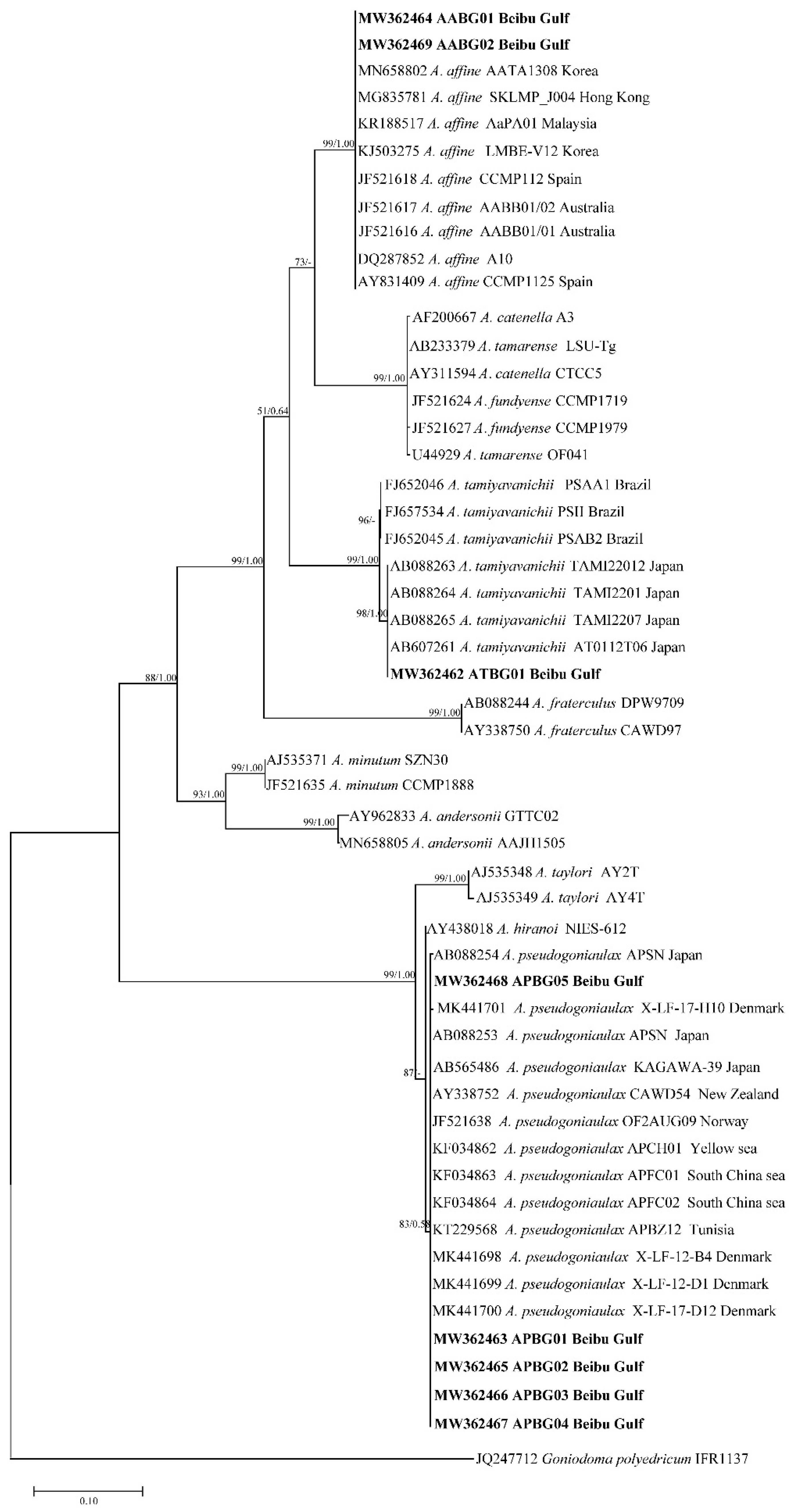
Figure 1. Phylogenetic tree constructed based on Alexandrium large subunit ribosomal DNA (LSU rDNA) sequences. Values at nodes indicate bootstrap values from the maximum likelihood method and posterior probabilities from the Bayesian inference method. Bootstrap values <50 and posterior probabilities <0.50 are not shown.
Toxins 13 00161 g001
Figure 2. Phylogenetic tree constructed based on Alexandrium small subunit ribosomal DNA (SSU rDNA). Values at nodes indicate bootstrap values from the maximum likelihood method and posterior probabilities from the Bayesian inference method. Bootstrap values <50 and posterior probabilities <0.50 are not shown.
Toxins 13 00161 g002
Figure 3. Phylogenetic tree constructed based on Alexandrium internal transcribed spacer (ITS) sequences. Values at nodes indicate bootstrap values from the maximum likelihood method and posterior probabilities from the Bayesian inference method. Bootstrap values <50 and posterior probabilities <0.50 are not shown.
Toxins 13 00161 g003
Figure 4. Sampling location.
Toxins 13 00161 g004
Table 1. Analysis of Alexandrium gene sequences used in a phylogenetic tree.
GeneAnalysis
Length
Average Content (%)Conserved SiteVariable SiteParsimonious Information SiteMonomorphic SiteConversion/Transversion Ratio
ATGC
LSU rDNA99626.931.525.616.04165553731821.0
SSU rDNA176927.729.425.217.71462275245291.9
ITS66624.734.023.917.4160494449450.7
Table 2. The components and content of paralytic shellfish toxins (PSTs) in Alexandrium spp. from the Beibu Gulf (fmol/cell).
ToxinsA. tamiyavanichiiA. pseudogonyaulax
ATBG01APBG04
GTX1, 40.88ND
GTX2, 31.2ND
GTX50.32ND
dcGTX2NDND
dcGTX3NDND
STX2.20ND
dcSTXNDND
neoSTXNDND
dcNEONDND
C1NDND
C2NDND
Total PST content4.60ND
Table 3. Paralytic shellfish toxins composition and contents in Alexandrium tamiyavanichii.
StrainLocalityToxin ComponentToxin Content (fmol/cell)References
Chula 5Gulf of ThailandC1-2, GTX1-5, STX16,200 a[47]
Chula 6Gulf of ThailandGTX1-5, STX7500 a[47]
Chula 8Gulf of ThailandC1-3, GTX1-5, STX18.9[48]
MBS8811-1Sagami Bay, JapanC1-4, GTX1-5, STX3.7[48]
MBS8811-3Sagami Bay, JapanC1-2, GTX1, 4, GTX5, STX66.3[48]
Seto Island, JapanC1-2, GTX1-5, STX 112.5[35]
Malaysia26[49]
CTCC23South AfricaC1-4, GTX1-4, STX, neoSTX, B1 0.26 b[50]
Western JapanJapanC1-2, GTX1-5, STX, neoSTX 40–424[51]
AcMS01Sebatu Malacca, MalaysiaC1-2, GTX1-5, STX 38–80[52]
AcMS01Sebatu Malacca, MalaysiaC1-2, GTX1-5, STX 60–180[53]
MalaysiaC2, GTX1-4, dcGTX3, STX 54[54]
ATY041106Seto Island, JapanC1-2, GTX1-4 38.7 ± 10.9 × 10⁻⁶ a[55]
ATY051018Seto Island, Japan111.5 × 10−6 a[55]
Fukuyama BaySeto Island, JapanC1-4, GTX1-5, neoSTX, STX244 ± 102[56]
Kasato BaySeto Island, JapanC1-4, GTX1-5, neoSTX, STX307 ± 83.8[56]
UchinoumiSeto Island, JapanC1-4, GTX1-5, neoSTX, STX328 ± 152[56]
Inokushi BayKyushu Island, JapanC1-4, GTX1-5, neoSTX, STX54.7 ± 5.32[56]
PSAA1BrazilGTX3-4, dcGTX2-3, neoSTX, STX16.85[36]
At2Seto Island, JapanC1-2, GTX1-6, neoSTX, STX2410[37]
At4Seto Island, JapanC1-2, GTX1-6, neoSTX, STX840[37]
At6-C1Seto Island, JapanC1-2, GTX1-6, neoSTX, STX289[37]
At6-C2Seto Island, JapanC1-2, GTX1-6, neoSTX, STX359[37]
At6-C3Seto Island, JapanC1-2, GTX1-6, neoSTX, STX264[37]
At6-C4Seto Island, JapanC1-2, GTX1-6, neoSTX, STX220[37]
KOSKP01Kuantan Port, MalaysiaGTX1-53070[57]
KOSKP02Kuantan Port, MalaysiaGTX1-55960.4[57]
KOSKP03Kuantan Port, MalaysiaGTX1-51027.2[57]
Kuantan Port, MalaysiaGTX1-53.07 × 10⁶[38]
Sebatu Malacca, MalaysiaGTX1-51.167[38]
ATBG01Weizhou Island, Beibu GulfGTX1-5, STX4.6This study
a MU/cell; b equivalent of Saxitoxin.
Table 4. PST contamination in shellfish in the Beibu Gulf, China.
Sampling DateLocalityMethodologyToxinsDetection Rate/Exceedance RateReferences
2001.11–2004.04Tieshan, Beihai, Fangcheng, WeizhouMBA, HPLCC1-2, GTX1-5, STXexceeding standard 2.15–3.54 times[58]
2003.03–2004.05BeihaiMBA, HPLCC1-2, GTX1-4, STXdetection rate 8.30%[59]
2005–2009Beibu GulfMBAexceedance rate 1.0%[28]
Qinzhou, Fangcheng, BeihaiAmerican ABRAXIS kita[60]
2007.11–2008.10BeihaiMBAb[61]
2014.09–2014.11Weizhou, Beihai, Fangcheng, QinzhouHPLCGTX1-5, dcGTX2-3, neoSTX, dcSTX, STXdetection rate 100%, exceedance rate 6.1%[29]
2015.09QinzhouMBA, HPLCGTX1, GTX4, neoSTX, STXdetection rate 86%, no exceedance[30]
MBA, Mouse Bioassay; HPLC, High Performance Liquid Chromatography; a positive results; b no PSP was found.
Table 5. Sampling information.
StrainsCollection DateLocation
ATBG012018-06-09S2 (21°02′51″ N, 109°08′43″ E)
APBG012018-09-15S2 (21°02′51″ N, 109°08′43″ E)
AABG012018-09-15S2 (21°02′51″ N, 109°08′43″ E)
AABG022018-12-20S8 (21°00′28″ N, 109°05′38″ E)
APBG022019-06-10S2 (21°02′51″ N, 109°08′43″ E)
APBG032019-06-10S4 (21°04′12″ N, 109°06′11″ E)
APBG042019-06-10S5 (21°03′39″ N, 109°05′06″ E)
APBG052019-06-10S5 (21°03′39″ N, 109°05′06″ E)
Table 6. Primers information.
GenePrimersPrimer SequencesReferences
LSU rDNAD1R5′-ACCCGCTGAATTTAAGCATA-3′[73]
D2C5′-TGATCCTTCTGCAGGTTCACCTAC-3′
SSU rDNA1F5′-AACCTGGTTGATCCTGCCAGT-3′[74]
1528R5′-TGATCCTTCYGCAGGTTCAC-3′’
ITSFA5′-CCAAGCTTCTAGATCGTAACAAGG(ACT)TCCGTAGGT-3′[75]
RB5′-CCTGCAGTCGACA(TG)ATGCTTAA(AG)TTCAGC(AG)GG-3′
Table 7. Multiple reaction monitoring conditions.
ToxinsQuantitative Transition m/zQualitative Transition m/zResidence Time (ms)Impact Voltage (V)Fragmentor Voltage (V)
GTX1412 > 332.1412 > 314.21006/20100/100
GTX2396.1 > 316.1396.1 > 297.81005/15110/110
GTX3396.1 > 316.1396.1 > 298.71005/15110/110
GTX4412 > 332.1412 > 314.21006/20100/100
GTX5380.1 > 300380.1 > 282.110011/1594/105
dcGTX2352.8 > 334.7352.8 > 254.91005/10120/120
dcGTX3352.8 > 334.7352.8 > 254.91005/10120/120
STX300 > 282300 > 20410010/20100/100
dcSTX257 > 222.1257 > 12610010/2080/80
neoSTX316.1 > 298316.1 > 22010010/20100/100
dcNEO273 > 225.1273 > 12610018/1880/80
C1474.1 > 251474.1 > 12210020/3060/60
C2474.1 > 351474.1 > 12210020/3060/60
Publisher’s Note: MDPI stays neutral with regard to jurisdictional claims in published maps and institutional affiliations.