Article

Ssu72 Regulates Fungal Development, Aflatoxin Biosynthesis and Pathogenicity in Aspergillus flavus

Key Laboratory of Pathogenic Fungi and Mycotoxins of Fujian Province, Key Laboratory of Biopesticide and Chemical Biology of Education Ministry, School of Life Sciences, Fujian Agriculture and Forestry University, Fuzhou 350002, China
*
Author to whom correspondence should be addressed.
Toxins 2020, 12(11), 717; https://doi.org/10.3390/toxins12110717
Submission received: 16 September 2020 / Revised: 2 November 2020 / Accepted: 3 November 2020 / Published: 13 November 2020
(This article belongs to the Section Mycotoxins)

Abstract

:
The RNA polymerase II (Pol II) transcription process is coordinated by the reversible phosphorylation of its largest subunit-carboxy terminal domain (CTD). Ssu72 is identified as a CTD phosphatase with specificity for phosphorylation of Ser5 and Ser7 and plays critical roles in regulation of transcription cycle in eukaryotes. However, the biofunction of Ssu72 is still unknown in Aspergillus flavus, which is a plant pathogenic fungus and produces one of the most toxic mycotoxins-aflatoxin. Here, we identified a putative phosphatase Ssu72 and investigated the function of Ssu72 in A. flavus. Deletion of ssu72 resulted in severe defects in vegetative growth, conidiation and sclerotia formation. Additionally, we found that phosphatase Ssu72 positively regulates aflatoxin production through regulating expression of aflatoxin biosynthesis cluster genes. Notably, seeds infection assays indicated that phosphatase Ssu72 is crucial for pathogenicity of A. flavus. Furthermore, the Δssu72 mutant exhibited more sensitivity to osmotic and oxidative stresses. Taken together, our study suggests that the putative phosphatase Ssu72 is involved in fungal development, aflatoxin production and pathogenicity in A. flavus, and may provide a novel strategy to prevent the contamination of this pathogenic fungus.
Key Contribution: The putative phosphatase Ssu72 is identified and shown to contribute to development, aflatoxin production and pathogenicity of A. flavus.

1. Introduction

Aspergillus flavus is a saprophytic plant pathogenic fungus that infects a range of seed crops (maize, peanuts, cottonseed and tree nuts) before and after harvest [1,2]. In addition, A. flavus is also a human opportunistic pathogen, causing invasive aspergillosis in mammals and humans [3,4]. Aflatoxin (AF), which is mainly synthesised by A. flavus, is one of the most toxic and carcinogenic secondary metabolites in nature, posing a huge threat to economic development, food safety and human health [5,6]. Long-term intake of low doses of AFs may result in a number of health problems, such as growth impairment, lung and liver cancer or even death in many mammals and humans [7,8]. Therefore, it is essential to explain and clarify the regulatory mechanism of this fungus in pathogenicity and aflatoxin biosynthesis.
Previous studies have shown that aflatoxin biosynthesis and pathogenicity of A. flavus are regulated by multiple factors, such as temperature [9], water activity [10] and post-translational modifications (PTMs) including phosphorylation [11], acetylation [12], succinylation [13], methylation [14] and SUMOylation [15]. Reversible phosphorylation catalyzed by kinases and phosphatases, is one of the most common PTMs, and has been shown to regulate various biological processes [16,17]. In eukaryotes, reversible phosphorylation mainly occurs on three amino acids (serine, threonine and tyrosine) [18,19]. Phosphatase-mediated dephosphorylation plays a critical role in signal transduction in eukaryotic cells [20]. Based on sequence homology, structure and catalytic specificity, phosphatases can be classified into two major superfamilies: serine/threonine (S/T) phosphatases and tyrosine phosphatases (PTPs) [21]. The PTPs have demonstrated to be involved in the regulation of various cellular processes in eukaryotes, including cell cycle, signal transduction, transcriptional activation, development and secondary metabolism [22,23,24].
In eukaryotes, transcription of all coding genes is mainly regulated by RNA polymerase II (RNAP II) complex, which consist of 12 different subunits [25]. The carboxy-terminal domain (CTD) is one of the largest RNAP II subunits and composed of the evolutionarily conserved reiterate heptapeptide sequence (YSPTSPS), and reversible phosphorylation of this sequence is involved in distinct stages of transcription [26,27]. Ssu72 is identified as a highly conserved CTD phosphatase and negatively regulate the phosphorylation of specific Ser(5) and Ser(7) [28]. In Saccharomyces cerevisiae, Ssu72 is well studied due to its role in transcription initiation, mRNA processing, transcription termination and sister-chromatid cohesion [29,30]. In fission yeast, phosphatase Ssu72 is known to be essential for growth [31]. In addition, Ssu72 is found to be involved in RNA 3′ processing and the regulation of phosphate homeostasis in Schizosaccharomyces pombe [32,33]. In plant pathogenic fungus Fusarium graminearum, it has been demonstrated that the ortholog of phosphatase Ssu72 is involved in asexual development and virulence [34]. Despite the roles of phosphatase Ssu72 in yeast have been well studied, the function of Ssu72 in Aspergillus species is still poorly understood.
In this study, we identified a putative phosphatase Ssu72 of A. flavus, and then systematically characterized the function of Ssu72 in A. flavus. Our results reveal that the putative phosphatase Ssu72 plays critical roles in the regulation of fungal development, aflatoxin biosynthesis, stresses response and pathogenicity in A. flavus. This will provide a new insight that Ssu72 may be used as a potential target for preventing the hazard of A. flavus.

2. Results

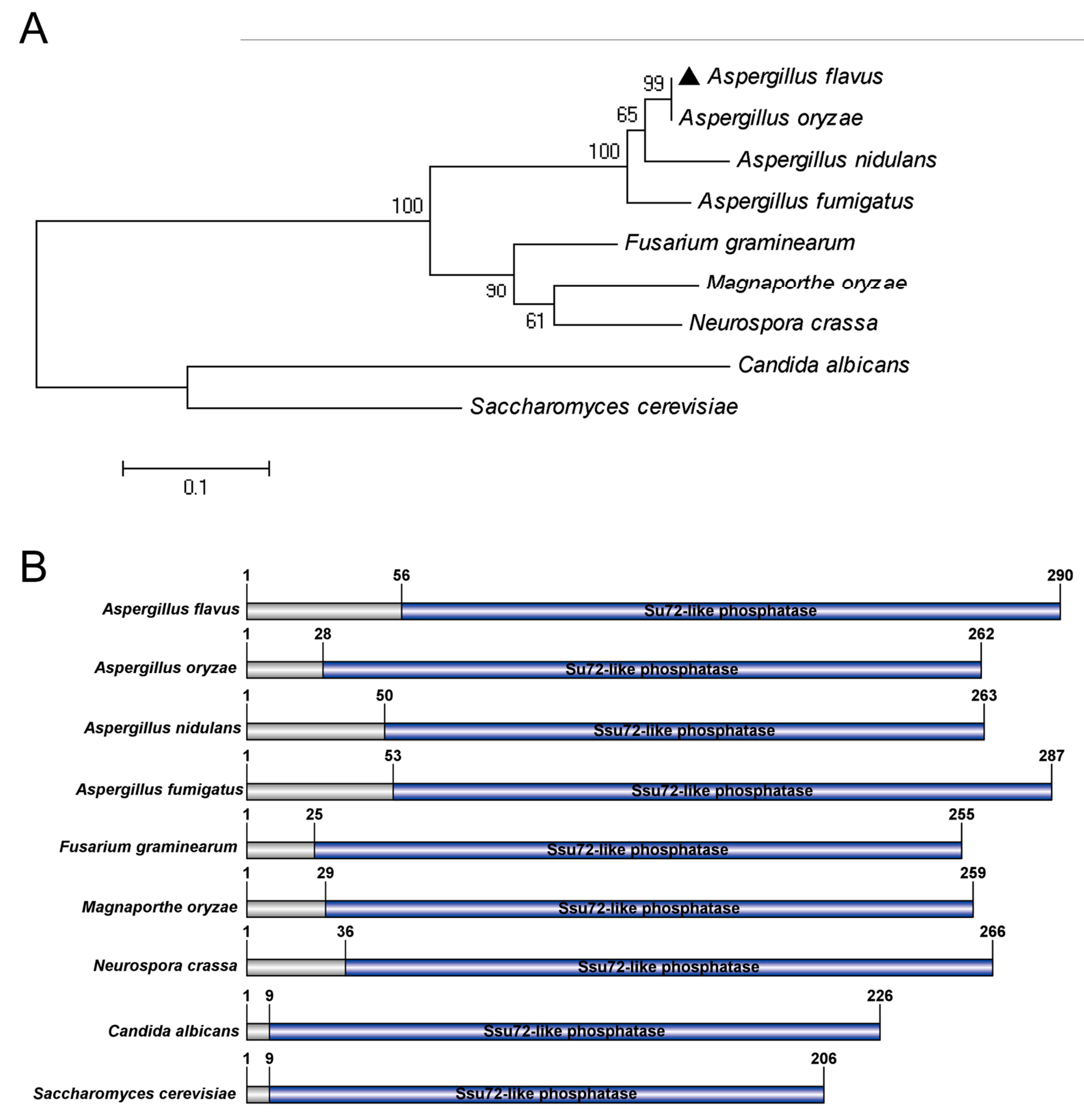
2.1. Identification of Putative Phosphatase Ssu72 in A. flavus

To identify the ortholog of Ssu72 phosphatase in A. flavus, the Ssu72 protein sequence of model organism S. cerevisiae was used to search with a basic local alignment search tool (BLAST) in the A. flavus genome database. A putative Ssu72 phosphatase protein in A. flavus was identified, predicting to encode 290 amino acids and present 45% homology with S. cerevisiae Ssu72. Then, a similar approach was performed to obtain Ssu72 homologous protein sequences in some fungi (A. nidulans, A. fumigatus, F. graminearum, Magnapoethe oryzae, Neurospora crassa, Candida albicans). Our sequence alignment results showed that the A. flavus Ssu72 protein displayed 99% similarity to A. oryzae, 95% similarity to A. nidulans, and 93% similarity to A. fumigatus. The phylogenetic analysis indicated that the Ssu72 protein is evolutionarily conserved in Aspergillus species (Figure 1A), and the A. flavus Ssu72 protein exhibited a high similarity with the ortholog of Ssu72 in Aspergillus species. Domain analysis revealed that the Ssu72 homologous proteins all contained a Ssu72-like phosphatase domain (Figure 1B), and this domain was mainly concentrated on the C-terminal of Ssu72 protein, suggesting that Ssu72-like phosphatase domain is highly conserved in fungi. These results indicate that the putative Ssu72 phosphatase protein is highly conserved in evolution and may have similar function in fungi.

2.2. Construction of the ssu72 Mutant Strains

To investigate the potential function of Ssu72 phosphatase gene in A. flavus, the ssu72 deletion (Δssu72) and the ssu72 complementation (Δssu72-Com) strains were constructed by a homologous recombination approach (Figure 2A). The specific primers and sequence lengths are displayed in Figure 2A. For knockout strain construction, three fragments (1392 bp ssu72 5′UTR, 1220 bp 3′UTR and 1890 bp pyrG) were amplified with the specific primers and then fused together. PyrG from A. fumigatus was used to replace the ssu72 gene, and open reading frame (ORF) of ssu72 gene was reinserted into the genome of the knockout strain to construct complementary strains. Then, the deletion and complementation transformants were verified by polymerase chain reaction (PCR) analysis (Figure 2B), and the result showed that ORF fragment could be detected in WT (wild type) and Δssu72-Com strains, but not in Δssu72 strain. While AP (5′UTR+pyrG) and BP (5′UTR+pyrG) fragments (Figure 2A) were amplified from ssu72 deletion and complementation mutants but not from WT strain. Furthermore, expression levels of ssu72 gene in WT, Δssu72 and Δssu72-Com strains were analyzed by reverse transcription-PCR (RT-PCR) and quantitative real-time PCR (qRT-PCR), and the results indicated that the transcripts of ssu72 were not detected in Δssu72 strain, whereas ssu72 gene was expressed in the WT and Δssu72-Com strains (Figure 2C,D). All these results showed that both ssu72 knockout mutant (Δssu72) and ssu72 complementation strain (Δssu72-Com) were successfully constructed.

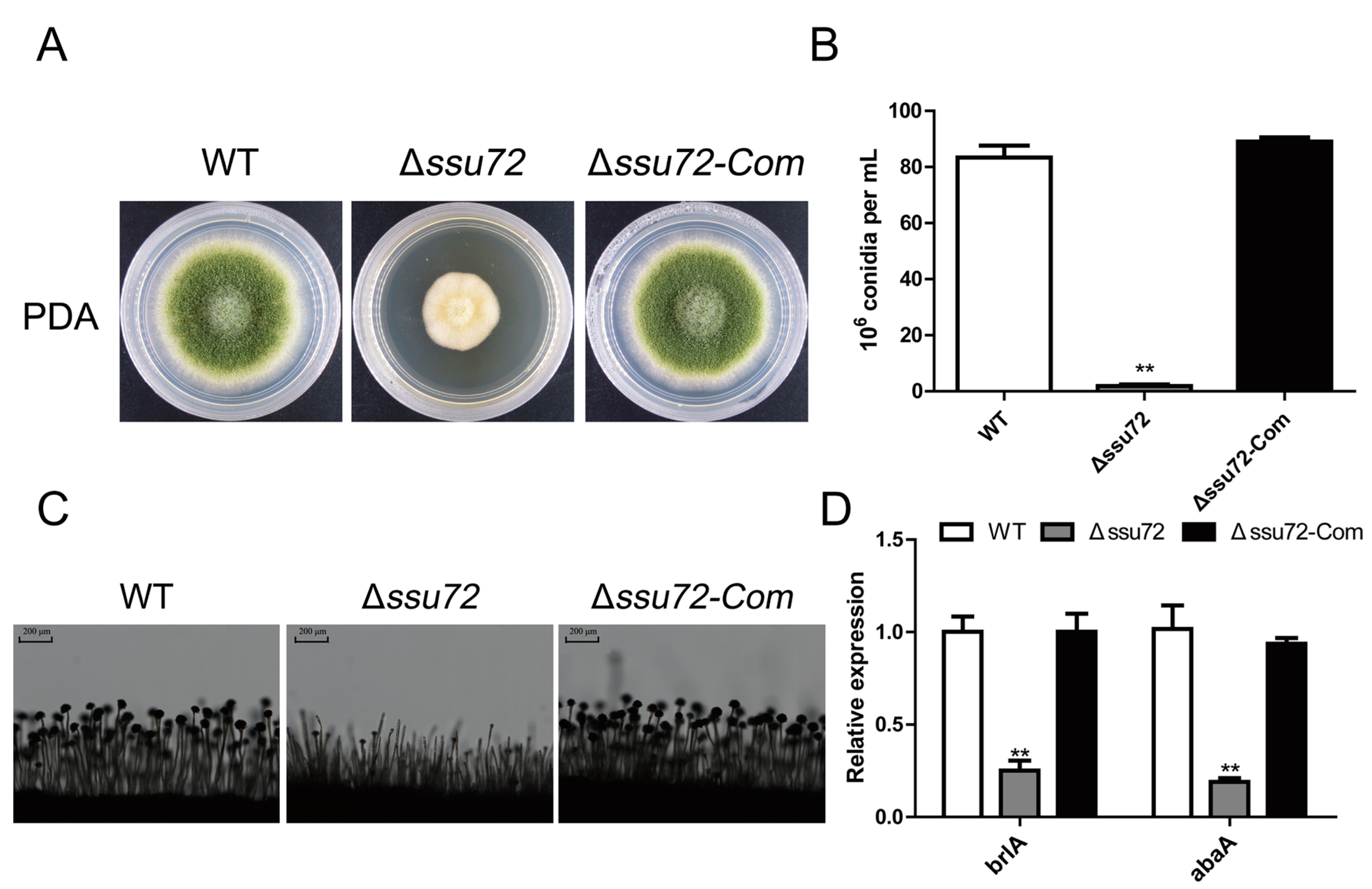
2.3. Ssu72 Is Involved in Vegetative Growth in A. flavus

To investigate the roles of ssu72 in vegetative growth of A. flavus, the WT, Δssu72 and Δssu72-Com strains were inoculated in yeast extract-sucrose (YES), potato dextrose agar (PDA) and yeast glucose trace-elements (YGT) media and cultured for 5 days. As shown in Figure 3A,B, our result revealed that the Δssu72 mutant displayed significant growth reduction on YES, PDA and YGT media when compared with WT and complementation strains (Δssu72-Com). Moreover, microscopic examination showed that abnormal and shorter aerial hyphal were observed in Δssu72 mutant, while the abnormal hyphal defect of Δssu72 mutant was restored in the Δssu72-Com strain (Figure 3C). These observations suggest that phosphatase Ssu72 plays important roles in vegetative growth of A. flavus.

2.4. Ssu72 Is Critical for Conidiation in A. flavus

Conidia is mainly produced by aerial hyphae and plays critical roles in asexual development and infection processes of A. flavus [35]. To assess the effect of ssu72 in conidiation, the WT, Δssu72 and Δssu72-Com strains were cultured on PDA medium for 5 days, and we found that the amount of conidia was obviously reduced in the Δssu72 mutant in comparison with WT and Δssu72-Com strains (Figure 4A). About one tenth of conidia was observed in the knockout strain, when compared with the WT and complementation strains (Figure 4B). Furthermore, microscopic examination revealed that abnormal head of conidiophores was observed in Δssu72 mutant (Figure 4C). To further explore the role of ssu72 in conidiation, the expression levels of two key transcript factors (brlA and abaA) were analyzed by qRT-PCR. The consequence showed that the expression levels of brlA and abaA were both significantly down-regulated in Δssu72 mutant when compared to WT and Δssu72-Com strains (Figure 2D). These results suggest that phosphatase Ssu72 is involved in the conidia production of A. flavus.

2.5. Ssu72 Is Required for Sclerotia Formation in A. flavus

In A. flavus, a sexual structure-sclerotia is formed to survive under harsh environment [5]. To investigate the bio-function of ssu72 in sclerotia formation, the WT, Δssu72 and Δssu72-Com strains were cultured on the Wickerham (WKM) medium in the dark for 7 days. As shown in Figure 5A,B, lots of sclerotia were discovered in WT and Δssu72-Com strains. However, no sclerotia was observed in Δssu72 strain. To further confirm these findings, qRT-PCR analysis was performed to analyze the transcript levels of two sclerotia formation key genes (nsdC and nsdD). The result indicated that the transcript levels of nsdC and nsdD were both dramatically reduced in the Δssu72 mutants when compared to WT and Δssu72-Com strains (Figure 5C). All the above findings suggest that phosphatase Ssu72 is required for sclerotia formation of A. flavus.

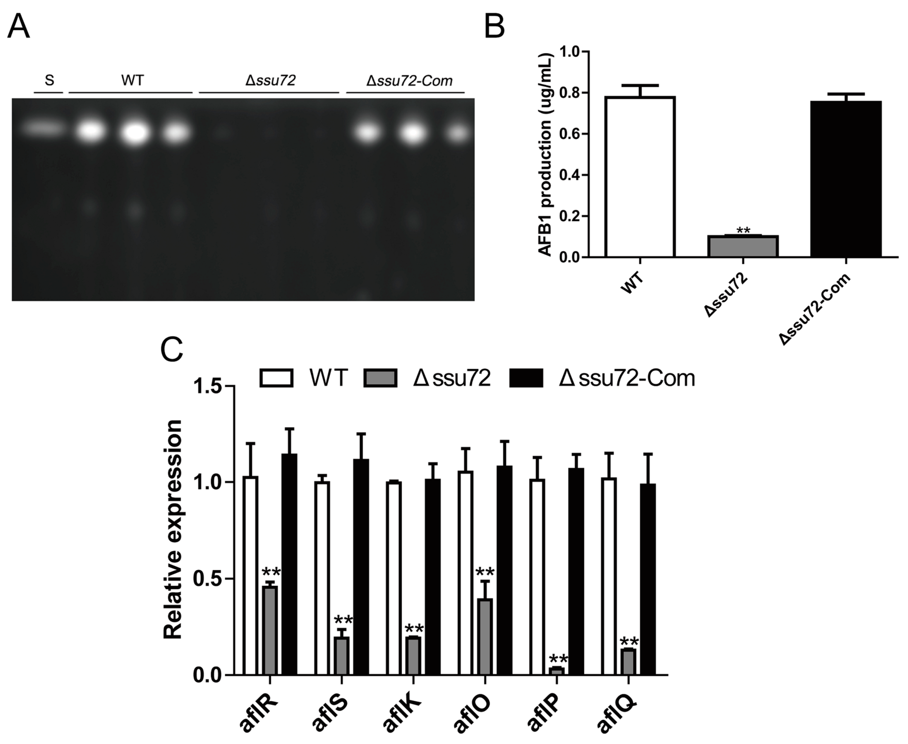
2.6. Ssu72 Plays a Positive Role in Regulation of Aflatoxin Biosynthesis

Aflatoxin (AF) is one of the most toxic secondary metabolites of A. flavus, and poses a huge threat to human health [36,37]. Thus, it is necessary to determine whether phosphatase Ssu72 is involved in aflatoxin biosynthesis. Then, the strains were cultured in YES liquid medium for 6 days, and thin-layer chromatography (TLC) analysis showed that deletion of ssu72 gene resulted in a significant decrease in aflatoxin B1 (AFB1) production when compared to WT and Δssu72-Com strains (Figure 6A,B). Subsequently, qRT-PCR was performed to analyze the expression levels of AFB1 biosynthesis cluster genes. We found that the transcript levels of some key genes (regulatory genes aflR and aflS, structural genes aflP, aflQ, aflO and aflK) in Δssu72 mutant were significantly lower than those in WT and Δssu72-Com strains (Figure 6C). Overall, these results indicate that phosphatase Ssu72 positively regulates AFB1 biosynthesis in A. flavus.

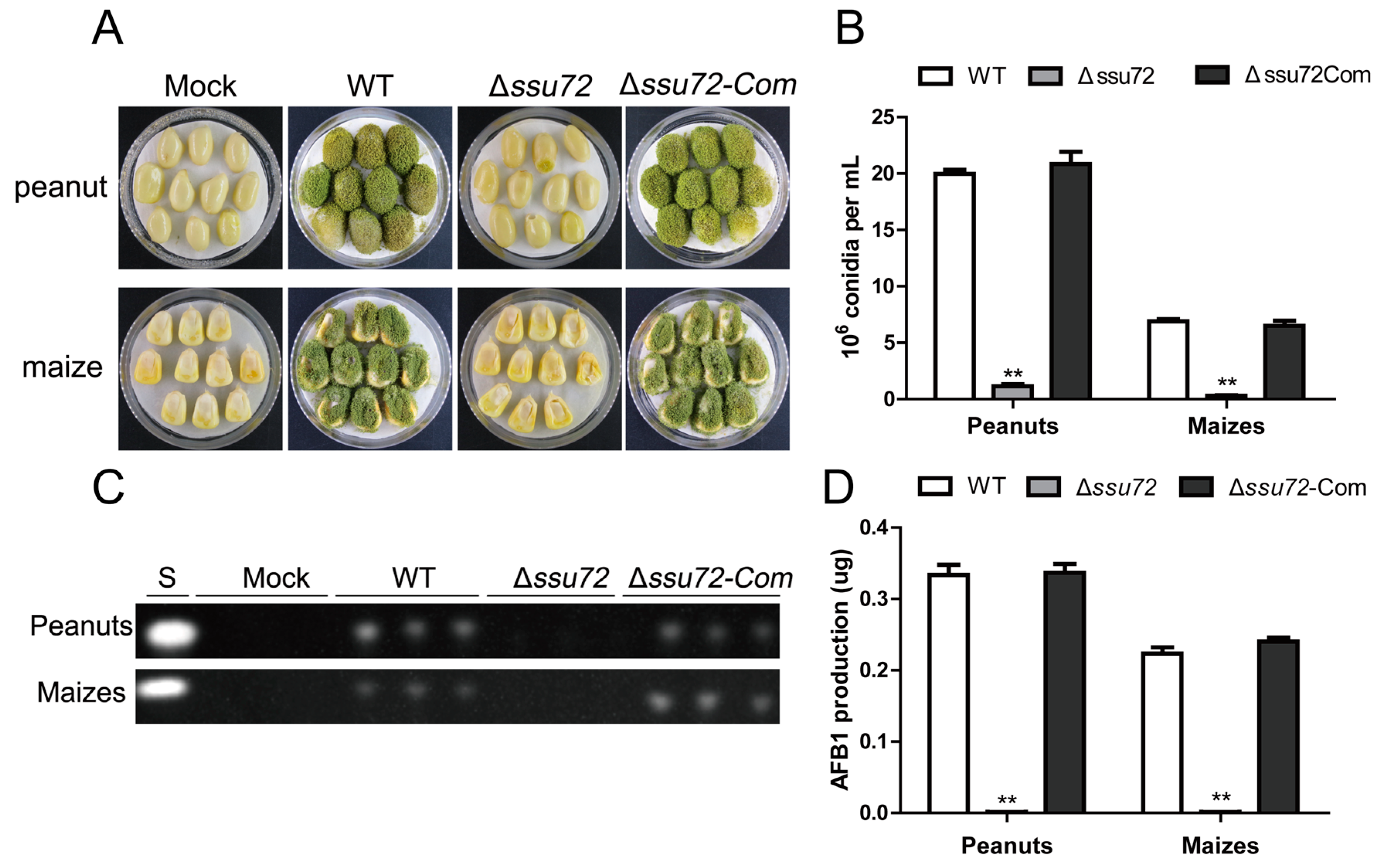
2.7. Ssu72 Contributes to Pathogenicity in Crop Seeds in A. flavus

Since A. flavus infects crops and causes huge economic losses, the effect of phosphatase Ssu72 on the pathogenicity to crop seeds was characterized in this study. After cultivation for 5 days, a lot of conidia on surface of peanut and maize seeds were found in the WT and Δssu72-Com strain (Figure 7A). However, no obvious colonization was observed on peanuts and maize seeds after inoculated with Δssu72 mutant (Figure 7A), indicating that deletion of ssu72 resulted in a significant decrease in pathogenicity to peanut and maize seeds. Conidia were then harvested from the infected seeds, and Δssu72 mutant produced less amount of conidia on infected seeds than WT and Δssu72-Com strain (Figure 7B). Subsequently, TLC assay was used to detect aflatoxin production in the infected seeds, and the result revealed that no AFB1 was observed in peanut and maize seeds infected with Δssu72 strain (Figure 7C,D). All these data suggest that phosphatase Ssu72 contributes to its pathogenicity to crop seeds in A. flavus.

2.8. Ssu72 Response to Multiple Stresses in A. flavus

In A. flavus, phosphatases have been proven to be involved in multiple stresses response [38,39]. To explore the role of putative phosphatase Ssu72 in response to osmotic and oxidative stresses, the strains (WT, Δssu72 and Δssu72-Com) were cultured on PDA medium supplemented with different stress reagents. As observed in Figure 8A,B, the relative inhibition of growth rate in Δssu72 induced by 1 M NaCl was obviously higher than that of WT and Δssu72-Com strains, suggesting that Δssu72 mutant displayed increased sensitivity to osmotic stresses. Additionally, the growth of Δssu72 mutant was completely inhibited after added with higher osmotic stresses (5 mM H2O2) (Figure 8A,B). All these results demonstrate that Ssu72 participates in osmotic and oxidative stresses in A. flavus.

3. Discussion

As one of the largest RNAP II subunits, carboxy-terminal domain (CTD) is known to be involved in the regulation of transcriptional initiation/termination and pre-mRNA processing in eukaryotes [25,40]. Ssu72 is identified as a conserved CTD-related phosphatase and well characterized in yeast [33]. However, the biological functions of Ssu72 in other fungi, especially in Aspergillus species, are still unclear. In this study, a putative phosphatase Ssu72 of A. flavus was identified. The C-terminus of the ssu72 protein in fungi all contained a conserved Ssu72-like phosphatase domain, indicating that the ssu72 protein is evolutionarily conserved, and may have similar functions in fungi. In S. cerevisiae, the ssu72 protein has been shown to play important roles in regulation of multiple processes, so we speculate that Ssu72 may be involved in various processes in A. flavus. Next ssu72 gene deletion and complementation strains were constructed to characterize its function in A. flavus. Our findings reveal that the putative phosphatase Ssu72 is involved in fungal development, aflatoxin production and pathogenicity of A. flavus.
Our previous studies have concluded that tyrosine phosphatases contribute to vegetative growth and conidiation in A. flavus [38]. In this study, our results demonstrated that knockout of putative phosphatase gene ssu72 resulted in a severe defect in vegetative growth (Figure 3A), which is consistent with the results in budding yeast [31]. Our microscopic observation on hyphae tips showed that shorter aerial hyphal was found in Δssu72 strain, which is in a good agreement with the above conclusion (Figure 3C). In addition, the amount of conidia was significantly reduced in Δssu72 strain (Figure 4A), which is consistent with the defect that was observed in ssu72 homolog mutants in F. graminearum and N. crassa [34,41]. This finding is well supported by the down-regulation of key transcription factors brlA and abaA [42]. Sclerotia is a sexual structure to survive in unfavorable environment in A. flavus [43]. However, no sclerotia was discovered in Δssu72 strain when compared to WT and complementation strains (Figure 5A), suggesting that phosphatase Ssu72 is essential for sclerotia formation. Furthermore, transcript levels of nsdC and nsdD genes, which have been known to be critical for sclerotia formation [44], was also significantly decreased in Δssu72 strain in this study. It is possible that deletion of ssu72 decrease the expression levels of nsdC and nsdD genes, and then regulate the sclerotia production. Therefore, phosphatase Ssu72 plays important roles in the regulation of vegetative growth, conidiation and sclerotia formation in A. flavus.
As one of the most toxic secondary metabolites in nature, aflatoxin contamination poses a serious threat to food safety, human and animal health [45]. Recent studies have revealed that kinases and phosphatases play vital roles in regulation of aflatoxin production in A. flavus [39,46,47]. Here, we observed that aflatoxin production was dramatically reduced in Δssu72 mutant, and this finding is in accordance with the down-regulation of aflatoxin biosynthesis regulatory and structural genes (Figure 6). A similar result was also found that deletion of ssu72 homolog gene led to a lower production of mycotoxin-DON in plant pathogenic fungi F. graminearum [34]. A 70-kb DNA cluster is associated with aflatoxin biosynthesis, and regulatory and structural genes play important roles in this cluster [48]. As a conserved CTD phosphatase in fungi, Ssu72 has been proven to be involved in multiple cellular processes, including mRNA processing, transcription initiation and termination [29,32]. So we speculated that phosphatase Ssu72 may affect the transcription process of some key aflatoxin biosynthesis related genes, and then regulate the production of aflatoxin. These findings suggest that phosphatase Ssu72 positively regulates AFB1 biosynthesis in A. flavus.
Tyrosine phosphatases are well conserved and play critical roles in fungal pathogenicity in filamentous fungi [24,39]. In F. graminearum, homologues of Ssu72 have been demonstrated to be essential for plant infection [34]. However, the role of phosphatase Ssu72 in seeds infection is still unknown in A. flavus. Here, seeds infection assays showed that deletion of ssu72 resulted in a severe defect in pathogenicity on maize and peanuts (Figure 7A), which is consistent with the previous finding in F. graminearum [34]. In addition, fewer conidia and aflatoxins were observed in the infected seeds of Δssu72 mutant (Figure 7B,C). In A. flavus, pathogenicity is related to various factors, such as vegetative growth, conidiation, mycotoxins and environment stresses [43]. Therefore, we believed that the severe defects in development and aflatoxin production are the main reasons for the decreased pathogenicity to seeds. Taken together, these data suggest that phosphatase Ssu72 displays a vital role in pathogenicity to seeds of A. flavus.
Mitogen-activated protein kinase (MAPK) related phosphatases have been identified to be involved in multiple stresses response by regulating the phosphorylation level of MAP kinases (Hog1, Slt2 and Fus3) in A. flavus [39]. In this study, we found that the putative phosphatase Ssu72 is critical to respond to osmotic stress (Figure 8A). Similarly, the orthologue of Ssu72 has also been known to response to osmotic stress in N. crassa, and a higher phosphorylation level of Hog1 kinase was observed in ssu72 deletion mutant [41]. Additionally, Δssu72 mutants exhibited more sensitive to oxidative stress than WT and Δssu72-Com strains (Figure 8B). Reactive oxygen species (ROS) have been characterized to be highly related to oxidative stress [43]. In plant pathogenic fungi, elimination of ROS is critical to seeds infection [34]. Due to lower pathogenicity found in Δssu72 strain, we speculate that ROS scavenging ability of Δssu72 strain may be reduced. And this hypothesis is consistent with the result that the Δssu72 mutant displayed more sensitive to oxidative stress in A. flavus.
In conclusion, a putative phosphatase Ssu72 was identified in A. flavus, and our results indicate that Ssu72 is involved in the regulation of development, aflatoxin biosynthesis and pathogenicity. This is the first report on the biofunction of phosphatase Ssu72 in Aspergillus species. We believe that our discoveries could improve the understanding of Ssu72 in filamentous fungi, and may provide a novel insight for developing new control strategies to this fungus.

4. Materials and Methods

4.1. Strains and Culture Conditions

Strains of A. flavus used and constructed were listed in Table 1. All strains were cultured on YES, PDA and YGT media for vegetative growth and conidiation analysis, and on WKM medium for sclerotia formation analysis [49]. YES liquid medium was used for aflatoxin production as described before [50].

4.2. Mutant Strains Construction

The homologous recombination approach was used to generate the ssu72 knockout mutant (Δssu72) [4]. Three fragments (1392 bp ssu72 5′UTR, 1220 bp 3′UTR and 1890 bp pyrG) were amplified with specific primers and fused together. Then, the fusion PCR products were transformed into A. flavus CA14 protoplasts as described earlier [51]. As the pyrG gene is inserted into the knockout strain, the knockout strain could grow in a medium without urea. This feature could be used to screen the knockout strain. Afterwards, the ssu72 complementation strain (Δssu72-Com) was constructed using a previously described method [50]. Briefly, an overlap PCR product which contains ssu72 coding region was introduced into the Δssu72 protoplasts. Finally, all the selected transformants were identified by PCR, and further confirmed by RT-PCR and qRT-PCR assays. At least two or three positive transformants were used for further phenotypic analysis. Primers are listed in Table 2.

4.3. Phenotpic Assays

To explore the roles of ssu72 in growth and conidiation in A. flavus, 104 spores were spotted on YES, PDA and YGT media, and cultured at 37 °C for 5 days. Colony diameter was measured after 5 days of cultivation. Subsequently, conidia were collected from PDA medium and quantified using a hemocytometer as previously described [52]. For sclerotia production assay, all strains were incubated on WKM medium at 37 °C for 7 days. Then, 75% ethanol was used to wash away the mycelia and conidia, and the sclerotia were harvested and counted by a light microscope as described earlier [53].

4.4. Aflatoxin Production Assays

To determine aflatoxin production, 104 conidia of strains were inoculated on YES liquid medium and cultured at 29 °C for 6 days in the dark, then aflatoxins were extracted by chloroform according to a previously described approach [38]. TLC was performed to analyze aflatoxin in a solvent system (chloroform:acetone = 9:1) and examined under 365 nm UV light [13].

4.5. Pathogenicity Assays

Pathogenicity analysis on peanuts and maize seeds were conducted as described earlier [46]. Briefly, the sterilized crop seeds were inoculated with 106 spores of each strain at 29 °C for 5 days. The infected seeds were collected and transferred to 50 mL centrifuge tubes with 15 mL sterile water. Then, the conidia amount and aflatoxin production of the infected seeds were analyzed as the methods mentioned before.

4.6. Stress Assay

To investigate the role of Ssu72 in various stresses response, the WT, Δssu72 and Δssu72-Com strains were inoculated onto PDA medium supplemented with 1 M NaCl or 5 mM H2O2 at 37 °C for 4 days. The inhibition of growth rates was calculated as described before [38].

4.7. RNA Extraction and Quantitative Real-Time PCR Analysis

RNA extraction and cDNA synthesis were conducted according to the published references in our lab [39]. Mycelia were collected from PDA and WKM media cultured for 48 h, then TRIzol reagent (Biomarker Technologies, Beijing, China) was used for total RNA isolation. cDNA was synthesized with First-Strand cDNA Synthesis Kit (TransGen Biotech, Beijing, China). Subsequently, cDNA was used as a template for qRT-PCR analysis. All the qRT-PCR primers used in this study are listed in Table 3. The relative transcript levels of related genes were calculated with the 2–ΔΔCt method [54], and β-actin was used as internal control.

Author Contributions

G.Y. and S.W. conceive and designed the experiments, X.C., L.Q., and L.Y. performed the experiments and analyzed the data. X.C. and R.H. contributed reagents and materials. G.Y., J.Y. and S.W. wrote and revised the manuscript. All authors have read and agreed to the published version of the manuscript.

Funding

This research was funded by the National Natural Science Foundation of China (No. 31772105) and the Scientific Research Foundation of Fujian Agriculture and Forestry University (324-1122yb042).

Acknowledgments

We would like to thank Perng Kuang Chang (Southern Regional Research Center, United States Department of Agriculture, New Orleans, USA) and Yang Liu (Institute of Food Science and Technology, CAAS) for kindly providing the strains.

Conflicts of Interest

The authors declare no conflict of interest.

References

  1. Zhang, F.; Huang, L.; Deng, J.; Tan, C.; Geng, L.; Liao, Y.; Yuan, J.; Wang, S. A Cell Wall Integrity-Related MAP Kinase Kinase Kinase AflBck1 Is Required for Growth and Virulence in Fungus Aspergillus flavus. Mol. Plant Microbe Interact. 2020, 33, 680–692. [Google Scholar] [CrossRef] [PubMed]
  2. Wang, X.; Zha, W.; Liang, L.; Fasoyin, O.E.; Wu, L.; Wang, S. The bZIP Transcription Factor AflRsmA Regulates Aflatoxin B(1) Biosynthesis, Oxidative Stress Response and Sclerotium Formation in Aspergillus flavus. Toxins 2020, 12, 271. [Google Scholar] [CrossRef] [PubMed] [Green Version]
  3. Fasoyin, O.E.; Yang, K.; Qiu, M.; Wang, B.; Wang, S.; Wang, S. Regulation of Morphology, Aflatoxin Production, and Virulence of Aspergillus flavus by the Major Nitrogen Regulatory Gene areA. Toxins 2019, 11, 718. [Google Scholar] [CrossRef] [PubMed] [Green Version]
  4. Yang, K.; Liu, Y.; Wang, S.; Wu, L.; Xie, R.; Lan, H.; Fasoyin, O.E.; Wang, Y.; Wang, S. Cyclase-Associated Protein Cap with Multiple Domains Contributes to Mycotoxin Biosynthesis and Fungal Virulence in Aspergillus flavus. J. Agric. Food Chem. 2019, 67, 4200–4213. [Google Scholar] [CrossRef]
  5. Chang, P.K.; Zhang, Q.; Scharfenstein, L.; Mack, B.; Yoshimi, A.; Miyazawa, K.; Abe, K. Aspergillus flavus GPI-anchored protein-encoding ecm33 has a role in growth, development, aflatoxin biosynthesis, and maize infection. Appl. Microbiol. Biotechnol. 2018, 102, 5209–5220. [Google Scholar] [CrossRef]
  6. Drott, M.T.; Satterlee, T.R.; Skerker, J.M.; Pfannenstiel, B.T.; Glass, N.L. The Frequency of Sex: Population Genomics Reveals Differences in Recombination and Population Structure of the Aflatoxin-Producing Fungus Aspergillus flavus. mBio 2020, 11, e00963-20. [Google Scholar] [CrossRef] [PubMed]
  7. Ibarra, B.A.; Lohmar, J.M.; Satterlee, T.; McDonald, T.; Cary, J.W.; Calvo, A.M. The 14-3-3 Protein Homolog ArtA Regulates Development and Secondary Metabolism in the Opportunistic Plant Pathogen Aspergillus flavus. Appl. Environ. Microbiol. 2018, 84, e02241-17. [Google Scholar] [CrossRef] [Green Version]
  8. Lan, H.; Wu, L.; Sun, R.; Keller, N.P.; Yang, K.; Ye, L.; He, S.; Zhang, F.; Wang, S. The HosA Histone Deacetylase Regulates Aflatoxin Biosynthesis Through Direct Regulation of Aflatoxin Cluster Genes. Mol. Plant Microbe Interact. 2019, 32, 1210–1228. [Google Scholar] [CrossRef] [PubMed]
  9. Bai, Y.; Wang, S.; Zhong, H.; Yang, Q.; Zhang, F.; Zhuang, Z.; Yuan, J.; Nie, X.; Wang, S. Integrative analyses reveal transcriptome-proteome correlation in biological pathways and secondary metabolism clusters in A. flavus in response to temperature. Sci. Rep. 2015, 5, 14582. [Google Scholar] [CrossRef]
  10. Zhang, F.; Guo, Z.; Zhong, H.; Wang, S.; Yang, W.; Liu, Y.; Wang, S. RNA-Seq-based transcriptome analysis of aflatoxigenic Aspergillus flavus in response to water activity. Toxins 2014, 6, 3187–3207. [Google Scholar] [CrossRef] [Green Version]
  11. Ren, S.; Yang, M.; Li, Y.; Zhang, F.; Chen, Z.; Zhang, J.; Yang, G.; Yue, Y.; Li, S.; Wang, S. Global Phosphoproteomic Analysis Reveals the Involvement of Phosphorylation in Aflatoxins Biosynthesis in the Pathogenic Fungus Aspergillus flavus. Sci. Rep. 2016, 6, 34078. [Google Scholar] [CrossRef]
  12. Yang, G.; Yue, Y.; Ren, S.; Yang, M.; Zhang, Y.; Cao, X.; Wang, Y.; Zhang, J.; Ge, F.; Wang, S. Lysine acetylation contributes to development, aflatoxin biosynthesis and pathogenicity in Aspergillus flavus. Environ. Microbiol. 2019, 21, 4792–4807. [Google Scholar] [CrossRef] [PubMed]
  13. Ren, S.; Yang, M.; Yue, Y.; Ge, F.; Li, Y.; Guo, X.; Zhang, J.; Zhang, F.; Nie, X.; Wang, S. Lysine Succinylation Contributes to Aflatoxin Production and Pathogenicity in Aspergillus flavus. Mol. Cell. Proteom. 2018, 17, 457–471. [Google Scholar] [CrossRef] [Green Version]
  14. Liang, L.; Liu, Y.; Yang, K.; Lin, G.; Xu, Z.; Lan, H.; Wang, X.; Wang, S. The Putative Histone Methyltransferase DOT1 Regulates Aflatoxin and Pathogenicity Attributes in Aspergillus flavus. Toxins 2017, 9, 232. [Google Scholar] [CrossRef] [Green Version]
  15. Nie, X.; Yu, S.; Qiu, M.; Wang, X.; Wang, Y.; Bai, Y.; Zhang, F.; Wang, S. Aspergillus flavus SUMO Contributes to Fungal Virulence and Toxin Attributes. J. Agric. Food Chem. 2016, 64, 6772–6782. [Google Scholar] [CrossRef]
  16. Pulido, R.; Lang, R. Dual Specificity Phosphatases: From Molecular Mechanisms to Biological Function. Int. J. Mol. Sci. 2019, 20, 4372. [Google Scholar] [CrossRef] [Green Version]
  17. Martín, H.; Flández, M.; Nombela, C.; Molina, M. Protein phosphatases in MAPK signalling: We keep learning from yeast. Mol. Microbiol. 2005, 58, 6–16. [Google Scholar] [CrossRef]
  18. Winkelströter, L.K.; Bom, V.L.; de Castro, P.A.; Ramalho, L.N.; Goldman, M.H.; Brown, N.A.; Rajendran, R.; Ramage, G.; Bovier, E.; Dos Reis, T.F.; et al. High osmolarity glycerol response PtcB phosphatase is important for Aspergillus fumigatus virulence. Mol. Microbiol. 2015, 96, 42–54. [Google Scholar] [CrossRef]
  19. Ariño, J.; Casamayor, A.; González, A. Type 2C protein phosphatases in fungi. Eukaryot. Cell 2011, 10, 21–33. [Google Scholar] [CrossRef] [Green Version]
  20. De Wulf, P.; Montani, F.; Visintin, R. Protein phosphatases take the mitotic stage. Curr. Opin. Cell Biol. 2009, 21, 806–815. [Google Scholar] [CrossRef]
  21. Offley, S.R.; Schmidt, M.C. Protein phosphatases of Saccharomyces cerevisiae. Curr. Genet. 2019, 65, 41–55. [Google Scholar] [CrossRef]
  22. Bohnert, S.; Heck, L.; Gruber, C.; Neumann, H.; Distler, U.; Tenzer, S.; Yemelin, A.; Thines, E.; Jacob, S. Fungicide resistance toward fludioxonil conferred by overexpression of the phosphatase gene MoPTP2 in Magnaporthe oryzae. Mol. Microbiol. 2019, 111, 662–677. [Google Scholar]
  23. Sacristán-Reviriego, A.; Martín, H.; Molina, M. Identification of putative negative regulators of yeast signaling through a screening for protein phosphatases acting on cell wall integrity and mating MAPK pathways. Fungal Genet. Biol. 2015, 77, 1–11. [Google Scholar] [CrossRef]
  24. Yang, Q.; Yu, F.; Yin, Y.; Ma, Z. Involvement of protein tyrosine phosphatases BcPtpA and BcPtpB in regulation of vegetative development, virulence and multi-stress tolerance in Botrytis cinerea. PLoS ONE 2013, 8, e61307. [Google Scholar] [CrossRef] [Green Version]
  25. Sanchez, A.M.; Garg, A.; Shuman, S.; Schwer, B. Genetic interactions and transcriptomics implicate fission yeast CTD prolyl isomerase Pin1 as an agent of RNA 3′ processing and transcription termination that functions via its effects on CTD phosphatase Ssu72. Nucleic Acids Res. 2020, 48, 4811–4826. [Google Scholar] [CrossRef]
  26. Schwer, B.; Sanchez, A.M.; Shuman, S. Inactivation of fission yeast Erh1 de-represses pho1 expression: Evidence that Erh1 is a negative regulator of prt lncRNA termination. RNA 2020, 10, 1334–1344. [Google Scholar] [CrossRef]
  27. Sanchez, A.M.; Garg, A.; Shuman, S.; Schwer, B. Inositol pyrophosphates impact phosphate homeostasis via modulation of RNA 3′ processing and transcription termination. Nucleic Acids Res. 2019, 16, 8452–8469. [Google Scholar] [CrossRef]
  28. Schwer, B.; Sanchez, A.M.; Shuman, S. RNA polymerase II CTD phospho-sites Ser5 and Ser7 govern phosphate homeostasis in fission yeast. RNA 2015, 21, 1770–1780. [Google Scholar] [CrossRef] [Green Version]
  29. Allepuz-Fuster, P.; O’Brien, M.J.; González-Polo, N.; Pereira, B.; Dhoondia, Z.; Ansari, A.; Calvo, O. RNA polymerase II plays an active role in the formation of gene loops through the Rpb4 subunit. Nucleic Acids Res. 2019, 47, 8975–8987. [Google Scholar] [CrossRef] [Green Version]
  30. Rosado-Lugo, J.D.; Hampsey, M. The Ssu72 phosphatase mediates the RNA polymerase II initiation-elongation transition. J. Biol. Chem. 2014, 289, 33916–33926. [Google Scholar] [CrossRef] [Green Version]
  31. Schwer, B.; Ghosh, A.; Sanchez, A.M.; Lima, C.D.; Shuman, S. Genetic and structural analysis of the essential fission yeast RNA polymerase II CTD phosphatase Fcp1. RNA 2015, 21, 1135–1146. [Google Scholar] [CrossRef] [Green Version]
  32. Escandell, J.M.; Carvalho, E.S.; Gallo-Fernandez, M.; Reis, C.C.; Matmati, S.; Luís, I.M.; Abreu, I.A.; Coulon, S. Ssu72 phosphatase is a conserved telomere replication terminator. EMBO J. 2019, 38, e100476. [Google Scholar] [CrossRef]
  33. Sanchez, A.M.; Shuman, S. RNA polymerase II CTD interactome with 3′ processing and termination factors in fission yeast and its impact on phosphate homeostasis. Proc. Natl. Acad. Sci. USA 2018, 115, E10652–E10661. [Google Scholar] [CrossRef] [Green Version]
  34. Yun, Y.; Liu, Z.; Yin, Y.; Jiang, J.; Chen, Y.; Xu, J.R.; Ma, Z. Functional analysis of the Fusarium graminearum phosphatome. New Phytol. 2015, 207, 119–134. [Google Scholar] [CrossRef]
  35. Abdel-Hadi, A.; Schmidt-Heydt, M.; Parra, R.; Geisen, R.; Magan, N. A systems approach to model the relationship between aflatoxin gene cluster expression, environmental factors, growth and toxin production by Aspergillus flavus. J. R. Soc. Interface 2012, 9, 757–767. [Google Scholar] [CrossRef] [Green Version]
  36. Lan, H.; Wu, L.; Sun, R.; Yang, K.; Liu, Y.; Wu, J.; Geng, L.; Huang, C.; Wang, S. Investigation of Aspergillus flavus in animal virulence. Toxicon 2018, 145, 40–47. [Google Scholar] [CrossRef]
  37. Chang, P.K. Genome-wide nucleotide variation distinguishes Aspergillus flavus from Aspergillus oryzae and helps to reveal origins of atoxigenic A. flavus biocontrol strains. J. Appl. Microbiol. 2019, 127, 1511–1520. [Google Scholar] [CrossRef]
  38. Yang, G.; Hu, Y.; Fasoyin, O.E.; Yue, Y.; Chen, L.; Qiu, Y.; Wang, X.; Zhuang, Z.; Wang, S. The Aspergillus flavus Phosphatase CDC14 Regulates Development, Aflatoxin Biosynthesis and Pathogenicity. Front. Cell. Infect. Microbiol. 2018, 8, 141. [Google Scholar] [CrossRef]
  39. Yang, G.; Cao, X.; Ma, G.; Qin, L.; Wu, Y.; Lin, J.; Ye, P.; Yuan, J.; Wang, S. MAPK pathway-related tyrosine phosphatases regulate development, secondary metabolism and pathogenicity in fungus Aspergillus flavus. Environ. Microbiol. 2020, 10, 1462–2920. [Google Scholar] [CrossRef]
  40. Krishnamurthy, S.; He, X.; Reyes-Reyes, M.; Moore, C.; Hampsey, M. Ssu72 Is an RNA polymerase II CTD phosphatase. Mol. Cell 2004, 14, 387–394. [Google Scholar] [CrossRef]
  41. Ghosh, A.; Servin, J.A.; Park, G.; Borkovich, K.A. Global analysis of serine/threonine and tyrosine protein phosphatase catalytic subunit genes in Neurospora crassa reveals interplay between phosphatases and the p38 mitogen-activated protein kinase. G3 2014, 4, 349–365. [Google Scholar] [CrossRef] [Green Version]
  42. Chang, P.K.; Scharfenstein, L.L.; Mack, B.; Ehrlich, K.C. Deletion of the Aspergillus flavus orthologue of A. nidulans fluG reduces conidiation and promotes production of sclerotia but does not abolish aflatoxin biosynthesis. Appl. Environ. Microbiol. 2012, 78, 7557–7563. [Google Scholar] [CrossRef] [Green Version]
  43. Amaike, S.; Keller, N.P. Aspergillus flavus. Annu. Rev. Phytopathol. 2011, 49, 107–133. [Google Scholar] [CrossRef]
  44. Cary, J.W.; Harris-Coward, P.Y.; Ehrlich, K.C.; Mack, B.M.; Kale, S.P.; Larey, C.; Calvo, A.M. NsdC and NsdD affect Aspergillus flavus morphogenesis and aflatoxin production. Eukaryot. Cell 2012, 11, 1104–1111. [Google Scholar] [CrossRef] [PubMed] [Green Version]
  45. Mupunga, I.; Izaaks, C.D.; Shai, L.J.; Katerere, D.R. Aflatoxin biomarkers in hair may facilitate long-term exposure studies. J. Appl. Toxicol. 2017, 37, 395–399. [Google Scholar] [CrossRef]
  46. Zhang, F.; Geng, L.; Deng, J.; Huang, L.; Zhong, H.; Xin, S.; Fasoyin, O.E.; Wang, S. The MAP kinase AflSlt2 modulates aflatoxin biosynthesis and peanut infection in the fungus Aspergillus flavus. Int. J. Food Microbiol. 2020, 322, 108576. [Google Scholar] [CrossRef]
  47. Tumukunde, E.; Li, D.; Qin, L.; Li, Y.; Shen, J.; Wang, S.; Yuan, J. Osmotic-Adaptation Response of sakA/hogA Gene to Aflatoxin Biosynthesis, Morphology Development and Pathogenicity in Aspergillus flavus. Toxins 2019, 11, 41. [Google Scholar] [CrossRef] [Green Version]
  48. Amare, M.G.; Keller, N.P. Molecular mechanisms of Aspergillus flavus secondary metabolism and development. Fungal Genet. Biol. 2014, 66, 11–18. [Google Scholar] [CrossRef]
  49. Lan, H.; Wu, L.; Fan, K.; Sun, R.; Yang, G.; Zhang, F.; Yang, K.; Lin, X.; Chen, Y.; Tian, J.; et al. Set3 Is Required for Asexual Development, Aflatoxin Biosynthesis, and Fungal Virulence in Aspergillus flavus. Front. Microbiol. 2019, 10, 530. [Google Scholar] [CrossRef]
  50. Hu, Y.; Yang, G.; Zhang, D.; Liu, Y.; Li, Y.; Lin, G.; Guo, Z.; Wang, S.; Zhuang, Z. The PHD Transcription Factor Rum1 Regulates Morphogenesis and Aflatoxin Biosynthesis in Aspergillus flavus. Toxins 2018, 10, 301. [Google Scholar] [CrossRef] [Green Version]
  51. Fasoyin, O.E.; Wang, B.; Qiu, M.; Han, X.; Chung, K.R.; Wang, S. Carbon catabolite repression gene creA regulates morphology, aflatoxin biosynthesis and virulence in Aspergillus flavus. Fungal Genet. Biol. 2018, 115, 41–51. [Google Scholar] [CrossRef] [PubMed]
  52. Wang, Y.; Wang, S.; Nie, X.; Yang, K.; Wang, S. Molecular and structural basis of nucleoside diphosphate kinase-mediated regulation of spore and sclerotia development in the fungus Aspergillus flavus. J. Biol. Chem. 2019, 294, 12415–12431. [Google Scholar] [CrossRef] [PubMed]
  53. Yuan, J.; Chen, Z.; Guo, Z.; Li, D.; Zhang, F.; Shen, J.; Zhang, Y.; Wang, S.; Zhuang, Z. PbsB Regulates Morphogenesis, Aflatoxin B1 Biosynthesis, and Pathogenicity of Aspergillus flavus. Front. Cell. Infect. Microbiol. 2018, 8, 162. [Google Scholar] [CrossRef] [Green Version]
  54. Livak, K.J.; Schmittgen, T.D. Analysis of relative gene expression data using real-time quantitative PCR and the 2(-Delta Delta C(T)) Method. Methods 2001, 25, 2–408. [Google Scholar] [CrossRef]
Figure 1. Identification of putative phosphatase Ssu72 in A. flavus. (A) Phylogenetic tree analysis of putative Ssu72 phosphatases from different organisms. The tree was generated by MEGA 5.1 software with Neighbour-joining and bootstrap method. (B) Domain structure analysis of putative Ssu72 phosphatase from different fungi. Protein structure was characterized by SMART and drew using DOG 1.0 software. The Ssu72-like phosphatase domain is shown in blue.
Toxins 12 00717 g001
Figure 2. Construction and verification of ssu72 mutants. (A) Strategy for deletion and complementation of phosphatase ssu72 gene by using homologous recombination. (B) PCR analysis of deletion and complementation strains. ORF: open reading frame; AP: 5′UTR+pyrG; BP: 5′UTR+pyrG. (C) RT-PCR analysis was used to detect the expression levels of ssu72 in different strains. Actin was used as reference gene. (D) qRT-PCR was performed to detect the transcript levels of ssu72 in different strains. ND means not detected.
Toxins 12 00717 g002
Figure 3. The function of ssu72 in vegetative growth in A. flavus. (A) The colony phenotype of WT, Δssu72 and Δssu72-Com strains grown on YES, PDA and YGT media for 5 days. All strains were cultured at 37 °C. (B) Colony diameter of all strains was measured on different media. Significant difference was analyzed by t test. ** p < 0.01 stands for significant difference. (C) Microscopic examination of mycelial tips in WT, Δssu72 and Δssu72-Com strains, scale bars = 200 μm. Each experiment was repeated at least three times. Standard deviation is indicated by error bars.
Toxins 12 00717 g003
Figure 4. Defects of conidiation in Δssu72 mutant. (A) Morphology analysis of WT, Δssu72 and Δssu72-Com strains grown on PDA medium at 37 °C for 5 days. (B) Statistical analysis of the amount of conidia produced on PDA medium. (C) Conidiophores of all strains were observed by light microscope, scale bars = 200 μm. (D) Transcript levels of conidia key genes (brlA and abaA) in different strains after cultured for 48 h. Actin was used as reference gene. (** p < 0.01 means significant difference by t test.) Each experiment was repeated at least three times. Standard deviation is indicated by error bars.
Toxins 12 00717 g004
Figure 5. Phosphatase Ssu72 is essential for sclerotia formation. (A) Sclerotia formation of WT and ssu72 mutants on WKM medium after 7 days. (B) Amount of sclerotia produced in different strains on WKM medium. (C) Expression levels of sclerotia formation key genes (nsdC and nsdD) in WT, Δssu72 and Δssu72-Com strains cultured for 48 h. Actin was used as reference gene. (** p ≤ 0.01 means significant difference by t test.) ND means not detected. Each experiment was repeated at least three times. Standard deviation is indicated by error bars.
Toxins 12 00717 g005
Figure 6. Analysis of aflatoxin production in the WT and Δssu72 mutants. (A) Aflatoxins in the YES liquid medium were extracted in YES liquid medium for 6 days at 29 °C, and TLC was used to detect the aflatoxin production in each strain. S means AFB1 standard. The concentration of the AFB1 standard is 0.1 mg/mL. (B) Gene Tools software was used for quantification analysis of AFB1 as in (A). (C) Relative expression levels of aflatoxin biosynthesis cluster genes cultured for 48 h. Actin was used as reference gene. (** p ≤ 0.01). Each experiment was repeated at least three times. Standard deviation is indicated by error bars.
Toxins 12 00717 g006
Figure 7. Analysis of seeds infection of WT, Δssu72 and Δssu72-Com strains. (A) Sterile peanut and maize seeds were infected with WT, Δssu72 and Δssu72-Com strains and cultured at 29 °C for 5 days. Mock means a blank control, peanuts and maize seeds were treated with the same amount of water instead of conidia. (B) Quantification analysis of conidia collected from the infected seeds. (C) TLC was used to detect the AFB1 production from the infected seeds. (D) Quantification of AFB1 as in (C). (** p ≤ 0.01). Each experiment was repeated at least three times. Standard deviation is indicated by error bars.
Toxins 12 00717 g007
Figure 8. The Ssu72 is involved in osmotic and oxidative stresses response. (A) Colony morphology of WT, Δssu72 and Δssu72-Com strains on PDA media added with 1 M NaCl or 5 mM H2O2 for 4 days. (B) Growth inhibition rate of all strains under osmotic and oxidative stresses. [inhibition of growth rate = (the diameter of untreated strain- the diameter of treated strain)/(the diameter of untreated strain) × 100%]. (** p ≤ 0.01 means significant difference by t test.) Each experiment was repeated at least three times. Standard deviation is indicated by error bars.
Toxins 12 00717 g008
Table 1. Strains used in the study.
StrainsGenotype DescriptionReference
A. flavus CA14 PTSku70, ∆pyrGOur lab
wild-type (WT)ku70, ∆pyrG::AfpyrGThis study
ssu72ku70, ∆pyrG::AfpyrG, ∆ssu72This study
ssu72-Comku70, ∆ssu72::Aflssu72, ∆pyrG::AfpyrGThis study
Table 2. Primers used in this study.
Primer NameSequence (5′–3′)Application
ssu72-AFAAACCGACCACGAAGACA5 ‘UTR of ssu72
ssu72-ARGGGTGAAGAGCATTGTTTGAGGCTCAAGGAGGCTGGAAGAT
ssu72-BFGCATCAGTGCCTCCTCTCAGACGGCTAGGGTCAACGAACA3′UTR of ssu72
ssu72-BRCCCTTCCCTCCTTCAGCA
pyrG-FGCCTCAAACAATGCTCTTCACCCfumigatus pyrG
pyrG-RGTCTGAGAGGAGGCACTGATGC
ssu72-NFGGTCCACTGGGTGGTAATFusion PCR
ssu72-NRGCACGATACAAGGCGATGG
ssu72-OFACCCAGGAGCAACAGTCAssu72 ORF verification
ssu72-ORCCAGCCTTCAGAGTTATTCG
P801-RCAGGAGTTCTCGGGTTGTCGVerification of AP and BP
P1020-FCAGAGTATGCGGCAAGTCA
ssu72-Com-AFAACTAGTGAACACATCTTC5′UTR of ∆ssu72-Com
ssu72-Com-ARGGGTGAAGAGCATTGTTTGAGGCCCAATGTTCGTTGACCCTA
ssu72-Com-BFGCATCAGTGCCTCCTCTCAGACCTGTATGCAGATCCAAAT3′UTR of ∆ssu72-Com
ssu72-Com-BRCTTCTAAAGCTCCTATCC
ssu72-Com-NFTTCTGTTGGCCTGCGTATFusion PCR
ssu72-Com-NRGTATGCCTCTTGACTCCC
Table 3. Primers used for qRT-PCR analysis.
Primer NameSequence (5′–3′)Application
ssu72-FGAGTCTTCAGACGGGACTGCssu72 detection
ssu72-RCACATTAGGTTGCGTGATGG
brlA-FGCCTCCAGCGTCAACCTTCbrlA qRT-PCR
brlA-RTCTCTTCAAATGCTCTTGCCTC
abaA-FTCTTCGGTTGATGGATGATTTCabaA qRT-PCR
abaA-RCCGTTGGGAGGCTGGGT
nsdC-FGCCAGACTTGCCAATCACnsdC qRT-PCR
nsdC-RCATCCACCTTGCCCTTTA
nsdD-FGGACTTGCGGGTCGTGCTAnsdD qRT-PCR
nsdD-RAGAACGCTGGGTCTGGTGC
aflR-FAAAGCACCCTGTCTTCCCTAACaflR qRT-PCR
aflR-RGAAGAGGTGGGTCAGTGTTTGTAG
aflS-FCGAGTCGCTCAGGCGCTCAAaflS qRT-PCR
aflS-RGCTCAGACTGACCGCCGCTC
aflK-FGAGCGACAGGAGTAACCGTAAGaflK qRT-PCR
aflK-RCCGATTCCAGACACCATTAGCA
aflQ-FGTCGCATATGCCCCGGTCGGaflQ qRT-PCR
aflQ-RGGCAACCAGTCGGGTTCCGG
aflO-FGATTGGGATGTGGTCATGCGATTaflO qRT-PCR
aflO-RGCCTGGGTCCGAAGAATGC
aflP-FACGAAGCCACTGGTAGAGGAGATGaflP qRT-PCR
aflP-RGTGAATGACGGCAGGCAGGT
actin-FACGGTGTCGTCACAAACTGGThe endogenos gene
actin-RCGGTTGGACTTAGGGTTGATAG
Publisher’s Note: MDPI stays neutral with regard to jurisdictional claims in published maps and institutional affiliations.

