Review

A Review of Nephrotoxicity of Microcystins

1
Hunan Provincial Key Laboratory of Clinical Epidemiology, Xiangya School of Public Health, Central South University, Changsha 410078, Hunan, China
2
School of Public Health, Xiangnan University, Chenzhou 423000, China
3
Chenzhou Center for Disease Control and Prevention, Chenzhou 423000, China
4
Hunan Province Key Laboratory of Typical Environmental Pollution and Health Hazards, School of Public Health, University of South China, Hengyang 421001, China
5
Department of Gastroenterology, Third Xiangya Hospital, Central South University, Changsha 410013, China
*
Authors to whom correspondence should be addressed.
These two authors contributed equally to this work.
Toxins 2020, 12(11), 693; https://doi.org/10.3390/toxins12110693
Submission received: 21 September 2020 / Revised: 9 October 2020 / Accepted: 28 October 2020 / Published: 31 October 2020
(This article belongs to the Special Issue Cyanobacterial Toxins: Their Occurrence, Detection and Removal)

Abstract

:
Cyanobacterial blooms triggered by eutrophication and climate change have become a global public health issue. The toxic metabolites microcystins (MCs) generated by cyanobacteria can accumulate in food chain and contaminate water, thus posing a potential threat to human and animals health. Studies have suggested that aside liver, the kidney may be another target organ of MCs intoxication. Therefore, this review provides various evidences on the nephrotoxicity of MCs. The review concludes that nephrotoxicity of MCs may be related to inhibition of protein phosphatases and excessive production of reactive oxygen species, cytoskeleton disruption, endoplasmic reticulum stress, DNA damage and cell apoptosis. To protect human from MCs toxic consequences, this paper also puts forward some directions for further research.
Key Contribution: In this work, we firstly highlight the role of microcystins and renal lesion outcome. It is a comprehensive review and will be a good read for the nephrotoxic researchers.

1. Introduction

Cyanobacterial blooms owing to global climatologic change and eutrophication have been propagated and widely reported [1,2,3]. The presence of these bacteria may produce a variety of toxins, such as cyanotoxins, which represent potential ecological and public health hazard. Cyanotoxins can be accumulated in aquatic organisms and transferred to higher trophic levels through the food chain [4].
Among the cyanotoxins, microcystins (MCs) are the most frequently studied due to their wide distribution and high toxicity. MCs are monocyclic heptapeptide hepatotoxins, which basically consist of seven amino acids with a unique side chain of amino acids. The general structure of MCs is cyclo-(-D-Ala-L-X-D-MeAsp-L-Z-Adda-D-Glu-Mdha) [5,6] (Figure 1A). Adda represents (3-amino-9-methoxy-2,6,8-trimethyl-10-phenyldeca-4,6-dienoicacid), a special amino acid necessary for the expression of toxin activity. R1 and R2 at position 2 and 4 represent different amino acids, respectively. MCs are chemically stable compounds and resistant to physical and chemical factors including extreme pH, high temperature and sunlight owing to their cyclic structure [4,6,7]. At present, at least 279 MC variants have been isolated and characterized [8]. On the basis of toxicity, microcystin-LR (MC-LR) is the most common and toxic isomer, followed by microcystin-RR (MC-RR) and microcystin-YR (MC-YR) [9,10] (Figure 1B,C).
Studies have shown that MCs are capable of impacting human health mainly through contaminated water and food, inhalation, body contact, dietary supplements, and hemodialysis [3,4,12,13,14]. In order to minimize the health hazards induced by MCs, the World Health Organization (WHO) emphasizes that MCs content in drinking water should not exceed 1μg/L [15]. Besides, numerous investigators have reported that MCs can be degraded and detoxified by microbial pathways [16,17].
The primary mechanism of MCs toxicity is the inhibition of protein phosphatases (PP1) and protein phosphatases (PP2A) [18]. It is well established that MCs cause animal and human hepatotoxicity [19,20,21], however, there is also substantial evidence that MCs-dependent impacts are found in the intestine [22,23], lung [24,25], heart [3,26], brain [27,28], testis [29], and kidney [30,31]. Particularly, more and more evidences have been generated suggesting kidney as another major target organ of MCs [32,33,34]. MCs can induce nephrotoxic effects including mitochondria dysfunction [35], endoplasmic reticulum (ER) disturbance [36], DNA damage [37], and cell apoptosis [34]. Nevertheless, this might even increase the possibility of malignant kidney lesions [38,39,40,41]. The purpose of this review was to summarize available evidence on the nephrotoxicity of mammals and fish after exposure to MCs. Besides, the associated challenges were elucidated and further future research that may contribute to protecting humans from MCs hazards were also put forward.

2. Distribution and Metabolism of MCs in the Kidney

2.1. Distribution of MCs in the Kidney

In most cases, MCs are not easily diffused through the plasma membrane owing to their water solubility, high molecular weight and complicated structure. However, several studies have shown that MCs could be detected in the kidney of mammals and fish under natural and laboratory conditions [42,43,44,45,46,47,48,49,50,51]. It is well known that due to the specificity of cells, a specific pathway from MCs to renal cells may exist [52]. Renal cells have a transport mechanism similar to that of hepatocytes. MCs can be transported into renal cells by multi-specific organic anion transporting polypeptides (OATPs) [53,54] and may generate negative impacts. Feurstein et al. [55] confirmed that the OATP was necessary for renal epithelial cells to actively uptake MCs. Zhang et al. [56] also established that MCs could accumulate in the kidney. 125I-MC-LR was injected into mice by three injection methods including intravenous(i.v.), intraperitoneal(i.p.) and peroral (p.o.). Isotope tracer showed that MC-LR was mainly distributed in the blood, liver and kidney. Interestingly, this result was consistent with a previous finding [45]. Wistar rats injected intravenously with extracted MCs at a dose of 80 μg MC-LR eq/kg bw for 1, 2, 4, 6, 12, and 24 h, indicated higher MCs concentrations in kidney compared with the liver [42]. The evidences suggest the kidney as a specific target organ of MC-LR aside the liver and that the toxin can be excreted directly through the kidney.
However, the distribution of MCs in fish is slightly different. Li et al. [43,44] injected 50 μg MCs extracts into crucian carp intraperitoneally and reported MCs accumulation in the blood, followed by liver and kidney. Interestingly, MC-RR rather than MC-LR was detected in renal tissue, which was consistent with the results of another study conducted by Li et al. [57]. Moreover, the authors speculated that the accumulation of MC-RR in the renal tissue was due to organ specificity for different MC variants. At the same time, content of MC-RR was negatively correlated with content of MC-RR in blood, indicating that blood might play an important role in the transport of MC-RR to renal excretion. In a recent study, Bi et al. [48] found that the accumulation of MCs in the liver was the highest, followed by the kidney in the market crucian carp. Jia et al. [49] noted that the MCs content in fish varied in different areas of Lake Taihu, China, and the toxins were mainly concentrated in the digestive organs such as liver and kidney when the bioaccumulation of MCs in four species of fish was assessed. The findings suggest that different species and tissues of fish may react differently to the accumulation degree of MCs. Similarly, Singh et al [50] demonstrated that species differences played a key role in the variation of MCs concentration in the digestive organs (liver and kidney) of common carp and catfish. Further research revealed that MC-LR was located at the nucleus of renal cortex. However, after exposure to 400 μg MC-LR eq/kg bw (MC-LR eq, MC-LR equivalent; bw, body weight) for 1 h in carp, the intracellular localization of MC-LR was in the top part of renal proximal tubular epithelial cells and increased with time [58]. It is worth-knowing that various other studies have also confirmed the presence of MCs in the kidney of swine [59,60] and canines [61,62,63]. Generally, the existing evidence shows that after exposure to MCs, besides the liver, higher MCs content may be detected in the kidney. Thus, the kidney is another target organ for specific distribution of MCs.

2.2. Metabolism of MCs in the Kidney

Glutathione (GSH) is an endogenous substance of detoxification metabolism, which plays a key role in the detoxification of MCs in both mammals and aquatic organism. Hermansky et al. [64] reported that GSH pretreatment could reduce toxic impacts of MCs in mice. Kondo et al. [65] confirmed the presence of microcystins-glutathione (MC-LR-GSH) and microcystins-cysteine conjugates (MC-LR-Cys) in the livers of mice and rats. Many studies have demonstrated alteration of MCs and their conjugates with MCs-GSH after exposure of MCs. Wu et al. [66] studied the metabolic distribution and accumulation of MC-RR-GSH/Cys in carp from Taihu Lake and revealed that the average value of MC-RR-Cys in the carp kidney was four times higher than that in liver. The ratio of MC-RR-Cys/MC-RR in liver/kidney was as high as 5.3/39.8, while the accumulation of MC-RR in kidney was lower, and the formation efficiency of MC-RR-Cys in kidney was higher than that in liver, which indicated that MC-RR-Cys accumulated significantly with the depletion of MC-RR, and selectively excreted further in the kidney. Further research carried out by the same team [67] showed higher MC-LR concentration in kidney of carp. However, the MC-LR-Cys/MC-LR ratio was higher in carp liver, and concentration of MC-LR-Cys in the liver was significantly correlated with MC-LR concentration, indicating that the liver was more active in detoxifying MC-LR through the formation of MC-LR-Cys. The authors believed that there may be a balance between MC-LR-Cys accumulation and purification/metabolism in the kidney. Besides, MC-LR-Cys could be formed directly in the kidney, or transported from the liver or other tissues, and eventually the kidney may be most related to dissociation or excretion of MC-LR. He et al. [68] also found that MCs-GSH conjugate was specific to kidney in herbivorous silver carp, and the kidney could effectively metabolize MCs into its cysteine conjugate. Interestingly, similar findings were observed in rat acute experiments reported by Chen et al. [69].
However, although quantitative analysis of these two conjugates, MCs-GSH and MCs-Cys, have been carried out in previous studies, the reason why the content of MCs-GSH in various animal organs was much lower than that of MCs-Cys is still unclear. Further studies are recommended to clarify this. He et al. [68] found that MC-LR and MC-RR were mainly excreted in the form of MC-LR/RR-Cys rather than MC-LR/RR-GSH, indicating that MCs-GSH may be rapidly transformed into more stable MCs-Cys as an intermediate metabolite. Chen et al. [69] and Li et al. [70] also believed that detoxification process of MCs in the kidney was as follows: The metabolite MCs-GSH produced by the GSH binding of MCs in the liver / kidney was transported to the kidney via blood circulation, and MC-GSH was rapidly converted into the downstream metabolite MC-Cys in the kidney to promote excretion. Further, Li et al. [71] proved that MCs were mainly excreted through urinary system, and MC-Cys was the main form of MCs in urine. It is generally speculated that kidney is the main organ of MCs metabolism, and the cysteine binding of MCs may be an important biochemical mechanism for mammals and fish to resist toxic cyanobacteria.

3. Nephrotoxicity Caused by MCs on Mammals

3.1. Evidences from Epidemiological Investigation

Epidemiological studies are used to clarify harmfulness of MCs, because it can reflect the direct relationship between human health state and MCs exposure. It will be particularly important to identify the impacts of MCs on humans. Table 1 presents a summary of nephrotoxicity caused by MCs in human population.
Proofs of epidemiological study on nephrotoxicity by MCs are limited. Earlier reports on acute renal damage were recorded in Brazil. Patients developed renal insufficiency after using water containing MCs for hemodialysis [72,73]. With the survey on high prevalence of chronic kidney disease of unknown etiology (CKDu) in the Girandurukotte area of Sri Lanka, Liyanage et al. [74] found that the presence of MCs in well water of this area was the reason behind the high incidence of CKDu. Chen et al. [75] also established a positive correlation between serum MCs and abnormal renal function indicators in fishermen. The authors revealed that blood uric acid (UA), blood urea ammonia (BUN), creatinine (SCr), and other indicators together changed in varying degrees, which suggested MCs involvement in kidney function damage on the fishermen. Additionally, a cross-sectional study revealed the relationship between MC-LR in drinking water and aquatic products and renal function damage. This study highlighted individual daily exposure (EDI) to MC-LR through drinking water, and aquatic products intake was found to be an independent risk factor for renal injury. More specifically, compared with the lowest exposure population, the highest exposure population developed BUN, SCr and abnormal filtration of glomerular rate (eGFR), which were 1.80 (95% CI = 1.34–2.42), 4.58 (95% CI = 2.92–7.21) and 4.41 (95% CI = 2.55–7.63), respectively [31]. The evidence suggests that drinking water and aquatic product intake of MCs may be one of the critical risk factors for renal function damage.

3.2. Evidences from in Vivo Studies

3.2.1. Microcystis Extracts (MCEs)

Table 2 shows a summary of nephrotoxicity of microcystins crude extracts in in vivo mammalian studies. Male rats were administered an intraperitoneal injection of Microcystis aeruginosa PCC7806 extracts at a dose of 0.5, 1.0 and 2.0 lethal dose 50 (LD50). Decreased albumin levels, increased BUN as well as SCr levels, followed by bilirubinuria, proteinuria and hematuria were manifested. In addition, decreased lactate dehydrogenase (LDHL) and aspartate aminotransferase (GOT) levels indicated that cyanobacteria extracts had potential nephrotoxicity [76]. In another acute study, male Kun-Ming (KM) mice were administered an intraperitoneal injection of Anabaena extracts from Dianchi Lake at a dose of 0.25, 0.5 and 1.0 g dry weight/kg bw for 24 h [77]. Mice exposed to crude extracts showed obvious toxic symptoms and died within 10–24 h after injection. Meanwhile, the biochemical indexes changed significantly in a dose-dependent manner, and severe histological damage occurred [77]. More interestingly, Humpage et al. [78] found that a new cyanotoxin (AC0243) could also induce acute damage to the kidney of mice. After sub-chronic exposure for 7 days, histological examination indicated that changes in the renal tubular were epithelial disintegration and fibrous deposition. Moreover, acute exposure within 24 h could cause extensive damage to renal cortex and medulla, and even destroy structure of the kidney.
The influence of MCs on the expression of profile of the protooncogenes (c-fos, c-jun and c-myc) was surveyed in the kidney of male Wistar rats injected intravenously with LD50 of MCEs including 86.7 μg MC-LR eq/kg bw [38]. It is worth pointing out that c-jun is a positive proliferation regulator, which has a positive regulatory effect on cell cycle progression. Nevertheless, c-fos has carcinogenic activity, which is overexpressed in tumor cells. C-myc could encode a transcription factor related to cell proliferation and cancerization. Hence, the overexpression of c-myc, c-jun and c-fos in the kidney were probable to clarify potential carcinogenicity induced by MCs [11]. Similarly, Hao et al. [39] reported the transcriptional alterations of tumor-related gene Stathmin in the kidney of male Wistar rats at a dose of 80 μg MC-LR eq/kg bw.
Xiong et al. [29] confirmed that exposure to MCs would lead to variations in antioxidant enzymes at the transcriptional level. Wistar rats were administered an intravenous injection with 80 μg MC-LR eq/kg bw, and the transcription abundance of antioxidant enzymes was modulated in the kidney. The results suggested an adaptative response to combat oxidative injury, confirming that oxidative stress was involved in the damage induced by MCs. Li et al. [79] also confirmed that the mRNA expression of glutathione S-transferase (GST)was inhibited in kidney of Wistar rats injected with MCE, suggesting that oxidative stress was involved in renal damage induced by MCs.
KM mice were fed with MCE extracted from Dianchi Lake for 1 month, and there was no obvious abnormality in renal anatomical morphology and renal coefficient. However, pathological results showed that the structure of renal tubule was incomplete due to cell exfoliation, and there was inflammatory cell infiltration in the intercellular space. MCs affected its physiological function by destroying the structure of nephron [80]. Hence, chronic low-dose exposure to MCE can induce renal injury. Adamovsky et al. [81] also reported nephrotoxicity caused by sub-chronic exposure to MCs. Rats were fed with cyanobacteria extract containing MCs for 28 days. The results showed that malonaldehyde (MDA) increased significantly in the high dose group, while there was no significant difference in biochemical indexes. Meanwhile, histopathology was dominated by the changes of renal cortical tubule system.

3.2.2. Pure Microcystins (-LR)

Nephrotoxicity of pure microcystin-LR in mammalian studies in vivo are summarized in Table 3. Sprague–Dawley (SD) rats and Balb/c mice were injected intraperitoneally with MC-LR for 24 h and 90 min, respectively [82]. In rats, the weight of kidney increased markedly. Pathological results showed that glomerular capillaries were filled with eosinophilic fibrous material after injection of MC-LR for 9 h, and the moderate vacuolation of proximal tubular epithelium was accompanied by mild tubular dilatation at 18 to 24 h. At the same time, serum analysis showed that alanine aminotransferase (ALT), alkaline phosphatase (ALP), total bilirubin (TBIL), BUN, and SCr increased significantly, respectively. However, no obvious serological and histological alterations were found in mice. The findings were consistent with that of Sun et al. [83]. Moreover, Lowe et al. [84] also reported the sub-chronic renal toxicities of MC-LR on Wistar rats via intraperitoneal injection. An increase in glomerular filtration rate, albuminuria and reactive oxygen species (ROS), along with a decrease in Na+ reabsorption was observed. Histological results showed increased fibrosis in interstitial space and collagen deposition. The authors believed that MC-LR was the reason for macroscopic changes found in renal parenchyma and renal physiology of the rats. Another acute experiment [85] indicated a significant increase in the renal index and UA levels after Balb/c mice were injected intraperitoneally with MC-LR at a dose of 22 and 43 μg/kg·bw. Xu et al. [86] also found a significant rise in BUN, SCr and MDA after male KM mice were intraperitoneally injected with MC-LR at a dose of 10 μg/kg bw for 13 days. The finding signifies that exposure to MC-LR may cause oxidative stress and kidney function damage. In contrast, Zhong et al. [87] noted no renal lesions in mice exposed to MC-RR. Interestingly Sun et al. [83] also revealed no abnormal changes in renal histopathology, blood biochemical indicators and urinary electrolytes when male KM mice were intraperitoneally injected with MC-RR at a dose of 35, 70 and 140 μg/kg bw for 60 days. It is therefore speculated that the difference in the expression type or amount of OATP family in kidney tissue may be one of the potential mechanisms leading to the difference in nephrotoxicity between rats and mice.
KM mice were given intraperitoneal injection at 3, 6 and 12 μg/kg bw MC-LR, once a day for 7 days [88]. Comparing with the control group, kidney weight of each treated group decreased significantly, while kidney coefficient considerably increased in the middle and high dose group. Meanwhile, it was observed that a certain dose of MC-LR could induce formation of DNA-protein crosslinking (DPC) and increase protein carbonyl content in mice kidney cells. This indicated the emergence of oxidative and DNA damage after MCs exposure. Gaudin et al. [89] also reported that MC-LR could lead to renal DNA fragmentation in mice. A study conducted by Qin et al. [36] revealed that MC-LR could induce renal cell apoptosis. Male ICR mice were treated with 20 μg/kg bw MC-LR for 21 days by intraperitoneal injection, and MC-LR significantly inhibited mRNA and protein expression of endoplasmic reticulum stress (ERS)-related molecules C/EBP homologous protein (CHOP) and caspase-12, whereas it induced weak up-regulation of B-cell lymphoma/leukemia-2 (Bcl-2) in kidney. Consequently, it was not considered that MC-LR can participate in kidney cell apoptosis induced by MC-LR through ERS pathway.
Male Wistar rats were injected intraperitoneally with MC-LR at a dose of 100 and 150 μg/kg bw, while control group was only treated with saline. MCs resulted in decreased expression of kidney antioxidant enzymes and up-regulation of lipid peroxidation levels, suggesting that oxidative stress played an important role in the pathogenesis of MC-LR-induced nephrotoxicity [90]. Similarly, KM mice treated with 5 μg/kg MC-LR for 15 days showed a significant decrease in antioxidants including GSH, catalase (CAT, superoxide dismutase (SOD) and glutathione peroxidase (GSH-Px), and an increase in the expression level of MDA [91]. However, oxidative damage caused by MC-LR treatment might be reversible. Mice injected intraperitoneally with 30μg/kg bw MC-LR indicated a decline in BUN levels after 1 hour, but increased significantly at 4 h post-exposure, and then after 8 h post-exposure, thus it basically returned to the control group level [92]. Sedan et al. [93] also found that alteration and recovery of the antioxidant system induced by sub-chronic exposure to MC-LR could decrease the activities of GSH, GPH-Px, SOD, and CAT in mice kidney, while nitrate (NOx) activities were significantly increased. It is worth noting that these indicators returned to the control level after the cleaning period. Generally, the sub-chronic experiment suggested that exposure to MC-LR disrupted the redox homeostasis of the kidney, resulting in a certain degree of damage to the antioxidant enzyme system, yet this change might be reversible.
Chronic low-dose exposure to MC-LR also induces renal injury and results in substantial toxicity to urinary system. MC-LR was orally administered to male mice at 1, 30, 60, 90, and 120 μg/L for 3 and 6 months [30]. In the 3-month group, there was no obvious change in SCr, however, BUN decreased significantly at doses of 90 and 120 μg/L. Pathological changes showed that the middle and high dose group had glomerular dilation, compressed bowman’s space, renal tubule dilatation, and interstitium full of eosinophils. Similar, but more pronounced effects were observed in the 6-month group [30]. Male Wistar rats also demonstrated renal tubules expansion with uniform eosinophilic materials, accompanied by collapsed glomerular and thickened basement membrane following MC-LR intraperitonal injection at a dose of 10 μg/kg for 8 months [94]. In further studies [95], rhodamine-podophyllotoxin labeling showed that there were cytoplasmic aggregation and accumulation of fibrous actin filaments in renal tubular epithelial cells. Terminal deoxynucleotidyl transferase-mediated dUTP nick end labeling (TUNEL) revealed that renal cortex and medulla TUNEL-positive cells increased, and the pathological changes induced by MC-LR were more serious than MC-YR. This indicated that chronic exposure to MCs could cause cytoskeleton disruption, apoptosis and necrosis in the kidney.

3.3. Evidences from in Vitro Studies

Table 4 represents a summary of nephrotoxicity of microcystins in in vivo mammalian studies. In previous research, Khan et al. [96] noted that high concentrations of MC-LR and long incubation times impacted rat kidney epithelial cells (ATCC 1571), and typical nuclear changes of apoptosis were observed in renal cells. Chen et al. [97] also demonstrated that exposure to MC-LR promoted apoptotic process by increasing the activation of the apoptosis cascade caspase-3 and Bax/Bcl-2 ratio, resulting in nephrotoxicity in male rats. In another interesting experiment, Nobre et al. [98] revealed that MC-LR could cause damage to isolated kidney. Isolated rats kidney perfused with MC-LR at a dose of 1 μg/ml for 120 min showed significant increase in urine flow and perfusion pressure, as well as significant decrease in glomerular filtration rate and sodium tubule transport fraction. These data indicated that MCs can directly affect kidney by inhibiting renal function. Dias et al. [99] also found that the Vero-E6 cell line (kidney epithelial cells derived from the African green monkey-Cercopithecus aethiops) can be used as a cell model to evaluate the nephrotoxicity of MCs. In a progressive study [40], the team further uncovered that MC-LR could stimulate the cellular process of Vero-E6 cell line. In MC-LR treated group, the activities of mitogen-activated protein kinases p38, c-Jun N-terminal kinase (JNK) and extracellular signal-regulated kinase 1/2 (ERK1/2) increased in a dose-dependent manner, indicating that the proliferation-promoting effect of MC-LR was related to the activation of ERK1/2 pathway. Alverca et al. [100] and Menezeset et al. [101] also demonstrated that MC-LR can reduce the viability of Vero-E6 and participate in the damage of renal cell cytoskeleton through the ERS pathway, resulting in autophagy, apoptosis or necrosis.
Some scholars have carried out studies on the toxicity of MCs in human kidney cells. Fischer et al. [54] found that low concentration of MCs could translate into human embryonic kidney cells (HEK293) through the OATP pathway and lead to renal lesions. In another study, Li et al. [102] revealed that exposure to MC-LR could induce ceramide production in HEK cells. It is worth-knowing that ceramide is an important second messenger in cells and regulates various cellular mechanisms. In this study, ceramide mediated the up-regulation of phosphatases 2A (PP2A) activity and its regulatory subunit protein in HEK293 cells induced by MC-LR. Further MC-LR reduced the polymerization of actin filaments and led to the contraction of tubulin and vimentin. The findings suggest that ceramide-mediated MC-LR may induce PP2A regulation and cytoskeleton instability. Similar, Fan et al. [41] also believed that MC-LR could inhibit the activity of PP2A in HEK293 cells. MC-LR increased the level of c-myc protein mainly by inhibiting the activity of PP2A, which altered the phosphorylation status of serine 62 on c-myc. In addition, MC-LR enhanced the activity of c-myc promoter, which might be related to tumorigenesis in animals and humans.
↑ = effect increase; ↓ = effect decrease.

4. Nephrotoxicity Caused by MCs on Fishes

4.1. Microcystis Extracts (MCEs)

Fischer et al. [58] treated carp with MCE at a dose of 400 μg MC-LR eq/kg bw for 72 h and reported that the toxin was mainly distributed in the apical part of the proximal tubular epithelial cells in kidney. Single renal tubular epithelial cell vacuolation, apoptosis, cell exfoliation, and finally protein casting appeared at the cortex-medulla junction, suggesting that exposure to MCs may lead to the death of carp kidney cells. Molina et al. [103] highlighted that MCs caused changes in the activities of acid phosphatase (ACP) and ALP. The histological findings included compressed bowman’s space, necrotic epithelial cells and renal tubules infiltrated by inflammatory cells. Mitsoura et al. [47] also observed similar pathological changes in carp.
In an acute study, Lei et al. [44] found invariable amounts of MCs (about 279–1592 ng/g dry weight) in the kidney of crucian carp after intraperitoneal injection of crude MCE containing 200 μg MC-LR equivalent/kg bw for 48h, using liquid chromatography-mass spectrometry (LC-MS). The content of MC-RR in the kidney was negatively correlated with that of MC-RR in blood, suggesting that blood played an important role in the transport of MC-RR to the kidney for excretion. Li et al. [104] conducted an interesting field study by collecting silver carp fish samples from natural lakes in different months. LC-MS showed that the concentration of MCs in the kidney reached the highest value in July, which was consistent with the time period of bloom outbreak. The renal ultrastructure recovered and MCs decreased or even disappeared after cyanobacteria bloom. It was observed that CAT and GST in the kidney of silver carp were significantly higher than those before and after the bloom. Glutathione content in liver and kidney was also high, indicating that silver carp had high resistance to MC exposure. Effective antioxidant defense may be an important mechanism of planktonic fish such as silver carp against toxic cyanobacteria blooms. In a follow-up laboratory study [105], bighead fish was intraperitoneally injected with crude MCE containing 200 and 500 μg MC-LR eq/kg bw. The results showed that the sensitive reaction of antioxidant enzymes and higher basic glutathione may be the reason for its strong resistance to MCs. Atencio et al. [106] also proved that MCs indeed caused oxidative damage in fish. Tenca were exposed to cyanobacteria at a dose of 5, 11, 25, and 55 μg MC-LR, and activities of CAT and SOD were decreased in a dose-dependent manner, while GSH level did not alter significantly. Pathological lesion on tenca kidney progressed in expanded bowman’s space and thickened basement membrane with slight bleeding. Qiu et al. [32] also studied the changes of antioxidants in the kidneys of various fish before and after blooms and found that CAT and GST during water bloom were significantly higher than before and after cyanobacterial bloom. Pathological lesions were renal ultrastructural alterations, including fusion of glomerular epithelial cell foot processes and swelled mitochondria of proximal tubules. The authors further believed that chronic exposure to toxic cyanobacterial blooms on kidney damage may be the first step, followed by liver failure. In the physiological state, compared with the liver, the antioxidant capacity of the kidney was weaker. In addition to the effective accumulation of MCs metabolites, the kidney was more susceptible to chronic exposure to MCs.

4.2. Pure Microcystins

Following Rainbows’ intraperitoneal injection with 400 and 1000 μg/kg MC-LR for 26 h, pathological changes of kidney fish composed of coagulated tubular necrosis and bowman’s space dilatation were noted [107]. Ma et al [108] also demonstrated that exposure to MC-LR significantly down-regulated the transcription level of PP2A in the kidney, after silver carp were exposed to 50 and 200 μg/kg bw MC-LR. The study suggested that PP2A may be involved in the nephrotoxicity of MCs in silver carp. Prieto et al. [109] exposed tilapia to MC-LR at a dose of 120 μg/kg for 7 consecutive days. Compared with the control group, activities of CAT, SOD, GSH-Px, and GR decreased significantly in MC-LR-treated group in the kidney of tilapia, suggesting that exposure of MC-LR may destroy the redox dynamic balance of tilapia kidney and cause nephrotoxicity. Puerto et al. [110] also observed the alterations of antioxidant enzymes after oral exposure of MCs in tilapia and noted that the toxin inhibited the expression of mRNA and proteins on GST and GSH-Px. The above two studies provided evidence that sub-chronic exposure to MC-LR may cause changes in antioxidant and detoxification enzymes, and the expression of GSH-Px and GST genes maybe a good indicator of MC-LR-induced nephrotoxicity in fish.
To better understand the relationship between miRNA and MCs-induced nephrotoxicity in fish, Feng et al. [111] injected MC-LR at a dose of 50 and 200 μg/kg bw for 48 h in silver carp and reported that the acute MC-LR exposure changed the expression profile of 7 miRNAs related to signal transduction, apoptosis, cell cycle, and fatty acid metabolism in the kidney. The results indicated that adjustments of miRNAs may be related to the nephrotoxicity of MC-LR. Huang et al. [112] confirmed that oxidative stress and cytoskeleton destruction were involved in apoptosis of MCs-induced Grass carp kidney cells (CIK) cells. CIK cells were exposed to 0, 1, 10, and 100 μg/L MC-LR for 48 h. MC-LR induced CIK cells to produce ROS; up-regulate the expression of MDA; and regulate the activities of antioxidant enzymes such as CAT, SOD and GSH, and these changes were more obvious in higher doses. At the same time, cell cycle analysis demonstrated that 1 and 10 μg/L MC-LR guided cell cycle from G1 phase to S phase, and G2/M phase, while 100 μg/L MC-LR reduced the number of cells in G2/M phase, indicating that MC-LR could significantly induce apoptosis in 100 μg/L groups. Furthermore, the transcriptional changes of cytoskeleton genes (β-actin, LC3a and keratin) and the damage of cytoskeleton structure were observed under laser scanning confocal microscope. Thus, oxidative stress and cytoskeleton destruction may interact with each other and jointly lead to apoptosis and nephrotoxicity. Wang et al [34] also reported that MC-LR induced renal cell apoptosis in female zebrafish after oral exposure to the toxin at a dose of 1, 5 and 25 μg/L for 60 days. Narrowing of the renal tubule space, which is full of eosinophils and blood infiltration, was observed in the histological damage. RNA-Seq analysis and TUNEL detection showed that exposure to MC-LR could significantly interfere with renal gene expression and induce renal cell apoptosis respectively. In addition, the negative changes of expression of apoptosis-related genes, proteins and enzyme activity in the kidney of zebra fish indicated that MC-LR can cause the production of ROS, and then induce kidney apoptosis through p53-bcl-2 and caspase-dependent pathway. Table 5 represents a summary of nephrotoxicity of microcystins in fishes.

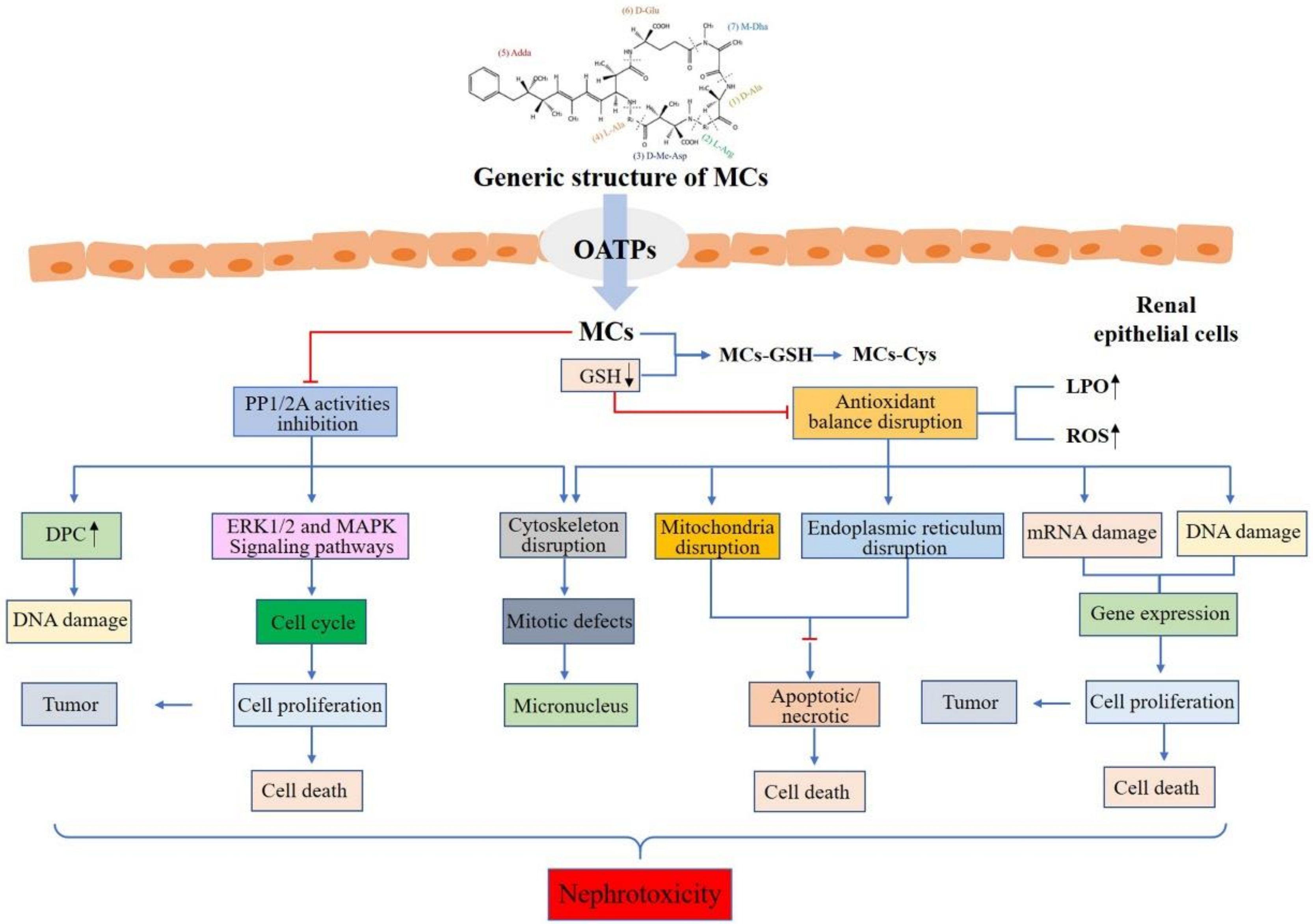
5. Potential Mechanisms of Action

MCs can not only induce damage to renal cells in vitro, but also induce pathological injury in vivo, leading to the inhibition of protein phosphatases, production of oxidative stress, cytoskeleton disruption, endoplasmic reticulum stress, DNA damage, and cell apoptosis. Figure 2 describes the toxic effect and nephrotoxicity mechanisms of MCs.

5.1. Cellular Uptake of MCs

In order to exert nephrotoxicity, a sufficient concentration of MCs must enter the urinary system. Cellular uptake of MCs has been shown to occur entirely through active transport, while passive transmembrane diffusion can be eliminated due to the high molecular weight and structure of MCs [52]. In fact, the active transmembrane transport of MCs is mediated by a specific organic anion transport peptide (OATP) [53,54,113]. Therefore, the systemic distribution of MCs in organs will depend on blood perfusion and the type and expression level of OATP carriers [52,87]. The presence of MCs in the kidney of rodents [42,46] and fishes [48,114] suggests that MCs were able to get across the renal plasma membrane. A meaningful experiment conducted by Sun et al. [83] revealed that after exposure to MC-RR, rats and mice showed different outcomes. In rats group, polyuria and hematuria appeared in the high dose group, and pathological findings were destruction of normal structure of kidney, while serological results showed decreased uric acid and increased creatinine. In contrast, there was no renal damage found in serum biochemical and renal histopathological examination in mice group. Further study found that there were differences in the type and quantity of Oatps expressed in rat and mice kidneys. Oatp1a1 was highly expressed in the kidneys of both rats and mice, while Oatp1a3 was highly expressed in the kidneys of rats. Hence, the difference in renal injury effect between rats and mice may be due to the difference in the expression of Oatps in kidney tissue. The uptake mechanisms of MCs by urinary systems need to be further investigated.

5.2. Modulation of PP2A Activities

MCs are highly effective and specific inhibitors of protein phosphatase 1/2A (PP1/2A), which can lead to phosphorylation/dephosphorylation imbalance of key control proteins [18,115,116]. MCs are found to interact with protein phosphatases (PPs) catalytic subunit through a two-step reaction including rapid linking and inactivation of the catalytic subunits, followed by an irreversible binding with cysteine. This combined model is why MCs can inhibit the catalytic activity of PPs and break the balance between protein phosphorylation and dephosphorylation [117]. Recent reports indicated the inhibition of PPs enzyme activity in kidney through both direct and indirect methods [41,102,108,118]. In addition, Dias et al. [40] showed that nephrotoxicity in members of mitogen-activated protein kinases p38, JNK and ERK1/2 of mitogen-activated protein kinase (MAPK) signaling pathways increased significantly under MC-LR exposure. Furthermore, impacts of MC-LR on cytoskeletal disruption, metabolic disorder, cell cycle arrest, abnormal cell proliferation, and cell death have been related to PPs activity and the increased phosphorylation of certain proteins [119].

5.3. Oxidative Stress

It is well known that the urinary system is highly sensitive to oxidative stress and lipid peroxidation (LPO), however, their significant increase may lead to renal toxicity [120]. Kidneys are rich in unsaturated lipids, thus they are vulnerable to peroxidation damage. On the one hand, protein carbonylation is widely used to evaluate the degree of oxidative damage of proteins in various biological organisms. After mice were injected with MC-LR for 7 days, Dong et al. [88] found that the content of protein carbonyl in kidney was remarkably increased in high-dose MC-LR group, suggesting that MC-LR had direct oxidative damage on kidney protein. On the other hand, alteration of the antioxidant enzyme system is another reliable evidence to exert oxidative stress. After exposure to MC-LR, the levels of antioxidant enzymes including SOD, CAT, GSH-Px, and GR decreased remarkably in Wistar rats kidney [29,90]. In the case of SD rats kidney, the elevated MDA contents as well as declined antioxidant enzymes involving CAT and SOD showed the occurrence of oxidative stress [121]. Similarly, Kim et al. [122] also discovered that some antioxidant enzymes could be disturbed by exposure to MCs in the kidneys of male Wistar rats. Jayaraj et al. [120] observed a downtrend of antioxidant enzymes in mice kidney exposed to MCs. MCs can also induce oxidative stress in fish, meanwhile, vitamin E can effectively remove ROS and exogenous substances and finally protect the kidney from damage successfully [109]. Similarly, Han et al. [91] also discovered that GSH may reach a certain protective effect on kidney by reducing the lipid peroxidation, improving the antioxidant activity and removing oxygen free radicals under exposure to MC-LR in mice. In vitro studies also showed that the oxidative stress by MCs might induce renal toxicity. MC-LR could induce CIK cells to produce expression of ROS and MDA and regulate the activities of antioxidant enzymes involving CAT, SOD and GSH [112]. However, it is not fully understood why and how MCs exposure can lead to excessive ROS formation that culminates in oxidative damage of the urinary system. This requires more research in the future to overcome this challenge.

5.4. Cytoskeleton Disruption

MCs exert a toxic role by causing blistering of cell membrane, loss of membrane integrity, cell coagulation, and the formation of apoptotic bodies, which lead to the disintegration of cytoskeleton [123,124]. The cytoskeleton consists of three elements: microfilaments (MFs), microtubules (MTs) and intermediate filaments (IFs) [125]. In renal cells, MCs exposure triggered the disintegration of IFs and MTs, followed by MFs. In addition, actin under the plasma membrane began to gather and condense into rose-like structures, which eventually collapsed into dense perinuclear bundles, and the cytoskeleton around the nucleus gradually collapsed [117]. Milutinovic et al. [95] revealed that sub-chronic treatment of MCs could induce cytoskeletal alterations. Huang et al. [112] also observed aggregation and collapse of MFs and MTs in CIK cells and even loss of some cytoskeleton structure. Moreover, the authors also detected transcriptional changes of cytoskeletal genes: β-actin, lc3a, and keratin.

5.5. DNA Damage

DNA-protein crosslinking (DPC) is a stable covalent compound formed by DNA and protein. As a molecular biomarker of the toxicity of foreign chemicals, DPC has attracted much attention in recent years [88]. DPC has a certain background level in normal cells. If the body is affected by external physical and chemical factors, it can induce excessive DPC, which will affect gene expression and destroy the structure of chromosomes. Xiong et al. [29] noted that the number of DPC increased significantly when MC-LR was exposed to 3 and 6 μg/kg bw, and the authors speculated that MC-LR exposure could cause DNA damage in mice renal cells. On the other hand, Dias et al. [126] found that DNA lesions were detected in renal micronucleus (Vero-E6) with 5 and 20 μM MC-LR, while the increased DNA damage only occurred temporarily 30 min after exposure to MC-LR by comet assay. In contrast, Gaudin et al. [89] detected that DNA damage in any case would be induced by MC-LR regardless of the route of administration in the kidney. If these increasing lesions occur in kidney, the progress of proliferation in normal renal cell would be affected, resulting in increasing the potential of renal tumorigenesis. Li et al. [38] showed that the expression of c-fos, c-jun and c-myc in kidney might be one possible mechanism for the tumor-promoting activity and initiating activity of MCs. Hao et al. [39] also found that tumor-associated stathmin was significantly correlated with the kidney concentration of MCs. Thus, the alteration of DNA or tumor-associated genes may provide a possible clue to the tumor-promoting potential of MCs in the kidney.

5.6. Apoptosis

There are strong evidences suggesting apoptosis can play a key role in MC-induced renal toxicity [95,112]. Chen et al. [97] detected that the expression of Bax protein increased in different exposure times when rat kidney cells were exposed to different concentrations of MC-LR. Pathmalal et al. [127] investigated the cytotoxicity and possible apoptotic influence of MC-LR on human embryonic kidney cell line (HEK-293) and human kidney adenocarcinoma cell line (ACHN). After exposure to MC-LR for 24 h, the activities of caspase3 and caspase9 in ACHN and HEK-293 cells increased significantly, indicating that the MC-LR induced cytotoxicity and an obvious apoptosis in both ACHN and HEK-293 kidney cell lines. Following exposure of M. aeruginosa equivalent to 400 μg/kg MC-LR over 72 h on carp, Fischer et al. [58] observed cavitation for single epithelial cells, cell shedding, apoptosis, and eventual protein-like exfoliation at the cortex-medulla junction. In addition, Wang et al. [34] showed that MCs exposure induced renal cell apoptosis by TUNEL. Additionally, the toxin caused dysfunction in protein processing, cell cycle and oxidative phosphorylation of endoplasmic reticulum. Moreover, Qin et al. [36] explored whether effects of MC-LR on apoptosis of renal cells were through the ERS pathway. The authors found that MC-LR inhibited expression of CHOP and caspase-12 in the kidney, followed by up-regulation of Bcl-2 mRNA, which suggests that apoptosis of renal cells induced by MC-LR may be through the ERS pathway.

6. Conclusions

MCs have become the most widely distributed and studied cyanotoxin, and it has become one of the biggest public health problems in the world. Long-term exposure to MCs is prone to cause nephrotoxicity. In the present review, the toxic effects of MCs on kidney were summarized. MCs can cross the glomerular filtration barrier by OATPs after exposure to multiple channels, which may lead to structural and functional changes in the kidney through various mechanisms. Exposure to MCs is highlighted to be fundamentally related to destroying PP2A, cytoskeleton and oxidative phosphorylation system; promote the formation of intracellular ROS; and further induce mitochondrial dysfunction and endoplasmic reticulum stress, which lead to miRNA dysfunction and DNA damage, and finally result in apoptosis or genotoxicity.
However, evidence from high-quality epidemiological study and expounded mechanisms of nephrotoxicity are still limited. Consequently, further studies are required to bridge the study gab. The following are suggested future research directions: (1) Even though many studies have been done on MCs, there is lack of consensus. Therefore, standardized animal model method(s) for assessing acute and chronic MCs exposures should be estimated and established to support direct comparisons between different studies and further to establish evidence weights to support risk assessment. (2) The current drinking water quality standards only provide information to control a few limited microcystins, but there is a relative lack of information on the health effects of a large number of other microcystin variants. Research should be strengthened to provide a basis for government officials to formulate laws and regulations. (3) To strengthen the combined effects of MCs and other environmental pollutants such as melamine, heavy metals, phthalates and pesticide residues to the kidney. (4) To develop accessible and advanced biodegradation technique to degrade MCs from cyanobacterial blooms. (5) Long-term exposure to MCs experiments in laboratory and large cohort studies in high risk area are necessary to verify the relationship between MCs exposure and high incidence of kidney disease. (6) To develop some chemical protective agents against nephrotoxicity caused by MCs. All these scientific challenges and practical values need to be addressed step by step.

Author Contributions

F.Y., S.X. and X.Y. conceived and designed the article; S.X., W.L. and C.Z. designed the tables and figures; L.T. contributed to the supervision and funding acquisition; S.X., X.Y. and I.Y.M. wrote the paper. All authors have read and agreed to the published version of the manuscript.

Funding

This research was funded by the Key Research and Development Projects in Hunan Province (2019SK2041), Hunan Province Excellent Youth Fund (2020JJ3053), National Natural Science Foundation (81773393, 81502787), Hunan Provincial Innovation Foundation for Postgraduate (CX20190238).

Conflicts of Interest

The authors declare that there are no conflicts of interest.

References

  1. Merel, S.; Walker, D.; Chicana, R.; Snyder, S.; Baurès, E.; Thomas, O. State of knowledge and concerns on cyanobacterial blooms and cyanotoxins. Environ. Int. 2013, 59, 303–327. [Google Scholar] [CrossRef] [PubMed]
  2. Boopathi, T.K.J. Impact of Environmental Factors on the Regulation of Cyanotoxin Production. Toxins 2014, 6, 1951–1978. [Google Scholar] [CrossRef] [PubMed] [Green Version]
  3. Alosman, M.; Cao, L.; Massey, I.Y.; Yang, F. The lethal effects and determinants of microcystin-LR on heart: A mini review. Toxin Rev. 2020, 1–10. [Google Scholar] [CrossRef]
  4. Massey, I.Y.; Yang, F.; Ding, Z.; Yang, S.; Guo, J.; Tezi, C.; Al-Osman, M.; Kamegni, R.B.; Zeng, W. Exposure routes and health effects of microcystins on animals and humans: A mini-review. Toxicon 2018, 151, 156–162. [Google Scholar] [CrossRef] [PubMed]
  5. Rastogi, R.P.; Sinha, R.P.; Incharoensakdi, A. The cyanotoxin-microcystins: Current overview. Rev. Environ. Sci. Biotechnol. 2014, 13, 215–249. [Google Scholar] [CrossRef]
  6. Yang, F.; Massey, I.Y.; Guo, J.; Yang, S.; Pu, Y.; Zeng, W.; Tan, H. Microcystin-LR degradation utilizing a novel effective indigenous bacterial community YFMCD1 from Lake Taihu. J. Toxicol. Environ. Health A 2018, 81, 184–193. [Google Scholar] [CrossRef]
  7. Yang, F.; Zhou, Y.; Sun, R.; Wei, H.; Li, Y.; Yin, L.; Pu, Y. Biodegradation of microcystin-LR and-RR by a novel microcystin-degrading bacterium isolated from Lake Taihu. Biodegradation 2014, 25, 447–457. [Google Scholar] [CrossRef]
  8. Bouaïcha, N.; Miles, C.O.; Beach, D.G.; Labidi, Z.; Djabri, A.; Benayache, N.Y.; Nguyen-Quang, T. Structural Diversity, Characterization and Toxicology of Microcystins. Toxins 2019, 11, 714. [Google Scholar] [CrossRef] [Green Version]
  9. Gupta, N.; Pant, S.C.; Vijayaraghavan, R.; Rao, P.V.L. Comparative toxicity evaluation of cyanobacterial cyclic peptide toxin microcystin variants (LR, RR, YR) in mice. Toxicology 2003, 188, 285–296. [Google Scholar] [CrossRef]
  10. Massey, I.Y.; Zhang, X.; Yang, F. Importance of bacterial biodegradation and detoxification processes of microcystins for environmental health. J. Toxicol. Environ. Health Part B 2018, 21, 357–369. [Google Scholar] [CrossRef]
  11. Chen, L.; Chen, J.; Zhang, X.; Xie, P. A review of reproductive toxicity of microcystins. J. Hazard. Mater. 2016, 301, 381–399. [Google Scholar] [CrossRef]
  12. Massey, I.Y.; Wu, P.; Wei, J.; Luo, J.; Ding, P.; Wei, H.; Yang, F. A Mini-Review on Detection Methods of Microcystins. Toxins 2020, 12, 641. [Google Scholar] [CrossRef] [PubMed]
  13. Gilroy, D.J.; Kauffman, K.W.; Hall, R.A.; Huang, X.; Chu, F.S. Assessing Potential Health Risks from Microcystin Toxins in Blue-Green Algae Dietary Supplements. Environ. Health Perspect 2000, 108, 435–439. [Google Scholar] [CrossRef]
  14. Pouria, S.; de Andrade, A.; Barbosa, J.; Cavalcanti, R.L.; Barreto, V.T.; Ward, C.J.; Preiser, W.; Poon, G.K.; Neild, G.H.; Codd, G.A. Fatal microcystin intoxication in haemodialysis unit in Caruaru, Brazil. Lancet 1998, 352, 21–26. [Google Scholar] [CrossRef]
  15. Chen, L.; Giesy, J.P.; Xie, P. The dose makes the poison. Sci. Total Environ. 2018, 621, 649–653. [Google Scholar] [CrossRef] [PubMed]
  16. Yang, F.; Huang, F.; Feng, H.; Wei, J.; Massey, I.Y.; Liang, G.; Zhang, F.; Yin, L.; Kacew, S.; Zhang, X.; et al. A complete route for biodegradation of potentially carcinogenic cyanotoxin microcystin-LR in a novel indigenous bacterium. Water Res. 2020, 174, 115638. [Google Scholar] [CrossRef] [PubMed]
  17. Massey, I.Y.; Yang, F. A Mini Review on Microcystins and Bacterial Degradation. Toxins 2020, 12. [Google Scholar] [CrossRef] [Green Version]
  18. MacKintosh, C.; Beattie, K.A.; Klumpp, S.; Cohen, P.; Codd, G.A. Cyanobacterial microcystin-LR is a potent and specific inhibitor of protein phosphatases 1 and 2A from both mammals and higher plants. FEBS Lett. 1990, 264, 187–192. [Google Scholar] [CrossRef] [Green Version]
  19. Yang, F.; Wen, C.; Zheng, S.; Yang, S.; Chen, J.; Feng, X. Involvement of MAPK/ERK1/2 pathway in microcystin-induced microfilament reorganization in HL7702 hepatocytes. J. Toxicol. Environ. Health Part A 2018, 81, 1135–1141. [Google Scholar] [CrossRef]
  20. Li, Y.; Chen, J.A.; Zhao, Q.; Pu, C.; Qiu, Z.; Zhang, R.; Shu, W. A cross-sectional investigation of chronic exposure to microcystin in relationship to childhood liver damage in the Three Gorges Reservoir Region, China. Environ. Health Perspect 2011, 119, 1483–1488. [Google Scholar] [CrossRef] [Green Version]
  21. Zheng, C.; Zeng, H.; Lin, H.; Wang, J.; Feng, X.; Qiu, Z.; Chen, J.; Luo, J.; Luo, Y.; Huang, Y.; et al. Serum microcystin levels positively linked with risk of hepatocellular carcinoma: A case-control study in southwest China. Hepatology 2017, 66, 1519–1528. [Google Scholar] [CrossRef] [PubMed]
  22. Cao, L.; Huang, F.; Massey, I.Y.; Wen, C.; Zheng, S.; Xu, S.; Yang, F. Effects of Microcystin-LR on the Microstructure and Inflammation-Related Factors of Jejunum in Mice. Toxins 2019, 11, 482. [Google Scholar] [CrossRef] [PubMed] [Green Version]
  23. Wu, J.; Huang, H.; Yang, L.; Zhang, X.; Zhang, S.; Liu, H.; Wang, Y.; Yuan, L.; Cheng, X.; Zhuang, D.; et al. Gastrointestinal toxicity induced by microcystins. World J. Clin. Cases 2018, 6, 344–354. [Google Scholar] [CrossRef] [PubMed]
  24. Li, X.; Xu, L.; Zhou, W.; Zhao, Q.; Wang, Y. Chronic exposure to microcystin-LR affected mitochondrial DNA maintenance and caused pathological changes of lung tissue in mice. Environ. Pollut. 2016, 210, 48–56. [Google Scholar] [CrossRef] [PubMed]
  25. Soares, R.M.; Cagido, V.R.; Ferraro, R.B.; Meyer-Fernandes, J.R.; Rocco, P.R.M.; Zin, W.A.; Azevedo, S.M.F.O. Effects of microcystin-LR on mouse lungs. Toxicon 2007, 50, 330–338. [Google Scholar] [CrossRef]
  26. Cao, L.; Massey, I.Y.; Feng, H.; Yang, F. A Review of Cardiovascular Toxicity of Microcystins. Toxins 2019, 11, 507. [Google Scholar] [CrossRef] [Green Version]
  27. Hinojosa, M.G.; Gutiérrez-Praena, D.; Prieto, A.I.; Guzmán-Guillén, R.; Jos, A.; Cameán, A.M. Neurotoxicity induced by microcystins and cylindrospermopsin: A review. Sci. Total Environ. 2019, 668, 547–565. [Google Scholar] [CrossRef]
  28. Hu, Y.; Chen, J.; Fan, H.; Xie, P.; He, J. A review of neurotoxicity of microcystins. Environ. Sci. Pollut. Res. 2016, 23, 7211–7219. [Google Scholar] [CrossRef]
  29. Xiong, Q.; Xie, P.; Li, H.; Hao, L.; Li, G.; Qiu, T.; Liu, Y. Acute effects of microcystins exposure on the transcription of antioxidant enzyme genes in three organs (liver, kidney, and testis) of male Wistar rats. J. Biochem. Mol. Toxicol. 2010, 24, 361–367. [Google Scholar] [CrossRef]
  30. Yi, X.; Xu, S.; Huang, F.; Wen, C.; Zheng, S.; Feng, H.; Guo, J.; Chen, J.; Feng, X.; Yang, F. Effects of Chronic Exposure to Microcystin-LR on Kidney in Mice. Int. J. Environ. Res. Public Health 2019, 16, 5030. [Google Scholar] [CrossRef] [Green Version]
  31. Lin, H.; Liu, W.; Zeng, H.; Pu, C.; Zhang, R.; Qiu, Z.; Chen, J.; Wang, L.; Tan, Y.; Zheng, C.; et al. Determination of Environmental Exposure to Microcystin and Aflatoxin as a Risk for Renal Function Based on 5493 Rural People in Southwest China. Environ. Sci. Technol. 2016, 50, 5346–5356. [Google Scholar] [CrossRef] [PubMed]
  32. Qiu, T.; Xie, P.; Li, L.; Guo, L.; Zhang, D.; Zhou, Q. Nephrotoxic effects from chronic toxic cyanobacterial blooms in fishes with different trophic levels in a large Chinese lake. Environ. Toxicol. Pharmacol. 2012, 33, 252–261. [Google Scholar] [CrossRef] [PubMed]
  33. Sitprija, V.; Sitprija, S. Marine toxins and nephrotoxicity: Mechanism of injury. Toxicon 2019, 161, 44–49. [Google Scholar] [CrossRef] [PubMed]
  34. Wang, Z.; Li, G.; Wu, Q.; Liu, C.; Shen, J.; Yan, W. Microcystin-LR exposure induced nephrotoxicity by triggering apoptosis in female zebrafish. Chemosphere 2019, 214, 598–605. [Google Scholar] [CrossRef]
  35. La-Salete, R.; Oliveira, M.M.; Palmeira, C.A.; Almeida, J.; Peixoto, F.P. Mitochondria a key role in microcystin-LR kidney intoxication. J. Appl. Toxicol. 2008, 28, 55–62. [Google Scholar] [CrossRef] [PubMed] [Green Version]
  36. Qin, W.; Xu, L.; Zhang, X.; Wang, Y.; Meng, X.; Miao, A.; Yang, L. Endoplasmic reticulum stress in murine liver and kidney exposed to microcystin-LR. Toxicon 2010, 56, 1334–1341. [Google Scholar] [CrossRef]
  37. Li, X. Effects of Chronic Exposure to Microcystin-LR on Somatic Mitochondrial DNA in Mice. Ph.D. Thesis, Nanjing University, Nanjing, China, 2015. [Google Scholar]
  38. Li, H.; Xie, P.; Li, G.; Hao, L.; Xiong, Q. In vivo study on the effects of microcystin extracts on the expression profiles of proto-oncogenes (c-fos, c-jun and c-myc) in liver, kidney and testis of male Wistar rats injected i.v. with toxins. Toxicon 2009, 53, 169–175. [Google Scholar] [CrossRef]
  39. Hao, L.; Xie, P.; Li, H.; Li, G.; Xiong, Q.; Wang, Q.; Qiu, T.; Liu, Y. Transcriptional alteration of cytoskeletal genes induced by microcystins in three organs of rats. Toxicon 2010, 55, 1378–1386. [Google Scholar] [CrossRef]
  40. Dias, E.; Matos, P.; Pereira, P.; Batoréu, M.C.C.; Silva, M.J.; Jordan, P. Microcystin-LR activates the ERK1/2 kinases and stimulates the proliferation of the monkey kidney-derived cell line Vero-E6. Toxicol. In Vitro 2010, 24, 1689–1695. [Google Scholar] [CrossRef]
  41. Fan, H.; Cai, Y.; Xie, P.; Xiao, W.; Chen, J.; Ji, W.; Zhao, S. Microcystin-LR stabilizes c-myc protein by inhibiting protein phosphatase 2A in HEK293 cells. Toxicology 2014, 319, 69–74. [Google Scholar] [CrossRef]
  42. Wang, Q.; Xie, P.; Chen, J.; Liang, G. Distribution of microcystins in various organs (heart, liver, intestine, gonad, brain, kidney and lung) of Wistar rat via intravenous injection. Toxicon 2008, 52, 721–727. [Google Scholar] [CrossRef]
  43. Lei, H.; Xie, P.; Chen, J.; Liang, G.; Dai, M.; Zhang, X. Distribution of toxins in various tissues of crucian carp intraperitoneally injected with hepatotoxic microcystins. Environ. Toxicol. Chem. 2008, 27, 1167–1174. [Google Scholar] [CrossRef]
  44. Lei, H.; Xie, P.; Chen, J.; Liang, G.; Yu, T.; Jiang, Y. Tissue Distribution and Depuration of the Extracted Hepatotoxic Cyanotoxin Microcystins in Crucian Carp (Carassius carassius) Intraperitoneally Injected at a Sublethal Dose. Sci. World J. 2008, 8, 713–719. [Google Scholar] [CrossRef] [Green Version]
  45. Moutou, K.A.; Tsikogias, S.; Papadimitriou, T.; Kagalou, I. Oxidative stress in Cyprinus carpio to analyze microcystin impact in eutrophic shallow lakes: A preliminary study. J. Environ. Monit. 2012, 14, 2195–2203. [Google Scholar] [CrossRef] [PubMed]
  46. Sedan, D.; Giannuzzi, L.; Rosso, L.; Marra, C.A.; Andrinolo, D. Biomarkers of prolonged exposure to microcystin-LR in mice. Toxicon 2013, 68, 9–17. [Google Scholar] [CrossRef] [PubMed]
  47. Mitsoura, A.; Kagalou, I.; Papaioannou, N.; Berillis, P.; Mente, E.; Papadimitriou, T. The presence of microcystins in fish Cyprinus carpio tissues: A histopathological study. Int. Aquat. Res. 2013, 5, 1–16. [Google Scholar] [CrossRef] [Green Version]
  48. Bi, X.; Dai, W.; Wang, X.; Dong, S.; Zhang, S.; Zhang, D.; Wu, M. Microcystins distribution, bioaccumulation, and Microcystis genotype succession in a fish culture pond. Sci. Total Environ. 2019, 688, 380–388. [Google Scholar] [CrossRef]
  49. Jia, J.; Luo, W.; Lu, Y.; Giesy, J.P. Bioaccumulation of microcystins (MCs) in four fish species from Lake Taihu, China: Assessment of risks to humans. Sci. Total Environ. 2014, 487, 224–232. [Google Scholar] [CrossRef] [PubMed]
  50. Singh, S.; Asthana, R.K. Assessment of Microcystin Concentration in Carp and Catfish: A Case Study from Lakshmikund Pond, Varanasi, India. Bull. Environ. Contam. Toxicol. 2014, 92, 687–692. [Google Scholar] [CrossRef] [PubMed]
  51. Ni, W.; Zhang, J.; Luo, Y. Microcystin accumulation in bighead carp (Aristichthys nobilis) during a Microcystis-dominated bloom and risk assessment of the dietary intake in a fish pond in China. Environ. Sci. Pollut. Res. Int. 2015, 24, 8894–8902. [Google Scholar] [CrossRef]
  52. Campos, A.; Vasconcelos, V. Molecular Mechanisms of Microcystin Toxicity in Animal Cells. Int. J. Mol. Sci. 2010, 11, 268–287. [Google Scholar] [CrossRef] [PubMed] [Green Version]
  53. Lu, H.; Choudhuri, S.; Ogura, K.; Csanaky, I.L.; Lei, X.; Cheng, X.; Song, P.; Klaassen, C.D. Characterization of Organic Anion Transporting Polypeptide 1b2-null Mice: Essential Role in Hepatic Uptake/Toxicity of Phalloidin and Microcystin-LR. Toxicol. Sci. 2008, 103, 35–45. [Google Scholar] [CrossRef] [PubMed] [Green Version]
  54. Fischer, A.; Hoeger, S.J.; Stemmer, K.; Feurstein, D.J.; Knobeloch, D.; Nussler, A.; Dietrich, D.R. The role of organic anion transporting polypeptides (OATPs/SLCOs) in the toxicity of different microcystin congeners in vitro: A comparison of primary human hepatocytes and OATP-transfected HEK293 cells. Toxicol. Appl. Pharm. 2010, 245, 9–20. [Google Scholar] [CrossRef] [Green Version]
  55. Feurstein, D.; Holst, K.; Fischer, A.; Dietrich, D.R. Oatp-associated uptake and toxicity of microcystins in primary murine whole brain cells. Toxicol. Appl. Pharm. 2009, 234, 247–255. [Google Scholar] [CrossRef] [Green Version]
  56. Zhang, Z.; Yu, S.; Wei, G.; Chen, W. Study on the whole and cells level distribution of Microcystin LR in mice. J. Toxicol. 2002, 16, 5–8. [Google Scholar]
  57. Li, L.; Xie, P.; Lei, H.; Zhang, X. Renal accumulation and effects of intraperitoneal injection of extracted microcystins in omnivorous crucian carp (Carassius auratus). Toxicon 2013, 70, 62–69. [Google Scholar] [CrossRef]
  58. Fischer, W.J.; Dietrich, D.R. Pathological and Biochemical Characterization of Microcystin-Induced Hepatopancreas and Kidney Damage in Carp (Cyprinus carpio). Toxicol. Appl. Pharm. 2000, 164, 73–81. [Google Scholar] [CrossRef] [Green Version]
  59. Greer, B.; Meneely, J.P.; Elliott, C.T. Uptake and accumulation of Microcystin-LR based on exposure through drinking water: An animal model assessing the human health risk. Sci. Rep. UK 2018, 8, 4913. [Google Scholar] [CrossRef] [Green Version]
  60. Falconer, I.R.; Burch, M.D.; Steffensen, D.A.; Choice, M.; Coverdale, O.R. Toxicity of the blue-green alga (cyanobacterium) Microcystis aeruginosa in drinking water to growing pigs, as an animal model for human injury and risk assessment. Environ. Toxicol. Water Qual. 1994, 9, 131–139. [Google Scholar] [CrossRef]
  61. Foss, A.J.; Aubel, M.T.; Gallagher, B.; Mettee, N.; Miller, A.; Fogelson, S.B. Diagnosing Microcystin Intoxication of Canines: Clinicopathological Indications, Pathological Characteristics, and Analytical Detection in Postmortem and Antemortem Samples. Toxins 2019, 11, 456. [Google Scholar] [CrossRef] [Green Version]
  62. Bautista, A.C.; Moore, C.E.; Lin, Y.; Cline, M.G.; Benitah, N.; Puschner, B. Hepatopathy following consumption of a commercially available blue-green algae dietary supplement in a dog. BMC Vet. Res. 2015, 11. [Google Scholar] [CrossRef] [Green Version]
  63. Rankin, K.; Alroy, K.; Kudela, R.; Oates, S.; Murray, M.; Miller, M. Treatment of Cyanobacterial (Microcystin) Toxicosis Using Oral Cholestyramine: Case Report of a Dog from Montana. Toxins 2013, 5, 1051–1063. [Google Scholar] [CrossRef] [PubMed]
  64. Hermansky, S.J.; Stohs, S.J.; Eldeen, Z.M.; Roche, V.F.; Mereish, K.A. Evaluation of potential chemoprotectants against microcystin-LR hepatotoxicity in mice. J. Appl. Toxicol. 1991, 11, 65–73. [Google Scholar] [CrossRef]
  65. Kondo, F.; Ikai, Y.; Oka, H.; Okumura, M.; Ishikawa, N.; Harada, K.; Matsuura, K.; Murata, H.; Suzuki, M. Formation, characterization, and toxicity of the glutathione and cysteine conjugates of toxic heptapeptide microcystins. Chem. Res. Toxicol. 1992, 5, 591–596. [Google Scholar] [CrossRef]
  66. Wu, L.; Wang, Q.; Tao, M.; Chen, J.; Ma, Z.; Xie, P. Preliminary Study of the Distribution and Accumulation of GSH/Cys Metabolites of Hepatotoxic Microcystins-RR in Common Carp from a Lake with Protracted Cyanobacterial Bloom (Lake Taihu, China). Bull. Environ. Contam. Toxicol. 2013, 90, 382–386. [Google Scholar] [CrossRef] [PubMed]
  67. Wu, L.; Wang, S.; Tao, M.; Xie, P.; Chen, J. Quantitative analysis of glutathione and cysteine S-conjugates of microcystin-LR in the liver, kidney and muscle of common carp (Cyprinus carpio) in Lake Taihu. J. Water Health 2017, 15, 300–307. [Google Scholar] [CrossRef] [PubMed]
  68. He, J.; Chen, J.; Xie, P.; Zhang, D.; Li, G.; Wu, L.; Zhang, W.; Guo, X.; Li, S. Quantitatively evaluating detoxification of the hepatotoxic microcystins through the glutathione and cysteine pathway in the cyanobacteria-eating bighead carp. Aquat. Toxicol. 2012, 116–117, 61–68. [Google Scholar] [CrossRef]
  69. Chen, F.; Chen, J.; Xie, P.; Li, W.; Yu, Y.; Zhang, X.; Han, X.; Yu, X. Quantitative Study in the Process of Glutathione Detoxification of Microcystin. Asian J. Ecotoxicol. 2018, 13, 99–105. [Google Scholar]
  70. Li, W.; Chen, J.; Xie, P.; He, J.; Guo, X.; Tuo, X.; Zhang, W.; Wu, L. Rapid conversion and reversible conjugation of glutathione detoxification of microcystins in bighead carp (Aristichthys nobilis). Aquat. Toxicol. 2014, 147, 18–25. [Google Scholar] [CrossRef]
  71. Li, W.; He, J.; Chen, J.; Xie, P. Excretion pattern and dynamics of glutathione detoxification of microcystins in Sprague Dawley rat. Chemosphere 2018, 191, 357–364. [Google Scholar] [CrossRef] [PubMed]
  72. Soares, R.M.; Yuan, M.; Servaites, J.C.; Delgado, A.; Magalhães, V.F.; Hilborn, E.D.; Carmichael, W.W.; Azevedo, S.M.F.O. Sublethal exposure from microcystins to renal insufficiency patients in Rio de Janeiro, Brazil. Environ. Toxicol. 2006, 21, 95–103. [Google Scholar] [CrossRef] [PubMed]
  73. Hilborn, E.D.; Carmichael, W.W.; Soares, R.M.; Yuan, M.; Servaites, J.C.; Barton, H.A.; Azevedo, S.M.F.O. Serologic evaluation of human microcystin exposure. Environ. Toxicol. 2007, 22, 459–463. [Google Scholar] [CrossRef] [PubMed]
  74. Madhushankha, L.; Dhammika, M.A.; Charitha, P.; Tilak, A.; Lishantha, G. Cyanobacteria and cyanotoxins in well waters of the Girandurukotte, CKDu endemic area in Sri Lanka; do they drink safe water? J. Ecotechnol. Res. 2016, 1, 17–21. [Google Scholar]
  75. Chen, J.; Xie, P.; Li, L.; Xu, J. First Identification of the Hepatotoxic Microcystins in the Serum of a Chronically Exposed Human Population Together with Indication of Hepatocellular Damage. Toxicol. Sci. 2009, 108, 81–89. [Google Scholar] [CrossRef] [PubMed]
  76. R Bhattacharya, K.S.R.S. Toxicity Evaluation of Freshwater Cyanobacterium Microcystis Aeruginosa PCC 7806: II. Nephrotoxicity in Rats. Biomed. Environ. Sci. 1997, 10, 93–101. [Google Scholar]
  77. Pan, X.; Chang, F.; Liu, Y.; Li, D.; Xu, A.; Shen, Y.; Huang, Z. Mouse toxicity ofAnabaena flos-aquae from Lake Dianchi, China. Environ. Toxicol. 2009, 24, 10–18. [Google Scholar] [CrossRef]
  78. Humpage, A.; Falconer, I.; Bernard, C.; Froscio, S.; Fabbro, L. Toxicity of the cyanobacterium Limnothrix AC0243 to male Balb/c mice. Water Res. 2012, 46, 1576–1583. [Google Scholar] [CrossRef]
  79. Li, G.; Xie, P.; Li, H.; Hao, L.; Xiong, Q.; Qiu, T.; Liu, Y. Acute effects of microcystins on the transcription of 14 glutathione S-transferase isoforms in Wistar rat. Environ. Toxicol. 2011, 26, 187–194. [Google Scholar] [CrossRef]
  80. Yang, L.; Shen, F.; Lu, B.; Tong, X.; Qin, J.; Fu, Z. Study the Toxicity of Microcystin in Water of Dianchi Lake on Mice Kidney. J. Kunming Univ. 2012, 34, 52–54. [Google Scholar]
  81. Adamovsky, O.; Palikova, M.; Ondrackova, P.; Zikova, A.; Kopp, R.; Mares, J.; Pikula, J.; Paskerova, H.; Kohoutek, J.; Blaha, L. Biochemical and histopathological responses of Wistar rats to oral intake of microcystins and cyanobacterial biomass. Neuro Endocrinol. Lett. 2013, 34 (Suppl. 2), 11–20. [Google Scholar]
  82. Hooser, S.B.; Beasley, V.R.; Lovell, R.A.; Carmichael, W.W.; Haschek, W.M. Toxicity of microcystin LR, a cyclic heptapeptide hepatotoxin from Microcystis aeruginosa, to rats and mice. Vet. Pathol. 1989, 26, 246–252. [Google Scholar] [CrossRef] [Green Version]
  83. Sun, F. Comparison of the Effect of Microcytin-RR on Acute Kidney, Liver Injury and Their Possible Mechanisms in Rats and Mice. Master’s Thesis, Huazhong University of Science and Technology, Wuhan, China, 2017. [Google Scholar]
  84. Lowe, J.; Souza-Menezes, J.; Freire, D.S.; Mattos, L.J.; Castiglione, R.C.; Barbosa, C.M.L.; Santiago, L.; Ferrão, F.M.; Cardoso, L.H.D.; Da Silva, R.T.; et al. Single sublethal dose of microcystin-LR is responsible for different alterations in biochemical, histological and physiological renal parameters. Toxicon 2012, 59, 601–609. [Google Scholar] [CrossRef]
  85. Lamei, L.; Lirong, S. Acute toxicity of microcystin-LR in BALB/c mice. J. First Med. Coll. PLA 2005, 25, 565–566. [Google Scholar]
  86. Xu, C. Microcystin Contamination in Aquatic Environment of the Three Gorges Reservior and Antagonism Study on MC-LR Induced Toxicity. Master’s Thesis, Third Military Medical University, Chongqing, China, 2005. [Google Scholar]
  87. Zhong, Q. Effect of Microcystin-RR on Water Metabolism and Neurobehavior in Mice and Related Mechanism Exporation. Ph.D. Thesis, Huazhong University of Science and Technology, Wuhan, China, 2017. [Google Scholar]
  88. Dong, L. Study on Toxicity of Microcystin-LR on Liver, Kidney and Testicle Cells in Male Mice. Master’s Thesis, Zhengzhou University, Zhengzhou, China, 2008. [Google Scholar]
  89. Gaudin, J.; Huet, S.; Jarry, G.; Fessard, V. In vivo DNA damage induced by the cyanotoxin microcystin-LR: Comparison of intra-peritoneal and oral administrations by use of the comet assay. Mutat. Res. Genet. Toxicol. Environ. Mutagenes. 2008, 652, 65–71. [Google Scholar] [CrossRef] [PubMed]
  90. Moreno, I.; Pichardo, S.; Jos, A.; Gómez-Amores, L.; Mate, A.; Vazquez, C.M.; Cameán, A.M. Antioxidant enzyme activity and lipid peroxidation in liver and kidney of rats exposed to microcystin-LR administered intraperitoneally. Toxicon 2005, 45, 395–402. [Google Scholar] [CrossRef] [PubMed]
  91. Han, Z.; He, M.; Cui, D.; Xiong, W.; Zhang, Q. The protective effects of glutathione on renal oxidative damage induced by microcystin-LR on mice. Chongqing Med. 2016, 45, 320–322. [Google Scholar]
  92. Li, Z. Study on Mechanisms of Microcystin-LR Toxicity Based on Metabonomics. Master’s Thesis, Jiangnan University, Wuxi, China, 2009. [Google Scholar]
  93. Sedan, D.; Andrinolo, D.; Telese, L.; Giannuzzi, L.; de Alaniz, M.J.T.; Marra, C.A. Alteration and recovery of the antioxidant system induced by sub-chronic exposure to microcystin-LR in mice: Its relation to liver lipid composition. Toxicon 2010, 55, 333–342. [Google Scholar] [CrossRef]
  94. Milutinović, A.; Sedmak, B.; Horvat-Znidarsic, I.; Suput, D. Renal injuries induced by chronic intoxication with microcystins. Cell. Mol. Biol. Lett. 2002, 7, 139. [Google Scholar]
  95. Milutinović, A.; Živin, M.; Zorc-Pleskovič, R.; Sedmak, B.; Šuput, D. Nephrotoxic effects of chronic administration of microcystins -LR and -YR. Toxicon 2003, 42, 281–288. [Google Scholar] [CrossRef]
  96. Khan, S.A.; Ghosh, S.; Wickstrom, M.; Miller, L.A.; Hess, R.; Haschek, W.M.; Beasley, V.R. Comparative pathology of microcystin-lr in cultured hepatocytes, fibroblasts, and renal epithelial cells. Nat. Toxins 1995, 3, 119–128. [Google Scholar] [CrossRef]
  97. Chen, J.; Xu, L.; Fu, W.; Xing, M. Bax expression of rat nnephrocyte NRK induced by microcystin-LR. Acta Hydrobiol. Sin. 2004, 28, 450–451. [Google Scholar]
  98. Nobre, A.C.; Jorge, M.C.; Menezes, D.B.; Fonteles, M.C.; Monteiro, H.S. Effects of microcystin-LR in isolated perfused rat kidney. Braz. J. Med. Biol. Res. 1999, 32, 985–988. [Google Scholar] [CrossRef] [Green Version]
  99. Dias, E.; Andrade, M.; Alverca, E.; Pereira, P.; Batoréu, M.C.C.; Jordan, P.; Silva, M.J. Comparative study of the cytotoxic effect of microcistin-LR and purified extracts from Microcystis aeruginosa on a kidney cell line. Toxicon 2009, 53, 487–495. [Google Scholar] [CrossRef] [PubMed]
  100. Alverca, E.; Andrade, M.; Dias, E.; Sam Bento, F.; Batoréu, M.C.C.; Jordan, P.; Silva, M.J.; Pereira, P. Morphological and ultrastructural effects of microcystin-LR from Microcystis aeruginosa extract on a kidney cell line. Toxicon 2009, 54, 283–294. [Google Scholar] [CrossRef]
  101. Menezes, C.; Alverca, E.; Dias, E.; Sam-Bento, F.; Pereira, P. Involvement of endoplasmic reticulum and autophagy in microcystin-LR toxicity in Vero-E6 and HepG2 cell lines. Toxicol. In Vitro 2013, 27, 138–148. [Google Scholar] [CrossRef]
  102. Li, T.; Ying, L.; Wang, H.; Li, N.; Fu, W.; Guo, Z.; Xu, L. Microcystin-LR induces ceramide to regulate PP2A and destabilize cytoskeleton in HEK293 cells. Toxicol. Sci. 2012, 128, 147–157. [Google Scholar] [CrossRef] [Green Version]
  103. Molina, R.; Moreno, I.; Pichardo, S.; Jos, A.; Moyano, R.; Monterde, J.G.; Cameán, A. Acid and alkaline phosphatase activities and pathological changes induced in Tilapia fish (Oreochromis sp.) exposed subchronically to microcystins from toxic cyanobacterial blooms under laboratory conditions. Toxicon 2005, 46, 725–735. [Google Scholar] [CrossRef]
  104. Li, L.; Xie, P.; Chen, J. Biochemical and ultrastructural changes of the liver and kidney of the phytoplanktivorous silver carp feeding naturally on toxic Microcystis blooms in Taihu Lake, China. Toxicon 2007, 49, 1042–1053. [Google Scholar] [CrossRef]
  105. Li, L.; Xie, P.; Guo, L.; Ke, Z.; Zhou, Q.; Liu, Y.; Qiu, T. Field and Laboratory Studies on Pathological and Biochemical Characterization of Microcystin-Induced Liver and Kidney Damage in the Phytoplanktivorous Bighead Carp. Sci. World J. 2008, 8, 121–137. [Google Scholar] [CrossRef] [Green Version]
  106. Atencio, L.; Moreno, I.; Jos, A.; Pichardo, S.; Moyano, R.; Blanco, A.; Cameán, A.M. Dose-dependent antioxidant responses and pathological changes in tenca (Tinca tinca) after acute oral exposure to Microcystis under laboratory conditions. Toxicon 2008, 52, 1–12. [Google Scholar] [CrossRef]
  107. Kotak, B.G.; Semalulu, S.; Fritz, D.L.; Prepas, E.E.; Hrudey, S.E.; Coppock, R.W. Hepatic and renal pathology of intraperitoneally administered microcystin-LR in rainbow trout (Oncorhynchus mykiss). Toxicon 1996, 34, 517–525. [Google Scholar] [CrossRef]
  108. Ma, J.; Feng, Y.; Xie, W.; Li, X. PP2A (PR65) in Silver Carp: cDNA Cloning and Expression Analysis. J. Biochem. Mol. Toxicol. 2015, 29, 399–409. [Google Scholar] [CrossRef]
  109. Prieto, A.I.; Jos, A.; Pichardo, S.; Moreno, I.; Camean, A.M. Protective role of vitamin E on the microcystin-induced oxidative stress in tilapia fish (Oreochromis niloticus). Environ. Toxicol. Chem. 2008, 27, 1152–1159. [Google Scholar] [CrossRef] [PubMed]
  110. Puerto, M.; Gutiérrez-Praena, D.; Prieto, A.I.; Pichardo, S.; Jos, A.; Miguel-Carrasco, J.L.; Vazquez, C.M.; Cameán, A.M. Subchronic effects of cyanobacterial cells on the transcription of antioxidant enzyme genes in tilapia (Oreochromis niloticus). Ecotoxicology 2011, 20, 479–490. [Google Scholar] [CrossRef]
  111. Feng, Y.; Ma, J.; Xiang, R.; Li, X. Alterations in microRNA expression in the tissues of silver carp (Hypophthalmichthys molitrix) following microcystin-LR exposure. Toxicon 2017, 128, 15–22. [Google Scholar] [CrossRef] [PubMed]
  112. Huang, X.; Chen, L.; Liu, W.; Qiao, Q.; Wu, K.; Wen, J.; Huang, C.; Tang, R.; Zhang, X. Involvement of oxidative stress and cytoskeletal disruption in microcystin-induced apoptosis in CIK cells. Aquat. Toxicol. 2015, 165, 41–50. [Google Scholar] [CrossRef] [PubMed]
  113. Fischer, W.J.; Altheimer, S.; Cattori, V.; Meier, P.J.; Dietrich, D.R.; Hagenbuch, B. Organic anion transporting polypeptides expressed in liver and brain mediate uptake of microcystin. Toxicol. Appl. Pharm. 2005, 203, 257–263. [Google Scholar] [CrossRef] [Green Version]
  114. Xia, H.; Song, T.; Wang, L.; Jiang, L.; Zhou, Q.; Wang, W.; Liu, L.; Yang, P.; Zhang, X. Effects of dietary toxic cyanobacteria and ammonia exposure on immune function of blunt snout bream (Megalabrama amblycephala). Fish Shellfish Immun. 2018, 78, 383–391. [Google Scholar] [CrossRef]
  115. Jayaraj, R.; Lakshmana Rao, P.V. Protein phosphorylation profile and adduct formation in liver and kidney of microcystin-LR-treated mice. Toxicon. 2006, 48, 272–277. [Google Scholar] [CrossRef]
  116. Wang, H.; Liu, J.; Lin, S.; Wang, B.; Xing, M.; Guo, Z.; Xu, L. MCLR-induced PP2A inhibition and subsequent Rac1 inactivation and hyperphosphorylation of cytoskeleton-associated proteins are involved in cytoskeleton rearrangement in SMMC-7721 human liver cancer cell line. Chemosphere 2014, 112, 141–153. [Google Scholar] [CrossRef]
  117. He, Y.; Huang, X.; Ding, X. Advances in Research on Toxicity and Mechanism of Action of Microcystins. Food Sci. 2020, 41, 290–298. [Google Scholar]
  118. Tavares, D.; Paulino, M.G.; Terezan, A.P.; Batista Fernandes, J.; Giani, A.; Fernandes, M.N. Osmoregulatory disturbance in Neotropical fish exposed to the crude extracts of the cyanobacterium, Radiocystis fernandoi. Aquat. Toxicol. 2019, 216, 105315. [Google Scholar] [CrossRef]
  119. Chen, L.; Xie, P. Mechanisms of Microcystin-induced Cytotoxicity and Apoptosis. Mini Rev. Med. Chem. 2016, 16, 1018–1031. [Google Scholar] [CrossRef]
  120. Jayaraj, R.; Anand, T.; Rao, P.V.L. Activity and gene expression profile of certain antioxidant enzymes to microcystin-LR induced oxidative stress in mice. Toxicology 2006, 220, 136–146. [Google Scholar] [CrossRef] [PubMed]
  121. Li, X.; Mao, G. The Acute Toxic Effects of Oral Aministration of Microcystin LR in SD Rats. Food Res. Dev. 2013, 13, 13–16. [Google Scholar]
  122. Kim, Y.D.; Kim, W.J.; Shin, Y.K.; Lee, D.; Kim, Y.; Kim, J.K.; Rhee, J. Microcystin-LR bioconcentration induces antioxidant responses in the digestive gland of two marine bivalves Crassostrea gigas and Mytilus edulis. Aquat. Toxicol. 2017, 188, 119–129. [Google Scholar] [CrossRef] [PubMed]
  123. Chen, L.; Zhang, X.; Zhou, W.; Qiao, Q.; Liang, H.; Li, G.; Wang, J.; Cai, F. The interactive effects of cytoskeleton disruption and mitochondria dysfunction lead to reproductive toxicity induced by microcystin-LR. PLoS ONE. 2013, 8, e53949. [Google Scholar] [CrossRef] [Green Version]
  124. Zhou, M.; Tu, W.; Xu, J. Mechanisms of microcystin-LR-induced cytoskeletal disruption in animal cells. Toxicon 2015, 101, 92–100. [Google Scholar] [CrossRef]
  125. Ding, W.; Shen, H.; Ong, C. Microcystic Cyanobacteria Extract Induces Cytoskeletal Disruption and Intracellular Glutathione Alteration in Hepatocytes. Environ. Health Perspect 2000, 108, 605–609. [Google Scholar] [CrossRef]
  126. Dias, E.; Louro, H.; Pinto, M.; Santos, T.; Antunes, S.; Pereira, P.; Silva, M.J. Genotoxicity of Microcystin-LR in In Vitro and In Vivo Experimental Models. Biomed Res. Int. 2014, 2014, 1–9. [Google Scholar] [CrossRef] [Green Version]
  127. Pathmalal, M.M.; Tennekoon, K.H.; Piyathilaka, M.A.P.C.; De Silva, B.G.D.N.; Samarakoon, S.R.; Chanthirika, S. Microcystin-LR-induced cytotoxicity and apoptosis in human embryonic kidney and human kidney adenocarcinoma cell lines. Microbiology 2015, 161, 819–828. [Google Scholar] [CrossRef]
Figure 1. Schematic diagram of the chemical structure of microcystins. (A) generic structure of the MCs. (B) Microcystin-LR (MC-LR). (C) represents some of the most frequent MC congeners (reproduced from [11], 2016, Elsevier Ltd., Amsterdam, The Netherlands).
Toxins 12 00693 g001
Figure 2. Potential mechanisms of renal toxicity caused by microcystins. (reproduced from [119]).
Toxins 12 00693 g002
Table 1. Summary of nephrotoxicity of microcystins in population.
Country/YearSample SizeInvestigated EffectsCyanotoxins(Detection Method)ConclusionReferences
Brazil/199695Blood biochemical indicatorsMCs (ELISA, LC-MS)Incident—
Cyanotoxin poisoning
Hilborn et al. [73]
Brazil/200144Blood biochemical indicatorsMCs (ELISA, LC-MS)Incident—
Cyanotoxin poisoning
Soares et al. [72]
China/200576Renal function indicatorsMCs (LC-MS)Epidemiological study—
positive association
Chen et al. [75]
China/20135493Renal function indicatorsMC-LR (ELISA)Epidemiological study—
positive association
Lin et al. [31]
Sri Lanka/2016330Chronic kidney disease
of unknown etiology (CKDu)
MCs (LC-MS)Epidemiological study—
inconclusive
Liyanage et al. [74]
Table 2. Summary of nephrotoxicity of microcystins crude extracts in mammalian studies in vivo.
Test Organism/SystemExposureToxicantConcentration/DoseTime PointToxic EffectsReferences
Male ratsI.P.Microcystis cell extracts0.5, 1.0, 2.0 LD50-BUN↑, SCr↑, LDH↓, GPT↓, followed with hematuria, albuminuria and bilirubinuriaBhattacharya et al. [76]
Male KM miceI.P.Cyanobacterial crude extracts0.25, 0.5 and 1.0 g/kg 10, 24 hBUN↑, SCr↑, T-AOC↓, microstructural damagePan et al. [77]
Balb/c miceI.P.Cyanobacterial crude extracts180 and 195 mg/mL2, 4, 24 hMicrostructural damageHumpage et al. [78]
Male Wistar ratsI.V.Cyanobacterial crude extracts86.7 μg MC-LR eq/kg1, 2, 4, 6, 12, 24 hmRNA of c-fos ↑, c-jun↑ and c-mys↑; protein of c-fos↑, c-jun↑Li et al. [38]
Wistar ratsI.V.Cyanobacterial crude extracts80 μg MC-LR eq/kg1, 2, 4, 6, 12, 24 hModulation of CAT, Mn-SOD, Cu, Zn-SOD,GR, GPX, γ-GCS transcriptionXiong et al. [29]
Wistar ratsI.V.Cyanobacterial crude extracts87 μg MC-LR eq/kg1, 2, 4, 6, 12, 24 hModulation of 14 GSTs transcriptionLi et al. [79]
KM miceI.G.Cyanobacterial crude extracts0.1 mL1 monthMicrostructural damageYang et al. [80]
Wistar ratsP.O.Cyanobacterial crude extracts136 and 928 μg MC-LR eq/kg28 daysUltrastructural damage, MDA↑, GR↑ and LPO↑Adamovsky et al. [81]
I.P. = intraperitoneal; I.V. = intravenous; ↑ = effect increase; ↓ = effect decrease; - = not determined.
Table 3. Summary of nephrotoxicity of pure microcystin-LR in mammalian studies in vivo.
Test Organism/SystemExposureConcentration/DoseTime PointToxic EffectsReferences
SD rats I.P.20, 40, 80, 120, 160, 180, 200, or 400 μg/kg1, 12, 24 hBUN↑, SCr↑, ALT↑, AST↑, TBIL↑, followed with ultrastructural damageHooser et al. [82]
Wistar ratsI.P.55 μg/kg24 hGlomerular filtration rate↑, albuminuria↑, ROS↑ and Na+ reabsorption↓, microstructural and ultrastructural damageLowe et al. [84]
Balb/c miceI.P.22, 43 μg/kg0.5, 4 hKidney relative weight↑, UA↑Lei et al. [85]
KM miceI.P.10 μg/kg13 daysALT↑, AST↑, ALP↑,BUN↑, SCr↑, MDA↑, LPO↑ and microstructural damageXu et al. [86]
KM miceI.P.3, 6, 12 μg/kg7 daysKidney absolute weight↓, Kidney relative weight↑, DNA-protein crosslinking↑ and protein carbonyl↑Dong et al. [88]
ICR miceI.P.20 μg/kg21 daysApoptosis, CHOP↓, caspase-12↓, Bcl-2↑Qin et al. [36]
Wistar ratsI.P.100, 150 μg/kg8 hGSH-Px↓, GR↓, SOD↓, CAT↓, LPO↑Moreno et al. [90]
KM miceI.P.5 μg/kg15 daysMDA↑, GSH↓, SOD↓, CAT↓Han et al. [91]
KM miceI.P.30 μg/kg1, 4, 8 hALT↑, SOD↑, CAT↑, BUN first↓ then↑, and all the biochemical indicators are reversible Li et al. [92]
SD ratsI.P.30 μg/kg1, 3, 7, 12 hMicrostructural damageLi et al. [92]
Male miceI.P.25μg/kg1, 2 monthsKidney relative weight↑, GSH↓, GPH-Px↓, SOD↓, CAT↓, NOx↑ and partial of indicators are reversible after one month’s cleaning periodSedan et al. [93]
C57BL/6 miceP.O.1, 30, 60, 90,120 μg/L3, 6 monthsBUN↓, microstructural and ultrastructural damageYi et al. [30]
Wistar ratsI.P.10 μg/L8 monthsMicrostructural damageMilutinović et al. [94]
Wistar ratsI.P.10 μg/L8 monthsMicrostructural damage, apoptosis↑, cytoskeleton disruption↑Milutinović et al. [95]
I.P. = intraperitoneal; P.O. = peroral; ↑ = effect increase; ↓ = effect decrease.
Table 4. Summary of nephrotoxicity of microcystins in mammalian studies in vitro.
Test Organism/SystemToxicantConcentration/DoseTime PointToxic EffectsReferences
Rat kidney epithelial
cells (ATCC 1571)
MC-LR13.3 μM24 hApoptosis↑, ultrastructural damageKhan et al. [96]
Rat NRK cellMC-LR10, 100, 1000 nM4, 6, 7, 8 hBax↑Chen et al. [97]
Isolated rat kidneyMC-LR1 μg/L2 hUrine flow↑, perfusion pressure↑, glomerular filtration rate↑, sodium tubule transport fraction↓, microstructural damageNobre et al. [98]
Vero-E6 cellMC-LR, cyanobacterial crude extracts 1.4–175 nM24, 48, 72 hCell viability↓Dias et al. [99]
Vero-E6 cellMC-LR, cyanobacterial crude extracts5, 50, 500, 5000 nM24 hCell proliferation↑, P38↑, JNK↑, ERK1/2 activity↑Dias et al. [40]
Vero-E6 cellMC-LR1.3, 2.5, 5, 10, 20, 30, 40, 50, 75, 100, 150 μM24, 48, 72 hCell viability↓, autophagy, apoptosis, necrosis,
ultrastructural damage
Alverca et al. [100]
Vero-E6 cellMC-LR6, 12, 25, 50 μM24 hCell viability↓, autophagy, cytoskeleton disruption↑Menezes et al. [101]
HEK293MC-LR, MC-RR, MC-LW, MC- LF0.01, 0.1, 1, 10, 100, 1000 nM4 hCell viability↓, phosphatase activity↓, modulation of Oatps expressionFischer et al. [54]
HEK293MC-LR2, 10 μM24 hCeramide↑, PP2A activity↑, cytoskeleton disruption↑Li et al. [102]
HEK293MC-LR10 μM24 hPP2A activity↓, alteration of c-myc expression Fan et al. [41]
Table 5. Summary of nephrotoxicity of microcystins in fishes.
Test Organism/SystemExposureToxicantConcentration/DoseTime PointToxic EffectsReferences
Microcystins crude extracts
CarpI.G.cyanobacterial crude extracts400 μg/kg MC-LR3 daysMicrostructural damage, apoptosis, necrosis, cell shedding, proteinaceous casts↑ at the cortico-medullary junctionFischer et al. [58]
TilapiaImmersioncyanobacterial crude extracts60.0 μg MC-LR/fish14, 21 daysMicrostructural damage, ALP↑, ACP↑Molina et al. [103]
Silver carpImmersioncyanobacterial crude extracts--CAT↑, GST↑, GSH↑, GPX↑, SOD↑, ultrastructural changesLi et al. [104]
Bighead CarpI.P.cyanobacterial crude extracts200, 400 μg/kg MC-LR24 hCAT↑, microstructural damageLi et al. [105]
TencaImmersioncyanobacterial crude extracts5, 11, 25, 55 μg CAT↓, SOD↓, ultrastructural damageAtencio et al. [106]
Silver carp, Bighead Carp, Carassius auratus, Culter ilishaeformisImmersioncyanobacterial crude extracts--Microstructural damage, alterations of antioxidant enzymesQiu et al. [32]
Pure microcystins
Oncorhynchus mykissI.P.MC-LR400, 1000 μg/kg16 hMicrostructural damageKotak et al. [107]
Silver carpI.V.MC-LR50, 200 μg/kg1, 3, 8, 12 hDownregulated PP2A-A transcription Ma et al. [108]
TilapiaImmersionMC-LR120 μg/kg7 daysDysfunction in redox dynamic balance, CAT↓, SOD↓, GSH-Px↓, GR↓Prieto et al. [109]
TilapiaImmersionMC-LR60 μg MC-LR21 daysLPO↑, alterations of antioxidant enzymes, modulation of GPx and GST genes transcriptionPuerto et al. [110]
Silver carpI.V.MC-LR50 and 200 μg/kg8, 24, 48 hModulation of 7 miRNAs transcriptionFeng et al. [111]
ZebrafishImmersionMC-LR1, 5, 25 μg/L60 daysMicrostructural damage, modulation of genes transcription, apoptosis, ROS↑Wang et al. [34]
CIK cell-MC-LR1, 10, 100 μg/L24, 48 hCell viability↓, G2/M phase arrest, ROS↑, MDA↑, modulation of antioxidant enzymes including CAT and SOD, modulation of cytoskeletal genes (β-actin, lc3a, and keratin) transcriptionHuang et al. [112]
I.P. = intraperitoneal; I.V. = intravenous; I.G. = intragastrical; ↑ = effect increase; ↓ = effect decrease; - = not determined.
Publisher’s Note: MDPI stays neutral with regard to jurisdictional claims in published maps and institutional affiliations.

