Next Article in Journal
Ex Vivo Evaluation of the Sepsis Triple Therapy High-Dose Vitamin C in Combination with Vitamin B1 and Hydrocortisone in a Human Peripheral Blood Mononuclear Cells (PBMCs) Model
Previous Article in Journal
Supplementation of Morin Restores the Altered Bone Histomorphometry in Hyperglycemic Rodents via Regulation of Insulin/IGF-1 Signaling
 
 
Comment published on 12 November 2021, see Nutrients 2021, 13(11), 4053.
Article

Dietary Spray-Dried Porcine Plasma Reduces Neuropathological Alzheimer’s Disease Hallmarks in SAMP8 Mice

by
Cristina Rosell-Cardona
1,†,
Christian Griñan-Ferré
2,†,
Anna Pérez-Bosque
1,
Javier Polo
3,
Mercè Pallàs
2,
Concepció Amat
1,
Miquel Moretó
1 and
Lluïsa Miró
1,3,*
1
Department of Biochemistry and Physiology, Faculty of Pharmacy and Food Sciences, Institute for Nutrition and Food Safety, Universitat de Barcelona (UB), 08028 Barcelona, Spain
2
Department of Pharmacology, Toxicology, and Medicinal Chemistry, Faculty of Pharmacy and Food Sciences, Institute of Neurosciences, CIBERNED, Universitat de Barcelona (UB), 08028 Barcelona, Spain
3
APC Europe S.L.U., 08403 Granollers, Spain
*
Author to whom correspondence should be addressed.
These authors contributed equally to this work.
Nutrients 2021, 13(7), 2369; https://doi.org/10.3390/nu13072369
Submission received: 23 May 2021 / Revised: 4 July 2021 / Accepted: 8 July 2021 / Published: 10 July 2021
(This article belongs to the Section Nutritional Epidemiology)

Abstract

:
Alzheimer’s disease (AD) is characterized by the aberrant processing of amyloid precursor protein (APP) and the accumulation of hyperphosphorylated tau, both of which are accompanied by neuroinflammation. Dietary supplementation with spray-dried porcine plasma (SDP) has anti-inflammatory effects in inflammation models. We investigated whether dietary supplementation with SDP prevents the neuropathological features of AD. The experiments were performed in 2- and 6-month-old SAMP8 mice fed a control diet, or a diet supplemented with 8% SDP, for 4 months. AD brain molecular markers were determined by Western blot and real-time PCR. Senescent mice showed reduced levels of p-GSK3β (Ser9) and an increase in p-CDK5, p-tau (Ser396), sAPPβ, and the concentration of Aβ40, (all p < 0.05). SDP prevented these effects of aging and reduced Bace1 levels (all p < 0.05). Senescence increased the expression of Mme1 and Ide1 and pro-inflammatory cytokines (Il-17 and Il-18; all p < 0.05); these changes were prevented by SDP supplementation. Moreover, SDP increased Tgf-β expression (p < 0.05). Furthermore, in aged mice, the gene expression levels of the microglial activation markers Trem2, Ym1, and Arg1 were increased, and SDP prevented these increases (all p < 0.05). Thus, dietary SDP might delay AD onset by reducing its hallmarks in senescent mice.

1. Introduction

The prevalence of Alzheimer’s disease (AD), already the most common type of dementia, is increasing at an alarming rate due to the life expectancy improvements of recent decades [1]. This neurodegenerative disorder, which leads to cognitive decline, is characterized by aberrant amyloid precursor protein (APP) processing, the deposition of β-amyloid (Aβ) peptides such as Aβ40 and Aβ42, and the aggregation of hyperphosphorylated tau (p-tau) protein, all processes that are accompanied by neuronal loss [2]. Aβ peptides tend to self-aggregate as oligomers in β-amyloid or senile plaques, with extracellular Aβ accumulation caused by cleavage of APP by the β-secretase BACE1 [3].
Tau proteins are microtubule-associated proteins that stabilize microtubules in neuronal axons. Hyperphosphorylation of tau (i.e., p-tau) leads to the accumulation of neurofibrillary tangles (NFTs) and toxic species of soluble tau, which disrupt normal synaptic and neuronal function [4]. The kinases responsible for tau phosphorylation are glycogen synthase kinase 3β (GSK3β) and cyclin-dependent kinase 5 (CDK5). GSK3β, in addition to its role in the pathogenesis of NFTs, also promotes pro-inflammatory pathways through nuclear factor kappa B (NF-κB) activation. Indeed, it is inhibited when phosphorylated at Ser9, which stimulates its glucose synthesis-related physiological effects [5]. Under pathological conditions, CDK5 becomes hyperactive and causes aberrant hyperphosphorylation of tau proteins in NFTs [6] and interferes with the proteolytic processing of APP, modulating Aβ by affecting the secretases, which are critical for the hydrolysis of APP [7]. Therefore, these enzymes play major roles in AD by affecting amyloid processing and NFTs formation.
Another of the main features of AD pathogenesis is inflammation. During senescence, there is an increase in the low-intensity inflammatory state known as inflammaging [8]. The role of neuroinflammation in AD has been determined using genome-wide association studies that identified more than 20 gene variants as risk factors for developing AD [9]. For instance, p-tau can activate the MAPK pathway and the NF-κB transcription factor pathway, which in turn increase IL-6 levels [10]. Similarly, Aβ can activate microglia, increasing the expression of cytokines [11]. Microglial cells are the immune system of the central nervous system: they can act as phagocytes, removing Aβ, and they play an important role in neuronal homeostasis and synaptic plasticity [12]. Indeed, activated microglia represent a noteworthy feature in AD [13].
Dietary supplementation with spray-dried porcine plasma (SDP) is widely used in farming as an alternative to antibiotics [14]. SDP promotes growth performance, feed efficiency, and animal survival [15,16]. Furthermore, SDP exerts anti-inflammatory effects in different models of inflammation, such as in the intestinal [17,18,19,20], pulmonary [21], and genitourinary mucosae in a mouse model, inducing low pregnancy rates by stress [22]. It also stimulates the growth of probiotic species in the gut microbiota and the expression of mucosal regulators of anti-inflammatory pathways [23]. Moreover, SDP supplementation prevents cognitive decline in senescent mice [24].
This study aimed to determine whether SDP supplementation could exert a neuroprotective effect against AD hallmarks in SAMP8 mice, a model of age-related cognitive decline and AD. These mice present several neuropathological features of AD, such as aberrant APP processing [25], oxidative stress damage [26], and neuroinflammation [27,28,29]. Thus, we used this rodent model to study some of the most important molecular mechanisms associated with AD.

2. Materials and Methods

2.1. Animals, Experimental Design and Diets

Experiments were conducted using mice prone to senescence (SAMP8 strain) obtained from Envigo (Bresso, Italy). The colony generated was kept at the animal facility of the Faculty of Pharmacy and Food Sciences of the Universitat de Barcelona under stable temperature and humidity conditions, with a 12 h:12 h light/dark cycle. All animal experiments were performed following the Guide for the Care and Use of Laboratory Animals, and the protocols used in this study were approved by the Ethics Committee for Animal Experimentation of the Universitat de Barcelona and the Catalan government (ref. 484/16 and 9272, respectively). Animals were housed individually and fed experimental diets (Control or SDP) for 4 months.
SDP is a protein-rich ingredient obtained from industrial fractionation of blood from healthy pigs. Once the cellular fraction is separated, plasma is then concentrated and spray-dried, thus obtaining the so-called “spray-dried plasma” (SDP). During this process, plasma proteins are exposed to high temperatures for a very short period, and this has the advantage over conventional drying in that the proteins are not denaturalised and preserve their biological activity [30]. The SDP is considered a safe product, and its manufacturing process includes several biosafety steps [31]. Diets were balanced for energy and total N, and lysine and methionine were formulated to meet the National Research Council requirements [32] for laboratory animals. The composition of the experimental diets has been published previously [24] and is detailed in Table 1.
The experimental design of this study included 3 groups of 6–8 animals/group: 2M, 2-month-old mice fed commercial standard feed; 6M-CTL, 6-month-old mice fed with control diet for 4 months; and 6M-SDP, 6-month-old mice fed with SDP diet for 4 months.

2.2. Sample Collection

At the end of the experimental period, mice were anaesthetized with ketamine: xylazine (100 mg/kg:10mg/kg); blood was obtained by cardiac puncture and the animal was euthanized during the experimental intervention. The brain was collected, quickly frozen in liquid nitrogen, and stored at −80 °C until further analysis.

2.3. Sample Homogenization

Samples of brain tissue were homogenized using a Polytron (PRO Scientific Inc, Oxford, CT, USA) at 20,000 rpm in a lysis buffer containing 50 mM Tris-HCl, 150 mM NaCl, 2 mM EDTA, 1% Triton, 1 mM PMSF, 1 mM DTT, and 2% (v/v) inhibitor protease cocktail. The homogenate was centrifuged at 4 °C at 955× g for 20 min. All reagents were provided by Sigma-Aldrich (St. Louis, MI, USA).

2.4. Western Blot

Western blot procedure was performed, as described previously by Moretó et al. [23]. Briefly, samples of homogenized brain tissue were quantified for protein concentration using the Bradford method (Bio-Rad, Munich, Germany). Equal amounts of protein (50 μg) were separated on 5–18% SDS-PAGE and transferred to polyvinylidene difluoride membranes (Bio-Rad, Munich, Germany). Membranes were blocked by incubation for 90 min at room temperature in Tris-buffered saline containing 0.1% Tween 20 (TBST) and 5% dry milk. Afterwards, membranes were incubated overnight at 4 °C with primary antibodies: β-amyloid (1/500, Abcam, Cambridge, UK); tau (1/1000, Millipore, Billerica, MA, USA); p-tau (Ser396, 1/1000, Invitrogen, Carlsbad, CA, USA); CDK5 (1/1000, Santa Cruz Biotech, Dallas, TX, USA); p-CDK5 (1/1000, Abcam, Cambridge, UK); GSK3 (1/1000, Cell Signaling, Danvers, MA, USA); p-GSK3 (Ser9, Abcam, Cambridge, UK); sAPPα (1/1000, Covance, Princeton, NJ, USA); sAPPβ (1/1000, Covance, Princeton, NJ, USA); β-actin (1/30000, Sigma Aldrich, St. Louis, MI, USA). Membranes were washed and incubated with HRP-conjugated secondary antibodies (Sigma-Aldrich, St. Louis, MI, USA) for 2 h at room temperature. Protein bands were visualized using a chemiluminescence detection kit Clarity and ChemiDoc XRS+ instrument (both from Bio-Rad, Munich, Germany). After their detection, hybridization bands were quantified using the plug-in Gel Lanes fit from ImageJ software.

2.5. ELISA Assay

Cortical tissues were homogenized in cold 5 M guanidine-HCl/50 mM Tris buffer containing protease inhibitor cocktail (Sigma Aldrich, St. Louis, MI, USA). The quantification of amyloid-β40 and amyloid-β42 was performed with the mouse Aβ40 and Aβ42 ELISA Kits (both from Invitrogen, Carlsbad, CA, USA), following manufacturer’s instructions.

2.6. Real-Time PCR

RNA extraction of the cortex and reverse transcription were carried out as described previously [24]. The primers used are shown in Table 2. Product fidelity was confirmed by melt curve analysis. TaqMan gene expression assays (Applied Biosystems, Foster City, CA, USA) were used for the following genes: Disintegrin and metalloproteinase 10 (Adam10, Mm00545742_m1), β-secretase (Bace1, Mm00478664_m1), Insulin-degrading enzyme (Ide, Mm00473077_m1), and Neprilysin (also known as Membrane metallo-endopeptidase Mme, Mm00485028_m1), following the manufacturer’s instruction. Each PCR run included duplicates of reverse transcription for each sample and negative controls (reverse transcription-free samples, RNA-free sample). Quantification of the target gene transcripts was performed using hypoxanthine phosphoribosyltransferase 1 (Hprt1, Mm00446968_m1) gene expression as reference, and was carried out with the 2−ΔΔCT method [33].

2.7. Statistical Analysis

Results are presented as means ± SEM. A test was performed to detect outliers (Grubb test) and check the homogeneity of variance (Levene’s test) and data normality (Shapiro–Wilk test) for all groups. To compare the different groups, one-way ANOVA followed by the Fisher’s post hoc test was used when data were normally distributed; otherwise, the non-parametric Kruskal–Wallis test was carried out. When normally distributed data showed no sphericity, we applied an ANOVA with the Geisser–Greenhouse correction. All data were analyzed using GraphPad Prism® software Version 8 (GraphPad Software Inc.). Differences in tests were considered statistically significant when p < 0.05.

3. Results

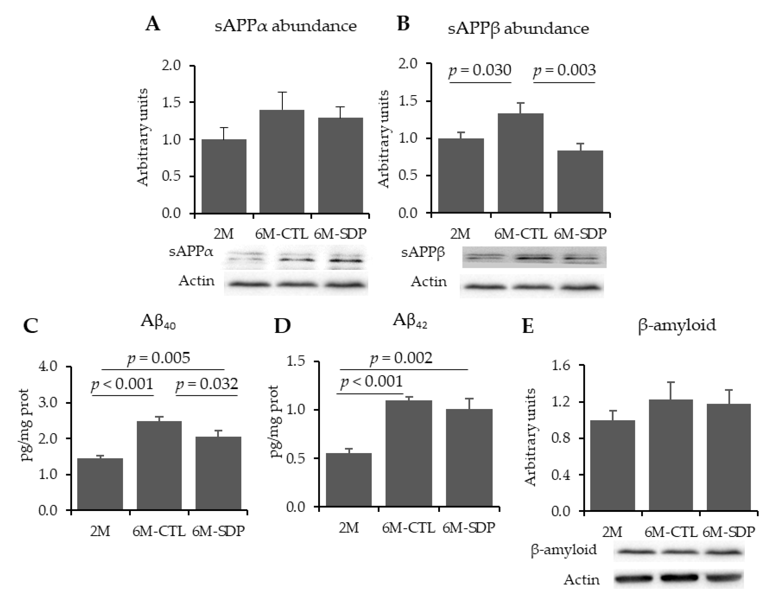
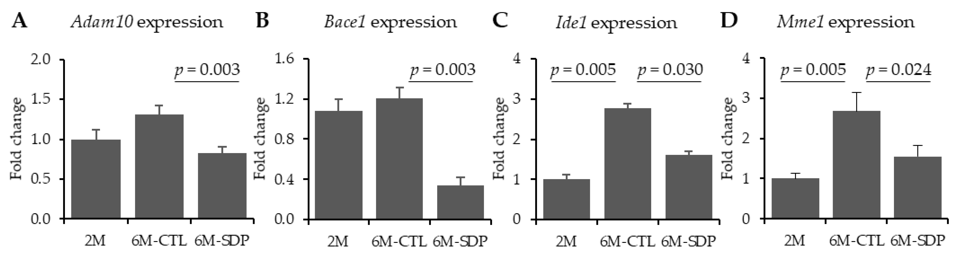
3.1. Amyloid Pathology

Neither aging nor SDP supplementation modified sAPPα abundance (Figure 1A). Aging increased the abundance of sAPPβ in the brain tissue compared to young mice (p = 0.030; Figure 1B). SDP supplementation reduced the abundance of this protein (p = 0.003). Levels of Aβ40 and Aβ42 were augmented in the cortex of 6-month-old mice (both, p < 0.001, Figure 1C,D, respectively), and SDP supplementation prevented Aβ40 increase (p = 0.032). Relative abundance of β-amyloid (Aβ) was not altered by senescence nor by SDP supplementation (Figure 1E).
Aging did not modify Adam10 and Bace1 gene expression (Figure 2A,B, respectively). Both genes were decreased in mice that were fed the SDP-supplemented diet (p = 0.003 for both enzymes). Gene expression of the Aβ degrading enzymes, Ide1 and Mme1, was higher in the cortex of 6-month-old SAMP8 mice than in young mice (both p = 0.005, Figure 2C,D), and these effects were prevented by SDP supplementation (p = 0.030 and p = 0.024, respectively).

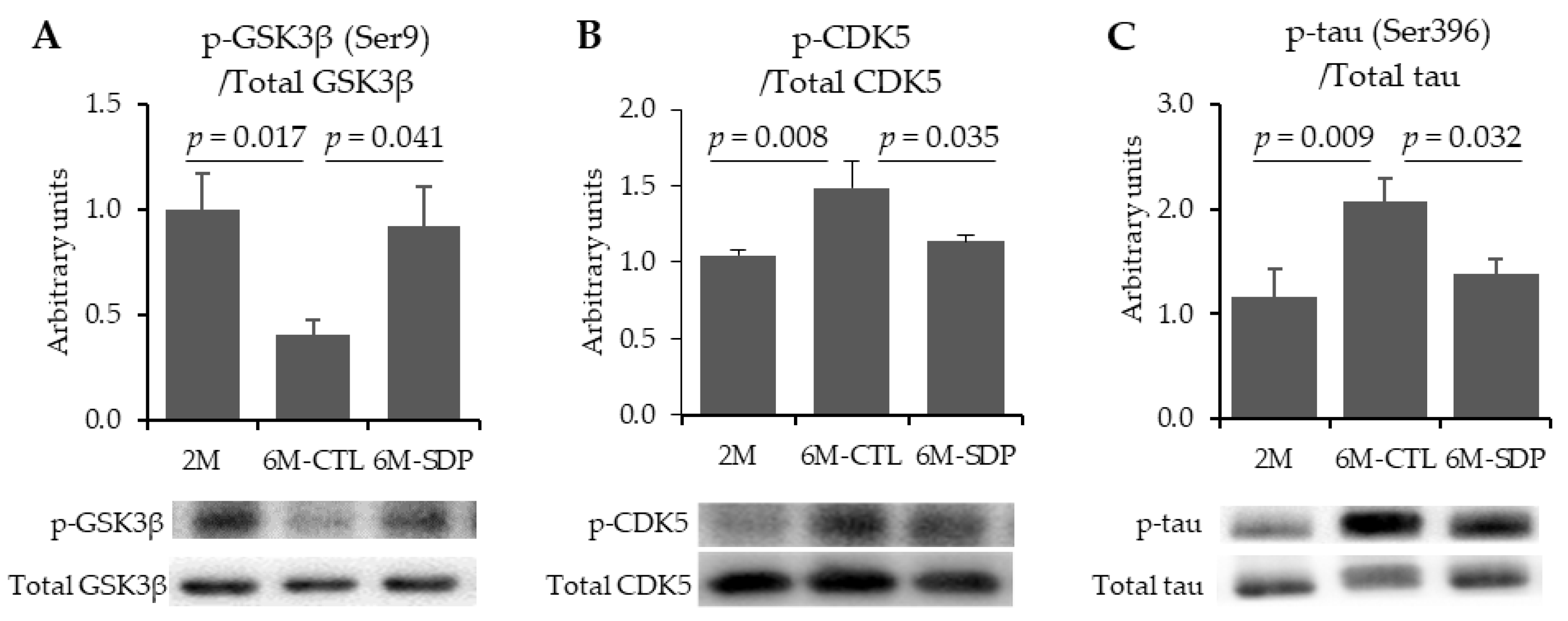
3.2. Neurofibrillary Tangles

The total abundance of GSK3β and CDK5 proteins was not altered by aging nor by SDP supplementation. Nevertheless, the abundance of phosphorylated GSK3β was reduced in aged mice compared with the younger mice (p = 0.017, Figure 3A), and dietary supplementation with SDP prevented this change (p = 0.041). Moreover, aging augmented the abundance of phosphorylated CDK5 (p-CDK5; p = 0.008, Figure 3B), and this increase was completely prevented by SDP supplementation (p = 0.035). Relative abundance of total tau was not changed, neither by aging nor by SDP supplementation. However, 6M-CTL mice showed increased abundance of phosphorylated tau on Ser196 (p-tau (Ser396); p = 0.009; Figure 3C), and SDP supplementation prevented this increase (p = 0.032).

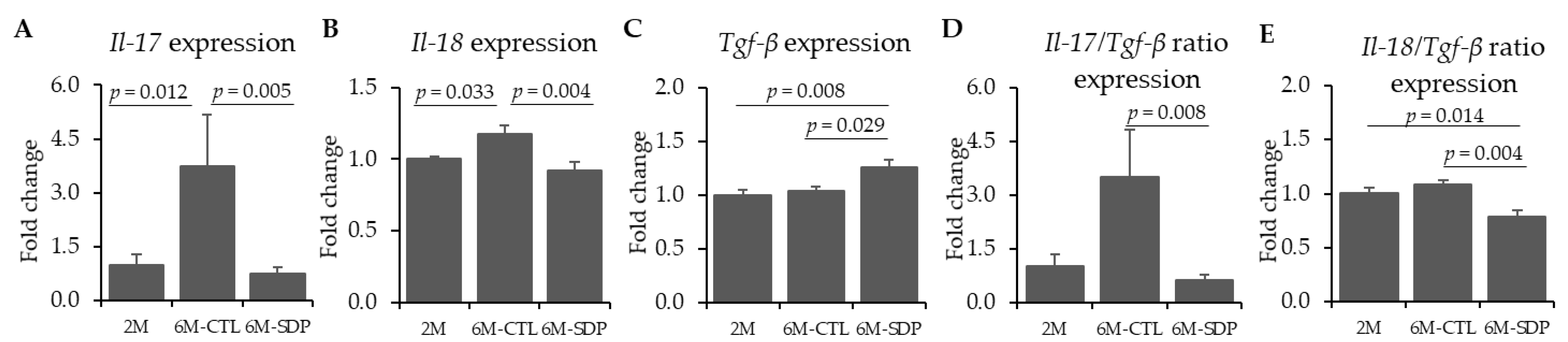
3.3. Neuroinflammatory Markers

The gene expression of the pro-inflammatory cytokines Il-17 and IL-18 was increased in 6M-CTL mice compared to 2M mice (p = 0.012 and p = 0.033, respectively, Figure 4A,B), whereas the SDP supplementation prevented it (p = 0.005 and p = 0.004, respectively). Furthermore, 6M-CTL mice showed a similar gene expression of the anti-inflammatory cytokines Tgf-β in the cortex compared to the 2M group, while SDP supplementation increased the gene expression of this anti-inflammatory cytokine in respect to 6M-CTL mice (p = 0.029) and young mice (p = 0.008, Figure 4C). SDP supplementation reduced the ratio Il-17/Tgf-β compared to 6M-CTL mice (p = 0.008, Figure 4D) and the ratio of Il-18/Tgf-β in respect to senescent control mice (p = 0.004, Figure 4E) and young mice (p = 0.014).

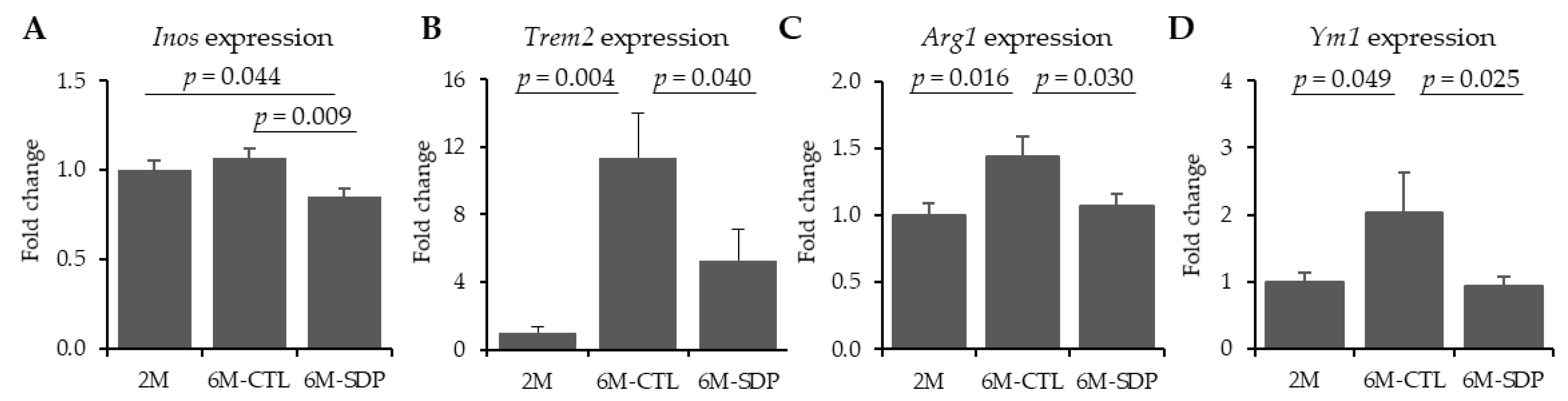
3.4. Microglia Markers
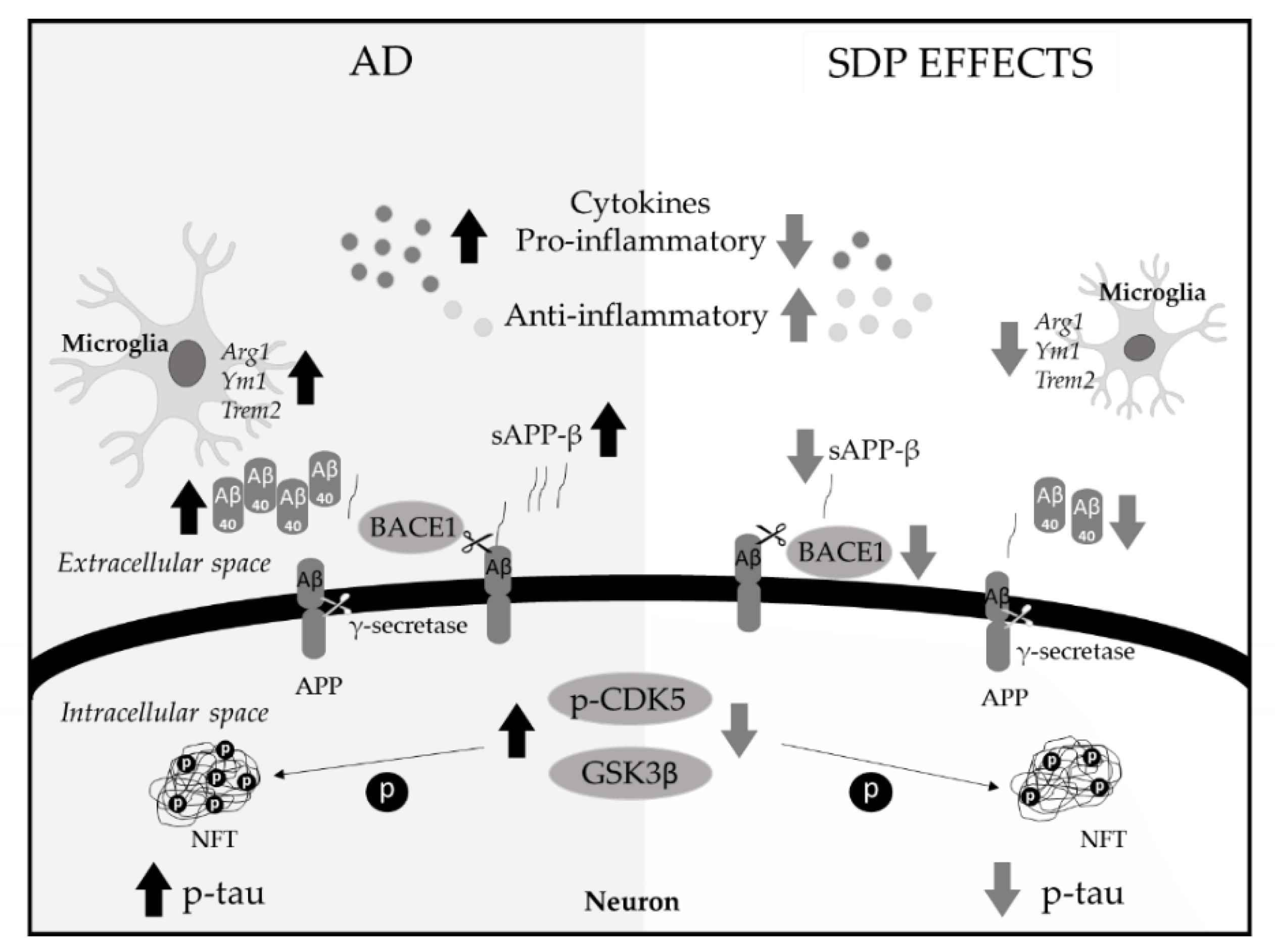
Gene expression of Inos was not altered by aging (Figure 5A), but SDP supplementation diminished it (p = 0.009). Gene expression of Trem2 in cortex of 6M-CTL mice was higher than in young mice (p = 0.004 Figure 5B), and SDP supplementation prevented it (p = 0.040). Moreover, the gene expression of Arg1 and Ym1 was also increased in aged mice (p = 0.016 and p = 0.049, Figure 5C,D, respectively), and SDP supplementation reduced these effects (p = 0.030 and p = 0.025, respectively).

4. Discussion

The recent approval of aducanumab, albeit with weak scientific evidence, has created a new drug for AD, as well as an anti-amyloid therapy [34]. However, no pharmacological therapy can effectively halt the decline in cognitive function associated with AD [1]. Therefore, there is growing interest in the study of protective measures to reduce disease incidence. Recent work shows that antioxidants and anti-inflammatory supplements may help decrease AD risk [35,36]. Accordingly, in this study, we examined the effects of dietary supplementation with SDP, a supplement with well-known anti-inflammatory properties. Previous studies have shown that SDP supplementation improves short- and long-term memory and increases synaptic integrity. These effects have been associated with reduced pro-inflammatory cytokine expression and increased IL-10 abundance in brain tissue [24]. Our current results show that SDP can reduce the main hallmarks of AD, such as aberrant APP processing and NFTs formation, which are substantial contributors to the disease [37].
As shown here, 6-month-old SAMP8 mice exhibited an increased amyloid and tau pathologies, as evaluated by several markers such as sAPPβ, Aβ40, Aβ42, and p-tau, indicating that they already had early molecular and clinical features of AD. These early events are consistent with cognitive impairment because 6-month-old SAMP8 mice already have both short- and long-term memory loss, as indicated by previous work conducted in our laboratory [24]. Activation of the amyloidogenic pathway of APP processing results in the production of sAPPβ and the generation of neurotoxic Aβ. Activation of this pathway involves the sequential cleavage of APP by β-secretase (BACE1) and γ-secretase, which results in the production of sAPPβ and the generation of neurotoxic Aβ peptides [38]. Furthermore, cleavage of APP by BACE1 is an essential first step in the production of Aβ that has been well recognized as an initial causal event of the disease [39]. Our results suggest that SDP supplementation reduces the levels of sAPPβ, as well as Aβ40 abundance, by decreasing the level of the amyloidogenic secretase BACE1, suggesting a reduction in the amyloidogenic pathway. These effects might explain the improved memory retention, in both the short- and the long-term, previously observed in 6-month-old SAMP8 mice supplemented with SDP [24]. Moreover, this effect of SDP is comparable to that of other nutritional interventions, which include supplementation with vitamin D, resveratrol, and lycopene, compounds, that also reduce the levels of Aβ-related biomarkers, such as APP, sAPPβ, and BACE1, in both mice and patients with AD [40,41,42].
Hyperphosphorylated tau is a neuropathological hallmark of AD that disrupts axonal transport and leads to NFTs accumulation [4]. In addition, tau phosphorylation is modulated by several protein kinases and phosphatases, such as GSK3β and CDK5 [43]. Of these kinases, CDK5 is particularly interesting because it may play a crucial role in the pathogenesis of the disease by potentially regulating the activity of other critical kinases, including GSK3β [6]. In addition, CDK5 and its potent activator p25 have been linked to the reduced synaptic density observed in early stages of AD [44]. In our hands, SDP reduced the abundance of p-tau and p-CDK5 while increasing that of p-GSK3β. Unlike CDK5, GSK3β is inactivated when phosphorylated in Ser9 [5]. Thus, GSK3β phosphorylation in Ser9 improves cell function and reduces NFTs levels [45]. Moreover, the effect of SDP associated with reduced abundance of activated CDK5 and p-tau is consistent with previous results showing that 6-month-old SAMP8 mice supplemented with SDP show a higher abundance of synaptophysin [24], which is indicative of higher synaptic density [46].
Although the two cardinal pathological features of AD and neuroinflammation were traditionally considered independent pathways, recent studies show an interaction between these processes, indicating that neuroinflammation might significantly affect the onset of AD [47]. Several studies indicate increased levels of pro-inflammatory cytokines in AD [48,49]. Moreover, pro-inflammatory cytokines and NF-κB signaling upregulate the BACE1 pathway, resulting in elevated Aβ production [50], and this correlates with the NF-κB increase found in aged SAMP8 mice [24]. For instance, IL-17 can activate glial cells [51], and IL-18 upregulates components of the γ-secretase complex, accelerating Aβ production, as well as GSK3β and CDK5 kinases, which promote the hyperphosphorylation of tau [52]. Moreover, reductions in microglial activation and neuronal loss are seen in IL-18 knockout mice [53]. In this study, we observed an increase in the gene expression of pro-inflammatory cytokines such as Il-17 (and to a lesser degree Il-18) with age, and SDP supplementation not only prevented this effect but also increased the expression of the anti-inflammatory factor Tgf-β. Consequently, SDP supplementation promoted the anti-inflammatory pathway, as shown by the reduction in the Il-17/Tgf-β and Il-18/Tgf-β ratios. This might be associated with the neuroprotective effects of SDP found in the aberrant amyloid processing pathway and in NFTs. These anti-inflammatory effects of SDP correlate well with previous results showing that the SDP diet reduced the levels of NF-κB and pro-inflammatory cytokines, such as IL-6, IL-1β and TNF-α, and increased IL-10 in 6-month-old SAMP8 mice [24]. Similarly, recent studies have reported that anti-inflammatory drugs play a significant role in the AD reduction strategy [54,55].
Activated microglial cells surrounding amyloid plaques and the presence of T-cells promote neuroinflammation in AD. Indeed, there is growing interest in analyzing the role of microglia in AD because activated microglia are another key hallmark of the pathology [9]. Activated microglia are localized around senile plaques [56] and NFTs in the brain tissue of AD patients and transgenic mouse models [57]. In our hands, 6-month-old SAMP8 mice have increased hallmarks of AD and neuroinflammation, which might elevate the levels of activated M2-type microglia. Indeed, recent studies confirm that, in early stage AD, M2 phenotype microglia are activated to prevent the overproduction of Aβ and further pathological damage [58,59]. Specifically, TREM2, which is dominantly expressed by microglia in the central nervous system, is also increased in 7-month-old SAMP8 mice from [60]. Moreover, Arg1+ microglia have been observed to increase during IL-1β neuroinflammation in a negative feedback mechanism [61], and this cytokine is increased in 6M-SAMP8 mice, as shown in previous work [24]. These facts correlate well with our findings of increased expression of the M2 microglial markers Trem2, Arg1, and Ym1 in aged mice. In a similar manner to our results, previous studies have shown that SDP supplementation reduces Inos gene expression in a mouse model of acute lung injury [21], and other natural products such as curcumin can inhibit this gene expression in microglia [62]. This relationship between neuroinflammation and activated microglia might explain the reduction in microglia markers by SDP supplementation, as SDP supplementation reduces both neuronal and systemic inflammation [19,20].
SDP is a complex mixture of functional proteins (albumin, transferrin, immunoglobulins, and other globulins), growth factors, bioactive peptides, and other biological components. Its mechanism of action may involve several of its functional components, rather than just one, which may act directly on the intestinal immune response [18], or it may promote the growth of a microbiota with anti-inflammatory effects, as demonstrated by Moretó et al. [23]. Recently, plasma protein-derived supplements have been observed to inhibit TNF secretion in rat T lymphocytes and monocytes in vitro, and to regulate the immune response of intestinal enterocytes through MyD88 and NFkB [63]. Stimulation of MyD88 and NF-κB-related receptors on enterocytes involves the activation of immunomodulatory complexes, including not only cytokine/chemokine secretion [64] but also the secretion of antibacterial peptides [65]. Conversely, there is growing evidence regarding the relationship between the gut microbiota, the digestive system, and the brain, as well as their association with inflammation [66,67,68]. Changes in the composition of the gut microbiota coextensive with aging likely contribute to immunosenescence and to the development of a pro-inflammatory phenotype [69]. In fact, SAMP8 mice at 6 months of age, showed a reduction in probiotic species, while the proinflammatory species of the intestinal microbiota increased [70]; these are able to secrete bacterial amyloids and lipopolysaccharides that are considered neurotoxic [71]. Moreover, SDP increases the presence of bacterial families that enhance the intestinal barrier function, as well as the growth of species that are well-known mediators of anti-inflammatory and tolerogenic responses [23,72]. These results suggest that some components in SDP may be effective in changing the profile of the microbiota, with potential implications for reducing intestinal and peripheral inflammation [19], and eventually communicating with to distant areas such as the brain [73].

5. Conclusions

In conclusion, SDP supplementation increases brain resilience against the key features of AD in SAMP8 mice, reducing the neuroinflammation and attenuating the microglial activation (Figure 6).

Author Contributions

Conceptualization and design of the experiments, C.G.-F., A.P.-B., J.P., M.P., C.A., M.M., and L.M.; methodology and data analysis, C.R.-C., L.M., A.P.-B.; contribution with essential reagents and materials, C.G.-F., J.P., M.P.; writing—original draft preparation, L.M. and C.R.-C., writing—review and editing, C.G.-F., A.P.-B., M.P., C.A., and M.M. All authors have read and agreed to the published version of the manuscript.

Funding

This research was funded by APC-Europe SLU (Granollers, Spain) by research contracts with the Bosch i Gimpera Foundation of the University of Barcelona. The research groups were also supported by grants 2017SGR945 and 2017SGR106 for Consolidated Research Groups, Generalitat de Catalunya, Spain. We also thank Ministerio de Economía y Competitividad of Spain and FEDER funds (PID2019-106285RB) for financial support. CR-C was supported by a grant from the Bosch i Gimpera Foundation (Universitat de Barcelona).

Institutional Review Board Statement

The study was conducted according to the Guide for the Care and Use of Laboratory Animals, and the protocols used in this study were approved by the Ethics Committee for Animal Experimentation of the Universitat de Barcelona and the Catalan government (ref. 484/16 and 9272, respectively).

Informed Consent Statement

Not applicable.

Data Availability Statement

The datasets generated during and/or analyzed during the current study are available from the corresponding author on reasonable request.

Conflicts of Interest

C.R.-C., C.G.-F., A.P.-B., M.P., C.A., M.M. and have no conflicts of interests. J.P. and L.M. are employed by A PC-Europe SLU. The funding sponsors had no role in the design of the study; in the collection, analysis, or interpretation of data; in the writing of the manuscript, and in the decision to publish the results.

References

  1. Soria-Lopez, J.A.; González, H.M.; Léger, G.C. Alzheimer’s disease. In Handbook of Clinical Neurology, 3rd ed.; DeKosky, S.T., Asthana, S., Eds.; Elsevier: Boston, MA, USA, 2019; Volume 167, pp. 231–255. [Google Scholar] [CrossRef]
  2. Ozben, T.; Ozben, S. Neuro-inflammation and anti-inflammatory treatment options for Alzheimer’s disease. Clin. Biochem. 2019, 72, 87–89. [Google Scholar] [CrossRef]
  3. Vassar, R. BACE1 inhibitor drugs in clinical trials for Alzheimer’s disease. Alzheimer’s Res. Ther. 2014, 6, 89. [Google Scholar] [CrossRef] [Green Version]
  4. Chong, F.P.; Ng, K.Y.; Koh, R.Y.; Chye, S.M. Tau proteins and tauopathies in Alzheimer’s disease. Cell. Mol. Neurobiol. 2018, 38, 965–980. [Google Scholar] [CrossRef] [PubMed]
  5. Souder, D.C.; Anderson, R.M. An expanding GSK3 network: Implications for aging research. Geroscience 2019, 41, 369–382. [Google Scholar] [CrossRef] [PubMed]
  6. Liu, S.L.; Wang, C.; Jiang, T.; Tan, L.; Xing, A.; Yu, J.T. The role of Cdk5 in Alzheimer’s disease. Mol. Neurobiol. 2016, 53, 4328–4342. [Google Scholar] [CrossRef] [PubMed]
  7. Lu, T.T.; Wan, C.; Yang, W.; Cai, Z. Role of Cdk5 in amyloid-beta pathology of Alzheimer’s disease. Curr. Alzheimer Res. 2019, 16, 1206–1215. [Google Scholar] [CrossRef] [PubMed]
  8. Ader, R.; Godbout, J.P.; Johnson, R.W. Aging, neuroinflammation and behavior. In Psychoneuroimmunology, 4th ed.; Ader, R., Ed.; Academic Press: Boston, MA, USA, 2009; pp. 322–345. [Google Scholar]
  9. Dansokho, C.; Heneka, M.T. Neuroinflammatory responses in Alzheimer’s disease. J. Neural Transm. 2018, 125, 771–779. [Google Scholar] [CrossRef] [PubMed]
  10. Medeiros, R.; LaFerla, F.M. Astrocytes: Conductors of the Alzheimer disease neuroinflammatory symphony. Exp. Neurol. 2013, 239, 133–138. [Google Scholar] [CrossRef] [PubMed]
  11. Rogers, J.; Strohmeyer, R.; Kovelowski, C.J.; Li, R. Microglia and inflammatory mechanisms in the clearance of amyloid beta peptide. Glia 2002, 40, 260–269. [Google Scholar] [CrossRef]
  12. Stephan, A.H.; Barres, B.A.; Stevens, B. The complement system: An unexpected role in synaptic pruning during development and disease. Annu. Rev. Neurosci. 2012, 35, 369–389. [Google Scholar] [CrossRef] [Green Version]
  13. Sarlus, H.; Heneka, M.T. Microglia in Alzheimer’s disease. J. Clin. Investig. 2017, 127, 3240–3249. [Google Scholar] [CrossRef]
  14. Torrallardona, D. Spray dried animal plasma as an alternative to antibiotics in weanling pigs—A review. Asian Australas. J. Anim. Sci. 2010, 23, 131–148. [Google Scholar] [CrossRef]
  15. Van Dijk, A.J.; Margry, R.J.C.F.; Van der Lee, A.G.; Hemke, G.; Beynen, A.C. Growth performance and health status in weanling piglets fed spray-dried porcine plasma under typical Northern European conditions. J. Anim. Physiol. Anim. Nutr. 2002, 86, 17–25. [Google Scholar] [CrossRef] [PubMed]
  16. Campbell, J.M.; Crenshaw, J.D.; González-Esquerra, R.; Polo, J. Impact of spray-dried plasma on intestinal health and broiler performance. Microorganisms 2019, 7, 219. [Google Scholar] [CrossRef] [PubMed] [Green Version]
  17. Pérez-Bosque, A.; Miró, L.; Polo, J.; Russell, L.; Campbell, J.; Weaver, E.; Crenshaw, J.; Moretó, M. Dietary plasma protein supplements prevent the release of mucosal proinflammatory mediators in intestinal inflammation in rats. J. Nutr. 2010, 140, 25–30. [Google Scholar] [CrossRef] [Green Version]
  18. Pérez-Bosque, A.; Miró, L.; Amat, C.; Polo, J.; Moretó, M. The anti-inflammatory effect of spray-dried plasma is mediated by a reduction in mucosal lymphocyte activation and infiltration in a mouse model of intestinal inflammation. Nutrients 2016, 8, 657. [Google Scholar] [CrossRef] [PubMed] [Green Version]
  19. Miró, L.; Garcia-Just, A.; Amat, C.; Polo, J.; Moretó, M.; Pérez-Bosque, A. Dietary animal plasma proteins improve the intestinal immune response in senescent mice. Nutrients 2017, 9, 1346. [Google Scholar] [CrossRef] [PubMed] [Green Version]
  20. Miró, L.; Amat, C.; Rosell-Cardona, C.; Campbell, J.M.; Polo, J.; Pérez-Bosque, A.; Moretó, M. Dietary supplementation with spray-dried porcine plasma attenuates colon inflammation in a genetic mouse model of inflammatory bowel disease. Int. J. Mol. Sci. 2020, 21, 6760. [Google Scholar] [CrossRef]
  21. Maijó, M.; Miro, L.; Polo, J.; Campbell, J.; Russell, L.; Crenshaw, L.; Weaver, E.; Moreto, M.; Pérez-Bosque, A. Dietary plasma proteins attenuate the innate immunity response in a mouse model of acute lung injury. Br. J. Nutr. 2012, 107, 867–875. [Google Scholar] [CrossRef] [Green Version]
  22. Song, M.; Liu, Y.; Lee, J.J.; Che, T.M.; Soares-Almeida, J.A.; Chun, J.L.; Campbell, J.M.; Polo, J.; Crenshaw, J.D.; Seo, S.W.; et al. Spray-dried plasma attenuates inflammation and improves pregnancy rate of mated female mice. J. Anim. Sci. 2015, 93, 298–305. [Google Scholar] [CrossRef] [Green Version]
  23. Moretó, M.; Miró, L.; Amat, C.; Polo, J.; Manichanh, C.; Pérez-Bosque, A. Dietary supplementation with spray-dried porcine plasma has prebiotic effects on gut microbiota in mice. Sci. Rep. 2020, 10, 2926. [Google Scholar] [CrossRef] [Green Version]
  24. Garcia-Just, A.; Miró, L.; Pérez-Bosque, A.; Amat, C.; Polo, J.; Pallàs, M.; Griñán-Ferré, C.; Moretó, M. Dietary spray-dried porcine plasma prevents cognitive decline in senescent mice and reduces neuroinflammation and oxidative stress. J. Nutr. 2020, 150, 303–311. [Google Scholar] [CrossRef]
  25. Morley, J.E.; Vijaya, S.A.; Kumar, B.; Armbrecht, H.J. The SAMP8 mouse: A model to develop therapeutic interventions for Alzheimer’s disease. Curr. Pharm. Des. 2012, 18, 1123–1130. [Google Scholar] [CrossRef]
  26. Griñan-Ferré, C.; Palomera-Ávalos, V.; Puigoriol-Illamola, D.; Camins, A.; Porquet, D.; Plá, V.; Aguado, F.; Pallàs, M. Behaviour and cognitive changes correlated with hippocampal neuroinflammaging and neuronal markers in female SAMP8, a model of accelerated senescence. Exp. Gerontol. 2016, 80, 57–69. [Google Scholar] [CrossRef] [Green Version]
  27. Pallàs, M. Senescence-accelerated mice P8: A tool to study brain aging and Alzheimer’s disease in a mouse model. ISRN Cell Biol. 2012, 1–12. [Google Scholar] [CrossRef] [Green Version]
  28. Miyamoto, M. Characteristics of age-related behavioral changes in senescence-accelerated mouse SAMP8 and SAMP10. Exp. Gerontol. 1997, 32, 139–148. [Google Scholar] [CrossRef]
  29. Takeda, T.; Hosokawa, M.; Higuchi, K.; Hosono, M.; Akiguchi, I.; Katoh, H. A novel murine model of aging, Senescence-Accelerated Mouse (SAM). Arch. Gerontol. Geriatr. 1994, 19, 185–192. [Google Scholar] [CrossRef]
  30. Borg, B.S.; Campbell, J.M.; Russel, L.E.; Rodríguez, C.; Ródenas, J. Evaluation of the chemical and biological characteristics of spray-dried plasma protein collected from various locations around the world. Am. Assoc. Swine Vet. 2002, 33, 97–100. [Google Scholar]
  31. Blázquez, E.; Rodríguez, C.; Ródenas, J.; Segalés, J.; Pujols, J.; Polo, J. Biosafety steps in the manufacturing process of spray-dried plasma: A review with emphasis on the use of ultraviolet irradiation as a redundant biosafety procedure. Porc. Health Manag. 2020, 6, 16. [Google Scholar] [CrossRef]
  32. National Research Council. Nutrient Requirements of Laboratory Animals, 4th ed.; National Academy Press: Washington, DC, USA, 1995. [Google Scholar]
  33. Schmittgen, T.D.; Livak, K.J. Analyzing real-time PCR data by the comparative CT method. Nat. Protoc. 2008, 3, 1101–1108. [Google Scholar] [CrossRef]
  34. Filling the void for new Alzheimer’s disease therapy. Nat. Aging 2021. [CrossRef]
  35. Griñán-Ferré, C.; Bellver-Sanchis, A.; Izquierdo, V.; Corpas, R.; Roig-Soriano, J.; Chillón, M.; Andres-Lacueva, C.; Somogyvári, M.; Sőti, C.; Sanfeliu, C.; et al. The pleiotropic neuroprotective effects of resveratrol in cognitive decline and Alzheimer’s disease pathology: From antioxidant to epigenetic therapy. Ageing Res. Rev. 2021, 67, 101271. [Google Scholar] [CrossRef]
  36. Canhada, S.; Castro, K.; Perry, I.S.; Luft, V.C. Omega-3 fatty acids’ supplementation in Alzheimer’s disease: A systematic review. Nutr. Neurosci. 2018, 21, 529–538. [Google Scholar] [CrossRef] [PubMed]
  37. Masters, C.L.; Bateman, R.; Blennow, K.; Rowe, C.C.; Sperling, R.A.; Cummings, J.L. Alzheimer’s disease. Nat. Rev. Dis. Primers 2015, 1, 15056. [Google Scholar] [CrossRef] [PubMed]
  38. Fisher, C.L.; Resnick, R.J.; De, S.; Acevedo, L.A.; Lu, K.P.; Schroeder, F.C.; Nicholson, L.K. Cyclic cis-locked phospho-dipeptides reduce entry of AβPP into amyloidogenic processing pathway. J. Alzheimer’s Dis. 2017, 55, 391–410. [Google Scholar] [CrossRef] [Green Version]
  39. Cai, Z.; Liu, Z.; Xiao, M.; Wang, C.; Tian, F. Chronic cerebral hypoperfusion promotes amyloid-beta pathogenesis via activating β/γ-secretases. Neurochem. Res. 2017, 42, 3446–3455. [Google Scholar] [CrossRef]
  40. Wang, J.; Li, J.; Wang, Z.; Cui, Y.; Tan, X.; Yuan, T.; Liu, Q.; Liu, Z.; Liu, Z. Supplementation of lycopene attenuates lipopolysaccharide-induced amyloidogenesis and cognitive impairments via mediating neuroinflammation and oxidative stress. J. Nutr. Biochem. 2018, 56, 16–25. [Google Scholar] [CrossRef]
  41. Corpas, R.; Griñán-Ferré, C.; Rodríguez-Farré, E.; Pallàs, M.; Sanfeliu, C. Resveratrol Induces Brain Resilience against Alzheimer Neurodegeneration through Proteostasis Enhancement. Mol. Neurobiol. 2019, 56, 1502–1516. [Google Scholar] [CrossRef] [Green Version]
  42. Jia, J.; Hu, J.; Huo, X.; Miao, R.; Zhang, Y.; Ma, F. Effects of vitamin D supplementation on cognitive function and blood Aβ-related biomarkers in older adults with Alzheimer’s disease: A randomised, double-blind, placebo-controlled trial. J. Neurol. Neurosurg. Psychiatry 2019, 90, 1347–1352. [Google Scholar] [CrossRef]
  43. Lee, S.; Hall, G.F.; Shea, T.B. Potentiation of tau aggregation by cdk5 and GSK3β. J. Alzheimer’s Dis. 2011, 26, 355–364. [Google Scholar] [CrossRef]
  44. Sheng, Y.; Zhang, L.; Su, S.C.; Tsai, L.H.; Zhu, J.J. Cdk5 is a new rapid synaptic homeostasis regulator capable of initiating the early Alzheimer-like pathology. Cereb. Cortex 2016, 26, 2937–2951. [Google Scholar] [CrossRef] [Green Version]
  45. Hermida, M.A.; Kumar, J.D.; Leslie, N.R. GSK3 and its interactions with the PI3K/AKT/mTOR signalling network. Adv. Biol. Regul. 2017, 65, 5–15. [Google Scholar] [CrossRef]
  46. Del Valle, J.; Bayod, S.; Camins, A.; Beas-Zárate, C.; Velázquez-Zamora, D.A.; González-Burgos, I.; Pallàs, M. Dendritic spine abnormalities in hippocampal CA1 pyramidal neurons underlying memory deficits in the SAMP8 mouse model of Alzheimer’s disease. J. Alzheimer’s Dis. 2012, 32, 233–240. [Google Scholar] [CrossRef] [Green Version]
  47. Korzhevskii, D.E.; Kirik, O.V. Brain Microglia and microglial markers. Neurosci. Behav. Physiol. 2016, 46, 284–290. [Google Scholar] [CrossRef]
  48. Park, J.C.; Han, S.H.; Mook-Jung, I. Peripheral inflammatory biomarkers in Alzheimer’s disease: A brief review. BMB Rep. 2020, 53, 10–19. [Google Scholar] [CrossRef]
  49. Shen, X.N.; Niu, L.D.; Wang, Y.J.; Cao, X.P.; Liu, Q.; Tan, L.; Zhang, C.; Yu, J.T. Inflammatory markers in Alzheimer’s disease and mild cognitive impairment: A meta-analysis and systematic review of 170 studies. J. Neurol. Neurosurg. Psychiatry 2019, 90, 590–598. [Google Scholar] [CrossRef] [PubMed]
  50. Sastre, M.; Dewachter, I.; Landreth, G.E.; Willson, T.M.; Klockgether, T.; van Leuven, F.; Heneka, M.T. Nonsteroidal anti-inflammatory drugs and peroxisome proliferator-activated receptor-gamma agonists modulate immunostimulated processing of amyloid precursor protein through regulation of beta-secretase. J. Neurosci. 2003, 23, 9796–9804. [Google Scholar] [CrossRef] [PubMed] [Green Version]
  51. Chen, J.; Liu, X.; Zhong, Y. Interleukin-17A: The key cytokine in neurodegenerative diseases. Front. Aging Neurosci. 2020, 12, 566922. [Google Scholar] [CrossRef] [PubMed]
  52. White, C.S.; Lawrence, C.B.; Brough, D.; Rivers-Auty, J. Inflammasomes as therapeutic targets for Alzheimer’s disease. Brain Pathol. 2017, 27, 223–234. [Google Scholar] [CrossRef]
  53. Sugama, S.; Wirz, S.A.; Barr, A.M.; Conti, B.; Bartfai, T.; Shibasaki, T. Interleukin-18 null mice show diminished microglial activation and reduced dopaminergic neuron loss following acute 1-methyl-4-phenyl-1,2,3,6-tetrahydropyridine treatment. Neuroscience 2004, 128, 451–458. [Google Scholar] [CrossRef]
  54. Griñán-Ferré, C.; Codony, S.; Pujol, E.; Yang, J.; Leiva, R.; Escolano, C.; Puigoriol-Illamola, D.; Companys-Alemany, J.; Corpas, R.; Sanfeliu, C.; et al. Pharmacological inhibition of soluble epoxide hydrolase as a new therapy for Alzheimer’s disease. Neurotherapeutics 2020, 17, 1825–1835. [Google Scholar] [CrossRef]
  55. Wang, J.; Tan, L.; Wang, H.F.; Tan, C.C.; Meng, X.F.; Wang, C.; Tang, S.W.; Yu, J.T. Anti-inflammatory drugs and risk of Alzheimer’s disease: An updated systematic review and meta-analysis. J. Alzheimer’s Dis. 2015, 44, 385–396. [Google Scholar] [CrossRef] [PubMed]
  56. Zotova, E.; Bharambe, V.; Cheaveau, M.; Morgan, W.; Holmes, C.; Harris, S.; Neal, J.W.; Love, S.; Nicoll, J.A.; Boche, D. Inflammatory components in human Alzheimer’s disease and after active amyloid-β42 immunization. Brain 2013, 136, 2677–2696. [Google Scholar] [CrossRef] [Green Version]
  57. Nilson, A.N.; English, K.C.; Gerson, J.E.; Whittle, T.B.; Crain, C.N.; Xue, J.; Sengupta, U.; Castillo-Carranza, D.L.; Zhang, W.; Gupta, P.; et al. Tau oligomers associate with inflammation in the brain and retina of tauopathy mice and in neurodegenerative diseases. J. Alzheimer’s Dis. 2017, 55, 1083–1099. [Google Scholar] [CrossRef] [PubMed] [Green Version]
  58. Li, L.; Li, L.; Zhang, J.; Huang, S.; Liu, W.; Wang, Z.; Liang, S.; Tao, J.; Chen, L. Disease stage-associated alterations in learning and memory through the electroacupuncture modulation of the cortical microglial M1/M2 polarization in mice with Alzheimer’s disease. Neural Plast. 2020, 8836173. [Google Scholar] [CrossRef] [PubMed]
  59. Zheng, H.; Cheng, B.; Li, Y.; Li, X.; Chen, X.; Zhang, Y.W. TREM2 in Alzheimer’s disease: Microglial survival and energy metabolism. Front. Aging Neurosci. 2018, 10, 395. [Google Scholar] [CrossRef] [PubMed] [Green Version]
  60. Jiang, T.; Yu, J.T.; Zhu, X.C.; Tan, M.S.; Gu, L.Z.; Zhang, Y.D.; Tan, L. Triggering receptor expressed on myeloid cells 2 knockdown exacerbates aging-related neuroinflammation and cognitive deficiency in senescence-accelerated mouse prone 8 mice. Neurobiol. Aging 2014, 35, 1243–1251. [Google Scholar] [CrossRef]
  61. Cherry, J.D.; Olschowka, J.A.; O’Banion, M.K. Arginase 1+ microglia reduce Aβ plaque deposition during IL-1β-dependent neuroinflammation. J. Neuroinflamm. 2015, 12, 203. [Google Scholar] [CrossRef] [PubMed] [Green Version]
  62. Ghasemi, F.; Bagheri, H.; Barreto, G.E.; Read, M.I.; Sahebkar, A. Effects of Curcumin on Microglial Cells. Neurotox. Res. 2019, 36, 12–26. [Google Scholar] [CrossRef] [PubMed]
  63. Hernández-Chirlaque, C.; Aranda, C.J.; Ocón, B.; Polo, J.; Martínez-Augustin, O.; Sánchez de Medina, F. Immunoregulatory Effects of Porcine Plasma Protein Concentrates on Rat Intestinal Epithelial Cells and Splenocytes. Animals 2021, 11, 807. [Google Scholar] [CrossRef] [PubMed]
  64. Günther, C.; Buchen, B.; Neurath, M.F.; Becker, C. Regulation and pathophysiological role of epithelial turnover in the gut. Semin. Cell Dev. Biol. 2014, 35, 40–50. [Google Scholar] [CrossRef]
  65. Menendez, A.; Willing, B.P.; Montero, M.; Wlodarska, M.; So, C.C.; Bhinder, G.; Vallance, B.A.; Finlay, B.B. Bacterial stimulation of the TLR-MyD88 pathway modulates the homeostatic expression of ileal Paneth cell alpha-defensins. J. Innate Immun. 2013, 5, 39–49. [Google Scholar] [CrossRef]
  66. Cattaneo, A.; Cattane, N.; Galluzzi, S.; Provasi, S.; Lopizzo, N.; Festari, C.; Ferrari, C.; Guerra, U.P.; Paghera, B.; Muscio, C.; et al. Association of brain amyloidosis with pro-inflammatory gut bacterial taxa and peripheral inflammation markers in cognitively impaired elderly. Neurobiol. Aging 2017, 49, 60–68. [Google Scholar] [CrossRef] [PubMed] [Green Version]
  67. Lin, C.; Zhao, S.; Zhu, Y.; Fan, Z.; Wang, J.; Zhang, B.; Chen, Y. Microbiota-gut-brain axis and toll-like receptors in Alzheimer’s disease. Comput. Struct. Biotechnol. J. 2019, 17, 1309–1317. [Google Scholar] [CrossRef] [PubMed]
  68. Liu, X.; Cao, S.; Zhang, X. Modulation of gut microbiota-brain axis by probiotics, prebiotics, and diet. J. Agric. Food Chem. 2015, 63, 7885–7895. [Google Scholar] [CrossRef]
  69. Vaiserman, A.M.; Koliada, A.K.; Marotta, F. Gut microbiota: A player in aging and a target for anti-aging intervention. Ageing Res. Rev. 2017, 35, 36–45. [Google Scholar] [CrossRef] [PubMed]
  70. Miró, L.; Moretó, M.; Amat, C.; Polo, J.; Pérez-Bosque, A. Aging effects on gut microbiota in SAMP8 mice. Proceedings 2020, 61, 25. [Google Scholar] [CrossRef]
  71. Askarova, S.; Umbayev, B.; Masoud, A.R.; Kaiyrlykyzy, A.; Safarova, Y.; Tsoy, A.; Olzhayev, F.; Kushugulova, A. The links between the gut microbiome, aging, modern lifestyle and Alzheimer’s disease. Front. Cell. Infect. Microbiol. 2020, 10, 104. [Google Scholar] [CrossRef] [Green Version]
  72. Torrallardona, D.; Conde, M.R.; Badiola, I.; Polo, J.; Brufau, J. Effect of fishmeal replacement with spray-dried animal plasma and colistin on intestinal structure, intestinal microbiology, and performance of weanling pigs challenged with Escherichia coli K99. J. Anim. Sci. 2003, 81, 1220–1226. [Google Scholar] [CrossRef]
  73. Caracciolo, B.; Xu, W.; Collins, S.; Fratiglioni, L. Cognitive decline, dietary factors and gut-brain interactions. Mech. Ageing Dev. 2014, 136–137, 59–69. [Google Scholar] [CrossRef]
Figure 1. Effects of SDP supplementation on amyloid pathology markers. Panels (AE) show the abundance of sAPPα, sAPPβ, Aβ40,42, and β-amyloid in brain tissue. Results are expressed as mean ± SEM (n = 5–6 mice). Statistics: ANOVA (Fisher’s multiple comparison test). Aβ: β-amyloid; APP: amyloid protein precursor.
Nutrients 13 02369 g001
Figure 2. Effects of SDP supplementation on gene expression of enzymes related to aberrant APP processing. Panels (AD) show the expression of Adam10, Bace1, Ide1, and Mme1, respectively, in cortex tissue. Results are expressed as mean ± SEM (n = 5–6 mice). Statistics: ANOVA (Fisher’s multiple comparison test). Adam10: disintegrin and metalloproteinase domain-containing protein 10; Bace1: beta-secretase 1; Ide1: insulin-degrading enzyme; Mme1: neprilysin.
Nutrients 13 02369 g002
Figure 3. Effect of SDP supplementation on neurofibrillary tangles. Panels (AC) show the abundance of p-GSK3β (Ser9), p-CDK5, and p-tau (Ser396) in the brain tissue, respectively. Results are expressed as mean ± SEM (n = 5–6 mice). Statistics: one-way ANOVA followed by Fisher’s multiple comparison test. GSK3β: glycogen synthase kinase 3β; CDK5: cyclin dependent kinase 5.
Nutrients 13 02369 g003
Figure 4. Effect of SDP supplementation on cytokine gene expression. Panels (AC) show the gene expression of Il-17, Il-18, and Tgf-β in cortex tissue, respectively. Panels (D,E) show the gene expression ratio Il-17/Tgf-β and Il-18/Tgf-β in cortex tissue, respectively. Results are expressed as mean ± SEM (n = 5–6 mice). Statistics: one-way ANOVA followed by Fisher’s multiple comparison test. Il: interleukin; Tgf-β: transforming growth factor beta.
Nutrients 13 02369 g004
Figure 5. Effect of SDP supplementation on markers of microglia. Panels (AD) show the gene expression of Inos, Trem2, Arg1, and Ym1 in cortex tissue, respectively. Results are expressed as mean ± SEM (n = 5–6 mice). Statistics: one-way ANOVA followed by Fisher’s multiple comparison test. Inos: inducible nitric oxide synthase; Trem2: triggering receptor expressed on myeloid cells 2; Arg1: arginase 1; Ym1: chitinase-like protein 3 (Chil3).
Nutrients 13 02369 g005
Figure 6. Representative scheme of the effects of SDP on AD hallmarks. SDP supplementation decreases the aberrant APP processing pathway and the hyperphosphorylation of tau. These preventive effects are accompanied by a reduction in the neuroinflammation and an attenuation in the activated microglia. APP: amyloid precursor protein, Arg1: arginase1, Aβ40: β-Amyloid 1-40, BACE1: β-secretase, CDK5: cyclin-dependent kinase 5, GSK3β: glycogen synthase kinase 3β, NFT: neurofibrillary tangles, P: phosphorylation, Trem2: triggering receptor expressed on myeloid cells 2, Ym1: chitinase-like protein 3 (Chil3).
Nutrients 13 02369 g006
Table 1. The composition of experimental diets.
IngredientsControl DietSDP 1 Diet
g/kg
SDP-80
Dried skim milk530.7340.5
Corn starch199.3308.8
Sucrose94.594.5
Soybean oil7070
Cellulose5050
AIN-93-G-MX (94046) 23535
AIN-93 VX (94047) 21515
Choline bitartrate33
Methionine2.53.2
1 SDP (spray-dried plasma) was provided by APC-Europe (Granollers, Spain). 2 AIN-93 MX, mineral mix AIN-93 VX, vitamin mix; both were provided by Envigo (Breso, Italy).
Table 2. Primers used for real-time PCR.
PrimerForward (5′–3′)Reverse (5′–3′)Size (bp)
Arg1GACAGGGCTCCTTTCAGGACGCCAAGGTTAAAGCCACTGC108
Il-17AGCAGCGATCATCCCTCAATTGCGCCAAGGGAGTTAAA104
Il-18GTTTACAAGCATCCAGGCACAGGAAGGTTTGAGGCGGCTTTC151
InosCCCAACAATACAAGATGACCCTAGGGATTCTGGAACATTCTGTG87
Tgf-bCAGGGTGAAGGGGAAAACTCAGTTCGGTCATTAGTCTCGC204
Trem2CCTGAAGAAGCGGAATGGGACTTGATTCCTGGAGGTGCTGT269
Ym1GTGGAGAAAGACATTCCAAGGCCAGTTCAGGGATCTTGTACCCA90
Arg1: arginase1; Il: interleukin; Inos: inducible nitric oxide synthase; Tgf-b: transforming growth factor beta; Trem-2: triggering receptor expressed on myeloid cells 2; Ym1: chitinase-like protein 3 (Chil3).
Publisher’s Note: MDPI stays neutral with regard to jurisdictional claims in published maps and institutional affiliations.

