Maternal Vitamin D and Newborn Telomere Length
Abstract
1. Introduction
2. Materials and Methods
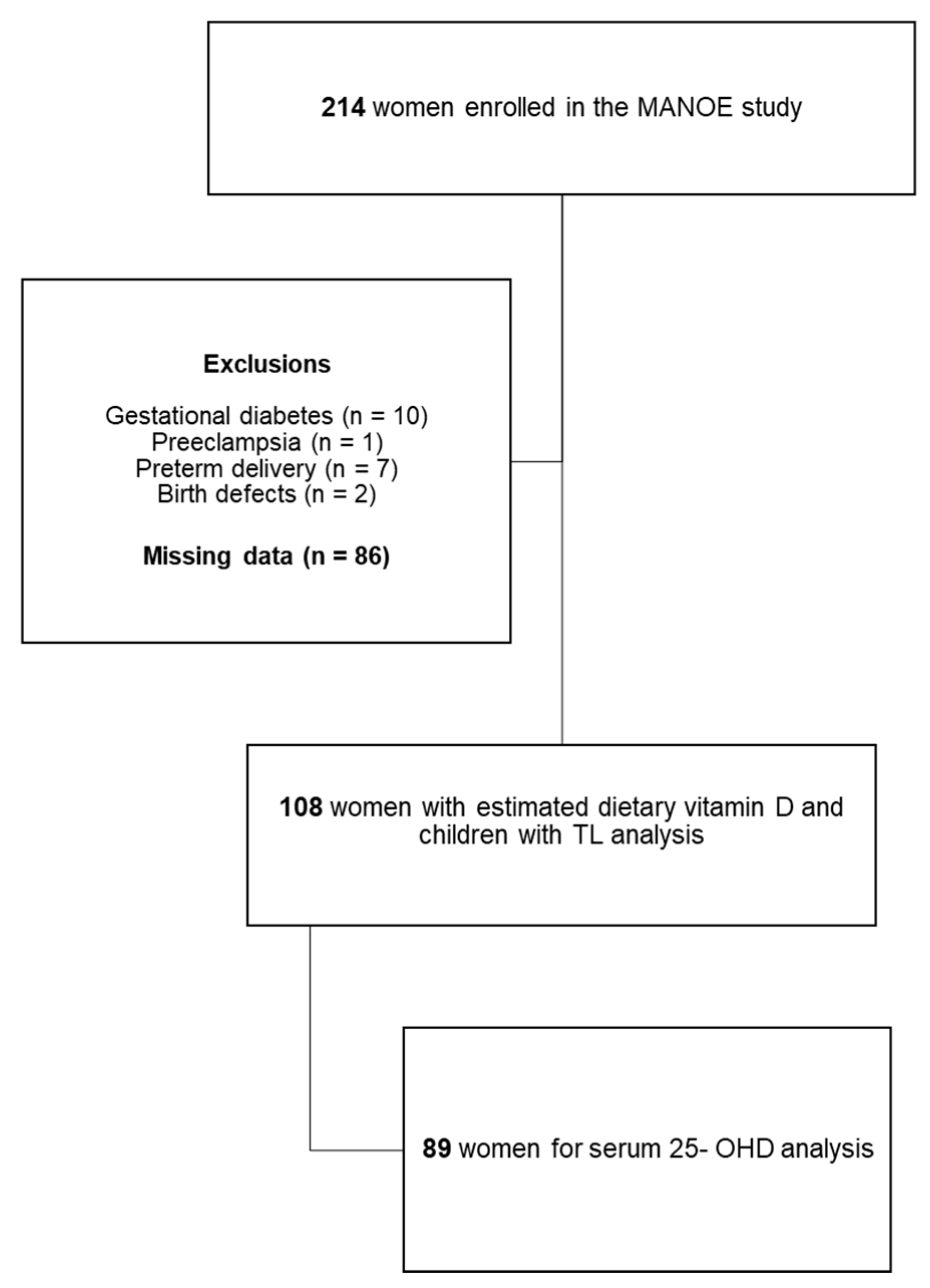
2.1. Study Participants
2.2. Maternal and Infant Measurements
2.3. Vitamin D
2.3.1. Dietary Vitamin D Intake
2.3.2. Serum 25-hydroxyvitamin D Concentration
2.4. Average Relative Telomere Length Measurements
2.5. Statistical Analysis
3. Results
3.1. Maternal and Infant Characteristics
3.2. Maternal Vitamin D
3.3. Offspring Telomere Length
3.4. Maternal Vitamin D and Average Relative Newborn TL
4. Discussion
5. Conclusions
Author Contributions
Funding
Institutional Review Board Statement
Informed Consent Statement
Data Availability Statement
Acknowledgments
Conflicts of Interest
References
- de Boo, H.A.; Harding, J.E. The developmental origins of adult disease (Barker) hypothesis. Aust. N. Z. J. Obstet. Gynaecol. 2006, 46, 4–14. [Google Scholar] [CrossRef] [PubMed]
- Lips, P.; Cashman, K.D.; Lamberg-Allardt, C.; Bischoff-Ferrari, H.A.; Obermayer-Pietsch, B.; Bianchi, M.L.; Stepan, J.; Fuleihan, G.E.-H.; Bouillon, R. Current vitamin D status in European and Middle East countries and strategies to prevent vitamin D deficiency: A position statement of the European Calcified Tissue Society. Eur. J. Endocrinol. 2019, 180, P23–P54. [Google Scholar] [CrossRef] [PubMed]
- Larqué, E.; Morales, E.; Leis, R.; Blanco-Carnero, J.E. Maternal and Foetal Health Implications of Vitamin D Status during Pregnancy. Ann. Nutr. Metab. 2018, 72, 179–192. [Google Scholar] [CrossRef]
- Vandevijvere, S.; Amsalkhir, S.; Van Oyen, H.; Moreno-Reyes, R. High prevalence of vitamin D deficiency in pregnant women: A national cross-sectional survey. PLoS ONE 2012, 7, e43868. [Google Scholar]
- Martens, D.S.; Plusquin, M.; Gyselaers, W.; De Vivo, I.; Nawrot, T.S. Maternal pre-pregnancy body mass index and newborn telomere length. BMC Med. 2016, 14, 148. [Google Scholar] [CrossRef]
- Martens, D.S.; Van Der Stukken, C.; Derom, C.; Thiery, E.; Bijnens, E.M.; Nawrot, T.S. Newborn telomere length predicts later life telomere length: Tracking telomere length from birth to child- and adulthood. EBioMedicine 2021, 63, 103164. [Google Scholar] [CrossRef]
- Okuda, K.; Bardeguez, A.; Gardner, J.P.; Rodriguez, P.; Ganesh, V.; Kimura, M.; Skurnick, J.; Awad, G.; Aviv, A. Telomere length in the newborn. Pediatr. Res. 2002, 52, 377–381. [Google Scholar] [CrossRef]
- Liu, B.; Song, L.; Zhang, L.; Wu, M.; Wang, L.; Cao, Z.; Xiong, C.; Zhang, B.; Li, Y.; Xia, W.; et al. Prenatal second-hand smoke exposure and newborn telomere length. Pediatr. Res. 2020, 87, 1081–1085. [Google Scholar] [CrossRef]
- Marchetto, N.M.; Glynn, R.A.; Ferry, M.L.; Ostojic, M.; Wolff, S.M.; Yao, R.; Haussmann, M.F. Prenatal stress and newborn telomere length. Am. J. Obstet. Gynecol. 2016, 215, 94.e1–94.e8. [Google Scholar] [CrossRef]
- Entringer, S.; Epel, E.S.; Lin, J.; Blackburn, E.H.; Buss, C.; Shahbaba, B.; Gillen, D.L.; Venkataramanan, R.; Simhan, H.N.; Wadhwa, P.D. Maternal Folate Concentration in Early Pregnancy and Newborn Telomere Length. Ann. Nutr. Metab. 2015, 66, 202–208. [Google Scholar] [CrossRef]
- Myers, K.O.; Ibrahimou, B.; Yusuf, K.K.; Mauck, D.E.; Salihu, H.M. The effect of maternal vitamin C intake on fetal telomere length. J. Matern. Fetal Neonatal. Med. 2019, 34, 1143–1148. [Google Scholar] [CrossRef]
- Kim, J.H.; Kim, G.J.; Lee, D.; Ko, J.H.; Lim, I.; Bang, H.; Koes, B.W.; Seong, B.; Lee, D.-C. Higher maternal vitamin D concentrations are associated with longer leukocyte telomeres in newborns. Matern. Child Nutr. 2018, 14, e12475. [Google Scholar] [CrossRef]
- Pauwels, S.; Duca, R.C.; Devlieger, R.; Freson, K.; Straetmans, D.; Van Herck, E.; Huybrechts, I.; Koppen, G.; Godderis, L. Maternal Methyl-Group Donor Intake and Global DNA (Hydroxy)Methylation before and during Pregnancy. Nutrients 2016, 8, 474. [Google Scholar] [CrossRef]
- Schmidt, M.D.; Freedson, P.S.; Pekow, P.; Roberts, D.; Sternfeld, B.; Chasan-Taber, L. Validation of the Kaiser Physical Activity Survey in pregnant women. Med. Sci. Sports Exerc. 2006, 38, 42–50. [Google Scholar] [CrossRef] [PubMed]
- Pauwels, S.; Ghosh, M.; Duca, R.C.; Bekaert, B.; Freson, K.; Huybrechts, I.; Langie, S.; Koppen, G.; Devlieger, R.; Godderis, L. Dietary and supplemental maternal methyl-group donor intake and cord blood DNA methylation. Epigenetics 2017, 12, 1–10. [Google Scholar] [CrossRef] [PubMed]
- Nubel VoedingsPlanner. v.z.w. NUBEL. 2010. Available online: https://www.nubel.be/ned/ (accessed on 10 June 2021).
- Thomas, R.L.; Jiang, L.; Adams, J.S.; Xu, Z.Z.; Shen, J.; Janssen, S.; Ackermann, G.; Vanderschueren, D.; Pauwels, S.; Knight, R.; et al. Vitamin D metabolites and the gut microbiome in older men. Nat. Commun. 2020, 11, 5997. [Google Scholar] [CrossRef]
- Green, M.R.; Sambrook, J. Molecular Cloning: A Laboratory Manual; Cold Spring Harbor Laboratory Press: New York, NY, USA, 2001. [Google Scholar]
- Cawthon, R.M. Telomere length measurement by a novel monochrome multiplex quantitative PCR method. Nucleic Acids Res. 2009, 37, e21. [Google Scholar] [CrossRef] [PubMed]
- Martens, D.S.; Janssen, B.G.; Bijnens, E.M.; Clemente, D.B.P.; Vineis, P.; Plusquin, M.; Nawrot, T.S. Association of Parental Socioeconomic Status and Newborn Telomere Length. JAMA Netw. Open 2020, 3, e204057. [Google Scholar] [CrossRef]
- Telomere Research Network. Available online: https://trn.tulane.edu/resources/study-design-analysis/ (accessed on 10 June 2021).
- Cabaset, S.; Krieger, J.P.; Richard, A.; Elgizouli, M.; Nieters, A.; Rohrmann, S.; Lötscher, K.C.Q. Vitamin D status and its determinants in healthy pregnant women living in Switzerland in the first trimester of pregnancy. BMC Pregnancy Childbirth 2019, 19, 10. [Google Scholar] [CrossRef] [PubMed]
- Moyersoen, I.; Teppers, E. Voedselconsumptiepeiling 2014–2015; Rapport 4. In Sciensano; Belgian Institute for Health: Brussels, Belgium, 2016. [Google Scholar]
- Gezondheidsraad, H. Voedingsaanbevelingen voor België—2016; Hoge Gezondheidsraad: Brussels, Belgium, 2016. [Google Scholar]
- Lukaszuk, J.M.; Luebbers, P.E. 25(OH)D status: Effect of D. Obes. Sci. Pract. 2017, 3, 99–105. [Google Scholar] [CrossRef]
- Agudelo-Zapata, Y.; Maldonado-Acosta, L.M.; Sandoval-Alzate, H.F.; Poveda, N.E.; Garcés, M.F.; Cortés-Vásquez, J.A.; Linares-Vaca, A.F.; Mancera-Rodríguez, C.A.; Perea-Ariza, S.A.; Ramírez-Iriarte, K.Y.; et al. Serum 25-hydroxyvitamin D levels throughout pregnancy: A longitudinal study in healthy and preeclamptic pregnant women. Endocr. Connect. 2018, 7, 698–707. [Google Scholar] [CrossRef] [PubMed]
- Lundqvist, A.; Sandström, H.; Stenlund, H.; Johansson, I.; Hultdin, J. Vitamin D Status during Pregnancy: A Longitudinal Study in Swedish Women from Early Pregnancy to Seven Months Postpartum. PLoS ONE 2016, 11, e0150385. [Google Scholar] [CrossRef] [PubMed]
- Hollis, B.W.; Wagner, C.L. New insights into the vitamin D requirements during pregnancy. Bone Res. 2017, 5, 17030. [Google Scholar] [CrossRef]
- Moon, R.J.; Harvey, N.C.; Cooper, C.; D’Angelo, S.; Crozier, S.R.; Inskip, H.M.; Schoenmakers, I.; Prentice, A.; Arden, N.K.; Bishop, N.J.; et al. Determinants of the Maternal 25-Hydroxyvitamin D Response to Vitamin D Supplementation During Pregnancy. J. Clin. Endocrinol. Metab. 2016, 101, 5012–5020. [Google Scholar] [CrossRef]
- Bodnar, L.M.; Simhan, H.N.; Powers, R.W.; Frank, M.P.; Cooperstein, E.; Roberts, J.M. High prevalence of vitamin D insufficiency in black and white pregnant women residing in the northern United States and their neonates. J. Nutr. 2007, 137, 447–452. [Google Scholar] [CrossRef] [PubMed]
- Hollis, B.W.; Johnson, D.; Hulsey, T.C.; Ebeling, M.; Wagner, C.L. Vitamin D supplementation during pregnancy: Double-blind, randomized clinical trial of safety and effectiveness. J. Bone Miner. Res. 2011, 26, 2341–2457. [Google Scholar] [CrossRef]
- Jiang, Y.; Zheng, W.; Teegarden, D. 1α, 25-Dihydroxyvitamin D regulates hypoxia-inducible factor-1α in untransformed and Harvey-ras transfected breast epithelial cells. Cancer Lett. 2010, 298, 159–166. [Google Scholar] [CrossRef] [PubMed]
- Brodowski, L.; Burlakov, J.; Myerski, A.C.; von Kaisenberg, C.S.; Grundmann, M.; Hubel, C.A.; Von Versen-Höynck, F. Vitamin D prevents endothelial progenitor cell dysfunction induced by sera from women with preeclampsia or conditioned media from hypoxic placenta. PLoS ONE 2014, 9, e98527. [Google Scholar] [CrossRef]
- Wojcicki, J.M.; Olveda, R.; Heyman, M.B.; Elwan, D.; Lin, J.; Blackburn, E.; Epel, E.S. Cord blood telomere length in Latino infants: Relation with maternal education and infant sex. J. Perinatol. 2016, 36, 235–241. [Google Scholar] [CrossRef]
- Sempos, C.T.; Binkley, N. 25-Hydroxyvitamin D assay standardisation and vitamin D guidelines paralysis. Public Health Nutr. 2020, 23, 1153–1164. [Google Scholar] [CrossRef]

| Newborn (n = 108) | Mean (SD) | Range |
|---|---|---|
| Gestational age, weeks | 39.7 (0.9) | 37.1–41.4 |
| Length, cm | 51.0 (1.8) | 47–56 |
| Birth weight, g | 3512.5 (412.9) | 2720–4750 |
| n | % | |
| Gender | ||
| Boys | 59 | 54.6 |
| Girls | 49 | 45.4 |
| Season of birth | ||
| Winter | 21 | 19.4 |
| Spring | 38 | 35.2 |
| Summer | 23 | 21.3 |
| Autumn | 26 | 24.1 |
| Mother (n = 108) | Mean (SD) | Range |
| Age, years | 30.7 (3.4) | 24–41 |
| Pre-pregnancy BMI, kg/m2 | 22.9 (3.2) | 17.9–33.0 |
| Gestational weight gain, kg | 14.6 (4.1) | 1.9–28.9 |
| Physical activity index | ||
| First trimester | 10.1 (1.3) | 6.7–13.2 |
| Second trimester | 9.8 (1.5) | 6.4–13.7 |
| Third trimester | 9.5 (1.5) | 5.6–12.7 |
| n | % | |
| Education | ||
| Low education | 14 | 13.0 |
| Medium education | 38 | 35.2 |
| High education | 56 | 51.9 |
| Smoking | ||
| First trimester | 3 | 2.8 |
| Second trimester | 2 | 1.9 |
| Third trimester | 2 | 1.9 |
| Maternal | Diet (μg) | Supplements (μg) | Total (μg) | Serum 25-OHD (ng/mL) | Correlation * | |||||||
|---|---|---|---|---|---|---|---|---|---|---|---|---|
| Vitamin D | Mean (SD) | Range | Mean (SD) | Range | (Diet + Supplement) | Mean (SD) | Range | Diet/Serum | Total/Serum | |||
| Mean (SD) | Range | R | p | R | p | |||||||
| Trimester 1 | 3.8 (2.3) | 0.2–10 | 4.4 (4.8) | 0–10 | 8.2 (5.4) | 0.2–17.9 | 22.4 (8.1) | 6.5–68 | 0.078 | 0.439 | 0.314 | 0.001 |
| Trimester 2 | 3.9 (2.8) | 0.5–14.5 | 5.3 (4.8) | 0–10 | 9.3 (5.6) | 0.7–22.3 | 23.0 (8.1) | 7.7–52 | 0.056 | 0.575 | 0.535 | <0.001 |
| Trimester 3 | 3.9 (2.5) | 0.1–10.7 | 5.4 (4.6) | 0–10 | 9.3 (5.5) | 0.4–20 | 24.8 (9.6) | 8.1–58.4 | 0.063 | 0.535 | 0.468 | <0.001 |
| Entire pregnancy | 3.9 (2.6) | 0.1–14.5 | 6.1 (4.4) | 0–10 | 8.9 (5.5) | 0.2–22.3 | 23.4 (6.8) | 10.6–45.5 | 0.047 | 0.656 | 0.573 | <0.001 |
| Entire Pregnancy | First Trimester | Second Trimester | Third Trimester | |||||
|---|---|---|---|---|---|---|---|---|
| β (95% CI) | p-Value | β (95% CI) | p-Value | β (95% CI) | p-Value | β (95% CI) | p-Value | |
| Unadjusted model | 0.004 (−0.005, 0.013) | 0.365 | 0.009 (0.001, 0.018) | 0.036 | −0.003 (−0.013, 0.007) | 0.534 | −0.001 (−0.010, 0.008) | 0.820 |
| Model 1 | 0.007 (−0.002, 0.015) | 0.132 | 0.012 (0.004, 0.020) | 0.005 | −0.004 (−0.013, 0.006) | 0.444 | −0.001 (−0.009, 0.008) | 0.828 |
| Model 2 | 0.007 (−0.002, 0.016) | 0.121 | 0.012 (0.003, 0.020) | 0.007 | −0.003 (−0.012, 0.007) | 0.569 | −0.001 (−0.010, 0.007) | 0.763 |
| Model 3 | 0.003 (−0.005, 0.010) | 0.498 | 0.012 (0.005, 0.019) | 0.001 | −0.002 (−0.010, 0.006) | 0.569 | −0.007 (−0.014, 0.000) | 0.065 |
| Entire Pregnancy | First Trimester | Second Trimester | Third Trimester | |||||
|---|---|---|---|---|---|---|---|---|
| β (95% CI) | p-Value | β (95% CI) | p-Value | β (95% CI) | p-Value | β (95% CI) | p-Value | |
| Unadjusted model | −0.017 (−0.041, 0.006) | 0.148 | 0.002 (−0.016, 0.020) | 0.832 | −0.011 (−0.026, 0.004) | 0.162 | −0.005 (−0.022, 0.012) | 0.542 |
| Model 1 | −0.014 (−0.037, 0.009) | 0.237 | 0.011 (−0.006, 0.029) | 0.202 | −0.017 (−0.032, −0.002) | 0.028 | −0.002 (−0.018, 0.013) | 0.782 |
| Model 2 | −0.019 (−0.043, 0.005) | 0.124 | 0.009 (−0.010, 0.028) | 0.345 | −0.015 (−0.030, 0.000) | 0.044 | −0.005 (−0.021, 0.011) | 0.510 |
| Model 3 | −0.018 (−0.038, 0.002) | 0.077 | 0.005 (−0.011, 0.020) | 0.547 | −0.008 (−0.021, 0.005) | 0.213 | −0.012 (−0.025, 0.002) | 0.088 |
| Entire Pregnancy | First Trimester | Second Trimester | Third Trimester | |||||
|---|---|---|---|---|---|---|---|---|
| β (95% CI) | p-Value | β (95% CI) | p-Value | β (95% CI) | p-Value | β (95% CI) | p-Value | |
| Unadjusted model | 0.009 (−0.0011, 0.018) | 0.050 | 0.010 (0.000, 0.020) | 0.062 | 0.002 (−0.012, 0.015) | 0.881 | −0.003 (−0.016, 0.010) | 0.654 |
| Model 1 | 0.010 (0.001, 0.018) | 0.024 | 0.010 (0.001, 0.020) | 0.039 | 0.005 (−0.008, 0.018) | 0.454 | −0.006 (−0.018, 0.007) | 0.367 |
| Model 2 | 0.010 (0.001, 0.019) | 0.028 | 0.010 (−0.002, 0.020) | 0.050 | 0.006 (−0.007, 0.019) | 0.388 | −0.005 (−0.017, 0.007) | 0.413 |
| Model 3 | 0.005 (−0.003, 0.012) | 0.250 | 0.012 (0.004, 0.021) | 0.005 | 0.003 (−0.008, 0.014) | 0.552 | −0.011 (−0.021, −0.001) | 0.038 |
| Entire Pregnancy | First Trimester | Second Trimester | Third Trimester | |||||
|---|---|---|---|---|---|---|---|---|
| β (95% CI) | p-Value | β (95% CI) | p-Value | β (95% CI) | p-Value | β (95% CI) | p-Value | |
| Unadjusted model | 0.004 (−0.003, 0.011) | 0.252 | 0.003 (−0.005, 0.012) | 0.466 | −0.003 (−0.013, 0.006) | 0.500 | 0.005 (−0.002, 0.011) | 0.166 |
| Model 1 | 0.003 (−0.003, 0.011) | 0.369 | 0.004 (0.004, 0.013) | 0.324 | −0.004 (−0.013, 0.005) | 0.422 | 0.004 (−0.003, 0.011) | 0.284 |
| Model 2 | 0.004 (−0.003, 0.010) | 0.306 | 0.003 (−0.006, 0.013) | 0.496 | −0.002 (−0.012, 0.009) | 0.716 | 0.003 (−0.006, 0.011) | 0.511 |
| Model 3 | 0.003 (−0.003, 0.009) | 0.311 | 0.003 (0.005, 0.011) | 0.517 | 0.000 (−0.009, 0.009) | 0.962 | 0.000 (−0.007, 0.008) | 0.934 |
Publisher’s Note: MDPI stays neutral with regard to jurisdictional claims in published maps and institutional affiliations. |
© 2021 by the authors. Licensee MDPI, Basel, Switzerland. This article is an open access article distributed under the terms and conditions of the Creative Commons Attribution (CC BY) license (https://creativecommons.org/licenses/by/4.0/).
Share and Cite
Daneels, L.; Martens, D.S.; Arredouani, S.; Billen, J.; Koppen, G.; Devlieger, R.; Nawrot, T.S.; Ghosh, M.; Godderis, L.; Pauwels, S. Maternal Vitamin D and Newborn Telomere Length. Nutrients 2021, 13, 2012. https://doi.org/10.3390/nu13062012
Daneels L, Martens DS, Arredouani S, Billen J, Koppen G, Devlieger R, Nawrot TS, Ghosh M, Godderis L, Pauwels S. Maternal Vitamin D and Newborn Telomere Length. Nutrients. 2021; 13(6):2012. https://doi.org/10.3390/nu13062012
Chicago/Turabian StyleDaneels, Lisa, Dries S. Martens, Soumia Arredouani, Jaak Billen, Gudrun Koppen, Roland Devlieger, Tim S. Nawrot, Manosij Ghosh, Lode Godderis, and Sara Pauwels. 2021. "Maternal Vitamin D and Newborn Telomere Length" Nutrients 13, no. 6: 2012. https://doi.org/10.3390/nu13062012
APA StyleDaneels, L., Martens, D. S., Arredouani, S., Billen, J., Koppen, G., Devlieger, R., Nawrot, T. S., Ghosh, M., Godderis, L., & Pauwels, S. (2021). Maternal Vitamin D and Newborn Telomere Length. Nutrients, 13(6), 2012. https://doi.org/10.3390/nu13062012


