Article

Two Simulated Spectral Databases of Lunar Regolith: Method, Validation, and Application

1
China University of Geosciences (Beijing), Beijing 100083, China
2
Hebei Bureau of Coal Geological Exploration, Shijiazhuang 050085, China
3
School of Geosciences, The University of Edinburgh, James Hutton Rd, King’s Buildings, Edinburgh EH9 3FE, UK
4
Faculty of Architecture, Civil and Transportation Engineering, Beijing University of Technology, Beijing 100083, China
*
Author to whom correspondence should be addressed.
Remote Sens. 2022, 14(2), 277; https://doi.org/10.3390/rs14020277
Submission received: 11 November 2021 / Revised: 31 December 2021 / Accepted: 1 January 2022 / Published: 7 January 2022

Abstract

:
Our simulated lunar regolith spectra database, based on the Hapke AMSA radiative transfer model (RTM), is a large supplement to the limited number of lunar spectra data. By analyzing the multiple solutions and applicable scopes of the Hapke model by means of Newton interpolation and the least square optimization method, an improved method was found for the simulation of spectra, but it remained challenging to use to invert mineral abundance. Then, we simulated the spectra, mineral abundance, particle size and maturity of 57 mare and highland samples of the Lunar Soil Characterization Consortium (LSCC) in size groups of 10 µm, 10–20 µm and 20–45 µm. The simulated and measured spectra fit well with each other, with correlation coefficients greater than 0.99 and root mean square errors at a magnitude of 10-3. The parameters of mineral abundance, particle size and maturity are highly consistent with the measured values. Having confirmed the reliability of our simulation method, we analyzed the mechanism, reliability and applicability of the “spectral characteristic angle parameter” proposed by Lucey using the simulated spectral data of lunar regolith. Lucey’s method is only suitable for macro analysis of the entire moon, and the error is large when it is used for areas with high abundance of forsterite or ilmenite. In the spectral simulation of lunar regolith, olivine was subdivided into forsterite and fayalite, and the two end-members were mixed to approximately estimate the effect of the chemical composition of olivine on the spectra, which has been confirmed to be feasible.

1. Introduction

Remote sensing is an important tool for the exploration and study of the Earth and other planets, providing information about the composition and physical structure of the observed target surfaces. The mineral composition of the lunar surface, identified by remotely sensed multispectral and hyperspectral data, provides evidence for the ‘magma ocean’ hypothesis, which is one of the mainstream evolution theories of the Moon (Lucey, 1995; Ohtake et al., 2009, 2012; Wu, 2012; Yamamoto et al., 2010, 2012a, 2012b, 2015; Lucey et al., 2014; Vinckier et al., 2019; Zhou et al., 2021; Peng et al., 2021) [1,2,3,4,5,6,7,8,9,10,11,12]. Furthermore, the mineral abundance and chemical composition, which can be retrieved by hyperspectral data more accurately than multispectral data, has played the main role in establishing the layer structure of the lunar upper crust (Lucey et al., 2014) [9].
The ground truth spectral data from the lunar regolith samples of Apollo and Luna sample-return missions have been used to process multispectral data, such as Clementine UVVIS, to retrieve the chemical composition of the lunar surface (Lucey, 1995, 1998, 2000; Blewett et al., 1997) [1,13,14,15]. Unfortunately, there are two main problems with the application of the truth data of the lunar samples. First, the number of samples is small, and the landing sites are so limited that the data cannot represent the compositional features of the lunar surface globally. Second, the spectral features of Apollo and Luna samples are the result of the combined effects of mineral composition, abundance, space weathering, particle size and viewing geometries (the zenith and azimuth angle of sun and sensor), thus making it difficult to understand how the spectral features are affected by each factor. Therefore, the radiative transfer model (RTM) (Shkuratov et al., 1999; Hapke, 1981, 1984, 1986, 1993, 2002, 2005, 2008) has been used increasingly frequently [16,17,18,19,20,21,22,23]. The RTM is an important method to extract information on mineral composition from visible and near-infrared (VIS-NIR) light (Denevi et al., 2008) [24], which is independent of the limited number of lunar samples as ground truth data and considers the effect of space weathering (Liu et al., 2015) [25]. It is an important method to investigate planets with thin air such as the moon and Mars by reproducing spectra of regolith with specific mineral abundance, space weathering, particle size and viewing geometries in use of the RTM. The RTM has been shown to be effective in spectral simulation and mineral abundance mapping (Lucey, 1998, 2004; Denevi et al., 2008; Yan et al., 2010; Li and Li, 2011; Lemilin et al., 2013; Liu et al., 2015) [13,24,25,26,27,28,29].
Lucey (2004) mapped lunar regolith minerals on a global scale using a look-up table. His interpolation interval of mineral abundance was 10%, with the least error being more than 10% [26]. Lucey (2004) simulated only minerals of clinopyroxene, orthopyroxene, olivine, plagioclase and metallic iron, and he did not take into account volcanic glass, agglutinate and ilmenite. These minerals have remarkable influence on spectra [26]. For example, the absorption features of spectra could be greatly weakened by agglutinate (Heiken et al., 1991; Taylor et al., 2003, 2010) [30,31,32]. This influence cannot be ignored; otherwise, it would result in large uncertainty during mineral abundance inversion (Li and Li, 2011) [28]. Denevi et al. (2008) reproduced the spectra of lunar mare regolith of the LSCC data and modeled mineral content successfully using olivine, pyroxene, plagioclase, ilmenite, agglutinate and volcanic glass [24]. Denevi et al. (2008) used a grid search to simulate LSCC data by varying the agglutinate abundance, particle size and lunar submicroscopic iron (abbreviated as SMFe) content in an effective range to simulate more than 100000 spectra, and the results with the best fitting with the measured spectra were obtained. It is notable that even a small change in the particle size or SMFe content of the minerals may have a major impact on the simulation results (Lucey, 1998; Warell and Davidsson, 2010; Trang et al., 2013), so their accuracy of grid search is still insufficient to meet the requirements of the simulation [13,33,34]. We speculate that this is the reason why the agglutinate abundance was underestimated while the particle size was approximately three times larger than the real value in their work. Li and Li (2011) and Liu et al. (2015) quantified lunar regolith mineral abundance, particle size and metallic Fe content using the method of Newton interpolation and least square optimization [25,28]. They chose mineral end-members of clinopyroxene, orthopyroxene, olivine, plagioclase, metallic Fe, ilmenite, agglutinate and volcanic glass and successfully simulated the LSCC lunar regolith spectra and the mineral abundance, particle size and metallic Fe content, although the precision of the simulated abundance of some minerals, such as olivine, ilmenite and volcanic glass, still needs to be improved. The likely reason is that the upper and lower limits of the iteration variable were set too wide or that two steps were used rather than the single, simultaneous step in the calculation of particle size, SMFe content and mineral abundance. Yan et al. (2010) mapped the distribution of lunar olivine, clinopyroxene, orthopyroxene, plagioclase and ilmenite with the Clementine UVVIS/NIR data in the RTM, while the role of space weathering was ignored [27]. Lemelin et al. (2013) successfully simulated the ilmenite abundance in Mare Australe and Mare Ingenii regions by means of the multivariate data of Clementine UVVIS/NIR and LRO WAC, even though he used 15 LSCC samples to verify the validity of the model and method without considering volcanic glass and agglutinate, which are of a large proportion in minerals [29].
There are two ways to use the RTM: one is to calculate a simulated spectral library of the lunar regolith serving as the “ground truth” (Lucey, 2004; Yan et al., 2010) [26,27]; the other way is to quantify the mineral abundance, particle size and metallic Fe content with the method of Newton interpolation and least square optimization, aiming to expand the method of remote sensing data (Li and Li, 2011, Liu et al., 2015) [25,28]; however, it was thought to be difficult to find an automatic approach that can be applied to any unknown spectrum due to nonunique solutions of the RTM equations (Deveni et al., 2008) [24]. In this paper, the two methods are called the simulation and inversion methods.
Although the RTM has been applied successfully, there are two main problems that need to be clarified. One is whether the inversion method is plausible. The other is how well the RTM is applied to simulate the spectral features of the lunar regolith. The reliability and accuracy of RTM was not tested at all in mineral mapping by simulating spectral libraries (Lucey, 2004; Yan et al., 2010) [26,27].
In this paper, we selected plagioclase, clinopyroxene, orthopyroxene, olivine, metallic iron, ilmenite, volcanic glass and agglutinate acquired from the RELAB spectral library as mineral endmembers. Based on the Hapke (2002) anisotropy of multiple scattering approximation (AMSA) radiative transfer model, we simulated spectra of lunar regolith with specific mineral abundance, space weathering degree (maturity), particle size and viewing geometries by means of Newton interpolation and the least square optimization method [21]. First, we chose sample 67461 of the Lunar Soil Characterization Consortium (LSCC) to analyze multiple RTM solutions. We simulated the reflectance spectra, mineral abundance, particle sizes and maturity of 57 LSCC samples and evaluated the accuracy of the simulation results to verify the validity and accuracy of the RTM. Having confirmed the reliability of our simulation method, we simulated two sets of lunar regolith spectra databases, which can be used to analyze the method, mechanism, accuracy and reliability of the abundance and composition of lunar regolith. By means of our simulated spectra, we tried to study the mechanism, reliability and application range of the “spectral characteristic angle parameter method” proposed by Lucey et al. (1995, 1998, 2000) [1,13,14]. Finally, our method was discussed in terms of the accuracy of the simulation of spectral parameters. We also probed the isomorphism of olivine.

2. Methods

2.1. Hapke Radiative Transfer Model

Our work is mainly based on Hapke’s semiempirical bidirectional reflectance model (Hapke, 1981, 1984, 1986, 1993, 2002, 2005, 2008) [17,18,19,20,21,22,23], which quantitatively describes the physical properties of electromagnetic radiation interacting with a semi-infinite particle medium. Hapke’s model is applicable in particle media whose particle size is much larger than the wavelength. The reflectance of the particle medium can be divided into single scattering and multiple scattering:
r ( μ 0 , μ , g ) = w 4 μ 0 μ 0 + μ [ ( 1 + B ( g ) ) p ( g ) + M ( μ 0 , μ ) ]
where μ 0 and μ are the cosine of the incidence angle and emittance angle, respectively, and g is the phase angle. As a single scattering albedo (SSA), w depends on wavelength, representing the combined contribution of particles in varieties of sizes per unit volume. w is independent of viewing geometries and can be mixed linearly by volume percentage of end-member minerals.
p ( g ) , the phase function, describes the distribution of scattering energy at different angles, reflecting the scattering properties of the particle medium. It is a function of the phase angle and wavelength, regardless of the condition of the particle surface. The second-order Legendre polynomial equation of p ( g ) is given by Mustard and Pieters (1989) [35]:
P ( g ) = 1 + b cos ( g ) + c [ 3 cos 2 ( g ) 1 ] / 2
b and c are often set as −0.4 and 0.25, respectively, which are the approximate average values of the forward scattering minerals of Mustard and Pieters (1989) [35].
B ( g ) is the backward scattering function used to explain the shadow-hiding opposition effect (SHOE) and a function of the phase angle ( g ), amplitude ( B o ) and angular width ( h ) of SHOE:
B ( g ) = B o 1 + tan ( g / 2 ) / h
B o represents the probability of light reflected by particles at a small phase angle, which is related to the single scattering albedo of particles. The empirical expression of B o is:
B o e w 2 / 2
h describes the range of the phase angle in SHOE, which is related to the filling factor ϕ of the particles:
h = 3 8 ln ( 1 ϕ )
M ( μ 0 , μ ) is a multiple scattering function. Hapke (1981, 1993) proposed the isotropic multiple scattering approximation function (IMSA) based on the assumption that the particle surface reflects isotropically [17,20], where
M ( μ 0 , μ ) = H ( μ ) H ( μ 0 ) 1
Hapke found that when the single scattering albedo is close to 1.00, the calculation error of absolute reflectance can reach 16% by means of the IMSA model, even if hot effects are not considered. He also indicated that if the particle media was highly anisotropically scattered, the absolute accuracy at certain angles was low. Having taken into account the above shortcomings, Hapke improved and optimized the H-function and determined the multiple scattering function of anisotropy. The new radiative transfer model based on the anisotropic multiple scattering approximation (AMSA), especially taking into account a new H-function, is able to express bidirectional reflectance more accurately (Hapke, 2002) [21]. The multiple scattering function of anisotropy is given by:
M ( μ 0 , μ ) = P ( μ 0 ) [ H ( μ ) 1 ] + P ( μ ) [ H ( μ 0 ) 1 ] + p [ H ( μ 0 ) 1 ] [ H ( μ ) 1 ]
H ( u ) is the H-function in Chandrasekhar’s (1960) theories [36], and Hapke (2002) provides a more accurate analytic approximation of it [21]:
H ( x ) = [ 1 w x ( r o + 1 2 r o x 2 ln 1 + x x ) ] 1
r o = ( 1 γ ) / ( 1 + γ )
γ = ( 1 w ) 1 / 2
The single scattering albedo (SSA) of the particle medium can be calculated by Equations (1)–(10) if the bidirectional reflectance and viewing geometries are known.

2.2. The Optical Constants of Minerals

The single scattering albedo is a function of optical constants (also called the complex index of refraction). Optical constants, independent of the size and shape of minerals (Lucey, 1998) [13], are basic physical properties that describe the propagation of light interacting with the particle medium. These constants are generally expressed as n = n ( 1 i k ) , where n and k are the real and imaginary parts of the optical constants, respectively, and characterize the refraction and absorption abilities of electromagnetic radiation when light enters the interior of minerals. In the VIS-NIR spectral range, the n values of mineral end-members can be seen to be approximately constant, while the k values of mineral end-members are usually < < 1 (Lucey, 1998; Denevi et al., 2008; Li and Li, 2011) [13,24,28]. It should be noted that the k values of metallic iron are usually greater than 1 (Yolken and Kruger, 1965; Johnson and Christy, 1974; Li and Li, 2011) [28,37,38]. Table 1 lists the n values of minerals used in this paper (Li and Li, 2011) [28].
Hapke (2005) described that SSA is a function of optical constants and the particle size of the particle medium [22]:
w = S E + ( 1 S E ) ( 1 S I ) Θ 1 S I Θ
S E = ( n 1 ) 2 + k 2 ( n + 1 ) 2 + k 2 + 0.05
S I = 1 4 n ( n + 1 ) 2
Θ = e D
where S E and S I are the surface reflectance coefficients for externally incident light and internally incident light, respectively, and Θ is the internal transmission factor of a semi-infinite medium.
D is the mean ray path length through a particle given by:
D = 2 3 ( n 2 1 n ( n 2 1 ) 3 / 2 ) D
where D is the particle size.
is the internal absorption coefficient given by:
= 4 π n k λ
where λ is the wavelength. In the calculation of , the effect of maturity on spectra should also be taken into consideration. Lunar submicroscopic iron (SMFe), as one of the main products of space weathering, is a good indicator of maturity, which can darken and redden the reflectance spectra and weaken absorption features (Heiken et al., 1991; Lucey et al., 1995; Starukhina and Shkuratov, 2001; Noble et al., 2001; Taylor et al., 2010) [1,32,42,43,44].
The absorption coefficients of minerals coated with SMFe are given (Hapke, 2001) [45]:
= 4 π n h k h λ + 36 π z M F e ρ h λ ρ F e
z = n h 3 n F e k F e ( n F e 2 k F e 2 + 2 n h 2 ) 2 + 4 n F e 2 k F e 2
where n h , k h , and ρ h are optical constants and densities of host minerals, respectively; n F e , k F e , and ρ F e are optical constants and densities of SMFe; and M F e is the mass fraction of SMFe.
On the basis of Hapke (2001)’s work [45], Lucey and Riner (2011) tested the effect of large SMFe grains (>50 nm) throughout the whole volume of agglutinate [46]:
= 4 π n h k h λ + 36 π z M c ρ h λ ρ F e + 3 q a M i n ρ h d F e ρ F e
where M c and M i n represents the content of the small SMFe grains covering the mineral surface and the content of the large SMFe grains throughout the entire agglutinate, respectively; q a is the absorption efficiency of SMFe; and d F e is the particle size of the large SMFe grains.
The optical constants of a particle medium can be inverted by Equations (11)–(16) if the single scattering albedo and particle size of a particle medium are given.
We chose some typical minerals and calculated the optical constants of each mineral end-member for spectral simulation. The first series of mineral end-members were selected from lunar samples and telluric samples of RELAB at Brown University (Taylor et al., 1999) [47], including typical lunar regolith minerals such as volcanic glass and agglutinate, part of which lacked chemical analysis (Table 2). The second series of mineral end-members were all lunar samples with chemical analysis (Table 3).
The optical constants of each mineral end-member in Table 2 and Table 3 can be inverted by means of the Hapke AMSA radiative transfer model. Optical constants, as a function of mineral composition, are not necessarily invariable because they depend on the quality of the model and selection of particle samples (Lucey, 1998) [13]. When calculating the optical constants of these minerals, we made a valid particle size of each mineral iterate in the range of measured particle sizes. The particle size and optical constants of each mineral were obtained when the simulated spectrum of each mineral was mostly close to the measured spectrum. Meanwhile, by using the concept of Monte Carlo simulation, we randomly set up the initial value within constraints to obtain the optimal solution over partly optimal solution when inverting optical constants of minerals, thus improving the accuracy of calculating optical constants by means of the Hapke model (Wu, 2014) [48]. Figure 1 shows the reflectance spectra and the inverted optical constants of each mineral end-member.

2.3. Simulation Method

Based on the calculated optical constants of mineral end-members, spectra of mixed minerals with various particle sizes, viewing geometries and abundances can be simulated by Equations (1)–(19). Although there are some errors existing in these optical constants retrieved from the Hapke model, these errors could be suppressed in the forward calculation model (simulating reflectance spectra by optical constants of minerals) and inversion calculation model (calculating optical constants of minerals by reflectance spectra) because of using the same model.
By means of Newton interpolation and least square estimation, we evaluate the accuracy of the simulation compared with that of the measured data. We iterated the mineral abundance, maturity and particle size using Newton interpolation in the effective range to simulate numerous spectra at 300–2600 nm. The error between the simulated and measured spectra was calculated as follows:
ε = i = 300 n = 2600 r mod e l e d D , S M F e , p r m e a s u r e d 2
where r mod e l e d and r m e a s u r e d are the simulated spectra and the measured spectra, respectively; D , S M F e , and p are the particle size, SMFe content and mineral abundance, respectively. The particle size, SMFe content, mineral abundance and spectra were selected from the simulated results when the error reached its minimum value. The simulated spectra and corresponding mineral abundance, particle size and SMFe content that were the closest to the measured data were selected.

3. Multiple Solutions and Application of the Model

Since Equations (11)–(19) are nonlinear, there are inevitably multiple solutions for the corresponding parameters, such as mineral abundance, particle size and SMFe content, when simulating the measured spectral data using Newton interpolation and the least squares method. Thus, these solutions are nonunique. We chose the LSCC data to analyze these solutions and the applications of the model.
The LSCC data (downloaded from http://apps.geology.brown.edu/RELAB.php www.planetary.brown.edu/relabdocs/LSCCsoil.html, accessed on 10 November 2021) are widely used in lunar research, including spectra, grain size, maturity, mineral composition and abundance of the lunar regolith. Samples of the LSCC data were divided into four groups (<10 µm, 10–20 µm, 20–45 µm, <45 µm) according to particle size. All the spectra of the LSCC data were measured in RELAB at Brown University at an incident angle 30°, emittance angle 0° and phase angle 30° with a wavelength range of 0.3–2.6 µm and spectral resolution of 5 nm. The LSCC data contain information on the modal abundance of minerals except for the size group of <45 µm (Taylor et al., 2001) [49]. The Is/FeO values, which can represent the relative maturity of samples, are also offered (Morris, 1978) [50].
As mentioned in Section 2.3, when we simulated the measured spectra of samples and obtained its mineral abundance, particle size and SMFe content, many spectra were simulated by varying parameters confined in the effective range in each iteration; the spectra and the corresponding variables of mineral abundance, particle size and SMFe content were selected, which had minimum error compared with the measured spectra. It is worth noting that this work depends on prior knowledge of the mineral abundance and particle size in the sample. For instance, the abundances of each mineral end-member are known, and they vary between 80% and 120% of the measured mineral abundance in simulation, simultaneously controlling the particle size within the range of 0–45 µm and SMFe content within the range of 0–0.01%. If variables are outside of a reasonable range, e.g., mineral abundance ranging from 0–100 wt. %, particle size from 0–100 µm, and SMFe content from 0–1 wt. %, then the simulated mineral abundance, particle size and SMFe content would lose their physical meaning even though the simulated spectra were extremely similar to the measured spectra.
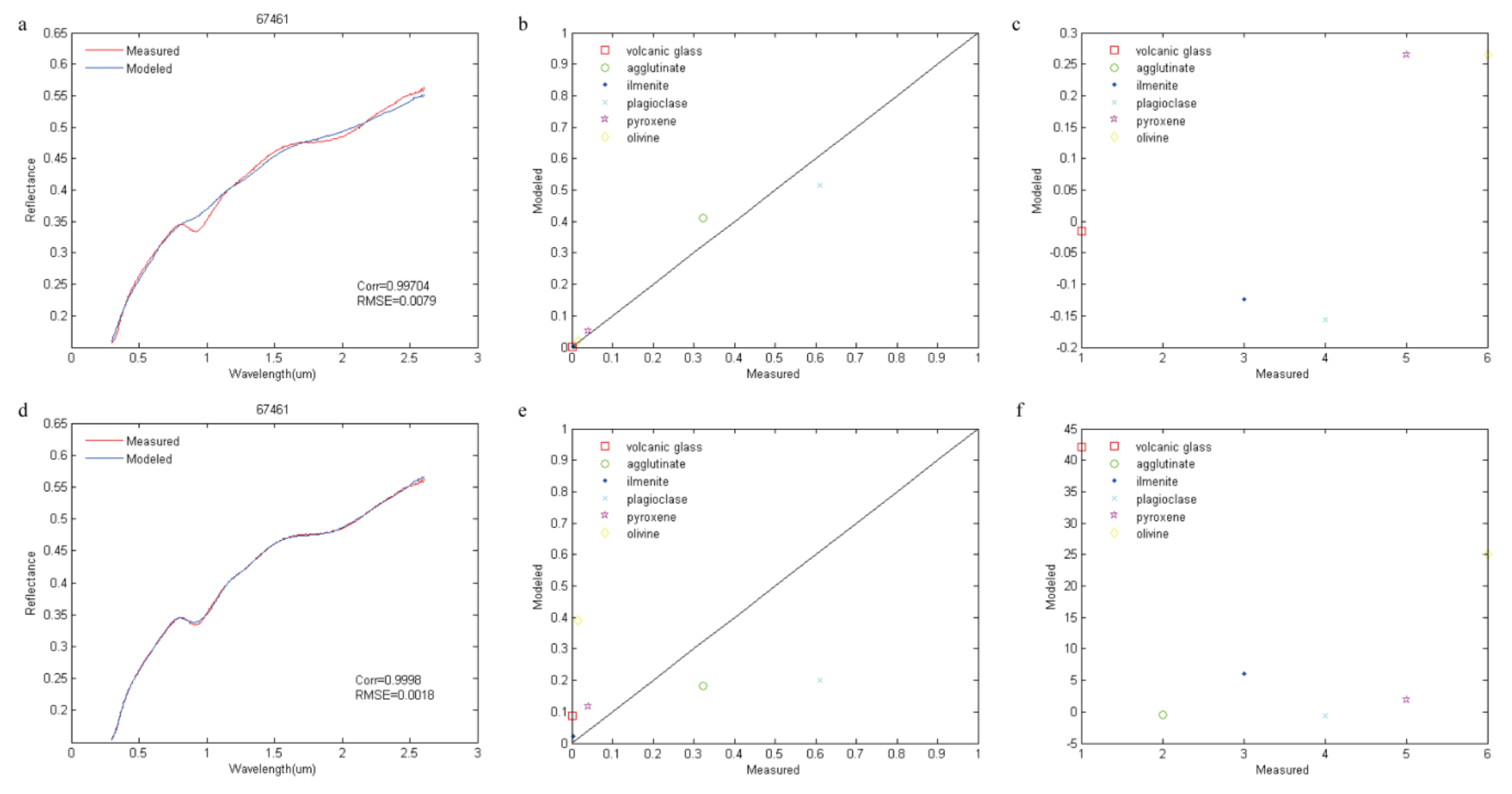
We chose the first series of mineral end-members in Table 2 and simulated the spectra of sample 67461 in LSCC data with corresponding variables such as mineral abundance and particle size. The simulated abundance of pyroxene is a combined effect of orthopyroxene and Mg-rich/Mg-poor clinopyroxene. The simulated abundance of olivine is the total abundance of forsterite and fayalite. The simulated abundance of volcanic glass is the sum abundance of black glass, orange glass and green glass. First, we strictly set up the upper and lower limits of the simulated variables and then obtained simulated spectra close to the measured data as well as the simulated mineral abundance (Figure 2a–c). If the upper and lower limits of the simulated variables were set up in a wider range (Table 4), only maintaining possible physical meaning, although the simulated and measured spectra showed good matching, the simulated variables gravely deviated from the measured data (Figure 2d–f). Table 5 shows the relative errors of the simulated mineral abundance in the two iterations, and the relative error of the second iteration is far greater than that of the first iteration.
Hence, the Hapke model is applicable to simulation work. In other words, we can simulate the spectra if the mineral abundance is known. This method, however, is unsuitable for estimation, and it is very difficult to estimate variables such as mineral abundance if given only the spectra of mixed minerals without prior knowledge of mineral abundance and particle size. From the above analysis, we can conclude that it is difficult to find an automatic approach to be applied to any unknown spectrum due to nonunique solutions of the RTM equations, and it is impossible to expand the method to remote sensing data, as expected by Li and Li (2011).

4. The Validation of Spectral Simulation

In this part, we use the LSCC data to evaluate the reliability of simulated lunar regolith spectra. Not only should the simulated spectra fit the measured spectra but also the information such as mineral abundance extracted from spectra is required to be consistent with the measured data.
We selected the mineral end-members in Table 2 with the following data available: simulated spectra, mineral abundance, maturity and particle size of the LSCC samples. The results were compared with the measurement given by Taylor et al. (2001) to evaluate the accuracy of the simulation [49]. Both Denevi et al. (2008) and Li and Li (2011) selected spectral data only within the size group of 10–20 µm when they performed similar work because they deemed this size group to be the most optically representative of lunar regolith attributes (Pieters et al., 1993) [24,28,51]. In this work, 57 samples were selected from Table 2. These samples from the mare and highlands fall into three size groups (20–45 µm, 10–20 µm and <10 µm) to ensure the reliability and applicability of the simulation.
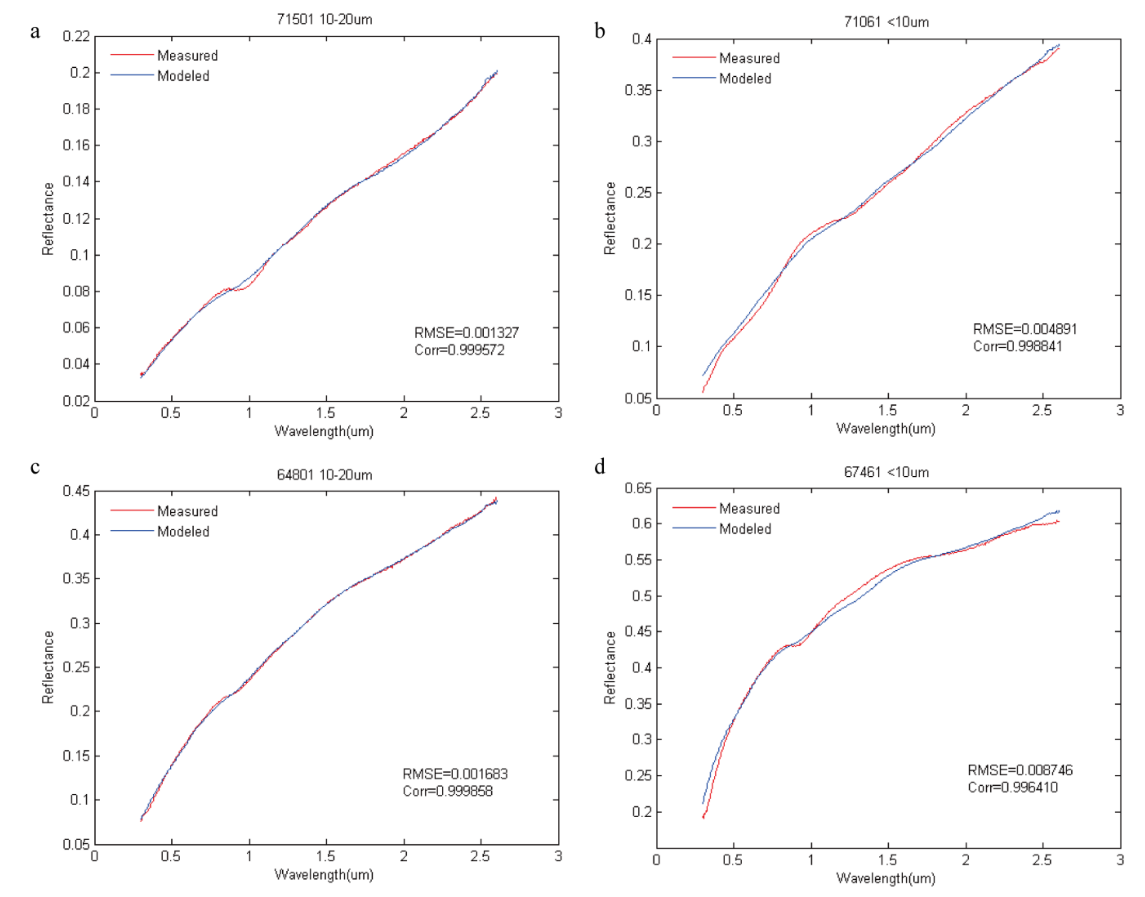
Table 6 and Table 7 and Figure 3 and Figure 4 present the root mean square error (RMSE) and correlation coefficient (Corr) between the simulated spectra and the measured spectra of the LSCC data. The correlation coefficient of each sample is greater than 0.99, and the RMSE is at a magnitude of 10-3. The results show an improvement compared with the findings in Denevi et al. (2008) and an accuracy of simulation close to that in Li and Li (2011) and Liu et al. (2015) [24,25,28]. This outcome may be attributed to the use of the least squares optimization method in the spectra simulation, which makes the interpolation interval smaller than that in the grid searching method (Denevi et al., 2008) [24]. In particular, the mare sample with the highest accuracy is sample 71501 with a size range of 10–20 µm, with an RMSE of 0.001327 and correlation coefficient of 0.999572. The mare sample with the lowest accuracy is sample 71061 in the size group of <10 µm, with an RMSE of 0.004891 and correlation coefficient of 0.998841. The highland sample with the highest accuracy is sample 64801 in the size group of 10–20 µm, with an RMSE of 0.001683 and correlation coefficient of 0.999858. The highland sample with the lowest accuracy is sample 67461 in the size group of <10 µm, with an RMSE of 0.008746 and correlation coefficient of 0.996410, as shown in Figure 5. The simulation accuracy of sample 71061 is not as high as that of other mare samples, and this inaccuracy also exists in the similar works of Denevi et al. (2008) and Li and Li (2011) [24,28]. This issue may be related to the small amount of black beads in sample 71061, adding to the uncertainty of the simulation results (Heiken and McKay, 1974) [42]. The simulation value of sample 67461 is much lower than the true value in the spectral range of 1.1 to 1.6 µm, which may be associated with the selection of plagioclase end-members. Plagioclase has obvious absorption at approximately 1250 nm, and the simulated spectra are dominated by this absorption in sample 67461 with large amounts of plagioclase (60.0 wt. %).
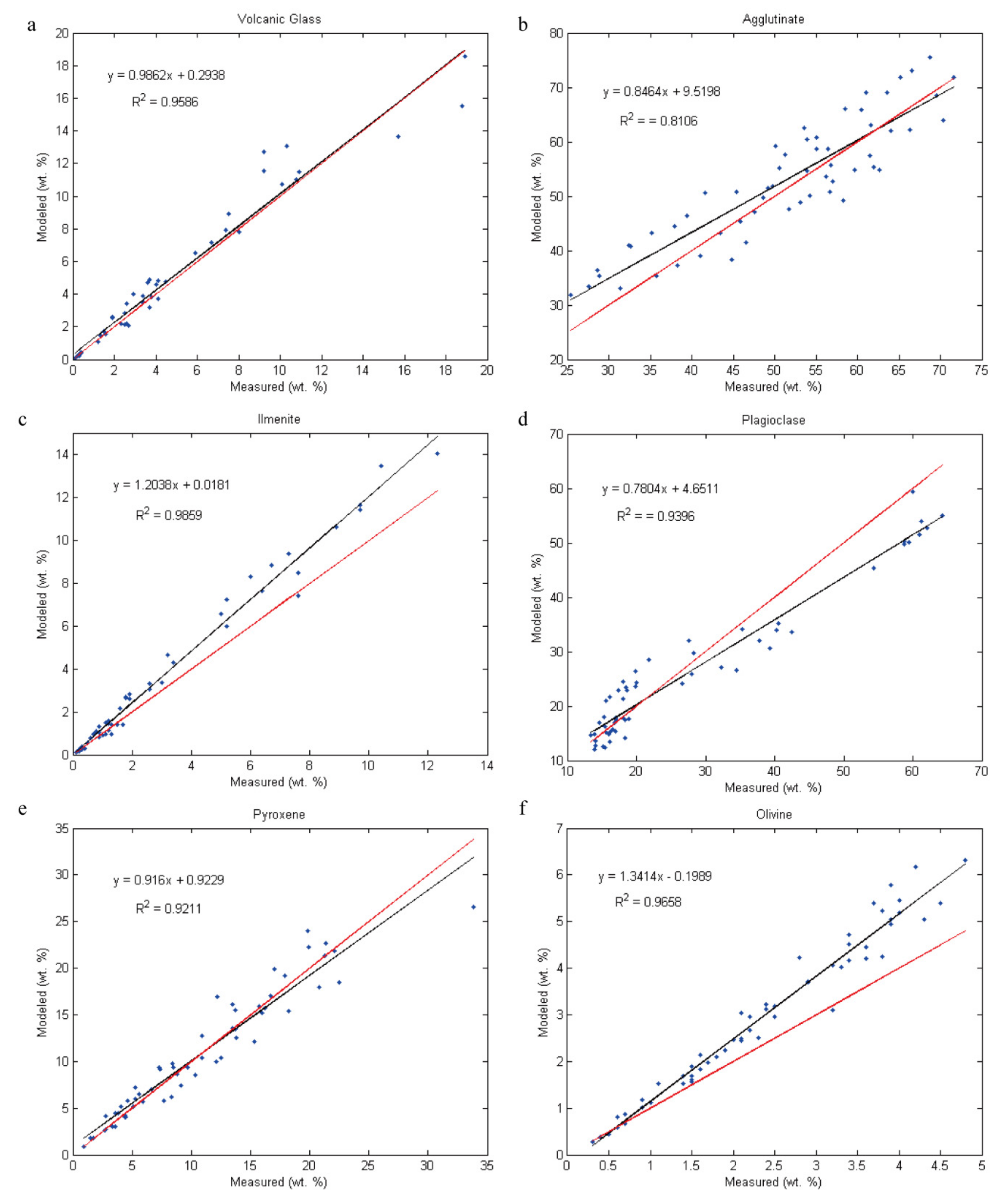
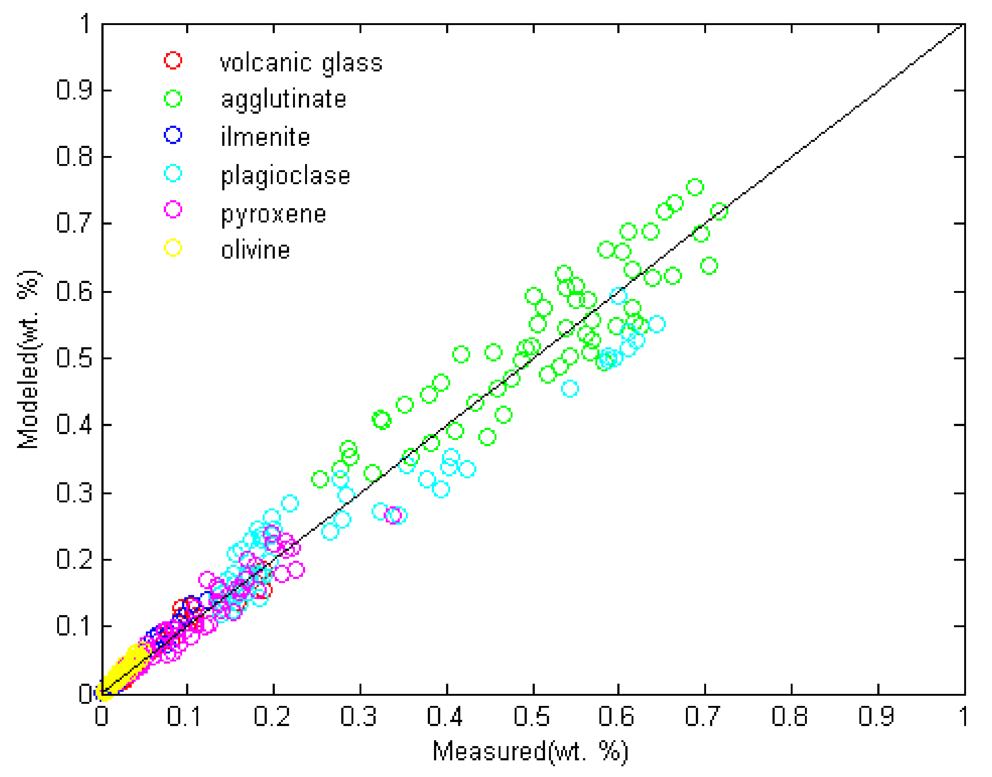
At the same time, we simulated the mineral abundance of volcanic glass, agglutinate, ilmenite, plagioclase, pyroxene and olivine and compared the simulated results with the available measurements (Taylor et al., 2001) (Figure 6 and Figure 7) [49]. The simulation results include 57 mature and immature samples from both mare and highland. The simulated and measured abundances of each mineral show a good linear correlation: the simulation accuracy of volcanic glass, ilmenite, plagioclase, pyroxene and olivine is relatively high, with R2 values larger than 0.92. The simulation accuracy of volcanic glass, ilmenite and olivine shows a significant improvement compared with that obtained by Li and Li (2011] and Liu et al. (2015) as a result of the more mineral end-members used in this work and simultaneously retrieving the particle size, SMFe content and mineral abundance [25,28]. Li and Li (2011)’s simulation of mineral abundance is appropriate for immature samples. Liu et al. (2015) considered the effect of larger diameter SMFe (>50 nm) throughout the entire agglutinate and simulated the mineral abundance of both mature and immature samples, although the simulation accuracy of volcanic glass, ilmenite and olivine was not very desirable. We suspected that this low accuracy may be caused by two separate steps in the simulation of mineral abundance, particle size and SMFe content: first, the reflectance spectra and mineral abundance were known, and the particle size and SMFe content were simulated; then, the reflectance spectra and the simulated particle size and SMFe content were known variables, and the mineral abundance was simulated. The method depends on prior knowledge of the mineral abundance, particle size and SMFe content, and the error of the first step will affect the results of the second step, which may be one of the reasons for the low simulation accuracy of volcanic glass, ilmenite and olivine. Our simulation accuracy of agglutinate is relatively poor, with an R2 close to 0.81, which is slightly lower than the results of Liu et al. (2015), who considered the effect of SMFe larger than 50 nm. Overall, the mineral abundance could be reproduced reliably by simulation.
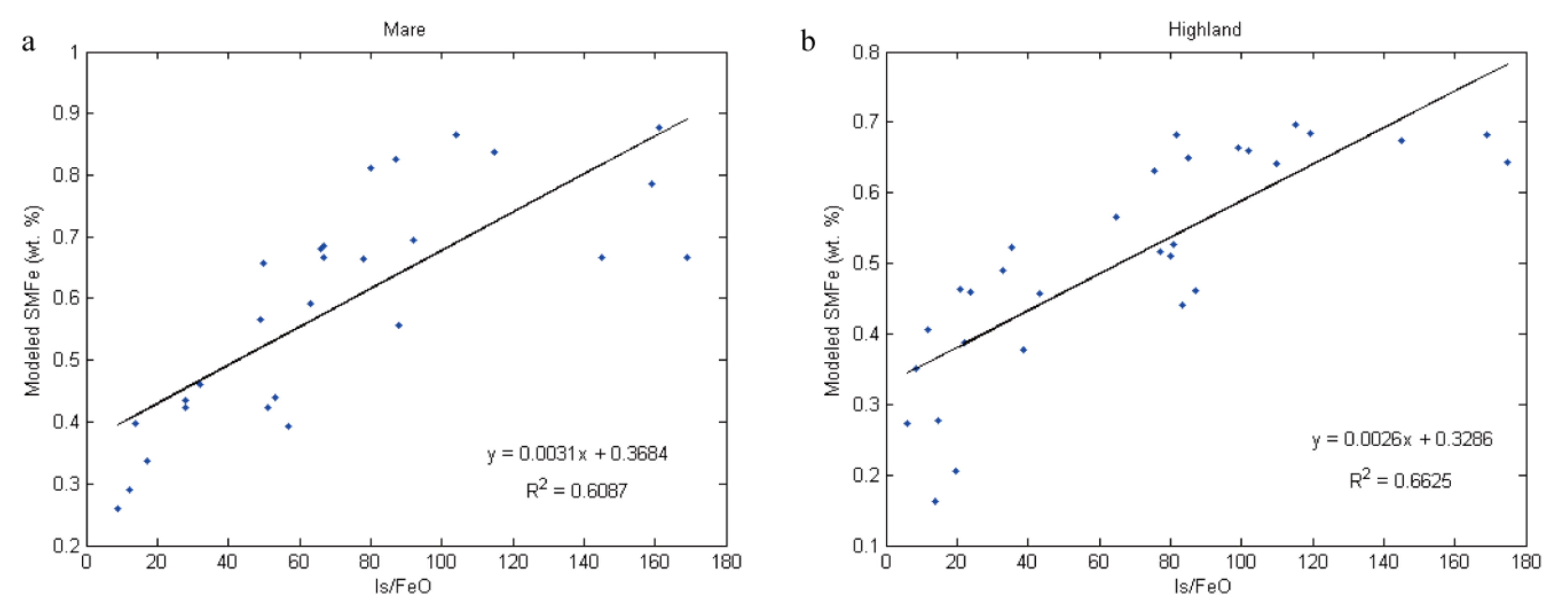
We simulated the SMFe content of each sample, which showed a significantly linear relationship with the relative maturity index Is/FeO (Figure 8). It is concluded that the highest SMFe content was 0.7% (wt. %) and 1% (wt. %) in the lunar highland and mare, respectively, and the SMFe concentrations that we simulated were all within the range by Morris (1980) [52]. It should be noted that the slope of the lunar mare data is steeper than that of the lunar highland data in Figure 8, which is identical to the conclusion of Li and Li (2011) and consistent with the observation of Morris (1980) that lunar regolith with a higher FeO content could accumulate metallic iron faster during the FeO reduction process.
The particle sizes that we simulated are basically consistent with the distribution range of the measured particle sizes, and we marked Y and N as the simulated particle size of each sample within the measured particle size range and out of the range in Table 8 and Table 9, respectively. There are 25 of the 27 lunar mare samples (except sample 10084 with particle size of <10 µm and sample 71501 of <10 µm) and 24 of the 30 lunar highland samples (except 61141 of 10–20 µm, 62231 of 20–45 µm, 62231 of 10–20 µm, 67461 of 20–45 µm, 67481 of 20–45 µm, 67481 of 10–20 µm), whose simulated particle sizes are within the measured particle size range. Even the eight samples whose simulated particle sizes are outside the measured particle size range still fall within the scope of <45 µm. We find that the mare samples with slightly large error are all from the <10 µm size group, and the simulated particle sizes are always overestimated. Compared to other samples, the ilmenite abundance is high in sample 10084 (<10 µm, 5 wt. %) and 71501 (<10 µm, 7.6 wt. %). There are large errors in the Hapke model when applied to opaque ilmenite (Li and Li, 2011), which may be the reason that the particle sizes of samples 10084 and 71501 in the size group <10 µm are overestimated. The highland samples with slightly large errors are all from the 10–20 µm and 20–45 µm size groups, and the simulated particle sizes are always underestimated. Compared to other samples, plagioclase abundance is high in sample 61141 (10–20 µm, 40.3 wt. %), 62231 (20–45 µm, 40.5 wt. %), 62231 (10–20 µm, 37.8 wt. %), 67461 (20–45 µm, 64.3 wt. %), 67481 (20–45 µm, 61.2 wt. %) and 67481 (10–20 µm, 62.0 wt. %). The underestimated particle size of the six highland samples might result from the presence of plagioclase. Overall, our simulation results of particle size could objectively represent the particle size of lunar regolith.
Above all, after the simulations of the reflectance spectra, mineral abundance, particle sizes, and maturity of 57 LSCC samples, good agreement exists between the simulation results and the measured values, indicating that the simulation method of lunar regolith is correct and feasible. Simulated spectra have provided good matches to measured spectra with a high correlation coefficient and small RMSE, and the inverted mineral abundance, particle sizes and maturity are nearly identical to the actual situation of lunar regolith. Of course, high correlation between measured and modeled data does not necessarily demonstrate model validity without further information. In future research, more datasets and experiments will be carried out regarding this section.

5. Simulated Spectral Datasets

After having inversed the optical constants of two series of mineral end-members in 2.2, we simulated two sets of lunar regolith spectra databases in various maturities and mineral abundances at an incident angle of 30°, emittance angle of 0° and phase angle of 30° with particle size of 15 µm (average value of the 10–20 µm size group). The data can be downloaded at the website http://pan.baidu.com/s/1c12FkkS (accessed on 10 November 2021).
Mineral end-members of the first spectra database were selected from lunar samples and telluric samples, part of which lack chemical analysis (Table 2). There are relatively more mineral end-members in the first spectra database, including typical lunar regolith minerals such as volcanic glass and agglutinate. The mineral end-members of olivine are forsterite and fayalite. The first spectral database includes more than 300,000 spectra with a wavelength range of 0.3–2.6 µm and a spectral resolution of 5 nm.
Mineral end-members of the second spectra database were all selected from lunar samples with chemical analysis, including plagioclase, orthopyroxene, clinopyroxene, forsterite and ilmenite (Table 3). More than 300,000 spectra were simulated with a wavelength range of 0.35–2.6 µm and a spectral resolution of 5 nm.

6. Application of Simulated Spectra

When mapping the FeO content of the entire moon by means of Clementine UVVIS/NIR data, Lucey et al. found that the spectral characteristics of lunar regolith had a certain relationship with both the FeO content and maturity in multiple dimensions of spectra. Based on the “spectral characteristic angle parameter” (Lucey et al., 1995, 1998, 2000; Blewett et al., 1997; Gillis et al., 2003) [1,13,14,15,53], a new method was proposed to separate the effects of maturity and FeO content, which has been widely applied to the analysis of lunar surfaces by means of remote sensing data on multiple scales (global, mare, important impact craters). Unfortunately, the method and model established by Lucey are based on Apollo and Luna data, which are limited to small landing site areas. Because the measured spectra of lunar samples are a comprehensive result of various factors, it is difficult to describe and clarify the influence of each factor on spectral characteristics. Accordingly, it is challenging to analyze the mechanism, scope of application and reliability of Lucey’s method. In our work, we chose the second set of mineral end-members and simulated spectra database and investigated the mechanism, scope of application and reliability of Lucey’s method based on the “spectral characteristic angle parameter”, which has been widely applied as one of the application cases of simulated spectra of lunar regolith.
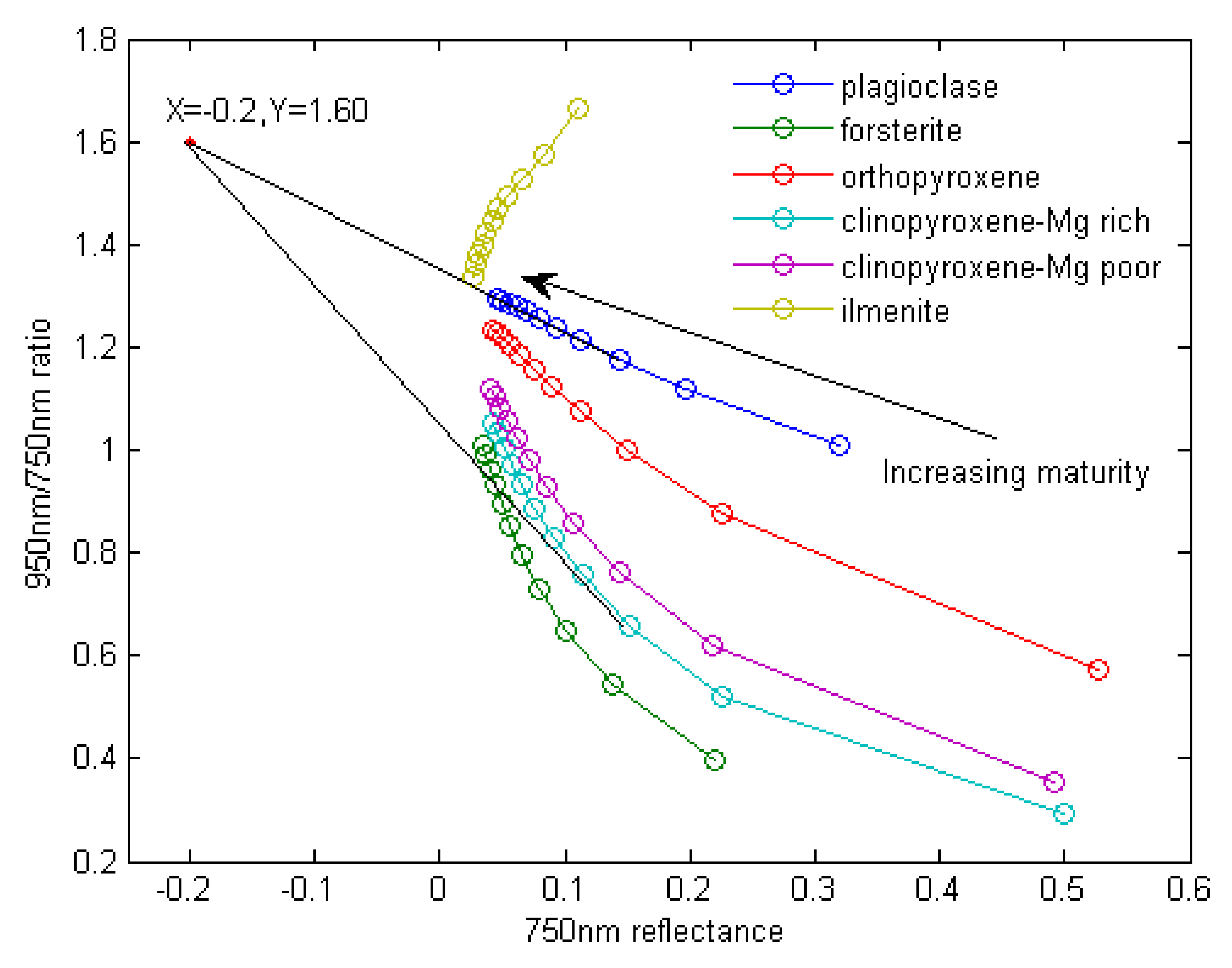
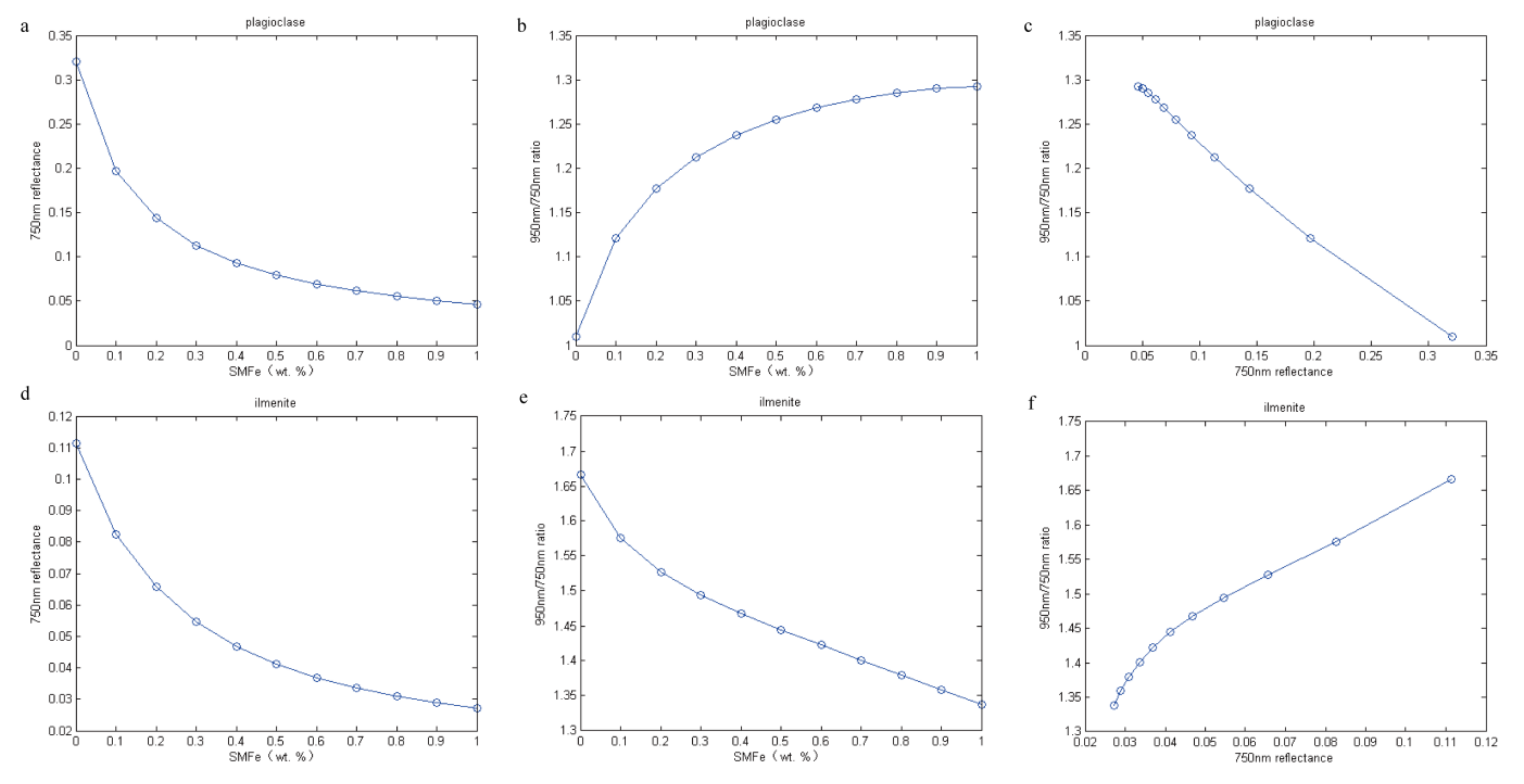
All Apollo and Luna samples are roughly scattered in a triangular region (Lucey et al., 1995, 1998, 2000) [1,13,14], and their trendlines tend to converge at a point on the top left corner in a two-dimensional scatterplot of the 750 nm–950/750 nm spectra. The distance between each sample and the converged origin is positively correlated with maturity, and the clockwise angle between each sample and the converged origin shows a good linear relationship with the FeO content. Here, a possible explanation is that when the lunar soil is exposed to the space environment, the continuous impact of micrometeorites, the continuous bombardment of high-energy particles of the sun and the charged particles of the universe will produce a series of space weathering and changes in composition. The various changes that occur when the lunar soil is exposed to the lunar surface are called maturation. If the lunar surface exposure time of the fresh lunar soil reaches a certain level, its composition will be relatively stable and reach a mature state. If the lunar surface is exposed for a short time and is quickly buried in the subsurface, it is in an immature state. Lunar soil elemental iron (SMFe), as one of the main products of spatial weathering, can better indicate the degree of maturity. SMFe makes the reflectance spectrum red, darkened, and weakened [1,30,32,43,44]. We simulated the spectra of pure mineral end-members in Table 3 with an SMFe content (indicating relative maturity) varying from 0 to 1% by an interpolation interval of 0.1%, and the 750 nm–950/750 nm spectra are plotted in Figure 9. The end-members of olivine, forsterite and ilmenite are not applicable to Lucey’s method of “spectral characteristic angle parameter”. The clockwise angle between forsterite and the converged origin is much larger than that of other mineral end-members; this observation of forsterite contradicts with its low FeO content. The slope of ilmenite is significantly different from that of other mineral end-members in the 750 nm–950/750 nm spectra plane. Then, we analyzed the influence of SMFe content on the 750 nm reflectance and the 950/750 nm reflectance. As the SMFe content increases, the 750 nm reflectance of all mineral end-members decreases, and the absorption features of the spectra become weaker. On the other hand, the 950/750 nm reflectance of plagioclase is high with red reflectance spectra. However, the relationship between the 950/750 nm reflectance and SMFe content for ilmenite is opposite to that of other mineral end-members, indicating that the influence of maturity on ilmenite is significantly different from that on other minerals (Figure 10).
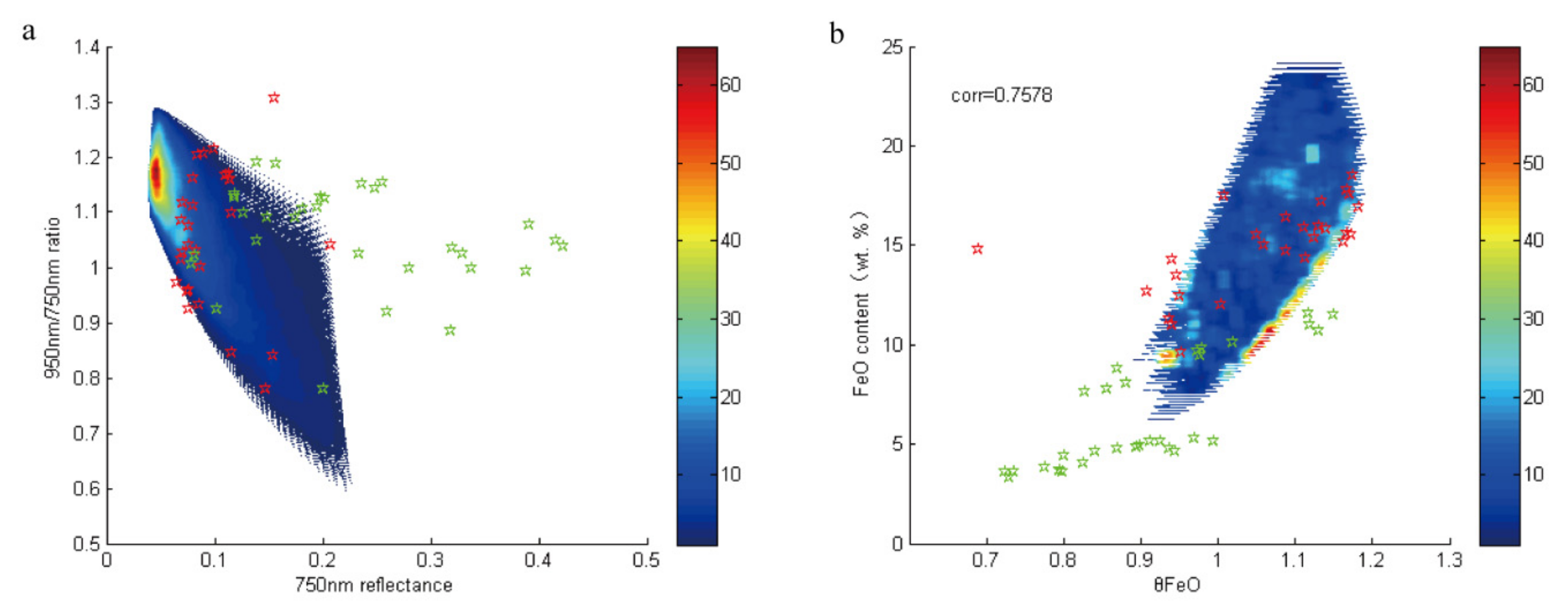
Therefore, mineral end-members of forsterite and ilmenite should not be considered when estimating the converged origin (X = −0.20, Y = 1.60) of the 750 nm–950/750 nm spectra plane (Figure 9). Six mineral end-members (plagioclase, orthopyroxene, Mg-rich clinopyroxene, Mg-poor clinopyroxene, forsterite (none or few) and ilmenite (none or few)) are used to simulate spectra of lunar regolith with different maturity degrees and mineral abundances. The clockwise angle between the simulated spectra and the converged origin of X = −0.20, Y = 1. 60 in the 750 nm–950/750 nm spectra plane showed a high correlation with the FeO content of the simulated samples with a correlation coefficient of 0.7594 (Figure 11). The LSCC mare and highland data in the size group of 10–20 µm, as reference data, are also plotted in the figure using the same method. In Figure 11, the plotted simulated data cover all the LSCC mare data, while they only partially cover the LSCC highland data. This discrepancy may be due to the presence of plagioclase, as the abundance of plagioclase in LSCC highland samples is relatively high, which we will strive to optimize in further work. We find that although the clockwise angle is positively correlated with the FeO content, the relationship between the two is not strictly linear. The spectra of Apollo and Luna samples, used to predict the FeO content by Lucey, might not be fully representative of the spectra of the entire moon.
From the above analysis, it can be seen that Lucey’s method, used to invert the FeO content based on the “spectral characteristic angle parameter”, can only be applied to mixed systems of plagioclase, clinopyroxene (Mg rich and Fe rich), orthopyroxene, forsterite (none or few) and ilmenite (none or few). This mixed system macroscopically constitutes the lunar surface composition except for some local areas, e.g., craters. Comprehensive analysis shows that Lucey’s method can only be applied to macroscopic analysis of the entire moon. Caution should be used when applying this method in areas with high contents of forsterite and ilmenite in the mixed system.

7. Discussion

7.1. Simulation of Spectral Parameters

The purpose of Section 7.2 is not to discuss errors of the model, but to try to discuss the influence of changes in chemical composition on the spectrum and approximate estimation methods. Denevi et al. (2008) pointed out that changes in the chemical composition of mineral endmembers have a great influence on the spectrum, which is more sensitive than mineral abundance. Liu et al. (2015) did not consider the influence of mineral chemical composition on the spectrum when selecting olivine end members. It is difficult to objectively and comprehensively represent the olivine on the moon. This may be the reason for its low accuracy in the simulation of olivine content. When simulating the spectrum of lunar soil, we subdivided olivine into forsterite and iron olivine end members, and approximated the chemical composition (Fo) of olivine by mixing forsterite and iron olivine end members in different proportions. The influence of the mixed spectrum.
Method 1: Calculate the Fo value of the mixed mineral of the two olivine end members by calculating the Fo value of the ferroolivine end member, the Fo value of the forsterite end member, and the mass percentage of the two olivine end members. Select the optical constant corresponding to the Fo value to simulate the reflectance spectrum of the mixed olivine.
Method 2: Simulate the reflectance spectrum of mixed olivine through the optical constants of the frond olivine end members, the optical constants of the forsterite end members, and the volume percentages of the two olivine end members.
Then, we compare and analyze the simulation results of the two methods.
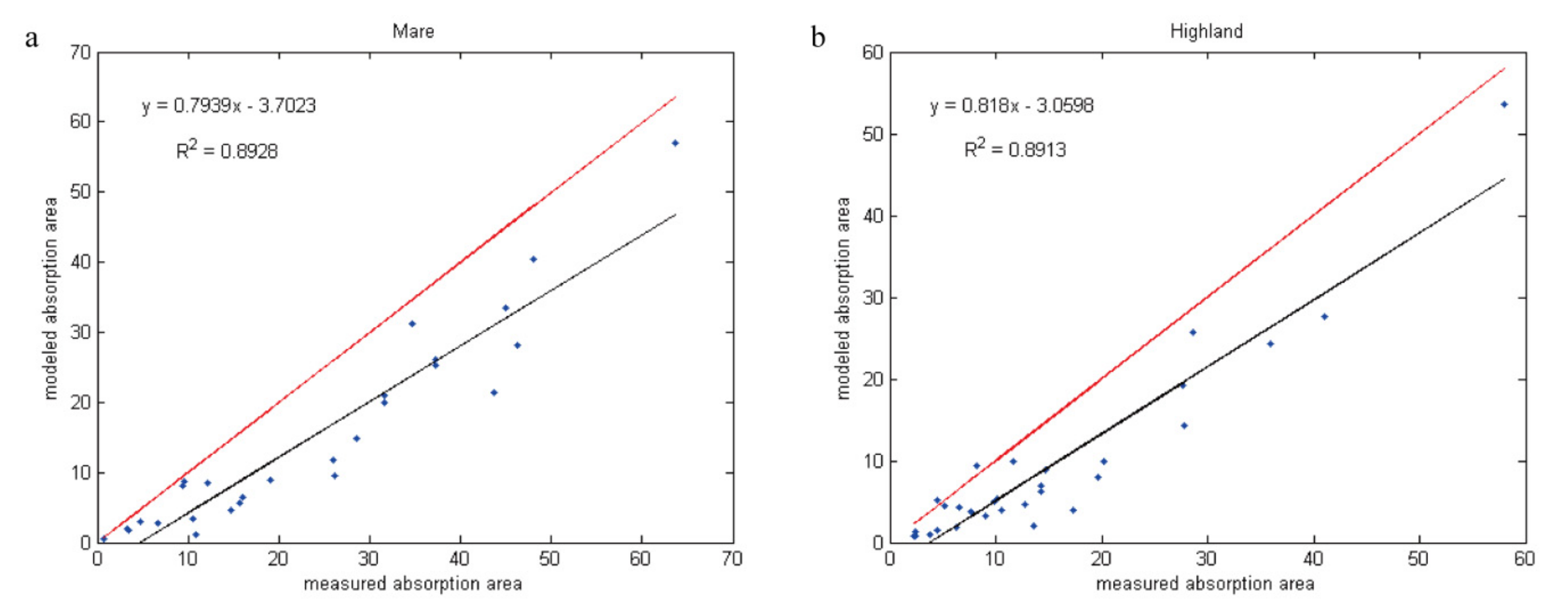
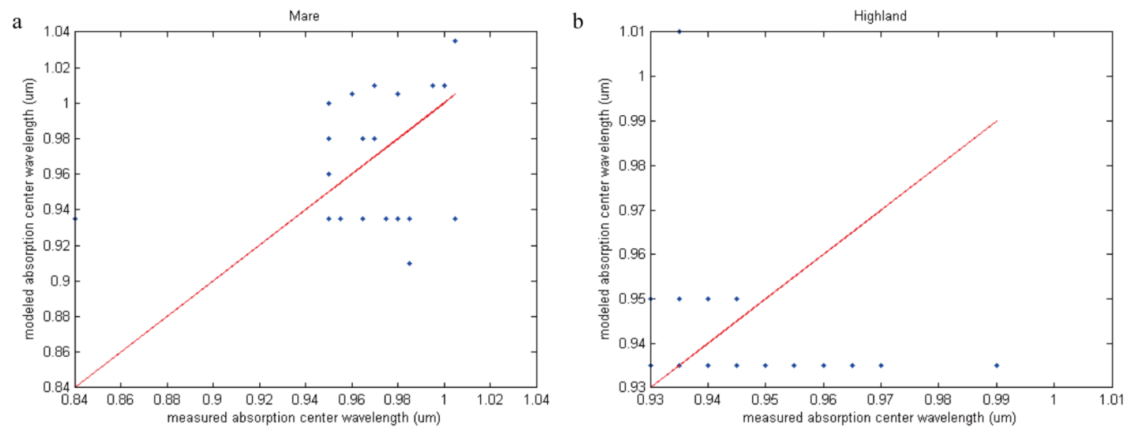
In this section, we simulate the spectra of 57 samples in LSCC data. The correlation coefficients between the simulated spectra and the measured spectra are all larger than 0.99, and the root mean square errors are at a magnitude of 10-3. However, it is not enough to evaluate the accuracy of simulation results just by correlation and RMSE. Spectral parameters, such as the absorption center wavelength and absorption area, are crucial parameters to describe spectral characteristics, with important significance for mineral identification. Here, we calculate the absorption area (Figure 12) and absorption center wavelength (Figure 13) of both the measured and modeled spectra of 57 LSCC samples to further evaluate the accuracy of our simulation results.
Figure 12 shows that the correlation of the absorption area between the modeled and measured spectra is high, even when the absorption area of the measured spectra is underestimated. In Figure 13, the absorption center wavelength data are discretely distributed around the y = x line. Table 10 shows the relative error of the simulated absorption center wavelength (the ratio of the difference between the simulated and measured absorption center wavelengths versus the measured data). From Figure 13, data are roughly correlated for Mare but not correlated for Highland. We think the reason for this is that the entire lunar surface is divided into the lunar sea and the lunar heights in topography. The lunar sea is mainly composed of dark minerals such as basalt, while the lunar highlands are mainly composed of light-colored minerals such as plagioclase. Plagioclase in the lunar soil is poor in sodium and rich in calcium, and has a relatively strong absorption near 0.65 µm and 1.25 µm, but it is not easy to observe in the lunar soil. This is because the absorption characteristics of plagioclase are weaker than other strong absorption minerals and can easily be concealed. Pyroxene has strong absorption characteristics near 1 µm and 2 µm, and with the decrease of Ca content, the absorption center position moves to the left [54,55]. The lunar highlands contain more plagioclase, while the lunar pyroxene contains relatively more. We speculate that this is one of the reasons why the data is roughly correlated for Mare but not correlated for Highland. On the other hand, spatial weathering weakens the spectral characteristics, making it difficult to accurately simulate the absorption position, which is also one of the possible reasons.
From Table 10, it can be seen that the relative error of the absorption center wavelength of sample 71061 is 11.31% and that its spectra can hardly be simulated due to the presence of a small amount of black beads. The relative error of the absorption center wavelength of the rest of the samples is no more than 8.02%, which has a maximum absolute error of 75 nm. Denevi et al. (2008) encountered a similar situation when simulating the spectra of LSCC data [24]. There is trouble matching the absolute reflectance and the continuum spectra, and this problem disappears when simulating the mixed spectra of two kinds of mineral end-members. In our speculation, SMFe, as a main product of space weathering, probably results in the large error of simulated spectral parameters.
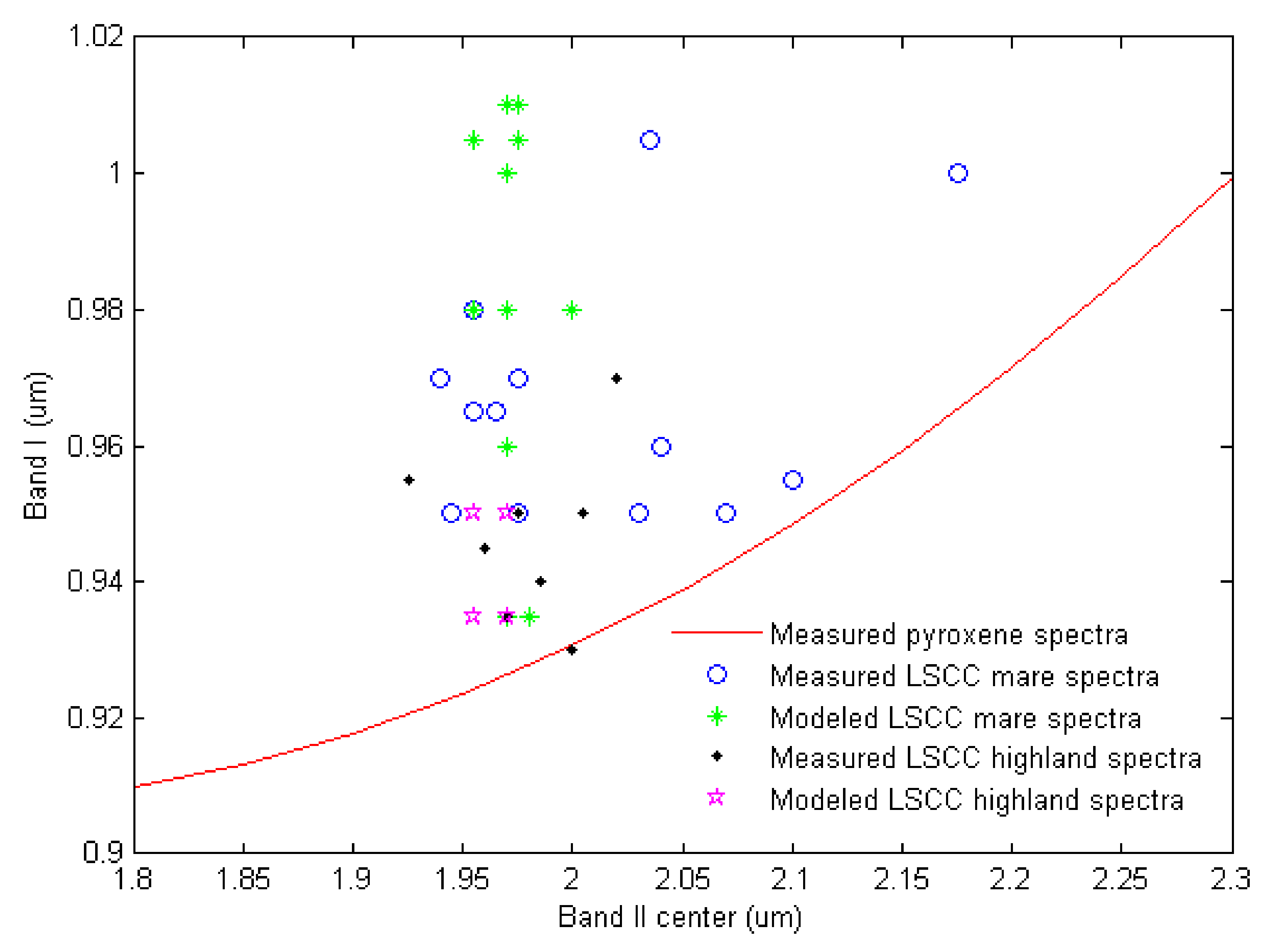
To test the above hypothesis, some spectra of LSCC data that have large absorption areas and absorption center wavelengths are plotted at 1 µm and 2 µm (called the Band I center and Band II center, respectively) of both the measured and modeled spectra. Pyroxene, as one of the most common minerals of lunar regolith, has a strong absorption feature at the Band I center and Band II center, so the scatter plot of the Band I center versus the Band II center is often used to distinguish orthopyroxene from clinopyroxene. The red line in Figure 14 is the approximate trend line of pyroxenes (Adams, 1974; Cloutis and Gaffey, 1991a) [54,55].
In Figure 14, the measured mare and highland data are slightly in the upper left part of the trend line, and there is no obvious linear relationship between the Band I center and Band II center plots. The spectral features of minerals (especially pyroxene) have been weakened by space weathering. The simulated and measured spectra for both mare and highland samples are widely scattered, although they plot roughly in the same area.
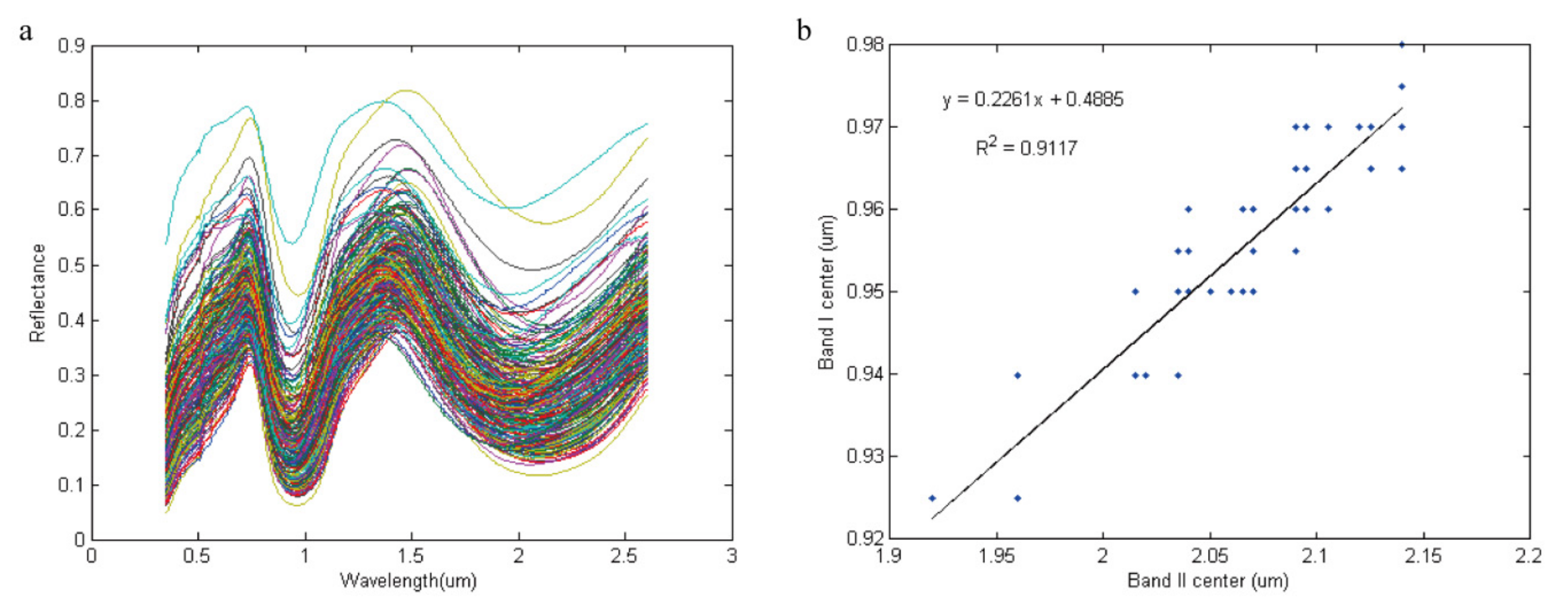
When space weathering is not considered, 231 spectra are simulated with three pyroxene end members mixed, and their Band I center versus Band II are plotted (Figure 15). The endmember pyroxenes in samples LR-CMP-168, LR-CMP-170 and LS-CMP-012 are from the RELAB library. Not taking into account the effect of space weathering, there is a linear relationship between the Band I center and Band II, and the absorption center wavelength shifts to a long wavelength with increasing Ca content of the mixed minerals.
In future work, more attention will be given to precisely simulating the parameters of spectral features. When optimizing the simulated spectra that are closest to the measured data with Newton interpolation and the least square fitting method, more efforts will be made to investigate the errors between the simulated and measured spectra at key band positions, such as the Band I center and Band II, thus improving the simulation accuracy of the spectral features.

7.2. The Isomorphism of Olivine

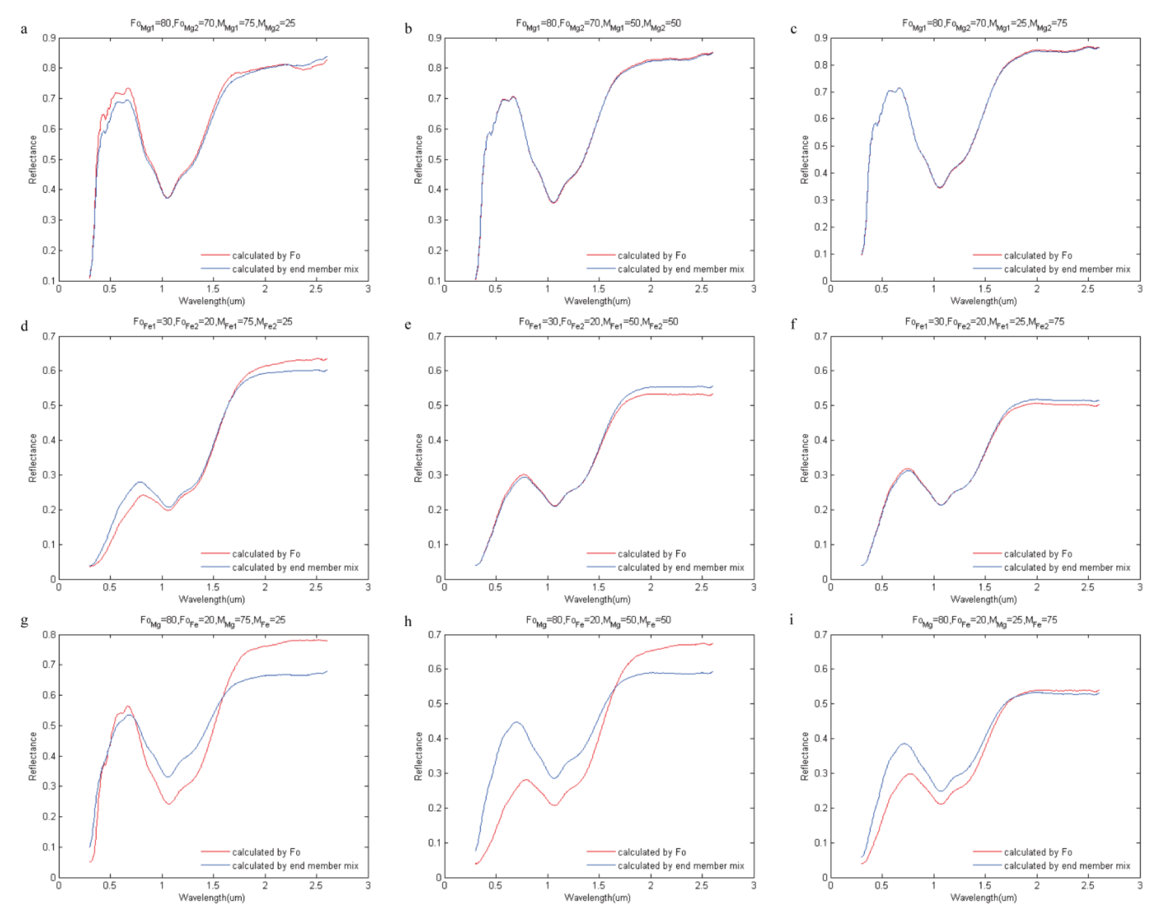
Yan et al. (2010) inversed the distribution of lunar olivine, clinopyroxene, orthopyroxene, plagioclase and ilmenite with the Clementine UVVIS/NIR data by the Hapke model [27]. The authors compared their results to those of Lucey (2004). There is a large uncertainty in the abundance and distribution range of lunar olivine. Denevi et al. (2008) indicated that the spectra were more sensitive to a change in the chemical composition of minerals than a change in mineral abundance. Liu et al. (2015) did not consider the effect of chemical composition (e.g., Mg number) on the spectra when selecting the mineral end-members, so it was difficult to objectively represent all lunar samples, which may be one of the reasons for the low accuracy of the simulated abundance of olivine. In our simulation of lunar regolith, the mineral end-member of olivine is subdivided into forsterite and fayalite, and the effect of the chemical composition of olivine on the spectra is approximated by a mixture of variable proportions of forsterite and fayalite (Figure 16).
Nine groups of olivine samples were selected from the RELAB spectra database with Fo values (molar Mg/(Mg + Fe)) from 90 to 10. The sample IDs are DD-MDD-037, DD-MDD-038, DD-MDD-039, DD-MDD-040, DD-MDD-041, DD-MDD-042, DD-MDD-043, DD-MDD-034, and DD-MDD-045. Some work has been done to retrieve the optical constants of each olivine end-member and to simulate the mixed spectra of two kinds of olivine with different Fo values for different mixed proportions. Considering the dominant role of chemical composition on the spectra, the Fo value of variable proportions of two kinds of olivine was calculated to invert corresponding optical constants and to forward model the spectra of the mixed mineral with Equations (1)–(16) (red lines in Figure 16). For the two end-members of olivine, one method is used to simulate spectra for the single scattering albedo of the two olivine end-members by their optical constants using Equations (11)–(16), and the other method is used for the mixed mineral by the mixed proportion with Equations (1)–(10) (blue lines in Figure 16). Figure 16 contrasts the two simulated spectra. The two pairs of mixed olivine end-members are as follows: Fo80 olivine—Fo70 olivine (Figure 16a–c), Fo30 olivine—Fo20 olivine (Figure 16d–f) and Fo80 olivine—Fo20 olivine (Figure 16g–i). The mixed proportions of the two olivine end-members are 75:25 (Figure 16a,d,g), 50:50 (Figure 16b,e,h) and 25:75 (Figure 16c,f,i).
If the Fo values of the two kinds of olivine are relatively close, the simulated spectra calculated by a mix of end-members can approximate those calculated by chemical composition (Figure 16). If there exists a large difference between the Fo values of the two kinds of olivine, the simulated spectra calculated by the mix of end-members deviate to a large extent from those calculated by chemical composition, while the difference in the main absorption features between the two methods is small.
DD-MDD-038 (Fo = 80, forsterite) and DD-MDD-044 (Fo = 20, fayalite) were chosen as two olivine end-members to simulate the first spectra database, in which case the simulated spectra calculated by the mix of two end-members may differ from the simulated spectra calculated by chemical composition. Given the low content of olivine in the LSCC lunar sample (less than 5%), however, it is possible to simulate the spectra. In future work, attention will be focused on the influence of chemical composition on the spectra to improve the accuracy of simulation and to analyze the mechanism and influencing factors of the spectra of lunar regolith.

8. Conclusions

In our work, two sets of lunar regolith spectra databases were simulated. Mineral end-members of the first set were selected from lunar and telluric samples, part of which lacks chemical analysis. Mineral end-members of the second set were from lunar samples with chemical analysis. More mineral end-members are available to model the spectra in the first set than in the second set. The two spectral databases can be used to analyze the method, mechanism, accuracy and reliability of the abundance and composition of lunar regolith.
First, multiple solutions and the applicability of the Hapke model were investigated by means of Newton interpolation and the least square optimization method. This method is suitable for the simulation of spectra, although it still has difficulty in the inversion of mineral abundance.
Then, simulations were performed on the spectra, mineral abundance, particle size and SMFe content of 57 mare and highland samples of LSCC in the size groups of 10 µm, 10–20 µm and 20–45 µm to verify the reliability of our simulation method. Full consideration was given in the simulation work when selecting mineral end-members; in particular, volcanic glass, pyroxene, and olivine were subdivided into more mineral end-members by chemical composition. The role of space weathering was also considered. We simulated spectra of lunar regolith with specific mineral abundance, particle size, SMFe content and viewing geometries using Newton interpolation and the least square optimization method based on the Hapke (2002) AMSA radiative transfer model, and the results were compared to the measured LSCC data. The simulated spectra match well with the measured spectra, with correlation coefficients greater than 0.99 and a root mean square error at magnitudes of 10-3. Both mature and immature samples were included in the simulation of mineral abundance of 57 mare and highland LSCC samples, and the simulated abundance shows a good linear relation with the measured abundance. The simulation accuracies of volcanic glass, ilmenite, plagioclase, pyroxene and olivine are relatively high, with R2 values higher than 0.92, and the simulation accuracy of agglutinate is relatively poor, with R2 values close to 0.81. The simulated SMFe content has a distinct linear relationship with the relative maturity index Is/FeO. The simulated particle size of 86% of the samples falls within the measured particle size range, and no particle size is beyond 45 µm. Overall, the spectra and mineral abundance, particle size, and maturity of LSCC can be simulated with small errors, verifying that the method that we used to simulate lunar regolith is correct and feasible.
Having confirmed the validity of the simulation of lunar regolith, we studied the mechanism, reliability and scope of application of the “spectral characteristic angle parameter method” proposed by Lucey et al. (1995, 1998, 2003) [1,13,56] as one of the application cases of simulated spectra of lunar regolith. We conclude that this method is only suitable for the macro analysis of the entire moon. More attention should be given to areas with relatively high abundances of forsterite and ilmenite in the mixed minerals.
In the LSCC simulation, the absorption center wavelength results are not satisfactory, although overall, the simulated and measured spectra fit well. These results may be due to space weathering, which suppresses the spectral features, making the spectral absorption position difficult to simulate.
Finally, the isomorphism of olivine has been discussed. In the spectral simulation of lunar regolith, olivine was subdivided into forsterite and fayalite, and the mixed spectra were calculated to approximately estimate the spectra calculated by the chemical composition of olivine. There are some errors in this method. The low content of olivine in the lunar regolith has little effect on the simulation results, and the approximation method is still valid for simulation. Moreover, the mineral abundance could be crucial information when selecting a suitable landing site for programs of in situ resource utilization.
In future work, effort will be made to investigate the influence of spinel on simulated lunar regolith spectra. Spinels (Mg-spinel, Fe/Cr-rich spinels), discovered by hyperspectral data in recent years, show a significant absorption feature near 2 µm (Pieters et al., 2011; Dhingra et al., 2011; Yamamoto et al., 2013) [57,58,59], which has important significance for analyzing the evolution of the moon. At the same time, we will consider the mechanism and influencing factors of chemical composition to further improve the simulation accuracy of spectra and corresponding mineral abundance, particle size and maturity, especially to reduce the simulation error of spectral parameters, such as absorption position and absorption area. If the large-scale roughness that describes the effects of topography were considered, our simulation method could be applied to remote sensing image data on the whole moon scale or in a specific region.

Author Contributions

P.Z. and Z.Z. are equally contributed to this paper, they are co-first authors. The specific author contribution is stated as follow: Conceptualization, P.Z. and Z.Z.; methodology, P.Z., Z.Z. and H.-Y.H.; software, Z.Z. and G.W.; validation, P.Z. and Z.Z.; analysis, Z.Z., P.Z. and H.-Y.H.; resources and code, Z.Z.; data curation, Z.Z., P.Z. and H.-Y.H.; writing—original draft preparation, P.Z. and Z.Z.; writing—review and editing, H.-Y.H.; visualization, P.Z. and Z.Z.; supervision, H.-Y.H.; project administration, H.-Y.H.; funding acquisition, H.-Y.H. All authors have read and agreed to the published version of the manuscript.

Funding

This research was funded by National Natural Science Foundation of China, grant number 41701425, and by Scientific Research Project of Beijing Municipal Educational Commission, grant number KM202110005017.

Institutional Review Board Statement

Not applicable.

Informed Consent Statement

Not applicable.

Data Availability Statement

The results of simulated spectra data can be freely downloaded and freely accessed through the website http://pan.baidu.com/s/1c12FkkS.

Acknowledgments

We would like to thank Bruce Hapke for his constructive model. Many thanks to NASA RELAB facility in Brown University for the spectra data. We also thank Baidu SkyDrive for storing the simulated spectra data at website http://pan.baidu.com/s/1c12FkkS.

Conflicts of Interest

The authors declare no conflict of interest.

References

  1. Lucey, P.G.; Taylor, G.J.; Malaret, E. Abundance and Distribution of Iron on the Moon. Science 1995, 268, 1150–1153. [Google Scholar] [CrossRef] [PubMed]
  2. Ohtake, M.; Matsunaga, T.; Haruyama, J.; Yokota, Y.; Morota, T.; Honda, C.; Ogawa, Y.; Torii, M.; Miyamoto, H.; Arai, T.; et al. The global distribution of pure anorthosite on the Moon. Nature 2009, 461, 236–240. [Google Scholar] [CrossRef] [PubMed]
  3. Ohtake, M.; Takeda, H.; Matsunaga, T.; Yokota, Y.; Haruyama, J.; Morota, T.; Yamamoto, S.; Ogawa, Y.; Hiroi, T.; Karouji, Y.; et al. Asymmetric crustal growth on the Moon indicated by primitive farside highland materials. Nat. Geosci. 2012, 5, 384–388. [Google Scholar] [CrossRef]
  4. Wu, Y. Major elements and Mg# of the Moon: Results from Chang’E-1 Interference Imaging Spectrometer (IIM) data. Geochim. Cosmochim. Acta 2012, 93, 214–234. [Google Scholar]
  5. Yamamoto, S.; Nakamura, R.; Matsunaga, T.; Ogawa, Y.; Ishihara, Y.; Morota, T.; Hirata, N.; Ohtake, M.; Hiroi, T.; Yokota, Y.; et al. Possible mantle origin of olivine around lunar impact basins detected by SELENE. Nat. Geosci. 2010, 3, 533–536. [Google Scholar] [CrossRef]
  6. Yamamoto, S.; Nakamura, R.; Matsunaga, T.; Ogawa, Y.; Ishihara, Y.; Morota, T.; Hirata, N.; Ohtake, M.; Hiroi, T.; Yokota, Y.; et al. Massive layer of pure anorthosite on the Moon. Geophys. Res. Lett. 2012, 39, 1–6. [Google Scholar] [CrossRef] [Green Version]
  7. Yamamoto, S.; Nakamura, R.; Matsunaga, T.; Ogawa, Y.; Ishihara, Y.; Morota, T.; Hirata, N.; Ohtake, M.; Hiroi, T.; Yokota, Y.; et al. Olivine-rich exposures in the South Pole-Aitken Basin. Icarus 2011, 218, 331–344. [Google Scholar] [CrossRef]
  8. Yamamoto, S.; Nakamura, R.; Matsunaga, T.; Ogawa, Y.; Ishihara, Y.; Morota, T.; Hirata, N.; Ohtake, M.; Hiroi, T.; Yokota, Y.; et al. Global occurrence trend of high-Ca pyroxene on lunar highlands and its implications. J. Geophys. Res. Planets 2015, 120, 831–848. [Google Scholar] [CrossRef] [Green Version]
  9. Lucey, P.G.; Norman, J.A.; Crites, S.T.; Taylor, J.G.; Hawke, B.R.; Lemelin, M.; Melosh, H.J. A large spectral survey of small lunar craters: Implications for the composition of the lunar mantle. Am. Mineral. 2014, 99, 2251–2257. [Google Scholar] [CrossRef]
  10. Vinckier, Q.; Hardy, L.; Gibson, M.; Smith, C.; Putman, P.; Hayne, P.O.; Sellar, R.G. Design and Characterization of the Multi-Band SWIR Receiver for the Lunar Flashlight CubeSat Mission. Remote Sens. 2019, 11, 440. [Google Scholar] [CrossRef] [Green Version]
  11. Zhou, P.; Zhao, Z.; Huo, H.-Y.; Liu, Z. Retrieval of Photometric Parameters of Minerals Using a Self-Made Multi-Angle Spectrometer Based on the Hapke Radiative Transfer Model. Remote Sens. 2021, 13, 3022. [Google Scholar] [CrossRef]
  12. Peng, M.; Di, K.; Wang, Y.; Wan, W.; Liu, Z.; Wang, J.; Li, L. A Photogrammetric-Photometric Stereo Method for High-Resolution Lunar Topographic Mapping Using Yutu-2 Rover Images. Remote Sens. 2021, 13, 2975. [Google Scholar] [CrossRef]
  13. Lucey, P.G. Model near-infrared optical constants of olivine and pyroxene as a function of iron content. J. Geophys. Res. Earth Surf. 1998, 103, 1703–1713. [Google Scholar] [CrossRef]
  14. Lucey, P.G.; Blewett, D.; Taylor, G.J.; Hawke, B.R. Imaging of lunar surface maturity. J. Geophys. Res. Earth Surf. 2000, 105, 20377–20386. [Google Scholar] [CrossRef]
  15. Blewett, D.; Lucey, P.G.; Hawke, B.R.; Jolliff, B.L. Clementine images of the lunar sample-return stations: Refinement of FeO and TiO2 mapping techniques. J. Geophys. Res. Earth Surf. 1997, 102, 16319–16325. [Google Scholar] [CrossRef]
  16. Shkuratov, Y.; Starukhina, L.; Hoffmann, H.; Arnold, G. A Model of Spectral Albedo of Particulate Surfaces: Implications for Optical Properties of the Moon. Icarus 1999, 137, 235–246. [Google Scholar] [CrossRef]
  17. Hapke, B. Bidirectional reflectance spectroscopy: 1. Theory. J. Geophys. Res. Earth Surf. 1981, 86, 3039–3054. [Google Scholar] [CrossRef]
  18. Hapke, B. Bidirectional reflectance spectroscopy: 3. Correction for macroscopic roughness. Icarus 1984, 59, 41–59. [Google Scholar] [CrossRef] [Green Version]
  19. Hapke, B. Bidirectional reflectance spectroscopy: 4. The extinction coefficient and the opposition effect. Icarus 1986, 67, 264–280. [Google Scholar] [CrossRef] [Green Version]
  20. Hapke, B. Theory of Reflectance and Emittance Spectroscopy; Cambridge Univ. Press: Cambridge, UK, 1993; p. 455. [Google Scholar]
  21. Hapke, B. Bidirectional reflectance spectroscopy: 5. The coherent backscatter opposition effect and anisotropic scattering. Icarus 2002, 157, 523–534. [Google Scholar] [CrossRef] [Green Version]
  22. Hapke, B. Theory of Reflectance and Emittance Spectroscopy; Cambridge University Press: Cambridge, UK, 2005; 472p. [Google Scholar]
  23. Hapke, B. Bidirectional reflectance spectroscopy: 6. Effects of porosity. Icarus 2008, 195, 918–926. [Google Scholar] [CrossRef] [Green Version]
  24. Denevi, B.W.; Lucey, P.G.; Sherman, S.B. Radiative transfer modeling of near-infrared spectra of lunar mare soils: Theory and measurement. J. Geophys. Res. Space Phys. 2008, 113, E02003. [Google Scholar] [CrossRef]
  25. Liu, D.; Li, L.; Sun, Y. An improved radiative transfer model for estimating mineral abundance of immature and mature lunar soils. Icarus 2015, 253, 40–50. [Google Scholar] [CrossRef]
  26. Lucey, P.G. Mineral maps of the Moon. Geophys. Res. Lett. 2004, 31, L08701. [Google Scholar] [CrossRef]
  27. Yan, B.; Wang, R.; Gan, F.; Wang, Z. Minerals mapping of the lunar surface with Clementine UVVIS/NIR data based on spectra unmixing method and Hapke model. Icarus 2010, 208, 11–19. [Google Scholar] [CrossRef]
  28. Li, S.; Li, L. Radiative transfer modeling for quantifying lunar surface minerals, particle size, and submicroscopic metallic Fe. J. Geophys. Res. Space Phys. 2011, 116, E09001. [Google Scholar] [CrossRef]
  29. Lemelin, M.; Morisset, C.-E.; Germain, M.; Hipkin, V.; Goïta, K.; Lucey, P.G. Ilmenite mapping of the lunar regolith over Mare Australe and Mare Ingenii regions: An optimized multisource approach based on Hapke radiative transfer theory. J. Geophys. Res. Planets 2013, 118, 2582–2593. [Google Scholar] [CrossRef]
  30. Heiken, G.H.; Vaniman, T.; French, B.M. Lunar Sourcebook: A User’s Guide to the Moon; Cambridge Univ. Press: New York, NY, USA, 1991. [Google Scholar]
  31. Taylor, L.A.; Pieters, C.M.; Patchen, A.; Taylor, D.S.; Morris, R.V.; Keller, L.P.; McKay, D.S. Mineralogical characterization of lunar highland soils. Lunar Planet. Sci. 2003, XXXIV, 1774. [Google Scholar]
  32. Taylor, L.A.; Pieters, C.; Patchen, A.; Taylor, D.H.S.; Morris, R.V.; Keller, L.P.; McKay, D.S. Mineralogical and chemical characterization of lunar highland soils: Insights into the space weathering of soils on airless bodies. J. Geophys. Res. Planets 2010, 115, E02002. [Google Scholar] [CrossRef]
  33. Warell, J.; Davidsson, B. A Hapke model implementation for compositional analysis of VNIR spectra of Mercury. Icarus 2010, 209, 164–178. [Google Scholar] [CrossRef]
  34. Trang, D.; Lucey, P.G.; Gillis-Davis, J.J.; Cahill, J.T.S.; Klima, R.L.; Isaacson, P.J. Near-infrared optical constants of naturally occurring olivine and synthetic pyroxene as a function of mineral composition. J. Geophys. Res. Planets 2013, 118, 708–732. [Google Scholar] [CrossRef]
  35. Mustard, J.F.; Pieters, C.M. Photometric phase functions of common geologic minerals and applications to quantitative analysis of mineral mixture reflectance spectra. J. Geophys. Res.–Solid Earth 1989, 94, 13619–13634. [Google Scholar] [CrossRef]
  36. Chandrasekhar, S. Radiative Transfer; Dover Publications Inc.: Mineola, NY, USA, 2013. [Google Scholar]
  37. Yolken, H.T.; Kruger, J. Optical constants of iron in the visible region. J. Opt. Soc. Am. 1965, 55, 842–844. [Google Scholar] [CrossRef]
  38. Johnson, P.B.; Christy, R.W. Optical constants of transition metals: Ti, V, Cr, Mn, Fe, Co, Ni, and Pd. Phys. Rev. 1974, B9, 5056–5070. [Google Scholar] [CrossRef]
  39. Bell, P.M.; Mao, H.K.; Weeks, R.A. Optical spectra and electron paramagnetic resonance of lunar and synthetic glasses: A study of the effects of controlled atmosphere, composition, and temperature. In Proceedings of the Lunar Science: Conference Proceedings: 7th, Houston, TX, USA, 15–19 March 1976; pp. 2543–2559. [Google Scholar]
  40. Masson, C.R.; Smith, I.B.; Jamieson, W.D.; McLachlan, J.L.; Volborth, A. Chromatographic and mineralogical study of Apollo 14 fines. In Proceedings of the Lunar Science: Conference Proceedings: 3rd, Houston, TX, USA, 10–13 January 1972; pp. 1029–1036. [Google Scholar]
  41. Egan, W.G.; Hilgeman, T. The interstellar medium: UV complex index of refraction of several candidate materials. Astron. J. 1975, 80, 587–594. [Google Scholar] [CrossRef]
  42. Heiken, G.; McKay, D.S. Petrography of Apollo 17 soils. In Proceedings of the Lunar Science: Conference Proceedings: 5th, Houston, TX, USA, 18–22 March 1974; pp. 843–860. [Google Scholar]
  43. Starukhina, L.V.; Shkuratov, Y.G. A theoretical model of lunar optical maturation: Effects of submicroscopic reduced iron and particle size variations. Icarus 2001, 152, 275–281. [Google Scholar] [CrossRef]
  44. Noble, S.K.; Pieters, C.M.; Taylor, L.A.; Morris, R.V.; Allen, C.C.; McKAY, D.S.; Keller, L.P. The optical properties of the finest fraction of lunar soil: Implications for space weathering. Meteorit. Planet. Sci. 2001, 36, 31–34. [Google Scholar] [CrossRef]
  45. Hapke, B. Space weathering from Mercury to the asteroid belt. J. Geophys. Res. Space Phys. 2001, 106, 10039–10073. [Google Scholar] [CrossRef]
  46. Lucey, P.G.; Riner, M.A. The optical effects of small iron particles that darken but do not redden: Evidence of intense space weathering on Mercury. Icarus 2011, 212, 451–462. [Google Scholar] [CrossRef]
  47. Taylor, L.A.; Pieters, C.M.; Morris, R.V.; Keller, L.P.; McKay, D.S.; Parchen, A.; Wentworth, S.J. Integration of the chemical and mineralogical characteristics of lunar soils with reflectance spectroscopy. Lunar Planet. Sci. Conf. 1999, XXX, 1859. [Google Scholar]
  48. Wu, Y.Z. Reflectance spectroscopy of the Moon and its application. Earth Sci. Front. 2014, 21, 074–087. [Google Scholar]
  49. Taylor, L.A.; Pieters, C.M.; Keller, L.P.; Morris, R.V.; McKay, D. S Lunar mare soils: Space weathering and the major effects of surface-correlated nanophase Fe. J. Geophys. Res. 2001, 106, 27985–27999. [Google Scholar] [CrossRef]
  50. Morris, R.V. The surface exposure (maturity) of lunar soils: Some concepts and Is/FeO complication. In Proceedings of the Lunar Science: Conference Proceedings: 9th, Houston, TX, USA, 13–17 March 1978; pp. 2287–2297. [Google Scholar]
  51. Pieters, C.M.; Fischer, E.M.; Rode, O.; Basu, A. Optical effects of space weathering: The role of the finest fraction. J. Geophys. Res. Earth Surf. 1993, 98, 20817–20824. [Google Scholar] [CrossRef] [Green Version]
  52. Morris, R.V. Origins and size distribution of metallic iron particles in the lunar regolith. In Proceedings of the Lunar Science: Conference Proceedings: 11th, Houston, TX, USA, 17–21 March 1980; pp. 1697–1712. [Google Scholar]
  53. Gillis, J.J.; Jolliff, B.L.; Elphic, R.C. A revised algorithm for calculating TiO2 from Clementine UVVIS data: A synthesis of rock, soil and remotely sensed TiO2 concentrations. J. Geophys. Res. 2003, 108, 5009. [Google Scholar]
  54. Adams, J.B. Visible and near-infrared diffuse reflectance spectra of pyroxenes as applied to remote sensing of solid objects in the solar system. J. Geophys. Res. Earth Surf. 1974, 79, 4829–4836. [Google Scholar] [CrossRef]
  55. Cloutis, E.A.; Gaffey, M.J. Pyroxene spectroscopy revisited: Spectral-compositional correlations and relationship to geothermometry. J. Geophys. Res. Space Phys. 1991, 96, 22809–22826. [Google Scholar] [CrossRef]
  56. Lucey, P.G.; Steutel, D. Global mineral maps of the Moon. Lunar Plant. Sci. 2003, XXXIV, 1051. [Google Scholar]
  57. Pieters, C.M.; Besse, S.; Boardman, J.; Buratti, B.; Cheek, L.; Clark, R.N.; Combe, J.P.; Dhingra, D.; Goswami, J.N.; Green, R.O.; et al. Mg-spinel lithology: A new rock type on the lunar farside. J. Geophys. Res. Space Phys. 2011, 116, 1–14. [Google Scholar] [CrossRef] [Green Version]
  58. Dhingra, D.; Pieters, C.M.; Boardman, J.W.; Head, J.W.; Isaacson, P.J.; Taylor, L.A. Compositional diversity at Theophillus Crater: Understanding the geological context of Mg-spinel bearing central peaks. Geophys. Res. Lett. 2011, 38, 1–4. [Google Scholar] [CrossRef] [Green Version]
  59. Yamamoto, S.; Nakamura, R.; Matsunaga, T.; Ogawa, Y.; Ishihara, Y.; Morota, T.; Hirata, N.; Ohtake, M.; Hiroi, T.; Yokota, Y.; et al. A new type of pyroclastic deposit on the Moon containing Fe-spinel and chromite. Geophys. Res. Lett. 2013, 40, 4549–4554. [Google Scholar] [CrossRef]
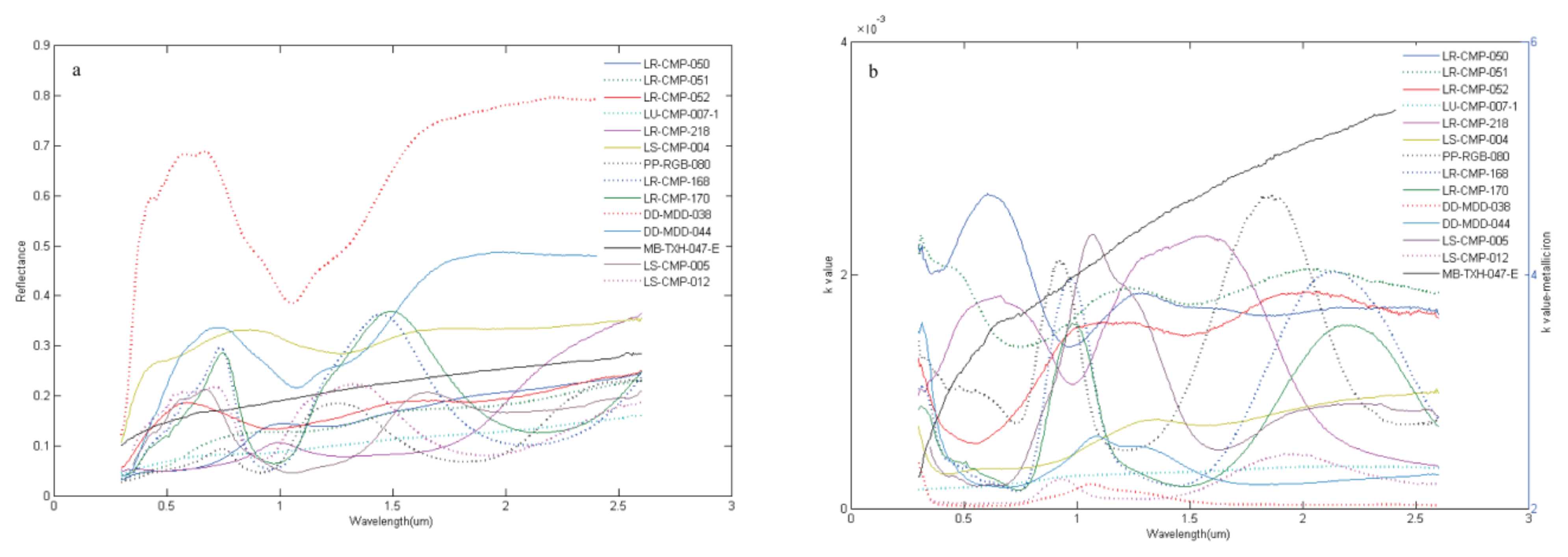
Figure 1. (a) Spectra and (b) k value of mineral end-members are involved in this study. The k values of mineral end-members are almost all in a magnitude of 10−3 except metallic iron, which is larger than 1.
Remotesensing 14 00277 g001
Figure 2. (ac) are the results of variable range with top limits, (df) are the corresponding results of variable range with bottom limits. (a,d) Comparison between simulated and measured spectra. (b,e) Comparison between simulated mineral abundance and measured value. (c,f) The relative error of simulated mineral abundance (the ratio of the difference between simulated and measured abundance versus the one of measured abundance). Although simulated spectra and measured spectra showed a good fitting precision when variable range was set up widely, the variables like simulated mineral abundance gravely deviated from the measured data. The solid line is the y = x line.
Remotesensing 14 00277 g002
Figure 3. RMSE between modeled and measured spectra of LSCC samples. RMSE is the root mean square error, in a magnitude of 10−3. Points labeled with asterisks and stars respectively are mare and highland samples. Points colored in red, green and blue respectively are samples in the size group of 20–45 μm, 10–20 μm and <10 μm.
Remotesensing 14 00277 g003
Figure 4. Corr between modeled and measured spectra of LSCC samples. Corr is correlation coefficient, larger than 0.99. Points labeled with asterisks and stars respectively serve as mare and highland samples. Points colored in red, green and blue serve as samples in the size group of 20–45 μm, 10–20 μm and <10 μm, respectively.
Remotesensing 14 00277 g004
Figure 5. Comparisons between modeled and measured spectra of LSCC samples. (a,b) The mare sample with the highest and lowest accuracy. (c,d) The highland sample with the highest and lowest accuracy. RMSE is root mean square error and Corr is correlation coefficient.
Remotesensing 14 00277 g005
Figure 6. Comparisons between modeled and measured mineral abundance of LSCC samples including volcanic glass (a), agglutinate (b), ilmenite (c), plagioclase (d), pyroxene (e), and olivine (f). The simulated abundance of pyroxene, olivine and volcanic glass are the sum abundance of their multiple end-members. In addition to agglutinitic with R2 near 0.81, other minerals’ R2 is larger than 0.92. The black line is the trend line and the red line is the y = x line.
Remotesensing 14 00277 g006
Figure 7. Comparisons between modeled and measured mineral abundance of LSCC samples. The solid line is the y = x line.
Remotesensing 14 00277 g007
Figure 8. Correlation between modeled SMFe content and Is/FeO of LSCC (a) mare and (b) highland samples. The slope of mare is a little steeper than that of highland. The solid line is the trend line.
Remotesensing 14 00277 g008
Figure 9. The simulated spectra of lunar regolith mineral with different maturity plotted in the 750 nm–950/750 nm two-dimension spectra plane. The angle between the horizontal line and the line linked by the value of each mineral end-member in the 750 nm–950/750 nm (the 750 nm reflectance is the x-axis, and the 950 nm/750 nm is the y-axis) spectra plane and the converged origin becomes larger with the increasing FeO content, except for ilmenite and forsterite. Ignoring the mineral end-member of ilmenite and forsterite, the converged origin of the 750 nm–50/750 nm spectra plane is set as: X = −0.20, Y = 1. 60.
Remotesensing 14 00277 g009
Figure 10. The effect of maturity on the 750 nm reflectance spectra and the 950/750 nm reflectance spectra of (ac) plagioclase and (df) ilmenite. The 750 nm reflectance of both plagioclase and ilmenite decreases and the absorption features become weaker as the SMFe content increases. With the increased SMFe content, the 950/750 nm reflectance of plagioclase increases, in other words, the spectra becoming redden; while ilmenite goes to the opposite trend.
Remotesensing 14 00277 g010
Figure 11. (a) Scatterplot of the simulated spectra at the 750 nm–950/750 nm spectra plane (the 750 nm reflectance is the x-axis, and the 950 nm/750 nm is the y-axis). (b) Scatterplot of the clockwise angles between the simulated spectra and the converged origin versus FeO content. The color in the figure represents the density of the data. The red and the green stars are served as LSCC mare and highland data in the size group of 10–20 μm. The converged origin of the 750 nm–950/750 nm spectra plane is set as X = −0.20 and Y = 1. 60, showing a high correlation with the FeO content of the simulated samples with correlation coefficient 0.7594 when it comes to the clockwise angles between the simulated spectra and the converged origin. The relationship between them, however, is not strictly linear. The solid line is the trend line.
Remotesensing 14 00277 g011
Figure 12. Correlation of absorption area between modeled and measured spectra of LSCC (a) mare and (b) highland samples. The black line is the trend line and the red line is the y = x line.
Remotesensing 14 00277 g012
Figure 13. Correlation of absorption center wavelength between modeled and measured spectra of LSCC (a) mare and (b) highland samples. The red line is the y = x line.
Remotesensing 14 00277 g013
Figure 14. Band I center versus Band II center for part of LSCC data. The red line is the approximate trend line of pyroxenes from Adams (1974) and Cloutis and Gaffey (1991a).
Remotesensing 14 00277 g014
Figure 15. (a) Modeled spectra and (b) Band I center versus Band II center for pyroxene. The solid line is the trend line.
Remotesensing 14 00277 g015
Figure 16. Mixed spectra of two olivine end-members at different mixed proportion. The red lines are served as the simulated spectra calculated by chemical composition (Fo value) and the blue lines as the simulated spectra calculated by the mix of end-members where FoMg and FoFe are respectively served as the Fo value of forsterite end-member and fayalite end-member and respectively as mixed proportion of forsterite end-member and fayalite end-member. Fo80 olivine—Fo70 olivine (ac), Fo30 olivine—Fo20 olivine (df) and Fo80 olivine—Fo20 olivine (gi). The mixed proportions of the two olivine end-members are 75:25 (a,d,g), 50:50 (b,e,h) and 25:75 (c,f,i).
Remotesensing 14 00277 g016
Table 1. The real index of the optical constants of minerals used in this study.
MineralReal Index (n)Reference
agglutinate1.49Bell et al. (1976) [39]
volcanic glass1.64Masson et al. (1972) [40]
plagioclase1.56Egan and Hilgeman (1975) [41]
olivine1.83Lucey (1998) [13]
orthopyroxene1.77Lucey (1998) [13]
clinopyroxene1.73Lucey (1998) [13]
ilmenite2.13Johnson and Christy (1974) [38]
metallic iron2.25–3.36Johnson and Christy (1974) [38]
Table 2. The first series of mineral end-members. Olivine was divided into forsterite and fayalite; Pyroxene was divided into orthopyroxene, clinopyroxene of Mg rich, clinopyroxene of Mg poor; Volcanic glass was divided into black, orange, green glass. They were lunar samples and telluric samples, part of which was lack of chemical analysis.
Mineral Sample ID
Volcanic glassblack glassLR-CMP-050
orange glassLR-CMP-051
green glassLR-CMP-052
Agglutinate LU-CMP-007-1
OlivineForsteriteDD-MDD-038
FayaliteDD-MDD-044
Ilmenite LR-CMP-218
ClinopyroxeneMg richLR-CMP-168
Mg poorLR-CMP-170
Orthopyroxene PP-RGB-080
Plagioclase LS-CMP-004
Metallic iron MB-TXH-047-E
Table 3. The second series of mineral end-members. They were all lunar samples with chemical analysis.
Mineral Sample IDPlagioclaseForsteriteOrthopyroxeneClinopyroxeneClinopyroxeneIlmenite
Oxide (wt. %) LS-CMP-004LS-CMP-005LS-CMP-012(Mg Rich)(Mg Poor)LR-CMP-218
FeO4.9911.3411.1620.3025.4041.40
MgO19.0943.6127.5218.3011.003.06
TiO20.050.030.320.611.1553.70
SiO242.8839.9353.9051.0048.300.03
Al2O320.731.533.441.351.520.21
Cr2O30.110.340.520.550.291.04
Fe2O30.000.000.000.000.000.00
MnO0.070.130.200.350.380.36
CaO11.411.143.266.8010.800.03
Na2O0.230.020.050.030.030.00
K2O0.030.000.010.000.000.00
P2O50.030.040.040.000.000.00
Table 4. With upper and lower limits of the simulated variables.
Simulated VariablesVolcanic Glass
(wt. %)
Agglutinate
(wt. %)
Ilmenite
(wt. %)
Plagioclase
(wt. %)
Pyroxene
(wt. %)
Olivine
(wt. %)
SMFe Content
(wt. %)
Particle Size
(μm)
Upper limits18.971.612.364.333.84.800
Lower limits0.125.40.113.40.90.3145
Table 5. The relative error of the simulated mineral abundance with strictly and widely variable range in simulation. The relative error is the ratio of difference between simulated and measured abundance versus the one of measured abundance.
Relative Error (%)Volcanic GlassAgglutinateIlmenitePlagioclasePyroxeneOlivine
Strict (Simulation)−0.0158000.265300−0.122900−0.1564000.2653000.265200
Wide (Inversion)42.114200−0.4353006.062300−0.6706001.89420025.003000
Table 6. RMSE and Corr between modeled and measured spectra of LSCC mare samples. RMSE is root mean square error and Corr is correlation coefficient. RMSE is less than 0.005 and Corr is more than 0.996.
Samples20–45 μm10–20 μm<10 μm
RMSECorrRMSECorrRMSECorr
100840.0017760.9984690.0027870.9990620.0023180.998898
120010.0032920.9969860.0021080.9993310.003840.998635
120300.0039550.9963760.0038310.9978650.003070.999263
150410.0024370.9985610.0022020.9991350.0034840.999034
150710.0036690.9974470.0017730.9995510.0024370.999466
701810.0024530.9981410.0023960.9990360.0028110.999340
710610.0039690.9961960.0047990.9978080.0048910.998841
715010.0014330.9992210.0013270.9995720.0021890.999154
792210.0019420.9987440.0026610.9986920.001950.999503
100840.0017760.9984690.0027870.9990620.0023180.998898
Table 7. RMSE and Corr between modeled and measured spectra of LSCC mare samples. RMSE is root mean square error and Corr is correlation coefficient. RMSE is less than 0.005 and Corr is more than 0.996.
Samples20–45 μm10–20 μm<10 μm
RMSECorrRMSECorrRMSECorr
141410.0043480.9978650.0044950.9986540.0047450.998499
141630.0053380.9954300.0033700.9990380.0029570.999457
142590.0034440.9978750.0035130.9994900.004890.998793
142600.0047920.9961950.0034940.9993820.0047970.998883
611410.0052530.9975110.0035360.9993670.0060090.998613
612210.0083830.992670.0031940.9990380.0037480.998928
622310.0057500.9969930.0043720.9990710.0046150.999118
648010.0045290.998630.0016830.9998580.0065720.998465
674610.008400.9964750.0079050.9970420.0087460.996410
674810.0043860.9985960.008510.9984720.0069890.998281
Table 8. Comparisons between modeled and measured particle sizes of LSCC mare samples. Y means the simulated particle size is within the measured particle size range while N means not.
Samples20–45 μm10–20 μm<10 μm
1008429.357330Y19.090910Y15.791520N
1200124.869890Y14.424440Y9.875270Y
1203023.022140Y12.520270Y7.269090Y
1504123.079240Y14.659100Y9.988960Y
1507120.153500Y12.690960Y9.999870Y
7018125.662460Y14.788270Y9.793450Y
7106121.518310Y12.042240Y6.879350Y
7150125.987210Y15.509300Y18.572280N
7922124.609920Y16.360630Y5.111250Y
1008429.357330Y19.090910Y15.791520N
Table 9. Comparisons between modeled and measured particle sizes of LSCC highland samples. Y means the simulated particle size is within the measured particle size range while N means not.
Samples20–45 μm10–20 μm<10 μm
1414135.071200Y18.91996Y4.344820Y
1416324.593370Y11.55559Y7.464690Y
1425925.062550Y11.04594Y7.625370Y
1426027.741030Y11.17127Y7.393020Y
6114144.990720Y7.62445N5.062500Y
6122144.340120Y13.58927Y3.844930Y
6223112.251470N6.60201N4.633810Y
6480145.000000Y19.97687Y4.621100Y
674616.027510N10.45335Y3.272480Y
674817.768130N3.77796N2.349130Y
Table 10. The relative error (%) of simulated absorption center wavelength (the ratio of difference between simulated and measured absorption center wavelength versus that between measured absorption center wavelength).
Mare20–45 μm10–20 μm<10 μmHighland20–45 μm10–20 μm<10 μm
100841.001.512.99141412.151.602.15
120014.694.125.08141632.090.531.58
120303.161.051.58142593.611.583.11
150415.262.094.10142601.580.531.58
150713.111.584.10611410.001.060.00
701816.976.976.97612210.531.068.02
710611.557.6111.31622315.562.092.60
715011.034.104.59648010.000.000.54
792212.556.976.97674611.580.000.53
674812.090.531.06
Publisher’s Note: MDPI stays neutral with regard to jurisdictional claims in published maps and institutional affiliations.