Article

Classification Endmember Selection with Multi-Temporal Hyperspectral Data

by
Tingxuan Jiang
*,
Harald van der Werff
and
Freek van der Meer
Faculty of Geo-Information Science and Earth Observation (ITC), University of Twente, 7514 AE Enschede, The Netherlands
*
Author to whom correspondence should be addressed.
Remote Sens. 2020, 12(10), 1575; https://doi.org/10.3390/rs12101575
Submission received: 24 April 2020 / Revised: 12 May 2020 / Accepted: 13 May 2020 / Published: 15 May 2020

Abstract

:
In hyperspectral image classification, so-called spectral endmembers are used as reference data. These endmembers are either extracted from an image or taken from another source. Research has shown that endmembers extracted from an image usually perform best when classifying a single image. However, it is unclear if this also holds when classifying multi-temporal hyperspectral datasets. In this paper, we use spectral angle mapper, which is a frequently used classifier for hyperspectral datasets to classify multi-temporal airborne visible/infrared imaging spectrometer (AVIRIS) hyperspectral imagery. Three classifications are done on each of the images with endmembers being extracted from the corresponding image, and three more classifications are done on the three images while using averaged endmembers. We apply image-to-image registration and change detection to analyze the consistency of the classification results. We show that the consistency of classification accuracy using the averaged endmembers (around 65%) outperforms the classification results generated using endmembers that are extracted from each image separately (around 40%). We conclude that, for multi-temporal datasets, it is better to have an endmember collection that is not directly from the image, but is processed to a representative average.

Graphical Abstract

1. Introduction

Hyperspectral remote sensing data have been used in many scientific fields for producing thematic maps through a diverse array of classification methods [1,2]. So-called spectral endmembers are used as reference for classifying a hyperspectral image [3,4,5]. Spectral endmembers are either extracted from an image or externally derived (e.g., from the field or an endmember library) [6,7]. The latter approach has been “mostly preferred and practiced” [3] and has “distinct advantages” [3]. These advantages include no influence from different characteristics between diverse sensors, closer to the mineral spectra in the study area than spectra from an existing spectral library and feasible for processing “large quantities of image data” [6,7,8]. Although these studies concluded that the classification of an image seems best done with endmembers derived from that image, it is unclear what is best for a multi-temporal image collection.
Multi-temporal remote sensing data are important for thematic analysis in diverse scientific fields, traditionally agriculture (e.g., [9,10,11]) and urban planning (e.g., [12,13,14]). It is also starting to be used in geological remote sensing (e.g., [15,16,17,18]). However, with the use of a multi-temporal dataset also, the complexity of a study grows [19]. Specifically, endmember extraction and selection is more complex, because extracted endmembers spectrally vary following the varying acquisition conditions one of each image [20]. There is neither a clear solution on how to extract endmembers from a multi-temporal dataset nor is it known what consequences there are of using extracted spectral endmembers for classifying a multi-temporal dataset.
This research evaluates the influence of spectral endmember extraction from a multi-temporal dataset on the consistency of classification results. We process three multi-temporal hyperspectral images with an identical atmospheric correction, use an automated endmember extraction method to classify the images, and evaluate the consistency. Specifically, we test how using a single set of spectral endmembers compares to using spectral endmembers extracted from each image separately.

2. Materials and Methods

2.1. Study Area

The study area is the Cuprite area in Nevada, USA (Figure 1). Two lithological units are present in the area: tertiary volcanic and volcaniclastic rocks of mainly rhyolitic ash-flow tuffs with some air-fall tuff [21]. Hydrothermal alteration has widely metamorphosed these units into three alteration zones, which are argillaceous alteration zone, silicified alteration zone, and the opal alteration zone [22]. Swayze et al. [23] refer to the hydrothermal alteration zones in Cuprite as the “eastern alteration center” and the “western alteration center” (as shown in Figure 2).
For three reasons, we chose the eastern alteration center as the study area in this research. First, the eastern alteration center hosts abundant hydrothermally altered minerals that can be identified by spectroscopy. Swayze et al. [23] map these minerals as Alunite, Kaolinite, Hydrated Silica, Montmorillonite, Buddingtonite, and a class as Kaolinite + Alunite, because of a high mixture of these two minerals. Second, only sparse vegetation is observed in the Cuprite area [25,26] and minerals should therefore be well exposed. Third, the Jet Propulsion Laboratory (JPL) uses the Cuprite area for calibrating and testing its spectrometers [27,28] and, as a result, several multi-temporal hyperspectral scenes with similar spectral and spatial resolution are available.

2.2. Hyperspectral Data

Three hyperspectral images that were acquired by the airborne visible/infrared imaging spectrometer (AVIRIS) over three different years were chosen (Table 1). AVIRIS has 224 spectral bands covering a 400–2500 nm wavelength range. These images are downloaded at L1-B pre-processing level, which means that they were radiometrically calibrated to radiance-at-sensor values and orthorectified with a digital elevation model by JPL [29]. In our paper, we refer to the three images after the acquisition year: “2006”, “2008”, and “2010”.

2.3. Pre-Processing

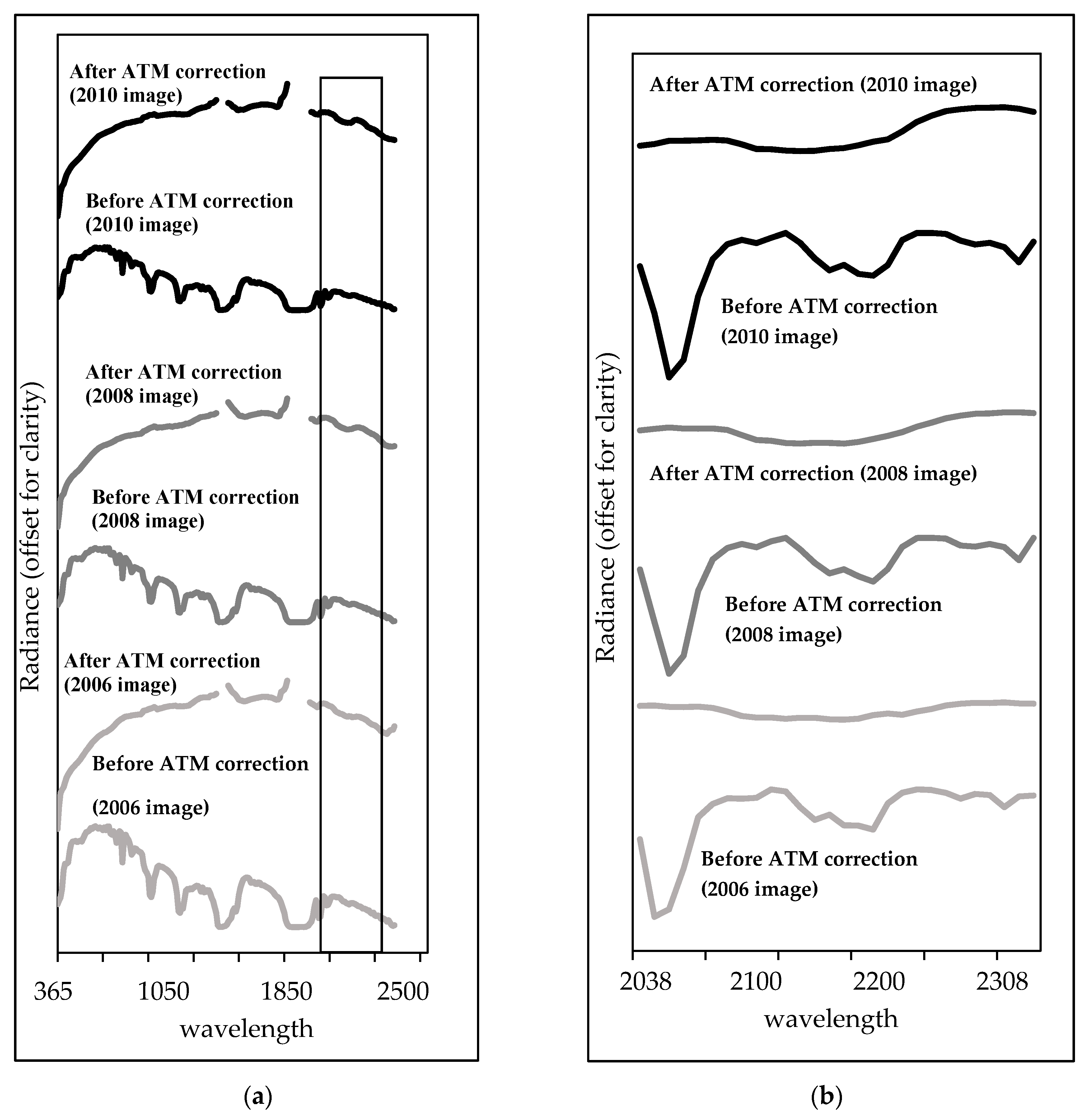
Atmospheric Correction

We applied the “fast line-of-sight atmospheric analysis of hypercubes” (FLAASH) software [30] in ENVI to atmospherically correct three images and convert the data from radiance-at-sensor to surface reflectance. FLAASH is based on MODTRAN4 and it was selected because it is frequently used to atmospherically correct AVIRIS images (e.g., [31,32,33]).
Table 1 shows ancillary information used for FLAASH.

2.4. Processing

2.4.1. Data Subset

The atmospherically corrected images were subset spectrally and spatially for subsequent processing. For three reasons, the images were spectrally subset to a 2048–2308 nm wavelength range. First, we focus on minerals whose diagnostic absorption features are located in the short-wave infrared (SWIR). Second, bands that were shorter than 2048 nm were influenced by atmospheric absorption around 1900 nm. Third, the diagnostic absorption features of all six mineral types referred by Swayze et al. [23] are all in a 2048–2308 nm wavelength range (as shown in Figure 3 and Figure 4). Apart from the spectral subset, all three images were spatially limited to the eastern alteration center (the red box in Figure 1).

2.4.2. NDVI

We used the normalized difference vegetation index (NDVI) [34] to detect the presence of any vegetation. Additionally, we analyzed a possible change in vegetation cover over time and, as such, judged whether it could influence our results.

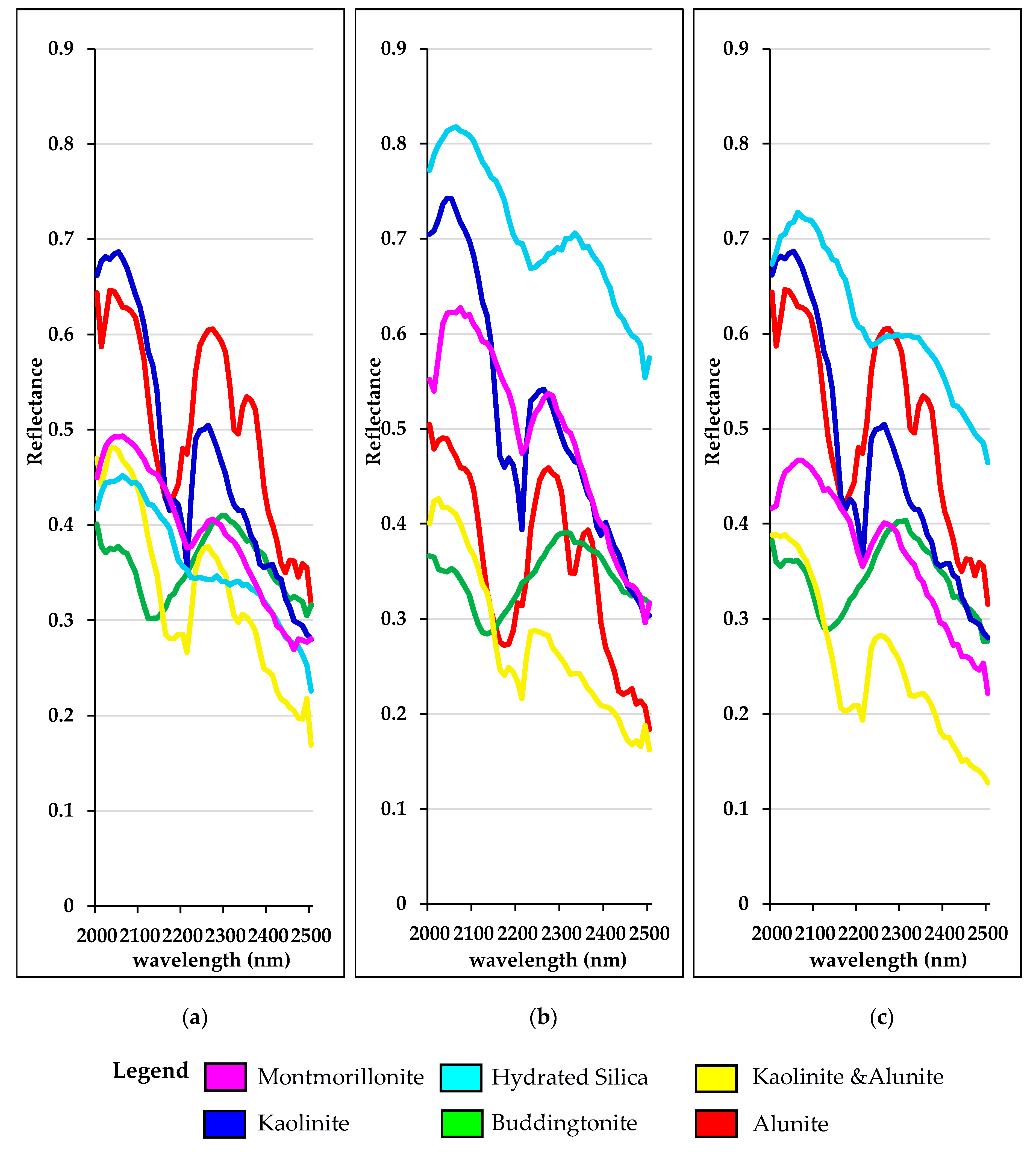
2.4.3. Endmember Libraries

Classification endmember spectra were extracted from the three images with the spatial-spectral endmember extraction (SSEE) method [35], applied separately to each image. We compared all of the extracted endmembers with the mineral spectral library of the United States Geologic Survey (USGS) [36] and manually selected endmembers for subsequent classification. The criteria of the endmember selection mainly relate with the wavelength position and the absorption depth of diagnostic absorption features. The only exception is the endmember of montmorillonite; SSEE extracted a montmorillonite spectrum only from the 2008 image, while no montmorillonite spectra could be extracted from the 2006 and 2010 images. Therefore, we manually extracted the spectra from the 2006 image and 2010 image, at the same pixel location where the SSEE-extracted montmorillonite endmember in the 2008 image came from. Based on these criteria, we selected endmembers for each of the six alteration minerals referred to by Swayze et al. [23]. We assembled three spectral endmember libraries that were named after the method used to create them: extracted endmember libraries.
In addition, we created a fourth “averaged” spectral endmember library, based on the arithmetic average of the three extracted endmember libraries. The purpose of the averaged library is to classify all three AVIRIS images with a single endmember set as well. Subsequently, the consistency of the classification done with the averaged endmember library is compared with the consistency of the classification done with the three extracted endmember libraries.
We used linear correlation [37] to compare all endmembers representing the same mineral for understanding the linear similarity among endmembers in different libraries. The linear correlation calculation was done with Microsoft Excel software [38].

2.4.4. SAM Classification

The spectral angle mapper (SAM) [39] classifier was selected to produce mineral maps. SAM was selected, because it is a “popular” [40] and “the most famous” [41] classification method. We performed the classification on the images before subsequent resampling for change detection analysis, as Zhou et al. [42] found that resampling leads to mixing of spectra and thus loss of information.
The main challenge for applying SAM classification is the selection of appropriate classification thresholds [3]. In this research, six mineral maps were generated with SAM, by using the same class membership thresholds for each image (Table 2). These thresholds were manually determined by “trial and error” [43] while using the mineral map from Swayze et al. [23] as reference; an approach that is “widely used” [44]. Each image was once classified with the “extracted endmembers” associated with that image, and once classified with the “averaged endmembers”, leading to six classified images in total.

2.5. Consistency Evaluation

For the evaluation of classification consistency on a pixel-by-pixel basis, the classification result of the 2010 image was resampled to the 3.3 m spatial resolution of the 2006 and 2008 images before collecting ground control points (GCPs) to undertake co-registration.
We used an image-to-image registration method [45] in IDL-ENVI software to spatially register all classified images. The image-to-image registration completes through resampling [45]. As indicated by Zhou et al. [42], resampling leads to a loss of information and leads to mixed spectra of pixels in the resampled images. Therefore, we registered classification results instead of the original images. The classification result of the 2006 image was set as the base image. GCPs were automatically collected from three images as ties to register the six classification results. The accuracy of automatically collected GCPs was tested through the root mean square error (RMSE) for each GCP and an overall RMSE for all GCPs [45,46]. For reducing the overall RMSE, we manually removed GCPs with an RMSE higher than 1 by “trial and error” and manually chose recognizable spots (e.g., street crossings) as GCPs.
We applied masking [47] to create maps with a single mineral class for comparing the classification results on a mineral-by-mineral basis (single-mineral map). With six mineral classes in six classified images, 36 single-mineral maps were produced in total (shown in Supplementary Materials: Figure S1–S36).
For evaluating the consistency of the 36 single-mineral maps, we used a change detection method [48] in ENVI. In change detection, we set a pair of single-mineral maps, as demonstrated in Figure 5. The change detection subtracts value of pixels in the “reference image” from value of pixels in the “test image”. Therefore, by revaluation, the classification differences between two single-mineral maps can be presented as four statuses: omission, reproduced, unidentified, and commission (Figure 5c).
We summarized the number of pixels in each of the four statuses between each pair of the compared single-mineral maps as shown below:
R = Re/(SU)
In this equation, R represents the consistency of certain mineral type, Re is the number of reproduced pixels, S is the amount of pixels in each of the compared single-mineral maps, and U is the number of unclassified pixels.

3. Results

Figure 6 shows the mean spectrum of each image before and after atmospheric correction. The atmospheric absorption around 1400 nm and 1900 nm were apparently overcorrected while the other atmospheric absorption features were corrected, as shown in Figure 6a. Figure 6b shows the part of the spectra used for classification, which falls outside the overcorrected atmospheric windows.
We used NDVI to check for vegetation presence and change over time. Figure 7 presents the NDVI images of all three images. 5799 out of 1,030,206 pixels in the 2006 image, 331 out of 1,024,152 pixels in the 2008 image, and 1529 out of 1,030,206 pixels in the 2010 image have an NDVI value higher than 0.2. Figure 8 shows the distribution of NDVI values in histograms.
Four spectral libraries were created with spectra that were extracted from the three images: one library for each image separately (three in total) and one library with averages of all endmember sets together (Figure 9). The extracted libraries consist of endmembers that were selected from all spectra that were extracted by SSEE from the three images.
The similarity of all endmembers is shown in Table 3 and Table 4.
The three classification results in Figure 10 were generated with the SAM while using the extracted libraries and manually set classification thresholds (Table 2). Several differences among these three classification results can be observed. For instance, 200,560 pixels in Figure 10b were identified as hydrated silica, while only 45,704 pixels in Figure 10a and 123,656 pixels in Figure 10c were identified as hydrated silica. Similarly, 194,416 pixels in Figure 10c were identified as alunite, while 121,578 pixels in Figure 10a and only 20,300 pixels in Figure 10b were identified as alunite.
The SAM classification results of the three images using the averaged library are presented in Figure 11. Overall, the spatial patterns are similar, except for montmorillonite, which has 5791 pixels in the 2006 image, 1462 pixels in the 2008 image, and 1277 pixels in the 2010 image.
Figure 12 presents the consistency of the SAM classification results that were obtained using the three extracted endmember libraries. Kaolinite shows a consistency of approximately 70% over all three classification results. Alunite shows a consistency of approximately 70%, but only between the 2006 and 2010 images, while between the 2006 and 2008 images as well as the 2008 and 2010 images is only of approximately 10%. The consistency of other classes in Figure 12 also vary following the conversion of compared results and show consistency of 0.16–50% over all three classification results. Figure 13 displays the consistency of SAM classification results produced while using averaged endmember library. Montmorillonite shows a consistency of 6–19% over the three classification results. Buddingtonite shows a consistency of approximately 30% between 2006 and 2008 images, 40% between 2006 and 2010 images, and 60% between 2008 and 2010 images. The other classes show a consistency of approximately 60–80% over all three classification results.

4. Discussion

We calculated NDVI to check whether vegetation changes over time and if masking would be necessary. The 2006 image was acquired on 2nd May, the 2008 image was acquired on 20th September, and the 2010 image was acquired on 14th October, covering spring to autumn. Results show that over 99.5% pixels of the three images present an NDVI value lower than 0.2: only approximately 0.5% pixels in the 2006 image, 0.03% pixels in the 2008 image, and 0.1% pixels of the 2010 image show a value higher than 0.2. This low NDVI value indicates that there is only sparse vegetation [34,49,50]. We can assume that vegetation cover in the Cuprite area remains sparse across different seasons.
FLAASH was used to atmospherically correct the three images, using the same atmospheric settings (Table 1) following Harris Geospatial Solution Inc. [51]. Nevertheless, the ATM correction remains an uncertainty in this research, as atmospheric conditions keep changing. However, the classification results that were derived from the ATM corrected images using the averaged endmembers (Figure 11) show a high reproducibility (60–80%, except for montmorillonite). On the other hand, the comparison between the overall spectra derived from images between and after ATM correction (Figure 6) illustrates that the atmospheric absorption curves have been corrected. Therefore, we conclude that the ATM correction does not play a major role in the reproducibility of classification.
As shown in Figure 3 and Table 3, endmembers of one mineral extracted from the three images have a similar shape but endmembers of certain minerals (e.g., hydrated silica) show different brightness (as shown in Figure 4). The brightness differences might be related to factors (e.g., illumination differences, different soil moisture conditions, etc.) that typically change with the acquisition of a multi-temporal dataset. It is necessary to properly select endmembers for classifying the multi-temporal dataset, given that the influence of the different brightness between endmembers to multi-temporal classification is hardly known.
In all classification results, Montmorillonite has a low abundance and also a low consistency. This could be a result of endmember selection: the SSEE only extracted a montmorillonite spectrum from the 2008 image and the montmorillonite endmembers in the other two extracted libraries were manually extracted from the same spot where the SSEE-extracted montmorillonite endmember came from.
We applied image-to-image registration to align the classification results pixel-by-pixel. An overall RMSE that indicates the average spatial shift between a pair of registered images was calculated after GCP collection [45]. Barazzetti et al. [46] indicated that image-to-image registration is inaccurate when the RMSE is higher than the ground sampling distance. Here, the RMSE of GCPs (0.508 m between 2006 image and 2008 image and 0.642 m between 2006 image and 2010 image) is approximately factor 6 lower than the ground sampling distance (3.3 m).
We decided to use classification consistency to observe the consequences of classifying multi-temporal images while using extracted endmembers. This decision was based on two assumptions. First, the consequences of endmember selection will be highlighted when only endmembers are derived from different sources, while the same methods and parameter settings are used in every other link of multi-temporal images classification chain. We therefore did not change any parameter setting (including classification thresholds) or processing method for classifying the three images with a purpose for only focusing on endmembers over time. Second, the distribution of well-exposed minerals is rather stable over times. Thus, multi-temporal classification results should present similar mineral distribution, unless the endmember selection was influenced.
There will be an impending demand on a strategy to classify multi-temporal images consistently as both availability of hyperspectral datasets and the attention of multi-temporal analysis are growing. In this research, we find differences between the three classifications while using extracted endmembers (Figure 10), while the three classifications (Figure 11) with the averaged endmember library show a better consistency. Therefore, we state that it is better to have an endmember collection as a representative average instead of using directly extract endmembers for a consistent classification of multi-temporal hyperspectral images.

5. Conclusions

With growing availability of hyperspectral datasets and attention to multi-temporal analysis, there is impending demand on a strategy to process multi-temporal hyperspectral datasets. The use of classification endmembers is a significant part of hyperspectral image processing chain, and endmember selection when dealing with multi-temporal hyperspectral analysis is therefore important for remote sensing studies.
The reproducibility of classification results that were created using the averaged endmember library outperforms the reproducibility of classification results generated by endmembers extracted from each image separately. This conclusion is contrary to the findings that were obtained on single images. The classification results produced using the extracted endmember libraries are different, although the endmembers of the extracted libraries are statistically highly correlated.
Although the advantage of using extracted endmembers has been shown for hyperspectral image classification, we conclude that an external library, which could be made by averaging multiple endmember libraries, leads to a set of more consistent and/or reproducible classification results when dealing with multi-temporal hyperspectral data.

Supplementary Materials

The following are available online at https://www.mdpi.com/2072-4292/12/10/1575/s1, Figure S1: Change between alunite in 2006 and 2008 images separately produced using the extracted endmembers; Figure S2. Change between alunite in 2006 and 2008 images separately produced using the averaged endmembers; Figure S3. Change between alunite in 2006 and 2010 images separately produced using the extracted endmembers; Figure S4. Change between alunite in 2006 and 2010 images separately produced using the averaged endmembers; Figure S5. Change between alunite in 2008 and 2010 images separately produced using the extracted endmembers; Figure S6. Change between alunite in 2008 and 2010 images separately produced using the averaged endmembers; Figure S7. Change between buddingtonite in 2006 and 2008 images separately produced using the extracted endmembers; Figure S8. Change between buddingtonite in 2006 and 2008 images separately produced using the averaged endmembers; Figure S9. Change between buddingtonite in 2006 and 2010 images separately produced using the extracted endmembers; Figure S10. Change between buddingtonite in 2006 and 2010 images separately produced using the averaged endmembers; Figure S11. Change between buddingtonite in 2008 and 2010 images separately produced using the extracted endmembers; Figure S12. Change between buddingtonite in 2008 and 2010 images separately produced using the averaged endmembers; Figure S13. Change between kaolinite in 2006 and 2008 images separately produced using the extracted endmembers; Figure S14. Change between kaolinite in 2006 and 2008 images separately produced using the averaged endmembers; Figure S15. Change between kaolinite in 2006 and 2010 images separately produced using the extracted endmembers; Figure S16. Change between kaolinite in 2006 and 2010 images separately produced using the averaged endmembers; Figure S17. Change between kaolinite in 2008 and 2010 images separately produced using the extracted endmembers; Figure S18. Change between kaolinite in 2008 and 2010 images separately produced using the averaged endmembers; Figure S19. Change between kaolinite + alunite in 2006 and 2008 images separately produced using the extracted endmembers; Figure S20. Change between kaolinite + alunite in 2006 and 2008 images separately produced using the averaged endmembers; Figure S21. Change between kaolinite + alunite in 2006 and 2010 images separately produced using the extracted endmembers; Figure S22. Change between kaolinite + alunite in 2006 and 2010 images separately produced using the averaged endmembers; Figure S23. Change between kaolinite + alunite in 2008 and 2010 images separately produced using the extracted endmembers; Figure S24. Change between kaolinite + alunite in 2008 and 2010 images separately produced using the averaged endmembers; Figure S25. Change between hydrated silica in 2006 and 2008 images separately produced using the extracted endmembers; Figure S26. Change between hydrated silica in 2006 and 2008 images separately produced using the averaged endmembers; Figure S27. Change between hydrated silica in 2006 and 2010 images separately produced using the extracted endmembers; Figure S28. Change between hydrated silica in 2006 and 2010 images separately produced using the averaged endmembers; Figure S29. Change between hydrated silica in 2008 and 2010 images separately produced using the extracted endmembers; Figure S30. Change between hydrated silica in 2008 and 2010 images separately produced using the averaged endmembers; Figure S31. Change between montmorillonite in 2006 and 2008 images separately produced using the extracted endmembers; Figure S32. Change between montmorillonite in 2006 and 2008 images separately produced using the averaged endmembers; Figure S33. Change between montmorillonite in 2006 and 2010 images separately produced using the extracted endmembers; Figure S34. Change between montmorillonite in 2006 and 2010 images separately produced using the averaged endmembers; Figure S35. Change between montmorillonite in 2008 and 2010 images separately produced using the extracted endmembers; Figure S36. Change between montmorillonite in 2008 and 2010 images separately produced using the averaged endmembers.

Author Contributions

Conceptualization, T.J., H.v.d.W and F.v.d.M.; Data curation, T.J.; Formal analysis, T.J.; Methodology, T.J., H.v.d.W. and F.v.d.M.; Writing – original draft, T.J.; Writing – review & editing, T.J., H.v.d.W. and F.v.d.M. All authors have read and agreed to the published version of the manuscript.

Funding

This research received no external funding.

Acknowledgments

The authors want to express our gratitude to Chris Hecker (University of Twente) for personal communication on the spectral angle mapper thresholds setting, Exaud Jeckonia Humbo for personal communication on the geological settings in the Cuprite area, Amarjargal Davaadorj for her personal communication on image registration, Fadard Maghsoudi Moud and Jonathan Franco Hempenius for ArcGIS software assistance, and Na Chen for her assistance on NDVI issues.

Conflicts of Interest

The authors do not perceive any financial or affiliation-related conflict of interest with respect to this study.

References

  1. Foody, G.M. Thematic map comparison: Evaluating the statistical significance of differences in classification accuracy. Photogramm. Eng. Remote Sens. 2004, 70, 627–633. [Google Scholar] [CrossRef]
  2. Olofsson, P.; Foody, G.M.; Herold, M.; Stehman, S.V.; Woodcock, C.E.; Wulder, M.A. Good practices for estimating area and assessing accuracy of land change. Remote Sens. Environ. 2014, 148, 42–57. [Google Scholar] [CrossRef]
  3. Asadzadeh, S.; de Souza Filho, C.R. A review on spectral processing methods for geological remote sensing. Int. J. Appl. Earth Obs. Geoinf. 2016, 47, 69–90. [Google Scholar] [CrossRef]
  4. Bioucas-Dias, J.M.; Plaza, A.; Camps-Valls, G.; Scheunders, P.; Nasrabadi, N.M.; Chanussot, J. Hyperspectral remote sensing data analysis and future challenges. IEEE Geosci. Remote Sens. Mag. 2013, 1, 6–36. [Google Scholar] [CrossRef] [Green Version]
  5. Schowengerdt, R.A. Remote Sensing. Models and Methods for Image Processing, 2nd ed.; Cambridge University Press: New York, NY, USA, 1997. [Google Scholar]
  6. Zortea, M.; Plaza, A. Spatial preprocessing for endmember extraction. IEEE Trans. Geosci. Remote Sens. 2009, 47, 2679–2693. [Google Scholar] [CrossRef]
  7. Chang, C.-I. Hyperspectral Data Processing: Algorithm Design and Analysis; KLUwer Acdemic publisher: New York, NY, USA, 2013. [Google Scholar]
  8. Veganzones, M.A.; Graña, M. Endmember Extraction Methods: A Short Review. In Proceedings of the Knowledge-Based Intelligent Information and Engineering Systems, Zagreb, Croatia, 3–5 September 2008; pp. 400–407. [Google Scholar]
  9. Holman, F.H.; Riche, A.B.; Michalski, A.; Castle, M.; Wooster, M.J.; Hawkesford, M.J. High throughput field phenotyping of wheat plant height and growth rate in field plot trials using UAV based remote sensing. Remote Sens. 2016, 8, 1031. [Google Scholar] [CrossRef]
  10. Veloso, A.; Mermoz, S.; Bouvet, A.; Le Toan, T.; Planells, M.; Dejoux, J.F.; Ceschia, E. Understanding the temporal behavior of crops using Sentinel-1 and Sentinel-2-like data for agricultural applications. Remote Sens. Environ. 2017, 199, 415–426. [Google Scholar] [CrossRef]
  11. Youssef, A.M.; Abu Abdullah, M.M.; Pradhan, B.; Gaber, A.F.D. Agriculture sprawl assessment using multi-temporal remote sensing images and its environmental impact; Al-Jouf, KSA. Sustainability 2019, 11, 4177. [Google Scholar] [CrossRef] [Green Version]
  12. Shen, H.; Huang, L.; Zhang, L.; Wu, P.; Zeng, C. Long-term and fine-scale satellite monitoring of the urban heat island effect by the fusion of multi-temporal and multi-sensor remote sensed data: A 26-year case study of the city of Wuhan in China. Remote Sens. Environ. 2016, 172, 109–125. [Google Scholar] [CrossRef]
  13. Pal, S.; Ziaul, S. Detection of land use and land cover change and land surface temperature in English Bazar urban centre. Egypt. J. Remote Sens. Space Sci. 2017, 20, 125–145. [Google Scholar] [CrossRef] [Green Version]
  14. Frick, A.; Tervooren, S. A framework for the long-term monitoring of urban green volume based on multi-temporal and multi-sensoral remote sensing data. J. Geovis. Spat. Anal. 2019, 3, 6. [Google Scholar] [CrossRef]
  15. Mielke, C.; Boesche, N.K.; Rogass, C.; Kaufmann, H.; Gauert, C.; de Wit, M. Spaceborne mine waste mineralogy monitoring in South Africa, applications for modern push-broom missions: Hyperion/OLI and EnMAP/Sentinel-2. Remote Sens. 2014, 6, 6790–6816. [Google Scholar] [CrossRef] [Green Version]
  16. Gorji, T.; Sertel, E.; Tanik, A. Monitoring soil salinity via remote sensing technology under data scarce conditions: A case study from Turkey. Ecol. Indic. 2017, 74, 384–391. [Google Scholar] [CrossRef]
  17. Wei, J.; Liu, X.; Ding, C.; Liu, M.; Jin, M.; Li, D. Developing a thermal characteristic index for lithology identification using thermal infrared remote sensing data. Adv. Space Res. 2017, 59, 74–87. [Google Scholar] [CrossRef] [Green Version]
  18. Jakob, S.; Zimmermann, R.; Gloaguen, R. The need for accurate geometric and radiometric corrections of drone-borne hyperspectral data for mineral exploration: Mephysto-a toolbox for pre-processing drone-borne hyperspectral data. Remote Sens. 2017, 9, 88. [Google Scholar] [CrossRef] [Green Version]
  19. Glanz, H.; Carvalho, L.; Sulla-Menashe, D.; Friedl, M.A. A parametric model for classifying land cover and evaluating training data based on multi-temporal remote sensing data. ISPRS J. Photogramm. Remote Sens. 2014, 97, 219–228. [Google Scholar] [CrossRef]
  20. Thouvenin, P.A.; Dobigeon, N.; Tourneret, J.Y. Hyperspectral unmixing with spectral variability using a perturbed linear mixing model. IEEE Trans. Signal Process. 2016, 64, 525–538. [Google Scholar] [CrossRef] [Green Version]
  21. Kruse, F.A.; Kierein-Young, K.S.; Boardman, J.W. Mineral mapping at Cuprite, Nevada with a 63-channel imaging spectrometer. Photogramm. Eng. Remote Sens. 1990, 56, 83–92. [Google Scholar]
  22. Wei, J.; Ming, Y.; Jia, Q.; Yang, D. Simple mineral mapping algorithm based on multitype spectral diagnostic absorption features: A case study at Cuprite, Nevada. J. Appl Remote Sens. 2017, 11, 026015. [Google Scholar] [CrossRef]
  23. Swayze, G.A.; Clark, R.N.; Goetz, A.F.H.; Livo, K.E.; Breit, G.N.; Kruse, F.A.; Sutley, S.J.; Snee, L.W.; Lowers, H.A.; Post, J.L.; et al. Mapping advanced argillic alteration at cuprite, Nevada, using imaging spectroscopy. Econ. Geol. 2014, 109, 1179–1221. [Google Scholar] [CrossRef]
  24. JPL JPL | AVIRIS Data Portal. Available online: https://aviris.jpl.nasa.gov/dataportal/ (accessed on 18 February 2020).
  25. Goetz, A.F.H.; Srivastava, V. Mineralogical Mapping in the Cuprite Mining District, Nevada; NTRS: Chicago, IL, USA, 1985. [Google Scholar]
  26. Abrams, M.; Hook, S.J. Simulated ASTER data for geologic studies. IEEE Trans. Geosci. Remote Sens. 1995, 33, 692–699. [Google Scholar] [CrossRef]
  27. Van der Meer, F.D.; van der Werff, H.M.A.; van Ruitenbeek, F.J.A.; Hecker, C.A.; Bakker, W.H.; Noomen, M.F.; van der Meijde, M.; Carranza, E.J.M.; de Smeth, J.B.; Woldai, T. Multi- and hyperspectral geologic remote sensing: A review. Int. J. Appl. Earth Obs. Geoinf. 2012, 14, 112–128. [Google Scholar] [CrossRef]
  28. Clark, R.N.; Swayze, G.A. Evolution in imaging spectroscopy analysis and sensor signal-to-noise: An examination of how far we have come. In Proceedings of the Summaries of the Sixth Annual JPL Airborne Earth Science Worksho, Orlando, FL, USA, 4–8 March 1996; JPL Publication: Pasadena, CA, USA, 1996; pp. 4–8. [Google Scholar]
  29. Gowey, K.; Lundeen, S. AVIRIS—Airborne Visible/Infrared Imaging Spectrometer—Data Processing. Available online: https://aviris.jpl.nasa.gov/aviris/data_facility.html (accessed on 27 January 2020).
  30. Cooley, T.; Anderson, G.P.; Felde, G.W.; Hoke, M.L.; Ratkowski, A.J.; Chetwynd, J.H.; Gardner, J.A.; Adler-Golden, S.M.; Matthew, M.W.; Berk, A.; et al. FLAASH, a MODTRAN4-based atmospheric correction algorithm, its applications and validation. Int. Geosci. Remote Sens. Symp. 2002, 3, 1414–1418. [Google Scholar]
  31. Ustin, S.L.; Lay, M.C.; Li, L. Remote sensing of wetland conditions in West Coast salt marshes. Remote Sens. Model. Ecosyst. Sustain. 2004, 5544, 159. [Google Scholar]
  32. Adler-Golden, S.M.; Acharya, P.K.; Berk, A.; Matthew, M.W.; Gorodetzky, D. Remote bathymetry of the littoral zone from AVIRIS, LASH, and QuickBird imagery. IEEE Trans. Geosci. Remote Sens. 2005, 43, 337–347. [Google Scholar] [CrossRef]
  33. Kayadibi, Ö.; Aydal, D. Quantitative and comparative examination of the spectral features characteristics of the surface reflectance information retrieved from the atmospherically corrected images of Hyperion. J. Appl. Remote Sens. 2013, 7, 073528. [Google Scholar] [CrossRef]
  34. Rouse, J.W.; Hass, R.H.; Schell, J.A.; Deering, D.W. Monitoring vegetation systems in the great plains with ERTS. In Proceedings of the Third Earth Resources Technology Satellite (ERTS) Symposium, Greenbelt, MD, USA, 10–14 December1973; Volume 1, pp. 309–317. [Google Scholar]
  35. Rogge, D.M.; Rivard, B.; Zhang, J.; Sanchez, A.; Harris, J.; Feng, J. Integration of spatial—Spectral information for the improved extraction of endmembers. Remote Sens. Environ. 2007, 110, 287–303. [Google Scholar] [CrossRef]
  36. Kokaly, R.F.; Clark, R.N.; Swayze, G.A.; Livo, K.E.; Hoefen, T.M.; Pearson, N.C.; Wise, R.A.; Benzel, W.M.; Lowers, H.A.; Driscoll, R.L.; et al. Base Spectra (splib07a). Available online: https://crustal.usgs.gov/speclab/QueryAll07a.php (accessed on 16 April 2019).
  37. Pearson, P.K. Mathematical Contributions to the Theory of Evolution.—on A Form of Spurious Correlation Which May Arise When Indices Are Used in the Measurement of Organs. Proc. Royal Soc. Lond. 1897, 60, 273–283. [Google Scholar]
  38. Microsoft Spreadsheet Software—Excel Free Trial—Microsoft Excel. Available online: https://products.office.com/en-us/excel (accessed on 31 March 2020).
  39. Kruse, F.A.; Lefkoff, A.B.; Boardman, J.W.; Heidebrecht, K.B.; Shapiro, A.T.; Barloon, P.J.; Goetz, A.F.H. The spectral image processing system (SIPS)-interactive visualization and analysis of imaging spectrometer data. Remote Sens. Environ. 1993, 44, 145–163. [Google Scholar] [CrossRef]
  40. Dennison, P.E.; Halligan, K.Q.; Roberts, D.A. A comparison of error metrics and constraints for multiple endmember spectral mixture analysis and spectral angle mapper. Remote Sens. Environ. 2004, 93, 359–367. [Google Scholar] [CrossRef]
  41. EL_Rahman, S.A. Performance of spectral angle mapper and parallelepiped classifiers in agriculture hyperspectral image. Int. J. Adv. Comput. Sci. Appl. 2016, 7, 55–63. [Google Scholar]
  42. Zhou, Q.; Jing, Z.; Jiang, S. Remote Sensing Image Fusion for Different Spectral and Spatial Resolutions with Bilinear Resampling Wavelet Transform. In Proceedings of the IEEE Conference on Intelligent Transportation Systems (ITSC), Shanghai, China, 12–15 October 2003; Volume 2, pp. 1206–1213. [Google Scholar]
  43. Bruzzone, L.; Prieto, D.F. Automatic analysis of the difference image for unsupervised change detection. IEEE Trans. Geosci. Remote Sens. 2000, 38, 1171–1182. [Google Scholar] [CrossRef] [Green Version]
  44. Shahriari, H.; Honarmand, M.; Ranjbar, H. Comparison of multi-temporal ASTER images for hydrothermal alteration mapping using a fractal-aided SAM method. Int. J. Remote Sens. 2015, 36, 1271–1289. [Google Scholar] [CrossRef]
  45. Jin, X. ENVI Automated Image Registration Solutions; Harris Geospatial Solution Inc.: Broomfild, Colorado, USA, 2018. [Google Scholar]
  46. Barazzetti, L.; Roncoroni, F.; Brumana, R.; Previtali, M. Georeferencing Accuracy Analysis of A Single Worldview-3 Image Collected over Milan. In Proceedings of the International Archives of the Photogrammetry, Remote Sensing and Spatial Information Sciences—ISPRS Archives, Prague, Czech Republic, 12–19 July 2016; pp. 429–434. [Google Scholar]
  47. Harris Geospatial Solution Inc. Masks. Available online: https://www.harrisgeospatial.com/docs/masks.html (accessed on 21 January 2020).
  48. Harris Geospatial Solution Inc. Change Detection Analysis. Available online: https://www.harrisgeospatial.com/docs/ChangeDetectionAnalysis.html (accessed on 21 January 2020).
  49. Weier, J.; Herring, D. Measuring Vegetation (NDVI & EVI). Available online: https://earthobservatory.nasa.gov/features/MeasuringVegetation (accessed on 8 January 2019).
  50. Hashim, H.; Abd Latif, Z.; Adnan, N.A. Urban Vegetation Classification With Ndvi Threshold Value Method With Very High Resolution (Vhr) Pleiades Imagery. In Proceedings of the ISPRS—International Archives of the Photogrammetry, Remote Sensing and Spatial Information Sciences, Kuala Lumpur, Malaysia, 1–3 October 2019; Volume XLII-4/W16, pp. 237–240. [Google Scholar]
  51. Harris Geospatial Solution Inc. Fast Line-of-sight Atmospheric Analysis of Spectral Hypercubes. Available online: https://www.harrisgeospatial.com/docs/FLAASH.html (accessed on 12 August 2018).
Figure 1. True color composite (650, 550, 450 nm, RGB) of Cuprite, Nevada derived from airborne visible/infrared imaging spectrometer (AVIRIS) data (downloaded from: JPL [24]). The red box shows the study area of this research.
Remotesensing 12 01575 g001
Figure 2. Tetracorder Mineral Map of Cuprite, Nevada derived from AVIRIS Data, showing Clays, Micas, Sulphates, and Carbonates (modified with permission from: Swayze et al. [23]). The red box shows the study area of this research.
Remotesensing 12 01575 g002
Figure 3. Spectra for each mineral class extracted from (a) the 2006 image, (b) the 2008 image, and (c) the 2010 image.
Remotesensing 12 01575 g003
Figure 4. Comparison of spectra from three images for each of the six minerals: (a) Alunite; (b) Buddingtonite; (c) Kaolinite; (d) Kaolinite and Alunite; (e) Hydrated silica; (f) Montmorillonite. In (a,c), the spectra from 2006 and 2010 image are overlapping.
Remotesensing 12 01575 g004
Figure 5. (a) demonstrates for single-mineral map in “test image” the identified pixels were revalued as “2” and unidentified pixels were revalued as ”0”; (b) shows an example for single-mineral map in “reference image”, of which the identified pixels were set as “1” and unidentified pixels were set as ”0”; (c) demonstrates the result of change detection, “-2” indicates a pixel identified in graph (b) but not in graph (a) (omission); “-1” indicates a pixel identified in both graph (a,b) (reproduced); “0” indicates a pixel unidentified in both of graph (a,b) (unidentified); “1” indicate a pixel identified in graph (a) but not in graph (b) (commission).
Remotesensing 12 01575 g005
Figure 6. (a) shows the comparison of overall spectra (400–2500 nm) derived from the three images before and after the atmospheric correction; (b) subset of the spectra in the black outline of figure (a) which is the comparison of spectra of wavelength range 2048–2308 nm that hosts diagnostic spectral features of the targeted mineral classes.
Remotesensing 12 01575 g006
Figure 7. Normalized difference vegetation index (NDVI) images derived from (a) the 2006 image; (b) the 2008 image; and, (c) the 2010 image.
Remotesensing 12 01575 g007
Figure 8. Histograms of NDVI values in (a) the 2006 image; (b) the 2008 image; and, (c) the 2010 images.
Remotesensing 12 01575 g008
Figure 9. The endmember spectra extracted from (a) the 2006 image; (b) the 2008 image; and, (c) the 2010 image. Figure (d) is the averaged library.
Remotesensing 12 01575 g009
Figure 10. (ac) are classification results derived from 2006, 2008, and 2010 image, respectively, obtained by spectral angle mapper (SAM) using extracted spectral endmember libraries.
Remotesensing 12 01575 g010
Figure 11. (ac) are classification results derived from 2006, 2008 and 2010 image, respectively, obtained by SAM using the averaged spectral endmember library.
Remotesensing 12 01575 g011
Figure 12. Consistency of each class of SAM classification results produced using the extracted endmember libraries.
Remotesensing 12 01575 g012
Figure 13. Consistency of each class of SAM classification results produced using the averaged endmember library.
Remotesensing 12 01575 g013
Table 1. Specifications of the three AVIRIS images used in this study and ancillary information of the images used in fast line-of-sight atmospheric analysis of hypercubes (FLAASH). The basic information was taken from JPL [24], the provider of the images. The image center location was automatically derived from three images by FLAASH. We derived the mean sensor altitude from JPL [24], while the ground elevation is an average value taken from Swayze et al. [23].
Image200620082010
Basic information Product ID f060502t01p00r05f080920t01p00r04f101014t01p00r04
Pixel Size (m)3.33.33.2
ProjectionUTM-11UTM-11UTM-11
DatumWGS-84WGS-84WGS-84
Image acquisition informationDate2 May 200620 September 200814 October 2010
Time (UTC)19:0218:3920:22
SensorAVIRISAVIRISAVIRIS
Center locationLat37°30′59.94″37°32′46.70″37°32′20.91″
Lon−117°10′38.88″−117°10′42.54″−117°10′44.73″
Sensor altitude (m)533453345364
Ground elevation (m)140014001400
Atmospheric modelU.S standardU.S standardU.S standard
FLAASH atmospheric settingsWater retrievalYesYesYes
Water absorption (nm)113511351135
Aerosol retrieval2-Band (KT)2-Band (KT)2-Band (KT)
Aerosol modelRuralRuralRural
Table 2. Spectral angle mapper (SAM) classification thresholds used in this study.
Classified byMineralThresholdMineralThreshold
Minimum ValueAlunite0.08Kaolinite + Alunite0.11
Buddingtonite0.09Hydrated silica0.035
Kaolinite0.11Montmorillonite0.02
Table 3. Linear correlation calculation of extracted Spectral endmembers.
2006 vs. 2008 2006 vs. 2010 2008 vs. 2010
Alunite0.9951.0000.995
Buddingtonite0.9930.9970.995
Kaolinite0.9991.0000.999
Kaolinite + Alunite0.9510.9970.966
Hydrated Silica0.9900.9970.996
Montmorillonite0.9750.9850.994
Table 4. Linear correlation calculation of extracted endmember libraries vs. averaged library.
2006 vs. Averaged2008 vs. Averaged2010 vs. Averaged
Alunite0.9990.9980.999
Buddingtonite0.9980.9980.999
Kaolinite0.9990.9990.999
Kaolinite + Alunite0.9920.9810.997
Hydrated Silica0.9970.9980.999
Montmorillonite0.9890.9960.998

