Article

Manganese Ferrite Nanoparticle-Assisted Enhancement of Photosynthetic Carbon Sequestration in Microalgae

1
Beijing Key Laboratory of Biomass Waste Resource Utilization, School of Biochemical Engineering, Beijing Union University, Beijing 100023, China
2
Center for Biorefining, Department of Bioproducts and Biosystems Engineering, University of Minnesota, 1390 Eckles Ave., St. Paul, MN 55108, USA
*
Authors to whom correspondence should be addressed.
Sustainability 2025, 17(10), 4303; https://doi.org/10.3390/su17104303
Submission received: 6 April 2025 / Revised: 30 April 2025 / Accepted: 6 May 2025 / Published: 9 May 2025
(This article belongs to the Section Air, Climate Change and Sustainability)

Abstract

:
With increasing global greenhouse gas emissions, carbon dioxide (CO2) reduction and fixation has become an important issue for global environmental protection. The use of microalgae photosynthesis to fix CO2 is a green method to reduce carbon emissions. This can also realize the resourceful use of carbon, which is in line with a sustainable development strategy. This study addresses the problem of limited light absorption and utilization efficiency of microalgae. This can result in low photosynthetic carbon sequestration efficiency. How to enhance the photosynthetic carbon sequestration performance of microalgae is the core of this study. We constructed a microalgae carbon sequestration reaction system and added manganese ferrite nanomaterials to the microalgae reaction system to improve the photosynthetic carbon sequestration efficiency of the microalgae. The results show that the addition of 90 mg/L of manganese ferrite nanoparticles offered a significant growth advantage for microalgae. This increased the photosynthetic reaction activity by promoting the electron transfer rate. This significantly enhanced the photosynthetic carbon fixation efficiency of the microalgae, when held under a 40% CO2 environment. The results of this study may provide a possible breakthrough for microalgal carbon sequestration. This may advance the feasibility of industrial applications for microalgal carbon sequestration.

1. Introduction

With the rapid development of the globalized economy, human demand for fossil fuels is increasing, resulting in a gradual increase in the concentration of CO2 emitted into the atmosphere [1]. This leads to environmental problems, seriously affecting the survival and development of human beings [2]. The capture and utilization of CO2 has become an important way to mitigate climate change. Microalgae have attracted attention due to their rapid growth and their ability to fix huge amounts of CO2 [3]. The use of microalgae to fix CO2 is an economical and feasible technology [4]. However, the limited efficiency of microalgae in absorbing, converting, and utilizing light leads to a reduced photosynthetic efficiency of microalgae [5]. The low light-absorption efficiency of microalgae is mainly limited by the narrow light-absorption range of their photosynthetic pigments, the limited depth of light penetration, and the energy loss during light energy conversion. Therefore, improving the photosynthetic carbon sequestration efficiency of microalgae may be an effective way to realize bioenergy production and enhance the utilization of industrial exhaust gases containing CO2 [6].
To overcome the low light-absorption efficiency of microalgae, previous studies have tried the following strategies: 1. The range of light absorption is broadened by cultivating algal strains rich in different pigments or adding exogenous pigments, but pigment synthesis is limited by metabolic regulation and may increase the energy burden. 2. The reactor structure is optimized to enhance light distribution, but it is difficult to completely solve the shading problem of deep cells. 3. Pulsed light or dynamic light is used to reduce the light saturation effect, but this requires a complex control system and causes high costs. To address the limitations of these experiments, it was found that adding a certain amount of nanomaterials to microalgae can capture a broad spectrum and convert ultraviolet or infrared light into visible light available to microalgae through surface plasmon resonance or upconversion effects, and that the scattering properties of the nanoparticles can disperse the light path, reduce the light masking between the cell layers, and improve the utilization of light energy to enhance light penetration. Some nanomaterials can be used as “artificial antennae” to directly transfer excited-state electrons to the photosynthetic system, reducing energy loss and improving energy transfer efficiency. Nanomaterials can also achieve targeted binding through surface modification and have better resistance to photodegradation than natural pigments, with a certain stability and controllability. Despite the potential toxicity and cost issues of nanomaterials, their customizable optical properties and efficient energy transfer capabilities make them a promising emerging direction to break through the bottleneck of microalgae light efficiency, which is of great significance for improving the photosynthetic efficiency and carbon sequestration efficiency of microalgae [7].
Table 1 shows the current status of research on the effects of different types of nanoparticles on microalgae. In recent years, more studies have been conducted on the growth of microalgae and lipid accumulation of carbon fixation products, using low concentrations of nanomaterials. It has been shown that nanomaterials can increase the relative electron transfer rate of microalgae photosynthesis system II, thus improving photosynthetic efficiency. However, there are fewer analytical studies on nanomaterials to improve photosynthetic efficiency. So, in this study, we evaluate different concentrations of nanomaterials on microalgae photosynthetic efficiency. Also, pigment accumulation and other indicators are studied to determine the optimal conditions for increasing the microalgae’s light transfer efficiency and strengthening the photosynthetic carbon sequestration efficiency.
The raw nanomaterial chosen for this experimental study was manganese ferrite nanomaterial (MnFe2O4), which has a specific surface area of 45 m2/g and a particle size of 20 nm, and its TEM, XRD, and magnetization curves are shown in Figure 1. Manganese ferrite nanoparticles (MnFe2O4) are a spinel-structured material with unique magnetic and optical properties [19]. Compared with the various nanomaterials in Table 1, the iron and manganese in the raw material of manganese ferrite nanoparticles are cheaper, the preparation method is relatively simple, the cost is lower than that of the precious metals Au, Ag, and CdSe, their magnetic and photocatalytic properties make them more multifunctional in applications, and their cost-effectiveness is superior to that of high-cost particles. The high efficiency of manganese ferrite nanoparticles at high CO2 concentrations indicates their potential for practical applications, especially when compared to other low-cost particles. Manganese ferrite nanoparticles strike a good balance between cost and performance, especially under high CO2 conditions, and have high potential for industrial applications.
Manganese ferrite nanoparticles have unique advantages over other materials. Firstly, it has spectral adaptability; its narrow bandgap can absorb 500–700 nm visible light, while most nanomaterials such as TiO2 only respond to ultraviolet light, and the Fe3+/Mn2+ d-d leap of manganese ferrite nanoparticles can directly match the electron transport chain of photosynthetic system II. Secondly, MnFe nanoparticles have the ability of electron mediation; the surface variable valence states (Mn2+/Mn3+, Fe2+/Fe3+) can be used as an “electron bridge” to promote the transfer of photogenerated electrons to the membrane of microalgal vesicles, which can increase the rate of oxygen release from PSII by 40%, and the efficiency of the wide bandgap electron transfer is 3–5 times higher than that of other nanomaterials, such as TiO2. Finally, MnFe2O4 nanoparticles are biocompatible; it has a significantly lower dissolution rate than other nanomaterials such as ZnO in neutral environments, and Fe/Mn is a natural cofactor for photosynthesizing enzymes, avoiding the problem of heavy metal toxicity, a property that makes it both efficient and stable in long-term culture. Therefore, the addition of a certain amount of MnFe2O4 nanoparticles has more obvious advantages compared with other nanomaterials.
The core principle of manganese ferrite nanomaterials to improve the photosynthetic carbon sequestration efficiency of microalgae is mainly based on their unique physicochemical properties and the multiscale interactions of the photosynthetic system. The narrow bandgap semiconductor property of manganese ferrite gives it a significant light-absorption ability in the visible region, which can produce near-field enhancement through the focal surface plasmon resonance effect to make up for the short absorption of the microalgal pigment molecules in the green light region. Meanwhile, the interfacial coupling of the nanoparticles to the photosynthetic membrane system induces Forster resonance energy transfer, which efficiently transfers exciton energy to the PSII reaction center. The valorization property of Mn2+/Fe3+ confers material-like superoxide dismutase activity that scavenges overproduced ROS in the photosynthetic electron transport chain in situ, reducing the risk of photoinhibition. The negative electronegativity interface formed after hydroxylation on the surface of nanomaterials selectively adsorbs HCO3, the main inorganic carbon source of microalgae, and enhances the substrate concentration in the carboxylation site of Rubisco by shortening the CO2 diffusion path. The simultaneous Mn2⁺ dissolution effect also activates carbonic anhydrase activity, accelerates CO2/HCO3 dynamic equilibrium, and expands the intracellular dissolved inorganic carbon reservoir capacity by 1.5–2-fold. The ferromagnetic properties of ferrite can produce a magnetothermal effect with the assistance of an external magnetic field, which enhances the fluidity of the bursa-like membrane and promotes the conformational change of ATP synthase. This non-thermal effect enhances photosynthetic phosphorylation efficiency by more than 15%, providing a more adequate supply of ATP/NADPH to the Calvin cycle, thereby increasing the photosynthetic carbon sequestration efficiency of microalgae.
Manganese ferrite nanoparticles show significant “green” potential for microalgae carbon sequestration applications due to their unique magnetic and optical properties. From an environmental point of view, their sourcing process can be achieved through sustainable chemical synthesis methods and the availability of abundant and low-cost raw materials (iron and manganese), which reduces pressure on resources. In terms of recycling potential, the magnetic properties of manganese ferrite nanoparticles make them easy to be separated from the culture system by an applied magnetic field and reused, reducing the risk of material waste and secondary contamination. In addition, the study showed that low concentrations of manganese ferrooxides significantly promoted microalgae growth and photosynthetic efficiency, and no significant ecotoxicity was observed, suggesting that the environmental risk is controllable within a reasonable range of use. Overall, manganese ferrite nanoparticles enhance the carbon sequestration efficiency of microalgae while combining recyclability and low environmental risk, which is in line with the development of green technology.

2. Materials and Methods

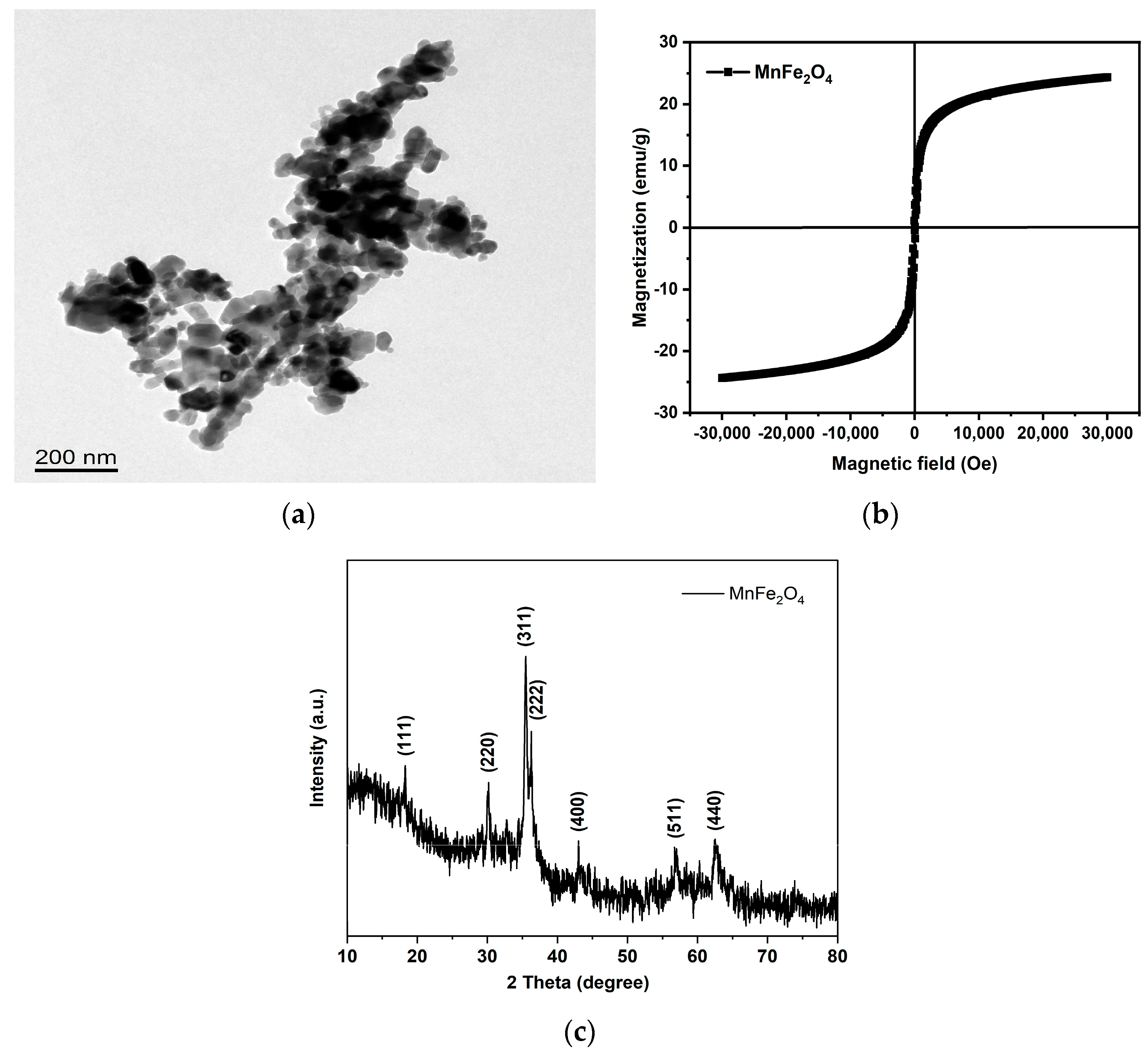
2.1. Experimental Materials

2.1.1. Sources of Algae Species and Culture Media

The algal strain (Chlorella ellipsoidea, FACHB-41) for this experiment was purchased from the University of Texas Algae Bank, which is tolerant of a 40% CO2 environment. It was screened in a previous test for CO2 gradient domestication. To study the efficiency of CO2 fixation by microalgae, the only inorganic carbon source used throughout the culture was CO2. Also, a modified BG-11 medium was used for the culture. The medium formulations are shown in Table 2 and Table 3.

2.1.2. Experimental Reagents

The reagents used in this experiment are shown in Table 4.

2.1.3. Main Instrumentation of the Experiment

The instrumentation, models, and manufacturers used in this experiment are shown in Table 5.

2.2. Construction of the Experimental Setup

This experiment was carried out using a homemade device, the construction of which is shown in Figure 2. The experiments involved in this study all used this set of devices. A microalgae carbon sequestration reactor with a height of 0.4 m and diameter of 0.25 m was used. The upper part is equipped with an air inlet, and the air inlet pipe goes straight to the bottom of the reactor; this enables the CO2 gas and microalgae algal liquid to react fully. The upper right and lower left of the outer wall of the reactor are equipped with an air outlet and a feed port, respectively. The feed port is connected to a hose and a valve; this valve controls the amount of feed liquid. For the aeration method, CO2 gas is mixed with air (filtered through the bacterial membrane) in a buffer bottle through a sevenstar flow control meter; this controls the flow rate, and the mixed gas is passed through a three-way valve, or a four-way valve, through a rotor flow control meter into the microalgae reactor. This distributes the microalgae carbon sequestration reaction into several vessels evenly.

2.3. Experimental Design

In this study, the initial inoculum concentration of microalgae was first diluted to OD680 = 0.4 using microalgae 41 after 4 generations of continuous culture at 40% CO2 concentration. The toxicity threshold of manganese ferrite nanoparticles on microalgae was used as a safety baseline, and within the safety range, different concentrations of manganese ferrite nanoparticles were set to be added to determine the peak concentration of photosynthetic carbon sequestration efficiency, to reduce the toxicity of the experimental group to no acute toxicity at a low concentration, and to determine the long-term stability by testing the microalgae’s growth and physiological changes over 12 days and the chlorophyll fluorescence parameter, etc. Eventually, 30 mg/L, 60 mg/L, 90 mg/L, and 120 mg/L concentrations of manganese ferrite nanomaterials were added to the microalgae carbon fixation reactor. Three experimental groups were set up in parallel, each with 40% CO2 to control the flow rate of 0.1 vvm, at 25 °C. Light intensity was set at 4500 Lux, with continuous light cultivation for 12 days. The growth of microalgae, carbon fixation rate, pigment accumulation, chlorophyll fluorescence parameters, and the expression of key photosynthetic genes were tested and analyzed. The optimal treatment conditions were compared to promote the conversion and absorption of light by microalgae and improve the photosynthetic carbon fixation efficiency.

2.4. Analysis and Calculations

2.4.1. Methods of Biomass Determination

The acetate filter membrane of 0.45 um pore size was soaked in a sufficient amount of distilled water for 6 h [20]; then, the surface water was drained off, and the membrane was dried in a constant-temperature drying oven at 80 °C until constant weight, cooled in a desiccator, and weighed (M1). A 10 mL sample of algal liquid was poured into a filter, and then pumped dry. Then, distilled water was used to drench the filter membrane three times and then, it was pumped dry; the filter membrane was put into a constant-temperature drying oven at 80 °C and dried to a constant weight. The microalgae was filtered in a membrane and then thoroughly dried and dehydrated. Finally, the sample was cooled in a desiccator and weighed (M2). The calculation formula for biomass determination is as shown in Equation (1):
Y(g/L) = (M2 − M1)/V
where M1 is the weight of the filter membrane (mg), M2 is the weight of the algae and filter membrane (mg), and V is the volume of the microalgae algal solution (L).

2.4.2. Measurement of Growth Rate

Growth rate is calculated by Equation (2):
V(mg/L/d) = DW2 − DW1/t1 − t2
where DW1 is the biomass concentration at time t1 (mg/L), and DW2 is the biomass concentration at time t2 (mg/L).

2.4.3. Determination of Carbon Dioxide Fixation Rate

The carbon dioxide fixation rate R (mg/L/d) is as shown in Equations (3) and (4):
R = Cc × (MCO2/Mc) × P
where Cc is the carbon content (%, w/w) in microalgal cell biomass, Cc is 50%; MCO2 is the molar molecular weight of carbon dioxide, value 44 g/moL; Mc is the molar mass of elemental carbon, with a value of 12 g/moL; and P is the microalgal cell biomass yield (mg/L/d), which was calculated as follows:
P = (BCt/BC0)/Δt
where BCt is the biomass concentration of microalgal cells at day (mg/L), BC0 is the bio- mass concentration of microalgae cells in the initial state (mg/L), and Δt is the incubation time (d).

2.4.4. Determination of pH

The algal solution was inserted into a Ray magnetic pH meter and recorded when the reading stabilized.

2.4.5. Measurement Method of Chlorophyll Content

A 5 mL sample of algal liquid was centrifuged; then, 5 mL of 95% ethanol was added, and the sample was placed in a 75 °C water bath. It reacted for 15 min. When the reaction was complete, the sample was cooled and then centrifuged at 5000 r/min for 5 min; the supernatant was collected. Using a UV–visible spectrophotometer, the OD values at 665 nm and 649 nm were read, and the test was set up with two repetitions. The chlorophyll concentration was calculated according to following Equations (5) and (6) [21]:
Ca (mg/L) = 13.95 × OD665 − 6.88 × OD649
where OD665 is the absorbance value of microalgae at 665 nm, and OD649 is the absorbance value of microalgae at 649 nm.
Cb (mg/L) = 24.96 × OD649 − 7.32 × OD665
where OD649 is the absorbance value of Chlorella at 649 nm, and OD665 is the absorbance value of Chlorella at 665 nm.

2.4.6. Determination Method of Chlorophyll Fluorescence Parameters

Two samples of 3.5 mL of algal cell culture solution in the dark were collected and dark-treated for more than 20 min each, to ensure that the reaction center of light was completely open and the electron transfer was completely oxidized. Then, the samples were put in the AP110-C Chlorophyll Fluorescence Instrument to determine relevant photosynthetic parameters; the meaning of each chlorophyll fluorescence parameter is shown in Table 6.

2.4.7. Fluorescence Quantitative PCR Assays

Selection of Target Genes

The target genes determined in this experiment were PsbA, PsbC, LHCB2, LHCB5, PetC, PetF, PetH, LHCA2, LHCA5, and RbcL.
PsbA and PsbC encode the reaction center D1 protein subunit of the PSII core complex and the CP43 protein subunit at the antenna splice site, respectively. LHCB2 and LHCB5 are genes coding for LHCII light-harvesting complex II. Light-harvesting pigment protein complex (LHCII) is a pigment protein centered on light energy capture and transfer and energy dissipation and photoprotective function under light stress. PetC is an iron–sulfur protein of the cytochrome b6-f complex, which encodes an iron–sulfur protein that acts as an electron transporter and is involved in various physiological processes in plants. The PetF gene encodes ferredoxin (Fd), which is involved in the electron transfer process of photosynthesis. The PetH gene encodes ferredoxin- NADP+ reductase (FNR), which is responsible for the acquisition of electrons and H+ from Fd and the conversion of NADP+ to NADPH during the Calvin cycle. LHCA2 and LHCA5 encode LHCI light-harvesting complex I. LHCI is mainly responsible for transferring light energy to the active center of the PSI core subunit, which leads to charge separation and electron transfer reactions. RbcL, the gene encoding Rubisco enzyme, is a key gene in the microalgae dark reaction Calvin cycle, which absorbs CO2 for the dark reaction and directly determines CO2 fixation capacity.

Fluorescence Quantitative PCR Procedure

Sample RNA was first extracted using the RNA-prep pure total plant RNA extract- ion kit; then, the extracted RNA from the previous step was reverse-transcribed into cDNA, using a reverse transcription kit. Finally, primers were designed to detect the expression by RT-PCR. A total of 7.5 mL of microalgae was added to a centrifuge tube and centrifuged to remove supernatant. Then, 1 mL of distilled water was added for resuspension and concentration in a 1.5 mL centrifuge tube, and then, this was centrifuged to remove the supernatant. Then, wall-breaking grinding was carried out with a grinding stick, followed by RNA extraction, and the steps of RNA-prep pure plant total RNA extraction kit were followed. Then, the steps for DNA removal were carried out with a DNA digestion kit; finally, the extracted RNA was collected for spare use.
The RT-RCR program is shown in Table 7 below.
Primers were designed using the NCBI website, and 18sRNA was selected as the internal reference gene; the primer design for the internal reference gene and the target gene is shown in Table 8.

3. Results

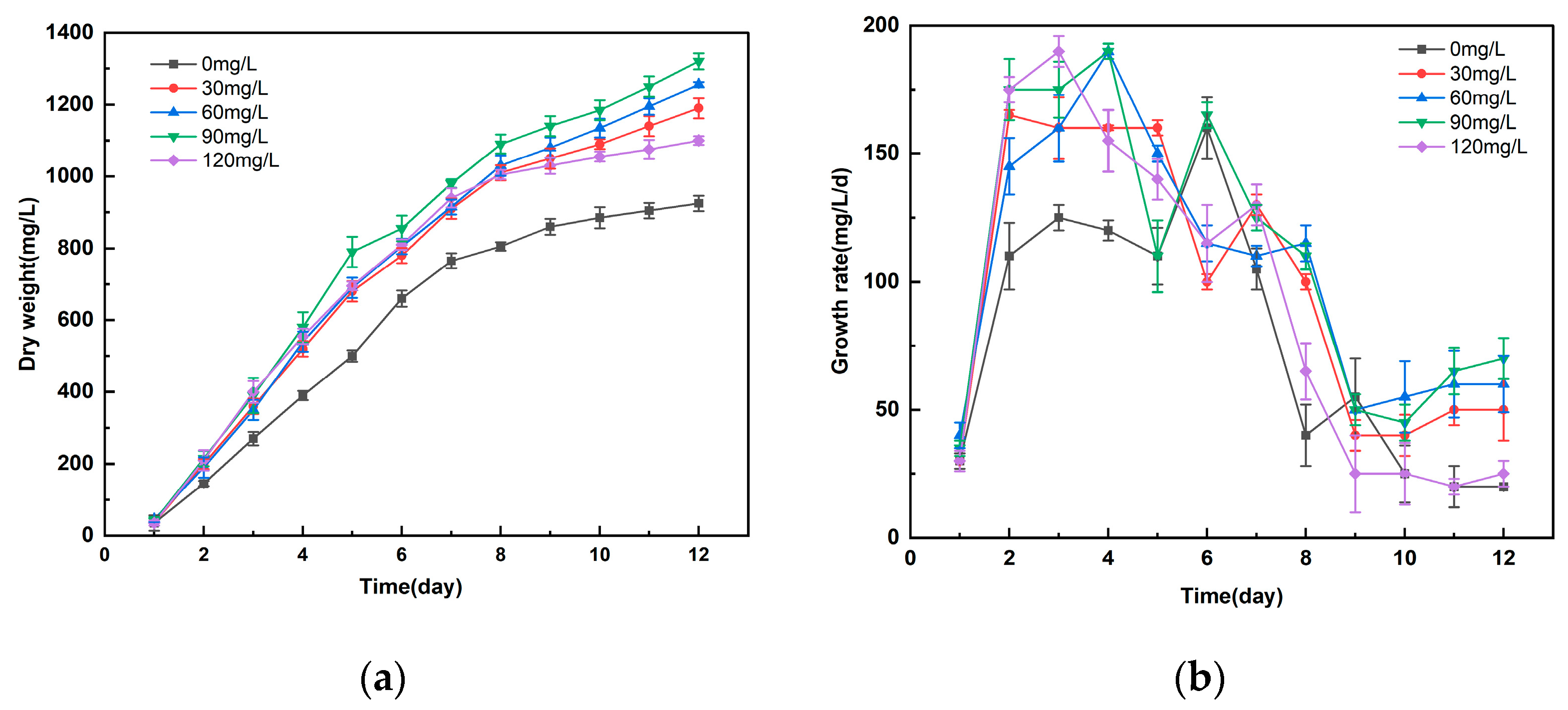
3.1. Study on the Effect of Different Concentrations of Manganese Ferrite Nanoparticles on the Growth of Microalgae

As can be seen from Figure 3a, the addition of different concentrations of nanomaterials helped to enhance the biomass of common microalgae. The microalgae with 90 mg/L manganese ferrite nanoparticles had an obvious growth advantage. The experimental group had the largest biomass of 1320 mg/L on day 12, which was 42.7% higher than that of the control group. As can be seen from Figure 3b, the growth of microalgae with different concentrations of nanomaterials added was relatively slow on the first day, probably due to the fact that the microalgae had just entered the high concentration of CO2 and conditions without which it would not have adapted; however, it began to grow rapidly into the logarithmic growth period on days 2–8, and entered into a growth plateau on days 9–12.

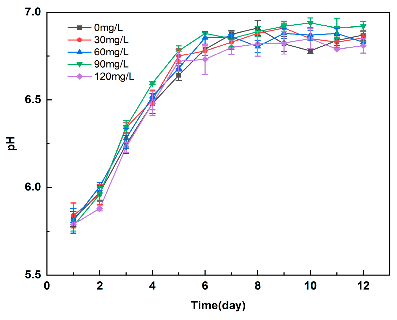
3.2. Effect of Different Concentrations of Manganese Ferrite Nanoparticles on the pH of Microalgal Algal Blooms

pH is an important factor that influences the growth of microalgae for efficient carbon sequestration [22]. As seen in Figure 4, on adding different concentrations of manganese ferrite nanoparticles to microalgae No. 41, the pH of the algal solution decreased sharply on the first day of growth (initial pH of 6.8), but then the pH gradually increased and stabilized at about 6.9. This was attributed to the increase in algal pH caused by the regulation of cellular metabolic action of the microalgae, when the microalgae were grown in the conditions of high CO2 concentration, after the cells became adapted. When microalgae grow under high CO2 concentration, the cell adapts to the strategy by reducing the activity of PSII system. The energy allocation of non-cyclic photosynthetic phosphorylation and increasing the energy ratio of cyclic photosynthetic phosphorylation of PSI system reduce normal organic carbon synthesis. At the same time, this provides and allocates more ATP for the maintenance of intracellular pH stability. So, after the metabolic adjustments in the delayed period of 1–2 days, the microalgae is able to accelerate growth, and gradually returns to a normal growth rate and enters the logarithmic growth period [23]. Finally, the algal ions reach equilibrium and the pH stabilizes. Therefore, the nano-manganese ferrite 1–7 adjusts the pH value of the algal fluid by promoting the metabolism of the microalgae, so that the pH value is slightly increased and stabilized in the weakly alkaline zone, in order to maintain a suitable environment for the microalgae, which is conducive to the growth of the microalgae.

3.3. Study on the Effect of Different Concentrations of Manganese Ferrite Nanoparticles on the Carbon Sequestration Rate of Microalgae

As shown in Figure 5, the rate of CO2 fixation in microalgae increased with the increase in Mn nano ferrite concentration (0–90 mg/L) in the first 7 days of incubation. The 90 mg/L Mn nano ferrite showed the greatest CO2 fixation rate, from the beginning of the incubation to the 6th day, which was 260 mg/L/d, and 30.0% higher than that of the control group. The CO2 fixation rate of microalgae with the addition of 120 mg/L Mn nano ferrite was slightly decreased, compared to that of 90 mg/L of manganese ferrite nanoparticles. However, this decrease in the carbon fixation rate of microalgae was still higher than the carbon fixation rate of the blank control group. With the prolongation of the incubation time, at 8–12 days, both the experimental group and the control group showed a decreasing trend. The reason for this result may be that the microalgae in this experiment belongs to continuous cultivation, and the nutrient salts, such as nitrogen and phosphorus sources, in the culture solution were limited. With the increase in the incubation time, the nutrient salts were gradually consumed, so the microalgae biomass yield gradually decreased, and therefore, the rate of CO2 fixation decreased at the end of the growth period.

3.4. Study on the Effect of Different Concentrations of Manganese Ferrite Nanoparticles on Chlorophyll Accumulation in Microalgae

As shown in Figure 6, the chlorophyll concentration in the microalgae, with 90 mg/L of manganese ferrite nanoparticles added, was significantly higher than that in the control group. The chlorophyll concentration reached 19.86 mg/L, which was 1.21 times higher than that in the control group. This suggests that the incorporation of nanomaterials can significantly promote the accumulation of chlorophyll in microalgae. Low concentrations of nanomaterials provide microalgae with key nutrients required for chlorophyll synthesis by releasing trace iron [24], while their photocatalytic properties enhance the efficiency of light energy utilization in the algal solution, thus accelerating the photosynthesis process.

3.5. Study on the Effect of Different Nanomaterials on Chlorophyll Fluorescence Parameters of Microalgae

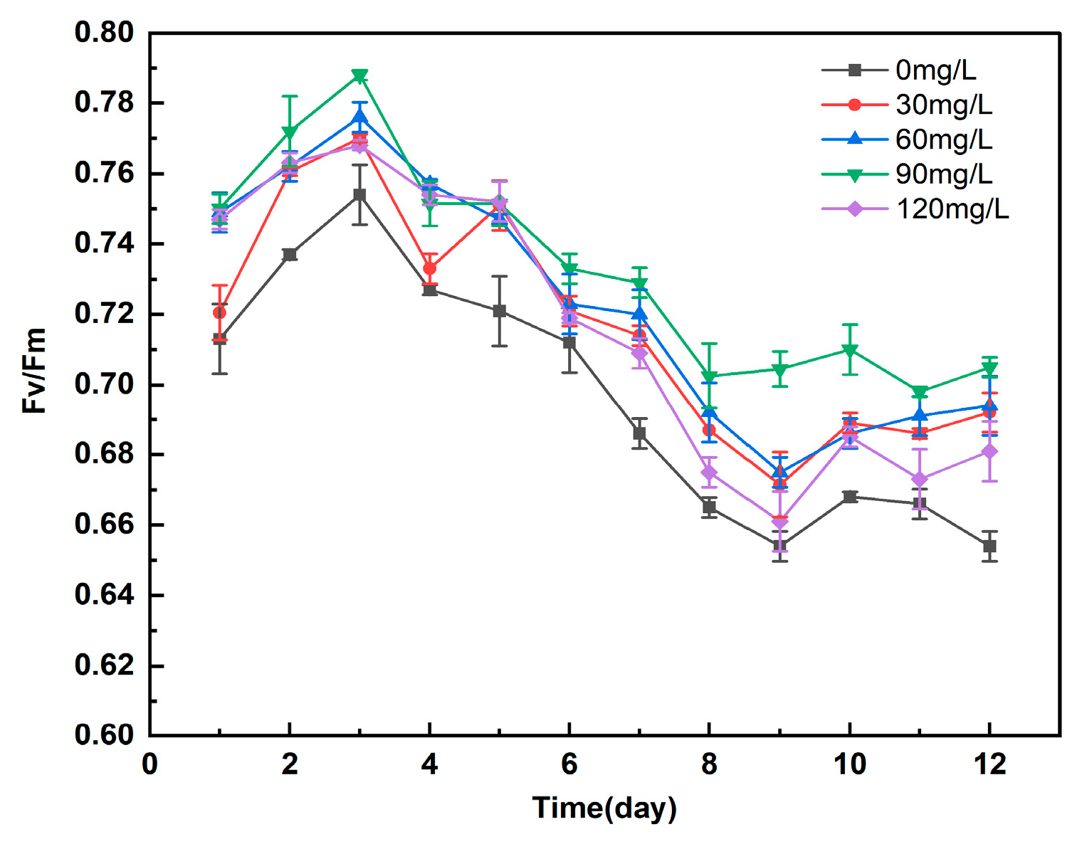
3.5.1. Study on the Effect of Different Concentrations of Manganese Ferrite Nanomaterials on Fv/Fm of Microalgae

Fv/Fm is the PSII photochemical maximum quantum yield, which reflects the potential photochemical capacity of PSII in algal cells [25]. As can be seen from Figure 7, the Fv/Fm of the added nanomaterials showed a trend of rising and then falling and did not show a rapid and substantial decrease, indicating that the microalgae did not undergo stress. The addition of a certain concentration of nanomaterials could increase the photosynthetic phosphorylation level of microalgae, increase ATP synthesis, increase the efficiency of the Calvin cycle, and promote the synthesis and regeneration of NADP+ and NADPH, increasing the rate of carbon sequestration by microalgae. This contributed to the enhancement of the functional activity of PSII, which was manifested in the increase in the photochemical efficiency and quantum yield of PSII, and the increase in the Fv/Fm ratio [26]. The small decrease in Fv/Fm value was because the Fv/Fm value was proportional to the chlorophyll content, and the microalgae adapted to the high concentration of CO2 in the late stage. This intensified the nutrient depletion of the microalgae, and the algal cell growth was faster. The color became darker green, which led to insufficient light, and the carbon source was used for the increase in the cellular biomass to the extent that the chlorophyll nutrient was lacking. This led to the obstruction of chlorophyll synthesis, the decrease in the chlorophyll content, and the decrease in the Fv/Fm ratio value [27]. However, the Fv/Fm values of the experimental group were still higher than those of the blank control group. It can be shown that the added manganese ferrite nanomaterials can improve the light energy capture efficiency, provide more ATP and NADPH for the Calvin cycle, and enhance the photosynthetic carbon sequestration efficiency of microalgae.

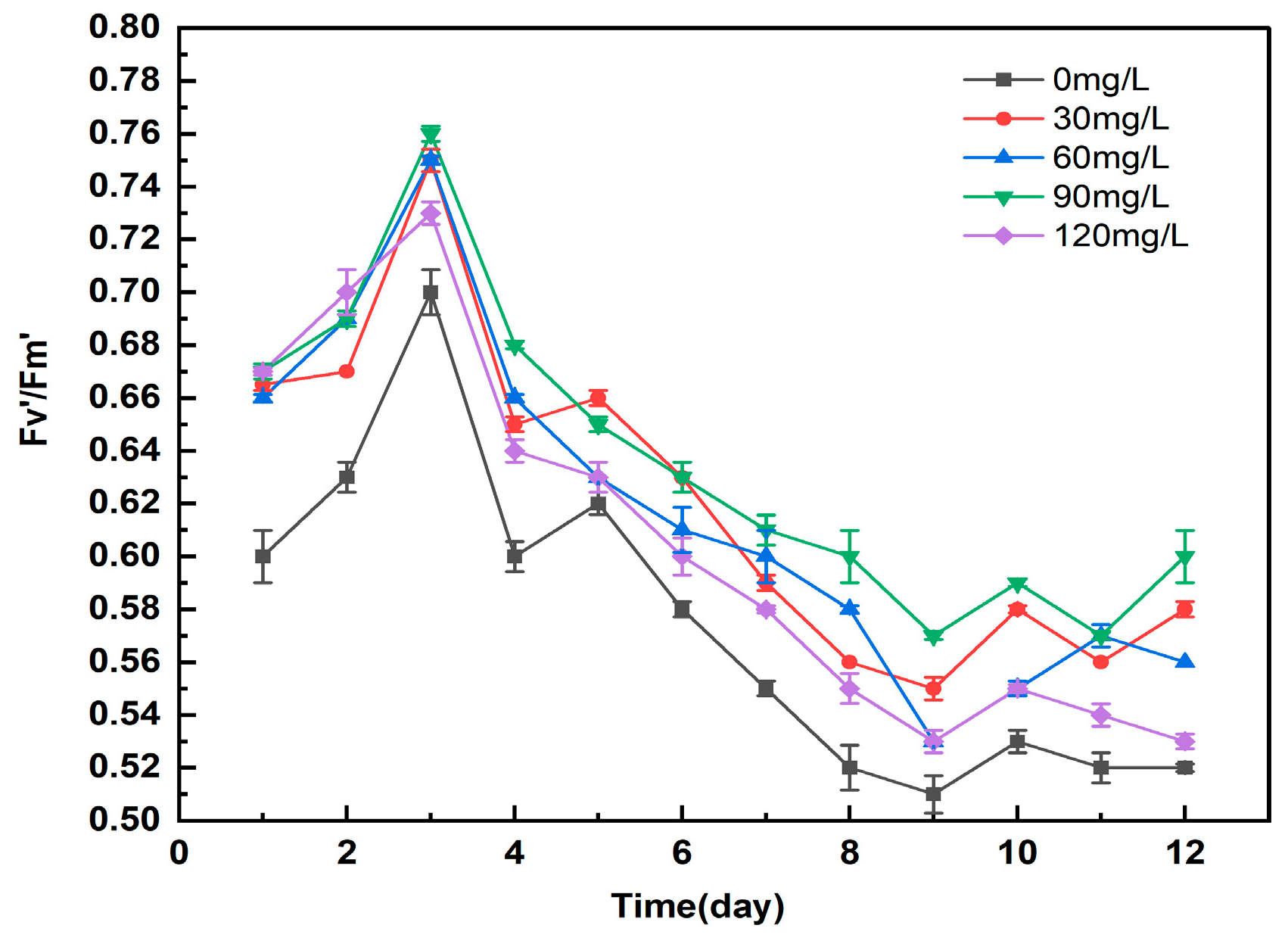
3.5.2. Study on the Effect of Different Concentrations of Manganese Ferrite Nanoparticles on Microalgae Fv′/Fm′

Fv′/Fm′ is the actual light energy conversion efficiency, which represents the quantum efficiency of linear electron transfer and reflects the efficiency of primary light energy capture in open PSII reaction centers. However, CO2 protects both the excitation energy capture efficiency and the photochemical reaction from being less affected by the photoprotection mechanism. As can be seen from Figure 8, the Fv′/Fm′ ratio of the experimental group with different concentrations of nanomaterials showed a trend of first increasing and then decreasing. But the overall change was not significant, and there was no rapid and large-scale decline, suggesting that smooth linear electron transfer, which supports the continued operation of the Calvin cycle, can contribute to the efficiency of carbon sequestration by microalgae, indicating that the microalgae did not undergo stress. Thus, the microalgae could grow normally and stably, after the addition of nanomaterials with different concentrations.

3.5.3. Study on the Effect of Different Concentrations of Manganese Ferrite Nanoparticles on Microalgae qP

qP is the photochemical bursting coefficient, which represents the photochemical quenching of variable chlorophyll fluorescence quenching. It is commonly used to reflect the degree of opening of PSII reaction centers [28]. As shown in Figure 9, the trend of qP in both the experimental group and the blank control group first increased and then decreased; this was the same as the trend of the Fv/Fm ratio. The qP values of each experimental group ranged from about 0.7 to 0.9. The synthesis of microalgae growth, carbon sequestration, and other indicators can be learned from the data. The addition of manganese ferrite nanoparticles in the first three days led to an increase in the activity of the reaction centers, opened by microalgae PSII and an increase in the number of electrons involved in the fixation of CO2, suggesting that more electrons are used for CO2 reduction rather than heat dissipation, directly contributing to carbon sequestration efficiency. This led to the enhancement of the photosynthetic electron transfer capacity and an increase in the photosynthetic efficiency, and thus the rise in qP in the first three days [29]. The decrease in qP indicates a decrease in the index of the proportion of open reaction centers in PSII reaction centers. This resulted in a blockage of electron transfer from the oxidized side of PSII to PSI reaction centers and a decrease in the number of electrons used to carry out photosynthesis. Thus, there was a decrease in the ability to use captured photon energy for photochemical reactions, resulted in a decrease in the ability to emit only light energy, dissipated as heat or in some other form, which is a kind of active defense mechanism to avoid algae from being damaged [30]. Overall, the addition of manganese ferrite nanoparticles resulted in enhanced photosynthetic electron transfer capacity and increased photosynthetic efficiency of the microalgae.

3.5.4. Study on the Effect of Different Concentrations of Manganese Ferrite Nanoparticles on NPQ of Microalgae

NPQ is the non-photochemical quenching coefficient, which represents the reduction in fluorescence yield due to non-photochemical reactions, and is often used to indicate how much heat is dissipated. As can be seen in Figure 10, the NPQ values of the experimental group were reduced compared to the control group, indicating that the heat dissipation in the experimental group with the addition of nanomaterials was reduced. Thus, more energy was used for the photochemical reaction. It can be concluded that the microalgae with added manganese ferrite nanomaterials had faster energy transfer during the photoreaction. The increased energy (electron) transfer rate of the photoreaction may synthesize more energetic compounds and reduce power, thus promoting the turnover rate of carbon flow in the Calvin cycle. This further explains the increase in the rate of carbon sequestration in the late stage of microalgae and the significant increase in the expression of key genes for carbon sequestration. The NPQs of all four experimental groups in Figure 10 tend to decrease and then increase throughout the experiment. NPQ experienced a decline from day 0 to 3, but then an upward trend began on day 4, at the same time as its Fv/Fm ratio change occurred.
The reason that NPQ is not 0 on the 1st day of inoculation may be because the algae species were transferred from a starvation medium for cultivation under a high CO2 concentration. This required an adaptation process, and the excess light energy could not be completely absorbed and transformed by ordinary Chlorella. To protect itself from being harmed, it had to emit the excess light energy in the form of heat, so the NPQ could not be 0. And from day 2–4, NPQ was 0. The reason is that the Fv/Fm ratio increased dramatically from day 2–4 when the algal cells were not yet limited by nutrient salts. The non-photochemical burst was at a lower level, the Calvin cycle of the algal cells was active, the utilization rate of the light energy was higher, the photosynthetic efficiency was improved, the absorption and conversion ability of the microalgae to light energy was not weakened drastically, and finally, the photosynthetic apparatus would not be damaged, so the NPQ remained at 0. This further indicates that the addition of manganese ferrite nanomaterials facilitates the capture and utilization of light energy by chlorophyll and does not result in excess light energy, suggesting that light energy is used more for photochemical reactions than for heat dissipation, thus enhancing carbon sequestration efficiency. The reason for the increasing NPQ from the fifth day is that the process of this change occurred at a late stage of the experiment. This indicated that the proportion of light energy absorbed by microalgae for heat dissipation had increased. The gradual increase in the microalgae NPQ to stabilization under the condition of high concentration of CO2 indicated that the degree of its inhibition of the activity of the Calvin cycle increased, and the potential for heat dissipation of PSII increased, which is a protective effect for the algal organism itself. With the prolongation of incubation time, nutrient salt limitation gradually manifested and became more and more obvious. The Calvin cycle was inhibited, the efficiency of photosynthesis was reduced, the utilization of chlorophyll after absorbing light energy was lower, and the excess light energy was emitted in the form of heat dissipation; this led to an increase in NPQ, thus avoiding or mitigating the damage of the photosynthetic apparatus caused by the absorption of excessive light energy by PSII. The addition of 90 mg/L of manganese ferrite nanoparticles ultimately had the smallest NPQ, probably because this concentration was the best in the experimental group. This indicated that its activity of the Calvin cycle was minimally inhibited, with high energy utilization. This resulted in less excess light energy and a higher photosynthetic and carbon fixation efficiency, which were all most suitable for the growth of the microalgae.

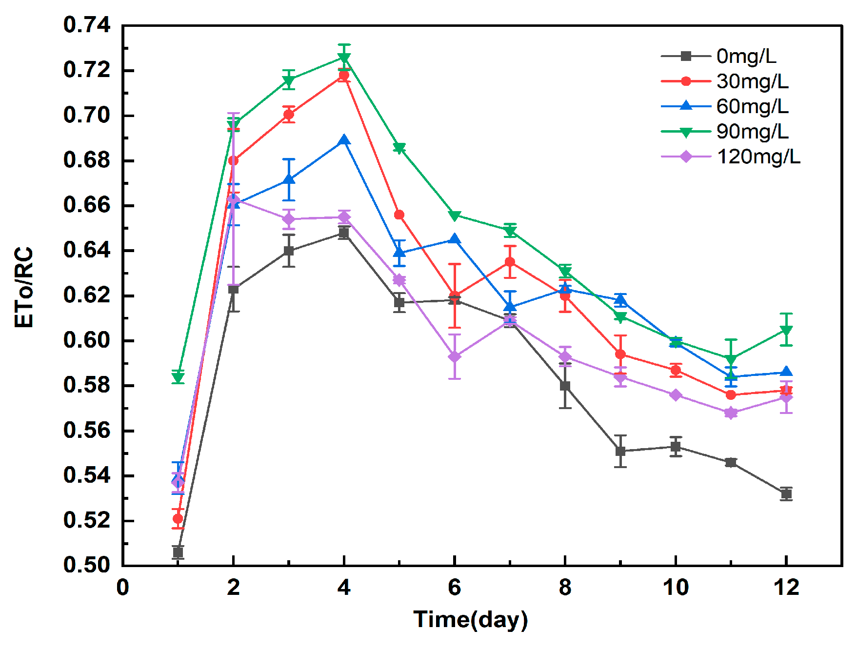
3.5.5. Study on the Effect of Different Concentrations of Manganese Ferrite Nanoparticles on ETo/RC of Microalgae

ETo/RC denotes the energy per unit area used for electron transfer. It can be seen from Figure 11 that ETo/RC shows a general trend of first increasing and then decreasing, and finally stabilizing. This result may be attributed to the fact that the addition of nanomaterials at the beginning will promote the Calvin cycle during photosynthesis. High ETo/RC indicates a strong activity of the electron transport chain, driving more NADPH production, providing reducing power for Rubisco enzymes, and promoting carbon sequestration efficiency. This makes the synthesis of microalgae chlorophyll and the rate of cell division increase, with an increase in ATP, so that the rate of electron transfer in the PSII reaction center may be enhanced, increasing microalgae photosynthetic efficiency. During this period, microalgae rapidly adapts to the high concentration of CO2 conditions, allowing the rate of CO2 fixation to increase. The increased carbon fixation rate of microalgae can exacerbate nutrient consumption and, thus, suppress the electron transfer rate of the PSII reaction center, which can lead to a decrease in photosynthetic efficiency. Microalgae, with different concentrations of nanomaterials added, can reach a dynamic equilibrium in electron transfer after short-term acclimatization to conditions of high CO2 concentration. The experimental groups with the addition of manganese ferrite nanomaterials all showed some increase in the energy of electron transfer, compared to the control group, implying that they have enhanced PSII reaction center activity and greater ability to drive more electrons for transfer. Also, higher electron transfer rates allow the microalgae cells to capture a gradual increase in the number of photo-quanta that can be utilized.

3.6. Study on the Effect of Different Concentrations of Manganese Ferrite Nanoparticles on Gene Expression of Microalgae

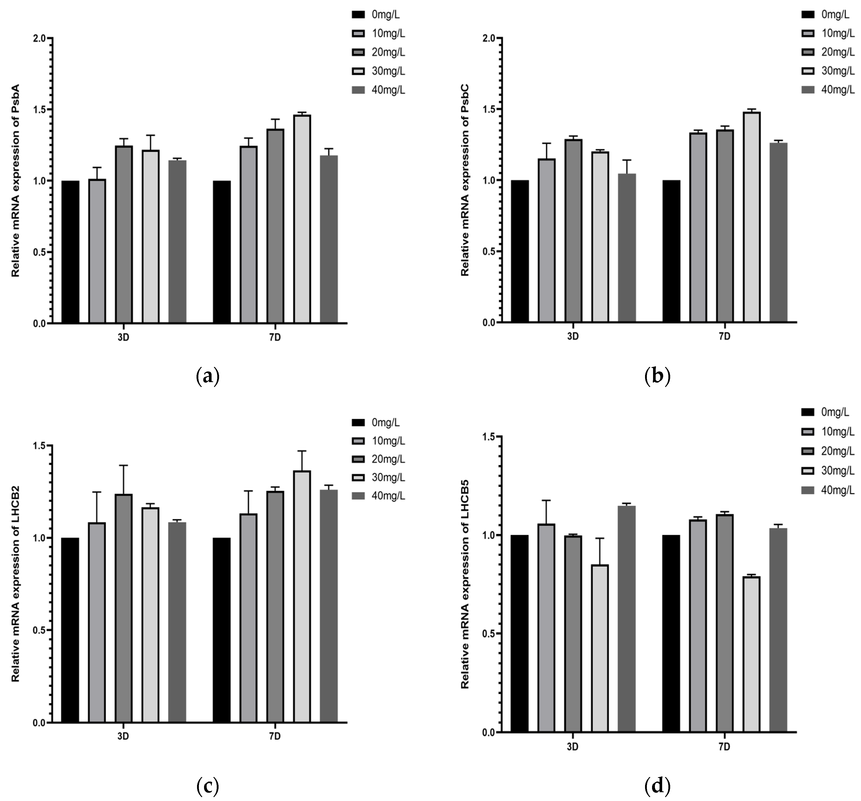
3.6.1. Study on the Effect of Two Nanomaterials on the Expression of Key Genes of Microalgae Photoresponsive PSII

PsbA and PsbC are important regulatory genes for the synthesis of response center proteins of the PSII core complex. LHCB2 and LHCB5 are genes coded for the LHCII light-trapping complex II, whose main functions include the capture and transfer of light energy and the regulation of excitation energy distribution between PSI and PSII [31]. As can be seen in Figure 12, the addition of different concentrations of nanomaterials significantly upregulated the gene expression of PsbA, PsbC, and LHCB2 proteins in microalgae. It has been shown that to achieve the most suitable living environment, photosynthetic organisms can better adapt to the quality and quantity of light energy by regulating the components of their photosynthesis system. Photosynthetic organisms regulate the size and composition of their light-trapping antenna system. Therefore, the manganese ferrite nanomaterials can cause a significant upregulation of LHCB2, the gene coding for the main protein of LHCII, in the face of high-CO2-concentration conditions. This suggests that the manganese ferrite nanomaterials have a greater effect on the transcriptional level of the LHCB2 protein. The light-trapping antennae proteins in the excited state can enhance the cell’s ability to capture light and increase the penetration of light energy to improve photosynthetic efficiency. However, the nanomaterials did not have a significant effect on LHCB5 protein transcription, so it is hypothesized that the main site of action of the nanomaterials on microalgae is LHCB2, and it is not obvious for LHCB5.

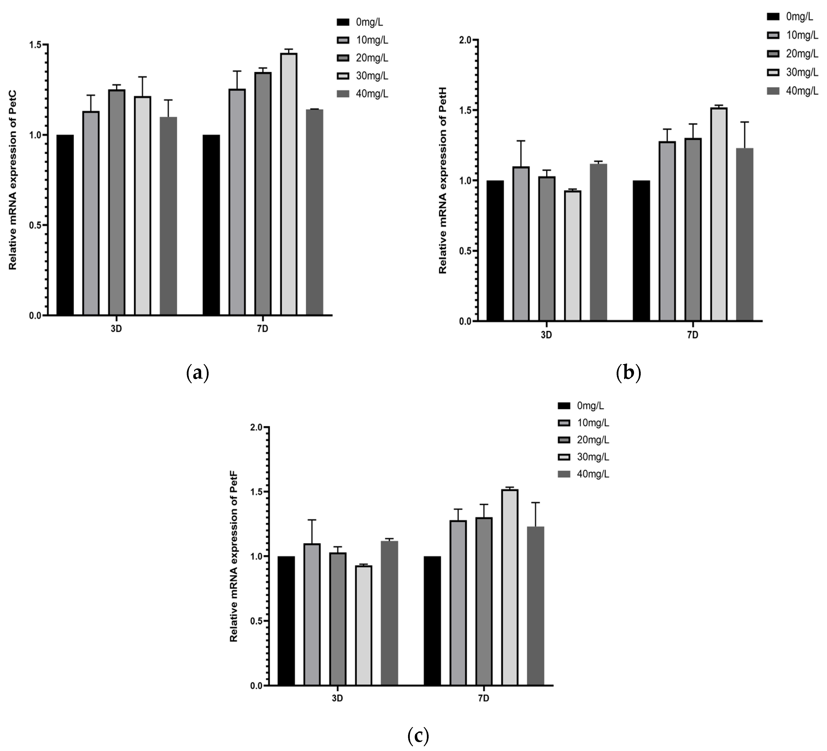
3.6.2. Study on the Effect of Different Concentrations of Manganese Ferrite Nanoparticles on the Expression of Key Genes for Light-Responsive Electron Transfer in Microalgae

PetC encodes the cytochrome b6-f complex iron–sulfur protein, whose primary role is to accept electrons and transfer them to the next electron acceptor. PetF encodes ferredoxin (Fd), which is involved in the electron transfer process of photosynthesis. PetH encodes ferredoxin-NADP+ reductase (FNR), the terminal oxidase of the photosynthetic electron transport chain. FNR catalyzes the end-point reaction of photosystem-I electron transport, converting NADP+ and H+ to NADPH.
As can be seen in Figure 13, the gene expression of PetC, PetF and PetH in the experimental group was significantly upregulated relative to the control group, indicating that the addition of different concentrations of manganese ferrite nanomaterials made the electron transfer in the microalgae cells very active. The ferredoxin-NADP+ reductase catalyzed the synthesis of more NADPH by electrons from the reduced flavin proteins, which provided more substrates of NADPH for the dark reaction, thereby enhancing the photosynthetic efficiency. Therefore, it can be shown that the addition of different concentrations of nanomaterials can increase the relative electron transfer rate of microalgae, thus enhancing the photosynthetic efficiency of microalgae.

3.6.3. Effects of Different Concentrations of Manganese Ferrite Nanoparticles on the Expression of Key Genes of Microalgae Photoresponsive PSI

PSI is composed of subunits such as reaction center complex and light-trapping complex, which contain a light-trapping complex and a light reaction center. The light-trapping complex that constitutes PSI is called LCHI [32]. LHCA2 and LHCA5 encode the LHCI light-trapping complex I. As can be seen from Figure 14, the addition of different concentrations of manganese ferrite nanoparticles resulted in the upregulation of light-trapping complex protein gene expression in the PSI reaction centers of microalgae. In particular, the experimental group with the addition of 90 mg/L of manganese ferrite nanoparticles was very different from the control group. Increased synthesis of photosynthetic antenna proteins allows for the formation of more light-trapping complexes with photosynthetic pigments, improving the light-trapping efficiency and energy transfer of PSI. Therefore, the addition of manganese ferrite nanomaterials increased the reactivity of PSI and PSII and caused a coordinated interaction between the two, to promote the transfer of energy in the photosynthetic electron transport chain. This increased the photosynthetic efficiency of the microalgae and thus promoted their biomass synthesis, while providing a basis for the rate of carbon fixation [33].

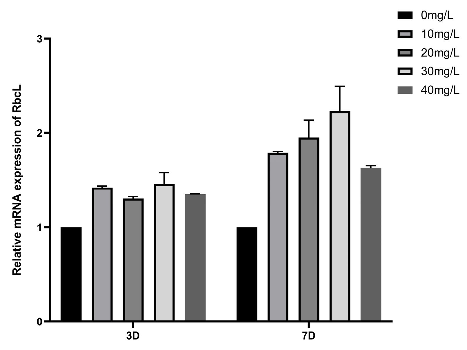
3.6.4. Study on the Effect of Different Concentrations of Manganese Ferrite Nanoparticles on the Expression of Key Rubisco Genes for Carbon Sequestration in Microalgae

RbcL encodes the Rubisco enzyme gene, the large subunit of ribulose-1,5-bisphosphate carboxylase/oxygenase of the Calvin cycle, which is a key enzyme in the CO2 fixation process [34]. As can be seen from Figure 15, the expression of the Rubisco enzyme in the experimental group was high, compared with that of the blank control group. This indicates that the addition of a certain amount of manganese ferrite nanomaterials may cause the presence of more activated forms of Rubisco enzyme. This led to the increase in the initial enzyme activity of the Rubisco enzyme. The activity of Rubisco-activating enzyme is positively correlated with the concentration of RuBP. The addition of a certain amount of manganese ferrite nanoparticles caused the microalgae to synthesize more RuBP, which in turn activated more Rubisco enzyme and increased the rate of CO2 fixation.

4. Conclusions

Based on the limited light absorption and utilization efficiency of microalgae during photosynthesis, the present study was conducted by adding different concentrations of manganese ferrite nanomaterials to microalgae No. 41 at 40% CO2 concentration. The growth and metabolism of the microalgae were investigated during the process of carbon sequestration. Chlorophyll fluorescence parameters and changes in the expression of key genes for photosynthesis and carbon sequestration were compared and analyzed to obtain the optimal concentration of nanomaterials to improve the light absorption and transmission efficiency of microalgae. Thus, the photosynthetic efficiency of microalgae was improved. These results are summarized as follows:
(1) The addition of 30–120 mg/L manganese ferrite nanoparticles helped to increase the biomass and carbon sequestration rate of the microalgae. Specifically, the addition of 90 mg/L manganese ferrite nanoparticles had an obvious growth advantage for the microalgae. The carbon sequestration rate of the experimental group reached the maximum on the 6th day, which was 40.72% higher than that of the control group. The biomass growth and the concentration of chlorophyll reached a maximum on the 12th day, which were 42.7% and 12.1% higher than that of the control group. At first, the pH of MnFePO4 nanomaterials decreased; then, the pH gradually increased and stabilized at about 6.9. It is shown that the nanomaterials promote the growth and metabolism of microalgae, thereby regulating the pH of the algal fluid to maintain a suitable growth environment for microalgae. The experimental group with the addition of 120 mg/L manganese ferrite nanoparticles was still better than the control group, but the effect was significantly lower than that of the 90 mg/L experimental group, and this trend of “increasing and then decreasing” indicates that the effect of adding manganese ferrite nanoparticles to the experimental group is still better than that of the control group. The light scattering effect of manganese ferrite nanoparticles was maximized at 90 mg/L. Higher concentrations, on the contrary, reduced the light penetration depth. The electron-mediated capacity was close to the carrying limit of the photosynthetic chain of microalgae, and the high concentration of nanoparticles might adsorb nutrient salts in the medium, exacerbating the nutrient limitation at the late stage of cultivation. Thus, the addition of 90 mg/L of manganese ferrite nanoparticles was the saturation point for light-absorption enhancement and electron transfer facilitation, with higher concentrations leading to diminishing returns.
(2) Microalgae photosynthetic activity parameters Fv/Fm, Fv′/Fm′, qP, and ETo/RC all showed a trend of increasing and then decreasing. However, NPQ showed the opposite effect, a trend of decreasing and then increasing. The photosynthetic activity of the microalgae in the experimental group was higher than that of the control group. The microalgae with the highest concentration of chlorophyll, the strongest ability to capture light, the strongest photosynthetic activity, and the fastest photoresponse was detected when 90 mg/L of manganese ferrite nanoparticles was added. The highest concentration of chlorophyll, the strongest light-trapping ability, the strongest photosynthetic activity, and the fastest electron transfer rate in the light reaction were all observed in the experimental group. It began to decline as it continued to rise up to 120 mg/L. NPQ was lowest in the experimental group with the addition of 90 mg/L of manganese ferrite nanoparticles, indicating the highest light energy utilization, whereas NPQ increased in the experimental group with the addition of 120 mg/L of manganese ferrite nanoparticles, suggesting that the excess light energy was dissipated in the form of heat, reflecting the beginning of the damage to the photosynthetic apparatus. The increase in NPQ and the decrease in Fv/Fm suggest that photosystem II may be subjected to oxidative damage at high concentrations. This indicated that the addition of an appropriate concentration of manganese ferrite nanoparticles could alleviate the inhibitory effect of the high concentration of CO2 on the photosynthesis of microalgae. This not only promoted the Calvin cycle of microalgae photosynthesis but also affected the synthesis of chlorophyll and the division of the cells, thus increasing the photosynthetic activity and the efficiency.
(3) From the analysis of gene expression, the addition of different concentrations of nanomaterials upregulated the gene expression of the light capture complex protein in the PSI reaction center of microalgae. The addition of manganese ferrite nanoparticles increased the reaction activity of microalgae PSI and PSII and coordinated the two to promote the transfer of energy in the photosynthetic electron transport chain. This improved the photosynthetic efficiency of the microalgae, promoted its biomass synthesis, and provided a basis for the improved carbon fixation rate. This study clearly supports the addition of 90 mg/L of manganese ferrite nanoparticles as the performance threshold, beyond which negative effects dominate, leading to diminishing returns, and a concentration window of 80–100 mg/L should be prioritized for industrial applications to balance efficiency and safety.

5. Prospects

In this study, it was found that the addition of a certain concentration of manganese ferrite nanomaterials could increase the relative electron transfer rate of microalgae PS II and improve the photosynthetic activity and photosynthetic efficiency of microalgae under controlled conditions. Although some research results have been achieved, due to conditions and time constraints, this study is relatively limited on the mechanism of how nanomaterials can improve the photosynthetic efficiency of microalgae, and its potential risks should not be ignored. If the nanomaterials are discharged with the algal solution into natural water bodies, they may be toxic to aquatic organisms and may enter microalgal cells through endocytosis, and their long-term accumulation may interfere with cell metabolism, leading to oxidative stress and damage to membrane structure. In the future, it is hoped that ecotoxicological studies can be invoked in research experiments or simulations can be carried out to increase the detection of intracellular nanoparticle distribution and functional effects on organelles, to quantify their effects on organisms at different trophic levels, to develop surface passivation or magnetic recycling techniques to reduce the risk of environmental residues, and to assess the residual amount of nanoparticles in biomass and their safety for downstream applications. The risk of carbon re-release due to cell lysis after microalgae death should be evaluated, and cell wall strengthening technologies should be developed to reduce degradation rates or achieve permanent carbon sequestration through deep geological sequestration to avoid secondary release of CO2 due to microbial decomposition after short-term carbon sequestration. Through multidisciplinary collaboration, nanomaterial-enhanced microalgae carbon sequestration technology is expected to be a key link in a negative emissions (CDR) system that promotes balance efficiency and ecological sustainability.
This experimental study of enhanced photosynthetic carbon sequestration by microalgae using manganese ferrite nanoparticles has multiple potential advantages. Firstly, manganese ferrite nanoparticles significantly enhanced the carbon sequestration rate of microalgae at 40% CO2 concentration by enhancing light energy absorption and electron transfer efficiency, which is both highly efficient and sustainable and avoids the problem of high energy consumption compared to traditional carbon sequestration methods and offers the possibility of direct utilization of industrial waste gas. Secondly, the method promotes the Calvin cycle and biomass accumulation by upregulating photosynthetic gene expression, which can synchronize carbon sequestration and bioenergy production; moreover, the low ecotoxicity of the nanomaterials provides a safety guarantee for their practical application. In practical application, it can be combined with an open-pool system or photobioreactor, using industrial waste gas as a carbon source, developing efficient gas diffusion technology to maintain the supply of carbon source, combining with dynamic light regulation to adapt to the natural fluctuation of light intensity, evaluating the stability of manganese ferrite nanoparticles in the open system, and reducing the cost and risk through the immobilization of nanomaterials technology. We also coupled the extraction of high-value-added products after microalgae harvesting to form a closed loop of “carbon capture-resourcing”, to promote its large-scale implementation in the carbon neutral and circular economy, and to gradually demonstrate the feasibility of large-scale implementation through pilot tests, so as to maximize the efficiency of carbon sequestration and to balance the economy and ecological safety.

Author Contributions

Conceptualization, T.C. and Y.C.; methodology, T.C.; software, H.S., N.Z. and W.H.; validation, T.C., X.W. and X.Z.; formal analysis, T.C. and W.W.; investigation, T.C. and S.W.; resources, Y.C. and C.Z.; data curation, T.C.; writing—original draft preparation, T.C.; writing—review and editing, Y.C., C.Z. and R.R.; visualization, T.C.; supervision, T.C. and Y.C.; project administration, T.C. and X.W.; funding acquisition, Y.C and C.Z. All authors have read and agreed to the published version of the manuscript.

Funding

This work was supported by the General Program of the Beijing Natural Science Foundation (Grant No. 8242025).

Institutional Review Board Statement

Not applicable.

Informed Consent Statement

Not applicable.

Data Availability Statement

The data used to support this study are available within the article.

Acknowledgments

The authors would like to thank the editor and anonymous reviewers for their valuable comments.

Conflicts of Interest

The authors declare no conflicts of interest.

References

  1. Parveen, A.; Bhatnagar, P.; Gautam, P.; Bisht, B.; Nanda, M.; Kumar, S.; Vlaskin, M.S.; Kumar, V. Enhancing the Bio-Prospective of Microalgae by Different Light Systems and Photoperiods. Photochem. Photobiol. Sci. 2023, 22, 2687–2698. [Google Scholar] [CrossRef] [PubMed]
  2. Hepburn, C.; Adlen, E.; Beddington, J.; Carter, E.A.; Fuss, S.; Mac Dowell, N.; Minx, J.C.; Smith, P.; Williams, C.K. The Technological and Economic Prospects for CO2 Utilization and Removal. Nature 2019, 575, 87–97. [Google Scholar] [CrossRef] [PubMed]
  3. Pugazhendhi, A.; Nagappan, S.; Bhosale, R.R.; Tsai, P.-C.; Natarajan, S.; Devendran, S.; Al-Haj, L.; Ponnusamy, V.K.; Kumar, G. Various Potential Techniques to Reduce the Water Footprint of Microalgal Biomass Production for Biofuel—A Review. Sci. Total Environ. 2020, 749, 142218. [Google Scholar] [CrossRef] [PubMed]
  4. Li, M.J.; Wang, R.L.; Yang, Y.W.; Chen, J.X. Numerical and Experimental Analysis of Optimized Conical Flask Photobioreactor Structures to Improve Liquid–Gas Two-Phase Distribution and Microalgae Carbon Sequestration. Appl. Therm. Eng. 2020, 180, 115855. [Google Scholar] [CrossRef]
  5. Levasseur, W.; Perré, P.; Pozzobon, V. A Review of High Value-Added Molecules Production by Microalgae in Light of the Classification. Biotechnol. Adv. 2020, 41, 107545. [Google Scholar] [CrossRef]
  6. Mohsenpour, S.F.; Hennige, S.; Willoughby, N.; Adeloye, A.; Gutierrez, T. Integrating Micro-Algae into Wastewater Treatment: A Review. Sci. Total Environ. 2021, 752, 142168. [Google Scholar] [CrossRef]
  7. Zhe, L.; Jie, W.; Yuan, H. The Effect of Place Attachment of Geographical Indication Agricultural Products on Repurchase Intention. J. Retail. Consum. Serv. 2023, 72, 103266. [Google Scholar] [CrossRef]
  8. He, M.; Yan, Y.; Pei, F.; Wu, M.; Gebreluel, T.; Zou, S.; Wang, C. Improvement on Lipid Production by Scenedesmus Obliquus Triggered by Low Dose Exposure to Nanoparticles. Sci. Rep. 2017, 7, 15526. [Google Scholar] [CrossRef]
  9. Pádrová, K.; Lukavský, J.; Nedbalová, L.; Čejková, A.; Cajthaml, T.; Sigler, K.; Vítová, M.; Řezanka, T. Trace Concentrations of Iron Nanoparticles Cause Overproduction of Biomass and Lipids during Cultivation of Cyanobacteria and Microalgae. J. Appl. Phycol. 2015, 27, 1443–1451. [Google Scholar] [CrossRef]
  10. Rudic, V.; Cepoi, L.; Gutsul, T.; Rudi, L.; Chiriac, T.; Miscu, V.; Sadovnic, D.; Nicorici, A. Red Algae Porphyridium cruentum Growth Stimulated by CdSe Quantum Dots Covered with Thioglycerol. J. Nanoelectron. Optoelectron. 2012, 7, 681–687. [Google Scholar] [CrossRef]
  11. Petrou, K.; Kranz, S.A.; Doblin, M.A.; Ralph, P.J. Photophysiological Responses of Fragilariopsis Cylindrus (Bacillariophyceae) to Nitrogen Depletion at Two Temperatures. J. Phycol. 2012, 48, 127–136. [Google Scholar] [CrossRef] [PubMed]
  12. Jeon, H.-S.; Park, S.E.; Ahn, B.; Kim, Y.-K. Enhancement of Biodiesel Production in Chlorella Vulgaris Cultivation Using Silica Nanoparticles. Biotechnol. Bioprocess Eng. 2017, 22, 136–141. [Google Scholar] [CrossRef]
  13. Li, X.; Sun, H.; Mao, X.; Lao, Y.; Chen, F. Enhanced Photosynthesis of Carotenoids in Microalgae Driven by Light-Harvesting Gold Nanoparticles. ACS Sustain. Chem. Eng. 2020, 8, 7600–7608. [Google Scholar] [CrossRef]
  14. Yang, L.; Su, Q.; Si, B.; Zhang, Y.; Zhang, Y.; Yang, H.; Zhou, X. Enhancing Bioenergy Production with Carbon Capture of Microalgae by Ultraviolet Spectrum Conversion via Graphene Oxide Quantum Dots. Chem. Eng. J. 2022, 429, 132230. [Google Scholar] [CrossRef]
  15. Eroglu, E.; Eggers, P.K.; Winslade, M.; Smith, S.M.; Raston, C.L. Enhanced Accumulation of Microalgal Pigments Using Metal Nanoparticle Solutions as Light Filtering Devices. Green Chem. 2013, 15, 3155. [Google Scholar] [CrossRef]
  16. Sun, Y.; Huang, Y.; Liao, Q.; Xia, A.; Fu, Q.; Zhu, X.; Fu, J. Boosting Nannochloropsis oculata Growth and Lipid Accumulation in a Lab-Scale Open Raceway Pond Characterized by Improved Light Distributions Employing Built-in Planar Waveguide Modules. Bioresour. Technol. 2018, 249, 880–889. [Google Scholar] [CrossRef]
  17. Ooms, M.D.; Jeyaram, Y.; Sinton, D. Wavelength-Selective Plasmonics for Enhanced Cultivation of Microalgae. Appl. Phys. Lett. 2015, 106, 063902. [Google Scholar] [CrossRef]
  18. Estime, B.; Ren, D.; Sureshkumar, R. Effects of Plasmonic Film Filters on Microalgal Growth and Biomass Composition. Algal Res. 2015, 11, 85–89. [Google Scholar] [CrossRef]
  19. Li, D.; Dong, H.; Cao, X.; Wang, W.; Li, C. Enhancing Photosynthetic CO2 Fixation by Assembling Metal-Organic Frameworks on Chlorella pyrenoidosa. Nat. Commun. 2023, 14, 5337. [Google Scholar] [CrossRef]
  20. Hajinajaf, N.; Rabbani, Y.; Mehrabadi, A.; Tavakoli, O. Experimental and Modeling Assessment of Large-Scale Cultivation of Microalgae Nannochloropsis Sp. PTCC 6016 to Reach High Efficiency Lipid Extraction. Int. J. Environ. Sci. Technol. 2022, 19, 5511–5528. [Google Scholar] [CrossRef]
  21. Zhang, Y.; Zhang, Y.; Guo, W.; Johnston-Peck, A.C.; Hu, Y.; Song, X.; Wei, W.D. Modulating Multi-Hole Reaction Pathways for Photoelectrochemical Water Oxidation on Gold Nanocatalysts. Energy Environ. Sci. 2020, 13, 1501–1508. [Google Scholar] [CrossRef]
  22. Da Silva Vaz, B.; Alberto Vieira Costa, J.; Greque De Morais, M. Physical and Biological Fixation of CO2 with Polymeric Nanofibers in Outdoor Cultivations of Chlorella Fusca LEB 111. Int. J. Biol. Macromol. 2020, 151, 1332–1339. [Google Scholar] [CrossRef] [PubMed]
  23. Ermis, H.; Guven-Gulhan, U.; Cakir, T.; Altinbas, M. Effect of Iron and Magnesium Addition on Population Dynamics and High Value Product of Microalgae Grown in Anaerobic Liquid Digestate. Sci. Rep. 2020, 10, 3510. [Google Scholar] [CrossRef]
  24. Xu, J.; Cheng, J.; Wang, Y.; Yang, W.; Park, J.-Y.; Kim, H.; Xu, L. Strengthening CO2 Dissolution with Zeolitic Imidazolate Framework-8 Nanoparticles to Improve Microalgal Growth in a Horizontal Tubular Photobioreactor. Chem. Eng. J. 2021, 405, 126062. [Google Scholar] [CrossRef]
  25. Zhang, W.; Deng, Z.; Wang, Q.; Li, J.; Qiu, X. Experimental Research on Chemical Desorption Based on CO2-Rich Absorption Solutions. Int. J. Greenh. Gas Control 2021, 109, 103356. [Google Scholar] [CrossRef]
  26. Kupriyanova, E.V.; Pronina, N.A.; Los, D.A. Adapting from Low to High: An Update to CO2-Concentrating Mechanisms of Cyanobacteria and Microalgae. Plants 2023, 12, 1569. [Google Scholar] [CrossRef]
  27. Gu, W.; Wu, S.; Liu, X.; Wang, L.; Wang, X.; Qiu, Q.; Wang, G. Algal-Bacterial Consortium Promotes Carbon Sink Formation in Saline Environment. J. Adv. Res. 2024, 60, 111–125. [Google Scholar] [CrossRef]
  28. Xu, P.; Li, J.; Qian, J.; Wang, B.; Liu, J.; Xu, R.; Chen, P.; Zhou, W. Recent Advances in CO2 Fixation by Microalgae and Its Potential Contribution to Carbon Neutrality. Chemosphere 2023, 319, 137987. [Google Scholar] [CrossRef]
  29. Beigbeder, J.-B.; Lavoie, J.-M. Effect of Photoperiods and CO2 Concentrations on the Cultivation of Carbohydrate-Rich P. Kessleri Microalgae for the Sustainable Production of Bioethanol. J. CO2 Util. 2022, 58, 101934. [Google Scholar] [CrossRef]
  30. Liu, B.; Kang, C.; Wang, X.; Bao, G. Physiological and Morphological Responses of Leymus Chinensis to Saline-Alkali Stress. Grassl. Sci. 2015, 61, 217–226. [Google Scholar] [CrossRef]
  31. You, X.; Chen, C.; Yang, L.; Xia, X.; Zhang, Y.; Zhou, X. Physiological and Morphological Responses of Chlorella pyrenoidosa to Different Exposure Methods of Graphene Oxide Quantum Dots. Sci. Total Environ. 2023, 854, 158722. [Google Scholar] [CrossRef] [PubMed]
  32. He, R.; Zhou, D.; Xiao, L.; Li, Y. Chlorella Vulgaris Extract-Decorated Gold Nanoparticle Hybridized Antimicrobial Hydrogel as a Potential Dressing. Gels 2022, 9, 11. [Google Scholar] [CrossRef] [PubMed]
  33. Comitre, A.A.; da Silva Vaz, B.; Costa, J.A.V.; de Morais, M.G. Renewal of Nanofibers in Chlorella Fusca Microalgae Cultivation to Increase CO2 Fixation. Bioresour. Technol. 2021, 321, 124452. [Google Scholar] [CrossRef] [PubMed]
  34. Zhou, Y.; Li, X.; Chen, G.-Y.; Zhou, W. Research Progress in Plant RuBisCO. Sci. Sin. Vitae 2023, 53, 1213–1229. [Google Scholar] [CrossRef]
Figure 1. Characterization of manganese ferrite nanomaterials. (a) TEM image of manganese ferrite nanomaterials; (b) magnetization profile of manganese ferrite nanomaterials; (c) RED plot of manganese ferrite nanomaterials.
Sustainability 17 04303 g001
Figure 2. Schematic diagram of the experimental setup. (1) CO2 cylinder; (2) air pump; (3) gas flow meter; (4) gas mixing cylinder; (5) microalgae reactor; (6) bacterial filtration membrane; (7) pressure-reducing valve; (8) four-way valve; (9) reactor inlet; (10) reactor outlet; (11) reactor take-up port.
Sustainability 17 04303 g002
Figure 3. Effects of different concentrations of manganese ferrite nanoparticles on the growth of microalgae. (a) Effect of different concentrations of manganese ferrite nanoparticles on the dry weight of microalgae; (b) effect of different concentrations of manganese ferrite nanoparticles on the growth rate of microalgae.
Sustainability 17 04303 g003
Figure 4. Changes in pH of microalgae with different concentrations of manganese ferrite nanoparticles.
Sustainability 17 04303 g004
Figure 5. Variation in carbon sequestration rate of microalgae by different concentrations of manganese ferrite nanoparticle.
Sustainability 17 04303 g005
Figure 6. Variation in chlorophyll concentration of microalgae by different concentrations of manganese ferrite nanomaterials.
Sustainability 17 04303 g006
Figure 7. Variation in microalgae Fv/Fm by different concentrations of manganese ferrite nanoparticles.
Sustainability 17 04303 g007
Figure 8. Variation in microalgae Fv′/Fm′ by different concentrations of manganese ferrite nanoparticles.
Sustainability 17 04303 g008
Figure 9. Changes in qP of microalgae by different concentrations of manganese ferrite nanoparticles.
Sustainability 17 04303 g009
Figure 10. Changes in NPQ of microalgae by different concentrations of manganese ferrite nanoparticles.
Sustainability 17 04303 g010
Figure 11. Changes in ETo/RC of microalgae by different concentrations of manganese ferrite nanoparticles.
Sustainability 17 04303 g011
Figure 12. Comparison of different concentrations of manganese ferrite nanoparticles on the expression of key genes of microalgae PSII. (a) Comparison of quantitative expression of PsbA; (b) comparison of quantitative expression of PsbC; (c) quantitative expression comparison of LHCB2; (d) quantitative expression comparison of LHCB5.
Sustainability 17 04303 g012
Figure 13. Comparison of different concentrations of manganese ferrite nanoparticles on the expression of key genes for electron transfer in microalgae. (a) Comparison of quantitative expression of PetC; (b) comparison of quantitative expression of PetH; (c) comparison of quantitative expression of PetF.
Sustainability 17 04303 g013
Figure 14. Comparison of different concentrations of manganese ferrite nanoparticles on the expression of key genes of microalgae PSI. (a) Comparison of quantitative expression of LHCA2; (b) comparison of quantitative expression of LHCA5.
Sustainability 17 04303 g014
Figure 15. Comparison of different concentrations of manganese ferrite nanoparticles on the expression of key Rubisco genes for carbon sequestration in microalgae.
Sustainability 17 04303 g015
Table 1. Current status of research on the effect of nanoparticles on microalgae.
Microalgae SpeciesNanomaterialsPropertyReference
MaterialSize
(nm)
Concentration (mg/L)Cost LevelIndexImprove Efficiency (%)CO2 Tolerance
Scenedesmus obliquusMgO<50.040.0LowLipid18.5010–15%[8]
Desmodesmus subspicatusZero-vzlent iron50.05.1LowLipid58.330–5%[9]
Porphyridium cruentumCdSe3.56.0HighBiomass47.500–5%[10]
Microcystis aeruginosaTiO2-0.8LowGrowth rate50.000–5%[11]
Chlorella vulgarisSiO2200.00.2 wt%LowGrowth rate177.0010–15%[12]
C.zofingiensisAu5.025.0HighCarotenoid42.700–5%[13]
C. pyrenoidosaGOQDs-100.0HighElectron transport rate50.002–10%[14]
C. reinhardtiiAg-20.0HighGrowth rate30.000–5%[15]
Raphidocelis subcapitataZero-vzlent iron50.05.1LowLipid45.200–5%[8]
Nannochloropsis oculataOrganosilicon compound-Internal waveguide dopingMediumLipid11.005–10%[16]
Synechococcus elongatusAu90.0PBRHighGrowth rate13.000–2%[17]
Chlamydomonas reinhardtiiAg±12.0PBRHighBiomass10.001–5%[18]
Table 2. Modified BG-11 medium formulation.
Serial NumberRestorativesConcentration
(g/L)
Dosage
(mL/L)
1NaNO3150.010.0
2K2HPO44.010.0
3MgSO4·7H2O7.510.0
4CaCl2·2H2O3.610.0
5Citric acid0.610.0
6Ferric ammonium citrate0.610.0
7EDTA-Na20.110.0
8A5Table 31.0
Table 3. A5 masterbatch formulations.
Serial NumberRestorativesConcentration
(g/L)
Dosage
(mL/L)
1H3BO32.91.0
2MnCl2·4H2O1.91.0
3ZnSO4·7H2O0.21.0
4Na2MoO4·2H2O0.41.0
5CuSO4·5H2O0.11.0
6Co(NO3)2·6H2O0.11.0
Note: pH adjusted to 7.1 with NaOH or HCl.
Table 4. Main experimental reagents.
Drug NameManufacturer
Sodium NitrateMcLean Biotechnology Co., Shanghai, China
Dipotassium Hydrogen PhosphateMcLean Biotechnology Co., Shanghai, China
Magnesium Sulfate HeptahydrateMcLean Biotechnology Co., Shanghai, China
Calcium Chloride DihydrateMcLean Biotechnology Co., Shanghai, China
Citric AcidMcLean Biotechnology Co., Shanghai, China
Ammonium Ferric CitrateMcLean Biotechnology Co., Shanghai, China
EDTA SaltMcLean Biotechnology Co., Shanghai, China
Sodium CarbonateMcLean Biotechnology Co., Shanghai, China
Boric AcidChemical Factory, Beijing, China
Manganese Chloride TetrahydrateMcLean Biotechnology Co., Shanghai, China
Zinc Sulfate HeptahydrateMcLean Biotechnology Co., Shanghai, China
Sodium Molybdate DihydrateMcLean Biotechnology Co., Shanghai, China
Copper Sulfate PentahydrateMcLean Biotechnology Co., Shanghai, China
Manganese ferrite NanometerHesimo New Material Co., Zhejiang, China
Cobalt Nitrate HexahydrateMcLean Biotechnology Co., Shanghai, China
Anhydrous ethanolZhiyuan Chemical Reagent Co., Tianjin, China
RNAprep pure Plant Total RNA Extraction Kit (Gene Column Type)Tiangen Biochemistry Technology Co., Beijing, China
FastKing cDNA First Strand Synthesis Kit (de-genomic)Tiangen Biochemistry Technology Co., Beijing, China
SuperReal Fluorescence Pre-mixing Kit (SYBR Green have)Tiangen Biochemistry Technology Co., Beijing, China
Carbon DioxideNanfei Gas Technology Development Co., Beijing, China
Table 5. Main instruments and equipment.
InstrumentModelManufacturer
Electronic balanceJB/T5374-1991Mettler, Zurich, Switzerland
Constant temperatureMS7IKAC-MAG, Staufen, Germany
AutoclaveGR60DRXiamen Zhiwei Instrument Co., Xiamen, China
High-speed centrifugeSorvall Lynx 60000Thermo Fisher Scientific, New York, NY, USA
Light oscillation incubatorZQZY-BGF8Zhichu Instrument Co., Shanghai, China
UV spectrophotometer7600Jinghua Science and Technology Instrument Co., Shanghai, China
Ultra-clean benchHeraguard ECO 1.8Thermo Fisher Scientific, New York, NY, USA
TGrade Lite Metal BathOSE-DB-05/06Tiangen Biochemical Technology Co., Beijing, China
Fluorescence Quantitative PCR InstrumentLightCycler 96Goldside Biotechnology Co., Shanghai, China
Enzyme labeling instrumentTecan SparkTecan Austria Ltd., Grodig, Austria
Gas flow indicatorD08-2FQixing Huachuang Flow Meter Co., Beijing, China
Gas flow controllerD07Qixing Huachuang Flow Meter Co., Beijing, China
Centrifuge3-18ksSigma, Osterode am Harz, Germany
Drying ovenDZF-6020Yiheng Technology Co., Shanghai, China
pH meterPHS-3CYidian Scientific Instrument Co., Shanghai, China
Chlorophyll fluorescence meterAP110FluorCam, Brno, Czech Republic
Table 6. Chlorophyll fluorescence parameters.
ParameterMeaning
Fv/FmMaximum photosynthetic efficiency under dark conditions in response to the potential photochemical capacity of PSII in algal cells
Fv′/Fm′Actual light energy conversion efficiency, reflecting the primary light energy capture efficiency of open PSII reaction center
ETo/RCEnergy used for electron transfer in the unit reaction center
qPPhotochemical quenching coefficient, reflecting the degree of opening of PSII reaction centers
NPQNon-photochemical quenching coefficient, how much reaction heat is dissipated
Table 7. qPCR amplification program.
StepTemperatureTimeNumber of Cycles
Step 195 °C30 s1
Step 295 °C5 s45
60 °C20 s
Step 395 °C1 min1
55 °C
95 °C
Table 8. Primer design of internal reference gene and target gene.
GenePrimer Sequence 5′-3′Gene Annotation
18sF: ACTTCTTAGAGGGACTATTGGCGGrowth reference genes
R: CCTTGTTACGACTTCTCCTTCCT
RbcLF: CTTGGACGACTGTATGGACTGRubisco enzyme large subunit gene
R: ATACCGTGAGGAGGACCTTG
PsbAF: TGCTTGGCCAGTTGTTGGTAPSII reaction center D1 protein subunit
R: ACGCTCGTGCATTACTTCCA
PsbCF: AGGT CCAGAAGCATCACACP43 protein subunit of PSII reaction center
R: AAT CCCAGAAACGCATAG
LHCB2F: CTACCTGACTGGCGAGTTCCLHCII light-harvesting complex II gene
R: CCTCCTGGAAGATCTGAGCA
LHCB5F: GACCTGGACAAGTGGTACGGLHCII light-harvesting complex II gene
R: CAGAGGGTCGTAGCCGTAGT
PetCF: TCGGTCACGATCAGGTAGGTCytochrome b6-f complex iron–sulfur protein gene
R: CCTACGCCCTGTTCTTCGTG
PetHF: CCACGTGGAGGTGTACTGAGFerredoxin-NADP+ reductase gene
GAGTGCCGAAAATAAGCGCC
PetFF: GTGATCACTATCAGCTACGAIron Oxygen Reduction Protein Gene
R: ACCTCCGACTACGCTGTCGT
LHCA2F: GTTGCGGATCTCGTTGGTCLHCI phototrapping complexl gene
R: CCCCTACACGCTGTTCTGG
LHCA5F: TGGGCGACTACGGCTTTLHCI photoconjugate complexl gene
R: CTCCAGGATGGCGAAGGT
Disclaimer/Publisher’s Note: The statements, opinions and data contained in all publications are solely those of the individual author(s) and contributor(s) and not of MDPI and/or the editor(s). MDPI and/or the editor(s) disclaim responsibility for any injury to people or property resulting from any ideas, methods, instructions or products referred to in the content.

