Article

Soil Physicochemical Properties, Metal Deposition, and Ultrastructural Midgut Changes in Ground Beetles, Calosoma chlorostictum, under Agricultural Pollution

1
Department of Zoology, Faculty of Science, Zagazig University, Zagazig 44519, Egypt
2
Department of Environmental Engineering, Faculty of Civil Engineering, Technical University of Kosice, 04200 Kosice, Slovakia
3
Department of Building Facilities, Faculty of Civil Engineering, Technical University of Kosice, 04200 Kosice, Slovakia
4
Department of Soil Science, Faculty of Agriculture, Zagazig University, Zagazig 44511, Egypt
*
Author to whom correspondence should be addressed.
Sustainability 2020, 12(12), 4805; https://doi.org/10.3390/su12124805
Submission received: 1 May 2020 / Revised: 30 May 2020 / Accepted: 8 June 2020 / Published: 12 June 2020
(This article belongs to the Special Issue Sustainable Water Resources Management and Waste Water Engineering)

Abstract

:
Unsustainable agricultural practices that minimize soil organic matter can promote the removal of heavy metal pollutants into the food chain. Such polluted soils can release contaminants into the groundwater, which leads to accumulation in plant tissue that is transferred to animals, birds, insects, and humans. Biomonitors of soil pollution with heavy metals can be identified by the ground beetles Calosoma chlorostictum (Coleoptera: Carabidae) as bioindicators of soil quality and its yield sustainability. The experiment was performed on two sites in Zagazig City (30.62° N, 31.44° E), Egypt. The physicochemical parameters indicated that soil moisture and organic matter had the highest differences in the polluted agricultural soil compared to the reference soil. However, there were no significant differences in chloride content. The atomic absorption analysis exhibited the highest concentration recorded for arsenic (As) and the lowest for selenium (Se) in the polluted soil and the insect’s midgut. Meanwhile, the differences between heavy metal concentrations in the total soil and midgut of C. chlorostictum from current sites indicated that the highest differences were in aluminum (Al) and mercury (Hg), while arsenic (As) and cadmium (Cd) were the lowest. Furthermore, the correlation between heavy metal concentrations in the soil and insect midgut was highest in As, while the lowest correlation was noticed in Al. We used transmission electron microscopy (TEM) that showed a more considerable disturbance in the C. chlorostictum midgut epithelial layer collected from the agricultural area than in the insects collected from the reference area. Evident ultrastructural alterations showed a rupture and distortion of microvilli, destruction of the columnar and regenerative cells, large separation between epithelial cells, and stretching of the cellular axis, as a result of which the lumen became very narrow. Moreover, a lot of vacuoles with little enzyme secretion were observed in the columnar epithelial cells. In addition, other manifestations due to pollution with heavy metals such as a pyknotic nucleus with abnormal chromatin, cytoplasmic vacuolization, disruptions, and vacuolation of mitochondria were detected, as well as the appearance of electron-dense vesicles, a lot of lysosomes, large myelin figures, and dilation of the rough endoplasmic reticulum on account of soil contamination. Potential counteractive health influence in such applications could be avoided if the soil was adequately treated.

1. Introduction

Soil contamination with heavy metals as one of the most serious outcomes of environmental pollution has variable effects on different living beings such as invertebrates that include insect groups which are an important part of the food chain. Heavy metals, pesticides, and airborne particles from industrial or agricultural sources have been detected as major human pollutants that have many toxic effects on insects [1,2,3]. Metal pollution usually causes extensive changes in the ecosystem function, while litter decomposition is delayed by the effect of heavy metals [4]. Many living organisms such as plants, animals (vertebrates and invertebrates), and microorganisms are used as model bioindicators for environmental contamination and provide admonition indexes for environmental changes in the ecosystem [5]. It is clear that the bioavailability of heavy metals is detected in three active processes [6]. These are the physicochemical desorption in the soil, physiological uptake of living organisms, and toxic redistribution within the body of the organism.
Insects provide a significant contribution to the terrestrial ecosystem functioning [7,8]. They are good monitoring models for the evaluation of environmental alterations and heavy metal pollution due to their popularity [9,10]. Beetles (Coleoptera, Insecta) are deemed as the most affluent order in the animal kingdom with roughly 400,000 species, gathering around 25% of all known animals and conquering most terrestrial environments [11]. Beetles are important pests and considerable groups, frequently with intercontinental importance. Many beetles are beneficial for ecosystems, and they are not deemed as destructive animals. Moreover, beetles have been applied as biological indexes of ecological contaminants, deeming that they have a highly adaptive capacity for extrinsic environmental states; they preserve long to live and constant inhabitants [12]. Coleopteran beetles (Coleoptera: Carabidae) are one of the largest beetle families, also considered as an indicator taxon [13]. Carabid beetle species help to enhance soil quality, and they prey on other insects to limit herbivore populations. The genus Calosoma is the second most species-rich genus of the subfamily; [14] recognized 170 species of Calosoma from all zoogeographic areas.
The extremely powerful regulatory body part for heavy metals in insects is the gut which is involved in the uptake, transmission, storage, and excretion of metals [15,16]. In addition, [17] proved that it is the first internal body part in the insects which is directly subjected to pollutants, while the midgut acts as the physical and chemical fence against the toxic matters consumed while feeding. Moreover, [18] proved that in the living organisms, all essential or nonessential metals become toxic to the organism, despite their concentration, and this is related to the concentration of the internal metabolically available heavy metals.
The present study aimed to examine the use of the ground beetle Calosoma chlorostictum as an indicator of soil pollution with heavy metals (As, Hg, Al, Cd, Se, and Pb) by determining the physicochemical characteristics of the soil and analyzing the heavy metal concentrations in the soil and beetle midgut. Finally, it investigated the ultrastructural changes in the midgut epithelial layer of the ground beetles which were collected from the reference and polluted agricultural sites.

2. Materials and Methods

2.1. Study Sites and Sampling Procedure

The experiment was performed on the ground beetle C. chlorostictum (Coleoptera: Carabidae), which are more dominant insects above other Carabidae in the study sites. Ground beetles were caught alive during May and June of 2019 by using pitfall traps in two sites at the Kafr El-Ashraf village, which is situated near Zagazig City (30.62° N, 31.44° E), Egypt. A total number of 32 adult beetles was caught at each site. The first site was deemed as a non-polluted environment because the soil of this site had previously been uncultivated for many years; the beetles collected from this site were named the reference samples. The second site was deemed as a polluted environment because the soil and crops on this site had been directly exposed to fertilizers and pesticides for a long time; the beetles collected from this site were named the polluted agricultural samples. The number of soil samples was 12 samples at each site, which were collected from eight areas (each 1 m × 1 m). Soil samples at a depth of 0–30 cm below the surface were collected from the two sites, then air-dried and passed through a 0.2 mm mesh sieve to eliminate gravel and debris.

2.2. The Insect Dissection

Stored insects were washed under tap water to remove any debris, then the ground beetles C. chlorostictum were anesthetized in absolute ethanol (95%). After cutting off the head, wings, and legs, the beetles were dissected in Petri dishes containing drops of 1% Ringer’s saline solution, and the guts of the beetles were removed from the body. After that, 23 isolated midguts were stored in 1% Ringer’s saline solution at 4 °C until they were analyzed for metal concentrations; nine midguts were stored in a 2.5% glutaraldehyde fixative for transmission electron microscopy (TEM) procedures.

2.3. Physicochemical Parameters of Soil Samples

Soil qualities were determined by analyzing the physicochemical characteristics of the soil samples. Soil salinity was identified by measuring the electrical conductivity (C) that was determined metrically according to [19]. The pH value was calculated by using a glass electrode pH-meter (Digital Mini-pH Meter model 55, Fisher Scientific, Denver, CO, USA). Moisture, chloride content, and alkalinity can be specific [20]. Total calcium carbonate (CaCO3) and organic matter were determined by standardized methods [21].

2.4. Analysis of Heavy Metal Concentrations in Soil Samples

A soil sample solution for metal analysis was prepared by treating 1 g of soil sample with 10 mL of concentrated nitric acid (HNO3) and 5 mL of perchloric acid (HClO4) 60% in a 100 mL Kjeldahl flask. The mixture was heated with median heat using a hot plate for nearly 15 min until white fumes manifested. The digest was refrigerated, then filtered (Whatman paper No. 44) into a 50 mL volumetric flask with rinsing in deionized water and made up to mark with deionized water [22]. Then the heavy metal residues were studied in the soil samples (n = 12) from the reference and polluted agricultural sites and analyzed by using an atomic absorption spectrophotometer (AAS), taking into account the recommended conditions and detection limits for each metal.

2.5. Analysis of Heavy Metal Concentrations in the Midgut of C. chlorostictum Samples

One gram of the prepared midgut sample was subjected to digestion by adding 5 mL of concentrated HNO3 into the beaker, covered with a watch glass, and gently heated until dryness on a hot plate, temperature not exceeding 300 °C. After refrigeration, 5 mL of concentrated sulphuric acid (H2SO4) was added, and the admixture was heated for one hour, then allowed to cool down to the room temperature. Then, 2 mL of 30% hydrogen peroxide (H2O2) solution was added to the contents of the beaker and reheated. The latter treatment was repeated until a clear solution was obtained. The beaker contents were quantitatively transmitted to a 50 mL volumetric flask using deionized water and preserved until measuring [23], then the heavy metal residues in the midgut of C. chlorostictum samples (n = 23) from reference and polluted agricultural sites were analyzed by using AAS.

2.6. Transmission Electron Microscopy (TEM) Examinations of the Midgut of C. chlorostictum Samples

Immersion fixation of the midguts of C. chlorostictum (n = 9) from the two reference and polluted agricultural sites were performed using a modified [24] solution “2.5% buffered glutaraldehyde and 2% paraformaldehyde in 0.1 M buffer sodium phosphate” (pH 7.4), then the midgut tissue was left overnight at 4 °C. Samples were washed three times for 15 min with 0.1 M buffer sodium phosphate and 0.1 M Sucrose, then postfixed for 90 min with 2% sodium phosphate-buffered “Oso4 osmium tetroxide” (pH 7.4). Next, fixation midgut tissues were dehydrated in ethanol series (2 × 15 min in 30%, 50%, 80%, 90%, and 96% ethanol, respectively), then 3 × 20 min in 100% ethanol and 2 × 15 min in acetone. Specimens were immersed in Epon mixture three times for 30 min in labeled beam capsules. Epon pure solution was left overnight at 4 °C, then put in an incubator at 70 °C for polymerization. Semithin section (0.5–1 µm) specimens were cut and stained with toluidine blue; sections were investigated by the binuclear light microscope to determine the orientation and the characteristic installation for TEM. Ultrathin sections were cut off with an ultramicrotome (50–100 nm), thereafter sections mounted on copper grids, then stained in “10 min 8% uranylacetate and 5 min 1% lead citrate” according to the standardized methods described by Reynolds [25] for the post-contrast. After drying for 15 min, ultrathin sections were observed and photographed by TEM at 160 kV using a JEOL JEM-2100 at the EM Unit, Mansoura University, Egypt.

2.7. Statistical Analysis

Data were statistically analyzed to obtain the independent samples t-test according to [26] for the determination of significant differences between the heavy metal concentrations in the soil and in the beetle midguts from two study sites by using SPSS software [27]. The recorded data were estimated with Pearson’s correlation coefficient (r) to check the possible relationship interactions between the heavy metal concentrations in the soil and beetle midguts.

3. Results and Discussion

3.1. Physicochemical Parameters of the Soil Samples

Overall results revealed the physicochemical parameters for the soil samples of the reference and polluted agricultural sites (Table 1). Data proved that the electrical conductivity in the polluted agricultural soil (9.019 ds/m) was higher than in the reference one (6.271 ds/m), and the difference was significant. Soil electrical conductivity (EC) is an important indicator of soil health; it is an indicator of soil management and its environmental media for sustainability. Results are consistent with Adviento et al. (2006) [28] who mentioned that soil EC affects the activity of soil microorganisms, plant nutrient availability, and crop yields. In addition, it is affected by cropping, irrigation, land use, and application of fertilizer, dung, and compost.
The present investigations observed that soil pH values of the polluted agricultural soil samples (7.515) were lower than in the reference ones (8.355), and the significance of the difference was low. This result is attributed to the increase in the levels of As, Al, Pb, and Hg which causes a reduction in soil pH and reflects desorption of heavy metals from the soil due to the overuse of phosphate fertilizers in the cultivated soils. Similar observations were obtained by [29,30]. According to Arias et al. (2005) [31], soil pH (alkalinity and acidity) reflect the most efficient parameter for the availability of nutrients to plants, insects, and the type of creatures present in the soil. In addition, [32] revealed that pH values also influence the metal solubility and, subsequently, its availability to plants.
The soil moisture of the polluted agricultural soil (2.512%) was higher compared with the moisture of the reference soil samples (1.956%), and the difference was highly significant. Soil moisture deemed as the volume of water included in the soil. Absorption of the essential elements by the soil depends on the moisture content of the soil and shows its effect on the soil texture [32]. According to Osuji and Nwoye (2007) [33], soil moisture is the amount of water staying in the soil drained to field capacity and the amount that is accessible to the functions of the soil type. Furthermore, observations of the present study disagreed with [34], and this may be attributed to the location of the soil on the surface.
Our data (Table 1) showed that the chloride content of polluted agricultural soil samples (0.018%) was higher compared with the reference ones (0.015%), but the difference was not significant. Chlorine exists as soluble chlorides in soil and is strongly connected with minerals and/or organic matter. It is always in soluble form in the soil solution and has the tendency to concentrate in the saline salts as it forms compounds with sodium, magnesium, and calcium. [35] mentioned that the accessibility of chlorides to plants is affected by the soil acidity, organic substance, and aeration. High limits of chloride ions lead to elevated concentrations in its take-up by plants, causing toxicity problems in crops and consequent decrease in the yield as chlorine is an essential component in photosynthesis. The observed concentrations of chloride ions in the soils were very low when compared with the values obtained by [36].
Current investigations showed that the total CaCO3 was slightly higher in polluted agricultural soil (3.254%) than in the reference one (2.891%), and the significance of the result was low. This result may be due to the increase in the levels of heavy metals in the polluted agricultural soil, and it is in accordance with Ayulungit et al. (2006) [37] who found that calcium carbonates act as cement which participates in the binding of the soil particles together and creates a stable soil structure. Clay is often more stable when free of CaCO3 which affects the dispersion and the permeability of the soil; meanwhile, in the presence of CaCO3 excess exchangeable sodium results in high pH of sodic soils. It is a useful and economic adjustment for rectifying the pH of acidic soils.
The obtained data revealed that the surface soil (0–30 cm) in the reference samples had the greatest amount of organic matter (1.951%) compared to the polluted agricultural soil samples (1.034%) at the same depth, and the result was highly significant, as shown in Table 1; this may be due to an increase in the levels of the heavy metals in the polluted agricultural soil. These results are in agreement with [38] who suggested that crop production worldwide has generally caused a reduction in soil organic matter levels and soil fertility.
The current study showed that the alkalinity of the polluted agricultural soil samples (4.825%) was lower than in the reference ones (6.024%), and it was a moderately significant result; this may be due to the increase in the levels of the heavy metals in the polluted agricultural soil which leads to a decreased alkalinity of the soil. Alkalinity is the percent of saline or salt which affects the soil; the pH value of the studied soils was >7. The present study showed lower alkalinity than that recorded by [30]; this may be due to the location and type of the soil.

3.2. Heavy Metal Concentrations in the Soil Samples

Under natural conditions, heavy metals are always present in certain amounts in different ecosystems. Their concentrations in the environment are increased because of human activities like active explosives, consumption of fossil fuels, agricultural fertilizers, pesticides, and insecticides [39]. In the present study, a comparison of heavy metal concentrations in the soil samples from the reference and polluted agricultural sites is recorded in Table 2.
The results revealed that all the studied heavy metals were more significant in the agricultural soil than the reference soil which could be due to the depositions of various pesticides used at the agricultural site that lead to environmental pollution. Moreover, Steevens and Benson (2001) [39] proved that pesticides have an additional role of maximizing the toxicity of heavy metals. Similar results were obtained by [40] who found that there are toxicological effects of metals on humans, particularly those of As, Cd, Hg, and Pb, according to their ecological effects on aquatic, agricultural, and forest ecosystems.
There was a larger concentration of arsenic in the reference and agricultural soil (9.075 and 13.235 µg/g−1, respectively) than other heavy metals in both study sites, and this metal showed a moderate significance between the two study sites. The concentration of As in the reference soil was below the permissible limits (11.3 µg/g−1) which was recommended by [41]. Meanwhile, it was higher in the agricultural soil due to excessive usage of pesticides and fertilizers in this site. These results concurred with [42] who suggested that As emerged from the implementation of organic and inorganic fertilizers, pesticides, soil sterility, and biosolids such as dung or wastewater.
Data in Table 2 revealed that Hg and Pb were relatively higher in the soil of the polluted agricultural site (1.178 and 1.218 µg/g−1, respectively) than in the reference one (0.808 and 0.918 µg/g−1, respectively) and showed significant differences between the two study sites. The Pb concentration was below the permissible limits (25.2 μg/g−1) at both study sites. However, Hg concentration was higher than the permissible levels (0.065 µg/g−1) reported by [29,41] at both study sites. This result disagreed with [43] who observed that mercury was found in lower concentration than the permissible world limit and considered Hg to be a global pollutant.
The obtained data suggest that cadmium was found in relatively high concentration in the agricultural soil than in the reference soil (0.905 and 0.322 µg/g−1, respectively), and Cd showed highly significant differences between them. The concentration of Cd in the reference soil was less than the permissible limits (0.6 μg/g−1) reported by [41,44]. However, it was higher in the polluted agricultural soil due to the heavy usage of fertilizers and wastewater in this site. Similar results were obtained by [40] who suggested that, beside atmospheric deposition, the major cause for Cd contamination of the agricultural soils worldwide is the utilization of phosphate fertilizers and sewage sludge. The obtained results revealed that aluminum was of higher concentration in the agricultural soil (6.918 µg/g−1) than in the reference site (2.780 µg/g−1), and this metal showed a high significance between the two study sites, as shown in Table 2. The concentration of Al was lower than the permissible levels (6.99 μg/g−1) recommended by [29,41] in both examined samples. These results agreed with [45] who suggested that heavy metals such as Al are less recurrent because they present in a small concentration in polluted soil.
In addition, the data shown in Table 2 proved that Se was of higher concentration in the polluted agricultural soil than in the reference one (0.512 and 0.386 µg/g−1, respectively), and Se showed fewer differences between two study sites. The concentration of Se in this investigation was lower in the reference soil than the permissible levels (0.50 μg/g−1) recommended by [29,41]; however, it had a high concentration in polluted agricultural soil. Similar results were obtained by [43,46] who suggested that Se in agricultural soil may affect agricultural productivity and human health in many countries.

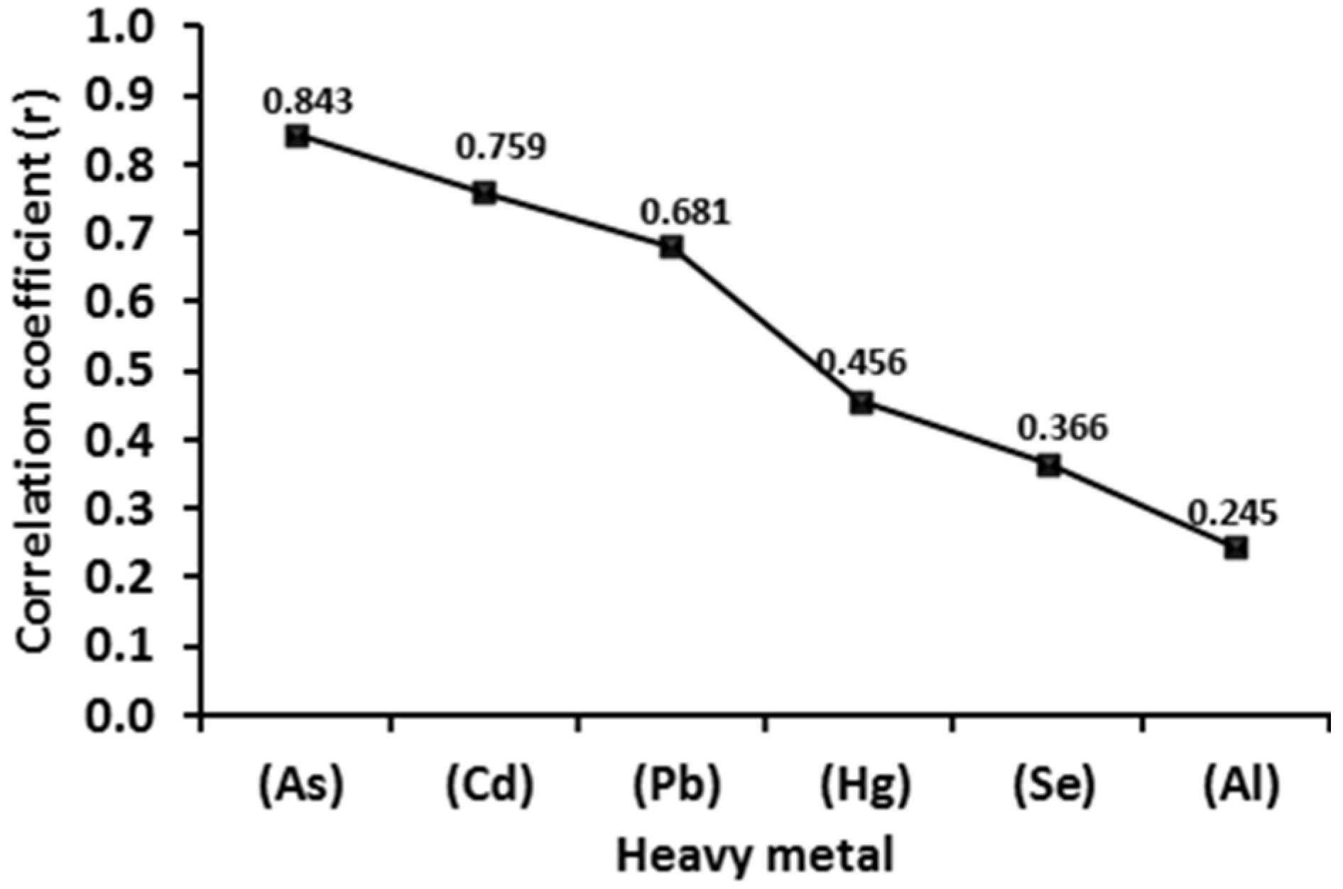
3.3. Heavy Metal Concentrations in the Midgut of C. chlorostictum

Soil acts as the major supplement of nutrients for many different organisms, including insects. Heavy metals are accumulated at the upper trophic level of the insect midgut as they are permanent and cannot be degraded by insect metabolism, according to Hopkein (1989) [15] and Agnieszka et al. (2016) [16]. The data in Table 3 indicate that there are significant differences between heavy metal concentrations in the midgut of the polluted agricultural C. chlorostictum compared with the reference one, in the descending order As, Al, Pb, Cd, Hg, and Se.
Our data (Table 3) proved that arsenic had a larger concentration in the polluted agricultural insect midgut (11.858 µg/g−1) than the reference one (6.903 µg/g−1), and this metal showed a moderate significance between the midgut of two study sites. Moreover, the heavy metal As showed a higher concentration than the other heavy metals in both sites. Similar results were obtained by [47] who indicated that the swelling of cells that lie next to the midgut lumen may be due to As stimulation and decadency in the deeper layers of the midgut.
The concentration of Al was higher in the polluted insect midgut (2.768 µg/g−1) than the reference midgut (0.933 µg/g−1), and aluminum showed a high significance between the midgut of the two study sites. Furthermore, the heavy metal Al did not affect the insect midgut which may be attributed to the fact that it is directly excreted; this result agreed with [48].
Data summarized in Table 3 proved that lead, cadmium, and mercury showed higher concentrations in the polluted agricultural insect midgut (0.849, 0.765, and 0.630 µg/g−1, respectively) than in the reference one (0.525, 0.278, and 0.209 µg/g−1, respectively). Moreover, Hg and Pb showed a moderately significant difference between the two study sites, while Cd showed a high significance; this is because the gut is the major heavy-metal-accumulating organ. These results are consistent with [49]. On the contrary, [50] observed low concentrations of lead in the ground beetle (Coleoptera: Staphylinidae). Furthermore, [51] suggested that the levels of accumulated heavy metals are high in animal organs with high metabolic activity such as the digestive tract. Moreover, Diener et al. (2015) [48] found that Cd accumulated in the larvae of the black soldier fly Hermetia illucens (Diptera: Stratiomyidae), whereas lead was excreted.
The concentration of selenium was slightly higher in the polluted agricultural insect midgut (0.293 µg/g−1) than in the reference one (0.201 µg/g−1); moreover, Se showed less significance between the midgut of the two study sites, as shown in Table 3. These are parallel with He and Yang’s (2005) [52] results which showed that some trace elements, such as Se, are not essential to plant growth but are desired by insects.
Data in Figure 1A,B show significant differences in all heavy metal concentrations of the soil compared to the insect’s midgut where the highest concentration was recorded for arsenic and the lowest for selenium, while the highest differences were observed for aluminum and the lowest differences for cadmium. In addition, the results of the correlation analyses between heavy metal concentrations of the soil and midgut of insects from both current sites, displayed in Figure 2, showed that the highest correlation was observed in As, while the lowest correlation was in Al. These results are consistent with Heikens et al. (2001) [53] who found that concentrations of internal heavy metals were high in Isopoda and low in Coleoptera because the beetles were weak accumulators of heavy metals in comparison to other arthropod groups. On the other hand, [54,55] reported that there were higher concentrations of cadmium and lead in ground beetles than in soil.

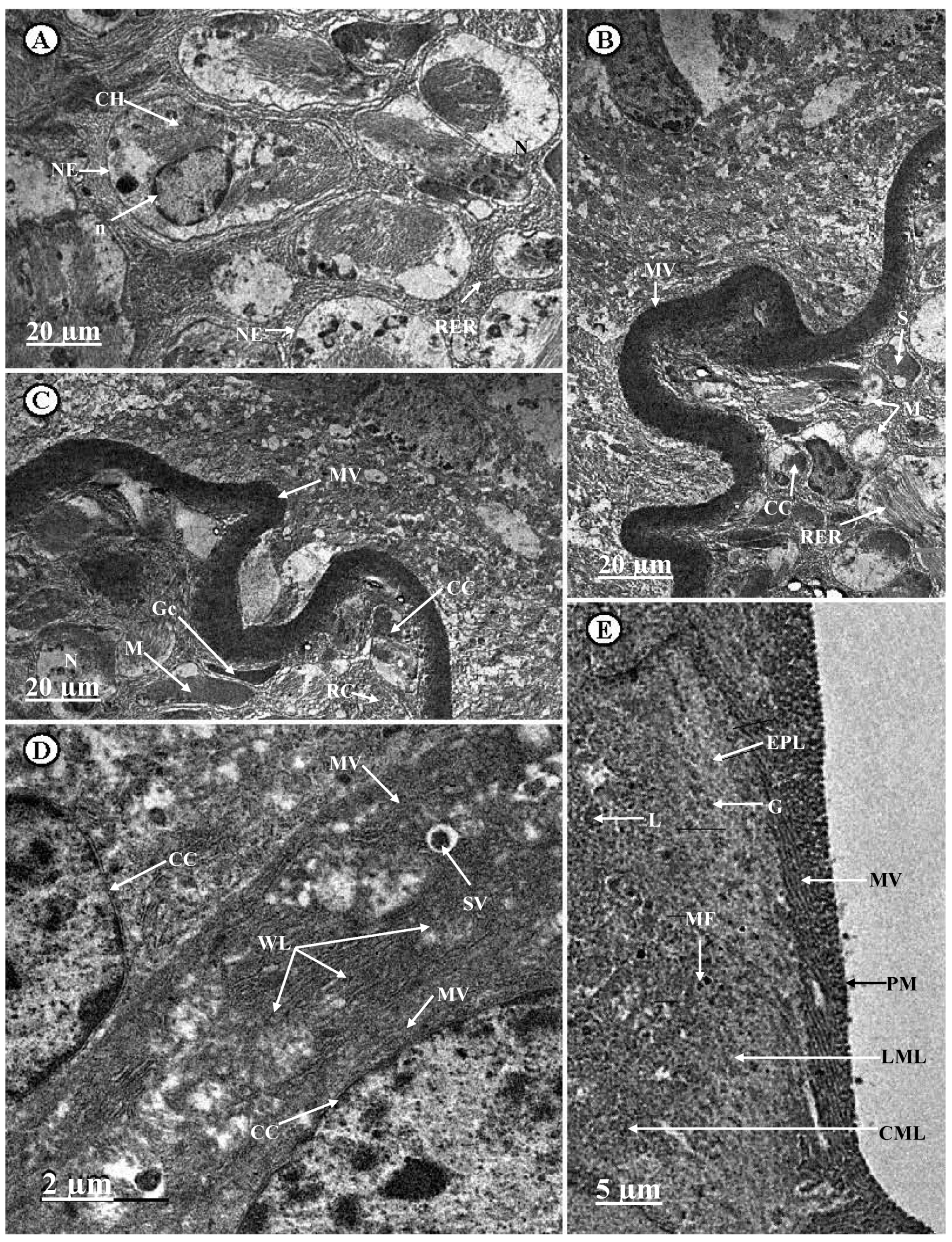
3.4. Ultrastructural Changes in the Midgut of C. chlorostictum Samples

The gut is considered an essential organ in the ecotoxicological studies of insects as the epithelial midgut is the earliest body part exposed to high levels of metals ingested by an insect [53]. The midgut epithelium, excluding its ends, is the only organ in an insect body that originates from endodermis [56]. The results in Figure 3 showed that the midgut of reference insects possessed no cuticular intima, which may be due to a healthy peritrophic membrane (PM) that surrounds the lumen, between the epithelial midgut and the lumen, and helps in the protection of the epithelium from accumulated food particles on it (Figure 3E). PM is encircled by normal circular (CML) and longitudinal (LML) muscle layers, respectively (Figure 3E). The epithelium surface is provided with border microvilli (MV) protruding into the midgut lumen (Figure 3B–E).
The midgut epithelium consists of a unicellular layer of digestive columnar epithelial cells (CCs) which help in the control of enzyme creation, assimilation, and secretion, while it is empty of Malpighian tubule excretion. Our results showed that the apical portion of CCs has a numerous secretory vesicles (Sv) or “spherites” (S) with plenty of digestive enzyme secretions released to the wider midgut lumen (WL), as depicted in Figure 3D. These results are in accordance with [57] who found that columnar digestive cells are responsible for digestion, secretion, absorption, storage of toxic substances, and excretion when Malpighian tubules are lacking.
The cytoplasm has distinct regionalization, filled with normal organelles such as a basal oval nucleus (N) with a regular nuclear envelope (NE) (Figure 3A,C). Moreover, a few lysosomes (L), glycogen granules (G), and a small myelin figure (MF) were observed, as shown in Figure 3E. In addition, rounded and elongated mitochondria (M) were observed, as depicted in Figure 3B,C, which were located in the apical and basal of the columnar digestive cells to support active transport of enzymes and digestive secretions, as indicated by [58]. The Golgi complex (Gc) (Figure 3C) and cisterns of the rough endoplasmic reticulum (RER), which lead to the synthesis of a large quantity of proteins in the columnar digestive cells, were also observed (Figure 3A,B); these observations agreed with the results obtained by [59].
The epithelial midgut was composed of regenerative cells (RC)—which are considered as stem cells, help in the renewal of cells, and may be located singly among epithelial cells or form groups called regenerative nests or crypts—which possessed a large nucleus (Figure 3B,C). Moreover, the normal regenerative cells were observed with normal nuclei which have a central nucleolus (n) and chromatin (CH) (Figure 3A); these results concurred with [60,61].
Something is known about ultrastructural alterations encouraged by heavy metal pollution in the midgut tissues of the insects, according to Yingmei et al. (2001) [62] and Polidori et al. (2018) [63]. The present study found that the midgut of the polluted agricultural C. chlorostictum showed several ultrastructural modifications, such as phagocytosis of apoptotic and necrosis of the epithelial cells, which appeared as destruction, complete lyses, and death of columnar (LCC) and regenerative epithelial cells (LRC) as an example of degradation, and are illustrated in Figure 4A and Figure 5B,C,E. These results agreed with [64] who observed that the apoptosis procedure in the insect midgut caused enhancement of the degenerative processes attributed to natural and anthropogenic agents like poisoning with heavy metals, then these cells were removed by the apoptosis and necrosis processes. These results are also consistent with [62] on German cockroach (Blatella germanica) and [17] on Egyptian cotton leafworm (Spodoptera littoralis). Furthermore, the stretching of the cellular axis of cells occurred due to pollution with the Hg and Se heavy metals, while the swelling and disintegration (apoptotic) of epithelial cells (columnar and regenerative) occurred due to pollution with the As, Cd, and Pb heavy metals which are considered as more toxic and destructive metals. This is parallel with Kabata and Mukherjee (2007) [65] who proved that Cd has no known biological function. It is also highly toxic at low levels and destroys the epithelial midgut after small exposure. Similar results were obtained by Bednarska et al. (2015) [66] who observed that Cd caused degenerative changes in the height of the midgut epithelium of crickets.
The present study showed an appearance of large separations (LS) between columnar epithelial cells and a rupture and curling in the microvilli border (rMV), as shown in Figure 5A that appeared to be a good sign of degradation due to pollution with heavy metals; these results agreed with [67]. Moreover, the current study revealed that the peritrophic membrane disappeared (Figure 5A); similar results were obtained by [68]. On the contrary, [67] noticed that there is a rupture in the peritrophic membrane. Consequently, the present study observed a dilated rough endoplasmic reticulum (dRER) which is depicted in Figure 4A and Figure 5B–D,F. The presence of large myelin figures (MF) can be seen in Figure 5A, and lyses of mitochondrial matrices such as dilated, vacuolated (VM), and destructed (dM) mitochondria are seen in Figure 4A and Figure 5F. These led to vacuolated areas in the cytoplasm which retracted to the effect of the heavy metals on the cytoplasmic membrane, leading to a high degradation of the cellular organelles. These results are in accordance with [69,70,71] who suggested that apoptosis is considered as a physiological process that enables the balance between the propagation rate and the removal of useless cells, whereas necrosis is a passive cell death resulting from acute cellular dysfunction after exposure to toxic heavy metals. The current study revealed that the damaged organelles were utilized and removed by the formation of considerable numbers of lysosomes (L) that are considered as an index of lyses due to pollution with heavy metals, as depicted in Figure 5D–F; these results agreed with [72] on the mosquito (Culex pipiens) midgut epithelium and [60] on (Archaeognatha, Lepismachilis notata, and Machilis hrabei) Coccinellidae. A lot of empty secretory vesicles (vacuoles) (ESV) without or with a few digestive enzyme secretions and spherites are presented in Figure 5A,D,E; there was a reduction of basophilic secretions towards the lumen that became very narrow (NL) due to enlargement and stretching of cells attributed to detoxification with heavy metals (Figure 5E). Furthermore, the accumulated heavy metals in the midgut of the polluted agricultural C. chlorostictum, due to degeneration when reaching their capacity for metal storage, intestinal cells break down, and the accumulation of heavy metals is shifted into the narrow midgut lumen, then eliminated by excretion from the insect. This is described by [15] who suggested that insects are able to protect themselves against the harmful effects of heavy metals. These results are consistent with [16] who proved that cell apoptosis of the midgut cells increased in the P. oblongopunctatus ground beetles when treated with a high level of heavy metal, followed by evacuation towards the gut lumen and excretion of metal assimilation, then removal from the body.
Autophagosomes (ACC) appeared as semitransparent cytoplasm and nuclei which were placed at different levels of the cellular body and varied in size and shape, as illustrated in Figure 4A and Figure 5D; this is considered as an index of lyses related to pollution with heavy metals and a type of cell death that enables degradation of organelles and elimination of heavy metals from the polluted agricultural C. chlorostictum midgut which are accumulated in the spherites. Other research has demonstrated that heavy metals assimilated at excess in insects are stored by metal-binding proteins and in spherites to prevent them from interfering with biochemical reactions in the tissues [63,73].
The current study found an occurrence of many electron-dense vesicles (EDV) in the C. chlorostictum midgut tissues, as indicated by Figure 5E; consequently, the heavy metals precipitated on the midgut epithelium. These results are parallel with [63]. The current study observed an occurrence of a pyknotic nucleus (PN) with an irregular shape, which was characterized by the presence of abnormal clumping chromatin (aCH) and the absence of nucleolus, as shown in Figure 5B; this resulted from pollution with the Cd and Pb heavy metals. In addition, the karyolysis of crypts of the regenerative cells (KRC) appeared as a narrow cytoplasm and small highly basophilic nuclei, as seen in Figure 4B; these results concurred with [74] who found that the midgut of larval Heliothis virescens suffered from apoptosis due to contamination with Cd and Pb, as well as cellular shrinkage, chromatin condensation, and blebbing of the cytoplasm.

4. Conclusions

From the present results, it can be concluded that the conditions of polluted agriculture soil are not secure for insects, animals, and human since the physicochemical parameters indicated that soil moisture and organic matter had the highest differences in the polluted agricultural soil compared to the reference soil. Meanwhile, there were no significant differences in chloride content. Moreover, AAS revealed the highest concentration in As and the lowest in Se in the polluted soil and the insect’s midgut. However, the differences between heavy metal concentrations in the total soil and insects midgut proved that the highest differences were in Al and Hg, while As and Cd were the lowest. Moreover, the correlation between heavy metal concentrations in the soil and the insect midgut showed that the highest correlation recorded was for As, while the lowest observed was for Al. The ultrastructure of midgut epithelial layer of ground beetles C. chlorostictum possessed great abnormalities and malformations due to accumulation of heavy metals, a hypothesis that could be supported by analyzing the soil physicochemical and heavy metal pollution levels in the soil and insect midgut. Our study confirms that midgut damages may be related to a probable decrease in the activity of protein synthesis due to damage in the rough endoplasmic reticulum and mitochondrial damages that cause decreasing in energetic efficiency and transport. Agricultural soil did not comply with the standard levels recommended by FAO, UNEP, and ISRIC, which greatly affected the ground beetles. Insects are efficient biomonitors in detecting soil contamination. An effort is necessary to diminish the perils resulting from the agricultural soil pollution, and the possibility of counteractive health effects could be avoided if the polluted soil is sufficiently treated.

Author Contributions

The research article included several contributions; conceptualizations were prepared by E.E.N., Z.Z.K., and M.A.-H. E.E.N. and Z.Z.K. provided the field survey. The data sample analysis and the investigations were prepared and analyzed by E.E.N., Z.Z.K., and M.A.-H. The original text draft of the article was written by E.E.N. and Z.Z.K. Writing—review and editing were finalized by M.Z., M.A.-H. and Z.V. The supervision and funding acquisition were supported by Z.Z.K., M.Z. and Z.V. The final version of the article was revised by M.A.-H., M.Z., and E.E.N. All authors have read and agreed to the published version of the manuscript.

Funding

This research received no external funding.

Acknowledgments

The authors gratefully thank Mahomoud Hassan, Mansoura University, Egypt, for his help in imaging the samples by electron microscopy. This work is thankful to be supported by the project of ERANETMED “Decentralized treatment wetlands for sustainable water management in rural and remote areas of semi-arid regions”. This work is thankful to be supported by project of the Ministry of Education of the Slovak Republic VEGA 1/0217/19 Research of Hybrid Blue and Green Infrastructure as Active Elements of a ‘Sponge City and the project od Slovak Research and Development Agency APVV-18-0360 Active hybrid infrastructure towards to sponge city.

Conflicts of Interest

The authors declare no conflict of interest.

References

  1. Sanchez-Bayo, F.; Wyckhuys, K.A.G. Worldwide decline of the entomofauna: A review of its drivers. Biol. Conserv. 2019, 232, 8–27. [Google Scholar] [CrossRef]
  2. Collison, E.; Hird, H.; Cresswell, J.; Tyler, C. Interactive effects of pesticide exposure and pathogen infection on bee health—A critical analysis. Biol. Rev. 2016, 91, 1006–1019. [Google Scholar] [CrossRef] [PubMed]
  3. Sanchez-Bayo, F.; Goulson, D.; Pennacchio, F.; Nazzi, F.; Goka, K.; Desneux, N. Are bee diseases linked to pesticides?—A brief review. Environ. Int. 2016, 89, 7–11. [Google Scholar] [CrossRef] [PubMed]
  4. Niklinska, M.; Laskowski, R.; Maryanski, M. Effect of heavy metals and storage time on two types of forest litter: Basal respiration rate and exchangeable metals. Ecotox Environ. Saf. 1998, 41, 8–18. [Google Scholar] [CrossRef] [PubMed]
  5. Siddig, A.A.; Ellison, A.M.; Ochs, A.N.; Villar, L.C.; Lau, M.K. How do ecologists select and use indicator species to monitor ecological change? Insights from 14 years of publication in Ecological Indicators. Ecol. Indic. 2016, 60, 223–230. [Google Scholar] [CrossRef] [Green Version]
  6. Hamelink, J.L.; Landrum, P.F.; Bergman, L.H.; Benson, W.H. Bioavailability: Physical, Chemical and Biological Interactions; CRC Press: Boca Raton, FL, USA, 1994. [Google Scholar]
  7. Hodkinson, I.D.; Jackson, J.K. Terrestrial and aquatic invertebrates as bioindicators for environmental monitoring, with particular reference to mountain ecosystems. Environ. Manag. 2005, 35, 649–666. [Google Scholar] [CrossRef] [PubMed]
  8. Skaldina, O.; Sorvari, J. Ecotoxicological effects of heavy metal pollution on economically important terrestrial insects. In Networking of Mutagenes in Terrestrial Ecotoxicology; Kesari, K., Ed.; Springer International Publishing: Cham, Switzerland, 2019; pp. 137–144. [Google Scholar]
  9. Kremen, C.N.; Colwell, R.K.; Erwin, T.L.; Murphy, D.D.; Noss, R.F.; Sanjayan, M.A. Terrestrial arthropod assemblages: Their use in conservation planning. Conserv. Biol. 1993, 7, 796–808. [Google Scholar] [CrossRef] [Green Version]
  10. Chen, T.B.; Zheng, Y.M.; Lei, M.; Huang, Z.C.; Wu, H.T.; Chen, H. Assessment of heavy metal pollution in surface soils of urban parks in Beijing, China. Chemosphere 2005, 60, 542–551. [Google Scholar] [CrossRef]
  11. Stork, N.E.; McBroom, A.J.; Claire, G.I.; Andrew, J.H. New approaches narrow global species estimates for beetles, insects, and terrestrial arthropods. Proc. Natl. Acad. Sci. USA 2015, 112, 7519–7523. [Google Scholar] [CrossRef] [Green Version]
  12. Zodl, B.; Wittmann, J. Effects of sampling, preparation and defecation on metal concentrations in selected invertebrates at urban sites. Chemosphere 2003, 52, 1095–1103. [Google Scholar] [CrossRef]
  13. Desender, K.; Maelfait, J.; Baert, L. Carabid beetles as ecological indicators in dune management. Coleotera Carabidae Elytron Suppl. 1991, 5, 239–247. [Google Scholar]
  14. Lorenz, W. Systematic List of Extant ground Beetles of the World (Insecta Coleoptera “Geadephaga”: Trachypachidae and Carabidae Incl. Paussinae, Cicindelinae, Rhysodinae), 2nd ed.; Wolfgang Lorenz: Tutzing, Germany, 2005; p. 530. [Google Scholar]
  15. Hopkin, S.P. Ecophysiology of Metals in Invertebrates; Elsevier, Applied Science: New York, NY, USA, 1989. [Google Scholar]
  16. Agnieszka, J.; Bednarska, A.; Laskowski, R.; Elżbieta, P.; Danuta, S.; Zuzanna, S.; Olga, W. Metal toxicokinetics and metal driven damage to the gut of the ground beetle. Pterostichus Oblongopunctatus Environ. Sci. Pollut. Res. 2016, 23, 22047–22058. [Google Scholar]
  17. Rawi, S.M.; Bakry, F.A.; Al Hazmi, M.A. Biochemical and histopathological effect of crude extracts on Spodoptera littoralis larvae. J. Evol. Biol. Res. 2011, 3, 67–78. [Google Scholar]
  18. Rainbow, R.B. Trace metal bioaccumulation: Models, metabolic availability and toxicity. Environ. Entomol. 2007, 33, 576–582. [Google Scholar] [CrossRef] [PubMed]
  19. Rowell, D.L. Soil sciences: methods and applications; John Wiley and Sons Inc.: New York, NY, USA, 1994. [Google Scholar]
  20. Gupta, P.K. Methods in environmental analysis water, soil and air. Agrobios 2000, 5, 1–400. [Google Scholar]
  21. Allen, S.; Grimshaw, L.; Parkinson, A.; Quarmby, C. Chemical Analysis of Ecological Materials; Blackwell Scientific Publications: Oxford, UK, 1974. [Google Scholar]
  22. Akubugwo, I.E.; Ofoegbu, C.J.; Ukwuoma, C.U. Physicochemical studies on Uburu Salt Lake Ebonyi State-Nigeria. Pak. J. Biol. Sci. 2007, 10, 3170–3174. [Google Scholar]
  23. Abd El-Shafee, M.E. Assessment of Some Trace Elements in Bladder Cancer Patients Associated with Bilharzia. M. Sc. Thesis, Faculty of Science, Zagazig University, Zagazig, Egypt, 2003. [Google Scholar]
  24. Karnovsky, M.J. A formaldehyde-glutaraldehyde fixative of high osmolality for use in electron microscopy. J. Cell Biol. 1965, 27, 137–138. [Google Scholar]
  25. Reynolds, E.S. The use of lead citrate at high pH as an electron-opaque stain in electron microscopy. J. Cell Biol. 1963, 17, 208–212. [Google Scholar] [CrossRef] [Green Version]
  26. Sokal, R.; Rohlf, F. Biometry: The Principles and Practice of Statistics in Biological Research, 2nd ed.; WH Freeman and Company: San Francisco, CA, USA, 1981. [Google Scholar]
  27. Norusis, M.J. SPSS 13.0 Statistical Procedures Companion; Prentice-Hall: Upper Saddle River, NJ, USA, 2005. [Google Scholar]
  28. Adviento-Borbe, M.A.; Doran, J.W.; Drijber, R.A.; Dobermann, A. Soil electrical conductivity and water content affect nitrous oxide and carbon dioxide emissions in intensively managed soils. J. Environ. Qual. 2006, 35, 1999–2010. [Google Scholar] [CrossRef] [Green Version]
  29. Allowary, B.J. Heavy Metals in Soils. Blackie Academic and Professional; Chapman and Hall: London, UK, 1995; p. 368. [Google Scholar]
  30. Soni, M. Analysis of soil samples for its physicochemical parameters from Abohar city. Pharma Innov. J. 2016, 5, 37–39. [Google Scholar]
  31. Arias, M.E.; Gonzalez-Perez, J.A.; Gonzalez-Villa, F.J.; Ball, A.S. Soil health: A new challenge for microbiologists and chemists. Int. Microbiol. 2005, 8, 13–21. [Google Scholar] [PubMed]
  32. Kekane, S.S.; Chavan, R.P.; Shinde, D.N.; Patil, C.L.; Sagar, S.S. A review on physicochemical properties of soil. Intern. J. Chem. Stud. 2015, 3, 29–32. [Google Scholar]
  33. Osuji, L.C.; Nwoye, I.M. An appraisal of the impact of petroleum hydrocarbons on soil fertility: The Owaza experience. Afr. J. Agric. Res. 2007, 2, 318–324. [Google Scholar]
  34. Edori, O.S.; Iyama, W.A. Assessment of Physicochemical Parameters of Soils from Selected Abattoirs in Port Harcourt Rivers State Nigeria. J. Environ. Anal. Chem. 2017, 4, 1–5. [Google Scholar]
  35. Schulte, E.E. Soil and Applied Chlorine; College of Agriculture and Life Sciences, University of Wisconsin-Madison; University of Wisconsin Extension, Cooperative Extension: Madison, WI, USA, 1999. [Google Scholar]
  36. Chukwu, U.J.; Anuchi, S.O. Impact of Abattoir Wastes on the Physicochemical Properties of Soils within Port Harcourt Metropolis. Int. J. Eng. Sci. 2016, 5, 17–21. [Google Scholar]
  37. Ayulungit, N.; Balwant, S.; Edith, L. Evaluation of Cadmium Toxicity to Collembola Proisotoma minuta Using Electron Microscopy. In Proceedings of the Australian New Zealand Soils Conference, Sydney, Australia, 5–9 December 2004. [Google Scholar]
  38. Abu-hashim, M.; Elsayed, M.; Belal, A.E. Effect of land-use changes and site variables on surface soil organic carbon pool at Mediterranean Region. J. Afr. Earth Sci. 2016, 114, 78–84. [Google Scholar] [CrossRef]
  39. Steevens, J.A.; Benson, W.A. Toxicokinetic interactions and survival of Hyalella azteca exposed to binary mixtures of chlorpyrifos, dieldrin and methyl mercury. Aquat. Toxicol. 2001, 51, 377–388. [Google Scholar] [CrossRef]
  40. Adriano, D.C. Trace elements in the Terrestrial Environments, 2nd ed.; Springer: New York, NY, USA, 2001. [Google Scholar]
  41. Food Agriculture Organization (FAO); International Soil reference Information Centre (ISRIC). Guiding Principles for the Quantitative Assessment of Soil Degradation with a Focus on Salinization, Nutrient Decline and Soil Pollution; FAO: Rome, Italy; ISRIC: Wageningen, The Netherlands, 2004. [Google Scholar]
  42. Wenzel, W.W. Arsenic. In Heavy Metals in Soils. Trace Metals and Metalloids in Soils and Their Bioavailability; Alloway, B.J., Ed.; Springer: Dordrecht, The Netherlands, 2013; pp. 241–282. [Google Scholar]
  43. Kabata, P.A. Trace Elements in Soils and Plants, 4th ed.; CRC Press: Boca Raton, FL, USA, 2011. [Google Scholar]
  44. United Nations Environment Programme. Final Review of Scientific Information on Cadmium—Version of December 2010; United Nations Geneva: Genève, Switzerland, 2010. [Google Scholar]
  45. Mandal, B.K.; Suzuki, K.T. Arsenic round the world: A review. Talanta 2002, 58, 201–235. [Google Scholar] [CrossRef]
  46. Allowary, B.J. Heavy Metals in Soils, 3rd ed.; Environmental Pollution Series 22; Springer: Dordrecht, The Netherlands, 2013; p. 615. [Google Scholar]
  47. Moriarty, M.M.; Koch, I.N.; Gordon, R.A.; Reimer, K.J. Arsenic speciation of terrestrial invertebrates. Environ. Sci. Technol. 2009, 43, 4818–4823. [Google Scholar] [CrossRef]
  48. Diener, S.; Zurbrugg, C.; Tockner, K. Bioaccumulation of heavy metals in the black soldier fly, Hermetia illucens and effects on its life cycle. J. Ins. Food Feed 2015, 1, 261–270. [Google Scholar] [CrossRef] [Green Version]
  49. Ge, S.; Haihua, W.; Ying, X.; Meiling, Y.; Enbo, M.; Yaping, G. Accumulation and distribution of cadmium in Oxya chinensis after feeding on wheat seedlings contaminated with cadmium. J. Agro. Environ. Sci. 2009, 28, 1812–1817. [Google Scholar]
  50. Bohac, J. Accumulation of heavy metals in the bodies of staphylinid beetles Coleoptera, Staphylinidae. In Proceedings of the 5th Internat Confer On Bioindicators in Deteriorisating Regions, Ceske Budejovice, Czech Republic, 23–27 May 1988; pp. 319–321. [Google Scholar]
  51. Menta, C.; Parisi, V. Metal concentrations in Helix pomatia, Helix aspersa and Arion rufus: A comparative study. Environ. Poll 2001, 115, 205–208. [Google Scholar] [CrossRef]
  52. He, Z.L.; Yang, X.E.; Stoffell, P.J. Trace elements in agroecosystems and impacts on the environment. J. Trace Elem. Med. Biol. 2005, 19, 125–140. [Google Scholar] [CrossRef] [PubMed]
  53. Heikens, A.; Peijnenburg, W.; Hendriks, J. Bioaccumulation of heavy metals in terrestrial invertebrates. Environ. Pollu. 2001, 113, 385–393. [Google Scholar] [CrossRef]
  54. Van Straalen, N.M.; Butovsky, R.O.; Pokarzhevskii, A.D.; Zaitsev, A.S.; Verhoef, C.S. Metal concentrations in soil and in invertebrates in the vicinity of a metallurgical factory near Tula (Russia). Pedobiol. Inter. J. Soil Biol. 2001, 45, 451–466. [Google Scholar] [CrossRef]
  55. Jelaska, S.; Blanusa, M.; Durbesic, P.; Jelaska, D. Heavy metal concentrations in ground beetles, leaf litter, and soil of a forest ecosystem. Ecotoxicol. Environ. Saf. 2007, 66, 74–81. [Google Scholar] [CrossRef]
  56. Chapman, R.F. The Insects: Structure and Function, 4th ed.; Cambridge, UK University Press: Cambridge, UK, 1998; pp. 38–66. [Google Scholar]
  57. Micchelli, C.A.; Perrimon, N.A. Evidence that stem cells reside in the adult Drosophila midgut epithelium. Nature 2006, 439, 475–479. [Google Scholar] [CrossRef]
  58. Billingsley, P.F.; Lehane, M.J. Structure and Ultrastructure of the Insect Midgut. Biology of the Insect Midgut; Springer: Dordrecht, The Netherlands, 1996; Volume 9, pp. 3–30. [Google Scholar]
  59. Geneser, F. Histologia: Sobre Bases Biomoleculare, 3rd ed.; Medica Panamericana: Mexico City, Mexico, 2000; p. 813. [Google Scholar]
  60. Rost Roszkowska, M.M.; Jansta, P.; Vilimova, J. Fine structure of the midgut epithelium in two Archaeognatha, Lepismachilis notata and Machilis hrabei (Insecta), in relation to its degeneration and regeneration. Protoplasma 2010, 247, 91–101. [Google Scholar] [CrossRef]
  61. Olivares, A.S.; Diaz, E.; Shibayama, M.; Tsutsumi, V.; Cisneros, R.; Zuniga, G. Ultrastructural study of the midgut and hindgut in eight species of the genus. Dendroctonus Erichson Coleopt. Scolytidae Ann. Entomol. Soc. Am. 2014, 96, 883–900. [Google Scholar] [CrossRef] [Green Version]
  62. Yingmei, Z.; Simonetta, L.; Mauro, F.; Carlo, G.; Aldo, G.; Ugo, L. Mortality and tissue damage by heavy metal contamination in the German cockroach. Bl. Ger. Bl. Bl. Ital J. Zool 2001, 68, 137–145. [Google Scholar]
  63. Polidori, C.; Pastorm, A.; Jorge, A.; Pertusa, J. Ultrastructural alterations of midgut epithelium, but not greater wing fluctuating asymmetry, in paper wasps Polistes dominula from urban environments. Microsc. Microanal 2018, 24, 183–192. [Google Scholar] [CrossRef] [PubMed] [Green Version]
  64. Rodrigues, A.; Cunha, L.; Amaral, A.; Medeiros, J.; Garcia, P. Bioavailability of heavy metals and their effects on the midgut cells of a phytophagous insect inhabiting volcanic environments. Sci. Total Environ. 2008, 406, 116–122. [Google Scholar] [CrossRef] [PubMed] [Green Version]
  65. Kabata, P.A.; Mukherjee, A.B. Trace Elements from Soil to Human; Springer: Berlin, Germany, 2007. [Google Scholar]
  66. Bednarska, A.J.; Opyd, M.; Żurawicz, E.; Laskowski, R. Regulation of body metal concentrations: Toxicokinetics of cadmium and zinc in crickets. Ecotoxicol. Environ. Saf. 2015, 119, 9–14. [Google Scholar] [CrossRef]
  67. Khan, I.; Qamar, A.; Mehdi, S.; Shahid, M. Histopathological effects of Datura alba leaf extract on the midgut of Periplaneta Americana. Biol. Med. 2011, 3, 260–264. [Google Scholar]
  68. Kohler, H.R. Localization of metals in cells of saprophagous soil arthropods Isopoda, Diplopoda, Collembola. Microsc. Res. Tech. 2002, 56, 393–401. [Google Scholar] [CrossRef] [PubMed]
  69. Zakeri, Z.; Lockshin, A. Cell death during development. J. Immunol. Methods 2002, 265, 3–20. [Google Scholar] [CrossRef]
  70. Schonthal, A.H. Endoplasmic reticulum stress: Its role in disease and novel prospects for therapy. Scientifica 2012. [Google Scholar] [CrossRef] [Green Version]
  71. Meyer, J.N.; Leung, M.C.; Rooney, J.P.; Sendoel, A.L.; Hengartner, M.O.; Kisby, G.E.; Bess, A.S. Mitochondria as a target of environmental toxicants. Toxicol. Sci. 2013, 134, 1–17. [Google Scholar] [CrossRef] [Green Version]
  72. Vaidyanathan, R.; Scott, T.W. Apoptosis in mosquito midgut epithelia associated with West Nile virus infection. Apoptosis 2006, 11, 1643–1651. [Google Scholar] [CrossRef]
  73. Lockshin, R.A.; Zakeri, Z. Apoptosis, autophagy and more. Int. J. Biochem. Cell Biol. 2004, 36, 2405–2419. [Google Scholar] [CrossRef] [PubMed]
  74. Loeb, M.J.; Hakim, R.S.; Martin, P.L.; Narang, N.R.; Goto, S.L.; Takeda, M.W. Apoptosis in cultured midgut cells from Heliothis virescens larvae exposed to various conditions. Arch. Insect. Biochem. Physiol. 2000, 45, 12–23. [Google Scholar] [CrossRef]
Figure 1. Comparison of differences between heavy metal concentrations in the soil and the midgut of C. chlorostictum from both current sites (A), and the significance differences between them (B). * Statistically significant at p ≤ 0.05.
Sustainability 12 04805 g001
Figure 2. Correlation coefficients between heavy metal concentrations in the soil and the midgut of C. chlorostictum from both current sites.
Sustainability 12 04805 g002
Figure 3. Photomicrographic details of the reference C. chlorostictum midgut, showing digestive columnar epithelial cells (CC), nucleus (N), nuclear envelope (NE), nucleolus (n), chromatin (CH), peritrophic membrane (PM), regenerative cells (RC), secretory vesicles (Sv), spherites (S), circular muscle layer (CML), longitudinal muscle layer (LML), epithelial layer (EPL), microvilli (MV), mitochondria (M), wider lumen (WL), rough endoplasmic reticulum (RER), Golgi complex (Gc), lysosomes (L), myelin figure (MF), and glycogen (G). Figure (A) shows CH, NE, n, and RER. Figure (B) shows MV, S, M, CC, and RER. Figure (C) shows MV, CC, Gc, M, N, and RC. Figure (D) shows MV, CC, SV, and W. Figure (E) shows EPL, G, L, MV, MF, PM, LML, and CML.
Sustainability 12 04805 g003
Figure 4. Photomicrographic details of the C. chlorostictum midgut (samples collected in polluted environments), showing dilated rough endoplasmic reticulum (dRER), lyses of columnar cells (LCC), large separation (LS), autophagosomes of columnar cells (ACC), vacuolated mitochondria (VM), and karyolysis of regenerative cells (KRC). Figure (A) shows LS, VM, LCC, dRER, and ACC. Figure (B) shows KRC.
Sustainability 12 04805 g004
Figure 5. Photomicrographic details of the C. chlorostictum midgut (samples collected in polluted environments), showing rupture microvilli (rMV), large separation (LS), epithelial layer (EPL), empty secretory vesicles (ESV), pyknotic nucleus (PN), abnormal chromatin (aCH), lyses of regenerative cells (LRC), dilated rough endoplasmic reticulum (dRER), lyses of columnar cells (LCC), lysosomes (L), myelin figures (MF), electron-dense vesicles (EDV), autophagocytic cell (AC), pigment (P), narrow lumen (NL), destructed mitochondria (dM), and vacuoles (V). Figure (A) shows rMV, LS, EPL, MF, ESV, V, LCC, and EDV. Figure (B) shows dRER, PN, aCH, LRC, and V. Figure (C) shows LCC and dRER. Figure (D) shows P, L, dRER, V, AC, and ESV. Figure (E) shows NL, MV, LRC, EDV, ESV, L, and LCC. Figure (F) shows dM, ADV, V, dRER, and L.
Sustainability 12 04805 g005
Table 1. Physicochemical parameters (mean ± SD) of the soil samples from the reference and polluted agricultural sites.
ParametersSoil SamplesIndependent T-test
Reference SitePolluted Agri. SiteT-Statistic p-Value
Conductivity (ds/m)6.271 ± 0.8329.019 ± 1.0077.288 *≤0.05
pH value8.355 ± 0.7707.515 ± 0.6832.827 *≤0.05
Moisture (%)1.956 ± 0.0782.512 ± 0.09615.571 *≤0.01
Chloride content (%)0.015 ± 0.0810.018 ± 0.0570.105>0.05
Total CaCO3 (%)2.891 ± 0.3793.254 ± 0.4022.276 *≤0.05
Organic matter (%)1.951 ± 0.2081.034 ± 0.20810.799 *≤0.01
Alkalinity (%)6.024 ± 0.7354.825 ± 0.3585.080 *≤0.05
* Statistically significant at p ≤ 0.05. T-Statistic: the value of the differences, using Independent t-test. p-value is used to determine the significance of differences. (n = 12).
Table 2. Heavy metal concentrations (mean ± SD) in the soil samples from the reference and polluted agricultural sites.
Heavy MetalsSoil Samples (µg/g−1)Independent T-Test
Reference SitePolluted Agri. SiteT-Statistic p-Value
Arsenic (As)9.075 ± 0.80813.235 ± 1.01411.115 *≤0.01
Mercury (Hg)0.808 ± 0.0441.178 ± 0.11310.570 *≤0.05
Aluminium (Al)2.780 ± 0.0976.918 ± 0.47529.568 *≤0.001
Cadmium (Cd)0.322 ± 0.0460.905 ± 0.08920.158 *≤0.01
Lead (Pb)0.918 ± 0.0711.218 ± 0.0958.762 *≤0.05
Selenium (Se)0.386 ± 0.0430.512 ± 0.0675.483 *≤0.05
* Statistically significant at p ≤ 0.05. T-Statistic: the value of the differences, using Independent t-test. p-value is used to determine the significance of differences. (n = 12).
Table 3. Heavy metal concentrations (mean ± SD) in the midgut of Calosoma chlorostictum samples from the reference and polluted agricultural sites.
Heavy MetalsBeetle Samples (µg/g−1)Independent T-Test
Reference SitePolluted Agri. SiteT-Statistic p-Value
Arsenic (As)6.903 ± 0.62811.858 ± 0.91221.460 *≤0.01
Mercury (Hg)0.209 ± 0.0360.630 ± 0.07723.753 *≤0.01
Aluminium (Al)0.993 ± 0.0772.768 ± 0.19939.895 *≤0.001
Cadmium (Cd)0.278 ± 0.0310.765 ± 0.06930.876 *≤0.001
Lead (Pb)0.525 ± 0.0450.849 ± 0.05721.396 *≤0.01
Selenium (Se)0.201 ± 0.0190.293 ± 0.03610.839 *≤0.05
* Statistically significant at p ≤ 0.05. T-Statistic: the value of the differences, using Independent t-test. p-value is used to determine the significance of differences. (n = 23).

