Article

In Vivo Cytotoxicity Induced by 60 Hz Electromagnetic Fields under a High-Voltage Substation Environment

by
J. Antonio Heredia-Rojas
1,
Abraham Octavio Rodríguez-De la Fuente
1,*,
Ricardo Gomez-Flores
1,
Omar Heredia-Rodríguez
1,
Laura E. Rodríguez-Flores
2,
Michaela Beltcheva
3 and
Ma. Esperanza Castañeda-Garza
1
1
Facultad de Ciencias Biológicas, Universidad Autónoma de Nuevo León, Av. Universidad S/N, Ciudad Universitaria, San Nicolás de los Garza 66455, Nuevo León, Mexico
2
Facultad de Medicina, Universidad Autónoma de Nuevo León, Av. Francisco I. Madero pte. S/N, Col. Mitras Centro, Monterrey 66460, Nuevo León, Mexico
3
Institute of Biodiversity and Ecosystem Research, Bulgarian Academy of Sciences, 1 Tzar Osvoboditel Blvd., 1000 Sofia, Bulgaria
*
Author to whom correspondence should be addressed.
Sustainability 2018, 10(8), 2789; https://doi.org/10.3390/su10082789
Submission received: 21 June 2018 / Revised: 26 July 2018 / Accepted: 30 July 2018 / Published: 7 August 2018
(This article belongs to the Special Issue Electromagnetic Waves Pollution)

Abstract

:
Living beings permanently receive electromagnetic radiation, particularly from extremely low-frequency electromagnetic fields (ELF-EMFs), which may cause adverse health effects. In this work, we studied the in vivo cytotoxic effects of exposing BALB/c mice to 60 Hz and 8.8 µT EMFs during 72 h and 240 h in a switchyard area, using animals exposed to 60 Hz and 2.0 mT EMFs or treated with 5 mg/kg mitomycin C (MMC) as positive controls. Micronucleus (MN) frequency and male germ cell analyses were used as cytological endpoints. ELF-EMF exposure was observed to significantly (p < 0.05) increase MN frequency at all conditions tested, with the 2 mT/72 h treatment causing the highest response, as compared with untreated control. In addition, increased sperm counts were observed after switchyard area ELF-EMF exposure, as compared with untreated control. In contrast, low sperm counts were obtained for 72 h/2.0 mT-exposed animals and for MMC-treated mice (p < 0.05), without altering male germ cell morphological characteristics.

1. Introduction

Electric, magnetic, and electromagnetic fields derive from nature. However, modern electrical devices and power lines have increased the levels of non-ionizing electromagnetic radiation, including extremely-low frequency electromagnetic fields (ELF-EMFs). In fact, it has been demonstrated that living in a major metropolitan region increases three times exposure to environmental EMFs at least three-fold compared with the exposure of organisms living in suburban or rural areas [1], although the degree depends on the proximity and time of exposure to a radiation source. Knowledge of ELF-EMFs’ interaction with life is progressing in many areas [2]. Today, the increasing amount of research related to the evaluation of magnetic fields’ cytotoxic and genotoxic effects led researchers to consider the potential risk associated with this exposure. ELF-EMFs are designated as “likely carcinogenic” by the International Agency for Research on Cancer (IARC) [3] and some have reported relationships between ELF-EMFs and DNA damage [4,5,6,7], but others have conflicting results [8,9,10,11].
Most reports on the cytotoxic and genotoxic effects of EMFs have been performed on somatic cells; however, the use of meiotic cells is increasingly accepted as a proper model to determine an association between ELF-EMFs and cytotoxicity [12,13,14]. In this regard, we previously used a male germ cell model to evaluate the in vivo effect of 60 Hz at 2.0 mT magnetic fields, in which genotoxicity in murine bone marrow (as measured by increased micronuclei (MN) frequency), but absence of meiotic chromosomes and sperm effects were reported [9,15]. However, the issue of the genotoxic potential of magnetic fields remains controversial. The lack of independent reproduction and definite outcomes has been a common characteristic of experimental studies searching for the biological effects of weak magnetic fields [16,17].
This study is relevant in view of the continuous conflicting results and disagreement among researchers regarding genotoxic and cytotoxic effects due to EMFs using diverse biological models. For this reason, an in vivo study was developed to evaluate the effects of 60 Hz and 8.8 µT magnetic field exposure for short (72 h) and extended (240 h) periods in a switchyard area in a murine model, determining micronuclei frequency and male germ cell alterations.

2. Materials and Methods

2.1. Animals

Three-month-old, 25–30 g male BALB/c mice raised in our laboratory were used in this study. After 15 days of quarantine, six mice were randomly placed into treatment and control groups. Water and food were given ad libitum. Animal handling and procedures were performed in accordance with the Guide for the Care and Use of Laboratory Animals published by the National Institutes of Health (Bethesda, MD, USA) and approved by the University Ethics and Animal Care Committee.

2.2. Experimental Design

The following treatment regimens were considered: (1) 72 h continuous 8.8 µT ELF-EMF exposure in the switchyard area, (2) 240 h continuous 8.8 µT ELF-EMF exposure in the switchyard area, (3) animals treated with 2.0 mT for 72 h continuous exposure in a standardized solenoid (positive control), (4) animals injected i.p. with 5 mg/kg mitomycin C (MMC) (positive control), and (5) untreated animals (negative control).

2.3. ELF-EMF Exposure

2.3.1. Switchyard Area

Plastic cages containing experimental animals were allocated in a high-voltage switchyard area (13,200 V substation), including three transformers (150 kVA, 225 kVA, and 500 kVA). Magnetic flux density (rms) was measured in the zone where the cages were placed by using an axial Hall-effect probe (Bell FW 6010 Gaussmeter, Orlando, FL, USA), with a value of 8.8 µT. The background magnetic field level was 0.3 µT and the local geomagnetic field had an average value of 20 µT, as determined by setting the Gaussmeter in DC mode and using an axial high-sensitivity Hall probe (Integrity Design IDR-321 geomagnetometer, Essex Junction, VT, USA).

2.3.2. Magnetic Field Exposure Facility (Standardized Solenoid)

A standardized home-made magnetic field exposure device was used, as previously reported [9,15,18]. Briefly, a coil was prepared by winding 552 turns of enamel-insulated copper wire (1.3 mm diameter), which produced a cylindrical solenoid with 13.5 cm radius and 71 cm length. It was connected to step-down and variable transformers and plugged to a 110 V AC source. Animals were then placed in the middle of this structure in a homogeneous magnetic field, and kept at 25 ± 0.2 °C and 45% humidity. Sham-treated animals were used as negative controls, which were placed in the same room, but with the magnetic field device turned off.
The magnetic flux density was determined using a Gaussmeter and an attached oscilloscope (BK Precision 20 MHz Oscilloscope, 2120 Model, Dynascan Corp., Chicago, IL, USA), which was required to monitor the resulting field. A 60 Hz alternating sinusoidal magnetic field was then generated. The frequency content was almost pure 60 Hz (<2% total harmonic distortion), and 0.3 µT and 20 µT values were observed for the respective background magnetic field and local geomagnetic field.
To maintain the exposure geometry, a plastic separator was inserted in the solenoid to allow the placement of mice in predetermined zones where the oscillating magnetic field rms value was 2.0 mT. Food and water were provided.

2.3.3. Micronucleus Test

At the end of the exposure time, the mice were sacrificed by cervical dislocation and the bone marrow micronucleated erythrocytes frequency was evaluated, as reported by Schmidt [19]. Briefly, femur bone marrow was flushed into a microfuge tube with 2.0 mL fetal calf serum (FCS, Sigma-Aldrich, St. Louis, MO, USA), using a 22 G needle and a 1 mL syringe. Cells were then obtained by centrifugation at 500 g for 10 min and the supernatant fluid was discarded. Next, the pellet was suspended in 100 μL FCS and spread on microscope coded slides. Air-dried smears were then stained with 5% May–Grünwald–Giemsa (Sigma-Aldrich) for 12–15 min and evaluated using a Leica DM2500 microscope (Leica Microsystems, Milton Keynes, UK) at a magnification of ×1000. To determine the MN frequency, 2000 consecutive polychromatic erythrocytes (PCEs) were scored for each animal, after which slides were decoded.

2.4. Male Germ Cell Analysis

An orchidectomy was performed by the open castration method [20] for sperm counting. Briefly, a midline or pre-scrotal incision was made and the testes were milked out at the incision site. The testicles were then exposed by incising the tunica vaginalis exposing the spermatic cord, which was ligated and incised. Next, semen samples were collected from the cauda epididymis and immediately analyzed. Spermatozoa were counted in a Neubauer hemocytometer (Deep 1/10 mm, LABART, Munich, Germany) [21].
Smears were prepared from the epididymis for sperm morphology analysis [22]. Spermatozoa were then stained using 1% Eosin Y for 1 h (Fisher Scientific Co., Fair Lawn, NJ, USA). Next, 100 cells per slide and 10 smears per animal, yielding a total of 5000 cells per group, were blindly evaluated at a magnification of ×1000 for morphological characteristics, including bicephalic or biflagellate forms, and shape abnormalities such as enlarged and amorphous heads. Results were expressed as a percentage of sperm morphology abnormalities.

2.5. Statistical Analysis
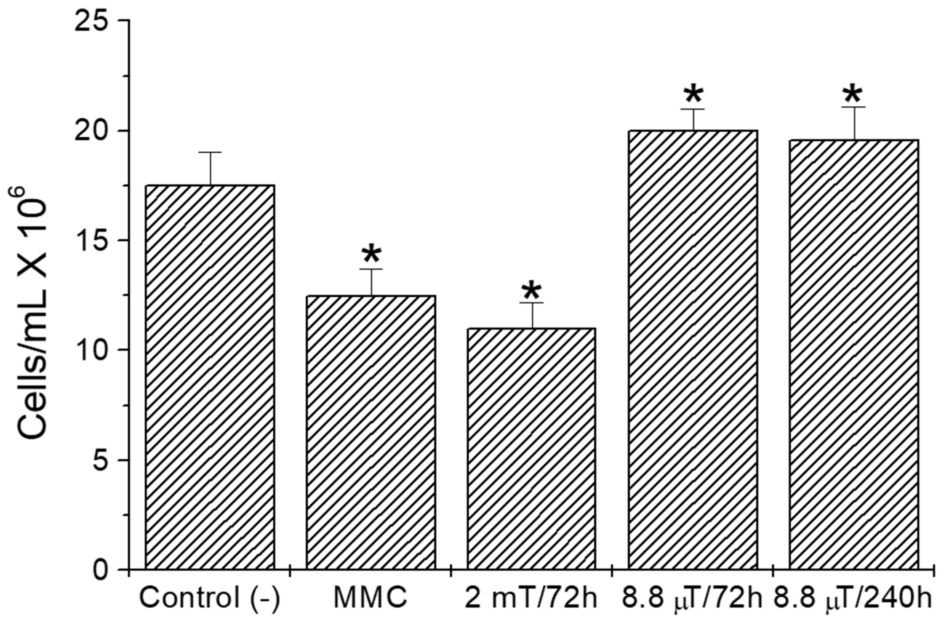
An analysis of variance was used to determine differences among groups. For morphology abnormality percentages, data were first transformed by the arcsin function, then an analysis of variance for normal distributions and a Tukey test were applied for establishing individual differences using the SPSS package version 22.0 (International Business Machines Corporation (IBM), Armonk, NY, USA); data normality was determined by the Kolmogorov–Smirnov test.

3. Results

The aim of this study was to evaluate the relationship between the exposure of mice to ELF-EMFs and bone marrow and male germ cell toxicity under switchyard area conditions. Figure 1 shows MN frequencies of mice exposed for 72 h and 240 h, and in a standardized solenoid. A significant (p < 0.05) increase in MN frequency was observed for all ELF-EMF exposure conditions (a 2 mT/72 h treatment induced the highest frequency of MN) and the MMC treatment, as compared with untreated animals (Figure 1).
In regard to the germ cell analysis, increased sperm counts were observed for 72 h/8.8 µT and 240 h ELF-EMF exposure in animals allocated in the switchyard area, compared with those of unexposed mice (Figure 2). In contrast, low sperm counts were obtained for 72 h/2.0 mT exposed animals and for MMC-treated mice (p < 0.05). Furthermore, no alterations in sperm morphology abnormality (SMA) percentages were found among groups (p > 0.05), except for the MMC-treated group, in which SMA alterations were observed (Figure 3).

4. Discussion

The electromagnetic spectrum of the non-ionizing band is becoming of great relevance in modern human life. An increasing use of overhead, high-voltage transmission lines and electrical substations has been noted in many countries. In this regard, speculations and serious research have raised concerns of possible health risks due to ELF-EMFs associated with power lines and substations. There is a general agreement that living organisms might be negatively affected by ELF-EMFs [1,2,11,14,15,23,24].
In this study, increased MN frequency in mice under 72 h and 240 h ELF-EMF exposure compared with those under negative controls was shown. These results concur with those of others [25,26], and with our recent study [15], suggesting a genotoxic effect related to magnetic fields. In this respect, Winker et al. [27] reported significant chromosomal damage in dividing human diploid fibroblasts, induced by low-frequency EMFs. Furthermore, Erdal et al. [28] showed that long-term (more than 24 h exposure time) ELF-EMFs caused an increased MN frequency in Wistar rat bone marrow cells. In contrast, there have been several reports showing no cell alterations after magnetic field exposure [29,30,31]. Furthermore, Chakraborty et al. [32] found no cytogenetic alterations in electricity substation attendants evaluated under occupational ELF-EMF exposures.
Conflicting reports on ELF-EMF cytotoxic effects derive in part from studies indicating that low-energy weak fields do not cause genotoxic effect or DNA damage, because the energy produced is not sufficient to affect chemical bonds [33]. However, others have shown the indirect effect of EMFs on DNA structure. In this respect, EMFs may cause secondary currents and electron movement in DNA [34], which in turn may generate guanine radicals that react with water to induce oxidative DNA damage [35]. Recently, Focke et al. [6] reported that human primary fibroblasts exposed to ELF-EMFs increased DNA fragmentation, which depended on cell proliferation, suggesting involvement in DNA replication. Another possibility is that EMFs might alter the rate or type of DNA repair mechanisms in the exposed cells. However, it has been proven that the DNA repair system is not affected [36].
Our results also showed that 72 h and 240 h magnetic field exposure increased sperm counts, suggesting an effect in cell cycle progression (Figure 2). An epidemiological survey reported that the offspring of high-voltage switchyard workers had a higher frequency of congenital malformations and fertility issues, as compared with other occupations [37]. In contrast, Lundsberg et al. [38] showed no association of occupational magnetic fields on sperm, and Aitken et al. [12] observed no alterations of sperm number, morphology, and vitality in mice treated with 900 MHz radio frequency electromagnetic radiation. However, Furuya et al. [39] observed that ELF-EMFs at 50 Hz, ranging from 1.0 mT up to 100 mT, altered murine spermatogonia proliferation and differentiation. Furthermore, Narra et al. [40] reported harmful effects on mice spermatogenesis under 1.5 T static magnetic fields. Similarly, Ramadan et al. [41] showed a significant decrease in murine sperm counts after a 20 mT treatment and after exposure of Sprague Dawley rats to 50 Hz and 25 µT for 18 continuous weeks [13], which is in agreement with our results depicted in Figure 2.
In this study, it was observed that 60 Hz and 2.0 mT magnetic field treatment did not alter the mouse male germ cell morphological characteristics, as previously reported [9]. In contrast, Roychoudhury et al. [42] showed that 50 Hz magnetic fields affected rabbit spermatozoa and fertilization rates; in humans, adverse effects on sperm quality have been demonstrated [14].
In addition, it has been demonstrated that static magnetic field exposure does not affect sperm morphology. For instance, Withers et al. [43] reported that 0.3 T static magnetic fields from a magnetic resonance device did not cause alterations in murine sperm heads, similar to the observations of Tablado et al. [44], who reported that a 0.7 T commercial permanent magnet did not alter sperm head size and the percentage of sperm with coiled tails or abnormal midpiece or tail.
In conclusion, this study demonstrated that continuous exposure to ELF-EMFs for 72 h or 240 h induced a clastogenic effect in murine bone marrow cells in a switchyard area. Moreover, it was found that magnetic fields may modify cell cycle progression by increasing sperm counts in exposed mice, although no alterations in sperm morphology were observed.
Although the mechanism of action for cytotoxicity and disease induced by ELF-EMFs has not yet been elucidated, reports have shown inherent electrical aspects related to biostructures and biological functions, including development, growth, and repair. Furthermore, living organisms appear to be sensitive to external electromagnetic fields of very weak intensity. The absence of mechanisms associating EMF exposure and biological events has resulted in a number of dubious investigations and conflicting results. More studies are needed to fully comprehend this phenomenon.

Author Contributions

J.A.H.-R. and A.O.R.-d.l.F.: conception and design of the work, statistical analysis and interpretation of work data. R.G.-F.: drafting the work and critically revising the manuscript. O.H.-R. and L.E.R.-F.: methodology (cytogenetic analysis), M.B. and M.E.C.-G.: revision of the manuscript and final approval of the version to be published.

Funding

This study was partially funded by the PAICYT grant CN 981-11 awarded to J. Antonio Heredia-Rojas.

Acknowledgments

We are grateful to César Elizondo-González from the Facultad de Ingeniería Mecánica y Eléctrica, Universidad Autónoma de Nuevo León, México for his assistance with magnetic field exposure facilities and measurements. We also thank Roumiana Metcheva from the Bulgarian Academy of Sciences for critically revising this manuscript.

Conflicts of Interest

The authors declare no conflict of interest.

References

  1. Gajšek, P.; Ravazzani, P.; Grellier, J.; Samaras, T.; Bakos, J.; Thuróczy, G. Review of Studies Concerning Electromagnetic Field (EMF) Exposure Assessment in Europe: Low Frequency Fields (50 Hz–100 kHz). Int. J. Environ. Res. Public Health 2016, 13, 875. [Google Scholar] [CrossRef] [PubMed]
  2. Cifra, M.; Fields, J.Z.; Farhadi, A. Electromagnetic cellular interactions. Prog. Biophys. Mol. Biol. 2011, 105, 223–246. [Google Scholar] [CrossRef] [PubMed]
  3. The International Agency for Research on Cancer. Non-Ionizing Radiation, Part I: Static and Extremely Low-Frequency (ELF) Electric and Magnetic Fields; The International Agency for Research on Cancer Press: Lyon, France, 2002; p. 80. Available online: https://www.ncbi.nlm.nih.gov/books/NBK390731/ (accessed on 3 August 2018).
  4. Ivancsits, S.; Pilger, A.; Diem, E.; Jahn, O.; Rüdiger, H.W. Cell type-specific genotoxic effects of intermittent extremely low-frequency electromagnetic fields. Mutat. Res. 2005, 583, 184–188. [Google Scholar] [CrossRef] [PubMed]
  5. Phillips, J.L.; Singh, N.P.; Lai, H. Electromagnetic fields and DNA damage. Pathophysiology 2009, 16, 79–88. [Google Scholar] [CrossRef] [PubMed]
  6. Focke, F.; Schuermann, D.; Kuster, N.; Schär, P. DNA fragmentation in human fibroblasts under extremely low frequency electromagnetic field exposure. Mutat. Res. 2010, 683, 74–83. [Google Scholar] [CrossRef] [PubMed]
  7. Yakymenko, I.; Tsybulin, O.; Sidorik, E.; Henshel, D.; Kyrylenko, O.; Kyrylenko, S. Oxidative mechanisms of biological activity of low-intensity radiofrequency radiation. Electromagn. Biol. Med. 2016, 35, 186–202. [Google Scholar] [CrossRef] [PubMed]
  8. Fairbairn, D.W.; O’Neill, K.L. The effect of electromagnetic field exposure on the formation of DNA single strand breaks in human cells. Cell Mol. Biol. 1994, 40, 561–567. Available online: https://www.ncbi.nlm.nih.gov/pubmed/8061573 (accessed on 3 August 2018). [PubMed]
  9. Heredia-Rojas, J.A.; Caballero-Hernández, D.E.; Rodríguez-De la Fuente, A.O.; Ramos-Alfano, G.; Rodríguez-Flores, L.E. Lack of alterations on meiotic chromosomes and morphological characteristics of male germ cells in mice exposed to a 60 Hz and 2.0 mT magnetic field. Bioelectromagnetics 2004, 25, 63–68. [Google Scholar] [CrossRef] [PubMed]
  10. Speit, G.; Schütz, P.; Hoffmann, H. Genotoxic effects of exposure to radiofrequency electromagnetic fields (RF-EMF) in cultured mammalian cells are not independently reproducible. Mutat. Res. 2007, 626, 42–47. [Google Scholar] [CrossRef] [PubMed]
  11. Udroiu, I.; Antoccia, A.; Tanzarella, C.; Giuliani, L.; Pacchierotti, F.; Cordelli, E.; Eleuteri, P.; Villani, P.; Sgura, A. Genotoxicity induced by foetal and infant exposure to magnetic fields and modulation of ionizing radiation effects. PLoS ONE 2015, 10, e0142259. [Google Scholar] [CrossRef] [PubMed]
  12. Aitken, R.J.; Bennetts, L.E.; Sawyer, D.; Wiklendt, A.M.; King, B.V. Impact of radio frequency electromagnetic radiation on DNA integrity in the male germline. Int. J. Androl. 2005, 28, 171–179. [Google Scholar] [CrossRef] [PubMed]
  13. Al-Akhras, M.A.; Darmani, H.; Elbetieha, A. Influence of 50 Hz magnetic field on sex hormones and other fertility parameters of adult male rats. Bioelectromagnetics 2006, 27, 127–131. [Google Scholar] [CrossRef] [PubMed]
  14. Li, D.K.; Yan, B.; Li, Z.; Gao, E.; Miao, M.; Gong, D.; Weng, X.; Ferber, J.R.; Yuan, W. Exposure to magnetic fields and the risk of poor sperm quality. Reprod. Toxicol. 2010, 29, 86–92. [Google Scholar] [CrossRef] [PubMed]
  15. Heredia-Rojas, J.A.; Beltcheva, M.; Rodríguez-de la Fuente, A.O.; Heredia-Rodríguez, O.; Metcheva, R.; Rodríguez-Flores, L.E.; Santoyo-Stephano, M.A.; Castañeda-Garza, E. Evidence of genotoxicity induced by 60 Hz magnetic fields on mice bone marrow as assessed by in vivo micronucleus test. Acta Zool. Bulg. Suppl. 2017, 8, 69–75. Available online: http://www.acta-zoologica-bulgarica.eu/downloads/acta-zoologica-bulgarica/2017/supplement-8-69-75.pdf (accessed on 3 August 2018).
  16. Crumpton, M.J.; Collins, A.R. Are environmental electromagnetic fields genotoxic? DNA Repair 2004, 11, 1385–1387. Available online: https://www.ncbi.nlm.nih.gov/pubmed/15336633 (accessed on 13 April 2018). [CrossRef] [PubMed]
  17. Vijayalaxmi, R.L.; Obe, G. Controversial cytogenetic observations in mammalian somatic cells exposed to extremely low frequency electromagnetic radiation: A review and future research recommendations. Bioelectromagnetics 2005, 26, 412–430. [Google Scholar] [CrossRef] [PubMed]
  18. Heredia-Rojas, J.A.; Rodríguez-de la Fuente, A.O.; Alcocer-González, J.M.; Rodríguez-Flores, L.E.; Rodríguez-Padilla, C.; Santoyo-Stephano, M.A.; Castañeda-Garza, E.; Tamez-Guerra, R.S. Effect of 60 Hz magnetic fields on the activation of hsp70 promoter in cultured INER-37 and RMA E7 cells. Vitro Cell Dev. Biol.-Anim. 2010, 46, 758–763. [Google Scholar] [CrossRef] [PubMed]
  19. Schmid, W. The micronucleus test for cytogenetic analysis in Chemical Mutagens. In Chemical Mutagens: Principles and Methods for Their Detection; Hollaender, A., Ed.; Springer Science and Business Media: New York, NY, USA, 1975; Volume 4, pp. 31–35. [Google Scholar]
  20. Oyeyemi, M.O.; Ubiogoro, O. Spermiogram and morphological characteristics in testicular and epididymal spermatozoa of Large White Boar in Nigeria. Int. J. Morphol. 2005, 23, 235–239. Available online: https://pdfs.semanticscholar.org/3e86/3438a072d41d9d4959f7f5bfed28a96a6a7f.pdf?_ga=2.63533223.941245367.1527095052-1250387518.1527095052 (accessed on 3 August 2018). [CrossRef]
  21. Pant, N.; Srivastava, S.P. Testicular and spermatotoxic effect of quinaphos in rats. J. Appl. Toxicol. 2003, 23, 271–274. [Google Scholar] [CrossRef] [PubMed]
  22. Wyrobeck, A.J. Changes in mammalian sperm morphology after X-ray and chemical exposures. Genetics 1979, 92, S105–S119. Available online: https://www.ncbi.nlm.nih.gov/pubmed/385431 (accessed on 3 August 2018).
  23. Feytching, M.; Ahlom, A.; Kheifets, L. EMF and health. Annu. Rev. Public Health 2005, 26, 165–189. [Google Scholar] [CrossRef]
  24. Bowman, J.D.; Tapas, K.R.; Park, R.M. Possible health benefits from reducing occupational magnetic fields. Am. J. Ind. Med. 2013, 56, 791–805. [Google Scholar] [CrossRef] [PubMed]
  25. Simkó, M.; Kriehuber, R.; Weiss, D.G.; Luben, R.A. Effects of 50 Hz EMF exposure on micronucleus formation and apoptosis in transformed and non-transformed human cell lines. Bioelectromagnetics. 1998, 19, 85–91. [Google Scholar] [CrossRef]
  26. Celikler, S.; Aydemir, N.; Vatan, O.; Kurtuldu, S.; Bilaloglu, R. A biomonitoring study of genotoxic risk to workers of transformers and distribution line stations. Int. J. Environ. Health. Res. 2009, 19, 421–430. [Google Scholar] [CrossRef] [PubMed]
  27. Winker, R.; Ivancsits, S.; Pilger, A.; Adlkofer, F.; Rüdiger, H.W. Chromosomal damage in human diploid fibroblasts by intermittent exposure to extremely low-frequency electromagnetic fields. Mutat. Res. 2005, 585, 43–49. [Google Scholar] [CrossRef] [PubMed]
  28. Erdal, N.; Gürgül, S.; Celik, A. Cytogenetic effects of extremely low frequency magnetic field on Wistar rat bone marrow. Mutat. Res. 2007, 630, 69–77. [Google Scholar] [CrossRef] [PubMed]
  29. Frahm, J.; Lantow, M.; Lupke, M.; Weiss, D.G.; Simkó, M. Alteration in cellular functions in mouse macrophages after exposure to 50 Hz magnetic fields. J. Cell Biochem. 2006, 99, 168–177. [Google Scholar] [CrossRef] [PubMed]
  30. Okudan, N.; Celik, I.; Salbacak, A.; Cicekcibasi, A.E.; Buyukmumcu, M.; Gökbel, H. Effects of long-term 50 Hz magnetic field exposure on the micronucleated polychromatic erythrocyte and blood lymphocyte frequency and argyrophilic nucleolar organizer regions in lymphocytes of mice. Neuro Endocrinol. Lett. 2010, 31, 208–214. Available online: https://www.ncbi.nlm.nih.gov/pubmed/20424591 (accessed on 3 August 2018). [PubMed]
  31. Zhu, K.; Lv, Y.; Cheng, Q.; Hua, J.; Zeng, Q. Extremely low frequency magnetic fields do not induce DNA damage in human lens epithelial cells in vitro. Anat. Rec. 2016, 299, 688–697. [Google Scholar] [CrossRef] [PubMed] [Green Version]
  32. Chakraborty, S.; Pithawala, M.; Gadhia, P. Cytogenetic studies of individuals with occupational exposures to extremely low frequency electromagnetic fields (ELF-EMF). Bull. Environ. Pharmacol. Life Sci. 2012, 1, 10–16. Available online: https://pdfs.semanticscholar.org/40f4/f972c991e25eb2ab5f1587843db5e1a333b9.pdf (accessed on 3 August 2018).
  33. Luceri, C.; De Filippo, C.; Giovanelli, L.; Blangiardo, M.; Cavalieri, D.; Aglietti, F.; Pampaloni, M.; Andreuccetti, D.; Pieri, L.; Bambi, F.; et al. Extremely low-frequency electromagnetic fields do not affect DNA damage and gene expression profiles of yeast and human lymphocytes. Radiat. Res. 2005, 164, 277–285. [Google Scholar] [CrossRef] [PubMed]
  34. Valberg, P.A.; Kavet, R.; Rafferty, C.N. Can low level 50/60 Hz electric and magnetic fields cause biological effects? Radiat. Res. 1997, 148, 2–21. [Google Scholar] [CrossRef] [PubMed]
  35. Giese, B. Electron transfer through DNA and peptides. Bioorg. Med. Chem. 2006, 14, 6139–6143. [Google Scholar] [CrossRef] [PubMed]
  36. Lambrozo, J.; Souques, M. Electricity and extremely low frequency electric and magnetic fields. In Electromagnetic Fields, Environment and Health; Perrin, A., Souques, M., Eds.; Springer: Berlin, Germany, 2012; p. 41. [Google Scholar]
  37. Nordstrom, S.; Birke, E.; Gustavsson, L. Reproductive hazards among workers at high voltage substations. Bioelectromagnetics 1983, 4, 91–101. [Google Scholar] [CrossRef] [PubMed]
  38. Lundsberg, L.S.; Bracken, M.B.; Belanger, K. Occupationally related magnetic field exposure and male subfertility. Fertil. Steril. 1995, 63, 384–391. [Google Scholar] [CrossRef]
  39. Furuya, H.; Aikawa, H.; Hagino, T.; Yoshida, T.; Sakabe, K. Flow cytometric analysis of the effects of 50 Hz magnetic fields on mouse spermatogenesis. Nippon Eiseigaku Zasshi 1998, 53, 420–425. Available online: https://www.ncbi.nlm.nih.gov/pubmed/9757758 (accessed on 15 April 2018). [CrossRef] [PubMed]
  40. Narra, V.R.; Howell, R.W.; Goddu, S.M.; Rao, D.V. Effects of a 1.5-Tesla static magnetic field on spermatogenesis and embryogenesis in mice. Investig. Radiol. 1996, 31, 586–590. Available online: https://www.ncbi.nlm.nih.gov/pubmed/8877496 (accessed on 25 January 2018). [CrossRef]
  41. Ramadan, L.A.; Abd-Allah, A.R.; Aly, H.A.; Saad-El-Din, A.A. Testicular toxicity effects of magnetic field exposure and prophylactic role of coenzyme Q10 and L-carnitine in mice. Pharmacol. Res. 2002, 46, 363–370. [Google Scholar] [CrossRef] [PubMed]
  42. Roychoudhury, S.; Jedlicka, J.; Parkanyi, V.; Rafay, J.; Ondruska, L.; Massanyi, P.; Bulla, J. Influence of a 50 Hz extra low frequency electromagnetic field on spermatozoa motility and fertilization rates in rabbits. J. Environ. Sci. Health Part A Tox./Hazard. Subst. Environ. Eng. 2009, 44, 1041–1047. [Google Scholar] [CrossRef]
  43. Withers, H.R.; Mason, K.A.; Davis, C.A. MR effect on murine spermatogenesis. Radiology 1985, 156, 741–742. [Google Scholar] [CrossRef] [PubMed]
  44. Tablado, L.; Pérez-Sánchez, F.; Núñez, J.; Núñez, M.; Soler, C. Effects of exposure to static magnetic fields on the morphology and morphometry of mouse epididymal sperm. Bioelectromagnetics 1998, 19, 377–383. Available online: https://www.ncbi.nlm.nih.gov/pubmed/9738528 (accessed on 14 February 2018). [CrossRef]
Figure 1. Effect of extremely low-frequency electromagnetic fields (ELF-EMFs) on micronuclei (MN) frequency/2000 polychromatic erythrocytes (PCEs). BALB/c mice were exposed in a switchyard area (8.8 µT/72 h and 240 h continuous exposure), and to 2.0 mT/72 h continuous exposure in a standardized solenoid (positive controls). Animals injected with 5 mg/kg of mitomycin C (MMC) were also used as positive controls. Negative controls included animals not exposed to any detectable magnetic field. Bars represent grouped means ± standard deviations. * p < 0.05, as compared with untreated control.
Sustainability 10 02789 g001
Figure 2. Effect of magnetic field exposure on sperm counts of BALB/c mice that were exposed in a switchyard area (8.8 µT/72 h and 240 h continuous exposure), and to 2.0 mT/72 h continuous exposure in a standardized solenoid (positive controls). Animals injected with 5 mg/kg of MMC were also used as positive controls. Negative controls included animals not exposed to any detectable magnetic field. Bars represent grouped means ± standard deviations. * p < 0.05, as compared with untreated control.
Sustainability 10 02789 g002
Figure 3. Percentage of sperm morphology abnormalities (SMAs) from male germ cells of BALB/c mice that were exposed in a switchyard area (8.8 µT/72 h and 240 h continuous exposure), and to 2.0 mT/72 h continuous exposure in a standardized solenoid (positive controls). Animals injected with 5 mg/kg of MMC were also used as positive controls. Negative controls included animals not exposed to any detectable magnetic field. Bars represent grouped means ± standard deviations. * p < 0.05, as compared with untreated control.
Sustainability 10 02789 g003

