Article

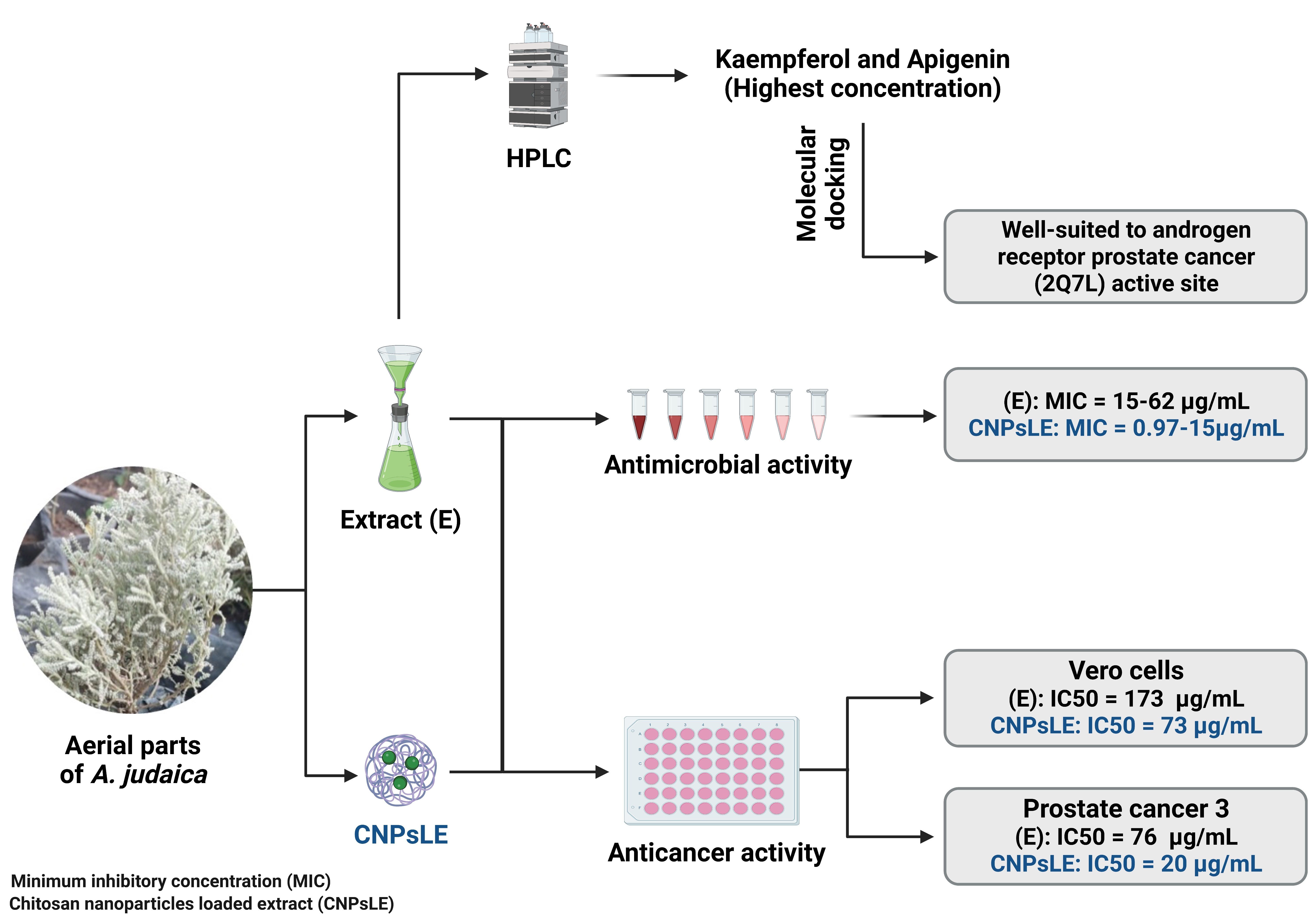
Phytochemical Characterization and Efficacy of Artemisia judaica Extract Loaded Chitosan Nanoparticles as Inhibitors of Cancer Proliferation and Microbial Growth

by
Husam Qanash
1,*,
Abdulrahman S. Bazaid
1,
Abdu Aldarhami
2,
Bandar Alharbi
1,
Majed N. Almashjary
3,4,
Mohannad S. Hazzazi
3,4,
Hashim R. Felemban
3,5 and
Tarek M. Abdelghany
6,*
1
Department of Medical Laboratory Science, College of Applied Medical Sciences, University of Ha’il, Hail 55476, Saudi Arabia
2
Department of Medical Microbiology, Qunfudah Faculty of Medicine, Umm Al-Qura University, Al-Qunfudah 21961, Saudi Arabia
3
Department of Medical Laboratory Sciences, Faculty of Applied Medical Sciences, King Abdulaziz University, Jeddah 22254, Saudi Arabia
4
Hematology Research Unit, King Fahd Medical Research Center, King Abdulaziz University, Jeddah 22254, Saudi Arabia
5
Special Infectious Agents Unit-BSL3, King Fahd Medical Research Center, King Abdulaziz University, Jeddah 21362, Saudi Arabia
6
Botany and Microbiology Department, Faculty of Science, Al-Azhar University, Cairo 71524, Egypt
*
Authors to whom correspondence should be addressed.
Polymers 2023, 15(2), 391; https://doi.org/10.3390/polym15020391
Submission received: 18 November 2022 / Revised: 6 January 2023 / Accepted: 9 January 2023 / Published: 11 January 2023
(This article belongs to the Special Issue Biomedical Applications of Polymer-Based Nanomaterials)

Abstract

:
Despite the advanced development in the field of drug discovery and design, fighting infectious and non-infectious diseases remains a major worldwide heath challenge due to the limited activity of currently used drugs. Nevertheless, in recent years, the approach of designing nanoparticles for therapeutic applications has gained more interest and promise for future use. Thus, the current study is focused on the evaluation of A. judaica extract and chitosan nanoparticles loaded extract (CNPsLE) for potential antimicrobial and anticancer activities. The HPLC analysis of the extract has shown the presence of various phenolic and flavonoid compounds, including kaempferol (3916.34 µg/mL), apigenin (3794.32 µg/mL), chlorogenic acid (1089.58 µg/mL), quercetin (714.97 µg/mL), vanillin (691.55 µg/mL), naringenin (202.14 µg/mL), and rutin (55.64 µg/mL). The extract alone showed higher MIC values against B. subtilis, E. coli, S. aureus, K. pneumonia, and C. albicans (62.5, 15.65, 15.62, 31.25, and 31.25 µg/mL, respectively), whereas lower MIC values were observed when the extract was combined with CNPsLE (0.97, 1.95, 3.9, 4.1, and 15.62 µg/mL, respectively). The extract exhibited low cytotoxicity against normal Vero cells with IC50 173.74 µg/mL in comparison with the cytotoxicity of the CNPsLE (IC50, 73.89 µg/mL). However, CNPsLE showed more selective toxicity against the human prostate cancer cell line (PC3) with IC50 of 20.8 µg/mL than the extract alone with 76.09 µg/mL. In the docking experiments, kaempferol and apigenin were revealed to be suitable inhibitors for prostate cancer (2Q7L). Overall, the obtained data highlighted the promising potential therapeutic use of CNPsLE as an anticancer and antimicrobial agent.

Graphical Abstract

1. Introduction

Plants are a rich source for diverse chemical compounds with various therapeutic properties that can be used alone or in combination with other medicines to treat infectious and non- infectious diseases [1,2,3]. Although numerous plant-derived products have been discovered and clinically used, this source still offers high probability for the discovery of new and promising therapeutic compounds for treating human illness. Artemisia is one of the largest genera within the Asteraceae family and the leading genus within the species, comprising between 200 and greater than 500 taxa at the species or sub-species level. For instance, mugwort, wormwood, and sagebrush are well-known species of Artemisia. Multiple species of Artemisia have gained great interest in various scientific fields in the search for promising heath and economical outcomes due to its reported nutritional and biological properties. In addition, numerous compounds are produced by species of this genera, including flavonoids, alkaloids, phenols, quinines, and terpenoids, exhibiting various biological activities [4]. This involves anticancer, antidepressant, anxiolytic, antidiabetic, antitubercular, hepatoprotective, gastroprotective, antihypertensive, antiepileptic, insecticidal, antiemetic, antihyperlipidemic, antiparasitic, and antiviral activities [5].
Artemisia judaica L. is one of the important species of Artemisia commonly grown in the Mediterranean area, including Saudi Arabia, Egypt, Algeria, Libya, and Jordan [6]. A. judaica was reported to exhibit great potential therapeutic use for gastrointestinal disorders, inflammatory disorders, sexual dysfunction, heart diseases, hyperglycemia, cancers, arthritis, oxidative stress, and wound healing [7,8]. In addition, in vitro investigations have reported anticancer and antioxidant activities of A. judaica extracts. Furthermore, antimicrobial activity of A. judaica oil was observed against Candida albicans, Cryptococcus neoformans [9], Bacillus cereus, and Aspergillus niger [8]. Moreover, both aqueous or alcoholic extracts of A. judaica have decreased the level of blood glucose in diabetic rats [10], showing higher antioxidant activity compared to ascorbic acid [11].
Recently, the field of nanotechnology has gained more interest regarding nanoparticle design and/or its incorporation with plant extract to enhance its biological activities [12]. The combination of certain natural extracts, such as oregano, savory, marjoram [13], sage [14], Leucas aspera [15], Pterocarpus marsupium [16], and aloe vera gel [17], with chitosan nanoparticles (CNPs) was performed and shown to increase the activity of the extract. The unique characteristics of CNPs, including surface their area, the existence of active functional groups, limited toxicity, great permeability into bacterial cell membranes, and cost effectiveness, have prioritized their potential therapeutic applications in comparison with bulk chitosan [18]. The antifungal activity of a Byrsonima crassifolia essential oils loaded with CNPs was reported against Colletotrichum gloeosporioides and Alternaria species [19]. In addition, the anticancer activity of essential oils from a species of Artemisia incorporated with CNPs against breast cancer lines was documented, while unloaded essential oils showed less anti-proliferative activity [20]. In addition, although Artemisia scoparia extract displayed limited toxicity towards hepatocarcinoma cell lines, its activity was notably improved when combined with chitosan-myristate nanogel [21]. Moreover, a previous in vitro study claimed the significant enhancement by CNPs for the anticancer activity of Zataria multiflora essential oil against breast and melanoma cells [22]. Although Artemisia species are well reported as being a rich source of compounds with promising nutritional and therapeutic properties, the enhancing, expanding, and stabilizing of desired biological activities is urgently required to overcome the resistance, ineffectiveness, and instability of currently and clinically used antimicrobial or anticancer compounds. Thus, the current study investigates the chemical composition of tested Artemisia species, evaluating the antimicrobial and anticancer activities of the extract alone, as well as in combination with chitosan nanoparticles.

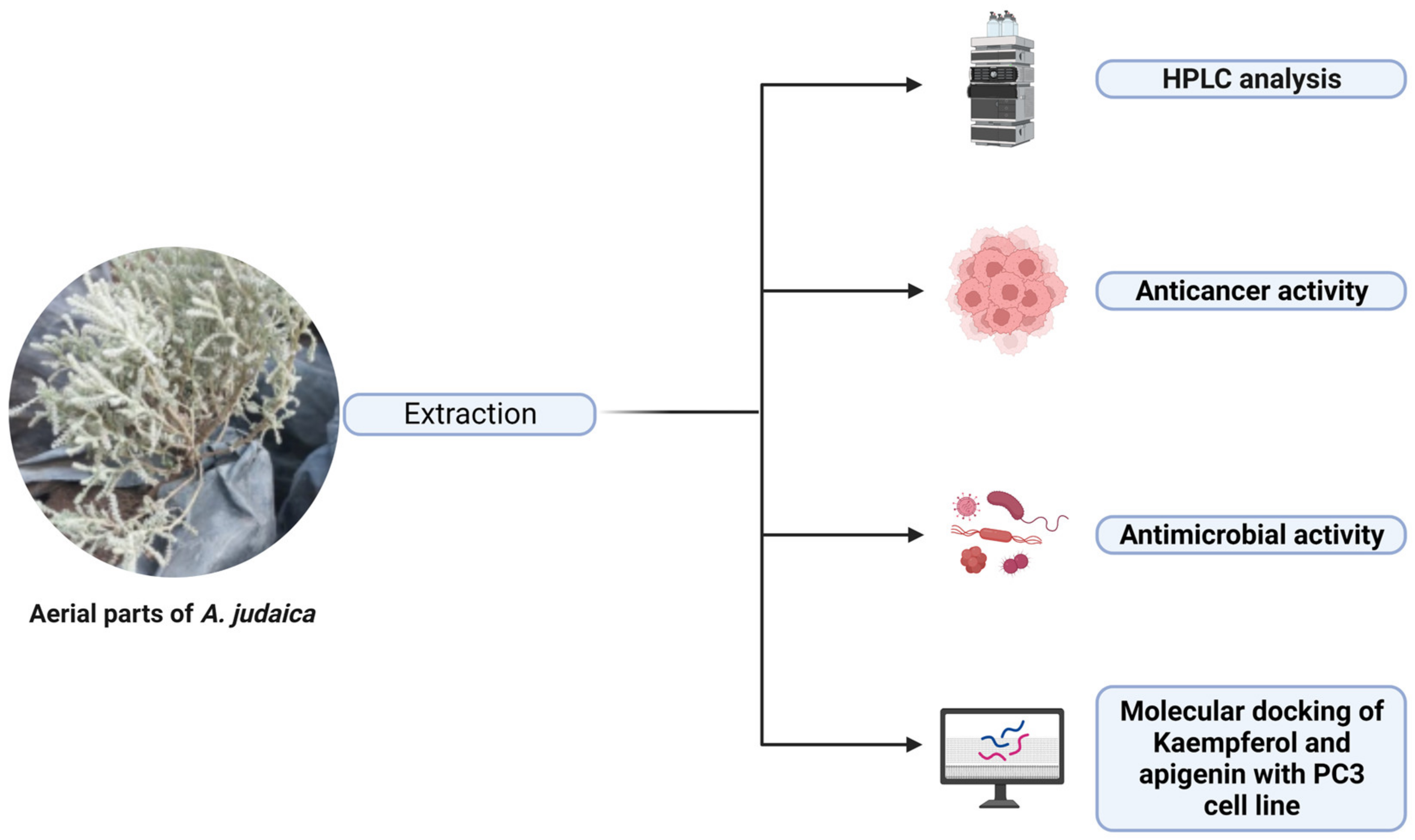
2. Materials and Methods

2.1. Source of Chitosan Nanoparticles

High quality chitosan nanoparticles (CNPs) powder was purchased from Primex, Siglufjordur, Iceland. Analytical grade solvents, buffers, and reagents used were obtained from Sigma-Aldrich (St. Louis, MS, USA), while Dimethyl Sulfoxide (DMSO) was purchased from Riedel-de Haën -Honeywell Research Chemicals (Seelze, Germany).

2.2. Plant Collection and Extraction

The aerial parts of Artemisia judaica were collected (May 2022) and dried, then ground into fine powder using a manual mortar. In a Soxhlet extractor (Fisher Scientific, Leicestershire, UK), about 5 g of powder was extracted using 250 mL of methanol over a one-day period. The extraction was repeated for three rounds, followed by evaporation via a rotary evaporator (Henan Lanphan Industrial Co., Ltd., China), yielding 0.6 mg g−1 as a crude extract. The extract was subjected to high-performance liquid chromatography (HPLC) analysis and the evaluation of its biological activities, including antibacterial, antifungal, and anticancer activities was determined.

2.3. Loading of A. judaica into Chitosan Nanoparticles

The loaded nanoparticles were purified by centrifugation, and the diameter of chitosan nanoparticles (CNPs) was then assessed. Briefly, CNPs solution was prepared through dissolving CNPs in an aqueous solution of acetic acid (1%, v/v), followed by the adding of CNPs to get 2% (w/v) as a final concentration. To the prepared solution, glycerol was added at a concentration of 2% (w/v) as a plasticizer. Then, Tween 20 (0.05%, v/v) was added to enhance the adhesion and wettability properties. To prepare extract incorporated with CNPs coating solution, the extract was incorporated into CNPs solution (stirred for 25 min) to obtain the final concentration (10%) of extract concentrate [17].

2.4. HPLC Analysis

Following the extraction, 5 μL of the A. judaica extract was injected into the HPLC machine (Agilent 1260 series, Agilent Technologies, Santa Clara, CA, USA). The Eclipse C18 column was used as follows: 4.6 mm × 250 mm i.d., and 5 μm that was maintained at 40 °C. Two buffers were used as the mobile phase—Buffer A (mili Q water + 0.05% trifluoroacetic acid) and Buffer B (acetonitrile + 0.05% trifluoroacetic acid—at a flow rate of 0.9 mL/minute. The mobile phase was programmed in a linear gradient for 20 min run as follows: 0 min (82% Buffer A); 0–5 min (80% Buffer A); 5–8 min (60% Buffer A); 8–12 min (60% Buffer A); 12–15 min (82% Buffer A); 15–16 min (82% Buffer A), and 16–20 (82% Buffer A). One wavelength at 280 nm was used by the ultraviolet (UV) detector (Icon Scientific Inc., North Potomac, M.D., USA) for the detection of phenolic and flavonoids contents. The phenolic and flavonoids contents of the extract were estimated qualitatively and quantitatively according to the methods of Abdelghany et al. [1], in comparison with the injected/used standards of phenolic flavonoid compounds.

2.5. Agar Well Diffusion Assay

Modified Mueller–Hinton agar and potato dextrose agar media were used for the growth of the tested bacteria and fungi, respectively. The agar well diffusion method was utilized to estimate the antimicrobial activity of the extract alone and when combined with CNPs. Bacterial isolates were collected from Ain Shams University Hospital, Egypt, while the mycology laboratory at Assiut University, Egypt, provided the research team with requested fungal isolates. The appropriate agar plate was individually inoculated with the tested microbe, including Staphylococcus aureus [ATCC 6538], Bacillus subtilis [ATCC 6633], Escherichia coli [ATCC 8739], Klebsiella pneumonia, Aspergillus niger, and Candida albicans [ATCC 10231], using sterile swabs. Next, a well (6 mm diameter) was formed using a sterile cork-borer, which was then filled with 100 µL of the tested sample. Then, agar plates inoculated with bacteria were incubated at 37 °C for 24 h, whereas about 72 h of incubation at 30 °C were used for fungal growth. The inhibitory zones around the wells were measured in millimeters (mm). Inhibitory zones caused gentamycin and clotrimazole against tested bacterial, and fungal isolates were used as positive controls, respectively [23].

2.6. Minimal Inhibitory Concentration (MIC)

The micro-dilution broth technique was applied to determine the minimal inhibitory concentration (MIC) (the lowest concentration of the sample that inhibits the growth of the tested microorganisms). A stock concentration was prepared to produce the multiple dilutions (0.98 to 1000 µg/mL) of the extract. A constant volume (200 µL) of each dilution was added to the designated well/s of a sterile 96-wells plate containing broth medium. The 0.5 McFarland suspension of each tested microorganism was prepared in 0.85% sterile sodium chloride (normal saline). About 2 µL of the prepared microbial suspension (containing 2 × 10⁸ of the bacterial colony forming units (CFU/mL, 2 × 10⁶ CFU/mL for yeasts and 4 × 10⁶ CFU/mL for filamentous fungi) was transferred to the designated well(s). Subsequently, the plates were incubated at the appropriate growth condition for bacteria and fungi, as mentioned above. Finally, MIC values were determined based on a visual observation of the microbial growth.

2.7. Cell Viability Assay

Prostate cancer (PC3) cells and normal Vero cells were obtained from Nawah Scientific (Inc., Cairo, Egypt), and about 1 × 10⁵ cells/mL (100 µL/ well) were inoculated in the 96-well tissue culture plate, then incubated at 37 °C, 5% CO2, for 24 h to develop a whole monolayer sheet. After that, the growth medium was decanted from the culture plate when the confluent sheet of the tested cells was formed. Then, the cell monolayer was washed using Roswell Park Memorial Institute (RPMI) medium with 2% serum (maintenance medium), which was amended with different dilutions (0.1 mL for each well) of the tested extract. The wells without any dilution but containing maintenance medium were used as controls. Plates were incubated at 37 °C, 5% CO2, overnight to investigate any possible cytopathic effects of treated cells. A solution of 3-(4,5-dimethylthiazol-2-yl)-2,5-diphenyltetrazolium bromide) (MTT) was prepared using phosphate buffered saline (PBS) as a solvent (5 mg/mL) [24]; then 20 µL of the prepared solution was added into each well, followed by well-mixing of prepared solution with the used medium through a shaking table at 150 rpm for 5 min [25]. The well plates were incubated at 37 °C for 4 h under 5% CO2 to initiate/enhance the metabolizing of MTT. Then, the metabolic product of MTT was resuspended in 200 µL of DMSO and left for 5 min on the shaking table at 150 rpm to enhance the mixture of the metabolic product with the solvent. At 560 nm and a subtracted background at 620 nm, the optical density was read to determine the viability of the treated cells [2,26]. Cell cytopathic effects were monitored using an inverted microscope.

2.8. Molecular Docking

The Molecular Operating Environment (MOE) is a set of software applications for the computer-aided design of the main detected kaempferol and apigenin flavonoids as a biologically active compounds in the A. judaica extract against the PC3 cell line. It enables the drawing of molecules and their minimization for the best possible conformations. The Androgen Receptor Prostate Cancer (2Q7L) was downloaded from the protein data bank (http://www.rcsb.org/pdb, assessed on 5 September 2022) [27]. The protein molecule performed 3D protonation after the water molecules were removed. To optimize the geometry of 2Q7L, the enzyme was shown using the MMFF94x force field implemented in the MOE software. The main chain was kept firm, while the side chains remained flexible. This approximation allowed the side chains of the proteins to find the position in which the interactions are most favorable. We then recorded the best score, that is, the one with the lowest energy corresponding to the best interactions between the ligands and the active site of the enzyme. The next indexes were used in the docking study: S = final score, which is the score of the last stage that was not set to none; rmsd = root mean square deviation of the pose, in Å, from the original ligand. This field occurred if the site description was the same as the description of the ligand; rmsd_refine = root mean square deviation from the earlier refinement and the later refinement of the pose; E_conf = energy of the conformer. If there was a refinement stage, this was the energy calculated at the end of the refinement. Note that for force field refinement, by default, this energy was calculated with the solvation option set to Born; E_place = score from the placement stage; E_score 1 and E_score 2 = scores from rescoring stages 1 and 2; E_refine = score from the refinement step, calculated to be the totality of the van der Waals electrostatic plus the solvation energies, according to the generalized Born solvation model (GB/VI).

2.9. Statistical Analysis

All measurements were performed in triplicate, and the results were calculated as mean values plus standard deviation (±SD).

3. Results and Discussion

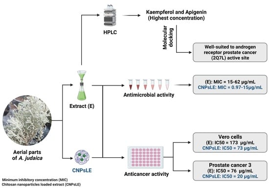
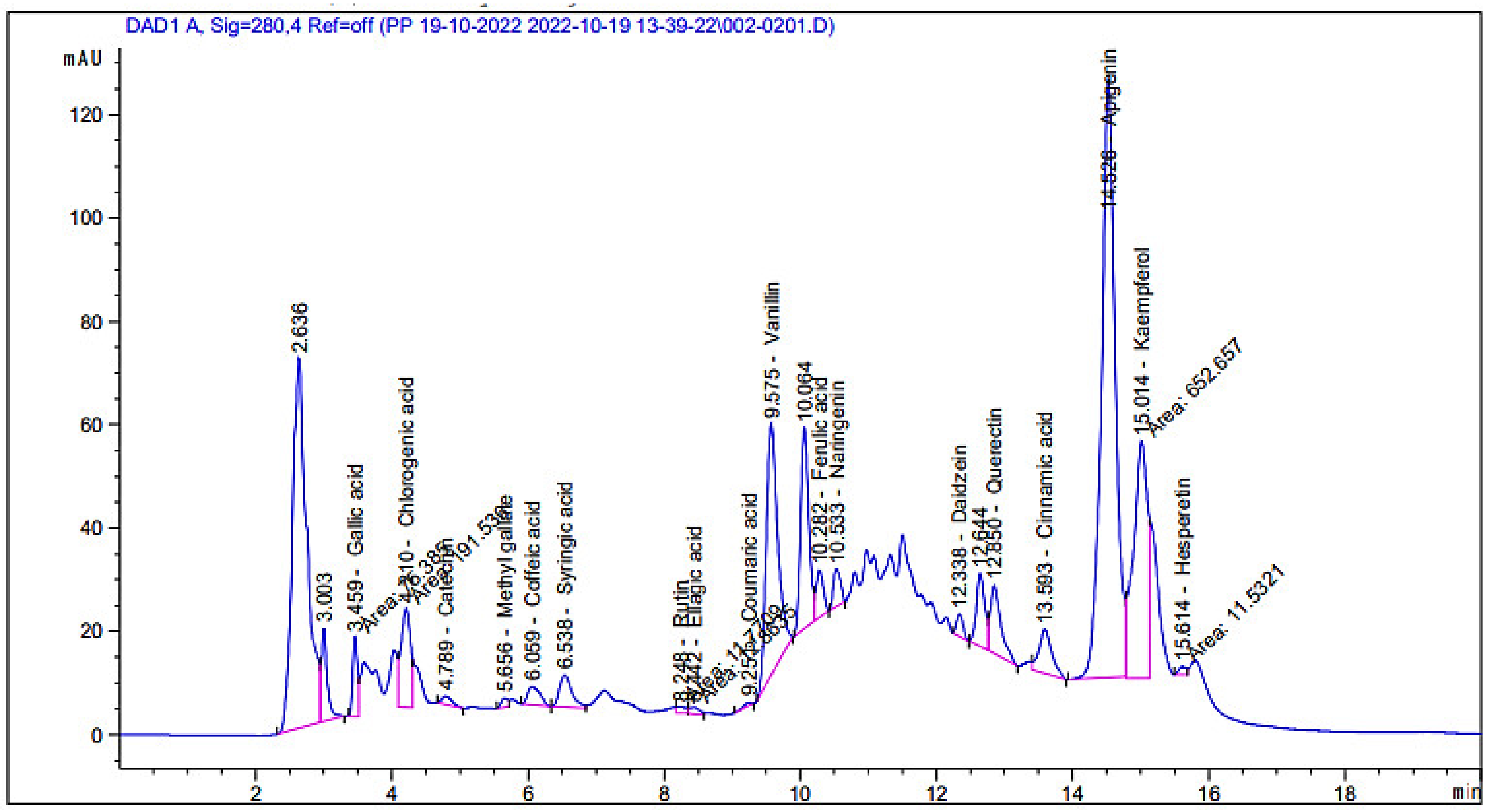
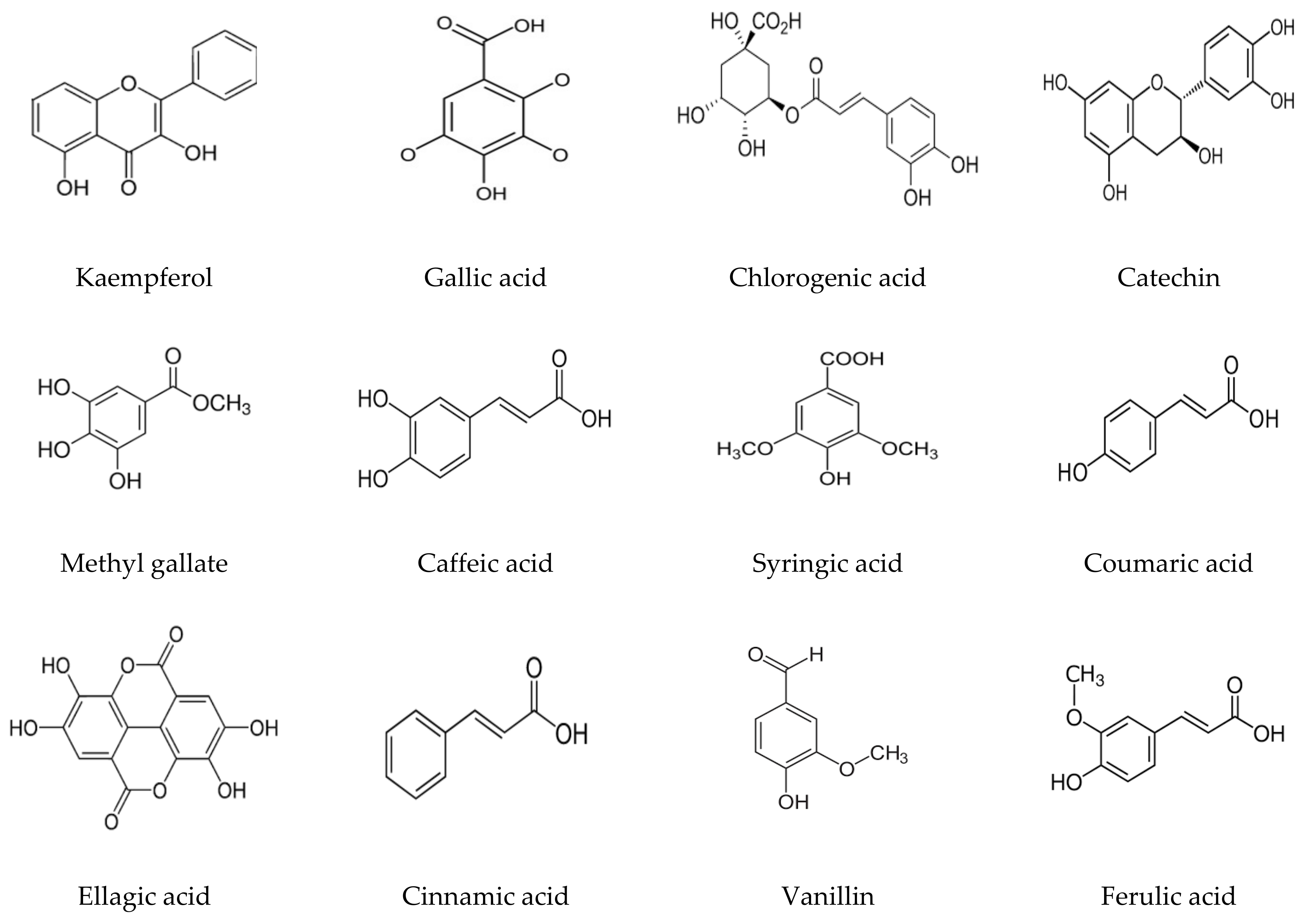
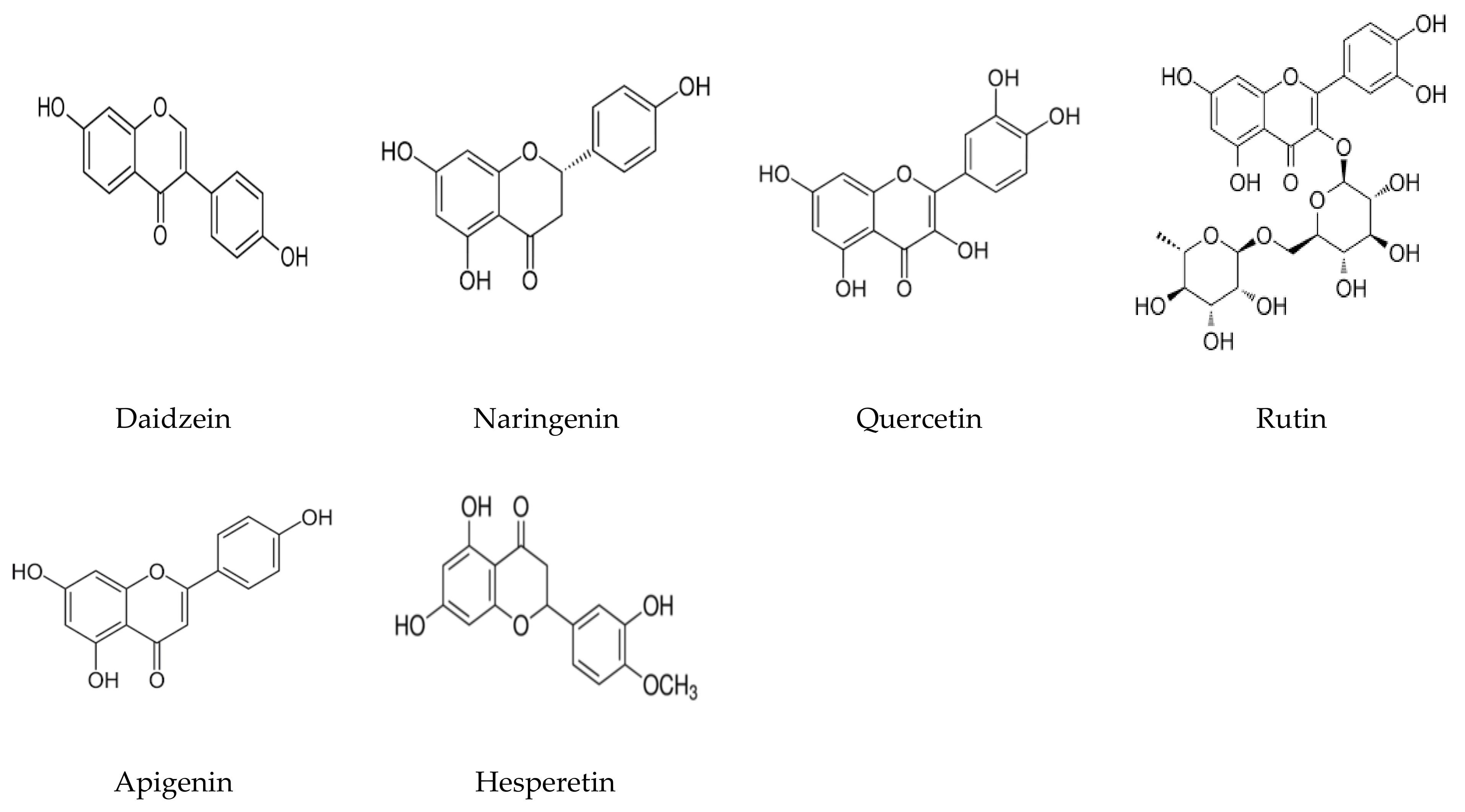
3.1. Phenolic and Flavonoid Analysis

Although the phytoconstituents of A. judaica were previously detected in several studies [28,29], they may differ based on growing conditions, climatic conditions, cultivar soil, the age of plant, and the flowering state; thus, the analysis of the plant extract was performed in this study. HPLC analysis for the A. judaica extract (aerial parts) revealed the presence of 18 known compounds (Figure 1), with their exact concentrations (Table 1 and Figure 2) and chemical formulas (Figure 3), but another 4 unknown compounds were also detected. Among the 18 known compounds, kaempferol, apigenin, and chlorogenic acid were revealed with the highest concentrations (3916.34, 3794.32, and 1089.58 µg/mL, respectively), while the lowest concentrations were noted for coumaric acid (5.46 µg/mL) and methyl gallate (27.68 µg/mL). The phytochemical profiles of the extract indicated that the aerial parts of A. judaica were very rich in phenolic and flavonoid compounds. Chlorogenic acid, luteolin, apigenin, rutin, and other compounds were reported in the extract of Artemisia gmelinii [4]. The identified compounds for A. judaica were reported, with various biological activities, i.e., kaempferol, which is a flavonoid compound, showed success toward the treatment of inflammatory responses, liver injuries, diabetes, malignancy, and obesity [30]. In addition, a recent study has claimed the promising potential therapeutic use of kaempferol, alone or in combination with colistin, to treat infections caused by drug resistant bacteria [31].
Apigenin is another compound identified with high concentrations in the A. judaica extract that was previously reported to exhibit anticancer, antioxidant, anti-inflammatory, and antimicrobial activities [32,33]. Similarly, multiple biological activities were linked with chlorogenic acid, including antibacterial activity towards Yersinia enterocolitica [34]. Other detected compounds in the current investigation, such as quercetin (714.97 µg/mL), naringenin (202.14 µg/mL), and rutin (55.64 µg/mL), were reported to possess great future possibilities for use as therapeutic agents for various illness. For instance, quercetin was shown to have antioxidant, anti-inflammatory, antidiabetic, anti-Alzheimer’s, and anticancer activities [35] [36]. Moreover, in vitro and in vivo studies of the naringenin compound have claimed its strong association with the suppression of cancer progression [37]. Furthermore, a recent report also proved multiple activities of rutin toward inflammation, oxidation, and thrombosis [38]. Although the above-mentioned activities noted for compounds within the current extract are not comprehensive, they should be adequate to state that A. judaica, especially its aerial parts, is very rich source for diverse compounds with various therapeutic properties.

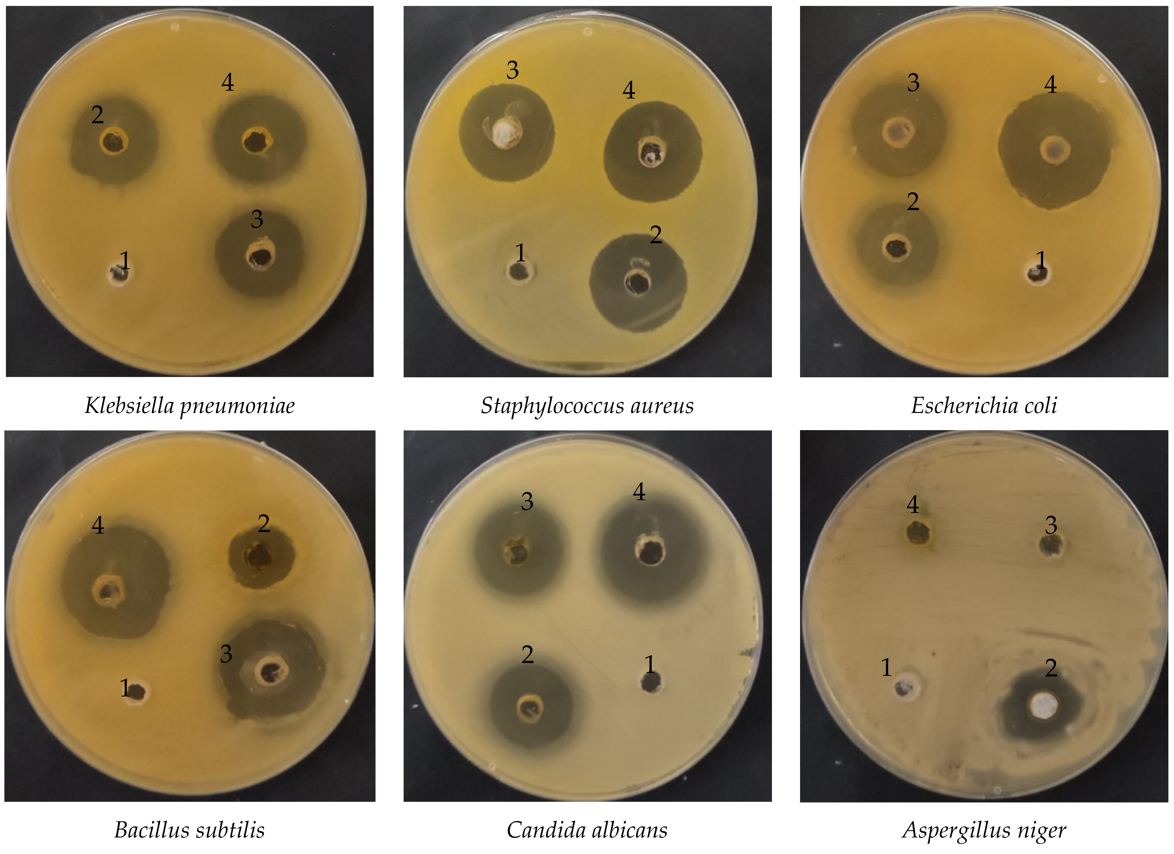
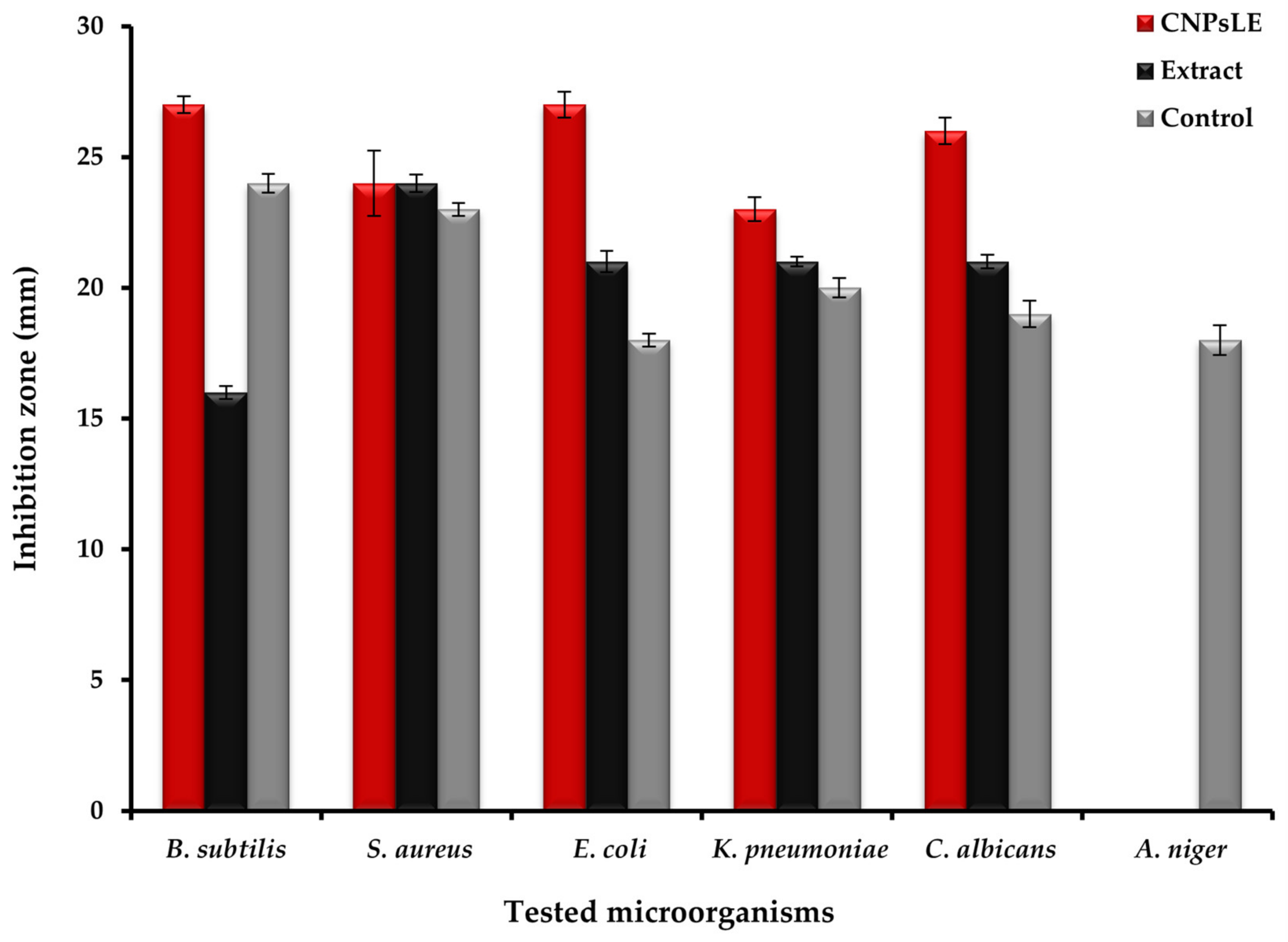
3.2. Antimicrobial Activity

Recent reports have claimed that CNPs possess a great promise for use in different medical applications through various technologies/approaches, including nanomedicine and biomedical engineering, for either enhancing their effectiveness or novel discovery [39,40]. CNPs loaded with Artemisia judaica extract (CNPsLE) were more effective than the extracts alone against all tested microorganisms, except Staphylococcus aureus (Figure 4 and Figure 5); this might be due to the differences in the cell wall compositions, virulence factors, or specific resistance mechanisms.
Judaica essential oils showed potent activity against the growth of Aspergillus fumigatus, Geotrichum candidum, Syncephalastrum racemosum, Candida albicans, Escherichia coli, Bacillus subtilis, and Streptococcus pneumonia, but failed to exhibit any activity towards the tested strain of Pseudomonas aeruginosa [41]. Nevertheless, another recent study [8], reported the high sensitivity of Aspergillus niger, Candida albicans, Salmonella typhimurium, and Bacillus cereus towards A. judaica extract, whereas E. coli, Klebsiella pneumonia, and Shigella flexneri were observed with limited susceptibility to the same extract [6]. This variation in the overall potency of the extract might be attributed to the solvent used in the extraction. For example, it was reported that no antifungal potential was observed on the tested fungi, with the exception of C. albicans. Moreover, ethyl acetate extract did not exhibit the growth of A. fumigatus and Mucor spp.; the growth of C. albicans and A. fumigatus, however, was detected by methyl alcohol extract. These outcomes are consistent with the current results. CNPsLE exhibited excellent MIC values against B. subtilis (0.97 µg/mL) E. coli (1.95 µg/mL), S. aureus (3.9 µg/mL), K. pneumonia (4.1 µg/mL), and C. albicans (15.62 µg/mL), but the same microbes showed higher MIC values—B. subtilis (62.5 µg/mL), E. coli (15.65 µg/mL), S. aureus (15.62 µg/mL), K. pneumonia (31.25 µg/mL), and C. albicans (31.25 µg/mL)—when treated with the extract only (Table 2). Extract from Artemisia judaica, without combination with any nanoparticle, was reported, with MIC between 2.5 and 5 mg/mL against Candida species, while species of Bacillus showed MIC between 1.25 and 5 mg/mL [4]. Currently, both the extract alone and the CNPsLE complex failed to inhibit the growth of A. niger (Figure 4 and Figure 5). This is in agreement with previous studies, in which either complete resistance (Aspergillus niger and Alternaria alternate) or limited sensitivity (Fusarium solani) of tested fungi towards A. judaica extract was noted [42]. In addition, the enhanced efficacy of the current extract, when combined with CNPs, towards the tested microbes was also observed for mint extract loaded with CNPs against the examined pathogenic fungi [43]. Moreover, Torreya grandis essential oil (EO) loaded on the CNPs (particle diameter, 349.6 nm) offered stronger antibacterial activity compared to the non-loaded EO [44]. Furthermore, Achillea millefolium extract loaded on the CNPs showed a three-fold activity against B. subtilis and P. aeruginosa compared to the extract alone [45]. Recently, CNPs incorporated with aloe vera gel demonstrated stronger activity against Helicobacter pylori [17]. This might be due to the small size and strong surface charge of the CNPs, which facilitates the easy and effective interaction between the microbial cell wall and the CNPs loaded with the active compound/s [46,47] Therefore, the use of the CNPs as carriers for plant extracts and other therapeutic agents would hold great promise for enhancing the activity of active agents or leading to the discovery of novel active compound(s), only when loaded into the CNPs.

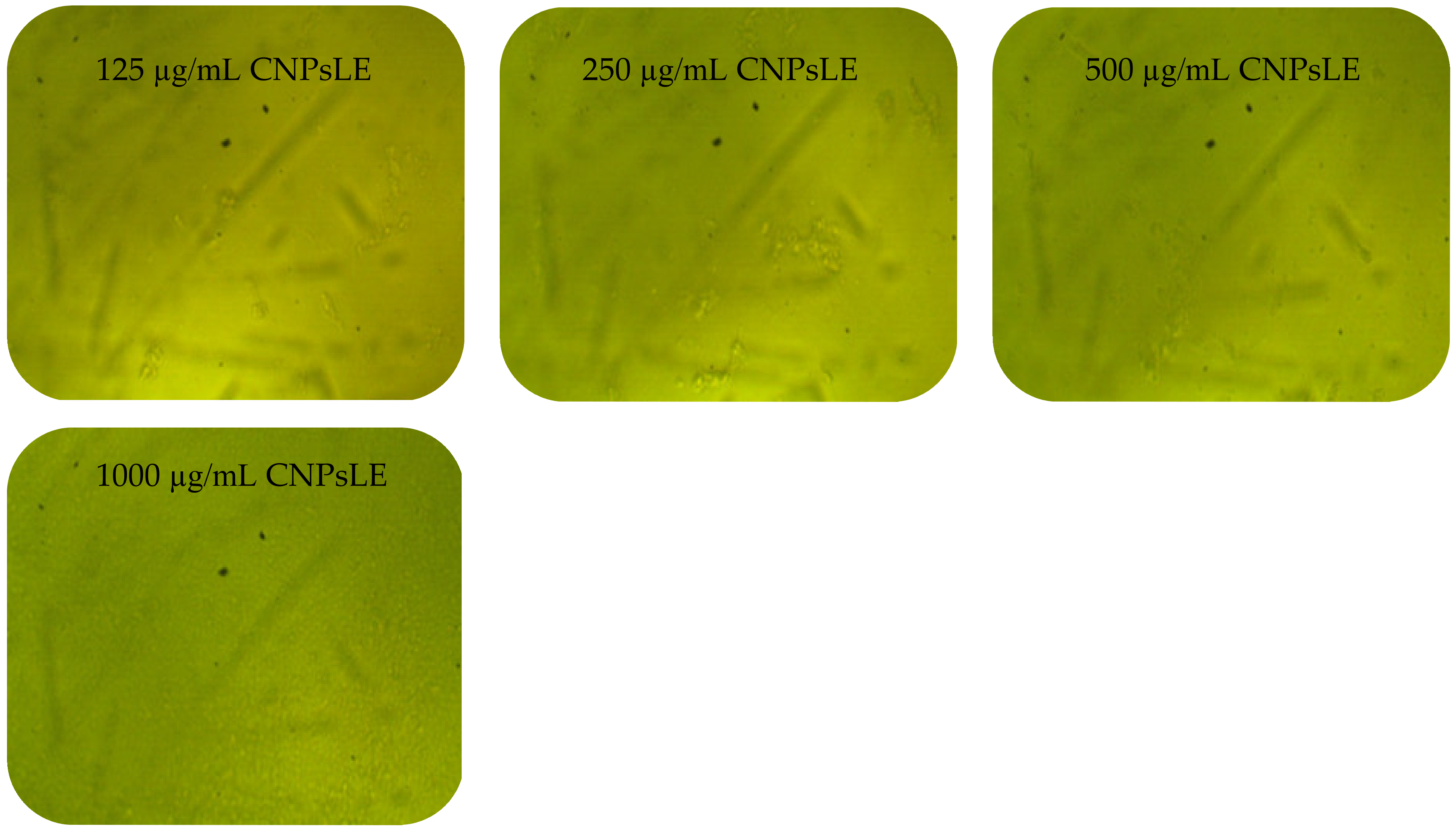
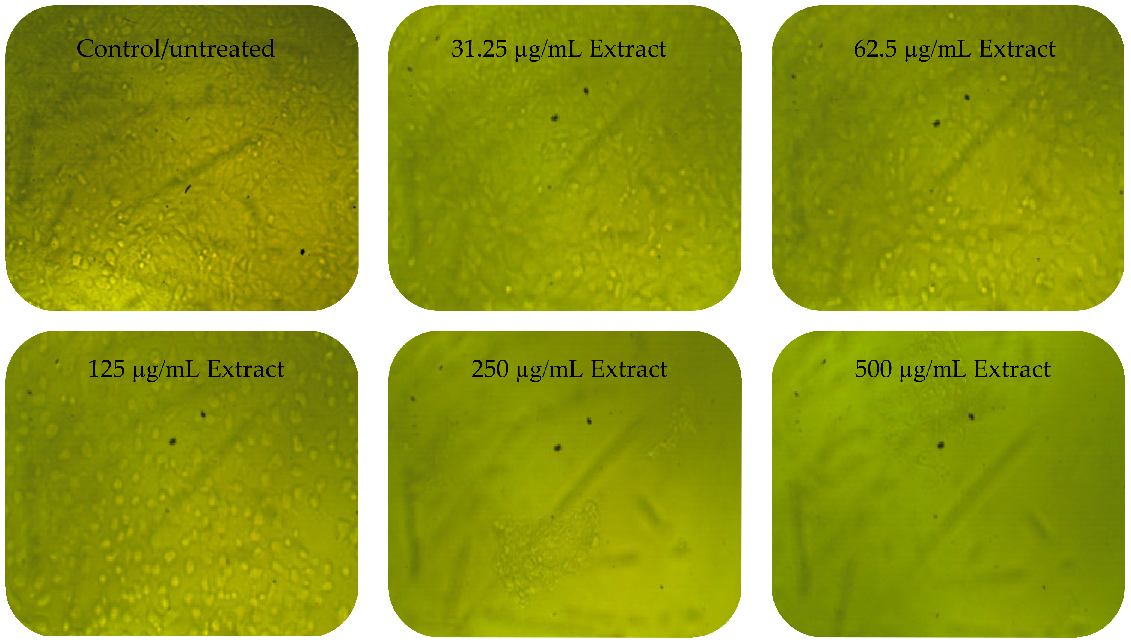
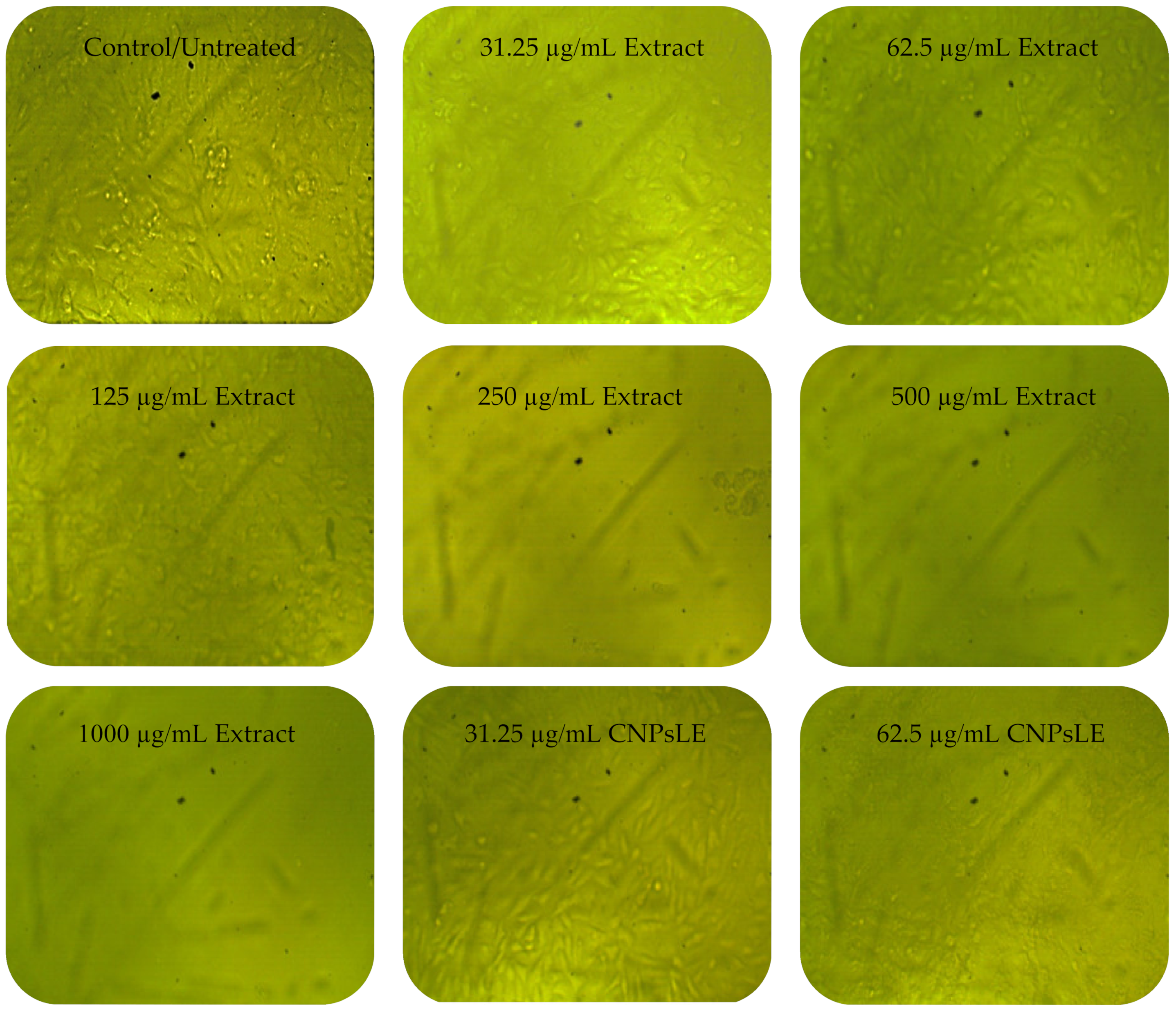
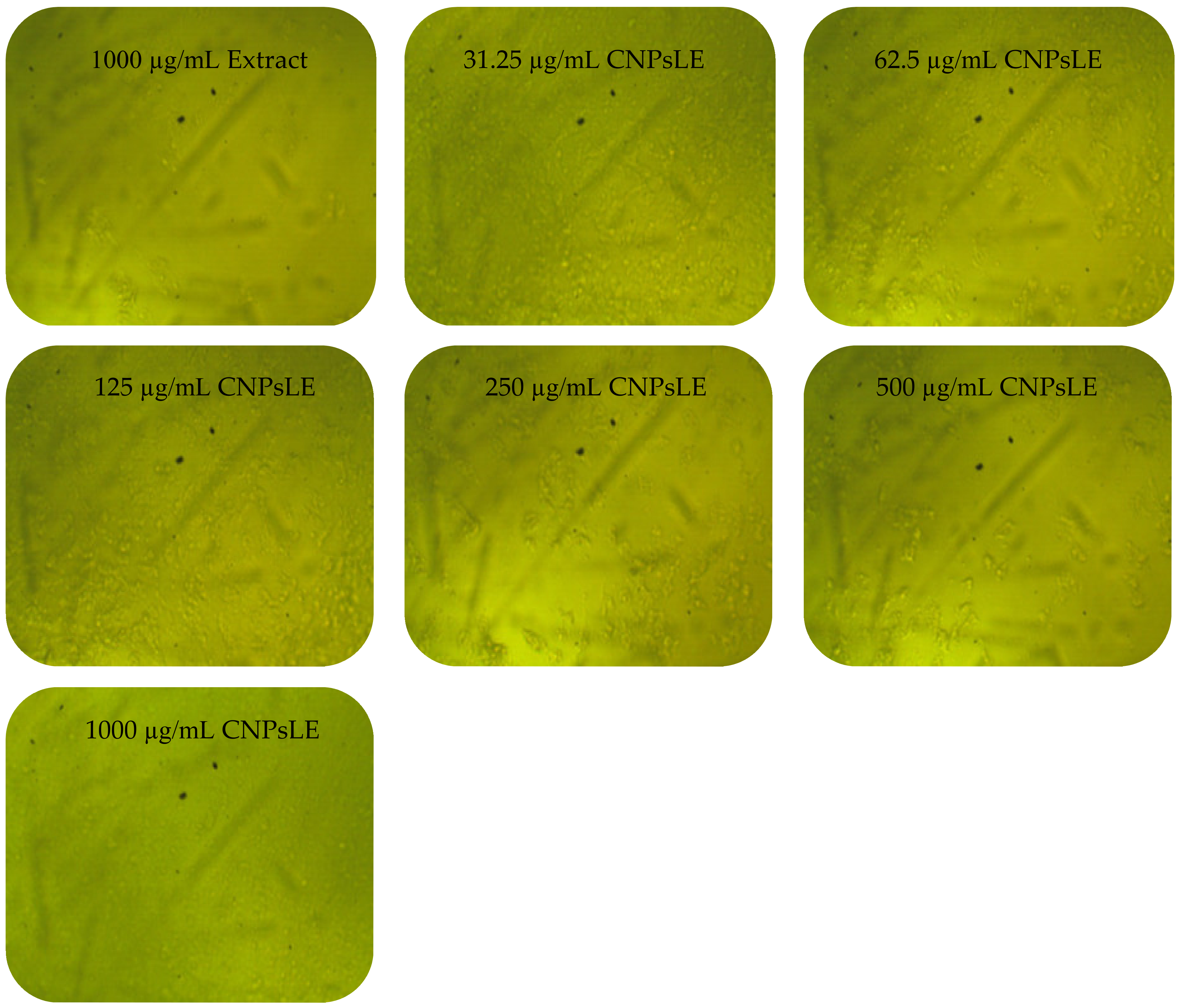
3.3. Anticancer Activity

To measure the chitosan’s toxicity, PC3 and Vero cells were treated with chitosan alone. A negligible cytotoxicity of chitosan against the PC3 cell line was observed, in which the viability of 98.35% and 88.47% was reported at 10 µg/mL and 1000 µg/mL, respectively. However, no cytotoxicity against chitosan-treated Vero cells was recorded as showing a viability of 100% at 10 µg/mL, but the viability was then decreased to 96.22% at 1000 µg/mL. The cytotoxicity of the A. judaica extract and the CNPs loaded extract was then evaluated against the cancer and normal cell line simultaneously to determine the possible selective toxicity to tumor cells, as well as to assess the suitability of these compounds for potential use in human medicine. The extract alone showed less activity against normal Vero cells than the extract loaded on the CNPs, particularly at concentrations ≤ 125 µg/mL (Table 3). The growth of Vero cells was reduced by 0.25, 1.15, and 28.97% or 6.11, 54.37, and 86.90% when treated by 31.25, 62.5, and 125 µg/mL of the extract alone or the extract loaded on the CNPs, respectively. Namely, the IC50 of the extract alone was higher (173.74 µg/mL) than that calculated for the extract loaded on the CNPs (73.89 µg/mL) against normal Vero cells.
Anticancer assays demonstrated a higher efficacy of the extract loaded on the CNPs, especially at concentrations ≤ 125 µg/mL, compared to the extract alone against PC3 cells (Table 4). A promising anticancer activity (toxicity of 73.80%) of the extract loaded on the CNPs compared with the anticancer activity (toxicity of 14.80%) of the extract only at the same concentration (31.25 µg/mL) (Table 4) was noted; therefore, the IC50 was calculated as 20.8 and 76.09 µg/mL for the extract loaded on the CNPs and extract alone, respectively. Artemisia quettensis extract was reported to exhibit anticancer activity against the human colorectal adenocarcinoma cell line (HT29), with IC50 around 31.54 µg/mL at 24 h of exposure [48]. Regarding the enhanced anticancer activity of the current extract when combined with the CNPs, multiple previous similar studies have documented this phenomenon for the compound of natural source (extract) formulated with CNPs compared to the bulk extract alone [49]. For instance, CNPs doped with the sodium tripolyphosphate enhanced the anti-proliferation activity toward the human ovarian cancer cell line (PA-1), while absent cytotoxicity was noted towards the normal mouse embryo cell lines (3T3) [50]. The anticancer activity in the current study of the tested compounds was evaluated using the positive control (Adriamycin), yielding IC50, 58.07 µg/mL against PC3.
Cytopathic effects (CPEs) were not observed on normal Vero cells treated with the A. judaica extract ≤ 62.5 µg/mL, while moderate CPEs were documented at 125 µg/mL (Figure 6). In addition, CPEs and the reduction of viable cells, or the complete absence and destruction of cells, was noted at 250–500 and 1000 µg/mL of the extract, respectively. The CNPsLE caused more morphological changes in the Vero cells at high concentrations 125–1000 µg/mL (Figure 6). Limited CPEs were observed on PC3 at lower concentrations of the A. judaica extract, but moderate morphological changes appeared, particularly at 250 to 1000 µg/mL (Figure 7). On the other hand, PC3 exposed to CNPsLE showed alteration in the shape, cell shrinkage, and membrane blebs at various tested concentrations. Apoptosis, the reduction of viable cells, and cytoplasmic condensation of treated PC3 cell with CNPsLE was observed at higher concentrations between 250 to 1000 µg/mL (Figure 7).
Although the A. judaica extract showed a lower affect towards all tested healthy and cancerous cells compared to the extract loaded on CNPs, both tested agents (A. judaica extract and CNPsLE) generally displayed more potent activity towards tested cancer cells (PC3) than the normal Vero cells. This potentially would indicate a possible selective toxicity of compounds detected from the A. judaica extract, although enhanced activity was observed when detected compounds from the A. judaica extract were loaded on the CNPs. Thus, detected compounds from the A. judaica extract, alone or combined with CNPs, show great promise for future use to treat prostate cancer, as their IC50 values are higher towards heathy cells (limited toxicity) compared to cancer cells.

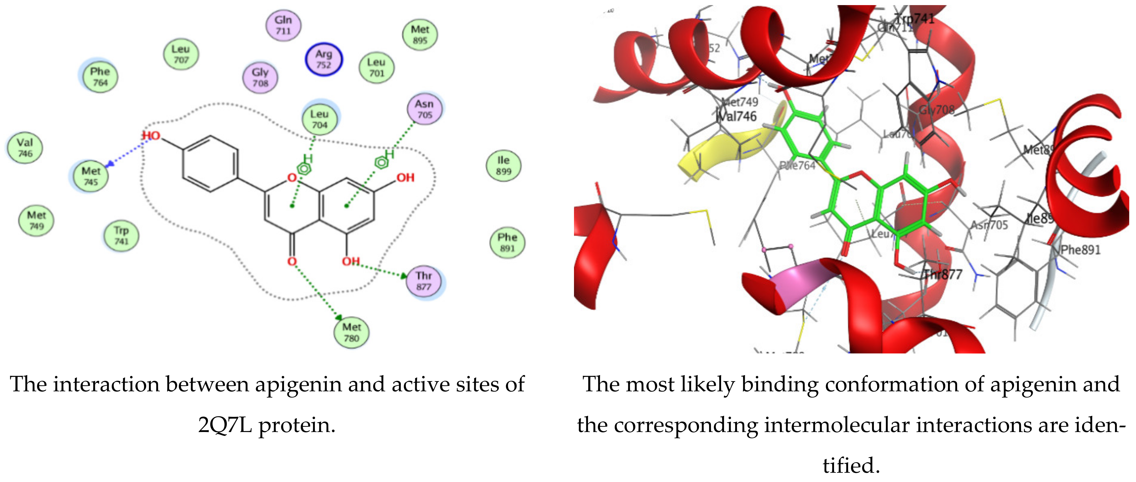
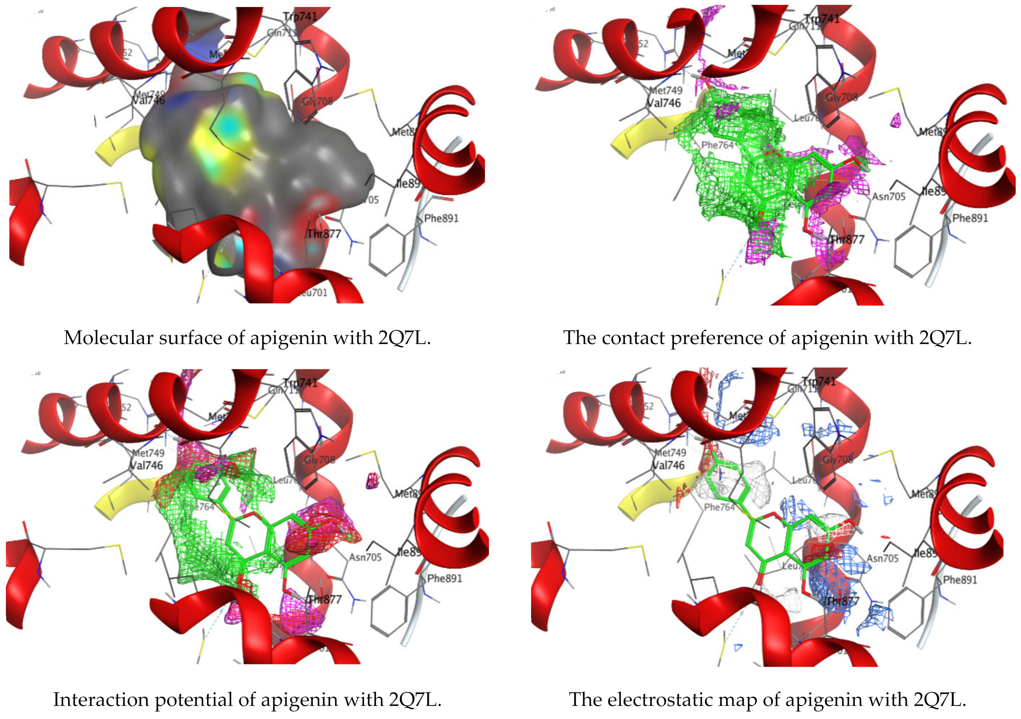
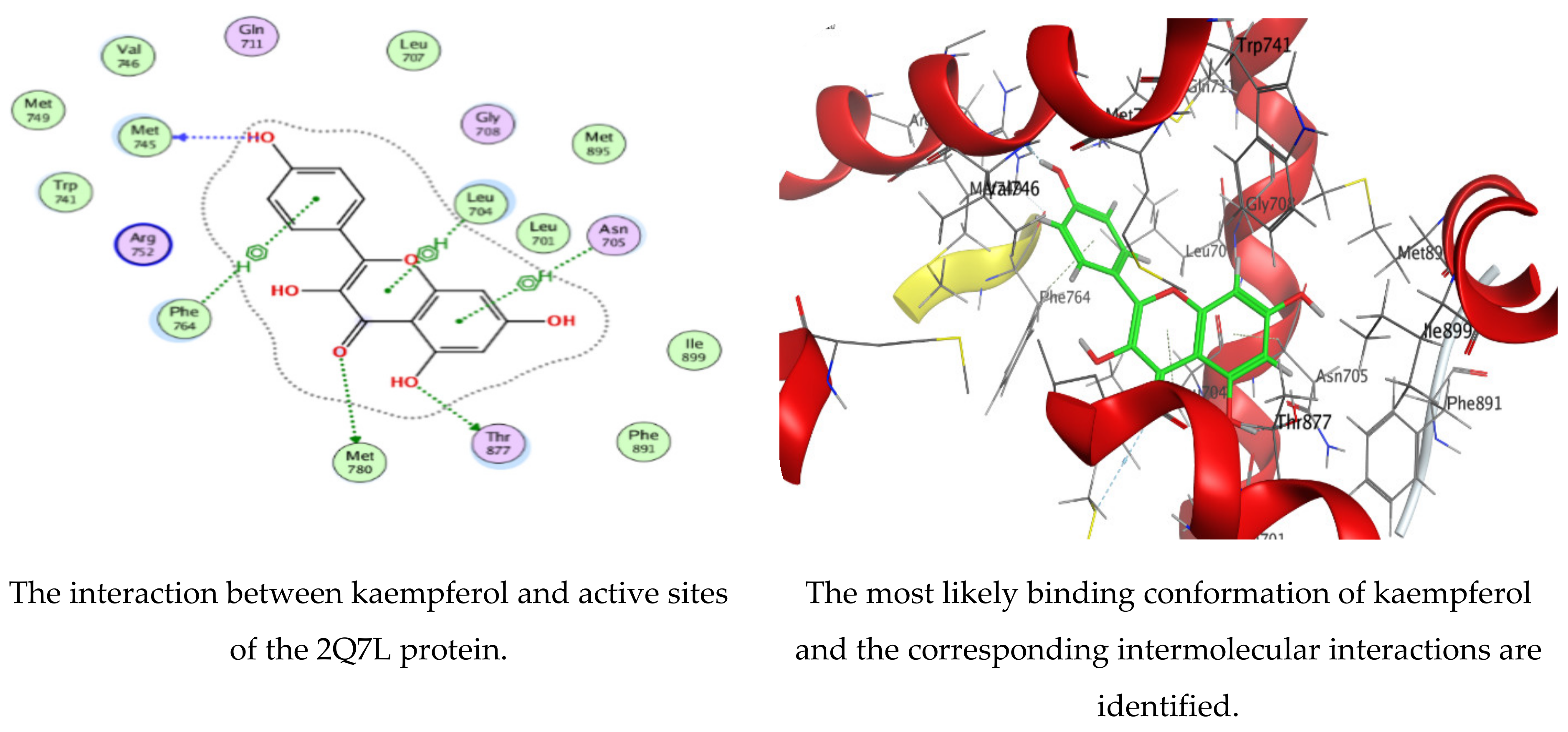
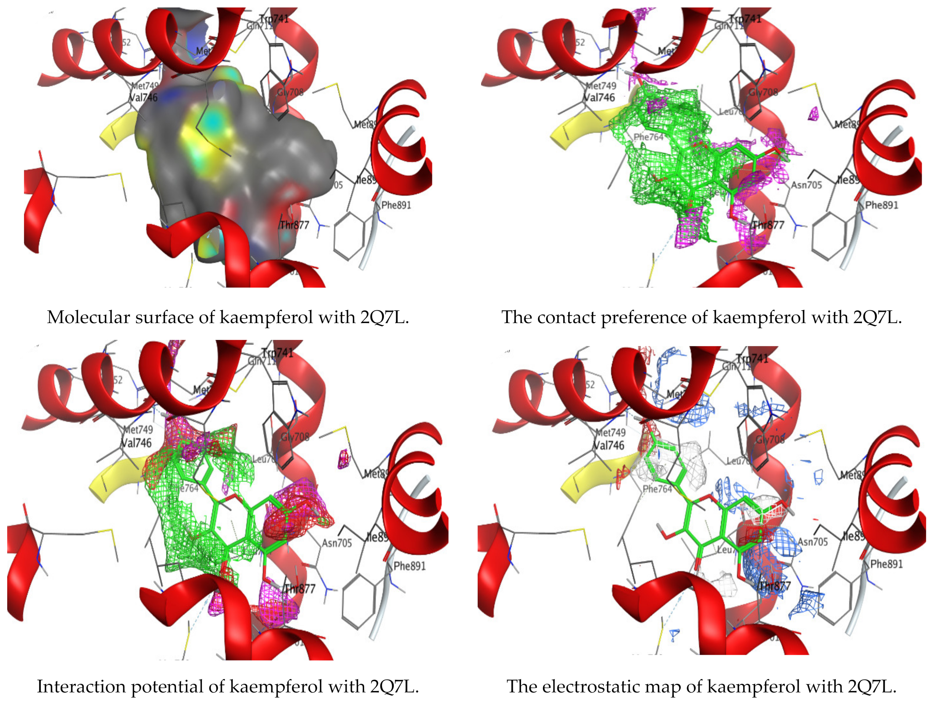
3.4. Molecular Docking of Kaempferol and Apigenin with Prostate Cancer (2Q7L)

The MOE-Dock program’s default parameters were employed for the molecular docking of the kaempferol and apigenin. The ligands were permitted to be flexible to determine the proper conformations of the ligands and to produce the least energy structures. The best ligand conformations were examined with Prostate Cancer (2Q7L) for their binding interaction. From the docking studies, it was observed that both ligands (kaempferol and apigenin) are good inhibitors of the 2Q7L enzyme, with low scores for binding free energy of −6.61458 kcal/mol and −6.47986 kcal/mol, respectively. The binding mode of the inhibitors kaempferol and apigenin are within the active site of 2Q7L (Figure 8 and Figure 9). Kaempferol was bound deeply to the binding cavity of Prostate Cancer (2Q7L), interacting with the residues MET 745, MET 780, THR 877, LEU 704, ASN 705, and PHE 764 via the O 23, O 25, O 26, and three 6-rings.
Likewise, apigenin showed similar interactions with the binding site residues MET 745, MET 780, THR 877, LEU 704, and ASN 705 via the O 24, O 2, O 27, and two 6-rings. The results are summarized in Table 5 and Table 6. In a recent investigation, the anticancer activity of chlorogenic acid, rutin, chrysoeriol, and kaempferol against breast cancer and PC3 cells, respectively, was documented via molecular docking protocol [51,52]. Another application of molecular docking was performed on the activity of the natural molecules of aloe vera gel doped with chitosan nanoparticles binding to Helicobacter pylori proteins [17]. Experimental findings by Qanash et al. [3] regarding the molecular docking of natural compounds such as chlorogenic acid confirmed its bacteriostatic action against Proteus vulgaris and antivirus activities against human corona virus (HCoV-229E). Moreover, natural exudates from some fungi were docked with Bacillus subtilis and Candida albicans [53] to elucidate its antimicrobial potential.

4. Conclusions

The phytochemical characterization of Artemisia judaica extract via HPLC showed several compounds with different concentrations belonging to phenolics and flavonoids. The antimicrobial activity of A. judaica extract was documented against B. subtilis, E. coli, S. aureus, K. pneumonia, and C. albicans, but not towards tested filamentous fungus (A. niger). In addition, the potent anticancer activity of the A. judaica extract against PC3 and the limited toxicity of normal Vero cells were revealed. Notably, the biological activities were found to be significantly enhanced when the extract was loaded with CNPs. Furthermore, the docking experiments showed an estimate of the ligand’s inhibitory actions. These demonstrated that the inhibitors kaempferol and apigenin were well-suited to the active site of the androgen receptor Prostate Cancer (2Q7L) and interacted with the residues essential for their biological activities. In short, A. judaica is a rich source for compounds with potential therapeutic properties, including antimicrobial and anticancer activities. The low toxicity of the extract towards Vero cells should highlight the promising future for the active compound(s) within the extract to be used in human medicine.

Author Contributions

Conceptualization, H.Q. and T.M.A.; formal analysis—antimicrobial activity, A.S.B., A.A. and H.R.F.; formal analysis—anticancer activity, H.Q., B.A., M.N.A. and M.S.H.; formal analysis—molecular docking, T.M.A.; investigation, H.Q., A.S.B., A.A., B.A., M.N.A., M.S.H., H.R.F. and T.M.A.; visualization and validation, H.Q., A.S.B., A.A., B.A., M.N.A., M.S.H., H.R.F. and T.M.A.; supervision, H.Q. and T.M.A.; funding acquisition, H.Q. and A.S.B.; writing—original draft preparation, H.Q. and T.M.A.; writing—review and editing; H.Q., A.S.B., A.A., B.A., M.N.A., M.S.H., H.R.F. and T.M.A. All authors have read and agreed to the published version of the manuscript.

Funding

This research was funded by the Deputy for Research and Innovation, Ministry of Education through Initiative of Institutional Funding at the University of Ha’il—Saudi Arabia, via project number IFP-22 052.

Institutional Review Board Statement

Not applicable.

Data Availability Statement

All data are available within the manuscript.

Acknowledgments

The authors thank the Deputy for Research and Innovation, Ministry of Education, through Initiative of Institutional Funding at the University of Ha’il—Saudi Arabia, for their grant through project number IFP-22 052. Graphical abstract and Figure 1 were created with BioRender.com.

Conflicts of Interest

The authors declare no conflict of interest.

References

  1. Abdelghany, T.; Yahya, R.; Bakri, M.M.; Ganash, M.; Amin, B.H.; Qanash, H. Effect of Thevetia peruviana seeds extract for microbial pathogens and cancer control. Int. J. Pharmacol. 2021, 17, 643–655. [Google Scholar] [CrossRef]
  2. Al-Rajhi, A.M.H.; Yahya, R.; Bakri, M.M.; Yahya, R.; Abdelghany, T.M. In situ green synthesis of Cu-doped ZnO based polymers nanocomposite with studying antimicrobial, antioxidant and anti-inflammatory activities. Appl. Biol. Chem. 2022, 65, 35. [Google Scholar] [CrossRef]
  3. Qanash, H.; Yahya, R.; Bakri, M.M.; Bazaid, A.S.; Qanash, S.; Shater, A.F.; Abdelghany, T.M. Anticancer, antioxidant, antiviral and antimicrobial activities of Kei Apple (Dovyalis caffra) fruit. Sci. Rep. 2022, 12, 5914. [Google Scholar] [CrossRef]
  4. Mamatova, A.S.; Korona-Glowniak, I.; Skalicka-Woźniak, K.; Józefczyk, A.; Wojtanowski, K.K.; Baj, T.; Sakipova, Z.B.; Malm, A. Phytochemical composition of wormwood (Artemisia gmelinii) extracts in respect of their antimicrobial activity. BMC Complement. Altern. Med. 2019, 19, 288. [Google Scholar] [CrossRef] [PubMed]
  5. Bisht, D.; Kumar, D.; Kumar, D.; Dua, K.; Chellappan, D.K. Phytochemistry and pharmacological activity of the genus artemisia. Arch. Pharmacal Res. 2021, 44, 439–474. [Google Scholar] [CrossRef] [PubMed]
  6. El-Amier, Y.A.; Al Borki, A.E.-N.S.; Elagami, S.A. Potential of wild plant Artemisia judaica L. as sustainable source of antioxidant and antimicrobial compounds. J. Exp. Sci. 2019, 10, 4–8. [Google Scholar] [CrossRef] [Green Version]
  7. Bora, K.S.; Sharma, A. The Genus Artemisia: A Comprehensive Review. Pharm. Biol. 2011, 49, 101–109. [Google Scholar] [CrossRef] [Green Version]
  8. Mohammed, H.A.; Qureshi, K.A.; Ali, H.M.; Al-Omar, M.S.; Khan, O.; Mohammed, S.A.A. Bio-Evaluation of the Wound Healing Activity of Artemisia judaica L. as Part of the Plant’s Use in Traditional Medicine; Phytochemical, Antioxidant, Anti-Inflammatory, and Antibiofilm Properties of the Plant’s Essential Oils. Antioxidants 2022, 11, 332. [Google Scholar] [CrossRef]
  9. Abu-Darwish, M.S.; Cabral, C.; Gonçalves, M.J.; Cavaleiro, C.; Cruz, M.T.; Zulfiqar, A.; Khan, I.A.; Efferth, T.; Salgueiro, L. Chemical composition and biological activities of Artemisia judaica essential oil from southern desert of Jordan. J. Ethnopharmacol. 2016, 191, 161–168. [Google Scholar] [CrossRef]
  10. Nofal, S.; Mahmoud, S.; Ramadan, A.; Soliman, G.; Abdel-Rahman, R. Anti-Diabetic Effect of Artemisia judaica Extracts. Res. J. Med. Med. Sci. 2009, 4, 42–48. [Google Scholar]
  11. Bakr, R.O. Microscopical and Phytochemical Investigation of Egyptian Artemisia judaica L. Var. Sinaitica tackholm and its Free Radical Scavenging Activity. Int. J. Pharmacogn. Phytochem. Res. 2014, 6, 698–703. [Google Scholar]
  12. Abdelghany, T.M.; Al-Rajhi, A.M.H.; Al Abboud, M.A.; Alawlaqi, M.M.; Ganash Magdah, A.; Helmy, E.A.M.; Mabrouk, A.S. Recent Advances in Green Synthesis of Silver Nanoparticles and Their Applications: About Future Directions. A Review. BioNanoScience 2018, 8, 5–16. [Google Scholar] [CrossRef]
  13. Keawchaoon, L.; Yoksan, R. Preparation, characterization and in vitro release study of carvacrol-loaded chitosan nanoparticles. Colloids Surf. B Biointerfaces 2011, 84, 163–171. [Google Scholar] [CrossRef]
  14. Da Silva, S.B.; Amorim, M.; Fonte, P.; Madureira, R.; Ferreira, D.; Pintado, M.; Sarmento, B. Natural extracts into chitosan nanocarriers for rosmarinic acid drug delivery. Pharm. Biol. 2015, 53, 642–652. [Google Scholar] [CrossRef] [PubMed] [Green Version]
  15. Devi, C.S.; Tarafder, A. EteeShishodiya, and Mohanasrinivasan. Encapsulation of staphylokinase and Leucasaspera plant extracts using chitosan nanoparticles. Int. J. PharmTech Res. 2015, 7, 654–661. [Google Scholar]
  16. Manne, A.A.; Vinay Viswanath, K.; Ajay Kumar, G.; Mangamuri, U.; Podha, S. Pterocarpus marsupium Roxb. heartwood extract synthesized chitosan nanoparticles and its biomedical applications. J. Genet. Eng. Biotechnol. 2020, 18, 19. [Google Scholar] [CrossRef]
  17. Yahya, R.; Al-Rajhi, A.M.H.; Alzaid, S.Z.; Al Abboud, M.A.; Almuhayawi, M.S.; Al Jaouni, S.K.; Selim, S.; Ismail, K.S.; Abdelghany, T.M. Molecular Docking and Efficacy of Aloe vera Gel Based on Chitosan Nanoparticles against Helicobacter pylori and Its Antioxidant and Anti-Inflammatory Activities. Polymers 2022, 14, 2994. [Google Scholar] [CrossRef]
  18. Antunes, J.C.; Domingues, J.M.; Miranda, C.S.; Silva, A.F.G.; Homem, N.C.; Amorim, M.T.P.; Felgueiras, H.P. Bioactivity of Chitosan-Based Particles Loaded with Plant-Derived Extracts for Biomedical Applications: Emphasis on Antimicrobial Fiber-Based Systems. Mar. Drugs 2021, 19, 359. [Google Scholar] [CrossRef]
  19. Barrera-Necha, L.; Correa-Pacheco, Z.; Bautista-Baños, S.; Hernández-López, M.; Jiménez, J.; Mejía, A. Synthesis and Characterization of Chitosan Nanoparticles Loaded Botanical Extracts with Antifungal Activity on Colletotrichum gloeosporioides and Alternaria species. Adv. Microbiol. 2018, 8, 286–296. [Google Scholar] [CrossRef]
  20. Tran, T.H.; Nguyen, T.D.; Poudel, B.K.; Nguyen, H.T.; Kim, J.O.; Yong, C.S.; Nguyen, C.N. Development and Evaluation of Artesunate-Loaded Chitosan-Coated Lipid Nanocapsule as a Potential Drug Delivery System Against Breast Cancer. AAPS PharmSciTech 2015, 16, 1307–1316. [Google Scholar] [CrossRef] [Green Version]
  21. Haghighi, S.M.; Tafvizi, F.; Mirzaie, A. Encapsulation of Artemisia scoparia extract in chitosan-myristate nanogel with enhanced cytotoxicity and apoptosis against hepatocellular carcinoma cell line (Huh-7). Ind. Crop. Prod. 2020, 155, 112790. [Google Scholar] [CrossRef]
  22. Alipanah, H.; Yarian, F.; Rasti, F.; Safari, M.; Hatami, S.; Osanloo, M. Cytotoxic effects of chitosan nanoparticles containing Zataria multiflora essential oil against human breast and melanoma cells. Beni-Suef Univ. J. Basic Appl. Sci. 2022, 11, 58. [Google Scholar] [CrossRef]
  23. Abdelghany, T. Stachybotrys chartarum: A Novel Biological Agent for The Extracellular Synthesis of Silver Nanoparticles and Their Antimicrobial Activity. Indones. J. Biotechnol. 2013, 18, 75. [Google Scholar] [CrossRef]
  24. Abdelghany, T.M.; Al-Rajhi, A.M.H.; Yahya, R.; Bakri, M.M.; Al Abboud, M.A.; Yahya, R.; Qanash, H.; Bazaid, A.S.; Salem, S.S. Phytofabrication of zinc oxide nanoparticles with advanced characterization and its antioxidant, anticancer, and antimicrobial activity against pathogenic microorganisms. Biomass Convers. Biorefinery 2022, 13, 417–430. [Google Scholar] [CrossRef]
  25. Bazaid, A.S.; Alamri, A.; Almashjary, M.N.; Qanash, H.; Almishaal, A.A.; Amin, J.; Binsaleh, N.K.; Kraiem, J.; Aldarhami, A.; Alafnan, A. Antioxidant, Anticancer, Antibacterial, Antibiofilm Properties and Gas Chromatography and Mass Spectrometry Analysis of Manuka Honey: A Nature’s Bioactive Honey. Appl. Sci. 2022, 12, 9928. [Google Scholar] [CrossRef]
  26. Elasbali, A.M.; Al-Soud, W.A.; Al-Oanzi, Z.H.; Qanash, H.; Alharbi, B.; Binsaleh, N.K.; Alreshidi, M.; Patel, M.; Adnan, M. Cytotoxic Activity, Cell Cycle Inhibition, and Apoptosis-Inducing Potential of Athyrium hohenackerianum (Lady Fern) with Its Phytochemical Profiling. Evid.-Based Complement. Altern. Med. 2022, 2022, 2055773. [Google Scholar] [CrossRef] [PubMed]
  27. Kitchen, D.B.; Decornez, H.; Furr, J.R.; Bajorath, J. Docking and scoring in virtual screening for drug discovery: Methods and applications. Nat. Rev. Drug Discov. 2004, 3, 935–949. [Google Scholar] [CrossRef]
  28. Moharram, F.A.; Nagy, M.M.; El Dib, R.A.; el-Tantawy, M.M.; El Hossary, G.G.; El-Hosari, D.G. Pharmacological activity and flavonoids constituents of Artemisia judaica L. aerial parts. J. Ethnopharmacol. 2021, 270, 113777. [Google Scholar] [CrossRef]
  29. Allam, H.; Benamar, H.; Ben Mansour, R.; Ksouri, R.; Bennaceur, M. Phenolic Composition, Antioxidant, and Antibacterial Activities of Artemisia judaica Subsp. Sahariensis. J. Herbs Spices Med. Plants 2019, 25, 347–362. [Google Scholar] [CrossRef]
  30. Ren, J.; Lu, Y.; Qian, Y.; Chen, B.; Wu, T.; Ji, G. Recent progress regarding kaempferol for the treatment of various diseases (Review). Exp. Ther. Med. 2019, 18, 2759–2776. [Google Scholar] [CrossRef] [Green Version]
  31. Zhou, H.; Xu, M.; Guo, W.; Yao, Z.; Du, X.; Chen, L.; Sun, Y.; Shi, S.; Cao, J.; Zhou, T. The Antibacterial Activity of Kaempferol Combined with Colistin against Colistin-Resistant Gram-Negative Bacteria. Microbiol. Spectr. 2022, 10, e0226522. [Google Scholar] [CrossRef] [PubMed]
  32. Song, L.; Hu, X.; Ren, X.; Liu, J.; Liu, X. Antibacterial Modes of Herbal Flavonoids Combat Resistant Bacteria. Front. Pharmacol. 2022, 13, 873374. [Google Scholar] [CrossRef] [PubMed]
  33. Imam, S.S.; Alshehri, S.; Altamimi, M.A.; Almalki, R.K.H.; Hussain, A.; Bukhari, S.I.; Mahdi, W.A.; Qamar, W. Formulation of Chitosan-Coated Apigenin Bilosomes: In Vitro Characterization, Antimicrobial and Cytotoxicity Assessment. Polymers 2022, 14, 921. [Google Scholar] [CrossRef]
  34. Chen, K.; Peng, C.; Chi, F.; Yu, C.; Yang, Q.; Li, Z. Antibacterial and Antibiofilm Activities of Chlorogenic Acid against Yersinia enterocolitica. Front. Microbiol. 2022, 13, 885092. [Google Scholar] [CrossRef] [PubMed]
  35. Salehi, B.; Machin, L.; Monzote, L.; Sharifi-Rad, J.; Ezzat, S.M.; Salem, M.A.; Merghany, R.M.; El Mahdy, N.M.; Kılıç, C.S.; Sytar, O.; et al. Therapeutic Potential of Quercetin: New Insights and Perspectives for Human Health. ACS Omega 2020, 5, 11849–11872. [Google Scholar] [CrossRef]
  36. Asgharian, P.; Tazekand, A.P.; Hosseini, K.; Forouhandeh, H.; Ghasemnejad, T.; Ranjbar, M.; Hasan, M.; Kumar, M.; Beirami, S.M.; Tarhriz, V.; et al. Potential mechanisms of quercetin in cancer prevention: Focus on cellular and molecular targets. Cancer Cell Int. 2022, 22, 257. [Google Scholar] [CrossRef]
  37. Stabrauskiene, J.; Kopustinskiene, D.M.; Lazauskas, R.; Bernatoniene, J. Naringin and Naringenin: Their Mechanisms of Action and the Potential Anticancer Activities. Biomedicines 2022, 10, 1686. [Google Scholar] [CrossRef]
  38. Chen, S.-C.; Yang, C.-S.; Chen, J.-J. Main Bioactive Components and Their Biological Activities from Natural and Processed Rhizomes of Polygonum sibiricum. Antioxidants 2022, 11, 1383. [Google Scholar] [CrossRef]
  39. Sharifi-Rad, J.; Quispe, C.; Butnariu, M.; Rotariu, L.S.; Sytar, O.; Sestito, S.; Rapposelli, S.; Akram, M.; Iqbal, M.; Krishna, A.; et al. Chitosan nanoparticles as a promising tool in nanomedicine with particular emphasis on oncological treatment. Cancer Cell Int. 2021, 21, 318. [Google Scholar] [CrossRef]
  40. El-Naggar, N.E.-A.; Saber, W.I.A.; Zweil, A.M.; Bashir, S.I. An innovative green synthesis approach of chitosan nanoparticles and their inhibitory activity against phytopathogenic Botrytis cinerea on strawberry leaves. Sci. Rep. 2022, 12, 3515. [Google Scholar] [CrossRef]
  41. Al-Wahaibi, L.H.N.; Mahmood, A.; Khan, M.; Alkhathlan, H.Z. Comparative study on the essential oils of Artemisia judaica and A. herba-alba from Saudi Arabia. Arab. J. Chem. 2020, 13, 2053–2065. [Google Scholar] [CrossRef]
  42. Mohamed, T.A.; Hegazy, M.F.; Abd El Aty, A.A.; Ghabbour, H.A.; Alsaid, M.S.; Shahat, A.A.; Paré, P.W. Antimicrobial sesquiterpene lactones from Artemisia sieberi. J. Asian Nat. Prod. Res. 2017, 19, 1093–1101. [Google Scholar] [CrossRef] [PubMed]
  43. El-Aziz, A.R.M.A.; Al-Othman, M.R.; Mahmoud, M.; Shehata, S.; Abdelazim, N. Chitosan Nanoparticles as a Carrier for Mentha longifolia Extract:Synthesis, Characterization and Antifungal Activity. Curr. Sci. 2018, 114, 2116–2122. [Google Scholar] [CrossRef]
  44. Chouhan, S.; Sharma, K.; Guleria, S. Antimicrobial Activity of Some Essential Oils-Present Status and Future Perspectives. Medicines 2017, 4, 58. [Google Scholar] [CrossRef] [Green Version]
  45. Kain, D.; Kumar, S. Synthesis and characterization of chitosan nanoparticles of Achillea millefolium L. and their activities. F1000Research 2020, 9, 1297. [Google Scholar] [CrossRef]
  46. Qi, L.; Xu, Z.; Jiang, X.; Hu, C.; Zou, X. Preparation and antibacterial activity of chitosan nanoparticles. Carbohydr. Res. 2004, 339, 2693–2700. [Google Scholar] [CrossRef]
  47. Sharma, D.; Rajput, J.; Kaith, B.S.; Kaur, M.; Sharma, S. Synthesis of ZnO nanoparticles and study of their antibacterial and antifungal properties. Thin Solid Film. 2010, 519, 1224–1229. [Google Scholar] [CrossRef]
  48. Bagheri Farahani, Z.; Mirzaie, A.; Ashrafi, F.; Rahimpour Hesari, M.; Chitgar, A.; Noorbazargan, H.; Rahimi, A. Phytochemical composition and biological activities of Artemisia quettensis Podlech ethanolic extract. Nat. Prod. Res. 2017, 31, 2554–2558. [Google Scholar] [CrossRef]
  49. Alipour, M.; Reza Bigdeli, M.; Aligholi, H.; Rasoulian, B.; Khaksarian, M. Sustained release of silibinin-loaded chitosan nanoparticle induced apoptosis in glioma cells. J. Biomed. Mater. Res. A 2020, 108, 458–469. [Google Scholar] [CrossRef]
  50. Sujima-Anbu, A.; Sahi, S.; Perumal, V. Synthesis of Bioactive Chemicals Cross-linked Sodium Tripolyphosphate (TPP)-Chitosan Nanoparticles for Enhanced Cytotoxic Activity against Human Ovarian Cancer cell Line (PA-1). J. Nanomed. Nanotechnol. 2016, 7, 1–9. [Google Scholar] [CrossRef]
  51. Al-Rajhi, A.M.H.; Yahya, R.; Abdelghany, T.M.; Fareid, M.A.; Mohamed, A.M.; Amin, B.H.; Masrahi, A.S. Anticancer, Anticoagulant, Antioxidant and Antimicrobial Activities of Thevetia peruviana Latex with Molecular Docking of Antimicrobial and Anticancer Activities. Molecules 2022, 27, 3165. [Google Scholar] [CrossRef] [PubMed]
  52. Al-Rajhi, A.M.H.; Qanash, H.; Almuhayawi, M.S.; Al Jaouni, S.K.; Bakri, M.M.; Ganash, M.; Salama, H.M.; Selim, S.; Abdelghany, T.M. Molecular Interaction Studies and Phytochemical Characterization of Mentha pulegium L. Constituents with Multiple Biological Utilities as Antioxidant, Antimicrobial, Anticancer and Anti-Hemolytic Agents. Molecules 2022, 27, 4824. [Google Scholar] [CrossRef] [PubMed]
  53. Al-Rajhi, A.M.H.; Mashraqi, A.; Al Abboud, M.A.; Shater, A.-R.M.; Al Jaouni, S.K.; Selim, S.; Abdelghany, T.M. Screening of Bioactive Compounds from Endophytic Marine-Derived Fungi in Saudi Arabia: Antimicrobial and Anticancer Potential. Life 2022, 12, 1182. [Google Scholar] [CrossRef] [PubMed]
Figure 1. Image showing aerial parts (above the soil) of Artemisia judaica (plant), which underwent successful extraction methods and multiple relevant assays to determine the phytochemical, antimicrobial, and antitumor activities of the extracted product. This figure was created with BioRender.com.
Polymers 15 00391 g001
Figure 2. High-performance liquid chromatography chromatogram of polyphenolic and flavonoid compounds detected from Artemisia judaica extract.
Polymers 15 00391 g002
Figure 3. Chemical formulas of compounds detected in Artemisia judaica extract by high-performance liquid chromatography analysis.
Polymers 15 00391 g003aPolymers 15 00391 g003b
Figure 4. Antimicrobial activity of Artemisia judaica extract, chitosan nanoparticles loaded extract (CNPsLE), and chitosan towards multiple pathogenic bacterial and fungal species.
Polymers 15 00391 g004
Figure 5. Well-diffusion assays to assess the [1] activity of chitosan, [2] positive controls (antibiotic/antifungal), [3] Artemisia judaica extract, and [4] chitosan nanoparticles (CNPs) loaded extract against pathogenic bacterial and fungal isolates.
Polymers 15 00391 g005
Figure 6. Images showing the cytopathic effect (CEP) of Vero cells treated with Artemisia judaica extract and Artemisia judaica extract loaded into chitosan nanoparticles (CNPsLE) at multiple concentrations.
Polymers 15 00391 g006aPolymers 15 00391 g006b
Figure 7. Images showing the cytopathic effects (CEP) of prostate cancer (PC3) cells treated with Artemisia judaica extract and Artemisia judaica extract loaded into chitosan nanoparticles (CNPsLE) at multiple concentrations.
Polymers 15 00391 g007aPolymers 15 00391 g007b
Figure 8. Molecular docking process of kaempferol with 2Q7L protein.
Polymers 15 00391 g008aPolymers 15 00391 g008b
Figure 9. Molecular docking process of apigenin with 2Q7L protein.
Polymers 15 00391 g009aPolymers 15 00391 g009b
Table 1. Polyphenolic and flavonoid compounds detected in Artemisia judaica extract.
CompoundRetention TimeAreaArea (%)Concentration (µg/mL)
Gallic acid3.4675.381.41225.96
Chlorogenic acid4.21191.543.601089.58
Catechin4.7916.970.32167.62
Methyl gallate5.6612.290.2327.68
Caffeic acid6.0644.820.84134.14
Syringic acid6.5480.261.51244.74
Rutin8.2511.770.2255.64
Ellagic acid8.4412.860.24134.23
Coumaric acid9.255.310.105.46
Vanillin9.58569.1010.68691.55
Ferulic acid10.2866.651.25139.74
Naringenin10.5353.461.00202.14
Daidzein12.3428.780.5469.63
Quercetin12.85147.662.77714.97
Cinnamic acid13.59109.082.0583.29
Apigenin14.531629.4730.583794.32
Kaempferol15.01652.6612.253916.34
Hesperetin15.6111.530.2225.09
Unknown2.641057.9319.86Undetected
Unknown3.00104.301.96Undetected
Unknown10.06327.026.14Undetected
Unknown12.64119.032.23Undetected
Table 2. Minimum inhibitory concentration (MIC) of Artemisia judaica extract and chitosan nanoparticles loaded extract (CNPsLE) towards multiple pathogenic bacterial and fungal species.
MicroorganismsMIC µg/mL
CNPsLEExtract
Bacillus subtilis0.9762.5
Staphylococcus aureus3.9015.65
Escherichia coli1.9515.62
Klebsiella pneumoniae4.1031.25
Candida albicans15.6231.25
Aspergillus niger--
Table 3. Cytotoxicity of different concentrations of Artemisia judaica extract and chitosan nanoparticles loaded extract against normal Vero cells.
Concentration µg/mLExtractChitosan Nanoparticles Loaded Extract
Mean ODViability %Toxicity %Mean ODViability %Toxicity %
00.786 ± 0.006100.00.000.786 ± 0.006100.00.00
31.250.784 ± 0.00399.750.250.738 ± 0.00393.896.11
62.50.777 ± 0.00998.851.150.358 ± 0.00445.6354.37
1250.558 ± 0.00571.0328.970.019 ± 0.03213.1086.90
2500.131 ± 0.00516.8083.290.774 ± 0.7887.5592.45
5000.019 ± 0.0002.4297.580.031 ± 0.0023.9496.06
10000.018 ± 0.0012.2597.750.024 ± 0.0033.1096.90
IC50 ± SD173.74 ± 2.13 µg/mL73.89 ± 0.8 µg/mL
OD; optical density.
Table 4. Cytotoxicity of different concentrations of Artemisia judaica extract and chitosan nanoparticles loaded extract (CNPsLE) against the PC3 cell line.
Concentration µg/mLExtractChitosan Nanoparticles Loaded Extract
Mean ODViability %Toxicity %Mean ODViability %Toxicity %
00.856 ± 0.003100.00.000.856 ± 0.003100.00.00
31.250.729 ± 0.00585.2014.800.224 ± 0.00426.2173.80
62.50.428 ± 0.00150.0449.960.118 ± 0.00613.7986.21
1250.182 ± 0.00821.3078.700.083 ± 0.0089.7490.26
2500.058 ± 0.0036.7893.220.052 ± 0.0036.1193.89
5000.020 ± 0.0012.3097.700.029 ± 0.0013.3596.65
10000.018 ± 0.0012.1097.900.025 ± 0.0012.4197.59
IC50± SD76.09 ± 0.6 µg/mL20.8 ± 0.23 µg/mL
OD; optical density.
Table 5. Docking scores and energies of kaempferol and apigenin with prostate cancer (2Q7L) receptors.
MolrseqmseqSrmsd_refineE_confE_placeE_score1E_refineE_score2
Kaempferol11−6.614581.2306874.854888−82.8657−12.7112−32.7628−6.61458
Kaempferol11−6.543831.1196536.537412−92.123−12.5656−28.8529−6.54383
Kaempferol11−6.543321.52694512.30441−89.1824−12.9152−27.8703−6.54332
Kaempferol11−6.44751.0460716.208238−85.183−13.053−30.3745−6.4475
Kaempferol11−6.373030.9162277.082326−80.5611−12.9719−31.4932−6.37303
Apigenin12−6.479860.861858−26.2797−84.5134−12.2088−32.4797−6.47986
Apigenin12−6.472442.015834−29.3779−72.5522−12.15−27.6243−6.47244
Apigenin12−6.277350.797183−25.3818−88.9263−13.4198−30.0022−6.27735
Apigenin12−6.173081.56109−25.6403−63.8105−12.9929−29.4521−6.17308
Apigenin12−6.166440.588309−27.4292−88.1041−12.1956−29.6247−6.16644
Table 6. Interaction of kaempferol and apigenin with Prostate Cancer (2Q7L) receptors.
MolLigandReceptorInteractionDistanceE (kcal/mol)
KaempferolO23OMET745(A)H-donor2.90−1.5
O25SDMET780(A)H-donor3.78−0.9
O26OG1THR877(A)H-donor3.00−0.5
6-ringCBLEU704(A)pi-H4.33−0.5
6-ringCAASN705(A)pi-H4.47−0.5
6-ringCD1PHE764(A)pi-H3.88−0.5
ApigeninO24OMET745(A)H-donor2.89−1.6
O26SDMET780(A)H-donor3.78−1.1
O27OG1THR877(A)H-donor3.07−0.5
6-ringCBLEU704(A)pi-H4.36−0.5
6-ringCAASN705(A)pi-H4.49−0.5
Disclaimer/Publisher’s Note: The statements, opinions and data contained in all publications are solely those of the individual author(s) and contributor(s) and not of MDPI and/or the editor(s). MDPI and/or the editor(s) disclaim responsibility for any injury to people or property resulting from any ideas, methods, instructions or products referred to in the content.

