Next Article in Journal
Effects of Polychlorinated Dibenzo-p-Dioxins and Polychlorinated Dibenzofurans on Phagocytic Response of Eisenia andrei Coelomocytes
Previous Article in Journal
Occurrence of Bisphenol A in Wastewater and Wastewater Sludge of CUQ Treatment Plant
 
 
Journal of Xenobiotics is published by MDPI from Volume 10 Issue 1 (2020). Previous articles were published by another publisher in Open Access under a CC-BY (or CC-BY-NC-ND) licence, and they are hosted by MDPI on mdpi.com as a courtesy and upon agreement with PAGEPress.
Article

Enzymatic Activity in Turkey, Duck, Quail and Chicken Liver Microsomes Against Four Human Cytochrome P450 Prototype Substrates and Aflatoxin B1

by
Hansen W. Murcia
*,
Gonzalo J. Díaz
and
Sandra Milena Cepeda
Laboratorio de Toxicología, Facultad de Medicina Veterinaria y de Zootecnia, Universidad Nacional de Colombia, Bogotá, DC, Colombia
*
Author to whom correspondence should be addressed.
J. Xenobiot. 2011, 1(1), e4; https://doi.org/10.4081/xeno.2011.e4 (registering DOI)
Submission received: 27 July 2011 / Accepted: 27 September 2011 / Published: 11 October 2011

Abstract

:
Cytochrome P450 enzymes (CYP) are a group of monooxygenases able to biotransform several kinds of xenobiotics including aflatoxin B1 (AFB1), a highly toxic mycotoxin. These enzymes have been widely studied in humans and others mammals, but there is not enough information in commercial poultry species about their biochemical characteristics or substrate specificity. The aim of the present study was to identify CYPs from avian liver microsomes with the use of prototype substrates specific for human CYP enzymes and AFB1. Biochemical characterization was carried out in vitro and biotransformation products were detected by high-performance liquid chromatography (HPLC). Enzymatic constants were calculated and comparisons between turkey, duck, quail and chicken activities were done. The results demonstrate the presence of four avian ortholog enzyme activities possibly related with a CYP1A1, CYP1A2, CYP2A6 (activity not previously identified) and CYP3A4 poultry orthologs, respectively. Large differences in enzyme kinetics specific for prototype substrates were found among the poultry species studied. Turkey liver microsomes had the highest affinity and catalytic rate for AFB1 whereas chicken enzymes had the lowest affinity and catalytic rate for the same substrate. Quail and duck microsomes showed intermediate values. These results correlate well with the known in vivo sensitivity for AFB1 except for the duck. A high correlation coefficient between 7-ethoxyresorufin-Odeethylase (EROD) and 7-methoxyresorufin- O-deethylase (MROD) activities was found in the four poultry species, suggesting that these two enzymatic activities might be carried out by the same enzyme. The results of the present study indicate that four prototype enzyme activities are present in poultry liver microsomes, possibly related with the presence of three CYP avian orthologs. More studies are needed in order to further characterize these enzymes.

Introduction

In order to estimate the activity of individual cytochrome P450 enzymes (CYP) in tissue samples it is necessary to search for specific substrates metabolized by a specific enzyme (or group of enzymes). However, only few pro- totype substrates have been found and most of them are intended for human CYPs.[1] Although mammalian and avian CYPs are not strictly orthologs, some substrates, inhibitors and expression inducers have been found to work for both groups[2] and even for fish.[3]
One of the most important advantages of using selective enzyme substrates is to be able to determine the role of an enzyme in a specif- ic biochemical reaction.[4,5] The use of this kind of molecules with differential specificities and the effect of these molecules on directed enzy- matic reactions has been a useful tool to iden- tify enzymes related with the biotransforma- tion of certain xenobiotics, endogenous metabolites and drugs.[6,7,8,9]
A widely used strategy to identify an enzyme involved in a metabolic pathway is to correlate the prototype enzymatic activity with the target substrate molecule activity.[10,11,12,13] In the same way, quantification of enzyme expression by immunoblot and correlation of the amount of protein expressed with the enzyme activity of interest has been used to characterize enzy- matic activities.[14]
The current knowledge about the role of poultry CYP enzymes in the metabolism of different xenobiotics and drugs is scarce.[15] Therefore it is necessary to find new methods and develop new tools in order to characterize enzyme expression and activity in these species. Among commercial poultry species only chicken[16] and turkey[17] genomes have been fully sequenced but there is still poor evidence of CYP genes and protein information in other avian species.
Of particular importance are the enzymes responsible for the bioactivation of compounds such as aflatoxin B1 (AFB1), a toxic compound produced by certain strains of Aspergillus flavus and A. parasiticus. This mycotoxin is bioactivated by CYP enzymes to a more toxic form called AFB1-8,9-exo-epoxide (AFBO) that causes deleterious effects on both poultry and human health.[18,19] CYP1A, CYP2A6 and CYP3A enzymes have been reported as responsible for the bioactivation of AFB1[20,21,22,23] in humans. The aim of the present study was to identify human ortholog activities in liver microsomes of four poultry species by the use of human CYP proto- type substrates and AFB1and to compare the KM and Vmax constants obtained.

Materials and Methods

Reagents

AFB1, AFB2, TRIS, Tween 20, glucose 6- phosphate sodium salt, glucose 6-phosphate dehydrogenase, ethylenediaminetetraacetic acid (EDTA), bicinchoninic acid, copper sul- fate, sucrose, glycine, NADP sodium salt hydrate, glycerol, bovine serum albumin, methoxyresorufin, α-naphthoflavone, 8-methoxypsoralen and 7-hydroxycoumarin were purchased from Sigma Chemical Co (St. Louis, MO, USA). Sodium chloride and magnesium chloride hexahydrate were from Mallinckrodt Baker (Phillipsburg, NJ, USA). Sodium dihy- drogen phosphate monohydrate and di-sodium hydrogen phosphate anhydrous were from Merck (Darmstadt, Germany). Nifedipine and oxidized nifedipine were purchased from BD- Biosciences (San Jose, CA, USA). Coumarin, ethoxyresorufin and resorufin sodium salt were purchased from MP Biomedicals (Solon, OH, USA). Methanol, acetonitrile, water and other solvents used in preparing mobile phas- es were all HPLC grade.

Liver samples

All experiments were carried out at 4°C. Six healthy 6-week old quail (three males and three females), six healthy 6-week old chickens (three males and three females), six healthy 6-week old Pekin ducks (three males and three females) and six healthy 6-week turkeys (three males and three females) obtained from local commercial growers were euthanized and their livers extracted immediately, according to international policies and ethics. The livers were washed with cold PBS buffer (20 mM phosphates pH 7.4, 100 mM NaCl) and stored at -70°C until processing.

Microsome extraction

For microsome extraction 5 g of frozen liver samples were carefully minced and homoge- nized with 10 mL of cold PBS buffer (20 mM phosphates pH 7.4 with 1 mM EDTA and 250 mM sucrose) for 30 s using a tissue homogenizer (IKA Ultra-Turrax, Staufen, Germany). The homogenate was then centrifuged at 10000 x g for 30 min at 4°C. The supernatant (10 mL approximately) was collected and transferred to ultracentrifuge tubes kept at 4°C and centrifuged for 90 min at 98,000 x g at 4 °C. Supernatants (cytosolic fraction) were collect- ed for further studies and the resulting pellet was resuspended in 3 mL of storage buffer (20 mM phosphate buffer pH 7.4, 1 mM EDTA, 250 mM sucrose and 20% glycerol) and aliquoted in microcentrifuge tubes. Aliquotes were stored at -70°C until in vitro assays were carried out. An aliquot was used to determine the protein content by the bicinchoninic acid protein quantification method.[24]

Microsomal incubations

Microsomal incubations were carried out in 1.5 mL microcentrifuge tubes at 39°C. Incubations contained 5 mM glucose 6-phosphate, 0.5 mM NADP+, 0.5 I.U. glucose 6-phosphate dehydrogenase, 2 µL of AFB1 or the different substrates dissolved in DMSO,20, 50, 75 or 100 µg of quail, turkey, duck and chicken microsomal protein, respectively and incuba- tion buffer (50 mM phosphate buffer, pH 7.4, 5 mM MgCl and 0.5 mM EDTA) to a final volume of 250 µL. Organic solvent concentration did not exceed 1%.[25] Blanks were used to verify the possible inhibiting effects of DMSO. Reactions were stopped after 10 min of incubation with 250 µL ice cold acetonitrile[26] and centrifuged at 12,000 x g for 10 min. Depending on the specific detector response of the substrate and/or product from the incubation; dilutions of the supernatant were made before injection into the HPLC as described below.

Enzymatic activity of selected CYP prototype substrates

The following prototype enzymatic activities were determined using HPLC: 7-ethoxyre- sorufin-O-deethylase (EROD) for CYP1A1 and 1A2 (CYP1A1/2), 7-methoxyresorufin-O- demethylase (MROD) for CYP1A2, coumarin 7- hydroxylase for CYP2A6, and nifedipine oxida- tion for CYP3A4. The amount of product formed by each enzymatic activity was quanti- fied using a Shimadzu HPLC Prominence System (Shimadzu Scientific Instruments, Columbia, MD, USA) equipped with a DGU- 20A3 Degasser, an LC-20AB pump, a SIL-20A HT autosampler, a CTO-20A column oven, an SPD-20AV UV-Vis detector, an RF-10A XL fluo- rescence detector, and a CBM-20A bus module, all controlled by LC Solutions software. All sep- arations were carried out with an Alltech Alltima HP C18 5 µm column, 150 mm x 3.0 mm (Alltech Associates Inc., Deerfield, IL, USA).

Chromatographic conditions

Determination of 7-ethoxyresorufin and its product resorufin (EROD activity) was carried out at room temperature, at a flow rate of 0.3 mL/min using an isocratic mobile phase con- sisting of 30% phosphate buffer (20 mM phos- phate, pH 7.4) and 70% methanol. The analytes were monitored using fluorescence detection at excitation and emission wavelengths of 530 and 580 nm, respectively. The incubation supernatant was diluted 1:10 with water and 5 µL of the dilute sample were injected into the chromatograph.
The same conditions were used for the determination of 7-methoxyresorufin and its product resorufin (MROD activity), except that only 2 µL of the dilute sample were injected into the chromatograph.
Determination of coumarin and its hydroxylated metabolite 7-hydroxycumarin (CYP2A6 activity) was carried out at room temperature, at a flow rate of 0.4 mL/min using an isocratic mobile phase consisting of 30% acetonitrile and 70% water. The two compounds were monitored using fluorescence detection at excitation and emission wavelengths of 325 nm and 452 nm, respectively. Supernatants were diluted 1:100 with water and 5 µL of the dilute sample were injected into the chromatograph.
Determination of nifedipine and its metabolite oxidized nifedipine (CYP3A4 activity) was carried out at room temperature, at a flow rate of 0.5 mL/min using an isocratic mobile phase consisting of 32% acetonitrile and 68% water. The analytes were monitored by UV detection at 270 nm and 10 µL of the supernatant were injected directly into the chromatograph with- out further dilution.
AFB1-dhd and AFB1 identification was carried out at 40°C, at a flow rate of 0.35 mL/min. The AFBO production was monitored as the AFB1-dhd adduct, which was quantified using AFB2 as standard (given the similar spectral properties of AFB1-dhd and AFB2α). The com- pounds were separated using a linear gradient of A: water-0.1% formic acid and B: methanol-0.1% formic acid, as follows: 0 min: 30% B, 2 min: 30% B, 7 min: 55% B; 7.01 min: 30% B. The two analytes were monitored by fluores- cence detection at excitation and emission wavelengths of 365 and 425 nm, respectively. A 1:100 dilution was made from which 2 µL were injected into the chromatograph.

Determination of enzymatic kinetic parameters

In order to estimate the relative KM and Vmax values of the selected CYP orthologs, each pro- totype substrate and AFB1 were incubated in three randomLy selected microsome samples from both male and female birds in duplicate. For EROD activity (CYP1A1/2) concentrations ranging from 4.8 to 0.3 µM of 7-ethoxyre- sorufin were used, for MROD activity (CYP1A2) concentrations from 5.88 to 0.36 µM of 7-metoxyresorufin were used, for coumarin 7-hydroxylase activity (CYP2A6) concentra- tions from 80 to 5 µM of coumarin were used, and for nifedipine oxidation (CYP3A4) concen- trations from 71.0 to 4.4 µM of nifedipine were used. To investigate AFB1 epoxidation activity, concentrations from 256 to 16 µM of AFB1 were used.

Statistical analysis
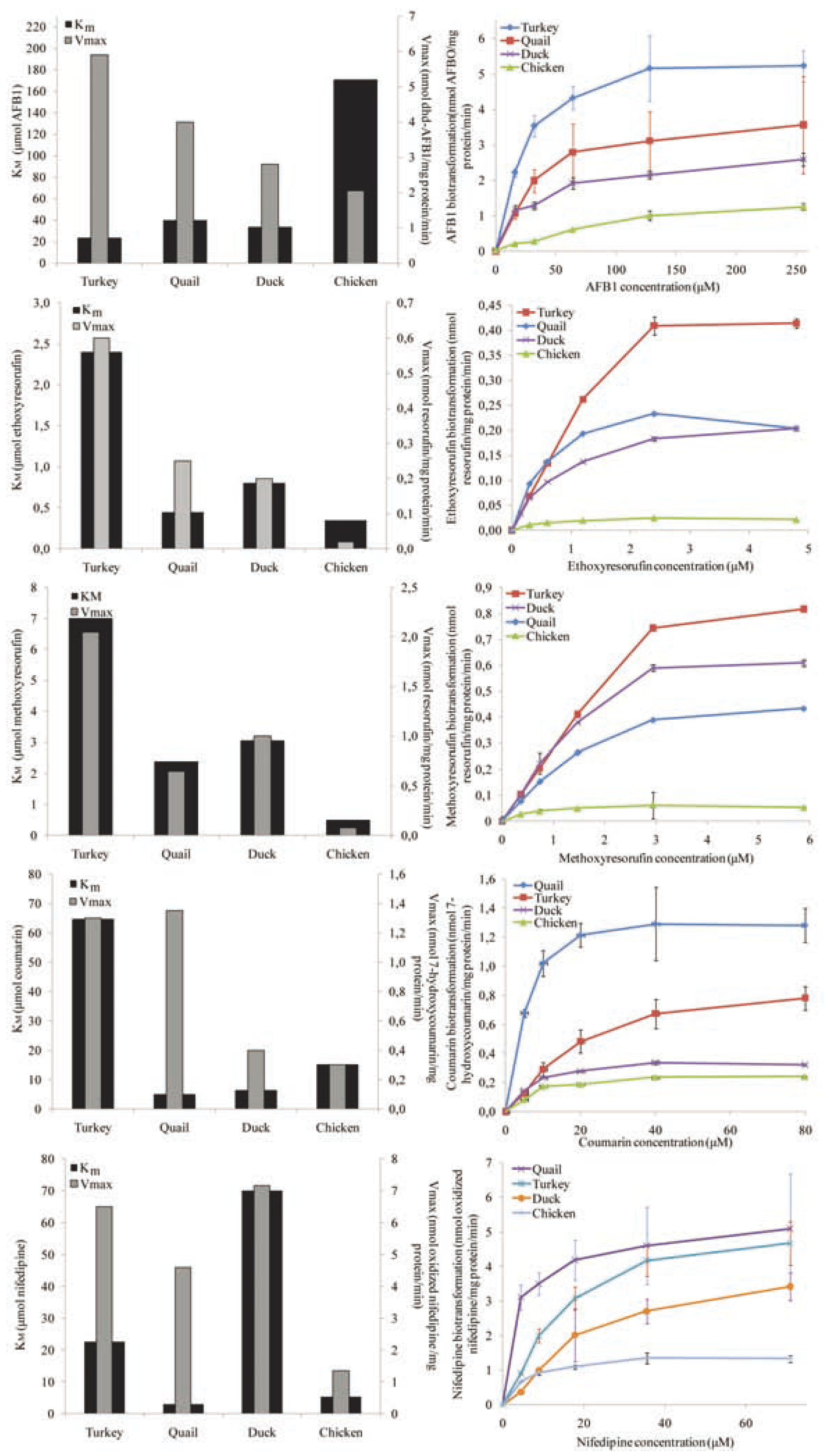
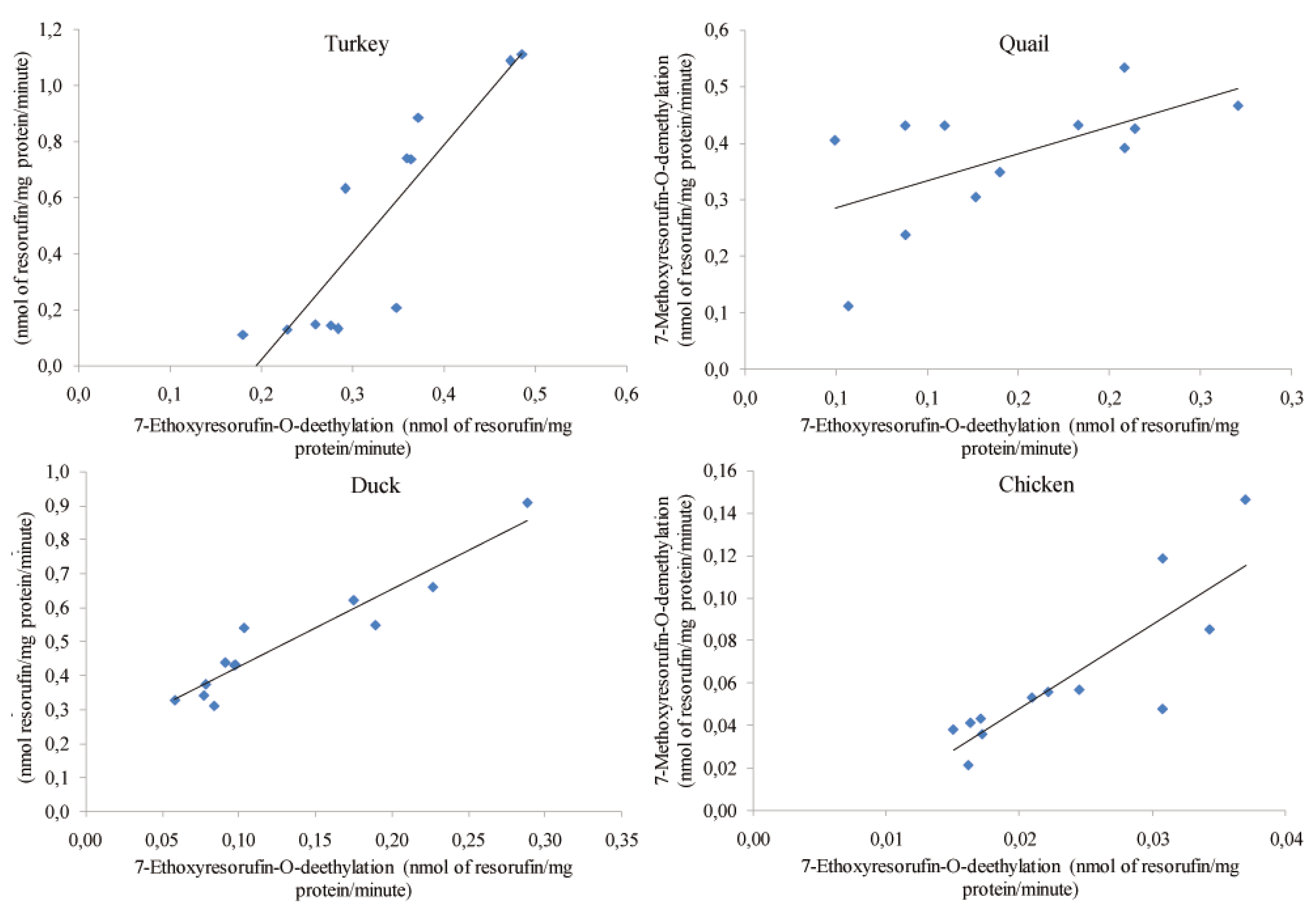
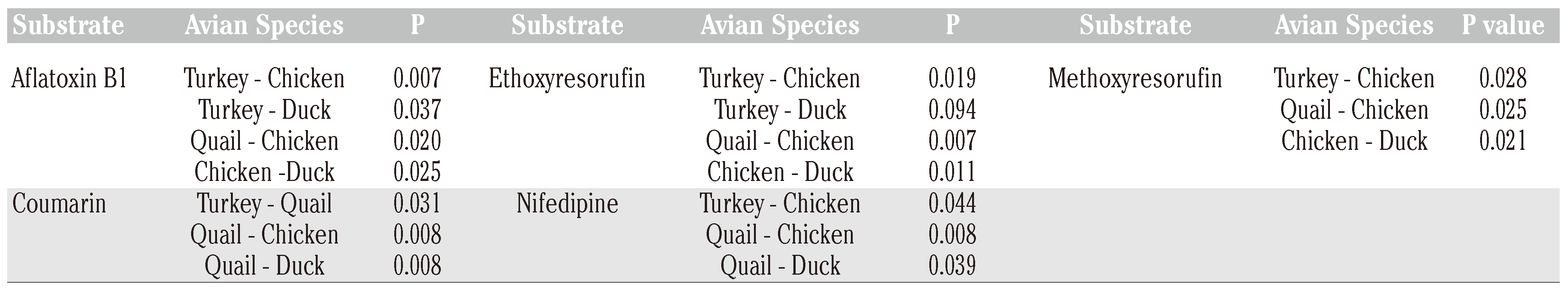
The enzymatic parameters KM and Vmax were determined by nonlinear regression using the Marquardt method adjusting the data to Michaelis-Menten enzyme kinetics using the equation: v = Vmax[S]/(KM+[S]), where v is the enzyme reaction velocity, [S] represents substrate concentration, Vmax represents maximal velocity and KM represents the Michaelis- Menten constant.[27,28] Nonlinear regression fitting was accomplished with the use of the following weighting function: ω = 1/yiα, where yi is the velocity for each substrate concentration and α the magnitude of relation between residuals and variance.[28,29]
The CYP mediated production of the different biotransformation products is expressed as the mean ± standard deviation of six birds (three males and three females). Comparisons were carried out between sexes and between species by a t-test with a significance level of 0.05. Pearson correlation coefficients were computed for EROD vs MROD activity in all species studied. Calculations were performed using the Statistical Analysis System software.[30]

Results

To compare activities between species, data from males and females were pooled since no significant differences between sexes were found. However, significant interspecies dif- ferences in enzyme kinetics by substrate were observed for several orthogonal comparisons (Table 1).
Figure 1 shows the enzymatic constants KM and Vmax and the enzyme kinetics for the four avian species studied. For AFB1 epoxidation, apparent KM and Vmax values ranged from 23.35 to 170.85 μM and from 2.05 to 5.90 nmol/mg protein/min, respectively. KM and Vmax values for EROD activity ranged from 0.39 to 2.55 μM and from 0.03 to 0.72 nmol/mg protein/min, respectively. MROD enzymatic activity resulted in KM values ranging from 0.46 to 7.37 μM and Vmax values from 0.06 to 2.27 nmol/mg pro- tein/min. The KM value for coumarin hydroxy- lation ranged from 4.9 to 64.7 μM, whereas the Vmax values ranged from 0.3 to 1.3 nmol/mg protein/min. Nifedipine oxidation enzyme con- stants ranged from 3.46 to 32.60 μM for KM and from 1.3 to 7.2 nmol/mg protein/min for Vmax.
The highest AFBO production rate was observed for turkey microsomes, while chicken showed the lowest rate. Duck and quail presented intermediate values. EROD activity was also highest in turkey microsomes, while chicken showed the lowest EROD activity. Duck and quail again presented intermediate values. This same trend was also observed for MROD activity. In the case of 7-coumarin hydroxylation, the quail and duck CYP2A6 orthologs had the highest affinity for this substrate but coumarin 7-hydroxylation activity was significantly higher in quail than in duck microsomes. The chicken and turkey orthologs presented medium and low affinity for the same enzymatic activity, respectively. Quail nifedipine oxidation activity was the highest followed by turkey and duck; chicken presented the lowest activity.
EROD and MROD activities showed a simi- lar trend in all species studied with higher enzyme constant values for the 7-methoxyre- sorufin-O-deethylation activity than for 7- ethoxyresorufin-O-deethylation activity. This finding prompted us to investigate the rela-tionship between these two enzymatic activi- ties by correlating 7-ethoxyresorufin-O-deethylase activity against 7-methoxyre- sorufin-O-deethylase activity (Figure 2). Pearson correlation coefficients and P values for turkey, quail, duck and chicken microsomes were 0.88 (P=0.0001), 0.60 (P=0.0400), 0.95 (P=0.0000) and 0.84 (0.0006), respectively.

Discussion

The results of the present study support the notion that phase I biotransformation by CYP enzymes plays a significant role in the large in vivo differences in susceptibility against the AFB1 observed among poultry species but that other variables must also play a role. For example, ducks are known for their extreme sensitivity to AFB1 in vivo and our results demonstrate that duck microsomes have a very high enzyme affinity for AFB1; however, the catalytic rate for AFBO production was lower com- pared to turkey or quail microsomes. These findings suggest that the extreme sensitivity of ducks to AFB1 cannot be explained solely on the basis of the bioactivation of AFB1 into AFBO (phase I metabolism). Future studies of enzymes involved in phase II metabolism are needed to elucidate and give a more precise explanation about the sensitivity shown by each poultry species against AFB1.
In regards to prototype substrate activities, studies with human liver microsomes have shown EROD activity KM and Vmax values of 0.162 μM and 0.028 nmol/mg protein/min, respectively; for MROD activity a KM value of 0.13 μM and a Vmax of 0.038 nmol resorufin/mg protein/min were reported.[31] These values are lower than those found in the present study for quail, turkey and duck liver microsomes but similar to those found for chicken liver microsomes. Our results also show that poultry enzymes have a much lower affinity for coumarin compared with human hepatic enzymes (KM=0.76 M) but their catalytic rate is comparable to that of human liver microsomes (Vmax=0.39 nmol/mg protein/min).[32]
The avian orthologs investigated showed a much lower nifedipine oxidation activity compared with human liver microsomes (Vmax=20.4 nmol/mg protein/min);[33] further, compared with the human CYP3A4 enzyme, which has been reported to have a KM value of 25.3 μM,[33] the enzyme affinity for nifedipine oxidation in the birds studied was similar to that of human in turkey microsomes (KM=22.70 μM), lower in duck microsomes (KM=32.60 M) and higher in chicken (KM=5.37 μM) and quail microsomes (KM=3.46 μM). This large variability in the avian CYP3A4 ortholog enzyme activity observed among the poultry species studied could have important implications from the therapeutic perspective since this enzyme is responsible for the biotransformation of about 70% of all drugs, at least in humans.34
In addition to the biochemical evidence shown in the present study, some CYP homol- ogous protein sequences have been reported in databases such as UniProtKB (http://www.uniprot.org) and the National Center for Biotechnology Information (http://www.ncbi.nlm.nih.gov). In terms of similarity with the human counterparts, the turkey CYP1A5 has 61% homology compared with the human CYP1A1 and 62% homology compared with the human CYP1A2. Human CYP1A1 has a 62.4% homology compared with chicken CYP1A1 and 60% with chicken CYP1A4; human CYP1A2 and chicken 1A4 have 56% homology. In regards to members of the 3A subfamily, the human CYP3A5 has a 59% homology compared with turkey CYP3A37, human CYP3A4 a 59% homology compared with turkey CYP3A37, human CYP3A7 a 58% homology compared with turkey CYP3A37, human CYP3A43 a 56% homology compared with turkey CYP3A37 and human CYP3A4 a 61.1% homology compared with chicken CYP3A80. The presence of enzymatic activity and homologous sequences in both humans and poultry strongly suggests the existence of the CYP orthologs studied in the present study in poultry liver microsomes. To have a better understanding of the role of these enzymes on the bioactivation of compounds such as AFB1it is necessary to further characterize the genes that encode these proteins in all poultry species, specially the gene that encodes the CYP2A6 ortholog since there is no information about this gene in poultry. An interesting find- ing of the present study is the high correlation between EROD and MROD activities in all poultry liver microsomes studied. This close relationship suggests either the existence of an overlapping activity of two separate enzymes or that MROD and EROD activities are catalyzed by a unique CYP1A avian ortholog with different affinities and catalytic rates against each prototype substrate. This issue needs to be further investigated.
The results of the present study show CYP activity against prototype substrates used in human experiments suggesting the existence of avian CYP orthologs of the human enzymes studied. These results also confirm that the differences in sensitivity to AFB1 among poul- try species can be partially explained by differ- ences in the relative enzymatic properties of the CYP avian orthologs responsible for AFB1 bioactivation.35 This study also shows that the turkey liver microsomes are the most efficient bioactivating AFB1 to AFBO, followed by quail, duck and chicken enzymes. In order to com- pletely understand how AFB1 is biotrans- formed in avian species and how metabolism determines in vivo sensitivity it is necessary to further investigate phase I and specially phase II metabolism of AFB1 in poultry.

Author Contributions

H.W.M., experimental procedures developing, data collecting and analyzing, manu- script drafting; G.J.D., article revising; S.M.C., exper- imental procedures supporting, data analyzing.

Acknowledgments

the authors would like to thank the International Foundation for Science (Stockholm, Sweden) for partial funding of our research on in vitro metabolism of aflatoxin B1, to all people involved in this work and to the National University of Colombia in Bogotá, our second home.

Conflicts of Interest

the authors report no conflicts of interest.

References

  1. Josephy, P.D. Molecular toxicology; Oxford University Press: New York, NY, USA, 1997. [Google Scholar]
  2. Chauret, N.; Gauthier, A.; Martin, J.; Nicoll-Griffith, D.A. In vitro comparison of cytochrome P450-mediated metabolic activities in human, dog, cat, and horse. Drug Metab. Dispos. 1997, 25, 1130–1136. [Google Scholar] [PubMed]
  3. Renauld, A.E.; Melancon, M.J.; Sordillo, L.M. Identification of in vitro cytochrome P450 modulators to detect induction by proto- type inducers in the mallard duckling (Anas platyrhynchos). Comp. Biochem. Physiol. C Pharmacol. Toxicol. Endocrinol. 1999, 122, 273–281. [Google Scholar] [CrossRef] [PubMed]
  4. Halpert, J.R.; Guengerich, F.P.; Bend, J.R.; Correia, M.A. Selective inhibitors of cytochromes P450. Toxicol. Appl. Pharmacol. 1994, 125, 163–175. [Google Scholar] [CrossRef] [PubMed]
  5. Pelkonen, O.; Turpeinen, M.; Hakkola, J.; et al. Inhibition and induction of human cytochrome P450 enzymes: Current status. Arch. Toxicol. 2008, 82, 667–715. [Google Scholar] [CrossRef]
  6. Henderson, M.C.; Miranda, C.L.; Stevens, J.U.F.; Deinzer, M.L.; Buhler, D.R. In vitro inhibition of human P450 enzymes by prenylated flavonoids from hops, Humulus lupulus. Xenobiotica 2000, 30, 235–251. [Google Scholar] [CrossRef]
  7. Donato, M.T.; Jiménez, N.; Castell, J.V.; Gómez-Lechón, M.J. Fluorescence-based assays for screening nine cytochrome P450 (P450) activities in intact cells expressing indi- vidual human P450 enzymes. Drug Metab. Dispos. 2004, 32, 699–706. [Google Scholar] [CrossRef]
  8. Peng, J.Z.; Remmel, R.P.; Sawchuk, R.J. Inhibition of murine cytochrome P4501A by tacrine: In vitro studies. Drug Metab. Dispos. 2004, 32, 805–812. [Google Scholar] [CrossRef]
  9. Appiah-Opong, R.; Commandeur, J.N.; van Vugt-Lussenburg, B.; Vermeulen, N.P. Inhibition of human recombinant cytochrome P450s by curcumin and cur- cumin decomposition products. Toxicology 2007, 235, 83–91. [Google Scholar] [CrossRef]
  10. McManus, M.E.; Boobis, A.R.; Minchin, R.F.; et al. Relationship between oxidative metab- olism of 2-acetylaminofluorene, debriso- quine, bufuralol, and aldrin in human liver microsomes. Cancer Res. 1984, 44, 5692–5697. [Google Scholar]
  11. Kenworthy, K.E.; Bloomer, J.C.; Clarke, S.E.; Houston, J.B. CYP3A4 drug interactions: Correlations of 10 in vitro probe substrates. Br. J. Clin. Pharmacol. 1999, 48, 716–727. [Google Scholar] [CrossRef]
  12. Sy, S.K.; Tang, B.; Pastrakuljic, A.; et al. Detail characterization of experimental derived human hepatic CYP1A1 activity and expression using differential inhibition of ethoxyresorufin O-deethylation by fluvox- amine. Eur. J. Pharmacol. 2001, 57, 377–386. [Google Scholar]
  13. Quintieri, L.; Geroni, C.; Fantin, M.; et al. Formation and antitumor activity of PNU- 159682, a major metabolite of nemoru- bicin in human liver microsomes. Clin. Cancer Res. 2005, 11, 1608–1617. [Google Scholar] [CrossRef] [PubMed]
  14. Díaz, G.J.; Squires, E.J. Metabolism of 3- methylindole by porcine liver microsomes: Responsible cytochrome P450 enzymes. Toxicol. Sci. 2000, 55, 284–292. [Google Scholar] [CrossRef] [PubMed]
  15. Šavlík, M.; Poláčkcová, L.; Szotáková, B.; et al. Activities of biotransformation enzymes in pheasant (Phasianus colchicus) and their modulation by in vivo administration of mebendazole and ubendazole. Res. Vet. Sci. 2007, 83, 20–26. [Google Scholar] [CrossRef]
  16. International Chicken Genome Sequencing Consortium. Sequence and comparative analysis of the chicken genome provide unique perspectives on vertebrate evolution. Nature 2004, 432, 695–716. [Google Scholar] [CrossRef]
  17. Dalloul, R.A.; Long, J.A.; Zimin, A.V.; et al. Multi-platform next-generation sequencing of the domestic turkey (Meleagris gallopavo): Genome assembly and analysis. PLoS Biol. 2010, 8, e1000475. [Google Scholar] [CrossRef]
  18. Leeson, S.; Díaz, G.J.; Summers, J.D. Poultry metabolic disorders and mycotoxins; University Books: Guelph, ON, USA, 1995. [Google Scholar]
  19. Hussein, H.S.; Brasel, J.M. Toxicity, metabolism, and impact of mycotoxins on humans and animals. Toxicol. 2001, 167, 101–134. [Google Scholar] [CrossRef]
  20. Eaton, D.L.; Gallagher, E.P. Mechanisms of aflatoxin carcinogenesis. Annu. Rev. Pharmacol. Toxicol. 1994, 34, 135–172. [Google Scholar] [CrossRef]
  21. Verma, R.J. Aflatoxin causes DNA damage. Int. J. Hum. Genet. 2004, 4, 231–236. [Google Scholar] [CrossRef]
  22. Do, J.H.; Choi, D. Aflatoxins: Detection, toxicity, and biosynthesis. Biotechnol. Bioprocess. Eng. 2007, 12, 585–593. [Google Scholar] [CrossRef]
  23. Ferguson, L.R.; Philpott, M. Nutrition and mutagenesis. Annu. Rev. Nutr. 2008, 28, 313–329. [Google Scholar] [CrossRef] [PubMed]
  24. Redinbaugh, M.G.; Turley, R.B. Adaptation of the bicinchoninic acid protein assay for the use with microtiter plates and sucrose gradient fractions. Anal. Biochem. 1986, 153, 267–71. [Google Scholar] [CrossRef] [PubMed]
  25. Busby, W.F.; Ackermann, J.M.; Crespi, C.L. Effect of methanol, ethanol, dimethyl sulphoxide and acetronitrile on in vitro activities of cDNA-expressed human cytochromes P-450. Drug Metab. Dispos. 1999, 27, 246–249. [Google Scholar] [PubMed]
  26. Blanchard, J. Evaluation of the relate efficacy of various techniques for deproteiniz- ing plasma samples prior to high-perform- ance liquid chromatography analysis. J. Chromatogr. 1981, 226, 455–460. [Google Scholar] [CrossRef]
  27. Bisswanger, H. Enzyme kinetics. In Principles and methods, 2nd ed.; Revised and Updated Edition; Wiley-VCH Verlag GmbH & Co., KGaA: Weinheim, Germany, 2003. [Google Scholar]
  28. Marangoni, A.G. Enzyme kinetics. A modern approach; Wiley Interscience. John Wiley & Sons, Inc: Hoboken, NJ, USA, 2008. [Google Scholar]
  29. Kakkar, T.; Boxenbaum, H.; Mayersohn, M. Estimation of Ki in a competitive enzyme- inhibition model: Comparisons among three methods of data analysis. Drug Metab. Dispos. 1999, 27, 756–762. [Google Scholar]
  30. SAS Institute Inc. SAS/STAT® 9.2 User’s Guide; SAS Institute Inc.: Cary, NC, USA, 2008. [Google Scholar]
  31. Hanioka, N.; Tatarazako, N.; Jinno, H.; et al. Determination of cytochrome P450 1A activities in mammalian liver microsomes by high-performance liquid chromatogra- phy with fluorescence detection. J. Chromatogr. B Biomed. Sci. Appl. 2000, 744, 399–406. [Google Scholar] [CrossRef]
  32. Yamasaki, H.; Tanaka, M.; Shimada, T. Highly sensitive high-performance liquid chromatographic assay for coumarin 7-hydrox-ylation and 7-ethoxycoumarin O-deethyla- tion by human liver cytochrome P450 enzymes. J. Chromatogr. B Biomed. Sci. Appl. 1999, 721, 13–19. [Google Scholar] [CrossRef]
  33. Patki, K.C.; von Moltke, L.L.; Greenblatt, D.J. In vitro metabolism of midazolam, triazolam, nifedipine, and testosterone by human liver microsomes and recombinant cytochromes P450: Role of CYP3A4 and CYP3A5. Drug Metab. Dispos. 2003, 31, 938–944. [Google Scholar] [CrossRef]
  34. Klaassen, C.D. (Ed). Casarett and Doull´s Toxicology-The Basic Science of Poisons, 7th ed.; McGraw Hill Medical: New York, NY, USA, 2008; pp. 180–190. [Google Scholar]
  35. Lozano, M.C.; Díaz, G.J. Microsomal and cytosolic bioactivation of aflatoxin B1 in four poultry species. Br. Poult. Sci. 2006, 47, 734–741. [Google Scholar] [CrossRef]
Figure 1. Enzymatic constants Km and Vmax obtained by nonlinear regression (left) and Michaelis-Menten kinetics obtained from experimental data (right) for CYP avian orthologs. Enzyme activities from top to bottom are AFB1 epoxidase, 7-ethoxyresorufin-O-deethylase (EROD), 7-methoxyresorufin-O-deethylase (MROD), coumarin 7-hydrox-ylase and nifedipine oxidase. Enzymatic constants KM and Vmax are graphed in black and gray, respectively. In Michaelis-Menten kinetics each point represents the mean ± standard deviation of 6 birds.
Jox 01 e4 g001
Figure 2. Linear regression of 7-Ethoxyresorufin-O-deethylase (EROD) vs 7-methoxyre- sorufin-O-deethylase (MROD) activities in liver microsomes (n=12) in four commercial poultry species. Correlation coefficients and level of significance are shown in the text.
Jox 01 e4 g002
Table 1. Orthogonal contrasts of enzyme kinetics among four commercial poultry species. Only values that are statistically different (P<0.05) are shown.
Jox 01 e4 i001
Disclaimer/Publisher’s Note: The statements, opinions and data contained in all publications are solely those of the individual author(s) and contributor(s) and not of MDPI and/or the editor(s). MDPI and/or the editor(s) disclaim responsibility for any injury to people or property resulting from any ideas, methods, instructions or products referred to in the content.

