1. Introduction
In thalassemia major patients, regular transfusion therapy leads to a deficit in the immune response and, thus, to susceptibility to infections and neoplastic events [
1]. In these patients, the immunological picture outlines the reduced functionality of phagocytes and natural killer cells; alterations in the IL-2-mediated lymphocyte response; defects in antigen presentation due to monocyte–macrophages; the altered production of white cell precursors in bone marrow; and severe changes in the major histocompatibility system (HLA) [
2]. Lymphocytopenia occurs in 96.1% of severe cases of SARS-CoV-2 infection [
3,
4]. COVID-19-related lymphopenia is due to various mechanisms, which lead to an increase in lymphocytic apoptosis [
5,
6] by upregulating Fas and Fas ligands (a type-II transmembrane protein that belongs to the tumor necrosis factor TNF family) in T cells, especially IFN-I sensitized virus-specific T cells (CD4+ and CD8+), or their redistribution caused by the chemotaxis of lung tissue [
7]. Post-COVID-19 lymphocytic quantitative and functional disorders may compromise the immune response and favor the onset of infections from opportunistic pathogens [
6], as described in our case.
2. Case History
A 50-year-old Caucasian woman affected by β thalassemia major, IVS1-6/IVS1-6 genotype, was subjected to regular hemotransfusions since she was 6 months of age to maintain a hemoglobin (Hb) concentration level of 9–10 gr/dL. At the time of the study, she received 2 units of packed red blood cells (RBC) every two weeks. Splenectomy and cholecystectomy were performed at ages 16 and 25 years, respectively. She had received regular chelation therapy with deferoxamine from 2 years of age to date at the dose of 2 g for 5 days a week with good compliance. However, her mean serum ferritin level was 1000 ng/mL, and the iron burden was equal to <155 mL blood/kg/year. At age 20, she tested as serum positive for hepatitis C and hepatitis B; she was treated with PEG interferon and ribavirin, achieving a good response. On 14 October 2020, she was admitted to our COVID-19 Medicine Internal Unit as she developed SARS-CoV-2 infection with bilateral pneumonitis. Laboratory tests showed an increased number of white blood cells (21,100/mmc), neutrophilia (91%), and lymphocytopenia (9%). Hemoglobin levels were good (10 gr/dL).
The patient was treated with a remdesivir therapy with a dose of 200 mg on the first day followed by 100 mg on the second day; dexamethasone, 8 mg/day; enoxaparin, 6.000 UI/day; doxycycline, 200 mg on the first day followed by 100 mg from the second day to the fifteenth day; vitamin C; antioxidants; and anti-inflammatory drugs. Moreover, the standard transfusion regimen had to be increased during hospitalization as hemoglobin levels tended to decrease; thus, she received a blood transfusion with packed RBC every week until discharge. The clinical, instrumental, and laboratory picture of bilateral pneumonia regressed after about one month, and the patient was discharged from the hospital.
On 27 December 2020, she developed a fever (T 39 °C); pharyngodynia; odynophagia; sinusitis; and a voluminous, hard, and painful pseudo-tumoral lesion adhering to the superficial and deep planes of the right lateral cervical region, which did not regress after therapy with amoxicillin and clavulanic acid. Molecular SARS-CoV-2 diagnostic molecular tests were negative. A marked post-COVID-19 lymphocytopenia persisted upon blood count (white blood cells: 16,300/mmc; neutrophils: 86%; lymphocytes: 4%; monocytes: 8%; eosinophils: 2%). She was admitted into the Otolaryngology Unit and subjected to Neck Contrast Computed Tomography (CT).
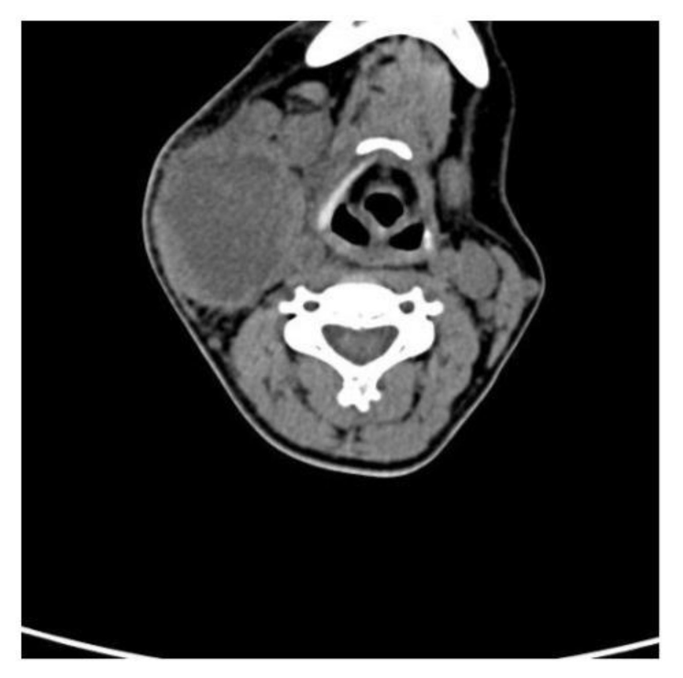
Contrast CT showed that the pseudo-tumoral lesion of the right lateral cervical region measured about 6 × 5 × 6 cm and was characterized by a hypodense probably necrotic–colliquative core, hyperdense internal shoots, thick walls, and jagged edges (
Figure 1).
Fine needle aspiration showed that the pseudo-tumoral lesion had a fibrous wall with lively vascular neogenesis and intense lymph plasma cell infiltration, without atypical elements. It found positivity for piperacillin- and tazobactam-sensitive Klebsiella Pneumoniae. The patient followed antibiotic therapy, resulting in the resolution of the infection. On 30 June 2020, we observed that lymphocytopenia resolved 6 months after discharge from the COVID-19 Medicine Internal Unit, as shown by the blood count (WB: 14,000/mmc N 65% L 30% M 5%). The patient’s transfusion requirements decreased with reasonable well-being.
3. Discussion and Conclusions
COVID-19 is a viral infection with a high impact on the hematopoietic system and hemostasis. Lymphopenia might be seen as a cardinal laboratory finding. A retrospective study showed that lymphopenia persisted in a remarkable percentage of patients who had recovered from SARS-CoV-2 infection [
8]. Post-COVID-19 lymphocytopenia is due to transient immune suppression, affecting innate and acquired immunity. Hence, there is a loss of self-tolerance characterized by the development of autoantibodies, and impaired immune reconstitution, which amplifies immune damage [
9]. It has also been found that regulatory T lymphocytes are suppressed by the activation of lymphocytes with lineages of self-reactivity [
10]. Furthermore, a high percentage of co-expression Tim −3 + PD-1 + T subset existed both in CD4+ and CD8+ T lymphocytes, especially in intensive care unit (ICU) patients, which suggested that T cells were in an exhausted state from activation, leading to a depletion of immune functions [
11].
Regular blood transfusion exerts an immunosuppressive effect on thalassemic patients. Dzik et al. showed at least two categories of immunosuppressive effects, HLA-dependent mechanisms directed against adaptive immunity and another unspecified against innate immunity. The non-specific mechanism could be due to the infusion of apoptotic blood cells and transforming growth factor beta (TGF-β) by blood transfusion [
12]. Recent studies have shown that regular and continuous blood transfusions cause the downregulation of cellular immune functions, with an increased frequency of relapses of solid tumors, post-operative bacterial infections, and greater severity of viral infection [
13]. Thalassemic patients also display enhanced susceptibility to infections as a consequence of several complex biological processes. The most important factors promoting thalassemia-induced alterations of the immune system are disease-related factors, such as decreased levels of complement, properdin, and lysozyme; reduced absorption and phagocytic abilities of polymorphonuclear neutrophils; disturbed chemotaxis; and altered intracellular metabolism and therapy-related factors, such as blood transfusions related to iron-overload and allogenic stimulation, iron chelation therapy and obviously splenectomy when applicable [
14]. Splenectomy also causes a predisposition to infections and changes in the immune system. It is a therapeutic intervention used in thalassemia in order to avoid an increased consumption of red blood cells caused by hypersplenism [
15]. The spleen is a primary organ of immunological surveillance as it is a reservoir of immunocompetent lymphocytes. Thus, its removal causes alterations in the immunological system, such as a decrease in the activity of natural killers and a weakened IgM memory B-cell response. In addition, an exacerbation of immunological alterations caused by multiple transfusions was found in splenectomized thalassemia patients [
16].
The observations in our case support the hypothesis that post-COVID-19 lymphocytopenia in thalassemia patients may facilitate the onset of severe infections from opportunistic pathogens. Additional studies performed on a large sample of patients are needed to replicate our findings.
Author Contributions
A.P., E.Q., P.S. and L.R. contributed equally to the study. All authors have read and agreed to the published version of the manuscript.
Funding
This article received no external funding.
Institutional Review Board Statement
Not applicable.
Informed Consent Statement
This manuscript contains an individual’s personal data, which we have consent to publish. This manuscript does not report on or involve the use of any animal or tissue.
Data Availability Statement
All data generated or analyzed during this report are included in this published article.
Conflicts of Interest
The authors have no other relevant affiliations or financial involvement with any organization or entity with a financial interest in or financial conflict with the subject matter or materials discussed in the manuscript.
References
- Grzelak, I.; Zaleska, M.; Olszewski, W. Blood transfusions downregulate hematopoiesis and subsequently downregulate the immune response. Transfusion 1998, 38, 1104–1114. [Google Scholar] [CrossRef] [PubMed]
- Mincheff, M.S.; Meryman, H.T.; Kapoor, V.; Alsop, P.; Wötzel, M. Blood Transfusion and Immunomodulation: A Possible Mechanism. Vox Sang. 1993, 65, 18–24. [Google Scholar] [CrossRef] [PubMed]
- Liu, X.; Zhang, R.; He, G. Hematological findings in coronavirus disease 2019: Indications of progression of disease. Ann. Hematol. 2020, 99, 1421–1428. [Google Scholar] [CrossRef] [PubMed]
- Li, T.; Qiu, Z.; Han, Y.; Wang, Z.; Fan, H.; Lu, W.; Xie, J.; Ma, X.; Wang, A. Rapid loss of both CD4+ and CD8+ T lymphocyte subsets during the acute phase of severe acute respiratory syndrome. Chin. Med. J. 2003, 116. [Google Scholar]
- Fan, B.E. Hematologic parameters in patients with COVID-19 infection. Am. J. Hematol. 2020, 95, E131–E134. [Google Scholar] [CrossRef] [PubMed] [Green Version]
- Wong, R.; Wu, A.; To, K.F.; Lee, N.; Lam, C.W.K.; Wong, C.K.; Chan, P.; Ng, M.H.L.; Yu, L.M.; Hui, D.; et al. Haematological manifestations in patients with severe acute respiratory syndrome: Retrospective analysis. BMJ 2003, 326, 1358–1362. [Google Scholar] [CrossRef] [PubMed] [Green Version]
- Channappanavar, R.; Fehr, A.R.; Vijay, R.; Mack, M.; Zhao, J.; Meyerholz, D.K.; Perlman, S. Dysregulated type I interferonand inflammatory monocyte-macrophage responses cause lethal pneumonia in SARS-CoV-infected mice. Cell Host. Microbe. 2016, 19, 181–193. [Google Scholar] [CrossRef] [PubMed] [Green Version]
- Varghese, J.; Sandmann, S.; Ochs, K.; Schrempf, I.M.; Frömmel, C.; Dugas, M.; Schmidt, H.H.; Vollenberg, R.; Tepasse, P.R. Persistent symptoms and lab abnormalities in patients who recovered from COVID-19. Sci. Rep. 2021, 11, 12775. [Google Scholar] [CrossRef] [PubMed]
- Cañas, C.A. The triggering of post-COVID-19 autoimmunity phenomena could be associated with both transient immunosuppression and an inappropriate form of immune reconstitution in susceptible individuals. Med. Hypotheses 2020, 145, 110345. [Google Scholar] [CrossRef]
- Qin, C.; Zhou, L.; Hu, Z.; Zhang, S.; Yang, S.; Tao, Y.; Xie, C.; Ma, K.; Shang, K.; Wang, W.; et al. Dysregulation of Immune Response in Patients With Coronavirus 2019 (COVID-19) in Wuhan, China. Clin. Infect. Dis. 2020, 71, 762–768. [Google Scholar] [CrossRef]
- Jin, H.-T.; Anderson, A.C.; Tan, W.G.; West, E.E.; Ha, S.-J.; Araki, K.; Freeman, G.J.; Kuchroo, V.K.; Ahmed, R. Cooperation of Tim-3 and PD-1 in CD8 T-cell exhaustion during chronic viral infection. Proc. Natl. Acad. Sci. USA 2010, 107, 14733–14738. [Google Scholar] [CrossRef] [PubMed] [Green Version]
- Dzik, W.H. Apoptosis, TGFβ and transfusion-related immunosuppression: Biologic versus clinical effects. Transfus. Apher. Sci. 2003, 29, 127–129. [Google Scholar] [CrossRef]
- Blumberg, N.; Heal, J.M. Transfusion-induced immunomodulation and its possible role in cancer recurrence and perioperative bacterial infection. Yale J. Biol. Med. 1990, 63, 429–433. [Google Scholar] [PubMed]
- Gluba-Brzózka, A.; Franczyk, B.; Rysz-Górzyńska, M.; Rokicki, R.; Koziarska-Rościszewska, M.; Rysz, J. Pathomechanisms of Immunological Disturbances in β-Thalassemia. Int. J. Mol. Sci. 2021, 22, 9677. [Google Scholar] [CrossRef] [PubMed]
- Farmakis, D.; Giakoumis, A.; Polymeropoulos, E.; Aessopos, A. Pathogenetic aspects of immune deficiency associated with beta-thalassemia. Med Sci. Monit. 2003, 9, 22. [Google Scholar]
- Zaninoni, A.; Fermo, E.; Vercellati, C.; Marcello, A.P.; Barcellini, W.; Bianchi, P. Congenital Hemolytic Anemias: Is There a Role for the Immune System? Front. Immunol. 2020, 11, 1309. [Google Scholar] [CrossRef] [PubMed]
| Publisher’s Note: MDPI stays neutral with regard to jurisdictional claims in published maps and institutional affiliations. |
© 2022 by the authors. Licensee MDPI, Basel, Switzerland. This article is an open access article distributed under the terms and conditions of the Creative Commons Attribution (CC BY) license (https://creativecommons.org/licenses/by/4.0/).


