Case Report

Pancytopenia Related to Splenic Angiosarcoma: A Case Report and Literature Review

Department of Pathology, Chair of Oncology, Medical University of Lodz, 92-213 Lodz, Poland
*
Author to whom correspondence should be addressed.
Hematol. Rep. 2024, 16(4), 648-655; https://doi.org/10.3390/hematolrep16040063
Submission received: 1 August 2024 / Revised: 14 October 2024 / Accepted: 15 October 2024 / Published: 18 October 2024

Abstract

:
Background: Angiosarcomas are highly aggressive malignancies with endothelial differentiation, presenting considerable challenges in oncology, especially when arising in rare locations such as the spleen. These tumors predominantly affect adults and are commonly found in the skin, breast, liver, or soft tissues, with more unusual occurrences in other organs. Angiosarcomas have a high propensity for metastasis, typically spreading to the liver, lungs, lymph nodes, and gastrointestinal tract. Splenic angiosarcoma, with fewer than 300 documented cases, is an especially rare and complex form of this malignancy. Case presentation: This report details a case of splenic angiosarcoma in a 45-year-old male, where bone marrow metastases were the first clinical presentation, initially mimicking myelodysplastic syndrome (MDS) due to persistent pancytopenia. Conclusions: The eventual identification of the splenic origin underscores the diagnostic difficulties and clinical challenges inherent in managing such atypical and rare presentations.

1. Introduction

Angiosarcoma is a malignant neoplasm with morphological or immunophenotypic features of endothelial differentiation. These aggressive tumors are generally found in adults, mainly in the skin, soft tissue, breast, liver, and, infrequently, in other organs such as the spleen [1]. To date, there are fewer than 300 reports of splenic angiosarcoma available in the literature [2] (Table 1). Angiosarcoma typically metastasizes to the liver, lungs, lymph nodes or bone [1,3,4]. Here, we present a rare case of angiosarcoma in the form of splenic angiosarcoma with bone marrow metastasis mimicking myelodysplastic syndrome.

2. Case Presentation

A 45-old-year male patient was admitted to the Hematology Department with a suspicion of myelodysplastic syndrome (MDS). He presented with petechiae, and blood tests showed a decreased hemoglobin level of 9.5 g/dL and thrombocytopenia at 24 k/mcL (2.4 × 10⁴/μL) (Table 2). Examination of FLT3 and NMP1 mutations was performed, but no abnormalities were detected. The presence of schistocytes in the peripheral blood smear, increasing liver enzyme levels, and the patient’s deteriorating condition were suggestive of acquired thrombotic thrombocytopenic purpura (aTTP). After a suspicion of aTTP, the following tests were performed: reticulocytes—0.110 × 10⁶/μL, INR—1.11, PT—12.8 s, APTT—23.8 s, LDH—978 U/L, haptoglobin—<0.1 g/L, total bilirubin—1.08 mg/dL, fibrinogen—188 mg/dL, and ADAMTS13 antibodies in the plasma, but they were not detected. Vitamin B12 and folic acid deficiencies were also excluded (1306 pg/mL and 6.15 ng/mL, respectively). The patient also experienced abdominal pain and periodic fever during hospitalization. The patient underwent three cycles of plasma exchange. However, due to the lack of improvement in blood morphology tests, a decision to discontinue the procedure was made.
Due to the occurrence of a dry tap during bone marrow aspiration, a trephine biopsy was performed. The histopathological examination showed widespread infiltration of the bone marrow by elongated and epithelioid neoplastic cells. These cells created numerous pseudovascular spaces. Additionally, reticular fibrosis of grade 3 was present (Figure 1A,B). Immunohistochemical profiling of the neoplastic cells revealed positivity for vascular markers (CD34, CD31, CD117, SMA), and negativity for cytokeratins, mastocytic, myeloid, and lymphoid markers (CK AE1/AE3, MPO, CD15, CD61, CD71, CD123, CD138, CD20, CD3, TdT, CD25, MCT) (Figure 1C–F).
Whole-body contrast-enhanced computed tomography (CT) revealed numerous hypodense foci exclusively in the spleen (Figure 1G). Magnetic resonance imaging (MRI) also identified the lesions, with the largest measuring 36 × 21 mm and 32 × 30 mm, containing hemorrhagic deposits (Figure 1H). All the above imaging tests, including of the skeletal system, revealed no signs of metastatic remodeling. Thus, the final diagnosis was splenic angiosarcoma metastatic to bone marrow.
The patient underwent palliative treatment with paclitaxel at a dose of 60 mg/m2 every 7 days with prophylaxis of febrile neutropenia using short-acting G-CSF starting in February 2022. Radiotherapy targeting the primary tumor in the spleen was added in July 2022. After receiving a total of 29 cycles of paclitaxel without an effective response, a weekly infusion of doxorubicin at a dose of 7.5 mg/m2 was recommended, with dose reduction due to the patient’s general condition. Nine months after the beginning of treatment, the patient died.

3. Discussion

In the presented case, a diagnosis of bone marrow metastatic angiosarcoma originating from the spleen is discussed, highlighting a distinctive clinico-pathological scenario due to the myelodysplastic syndrome-like symptoms induced by the bone marrow metastases. The uniqueness of this case is underscored by the rare occurrence of such metastases, emphasizing the biological complexity and variability in the clinical presentation of angiosarcoma [28].
A particularly rare manifestation in the patient we described was bone metastases located between the trabeculae, directly infiltrating the bone marrow and impacting blood count results. The thrombocytopenia and petechiae observed in the patient initially suggested a hematopoietic disorder, such as myelodysplastic syndrome (MDS), leukemia, or thrombocytopenic purpura. The rarity of this manifestation contributed to the delay in reaching the final diagnosis.
In our patient’s case, typical imaging tests and blood tests were performed for the diagnosis of angiosarcoma. Both MRI and CT imaging are standard diagnostic tools for these tumors. However, the biopsy in this case differed from the usual approach. Typically, for solid tumors, a biopsy of the primary lesion or a lymph node affected by metastasis is performed. In our case, the patient underwent a procedure more common for hematopoietic tumors, namely a trephine biopsy. This biopsy allowed for the collection of metastatic tumor cells, which were subsequently characterized using immunohistochemistry (IHC). The results of the imaging tests and IHC together confirmed the final diagnosis of angiosarcoma of splenic origin.
Upon review of the literature (Table 1), it is evident that splenic angiosarcoma can manifest across a wide age range. For instance, a case involving a 25-year-old woman demonstrates that younger populations can also be affected by this disease [5,16,23]. Most reported instances involve symptoms of abdominal discomfort, pain, or palpable masses in the left epigastrium, which can serve as early indicators of the disease [5,6,7,20,23,24,27]. Additionally, while the liver is the most frequent site for metastases of splenic angiosarcoma, occurrences have been documented in bones and bone marrow (as in the presented case), peritoneum, lungs, kidneys, and potentially the thyroid gland, illustrating the extensive metastatic potential of angiosarcoma [25,26,27].
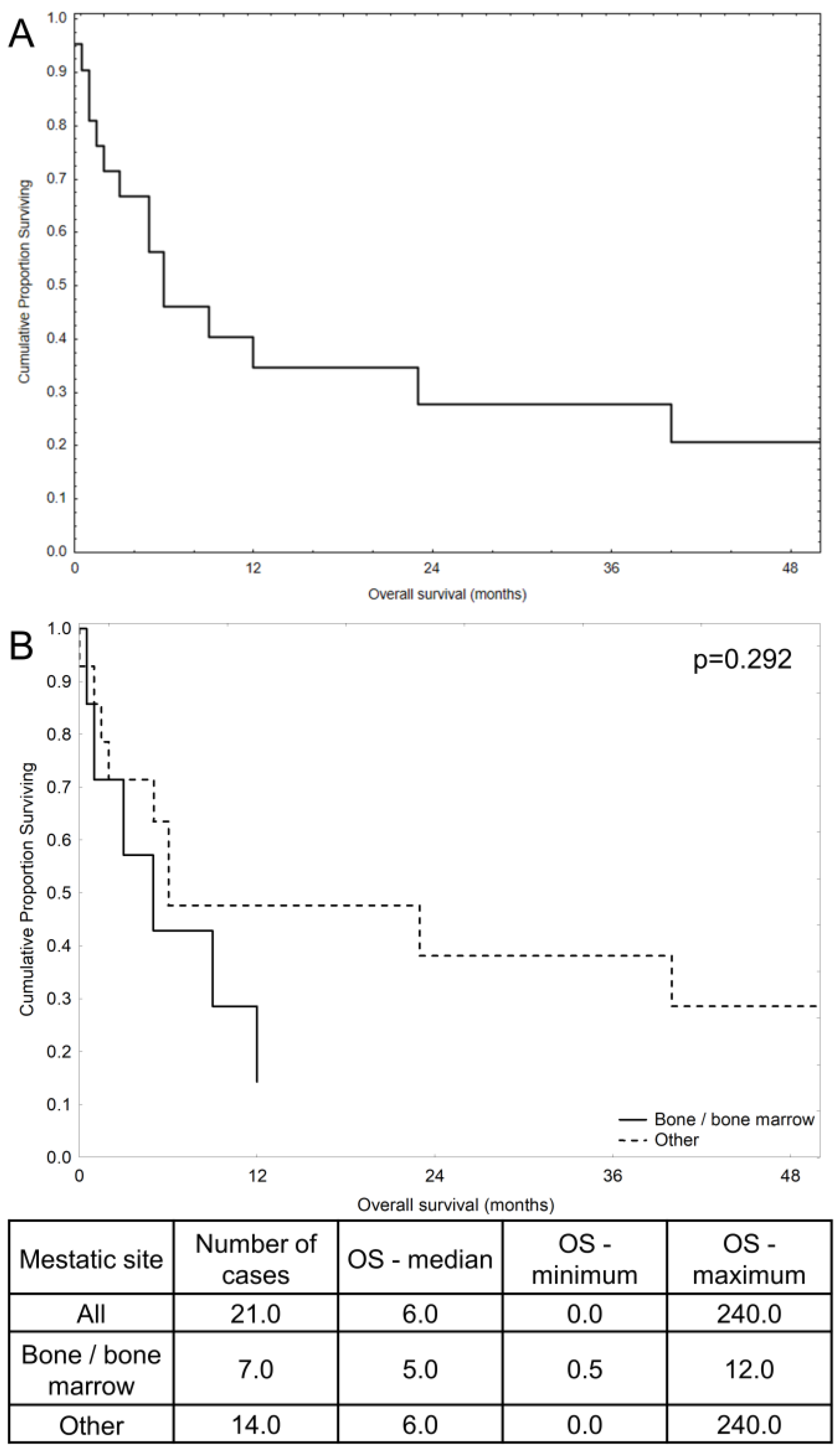
Current treatment approaches predominantly involve splenectomy followed by chemotherapy with agents such as paclitaxel [17,18], but the outcomes vary across patients with various presentations of the disease (Table 1, Figure 2). The presence of unique metastatic profiles and clinical presentations, such as in the bone marrow metastatic angiosarcoma case discussed here, accentuates the need for individualized treatment plans and further research into tailored therapeutic interventions. Notably, in cases with bone marrow metastases, regardless of patient age, survival rates are poor [8,23,26,27] (Figure 2). Our patient died within 9 months of treatment initiation, which is close to the median survival reported in the literature (Figure 2). This suggests that metastases to the bone marrow might be a significant prognostic factor, warranting further investigation into survival differences compared to other metastatic sites. An important aspect of both treatment and diagnostics in this case appears to be splenectomy. In the patient described, the disease primarily manifested as abnormalities in blood morphology tests, which were secondary to bone marrow metastases. These metastases led to the observed blood abnormalities. In cases of metastatic disease, surgery does not improve prognosis and additionally exposes the patient to potential complications. In this case, surgery was not feasible, not only due to the widespread nature of the disease, but also because of the blood abnormalities directly caused by bone marrow metastases. In summary, the use of palliative chemotherapy was the only viable option with a chance to prolong the patient’s life and slow the progression of the neoplastic disease.
Improving outcomes in this area remains a challenge for clinicians. In patients presenting with similar symptoms, it is worth considering immediate imaging tests to identify the primary tumor site. This approach enables the simultaneous diagnosis of both hematopoietic disorders and the potential development of a neoplastic process in other organs.

4. Conclusions

Abnormalities in blood counts should not raise suspicions solely of hematological diseases. Such an approach can significantly delay the diagnostic process, thereby reducing the patient’s chances for longer survival and better treatment outcomes. The changes we have described should raise oncological vigilance toward a potential proliferative process occurring in the organs, as seen in this case. Therefore, the most important takeaway is that imaging tests with high sensitivity in detecting neoplastic foci should be ordered early in the diagnostic process.
In conclusion, the case of bone marrow metastatic angiosarcoma originating from the spleen provides valuable insight into the diverse clinical manifestations and complex biological behavior of angiosarcoma. It underscores the imperative for precise diagnosis and treatment of this malignancy.

Author Contributions

Conceptualization, J.M. and M.B.; methodology, J.M.; software, J.M.; validation, M.B., J.M. and B.S.; formal analysis, N.S.; investigation, B.S.; resources, N.S.; data curation, M.M.; writing—original draft preparation, J.M.; writing—review and editing, M.B.; visualization, M.M.; supervision, M.B.; project administration, M.B. All authors have read and agreed to the published version of the manuscript.

Funding

The research was solely funded by the Department of Pathology, Chair of Oncology, Medical University of Lodz (503/1-034-03/503-11-001).

Institutional Review Board Statement

All procedures performed in studies involving human participants were in accordance with the ethical standards of the institutional and/or national research committee and with the 1964 Helsinki Declaration and its later amendments or comparable ethical standards.

Informed Consent Statement

Informed verbal consent was obtained from all subjects involved in the study, with a witness present.

Data Availability Statement

The authors confirm that the data supporting the findings of this study are available within the article.

Conflicts of Interest

The authors declare no conflicts of interest.

References

  1. Young, R.J.; Brown, N.J.; Reed, M.W.; Hughes, D.; Woll, P.J. Angiosarcoma. Lancet Oncol. 2010, 11, 9983–9991. [Google Scholar] [CrossRef] [PubMed]
  2. Damouny, M.; Mansour, S.; Khuri, S. Primary Angiosarcoma of the Spleen: An Aggressive Neoplasm. World J. Oncol. 2022, 13, 337. [Google Scholar] [CrossRef] [PubMed]
  3. Gaballah, A.H.; Jensen, C.T.; Palmquist, S.; Pickhardt, P.J.; Duran, A.; Broering, G.; Elsayes, K.M. Angiosarcoma: Clinical and imaging features from head to toe. Br. J. Radiol. 2017, 90, 20170039. [Google Scholar] [CrossRef]
  4. Tateishi, U.; Hasegawa, T.; Kusumoto, M.; Yamazaki, N.; Iinuma, G.; Muramatsu, Y.; Moriyama, N. Metastatic angiosarcoma of the lung: Spectrum of CT findings. Am. J. Roentgenol. 2003, 180, 1671–1674. [Google Scholar] [CrossRef]
  5. Frontario, S.C.N.; Goldenberg-Sandau, A.; Roy, D.; Sandau, R. Primary Splenic Angiosarcoma Presenting as Idiopathic Thrombocytopenic Purpura: A Case Report and Review of the Literature. Case Rep. Surg. 2016, 2016, 4173060. [Google Scholar] [CrossRef]
  6. Levy, A.C.J.; DeFilipp, M.; Blakely, M.; Asiry, S.; Jormark, S.; Goodman, A. Splenic Angiosarcoma Diagnosed on Bone Marrow Biopsy: Case Report and Literature Review. Radiol. Case Rep. 2019, 14, 390–395. [Google Scholar] [CrossRef]
  7. Gorzelak-Pabiś, P.; Zuszek-Frynas, A.; Broncel, M. Primary splenic angiosarcoma: A very rare and aggressive neoplasm with a poor prognosis. Pol. Arch. Intern. Med. 2020, 130, 142–144. [Google Scholar] [CrossRef] [PubMed]
  8. Plantinga, P.; Rahman, S.; Rizkalla, K.; Shepherd, J.G.; Phua, C.W. Splenic Angiosarcoma with Bone Marrow Involvement Initially Diagnosed as Systemic Mastocytosis: A Case Report. Cureus 2019, 11, e5804. [Google Scholar] [CrossRef] [PubMed]
  9. Sözel, H.; Yılmaz, F. Primary Splenic Angiosarcoma with Liver Metastasis Caused by Malign Transformation of Hemangioma: A Case Report and Literature Review. J. Gastrointest. Cancer 2021, 52, 1086–1089. [Google Scholar] [CrossRef]
  10. Kimura, Y.; Seno, H.; Matsumoto, Y.; Yamashita, Y. Primary splenic angiosarcoma. Intern. Med. Tokyo Jpn. 2014, 53, 1717–1719. [Google Scholar] [CrossRef]
  11. Deng, R.; Chang, W.; Wu, X.; Chen, J.; Tao, K.; Zhang, P. Primary splenic angiosarcoma with fever and anemia: A case report and literature review. Int. J. Clin. Exp. Pathol. 2015, 8, 14040–14044. [Google Scholar] [PubMed]
  12. Duan, Y.F.; Jiang, Y.; Wu, C.X.; Zhu, F. Spontaneous rupture of primary splenic angiosarcoma: A case report and literature review. World J. Surg. Oncol. 2013, 11, 53. [Google Scholar] [CrossRef] [PubMed]
  13. Zhao, S.; Zhu, L.; Tong, F.; Tinzin, L.; Huang, F.; Zhou, Y. Unexpected death due to spontaneous splenic rupture: A rare case in splenic angiosarcoma. Leg. Med. Tokyo Jpn. 2020, 47, 101785. [Google Scholar] [CrossRef] [PubMed]
  14. Batouli, A.; Fairbrother, S.W.; Silverman, J.F.; Muniz, M.D.L.A.; Taylor, K.B.; Welnick, M.A.; Mancini, S.A.; Hartman, M.S. Primary Splenic Angiosarcoma: Clinical and Imaging Manifestations of This Rare Aggressive Neoplasm. Curr. Probl. Diagn. Radiol. 2016, 45, 284–287. [Google Scholar] [CrossRef]
  15. Chen, F.; Jin, H.F.; Fan, Y.H.; Cai, L.J.; Zhang, Z.Y.; Lv, B. Case report of primary splenic angiosarcoma with hepatic metastases. World J. Gastroenterol. 2015, 21, 11199. [Google Scholar] [CrossRef]
  16. Wheelwright, M.; Spartz, E.J.; Skubitz, K.; Yousaf, H.; Murugan, P.; Harmon, J.V. Primary angiosarcoma of the spleen, a rare indication for splenectomy: A case report. Int. J. Surg. Case Rep. 2021, 82, 105929. [Google Scholar] [CrossRef]
  17. Fiorentino, M.D.; Monteiro, J.M.C.; de Siqueira, R.E.B.; Kim, E.I.M.; Curi, A.P.; Ferrreira, C.R.; Nardo, M.; de Campos, F.P. Primary splenic angiosarcoma: A rare entity often associated with rupture and hemoperitoneum. Autops. Case Rep. 2019, 9, e2019100. [Google Scholar] [CrossRef]
  18. de Azevedo, O.S.; do Nascimento Santos, B.; de Souza Liboni, N.; da Costa, J.F.; de Campos, O.D. Splenic Angiosarcoma: A Diagnostic Splenectomy Finding. Case Rep. Oncol. 2016, 9, 733–737. [Google Scholar]
  19. Kohutek, F.; Badik, L.; Bystricky, B. Primary Angiosarcoma of the Spleen: Rare Diagnosis with Atypical Clinical Course. Case Rep. Oncol. Med. 2016, 2016, 4905726. [Google Scholar] [CrossRef]
  20. Zou, R.Q.; Hu, H.J.; Li, F.Y. Primary Splenic Epithelioid Angiosarcoma with Hepatic Metastases. J. Gastrointest. Surg. 2021, 25, 3268–3269. [Google Scholar] [CrossRef]
  21. Hamid, K.S.; Rodriguez, J.A.; Lairmore, T.C. Primary splenic angiosarcoma. JSLS 2010, 14, 431–435. [Google Scholar] [CrossRef] [PubMed]
  22. Özcan, B.; Çevener, M.; Kargı, A.O.; Dikici, H.; Yıldız, A.; Özdoğan, M.; Gürkan, A. Primary splenic angiosarcoma diagnosed after splenectomy for spontaneous rupture. Turk. J. Surg. 2018, 34, 68–70. [Google Scholar] [PubMed]
  23. Anoun, S.; Marouane, S.; Quessar, A.; Benchekroun, S. Primary splenic angiosarcoma revealed by bone marrow metastasis. Turk. J. Hematol. 2014, 31, 408–410. [Google Scholar] [CrossRef] [PubMed]
  24. Takehara, M.; Miyamoto, H.; Fujino, Y.; Tomonari, T.; Taniguchi, T.; Kitamura, S.; Okamoto, K.; Sogabe, M.; Sato, Y.; Muguruma, N.; et al. Long-Term Survival due to Chemotherapy including Paclitaxel in a Patient with Metastatic Primary Splenic Angiosarcoma. Case Rep. Gastroenterol 2021, 15, 910–918. [Google Scholar] [CrossRef]
  25. Hu, S.; Bueso-Ramos, C.E.; Verstovsek, S.; Miranda, R.N.; Yin, C.C.; McDonnell, T.; Medeiros, L.J.; Lin, P. Metastatic splenic angiosarcoma presenting with thrombocytopenia and bone marrow fibrosis mimicking idiopathic thrombocytopenic purpura and primary myelofibrosis: A diagnostic challenge. Clin. Lymphoma Myeloma Leuk. 2013, 13, 629–633. [Google Scholar] [CrossRef]
  26. Xu, L.; Zhang, Y.; Zhao, H.; Chen, Q.; Ma, W.; Li, L. Well-differentiated angiosarcoma of spleen: A teaching case mimicking hemagioma and cytogenetic analysis with array comparative genomic hybridization. World J. Surg. Oncol. 2015, 13, 300. [Google Scholar] [CrossRef] [PubMed]
  27. Wang, C.; Rabah, R.; Blackstein, M.; Riddell, R.H. Bone marrow metastasis of angiosarcoma. Pathol. Res. Pract. 2004, 200, 551–555. [Google Scholar] [CrossRef]
  28. Goździk, P.Ł.; Piekarska, B.A.; Pawlik, L.S.; Szpotan, T.; Braun, M.; Kasznicki, J. Angiosarcoma: An Unexpected Cause of Fever of Unknown Origin. Pol Arch Intern Med. 2024, 16828, Epub ahead of print. [Google Scholar] [CrossRef] [PubMed]
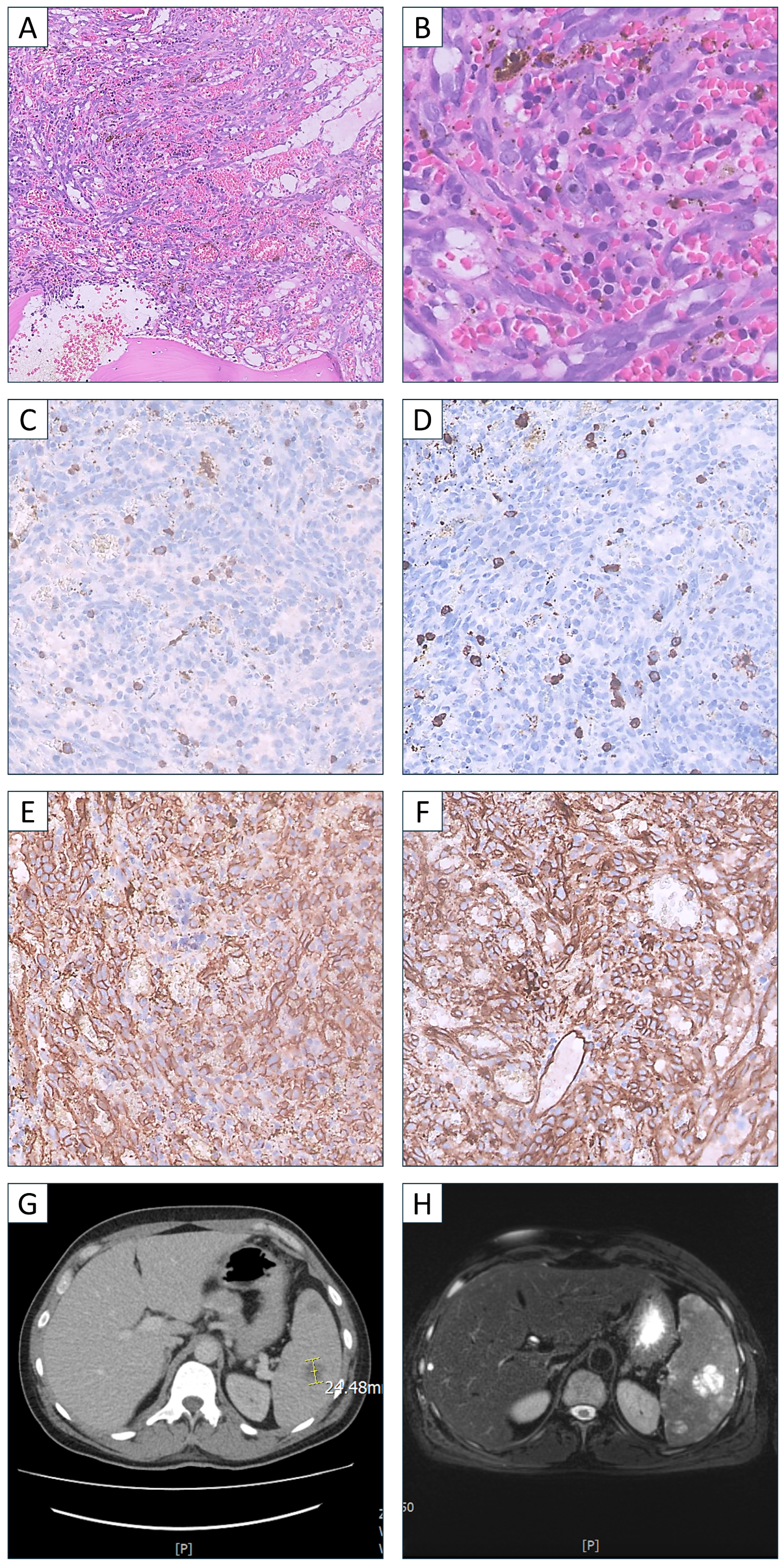
Figure 1. Histopathology examination diffuse infiltration of bone marrow by neoplastic cells. (A) Hematoxylin and eosin, 1× magnification; (B) Hematoxylin and eosin, 40× magnification. Immunohistochemical profiling of the neoplastic cells (C) MPO negative, 20× magnification; (D) CD15 negative, magnification 20×; (E) CD34 positive, 20×magnification; (F) CD31 positive, 20× magnification. A tumor lesion in a spleen in (G) computed tomography and (H) magnetic resonance imaging. The lesion in the central part of the spleen imitates cavernous angiomas. The heterogeneity of the lesions and the presence of hemorrhagic focus suggest a more aggressive clinical picture like angiosarcoma.
Hematolrep 16 00063 g001
Figure 2. Kaplan–Meier curves along with the table with median and ranges for overall survival (OS) in months based on the available OS data presented in Table 1. (A) OS of all 21 patients reported in Table 1 and this case. (B) Comparison of OS between patients with bone/bone marrow metastases and other metastatic sites.
Hematolrep 16 00063 g002
Table 1. Selected case reports of splenic angiosarcoma with associated abdominal symptoms, metastases, treatment and outcome.
CitationAge, SexAbdominal SymptomsMetastasesTreatmentFollow-UpOutcome
Levy et al. [5]83, FPainBoneSplenectomy + doxorubicin1 monthDied
Frontario et al. [6]56, FPainBoneElective splenectomy, adriamycin and ifosfamide with mesa, paclitaxelMore than 12 months from initial diagnosisAlive
Gorzelak-Pabis et al. [7]78, MPainLiver, infiltration in gastric and peritoneal regionSplenectomy2 monthsDied
Plantiga et al. [8]67, FNoLiver, bone marrowNot reported7 daysDied
Hasan Sözel et al. [9]65, MPainLiverSplenectomy + systemic chemotherapyNot reportedNot reported
Kimura et al. [10]77, FNoLiverSplenectomy1.5 monthsDied
Deng et al. [11]64, MVomitingNoSplenectomy + adjuvant chemotherapy9 monthsNot reported
Duan et al. [12]65, MPainLiverSplenectomy6 monthsDied
Zhao et al. [13]44, MDistentionLiverNot started8 hDied
Batouli et al. [14]45, FNot reportedLiverSplenectomy + chemotherapy5 monthsDied
Fang Chen et al. [15]72, FPainLiverNo data4 weeksDied
Wheelwright et al. [16]50, FPainNoSplenectomy + PLD and ifosfamide4 years, then no dataAlive
Wheelwright et al. [16]72, MNoSpineSplenectomy + PLD and ifosfamide5 years, then no dataAlive
Fiorentino et al. [17]80, FPainLiver, peritoneumSplenectomy + Paclitaxel, β-AR antagonists6 monthsDied
Schmidt de Azevedo et al. [18]57, FPainSpineSplenectomy + Paclitaxel + PazopanibMore than 3 yearsDied
Filip Kohutek et al. [19]65, FPainMultiple in bones, liver, axial skeleton and lungSplenectomy + Doxorubicin + Radiotherapy13 yearsAlive
Rui-Qi Zou et al. [20]67, FEnlarged spleenLiverHepato-splenectomy3 monthsNo data
Kamran S Hamid et al. [21]70, FDiscomfortLiver, lungsSplenectomy + chemotherapy8 monthsNo data
Barış Özcan et al. [22]65, FPain and distentionLiver, bonesSplenectomy + chemotherapyMore than 5 monthsNo data
Soumaya Anoun et al. [23] 25, FSplenomegalyBone marrowSplenectomy1 yearDied
Takehara et al. [24]62, FLeft flank painLiver, vertebraePaclitaxel, doxorubicin, pazopanib, docetaxel, gemcitabine plus docetaxel, ifosfamide23 monthsDied
Shimin Hu et al. [25]83, MNoBone marrow, presumably in thyroid glandHospice careNo dataNo data
Lichen Xu et al. [26]38, FPainLiver, bone marrowRefuse chemotherapy3 monthsDied
Wang et al. [27]36, MAbdominal mass localized to the left upper quadrantBone marrow, retroperitoneal lymph nodes, liver, lumbar spines, right kidneyAdriamycin, isophosphamide, VP-16, toxol24 years after diagnosisDied
Table 2. Complete blood count (CBC) on admission.
ParameterCBC on Admission
WBC1.86 × 103/μL
RBC3.15 × 106/μL
Hemoglobin9.5 g/dL
Hematocrite27.1%
MCV86.0 fL
MCH30.2 pg
MCHC35.1 g/dL
PLT24 k/mcL
RDW-SD51.6 fL
RDW-CV17.8%
NEU13.20 × 103/μL
LYM3.80 × 103/μL
MON1.34 × 103/μL
EOS0.21 × 103/μL
BAS0.03 × 103/μL
NRBC2.900 × 103/μL
IG4.00 × 103/μL
Disclaimer/Publisher’s Note: The statements, opinions and data contained in all publications are solely those of the individual author(s) and contributor(s) and not of MDPI and/or the editor(s). MDPI and/or the editor(s) disclaim responsibility for any injury to people or property resulting from any ideas, methods, instructions or products referred to in the content.