Case Report

Transient Stress Lymphocytosis in a Child: A Case Report and Systematic Review of the Literature

by
Alexander Placek
1,
Randall Y. Chan
2,
Maria Vergara-Lluri
1 and
Russell K. Brynes
1,*
1
Hematopathology Section, Department of Pathology, University of Southern California Keck School of Medicine, Los Angeles, CA 90033, USA
2
Hematology-Oncology Division, Department of Pediatrics, University of Southern California Keck School of Medicine, Los Angeles, CA 90033, USA
*
Author to whom correspondence should be addressed.
Hematol. Rep. 2024, 16(3), 431-439; https://doi.org/10.3390/hematolrep16030042
Submission received: 21 April 2024 / Revised: 11 June 2024 / Accepted: 28 June 2024 / Published: 3 July 2024

Abstract

:
Transient stress lymphocytosis (TSL) is an under-recognized phenomenon associated with an acute stressful event such as physical trauma or various emergency medical conditions. Lymphocytosis generally resolves within several hours to days of the stressor. While most reports of TSL predominantly involve adult patients, it has only rarely been reported in pediatric patients. Here, we describe the clinical course of a 9-year-old male who developed TSL following a traumatic fall from a second-story balcony and provide a systematic literature review of TSL.

1. Introduction

Lymphocytosis is a common finding in hospitalized patients. In children, the causes of lymphocytosis include infection, medications, thyrotoxicosis, and lymphoproliferative disorders [1,2,3]. Rarely, lymphocytosis may be associated with inheritable causes, such as the primary immunodeficiency known as B-cell expansion with NF-κB and T-cell anergy (i.e., BENTA) due to CARD11 mutations [3]. Distinguishing non-malignant lymphocytosis from a clonal lymphoproliferative disorder is essential and requires assessment of the clinical presentation, laboratory findings, and morphological review.
TSL, a poorly studied cause of lymphocytosis, is associated in adults with significant physical trauma or various emergency medical conditions including cardiac emergencies, gynecologic and obstetric emergencies, and sickle cell disease in crisis [4,5,6,7,8,9,10,11,12]. Resolution of the lymphocytosis typically occurs within 24–48 h and may be followed by neutrophilia. We report the case of a 9-year-old male with TSL due to serious physical trauma.

2. Case

A previously healthy 9-year-old male presented with multiple traumatic injuries after falling from a second-story balcony. On initial evaluation in the emergency department, he was tachycardic and hypertensive, with a Glasgow coma scale score of six. Computed tomography (CT) of the head revealed a right subdural hematoma measuring 9 mm with subfalcine herniation, impending transtentorial herniation, and displaced skull fractures through the occipital, bilateral parietal, and right temporal bones and superior sagittal and transverse sinus (Figure 1). CT of the thorax detected bilateral lung consolidations suggestive of aspiration, as well as a left clavicular fracture.
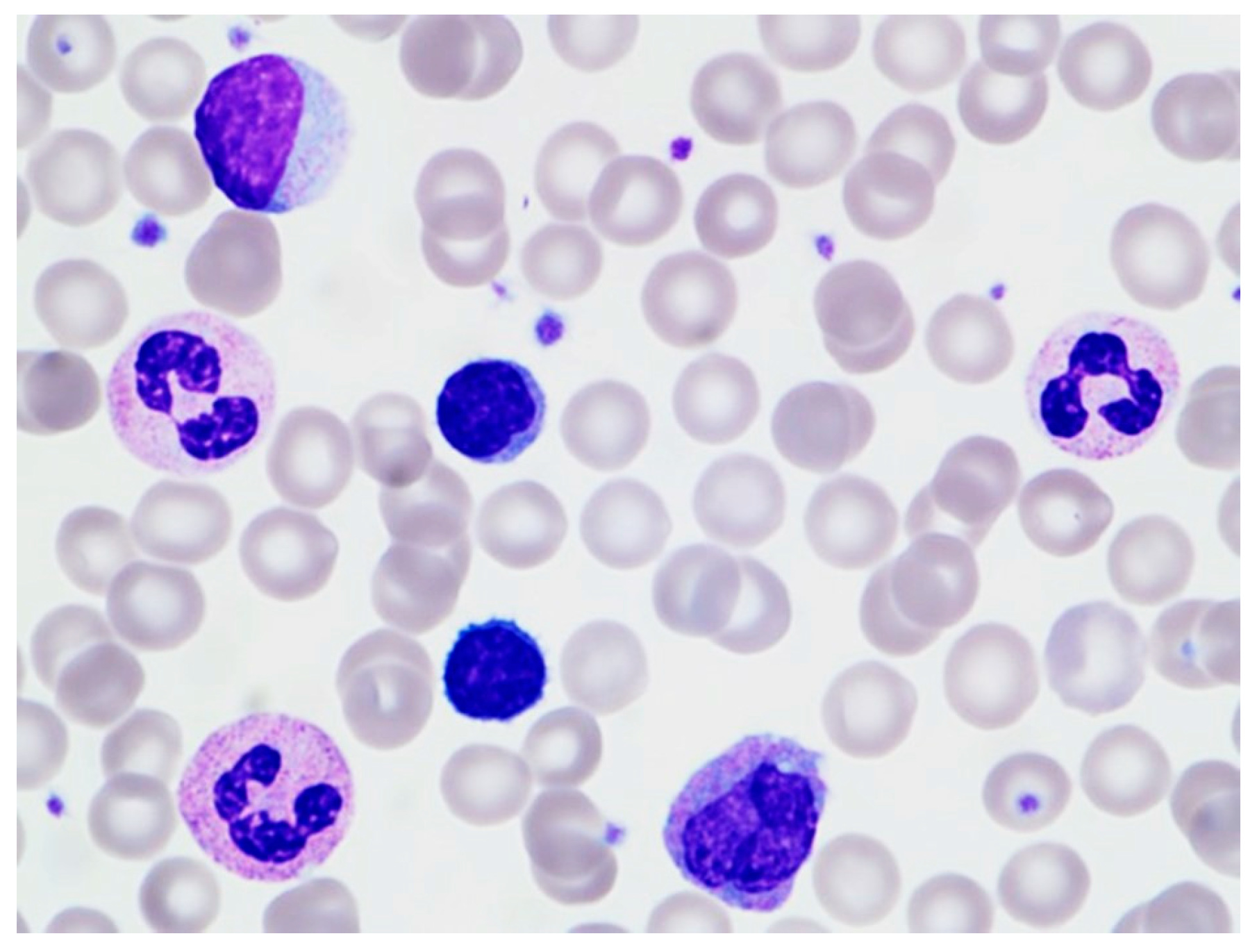
An initial complete blood count with differential (CBC/D), collected in the emergency department within 30 min of arrival, was notable for an elevated white blood cell (WBC) count of 32.3 × 10⁹/L, with an absolute neutrophil count (ANC) of 18.8 × 10⁹/L, absolute lymphocyte count (ALC) of 10.0 × 10⁹/L, monocyte count of 2.90 × 10⁹/L, eosinophil count of 0.50 × 10⁹/L, and basophil count of 0.2 × 10⁹/L. (The reference range cutoffs for lymphocytosis at our institution are ALC of 6.5 × 10⁹/L and 3.3 × 10⁹/L for 6–11-year-old pediatric patients and adult patients, respectively.) A morphologic review of the peripheral blood smear revealed predominantly small lymphocytes and neutrophilia. Large granular lymphocytes (LGLs) were present, but not increased (Figure 2).
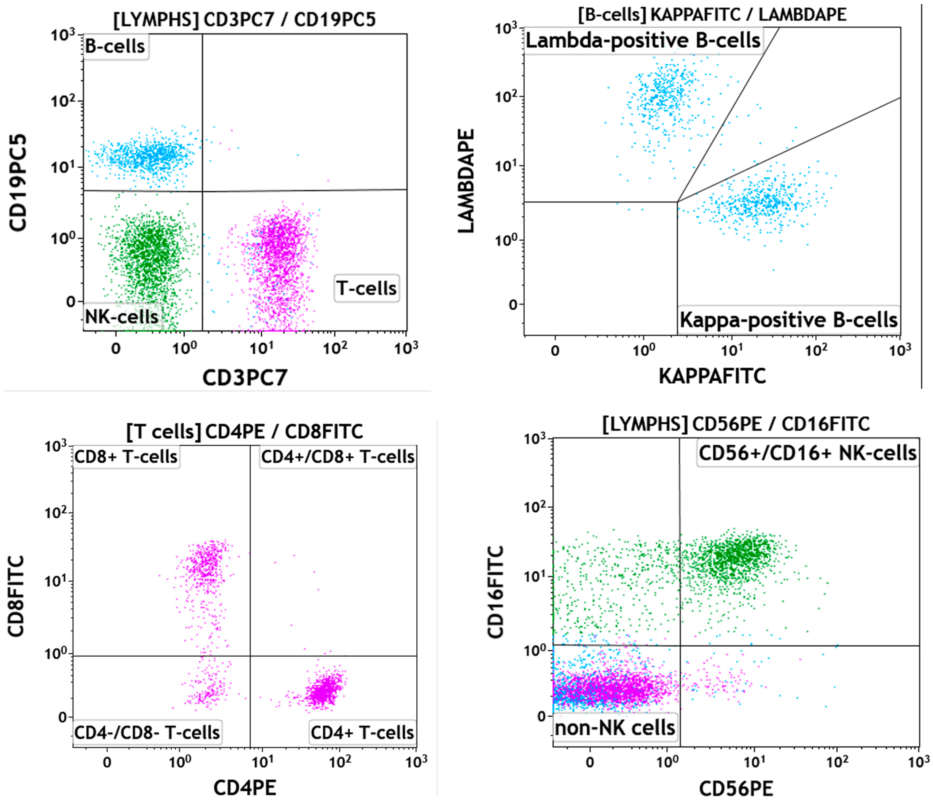
Flow cytometric immunophenotyping studies detected 36% lymphocytes composed of 18% B-cells (CD19+, CD20+), 41% T-cells (CD3+) with a CD4:CD8 ratio of 1.8, and 27% natural killer cells (CD3−, CD56+ and/or CD57+). Neither a monoclonal B-cell population nor an immunophenotypically abnormal T-cell population was identified (Figure 3).
A CBC/D collected four hours later showed resolution of the lymphocytosis, supporting the diagnosis of TSL (Figure 4).
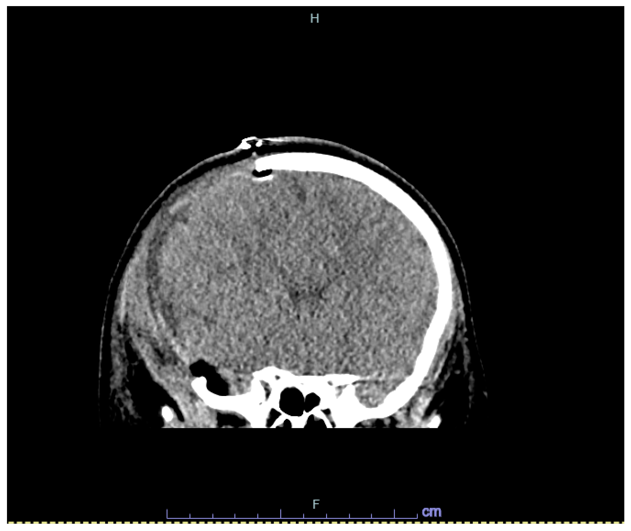
An emergent right hemicraniectomy with subdural hematoma evacuation was successfully performed. A subsequent head CT demonstrated improvement in the subdural hemorrhage and midline shift (Figure 5).
The WBC and ANC remained elevated for the duration of the admission, while the ALC remained within the reference range from 1.0 to 3.5 × 10⁹/L (Figure 4). Flow cytometric analysis on day 16 revealed a significant decrease in B-cells, T-cells, and NK-cells, all of which had returned to normal levels [13]. Neither a monoclonal B-cell population nor an immunophenotypically abnormal T-cell population was identified. The patient was transferred to a rehabilitation facility after two weeks with stable vital signs for continued care provided by neurosurgical and orthopedic specialists.

3. Systematic Review of the Literature

A systematic review of the literature was performed using the PubMed database (https://pubmed.ncbi.nlm.nih.gov (accessed on 20 April 2024)). The keywords used were Stress AND Lymphocytosis, OR Transient AND Lymphocytosis. The search was filtered for human subjects only. All articles reporting cases of transient and self-resolving lymphocytosis as a result of significant physical trauma (e.g., motor vehicle accident) or emergency medical conditions (e.g., sickle cell disease crisis and myocardial infarction) were included. Articles were excluded based on the following criteria: (1) lack of absolute lymphocytosis, (2) persistent lymphocytosis, (3) healthy or non-hospitalized patients, and (4) lymphocytosis unrelated to significant physical trauma or emergency medical conditions. The titles and abstracts were reviewed to identify relevant articles and exclude others as necessary. The selected reports include observational studies, a single case report, and letters to the editor (Figure 6).

4. Results

We identified nine reports that met our criteria (Table 1). All articles describe TSL as a phenomenon with an elevated ALC (>5 × 10⁹/L) that spontaneously resolved within 24–48 h of injury [4,5,6,7,8,9,10,11,12]. Resolution of the lymphocytosis may occur as quickly as 75 min [6]. Despite normalization of the ALC, an elevated WBC count that remains elevated for a longer duration may persist due to a delayed rise of the ANC; this finding was reported in 32 (16%) of the 196 total cases [4,6,8]. The articles describe etiologies including significant physical injuries [7,8,11,12] and a variety of emergency medical conditions including cardiac emergencies, seizure disorders, sickle cell disease with vaso-occlusive crisis, gynecologic/obstetric emergencies, thermal injury, acute pancreatitis, hypertensive crises, rupture of cerebral artery aneurysm, and anaphylaxis [4,5,6,8,10,12].
The morphologic appearance of the lymphocytes varied between studies. Karandikar et al. and Thommasen et al. describe mature-appearing small lymphocytes on microscopic examination and increased B-cells, T-cells, and natural killer cells and a preserved CD4:CD8 ratio on flow cytometry [4,8]. Karandikar et al., Vas et al., and Groom et al. describe reactive lymphocytes (Downey type II cells), or pleomorphic lymphoma-like cells (Downey type III reactive lymphocytes) [4,5,11]. Nevertheless, even in cases where “lymphoma-like” cells were noted, none of the patients were ultimately diagnosed with lymphoma or malignancy.
We found three articles in our systematic review that document TSL in the pediatric setting [4,7,8]. Except for the study by Karandikar et. al., which mentioned the presence of reactive and lymphoma-like cells, information is limited to the severity of the physical injury, without morphologic descriptions to assist in differentiating TSL from other causes of lymphocytosis [7].
Our formal literature review excluded non-traumatic/non-emergency descriptions of transient stress lymphocytosis. For completeness, we provide the following summary of the articles that were excluded, as they did not occur in traumatic/emergent settings. TSL was described in splenic contraction secondary to dasatinib [14], acute illness during chronic lymphocytic leukemia [15], exposure to catecholamines [16,17] and glucocorticoids [18], early-phase infection of human immunodeficiency virus [19], infection with human T-lymphotropic virus type 1 [20], reactivation of Epstein Barr virus or hepatitis B virus after allogeneic hematopoietic stem cell transplantation [21,22], as an effect of anti-cancer medications such as methotrexate [23] and tirabrutinib [24], and in the recovery phase of the recently described multisystem inflammatory syndrome in children (MIS-C) associated with COVID-19 infection [25]. One case described transient lymphocytosis, but no cause was assigned (idiopathic) [26]. Lymphocytosis was also described as part of an expansion of large granular lymphocytes and natural killer-associated cells, which could be persistent or transient [27].

5. Discussion

TSL is an underrecognized condition associated with serious events including physical trauma and various medical emergencies that typically resolves within 24 to 48 h. Lymphocytosis ensues following peripheralization from the spleen, bone marrow, lymphatic tissue, and lungs [16].
It is postulated that the mechanism is driven by catecholamines and resolves due to release of glucocorticoids. Immunologic studies have demonstrated increased circulating lymphocytes following the release of endogenous catecholamines and steroid hormones which are induced by various physical and psychological stressors [28,29]. Additionally, lymphocytosis has been observed following infusion with epinephrine [17]. In animal models, adrenaline injection swiftly induces lymphocytosis due to mobilization from the bone marrow, spleen, and lymphatics [30]. On the other hand, glucocorticoid administration has been shown to cause a rapid decrease in ALC due to redistribution of circulating lymphocytes to the bone marrow [31].
Given the rarity of TSL in children, effective exclusion of an underlying neoplastic lymphoproliferative process is paramount when this phenomenon is encountered. Particularly, patients with cytopenias (thrombocytopenia and anemia), not explained by their acute presentation, should raise suspicion of a lymphoproliferative disorder, as should lymphadenopathy, splenomegaly, and/or constitutional symptoms such as fevers and weight loss [32]. Such symptoms may be particularly challenging to elicit when a child is admitted for major trauma with multiple distracting injuries; additionally, hematologic derangements such as anemia and thrombocytopenia are common in critically ill victims of trauma [33].
In the assessment of lymphocytosis with the peripheral smear exam, pathologists and clinicians should be aware that TSL can show a spectrum of lymphocytes that includes small lymphocytes, reactive lymphocytes, and even lymphoma-like cells [11]. Reactive lymphocytes come in several forms, which have historically been categorized into three types: Downey type I, type II, and type III. Downey type I cells are smaller cells with round to reniform or lobated nuclei with visible nucleoli, and variably basophilic to foamy to vacuolated cytoplasm. Downey type II cells are characteristic of the so-called “atypical lymphocytes” associated with infectious mononucleosis; these are large lymphocytes with loosely clumped chromatin and abundant pale cytoplasm, with radial gradation of cytoplasmic basophilia and red cells indenting their cytoplasm. Downey type III cells are “lymphoma-like” cells as they are medium to large pleomorphic cells with sometimes irregular nuclear contours, moderately coarse chromatin, and prominent nucleoli; they have moderate to abundant and deeply basophilic cytoplasm [34]. Viewing a single Downey type III cell in isolation may lead to misinterpretation of lymphocytosis as circulating lymphoma cells.
Recognizing the heterogeneity and spectrum of morphology in the lymphocytes can aid in the correct interpretation of a reactive process, as lymphoma cells are generally monomorphous [35]. Another clue to support a reactive process is the simultaneous expansion of lymphocytes and monocytes. Time permitting, serial evaluation of peripheral blood counts and smears can be particularly enlightening. On the other hand, concerning findings on initial blood counts may co-exist with falsely alarming peripheral blood smear findings, such as monomorphous lymphocytes, atypical lymphocytes, or lymphoma-like cells (even when the visual exam is performed by experienced consultants), and serial evaluation alone may not be deemed optimal; this was true for our patient, as well as for several other cases in our review of the literature [5,6,12]. In such situations where a watch-and-wait approach may not be desirable, analysis of lymphocyte subsets via flow cytometry can quickly establish the absence of a clonal lymphoproliferative disorder [4].
In our patient, the presence of a relatively monomorphous population of lymphocytes raised the possibility of lymphoproliferative disease. Easily identifiable LGLs in our patient’s peripheral blood also raised concern. In pediatric patients, clonal T-cell LGLs are associated with a wide variety of immune dysregulation states and lymphoproliferative disorders, such as common variable immunodeficiency, Burkitt lymphoma, and Hodgkin lymphoma [36]. Careful consideration with further work-up may prevent the missed opportunity of diagnosing an underlying disease, particularly in the emergency department setting in which patients may be lost to follow-up. Ultimately, we chose to pursue flow cytometry early in the patient’s course to provide additional evidence to support reactive lymphocytosis rather than lymphoma, leukemia, or associated conditions such as immune dysregulation states. The absence of clonality or an immunophenotypically abnormal population was consistent with a reactive process. Furthermore, TSL is supported by flow cytometric detection of combined (rather than isolated) expansions of B-cell, T-cell, and NK-cell populations [4,5]. These findings may be reflected morphologically by increased small lymphocytes (B- and T-cells) and LGLs (NK- and T-cells), as seen here.
Our case draws attention to TSL in childhood and includes detailed laboratory, morphologic, and flow cytometric findings. TSL is an uncommon and overlooked cause of lymphocytosis in adults. Recognition of TSL in children is important because it is a reactive process that is self-limited, does not require further work-up, and can offer reassurance to patients and their families. Although flow cytometry may be the most sophisticated technique to distinguish TSL from a lymphoproliferative disorder, the more cost-effective approach is to trend the CBC/Ds. TSL will generally resolve within 48 h. However, persistent lymphocytosis should raise suspicion for an alternate etiology that may warrant further investigation. One can avoid misdiagnosis of lymphoproliferative disease by integrating clinical history, surveillance of the CBC with differential counts, and requesting a blood smear review by an experienced hematologist, hematopathologist, and/or laboratorian.

Author Contributions

Conceptualization, R.K.B. and R.Y.C.; data curation, A.P.; writing—original draft preparation, A.P. and R.K.B.; writing—review and editing, A.P., R.Y.C., M.V.-L. and R.K.B.; supervision, R.K.B. All authors have read and agreed to the published version of the manuscript.

Funding

This research received no external funding.

Institutional Review Board Statement

The University of Southern California Institutional Review Board waives review, patient consent, and approval for case reports which do not meet the federal definition of human subject research (i.e., lack identifiable patient information, report only facts, and have no plan to collect information that would not normally be placed in the records). The study was conducted in accordance with the Declaration of Helsinki.

Informed Consent Statement

Patient consent for case reports which do not meet the federal definition of human subject research (i.e., lack identifiable patient information, report only facts, and have no plan to collect information that would not normally be placed in the records).

Data Availability Statement

Dataset available on request from the authors.

Conflicts of Interest

The authors declare no conflicts of interest.

References

  1. Ferronato, A.E.; Leite, D.; Vieira, S.E. The role of respiratory virus infection in suspected pertussis: A prospective study. J. Microbiol. Immunol. Infect. 2021, 54, 379–384. [Google Scholar] [CrossRef]
  2. Allen, R.; Ezekowitz, B. Hematologic manifestations of systemic diseases. In Nathan and Oski’s Hematology of Infancy and Childhood, 7th ed.; Nathan, D.G., Orkin, S.H., Eds.; Saunders: Philadelphia, PA, USA; Elsevier: Philadelphia, PA, USA, 2009; pp. 1697–1714. [Google Scholar]
  3. Mims, M.P. Lymphocytosis, lymphocytopenia, hypergammaglobulinemia, and hypogammaglobulinemia. In Hematology: Basic Principles and Practice, 7th ed.; Hoffman, R., Benz, E.J., Eds.; Elsevier: Philadelphia, PA, USA, 2018; pp. 682–690. [Google Scholar]
  4. Karandikar, N.J.; Hotchkiss, E.C.; McKenna, R.W.; Kroft, S.H. Transient stress lymphocytosis: An immunophenotypic characterization of the most common cause of newly identified adult lymphocytosis in a tertiary hospital. Am. J. Clin. Pathol. 2002, 117, 819–825. [Google Scholar] [CrossRef]
  5. Groom, D.A.; Kunkel, L.A.; Brynes, R.K.; Parker, J.W.; Johnson, C.S.; Endres, D. Transient stress lymphocytosis during crisis of sickle cell anemia and emergency trauma and medical conditions. An immunophenotyping study. Arch. Pathol. Lab. Med. 1990, 114, 570–576. [Google Scholar]
  6. Teggatz, J.R.; Parkin, J.; Peterson, L. Transient atypical lymphocytosis in patients with emergency medical conditions. Arch. Pathol. Lab. Med. 1987, 111, 712–714. [Google Scholar]
  7. Pinkerton, P.H.; McLellan, B.A.; Quantz, M.C.; Robinson, J.B. Acute lymphocytosis after trauma–early recognition of the high-risk patient? J. Trauma 1989, 297, 49–751. [Google Scholar] [CrossRef]
  8. Thommasen, H.V.; Boyko, W.J.; Montaner, J.S.; Russell, J.A.; Johnson, D.R.; Hogg, J.C. Absolute lymphocytosis associated with nonsurgical trauma. Am. J. Clin. Pathol. 1986, 86, 480–483. [Google Scholar] [CrossRef]
  9. Kho, H.G.; van Egmond, J.; Zhuang, C.F.; Zhang, G.L.; Lin, G.F. The patterns of stress response in patients undergoing thyroid surgery under acupuncture anaesthesia in China. Acta Anaesthesiol. Scand. 1990, 34, 563–571. [Google Scholar] [CrossRef]
  10. Siddiqui, M.A.; Esmaili, J.H. Transient reactive lymphocytosis-associated with acute middle cerebral artery aneurysmal rupture. J. Natl. Med. Assoc. 1997, 89, 283–284. [Google Scholar]
  11. Vas, M.R.; Pantalony, D.; Carstairs, K.C. Transient ‘stress lymphocytosis’. Arch. Emerg. Med. 1990, 7, 126–127. [Google Scholar] [CrossRef]
  12. Wentworth, P.; Salonen, V.; Pomeroy, J. Transient stress lymphocytosis during crisis of sickle cell anemia. Arch. Pathol. Lab. Med. 1991, 115, 211. [Google Scholar]
  13. Schatorjé, E.J.; Gemen, E.F.; Driessen, G.J.; Leuvenink, J.; van Hout, R.W.N.M.; de Vries, E. Paediatric reference values for the peripheral T cell compartment. Scand. J. Immunol. 2012, 75, 436–444. [Google Scholar] [CrossRef] [PubMed]
  14. Marcos-Jiménez, A.; Carvoeiro, D.C.; Ruef, N.; Cuesta-Mateos, C.; Roy-Vallejo, E.; de Soria, V.G.-G.; Laganá, C.; del Campo, L.; Zubiaur, P.; Villapalos-García, G.; et al. Dasatinib-induced spleen contraction leads to transient lymphocytosis. Blood Adv. 2023, 13, 2418–2430. [Google Scholar] [CrossRef] [PubMed]
  15. Sandhu, I.; Baumann, M. Transient marked increase in lymphocytosis during acute illness in chronic lymphocytic leukemia. Leuk. Res. 2011, 35, e8–e9. [Google Scholar] [CrossRef] [PubMed]
  16. Benschop, R.J.; Rodriguez-Feuerhahn, M.; Schedlowski, M. Catecholamine-induced leukocytosis: Early observations, current research, and future directions. Brain Behav. Immun. 1996, 10, 77–91. [Google Scholar] [CrossRef] [PubMed]
  17. Toft, P.; Tønnesen, E.; Svendsen, P.; Rasmussen, J.W.; Christensen, N.J. The redistribution of lymphocytes during adrenaline infusion. An in vivo study with radiolabelled cells. APMIS 1992, 100, 593–597. [Google Scholar] [CrossRef] [PubMed]
  18. Bromberg, L.; Roufosse, F.; Pradier, O.; Delporte, C.; Van Antwerpen, P.; De Maertelaer, V.; Cogan, E. Methylprednisolone-induced lymphocytosis in patients with immune-mediated inflammatory disorders. Am. J. Med. 2016, 129, 746–752. [Google Scholar] [CrossRef] [PubMed]
  19. Smith, P.R.; Cavenagh, J.D.; Milne, T.; Howe, D.; Wilkes, S.J.; Sinnott, P.; E Forster, G.; Helbert, M. Benign monoclonal expansion of CD8+ lymphocytes in HIV infection. J. Clin. Pathol. 2000, 53, 177–181. [Google Scholar] [CrossRef]
  20. Ehrlich, G.D.; Han, T.; Bettigole, R.; Merl, S.A.; Lehr, B.; Tomar, R.H.; Poiesz, B.J. Human T-lymphotropic virus type I-associated benign transient immature T-cell lymphocytosis. Am. J. Hematol. 1988, 27, 49–55. [Google Scholar] [CrossRef] [PubMed]
  21. Isobe, Y.; Hamano, Y.; Ito, Y.; Kimura, H.; Tsukada, N.; Sugimoto, K.; Komatsu, N. A monoclonal expansion of Epstein-Barr virus-infected natural killer cells after allogeneic peripheral blood stem cell transplantation. J. Clin. Virol. 2013, 56, 150–152. [Google Scholar] [CrossRef] [PubMed]
  22. Adachi, Y.; Morio, S.; Hirasawa, A.; Wakita, H.; Oh, H.; Yoshida, S.; Aotsuka, N.; Asai, T.; Fukazawa, T.; Itaya, T. Transient B lymphocytosis associated with hepatitis B after allogeneic bone marrow transplantation. Rinsho Ketsueki 1991, 32, 884–889. [Google Scholar]
  23. Ureshino, H.; Kadota, C.; Kurogi, K.; Miyahara, M.; Kimura, S. Spontaneous regression of methotrexate-related lymphoproliferative disorder with T-cell large granular lymphocytosis. Intern. Med. 2015, 54, 2235–2239. [Google Scholar] [CrossRef]
  24. Toyama, T. A transient lymphocytosis after administration of tirabrutinib for leukemic Waldenstrom macroglobulinemia. Rinsho Ketsueki 2023, 64, 69–72. [Google Scholar]
  25. Okarska-Napierała, M.; Mańdziuk, J.; Feleszko, W.; Stelmaszczyk-Emmel, A.; Panczyk, M.; Demkow, U.; Kuchar, E. Recurrent assessment of lymphocyte subsets in 32 patients with multisystem inflammatory syndrome in children (MIS-C). Pediatr. Allergy Immunol. 2021, 32, 1857–1865. [Google Scholar] [CrossRef]
  26. Nakahara, K.; Utsunomiya, A.; Hanada, S.; Takeshita, T.; Uozumi, K.; Yamamoto, K.; Komatsu, H.; Nitta, M.; Ueda, R.; Tatsumi, E.; et al. Transient appearance of CD3+CD8+ T lymphocytes with monoclonal gene rearrangement of T-cell receptor beta locus. Br. J. Haematol. 1998, 100, 411–414. [Google Scholar] [CrossRef]
  27. Scott, C.S.; Richards, S.J.; Sivakumaran, M.; Short, M.; Child, J.A.; Hunt, K.M.; McEvoy, M.; Steed, A.J.; Balfour, I.C.; Parapia, L.A.; et al. Transient and persistent expansions of large granular lymphocytes (LGL) and NK-associated (NKa) cells: The Yorkshire Leukaemia Group Study. Br. J. Haematol. 1993, 83, 505–515. [Google Scholar] [CrossRef]
  28. Mills, P.J.; Berry, C.C.; Dimsdale, J.E.; Ziegler, M.; Nelesen, R.; Kennedy, B. Lymphocyte subset redistribution in response to acute experimental stress: Effects of gender, ethnicity, hypertension, and the sympathetic nervous system. Brain Behav. Immun. 1995, 9, 61–69. [Google Scholar] [CrossRef]
  29. Chi, D.S.; Neumann, J.K.; Mota-Marquez, M.; Dubberley, F.A. Effects of acute stress on lymphocyte beta 2-adrenoceptors in white males. J. Psychosom. Res. 1993, 37, 763–770. [Google Scholar] [CrossRef]
  30. Iversen, P.O.; Stokland, A.; Rolstad, B.; Benestad, H.B. Adrenaline-induced leucocytosis: Recruitment of blood cells from rat spleen, bone marrow and lymphatics. Eur. J. Appl. Physiol. Occup. Physiol. 1994, 68, 219–227. [Google Scholar] [CrossRef]
  31. Fauci, A.S. Mechanisms of corticosteroid action on lymphocyte subpopulations. I. Redistribution of circulating T and B lymphocytes to the bone marrow. Immunology 1975, 28, 669–680. [Google Scholar]
  32. Gribben, J.G. Clinical manifestations, staging, and treatment of follicular lymphoma. In Hematology: Basic Principles and Practice, 7th ed.; Hoffman, R., Benz, E.J., Eds.; Elsevier: Philadelphia, PA, USA, 2018; pp. 1288–1297. [Google Scholar]
  33. Drews, R.E. Critical issues in hematology: Anemia, thrombocytopenia, coagulopathy, and blood product transfusions in critically ill patients. Clin. Chest Med. 2003, 24, 607–622. [Google Scholar] [CrossRef]
  34. Perkins, S.L.; Bradley, K.T. Lymphocytes and plasma cells. In Color Atlas of Hematology: An Illustrated Field Guide Based on Proficiency Testing, 2nd ed.; Glassy, E.F., Ed.; College of American Pathologists: Northfield, MN, USA, 2018; pp. 260–261. [Google Scholar]
  35. Chabot-Richards, D.S.; George, T.I. Leukocytosis. Int. J. Lab. Hematol. 2014, 36, 279–288. [Google Scholar] [CrossRef] [PubMed]
  36. Savaşan, S.; Al-Qanber, B.; Buck, S.; Wakeling, E.; Gadgeel, M. Clonal T-cell large granular lymphocyte proliferations in childhood and young adult immune dysregulation conditions. Pediatr. Blood Cancer 2020, 67, e28231. [Google Scholar] [CrossRef] [PubMed]
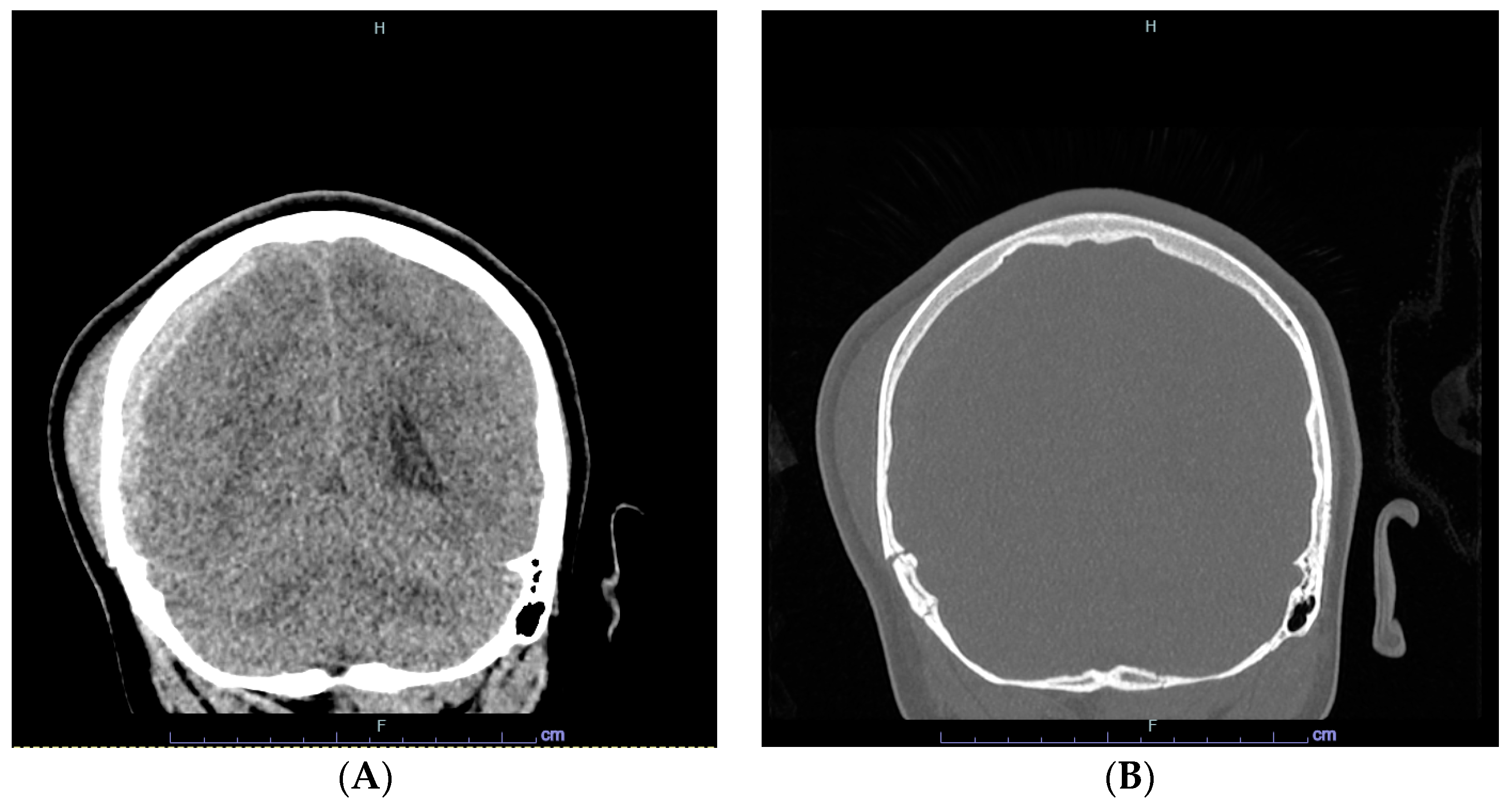
Figure 1. Head CT images of right acute subdural hematoma (A), and bone window of skull fractures (B).
Hematolrep 16 00042 g001
Figure 2. Peripheral blood morphology of transient stress lymphocytosis within 30 min of patient arrival. The smear shows small monomorphic mature-appearing lymphocytes, a large granular lymphocyte, a monocyte, and neutrophils (Wright–Giemsa stain, ×1000).
Hematolrep 16 00042 g002
Figure 3. Flow cytometry plots at presentation: There is an absolute lymphocytosis, composed of a combination of B-cells (light-blue events), T-cells (pink events), and NK-cells (green events). The CD19+ B-cells are polytypic/polyclonal with a kappa-to-lambda ratio of 1.2:1. CD3+ T-cells are an admixture of CD4+ and CD8+ T-cells, with a normal CD4:CD8 ratio of 1.8:1. NK-cells are CD56+ and CD16+. There are no immunophenotypically aberrant T-cell, B-cell, or NK-cell populations.
Hematolrep 16 00042 g003
Figure 4. WBC, ALC, and ANC values over the course of the hospitalization. WBC, white blood cell count; ANC, absolute neutrophil count; and ALC, absolute lymphocyte count.
Hematolrep 16 00042 g004
Figure 5. Post-hemicraniectomy head CT. Coronal view after subdural hematoma evacuation.
Hematolrep 16 00042 g005
Figure 6. Results of systematic review of the literature.
Hematolrep 16 00042 g006
Table 1. Summary of articles documenting transient stress lymphocytosis associated with trauma or acute medical conditions.
Author (Year)# CasesMean Age (Range)SexALC (× 109/L) (Mean)EtiologyMorphologyFlow
Cytometric Findings
Time to Resolution
Thommasen (1986) [8]24(13–65)->5Physical traumaSmall lymphocytes-6–24 h
Teggatz (1987) [6]26(43–93)-4.1–12.9 (6.8)Various acute medical conditionsSmall lymphocytes, reactive lymphocytes, LGLs-75 min (earliest)
Pinkerton (1989) [7]2338
(16–78)
17M
6F
4.5–8.8 (5.5)Physical trauma--
Groom (1990) [5]25(21–58)14M
11F
5.1–11.1Physical trauma,
Various acute medical conditions
Reactive lymphocytes, LGLs, lymphoma-like↑ B and T-cells
↓ CD4:CD8
10 to 24 h
Kho
(1990) [9]
1738.8-4–11.5 (6.9)Surgical removal of thyroid nodules--48 h
Vas
(1990) [11]
17 -5.0–13.4 (8.5)Various acute medical conditionsNormal, lymphoma-like, or reactive lymphocytes↑ B-cell
↑ CD4:CD8
24 h
Wentworth (1991) [12]8(27–66)-4.2–8.5Physical trauma,
Various acute medical conditions
--Majority 24 h
Siddiqui (1997) [10]178F5.4Middle cerebral artery aneurysmal ruptureReactive lymphocytes-36 h
Karandikar (2002) [4]5241.4
(15–89)
22M 30F4–10.4 (5.1)Physical trauma,
Various acute medical conditions
Small/medium sized lymphocytes
+/− Reactive/LGLs/lymphoma-like
↑ B, T and NK-cells
↑ CD4:CD8
32 h
Average
Acute medical conditions included cardiac emergencies (acute myocardial infarctions, cardiac arrest), gynecologic/obstetric emergencies, psychiatric emergencies, thermal trauma, seizure disorders, sickle cell disease with vaso-occlusive crisis, acute drug toxicity, acute infection, viral syndrome, cirrhosis, acute pancreatitis, anaphylaxis, hypertensive crisis, brain stem infarction, and gastroesophageal reflux disease. ALC, absolute lymphocyte count; CD, cluster of differentiation; F, female; h, hours; LGL, large granular lymphocyte; M, male; NK, natural killer; ↑, increased; ↓, decreased.
Disclaimer/Publisher’s Note: The statements, opinions and data contained in all publications are solely those of the individual author(s) and contributor(s) and not of MDPI and/or the editor(s). MDPI and/or the editor(s) disclaim responsibility for any injury to people or property resulting from any ideas, methods, instructions or products referred to in the content.