Article

Comprehensive Analysis of the Molecular Epidemiological Characteristics of Duck-Derived Salmonella in Certain Regions of China

1
Laboratory of New Veterinary Drug Development and Safety, College of Veterinary Medicine, Qingdao Agricultural University, Qingdao 266109, China
2
Qingdao Bolin Biotechnology Co., Ltd., Qingdao 266114, China
3
Qingdao Center for Animal Disease Prevention and Control, Qingdao 266033, China
*
Authors to whom correspondence should be addressed.
These authors contributed equally to this work.
Microbiol. Res. 2025, 16(8), 184; https://doi.org/10.3390/microbiolres16080184
Submission received: 26 June 2025 / Revised: 24 July 2025 / Accepted: 28 July 2025 / Published: 7 August 2025

Abstract

Salmonella is a major foodborne pathogen, yet real-time data on duck-derived strains in China remain scarce. This study investigated the epidemiology, antimicrobial resistance (AMR), gene profiles, and PFGE patterns of 114 Salmonella isolates recovered from 397 deceased ducks (2021–2024) across nine provinces (isolation rate: 28.72%). Fourteen serotypes were identified, with S. Typhimurium (23.68%), S. Indiana (21.93%), S. Kentucky (18.42%), and S. Enteritidis (12.28%) being predominant. Most isolates showed high resistance to β-lactams, tetracyclines, quinolones, and sulfonamides, with extensive multidrug resistance (MDR) observed—especially in S. Indiana, S. Typhimurium, and S. Kentucky. Among the 23 detected resistance genes, tet(B) had the highest prevalence (75.44%), particularly in S. Indiana. Biofilm formation was observed in 99.12% of isolates, with 84.21% demonstrating moderate to strong capacity. Eighteen virulence genes were detected; S. Enteritidis carried more spvB/C, sipB, and sodC1, while S. Indiana had higher cdtB carriage. PFGE revealed substantial genetic diversity among strains. This comprehensive analysis highlights the high AMR and biofilm potential of duck-derived Salmonella in China, emphasizing the urgent need for enhanced surveillance and control measures to mitigate public health risks.

1. Introduction

Salmonella is a widely distributed foodborne pathogen that causes significant economic losses to the poultry industry and poses a serious threat to human health. It can cause gastroenteritis through contaminated meat products, leading to diarrhea and even life-threatening conditions [1]. It has been included in the 2024 WHO Bacterial Priority Pathogens List (WHO BPPL) [2], attracting significant attention in the fields of global public health and food safety. Salmonella has a wide range of hosts and is often found in retail meat products, such as pork, beef, chicken, and lamb [3,4]. In China, ducks have become a primary economic animal [5], with duck meat products being highly popular. According to the Food and Agriculture Organization of the United Nations (FAO), China is the largest producer of duck meat, with an annual output of 3 million tons, and consumption is increasing every year [6]. The most common sources of infection in ducks include viral, bacterial, parasitic, and fungal pathogens. Among bacterial pathogens, Salmonella accounts for approximately 20% [7]. The main serotypes include S. Enteritidis, S. Typhimurium, and S. Kentucky [8,9]. Given that Salmonella accounts for a large proportion of all foodborne disease outbreaks, monitoring drug resistance in duck-derived Salmonella is crucial for public health and food safety [10].
Antibiotic treatment remains the primary strategy for controlling Salmonella outbreaks in poultry farming; however, the growing resistance to antibiotics has become a major global concern [11]. Accompanying the intensification of farming, antibiotic usage has remained at elevated levels. The estimated consumption of the top ten veterinary antibiotics in 2017 was as follows: China (45%), Brazil (7.9%), the United States (7.0%), Thailand (4.2%), India (2.2%), Iran (1.9%), Spain (1.9%), Russia (1.8%), Mexico (1.7%), and Argentina (1.5%), with an upward trend [12]. By 2020, the total consumption of antimicrobial agents in China’s food–animal sector (including poultry) reached 32,776.3 tonnes—equivalent to 165 g per tonne of animal product—which may have driven the continuous increase in bacterial resistance [13]. In recent years, there have been many reports of the high resistance of duck-derived Salmonella to various antibiotics, particularly quinolones and third-generation cephalosporins. Research demonstrated that the resistance rate of duck-derived Salmonella to cefotaxime was as high as 70.1%, and 8.7% of strains exhibited resistance to polymyxins [14]. The multidrug-resistant (MDR) characteristics of duck-derived Salmonella are attributed to several factors. Plasmid-mediated quinolone resistance (PMQR) involves the acquisition of various resistance genes (such as qnr genes, aac(6′)-Ib-cr, oqxAB, and qepA), facilitating the transmission of fluoroquinolone-resistant Salmonella at the human–animal interface [15,16,17]. Additionally, strong biofilm formation ability is a major factor affecting antibiotic resistance [18]. The irrational use of antibiotics in livestock production further exacerbates resistance development, leading to the emergence of MDR Salmonella and extensively drug-resistant (XDR) bacteria, which threaten food safety and public health [17,19].
The pathogenicity of Salmonella is primarily determined by its virulence factors, which enable the bacteria to adhere to host tissues, invade cells, replicate within the host environment, and ultimately destroy infected tissues, leading to disease. These virulence factors are encoded by genes located on bacterial chromosomes or plasmids, playing a crucial role in the infection process [20]. Numerous virulence factors have been identified in Salmonella, including those encoded by fimbrial virulence genes such as sefA, lpfA, lpfC, csgA, and pefA, which are involved in biofilm formation and environmental persistence [21]. The gene encoding spvB, a protein responsible for intracellular maintenance and the survival of the bacteria, is located on the virulence plasmid [22]. Additionally, genes related to the Type III Secretion System (TTSS), such as invA, orgA, sipB, prgH, and spaN, are closely associated with Salmonella’s pathogenicity [23].
However, in recent years, there have been notable gaps in surveillance studies of duck-derived Salmonella both within China and globally. In this study, we conducted a comprehensive epidemiological investigation of duck-derived Salmonella in selected regions across nine provinces in China. Our objectives were to study the epidemiological characteristics, antibiotic resistance profiles, prevalence of virulence genes, and genetic diversity of Salmonella. This research aimed to provide a detailed understanding of the prevalence and transmission patterns of duck-derived Salmonella, thereby contributing valuable insights for the development of effective prevention and control strategies in the context of public health and food safety.

2. Materials and Methods

2.1. Strain Isolation and Serotype Identification

In this study, from 2021 to 2024, we collected 397 samples from diseased and dead ducks across 27 farms in 9 provinces of China. These samples included feces, liver, intestine, heart, spleen, and legs (Supplementary Data S1). The Supplementary Data include the isolation time, isolation location, isolation site, serotype, biofilm formation ability, and MIC value of the strain. The isolation and identification of Salmonella were conducted according to the ISO-6579 standard (International Organization for Standardization, 2002) procedure [24]. Samples were inoculated onto XLT4 selective medium for culture, and 114 suspected Salmonella strains were isolated. Suspected colonies were inoculated onto Salmonella chromogenic medium and incubated at 37 °C for 12–16 h. Single red colonies were picked and inoculated into Luria–Bertani (LB) broth medium, and incubated at 37 °C for 16–18 h. Isolates with typical Salmonella phenotypes were further confirmed using API 20E test strips (bioMerieux, Marcy-l’Etoile, France). All confirmed Salmonella isolates were serotyped using Salmonella diagnostic antisera kits according to the White–Kauffmann–Le Minor scheme.

2.2. Antimicrobial Susceptibility Testing

Antimicrobial susceptibility testing was conducted using the micro-broth dilution method recommended by the Clinical and Laboratory Standards Institute (CLSI) guidelines [25]. Herein, we selected 19 antibiotics, including: Azithromycin (AZM) from the Macrolides; Cefotaxime (CTX), Ceftriaxone (CRO), Ceftazidime (CAZ) from the third-generation cephalosporins, Meropenem (MER) from the Carbapenem, and Aztreonam (ATM) from the Monocyclic β-lactams; Lomefloxacin (LOM), Norfloxacin (NOR), Gatifloxacin (GAT), Ciprofloxacin (CIP), Nalidixic acid (NAL), and Enrofloxacin (ENR) from the Quinolones; Amikacin (AMK), Apramycin (APR), and Gentamicin (GEN) from the Aminoglycosides; Sulfamethoxazole (SUL) from the Sulfonamides; Florfenicol (FFC) from the Chloramphenicols; and Tetracycline (TET) and Doxycycline (DOX) from the Tetracyclines. ATCC 25922 was used as a quality control strain. The minimum inhibitory concentration (MIC) was determined by referring to standards from CLSI documents M100-S34 (CLSI, 2024 edition). Based on the MIC values of antibiotics against various strains, the MIC50 (the minimum inhibitory concentration required to inhibit 50% of the tested strains) and the MIC90 (the minimum inhibitory concentration required to inhibit 90% of the tested strains) for each drug were calculated. The resistance rate of the test strains to different drugs was calculated according to known resistance breakpoints. A strain that is resistant to at least three types of antimicrobial drugs was referred to as a multidrug-resistant (MDR) strain [26].

2.3. Determination of Biofilm Formation Ability

Biofilm quantification testing was performed as previously described [27,28]. Briefly, strains were cultured by inoculating them into a 96-well sterile polystyrene microtiter plate and incubating at 28 °C for 48 h. Non-adherent bacteria were then removed from the microplates. The samples were fixed with anhydrous methanol, stained with crystal violet for biofilm staining, and rinsed with PBS. Finally, the samples were dissolved in glacial acetic acid. The absorbance at 595 nm was measured using a plate reader. Each strain was tested in three independent experiments; results are expressed as the mean of three biological replicates. The optical density cut-off value (ODc) was defined as the mean OD of three blank controls plus three times their standard deviation. Based on the OD values of the samples, biofilm-forming ability was classified as follows: OD > 4 × ODc indicated strong biofilm formation; 2 × ODc < OD ≤ 4 × ODc indicated moderate biofilm formation; ODc < OD ≤ 2 × ODc indicated weak biofilm formation; and OD ≤ ODc indicated no biofilm formation.

2.4. Detection of Drug Resistance Genes

Twenty-three antibiotic resistance genes (ARGs) were examined in all duck-derived Salmonella isolates. First, bacterial DNA was extracted using the standard boiling method [29]. Subsequently, based on data from the literature, drug resistance gene primers were designed and synthesized. Information on the drug resistance gene primers can be found in the Supplementary Material (Table S1) [6,30,31]. Bacterial DNA was used as a template for PCR amplification to detect various drug resistance genes. In addition, PCR products were randomly selected for sequencing, and the sequencing results were compared with sequences in the National Center for Biotechnology Information (NCBI) database (Bethesda, MD, USA) to verify the accuracy of the sequences.

2.5. Virulence Gene Detection

Eighteen virulence genes were detected in all duck-derived Salmonella isolates. The detection of virulence genes was conducted by designing and synthesizing the primers shown in the Supplementary Material (Table S2) [32,33,34,35] and based on the existing literature. Bacterial genomic DNA was used as a template, and PCR amplification was performed for 18 virulence genes. All primers were synthesized by Sangon Biotech Co., Ltd. (Shanghai, China).

2.6. Pulsed-Field Gel Electrophoresis (PFGE)

The PFGE test for Salmonella was conducted following the standard operating procedures of the US CDC, which included plug preparation, proteinase K digestion, and plug washing. Restriction enzyme XbaI was used for digestion at 37 °C for 4 h, followed by electrophoresis for 17.5 h. After staining, destaining, and imaging, the images and data were analyzed using the BioNumerics (version 7.6)software developed by Applied-Maths NV (Sint-Martens-Latem, Belgium).

2.7. Data Analysis and Visualization

Maps were generated using R (version 4.4.3; https://www.r-project.org/). Statistical analyses (ANOVA) were performed with GraphPad Prism (version 9.5; https://www.graphpad-prism.cn/). Cluster heatmaps were constructed using TBtools (version 2.138) [36], and basic graphical illustrations were created with Origin (version 2024; https://www.originlab.com/).

3. Results

3.1. Epidemiological Characteristics of Duck-Derived Salmonella

In this study, from 2021 to 2024, we collected 397 samples from diseased and dead ducks across 27 farms in 9 provinces of China. A total of 114 Salmonella strains were isolated and identified, with a positivity rate of 28.72%. The isolation rates among provinces ranged from 16.67% to 33.33%, with the highest rates observed in Anhui (33.33%), Henan (32.94%), and Guangdong (29.63%). The positivity rates from 2021 to 2024 were 25.58%, 22.55%, 27.27%, and 40.23%, respectively (Table S3). The regional distribution of duck-derived Salmonella strains, from highest to lowest, was as follows: Guangdong (56/114, 49.12%), Henan (28/114, 24.56%), Fujian (9/114, 7.89%), Guangxi (6/114, 5.26%), Shandong (5/114, 4.39%), Anhui (4/114, 3.51%), Jiangsu (3/114, 2.63%), Hubei (2/114, 1.75%), and Yunnan (1/114, 0.88%) (Figure 1A). The highest number of isolates was found in 2023 (Figure 1B), primarily isolated from the liver (40/114, 35.09%), feces (31/114, 27.19%), intestines (29/114, 25.44%), heart (13/114, 11.40%), and spleen (1/114, 0.88%) (Figure 1C).
The 114 Salmonella isolates were identified as 14 serotypes (Figure 1D). The 114 Salmonella isolates were identified as 14 serotypes (Figure 1D). The most common serotype was S. Typhimurium (27/114, 23.68%), followed by S. Indiana (25/114, 21.93%), S. Kentucky (21/114, 18.42%), and S. Enteritidis (14/114, 12.28%). A small amount of S. Agona (5/114, 4.39%), S. Gallinarum (5/114, 4.39%), S. London (5/114, 4.39%), S. Anatum (3/114, 2.63%), S. Pullorum (2/114, 1.75%), S. 1,4,[5],12:i:- (1/114, 0.88%), S. java (1/114, 0.88%), S. potsdam (1/114, 0.88%), S. Rissen (1/114, 0.88%), and S. Kottbus (1/114, 0.88%) was identified. Among them, S. Indiana, S. Typhimurium, and S. Kentucky were mainly sourced from Guangdong Province, while S. Enteritidis primarily came from Henan Province.

3.2. Biofilm Formation Ability

The results indicated that a substantial majority of the strains, specifically 99.12% (113/114), possessed the capability to form biofilms (Supplementary Data S1). Within this group, 14.91% (17/114) demonstrated a weak biofilm formation ability, 32.46% (37/114) exhibited a moderate biofilm formation ability, and a significant proportion, 51.75% (59/114), showcased a strong biofilm formation ability. Notably, strains with strong and moderate biofilm formation abilities were particularly prevalent among the four dominant serotypes: S. Typhimurium, S. Indiana, S. Kentucky, and S. Enteritidis (Figure 2).

3.3. Analysis of Drug Resistance

The 114 isolates exhibited high resistance to several classes of antibiotics (Figure 3A, Figure 4). Specifically, they showed high resistance to quinolones (NAL 100%, LOM 77.19%, CIP 75.44%, NOR 70.18%, ENR 55.26%, and GAT 50.00%), β-lactams (CRO 78.07%, CTX 71.93%, CAZ 58.77%, and ATM 44.74%), tetracyclines (TET 100% and DOX 87.72%), sulfonamides (SUL 100%), and amphenicols (FFC 65.79%). The isolates showed relatively lower resistance rates to aminoglycosides (GEN 46.49%, AMK 36.84%, and APR 11.40%) and macrolides (AZM 49.12%). Notably, the isolates showed moderate to high resistance to third-generation cephalosporins, including CRO, CTX, and CAZ, which are commonly used in clinical practice. Fortunately, no MER-resistant strains were detected (Supplementary Data S1).
All 114 (100%) strains were multidrug-resistant (MDR), with 76.32% (87/114) of the strains being resistant to five or more classes of antibiotics. Additionally, 28.07% (32/114) of the strains were resistant to more than seven classes of antibiotics, and 54.39% (62/114) of the strains were resistant to more than ten types of antibiotics (Figure 3B). The analysis of the correlation between predominant serotypes and antibiotic resistance indicated that S. Indiana, S. Typhimurium, and S. Kentucky exhibited relatively high resistance. Specifically, S. Indiana and S. Kentucky were resistant to an average of 15 and 12 antibiotics, respectively. In contrast, S. Enteritidis had a relatively low resistance rate, with an average of seven antimicrobial agents. Notably, except for the non-significant difference in the number of resistance traits between S. Kentucky and S. Typhimurium, all other serotypes showed significant (p < 0.05) or highly significant (p < 0.001) differences in the number of antimicrobial resistances (Figure 3C).

3.4. Antimicrobial Resistance Genes Profile

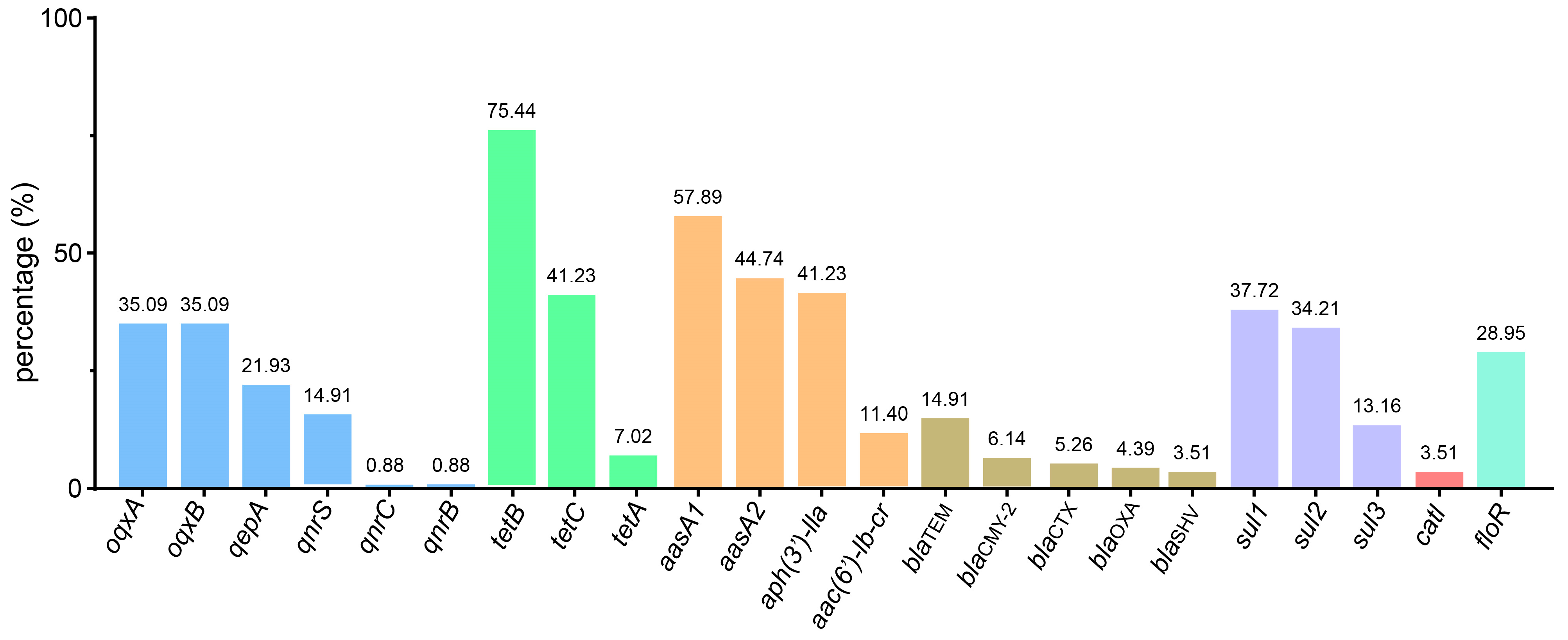
In this study, 23 ARGs were detected (Figure 4, Supplementary Data S2), including quinolone resistance genes (oqxA (35.09%, 40/114), oqxB (35.09%, 40/114), qnrB (0.88%, 1/114), qnrC (0.88%, 1/114), qnrS (14.91%, 17/114), and qepA (21.93%, 25/114)); tetracycline resistance genes (tetC (41.23%, 47/114), tetA (7.02%, 8/114), and tetB (75.44%, 86/114)); aminoglycoside resistance genes (aadA1 (57.89%, 66/114), aadA2 (44.74%, 51/114), aph(3′)-IIa (41.23%, 47/114), and aac(6′)-Ⅰb-cr (11.40%, 13/114)); β-lactam resistance genes (blaSHV (3.51%, 4/114), blaCMY-2 (6.14%, 7/114), blaCTX (5.26%, 6/114), blaOXA (4.39%, 5/114), and blaTEM (14.91%, 17/114)); sulphonamide resistance genes (sul1 (37.72%, 43/114), sul2 (34.21%, 39/114), and sul3 (13.16%, 15/114); a macrolide resistance gene (catl (3.51%, 4/114)); and a phenicol resistance gene (floR (28.95%, 33/114)).
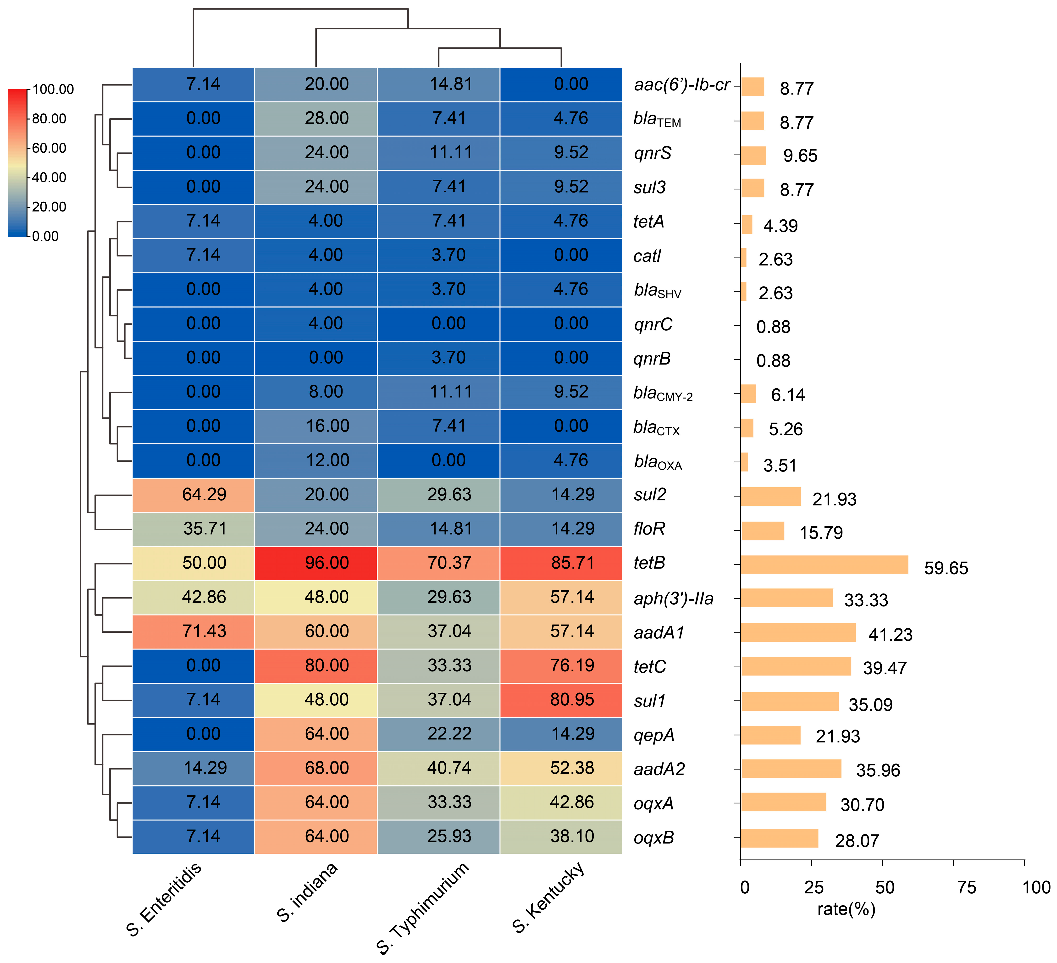
In addition, different serotypes of Salmonella exhibit variations in carrying drug resistance genes (Figure 5). S. Enteritidis mainly carries sul2, floR, tetB, aph(3′)-lla, and aadA1; S. Indiana, S. Typhimurium, and S. Kentucky have a broader spectrum of drug resistance genes, primarily carrying tetB, aph(3′)-IIa, aadA1, tetC, sul1, aadA2, oqxA, and oqxB. In addition, S. Indiana carries more plasmid-mediated quinolone resistance genes (qepA (64.00%) and broad-spectrum beta-lactamase resistance genes (blaTEM).

3.5. Virulence Gene Profile

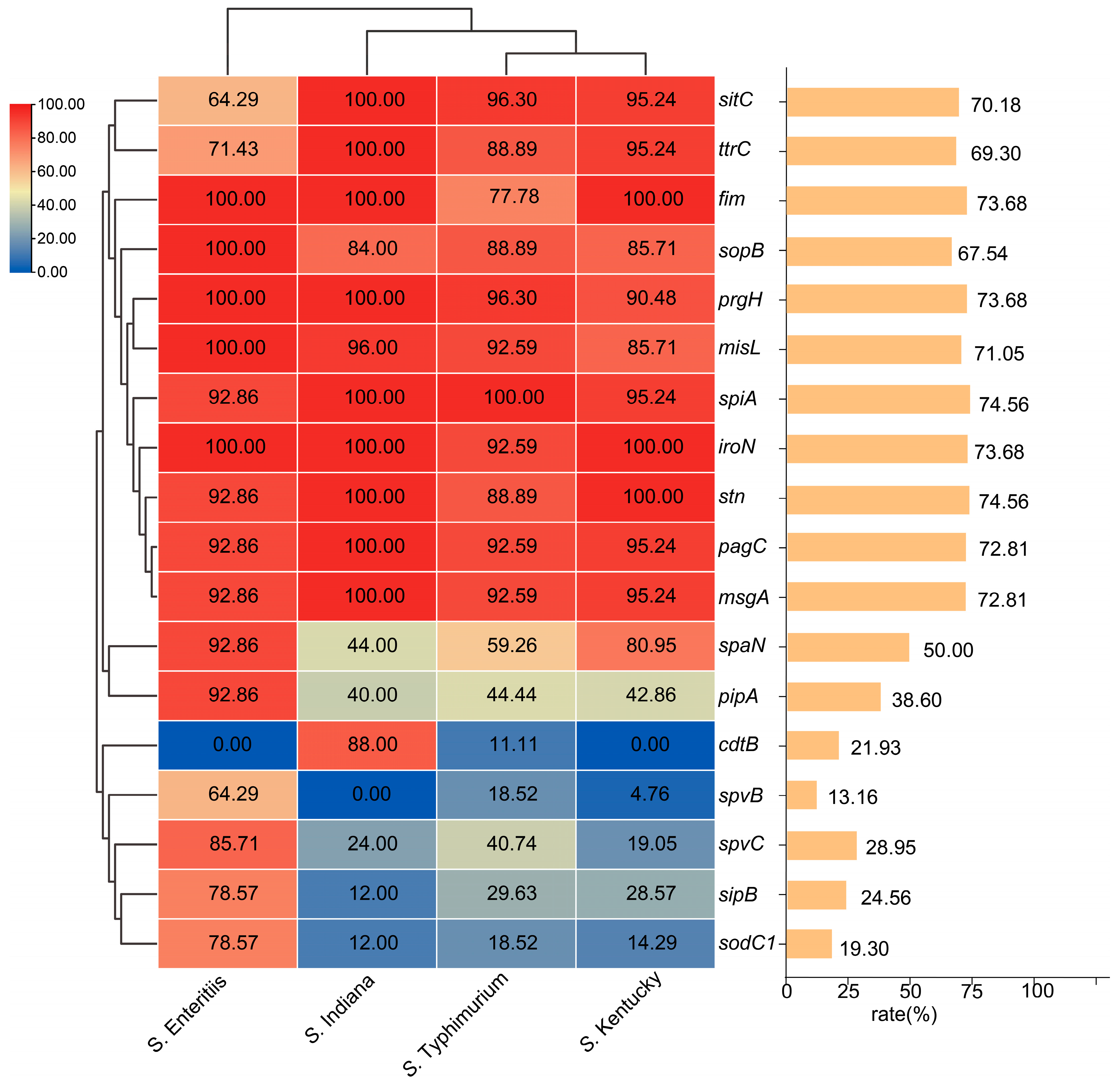
In this study, 18 virulence genes were detected (Figure 6, Supplementary Data S3), which were as follows: genes encoding factors related to cell invasion and adhesion (stn 96.49%, fim 95.61%, spaN 69.30%, and sipB 36.84%), genes encoding factors related to intracellular survival and escape (spiaA 96.49%, pagC 95.61%, msgA 94.74%, spvC 46.49%, sodC1 35.09%, spvB 26.32%, cdtB 22.81%), genes encoding factors related to resistance (prgH 97.37%, iroN 96.49%, sitC 89.47%, ttrC 85.96%), genes encoding factors related to motility and chemotaxis (sopB 90.35%, pipA 57.89%), and the gene encoding the autotransporter protein (misL 94.74%).
We analyzed the differences between virulence genes and the main serotypes (Figure 7). The results indicated that the main serotypes carry a high proportion of the virulence genes sopB, prgH, misL, fim, iroN, pagC, msgA, stn, and spiA. The proportion of spvB, spvC, sipB, sodC1, and pipA genes in S. Enteritidis strains was higher than that in in other serotypes. The proportions of spaN virulence genes in S. Indiana and S. Typhimurium were significantly lower than those in S. Enteritidis and S. Kentucky. Only S. Indiana carried a high proportion of the cdtB virulence gene.

3.6. PFGE Molecular Typing

The experimental results indicated that the PFGE patterns of Salmonella from ducks showed significant variability and numerous types, demonstrating significant genetic diversity among the strains. The strains from Henan (S459, S444, S457, S450, S453, S458, etc.), Fujian (S105, S109), and Anhui (S084) show close genetic relatedness (with similarity exceeding 80%) to the strains from Guangdong (Figure S1), suggesting the possibility of epidemiological transmission.

4. Discussion

Duck meat and its related products are especially popular in Asia, and even more so in China. China is the undisputed global leader in duck meat and egg production. According to 2023 statistics, China’s duck meat output accounted for 79% of the world total, and its duck egg production reached 83.2% [37]. Vietnam ranked second, contributing only 2.95% of duck meat and 1.5% of duck egg production [37]. The vast scale and high-density farming systems in China place great pressure on disease control, prompting widespread use of antibiotics for both prevention and treatment. The global dissemination of multidrug-resistant (MDR) bacteria has emerged as one of the most severe public health crises of the twenty-first century. Salmonella, as an important zoonotic pathogen, poses a significant challenge to global public health due to its serotype diversity and multidrug resistance [38,39,40]. In this study, 114 positive strains were isolated from the collected 397 samples, yielding an overall positive rate of 28.72%. A total of 14 serotypes were identified, highlighting the rich diversity of duck-derived Salmonella serotypes. The predominant serotypes (S. Typhimurium, S. Indiana, and S. Kentucky) were consistent with findings reported in previous studies [14,41]. Therefore, surveillance of Salmonella prevalence in ducks is critically important for food safety. The hosts of Salmonella are diverse, yet there are distinct differences in the distribution of serotypes among different hosts. For instance, the main prevalent serotypes of pig-derived Salmonella are S. Derby, S. Rissen, and S. Typhimurium [42], and the main prevalent serotypes of chicken-derived Salmonella are S. Enteritidis, S. Indiana, and S. Typhimurium [43,44], which are similar to the epidemiological characteristics of the duck-derived Salmonella serotypes presented in this study. Notably, two predominant serotypes of avian-derived Salmonella, S. Typhimurium and S. Enteritidis, are also the predominant serotypes in humans [45], indicating that avian-derived Salmonella is closely related to human health. Thus, tracking the epidemiological dynamics of duck-derived Salmonella is of great significance to public health safety.
Antibiotics are extensively utilized in the livestock industry for disease prevention, treatment, and growth promotion, which has led to the rapid emergence of drug-resistant Salmonella strains [46,47]. In our study, the 114 strains of duck-derived Salmonella demonstrated resistance to multiple drug classes, characterized by MDR or extensive drug resistance (XDR). It is particularly concerning that duck-derived Salmonella exhibited high resistance to quinolones (e.g., CIP 75.44% and LOM 77.19%), macrolides (e.g., AZM 49.12%), and third-generation cephalosporins (e.g., CRO 78.07%, CAZ 58.77%, and CTX 71.93%), which are currently the commonly used first-line drugs in clinical settings [48]. However, in a 2022 report from Shandong Province, China, 71.82% of 110 Salmonella isolates from duck farms were identified as multidrug-resistant (MDR) [49]. This finding suggests a potential rise in antimicrobial resistance among duck-derived Salmonella. In other regions, duck-derived Salmonella continue to exhibit high resistance levels; for example, in Vietnam, 82.4% of isolates were resistant to tetracycline and cefazolin [50]; in Egypt, 60% of strains were resistant to ≥3 antibiotic classes, with resistance rates to amoxicillin and clindamycin reaching 90% [51]; and in Nigeria, duck-derived strains showed high resistance to penicillin (100%), ampicillin (96%), and tylosin (93.9%) [52]. In our study, S. Indiana, S. Typhimurium, S. Kentucky, and S. Enteritidis exhibited high levels of antimicrobial resistance, with S. Indiana being resistant to as many as 15 antibiotics. Related studies have reported a large number Salmonella serotypes associated with animal meat products, particularly S. Derby, S. Indiana, S. Typhimurium, S. Kentucky, and S. Enteritidis. These serotypes are often associated with MDR [5,53,54]. In this study, a serotype 1,4,[5],12:i:- strain (a variant of S. Typhimurium) was found among the duck-derived Salmonella, which merits further investigation. In recent years, the emergence of the monophasic variant 1,4,[5],12:i:-, associated with colistin resistance in animal sources, has attracted attention [55], raising public health concerns. Therefore, scientifically managing the use of antibiotics in animal production holds great significance.
The factors influencing bacterial drug resistance are multifaceted and complex. In addition to the inherent characteristics of the bacteria, ARGs, plasmids, mobile genetic elements, and biofilms all play a role in determining bacterial sensitivity to antibiotics. Among them, the antibiotic resistance phenotype can be directly linked to ARGs. In this study, we identified 23 ARGs. Resistance to quinolone drugs is typically associated with PMQR genes. We detected oqxAB and qnrBC in our samples. The oqxAB gene is commonly found in Salmonella isolates and often coexists with extended-spectrum β-lactamase (ESBL)-encoding genes on multidrug-resistant plasmids [56,57], which partly explains the high resistance of duck-derived Salmonella to both β-lactams and quinolones observed in this study. The ESBL genes (blaTEM, blaCTX, blaSHV, and blaOXA) confer resistance to cephalosporins in bacteria. Among ESBL genes, blaTEM, blaCTX, and blaOXA are the most prevalent among Salmonella isolates from humans, food animals, retail food, and pets [58,59]. The protein encoded by blaTEM is known for its enhanced hydrolysis of cephalosporins and blaTEM is the primary gene encoding ESBL enzymes in Salmonella isolates [60]. Moreover, our study also observed other clinically relevant and significant ARGs (e.g., floR, aadA1, and sul3), which confer resistance to multiple classes of antibiotics in bacteria. Therefore, monitoring the prevalence of ARGs is essential to prevent and control drug-resistant Salmonella isolates.
Biofilms play a crucial role in bacterial drug resistance and virulence, being closely related to the long-term presence of bacteria in hosts, and are one of the reasons for the development of ‘tolerance’. The composition of biofilms includes extracellular polysaccharides, DNA, and proteins, which serve as a ‘protective coat’ for micro-organisms in extreme environments, shielding them from ultraviolet radiation, extreme temperatures, extreme pH values, high salinity, high pressure, malnutrition, and antibiotics [61,62]. Biofilms can induce bacterial ‘tolerance’ through various mechanisms. In addition to the classic mechanisms of drug resistance, bacteria can also exhibit ‘tolerance,’ which is the ability to survive short-term exposure to high concentrations of antibiotics [63]. One of the reasons biofilms generate ‘tolerance’ is because of their structure. Biofilms can resist the killing effects of antibiotics and the host immune system through mechanisms such as efflux pumps, resistance to antibiotic penetration, and the transfer of drug resistance genes. This leads to drug resistance and inflammation, causing persistent infections and leading to chronic diseases [64,65,66]. In addition, bacteria originating from water exhibit a stronger ability to form biofilms [67,68]. In our study, among the 114 duck-derived Salmonella strains, 113 (99.12%) were capable of producing biofilms, with 84.21% exhibiting strong and moderate biofilm-forming abilities. However, in Egypt, only 60% of duck-derived Salmonella strains were capable of forming biofilms, which is significantly lower than the proportion observed in the present study [51]. Studies have shown that the two serotypes, S. Typhimurium and S. Enteritidis, are the most common types of Salmonella outbreaks in humans and poultry, and they generally exhibit strong biofilm-forming abilities [69]. S. Indiana and S. Kentucky are commonly found in Salmonella strains isolated from ducks and wild geese in China and have been confirmed to possess strong biofilm-forming abilities [70]. Moreover, the biofilm formation ability of Salmonella in water sources is related to the physical, morphological, and chemical composition of the water. For example, river water can induce the formation of new biofilm morphologies in Salmonella, such as SPAM-type biofilms, which may be an adaptation strategy in stressful environments [71]. The strong biofilm-forming ability of Salmonella is a key factor in its persistence in farm environments, the spread of ARGs, and antibiotic treatment failure [72,73]. Biofilms hinder antibiotic penetration (e.g., reduced ciprofloxacin permeability), and the hypoxic conditions inside biofilms lower bacterial metabolism, weakening the efficacy of metabolism-dependent antibiotics like β-lactams [74]. This enables Salmonella to evade disinfection and treatment, posing an ongoing threat to public health and food safety [75,76]. Strengthening “One Health” strategies and regulating antibiotic use from farm to fork are essential to curb the emergence and spread of resistant bacteria.
Our data indicated that the isolated Salmonella strains contained 18 types of virulence factors. The spiA gene encodes an outer membrane component of the SPI-2 type III secretion system, which is crucial for virulence toward host cells, and is also essential for the invasion and transmission of Salmonella [77]. The pipA encodes a type III effector that targets components of the nuclear factor kappa B (NF-κB) signaling pathway to induce inflammation [78]. The fim gene is a key gene encoding fimbriae, which are the main mediators of the interaction between Salmonella and the host intestinal epithelium, playing a key role in the processes of infection and inflammation [79]. Most of our S. Enteritidis isolates carried plasmid-encoded spvBC operons, which encode effector proteins that disrupt host membrane transport [80]. Salmonella invasion protein B (sipB) initiates the invasion process in the Salmonella TTSS [81]. PFGE highlighted the genetic diversity of duck-derived Salmonella and indicated the potential transmission of strains across different regions (e.g., between Guangdong and other provinces), emphasizing the need for enhanced prevention and surveillance measures. Notably, in the second edition of China’s local Salmonella genome database constructed by Yanan Wang et al. [82], duck-derived Salmonella accounted for merely 2.82% (230/8159) of the total isolates. This striking underrepresentation underscores the urgent need for enhanced genomic surveillance of duck-derived Salmonella strains using whole-genome sequencing technology. Several study limitations should be acknowledged. For instance, the interactions between biofilm formation and antibiotic resistance, as well as the potential regulatory relationships between antimicrobial resistance genes and virulence genes, were not thoroughly investigated. Further experimental studies are required to validate and elucidate these underlying mechanisms.

5. Conclusions

This study comprehensively characterized the molecular epidemiological and biological characteristics of duck-derived Salmonella in China from 2021 to 2024. A total of 114 duck-derived Salmonella strains exhibited significant genetic diversity, with all isolates showing characteristics of MDR, 99.12% of isolates having the ability to form biofilms, and 84.21% of isolates possessing moderate or strong biofilm-forming ability. The 114 isolates carried 23 types of ARGs and 18 types of virulence genes, showing significant differences among serotypes. PFGE analysis revealed substantial genetic diversity among the isolates and indicated potential regional transmission patterns. Given the serotype diversity and multidrug-resistant characteristics of duck-derived Salmonella strains, it is recommended that duck farms be incorporated into the Global Antimicrobial Resistance and Use Surveillance System (GLASS) and relevant regional monitoring networks. Surveillance programs targeting antimicrobial resistance in waterfowl should prioritize dominant serotypes, such as S. Indiana and S. Typhimurium. Moreover, genome-based approaches are essential for the global monitoring of Salmonella in waterfowl to better track its epidemiological dynamics.

Supplementary Materials

The following supporting information can be downloaded at: https://www.mdpi.com/article/10.3390/microbiolres16080184/s1, Figure S1: Phylogenetic Analysis of Duck-Derived Salmonella. The provinces, isolation date, and serotype are annotated from left to right, respectively; Table S1: Primer sequences for resistance genes in this study; Table S2: Primer sequences for resistance genes in this study; Table S3. Sample collection volume and positivity rate in different provinces; Supplementary Data 1: Background information and resistance data of the 114 Salmonella strains in this study; Supplementary Data 2: Full ARGs information of 114 Salmonella strains; Supplementary Data 3: Full virulence genes information of 114 Salmonella strains.

Author Contributions

Conceptualization, J.C.; methodology, L.F.; validation, J.C., L.F. and X.L.; formal analysis, W.R.; investigation, Y.L. (Yanling Liu); resources, S.W.; data curation, J.C. and W.R.; writing—original draft preparation, J.C.; writing—review and editing, L.G., Y.L. (Yan Li) and Y.Z.; visualization, X.L.; supervision, Y.Z.; project administration, X.D.; funding acquisition, L.G. and Y.Z.; J.C., X.L. and Y.L. (Yanling Liu) contributed equally to this work. All authors have read and agreed to the published version of the manuscript.

Funding

This research was funded by grants from the Taishan Industrial Experts Program (TS20220701); the Qingdao Science and Technology for the People demonstration special (25-1-5-xdny-30-nsh, 24-1-8-xdny-4-nsh); the Central Guidance for Local Funds (YDZX2023123); and the Shandong Province Key R&D Projects (2024CXGC010910).

Institutional Review Board Statement

All procedures involving animals were conducted in accordance with the ethical standards and guidelines for animal care and use. The study protocol was reviewed and approved by the Animal Ethics Committee of Qingdao Agricultural University (Approval No. 2021-018).

Informed Consent Statement

Not applicable.

Data Availability Statement

The datasets presented in this article are not readily available because the data are part of an ongoing study. For access to the dataset, please contact the corresponding author.

Conflicts of Interest

Author Shuhua Wang and Yongda Zhao was employed by the company Qingdao Bolin Biotechnology Co., Ltd. The remaining authors declare that the research was conducted in the absence of any commercial or financial relationships that could be construed as a potential conflict of interest.

References

  1. Bae, D.H.; Dessie, H.K.; Baek, H.J.; Kim, S.G.; Lee, H.S.; Lee, Y.J. Prevalence and Characteristics of Salmonella spp. Isolated from Poultry Slaughterhouses in Korea. J. Vet. Med. Sci. 2013, 75, 1193–1200. [Google Scholar] [CrossRef] [PubMed]
  2. Jesudason, T. WHO Publishes Updated List of Bacterial Priority Pathogens. Lancet Microbe 2024, 5, 100940. [Google Scholar] [CrossRef]
  3. Threlfall, E.J. Antimicrobial Drug Resistance in Salmonella: Problems and Perspectives in Food- and Water-Borne Infections. FEMS Microbiol. Rev. 2002, 26, 141–148. [Google Scholar] [CrossRef] [PubMed]
  4. Anjum, M.F.; Duggett, N.A.; AbuOun, M.; Randall, L.; Nunez-Garcia, J.; Ellis, R.J.; Rogers, J.; Horton, R.; Brena, C.; Williamson, S.; et al. Colistin Resistance in Salmonella and Escherichia Coli Isolates from a Pig Farm in Great Britain. J. Antimicrob. Chemother. 2016, 71, 2306–2313. [Google Scholar] [CrossRef]
  5. Kang, X.; Wang, M.; Meng, C.; Li, A.; Jiao, X.; Pan, Z. Prevalence and Whole-Genome Sequencing Analysis of Salmonella Reveal Its Spread along the Duck Production Chain. Poult. Sci. 2022, 101, 101993. [Google Scholar] [CrossRef]
  6. Chen, Z.; Bai, J.; Wang, S.; Zhang, X.; Zhan, Z.; Shen, H.; Zhang, H.; Wen, J.; Gao, Y.; Liao, M.; et al. Prevalence, Antimicrobial Resistance, Virulence Genes and Genetic Diversity of Salmonella Isolated from Retail Duck Meat in Southern China. Microorganisms 2020, 8, 444. [Google Scholar] [CrossRef]
  7. Patil, S.S.; Shinduja, R.; Suresh, K.P.; Phukan, S.; Kumar, S.; Sengupta, P.P.; Amachawadi, R.G.; Raut, A.; Roy, P.; Syed, A.; et al. A Systematic Review and Meta-Analysis on the Prevalence of Infectious Diseases of Duck: A World Perspective. Saudi J. Biol. Sci. 2021, 28, 5131–5144. [Google Scholar] [CrossRef]
  8. Yang, J.; Ju, Z.; Yang, Y.; Zhao, X.; Jiang, Z.; Sun, S. Serotype, Antimicrobial Susceptibility and Genotype Profiles of Salmonella Isolated from Duck Farms and a Slaughterhouse in Shandong Province, China. BMC Microbiol. 2019, 19, 202. [Google Scholar] [CrossRef]
  9. Tang, B.; Elbediwi, M.; Nambiar, R.B.; Yang, H.; Lin, J.; Yue, M. Genomic Characterization of Antimicrobial-Resistant Salmonella Enterica in Duck, Chicken, and Pig Farms and Retail Markets in Eastern China. Microbiol. Spectr. 2022, 10, e01257-22. [Google Scholar] [CrossRef]
  10. Wang, C.; Chen, Q.; Zhang, C.; Yang, J.; Lu, Z.; Lu, F.; Bie, X. Characterization of a Broad Host-Spectrum Virulent Salmonella Bacteriophage Fmb-P1 and Its Application on Duck Meat. Virus Res. 2017, 236, 14–23. [Google Scholar] [CrossRef] [PubMed]
  11. Vinueza-Burgos, C.; Baquero, M.; Medina, J.; De Zutter, L. Occurrence, Genotypes and Antimicrobial Susceptibility of Salmonella Collected from the Broiler Production Chain within an Integrated Poultry Company. Int. J. Food Microbiol. 2019, 299, 1–7. [Google Scholar] [CrossRef] [PubMed]
  12. Tiseo, K.; Huber, L.; Gilbert, M.; Robinson, T.P.; Van Boeckel, T.P. Global Trends in Antimicrobial Use in Food Animals from 2017 to 2030. Antibiotics 2020, 9, 918. [Google Scholar] [CrossRef]
  13. Zhao, Q.; Jiang, Z.; Li, T.; Cheng, M.; Sun, H.; Cui, M.; Zhang, C.; Xu, S.; Wang, H.; Wu, C. Current Status and Trends in Antimicrobial Use in Food Animals in China, 2018–2020. One Health Adv. 2023, 1, 29. [Google Scholar] [CrossRef]
  14. Chu, Y.; Wang, D.; Hao, W.; Sun, R.; Sun, J.; Liu, Y.; Liao, X. Prevalence, Antibiotic Resistance, Virulence Genes and Molecular Characteristics of Salmonella Isolated from Ducks and Wild Geese in China. Food Microbiol. 2024, 118, 104423. [Google Scholar] [CrossRef]
  15. Robicsek, A.; Jacoby, G.A.; Hooper, D.C. The Worldwide Emergence of Plasmid-Mediated Quinolone Resistance. Lancet Infect. Dis. 2006, 6, 629–640. [Google Scholar] [CrossRef]
  16. Correia, S.; Poeta, P.; Hébraud, M.; Capelo, J.L.; Igrejas, G. Mechanisms of Quinolone Action and Resistance: Where Do We Stand? J. Med. Microbiol. 2017, 66, 551–559. [Google Scholar] [CrossRef]
  17. Cuypers, W.L.; Jacobs, J.; Wong, V.; Klemm, E.J.; Deborggraeve, S.; Van Puyvelde, S. Fluoroquinolone Resistance in Salmonella: Insights by Whole-Genome Sequencing. Microb. Genom. 2018, 4, e000195. [Google Scholar] [CrossRef]
  18. Xia, L.; Wang, J.; Chen, M.; Li, G.; Wang, W.; An, T. Biofilm Formation Mechanisms of Mixed Antibiotic-Resistant Bacteria in Water: Bacterial Interactions and Horizontal Transfer of Antibiotic-Resistant Plasmids. J. Hazard. Mater. 2025, 481, 136554. [Google Scholar] [CrossRef] [PubMed]
  19. Duarte, A.N. Pathology of Infectious Diseases: New Agents, Opportunistic, Neglectable, Emergent, Reemergent Diseases and Why Not Super Resistant Nosocomial Bacteria? Autops. Case Rep. 2019, 9, e2019126. [Google Scholar] [CrossRef] [PubMed]
  20. Raufu, I.A.; Ahmed, O.A.; Aremu, A.; Ameh, J.A.; Timme, R.E.; Hendriksen, R.S.; Ambali, A.G. Occurrence, Antimicrobial Resistance and Whole Genome Sequence Analysis of Salmonella Serovars from Pig Farms in Ilorin, North-Central Nigeria. Int. J. Food Microbiol. 2021, 350, 109245. [Google Scholar] [CrossRef] [PubMed]
  21. Kong-Ngoen, T.; Santajit, S.; Tunyong, W.; Pumirat, P.; Sookrung, N.; Chaicumpa, W.; Indrawattana, N. Antimicrobial Resistance and Virulence of Non-Typhoidal Salmonella from Retail Foods Marketed in Bangkok, Thailand. Foods 2022, 11, 661. [Google Scholar] [CrossRef]
  22. Webber, B.; Borges, K.A.; Furian, T.Q.; Rizzo, N.N.; Tondo, E.C.; Santos, L.R.d.; Rodrigues, L.B.; Nascimento, V.P.d. Detection of Virulence Genes in Salmonella Heidelberg Isolated from Chicken Carcasses. Rev. Inst. Med. Trop. Sao Paulo 2019, 61, e36. [Google Scholar] [CrossRef]
  23. Khajanchi, B.K.; Foley, S.L. Antimicrobial Resistance and Increased Virulence of Salmonella. Microorganisms 2022, 10, 1829. [Google Scholar] [CrossRef]
  24. Assaf, A.; Grangé, E.; Cordella, C.B.Y.; Rutledge, D.N.; Lees, M.; Lahmar, A.; Thouand, G. Evaluation of the Impact of Buffered Peptone Water Composition on the Discrimination between Salmonella Enterica and Escherichia Coli by Raman Spectroscopy. Anal. Bioanal. Chem. 2020, 412, 3595–3604. [Google Scholar] [CrossRef]
  25. Lewis, J.S. Performance Standards for Antimicrobial Susceptibility Testing; Clinical and Laboratory Standards Institute: Wayne, PA, USA, 2024; ISBN 9781684402205. [Google Scholar]
  26. Magiorakos, A.-P.; Srinivasan, A.; Carey, R.B.; Carmeli, Y.; Falagas, M.E.; Giske, C.G.; Harbarth, S.; Hindler, J.F.; Kahlmeter, G.; Olsson-Liljequist, B.; et al. Multidrug-Resistant, Extensively Drug-Resistant and Pandrug-Resistant Bacteria: An International Expert Proposal for Interim Standard Definitions for Acquired Resistance. Clin. Microbiol. Infect. 2012, 18, 268–281. [Google Scholar] [CrossRef] [PubMed]
  27. Stepanović, S.; Vuković, D.; Dakić, I.; Savić, B.; Švabić-Vlahović, M. A Modified Microtiter-Plate Test for Quantification of Staphylococcal Biofilm Formation. J. Microbiol. Methods 2000, 40, 175–179. [Google Scholar] [CrossRef] [PubMed]
  28. Jin, H.; Zhou, R.; Kang, M.; Luo, R.; Cai, X.; Chen, H. Biofilm Formation by Field Isolates and Reference Strains of Haemophilus Parasuis. Vet. Microbiol. 2006, 118, 117–123. [Google Scholar] [CrossRef] [PubMed]
  29. Guo, L.; Xiao, T.; Wu, L.; Li, Y.; Duan, X.; Liu, W.; Liu, K.; Jin, W.; Ren, H.; Sun, J.; et al. Comprehensive Profiling of Serotypes, Antimicrobial Resistance and Virulence of Salmonella Isolates from Food Animals in China, 2015–2021. Front. Microbiol. 2023, 14, 1133241. [Google Scholar] [CrossRef]
  30. Fang, L.-X.; Deng, G.-H.; Jiang, Q.; Cen, D.-J.; Yang, R.-S.; Feng, Y.-Y.; Xia, J.; Sun, J.; Liu, Y.-H.; Zhang, Q.; et al. Clonal Expansion and Horizontal Transmission of Epidemic F2:A1:B1 Plasmids Involved in Co-Spread of RmtB with QepA and BlaCTX-M-27 in Extensively Drug-Resistant Salmonella Enterica Serovar Indiana Isolates. J. Antimicrob. Chemother. 2019, 74, 334–341. [Google Scholar] [CrossRef]
  31. Maravić, A.; Skočibušić, M.; Šamanić, I.; Fredotović, Ž.; Cvjetan, S.; Jutronić, M.; Puizina, J. Aeromonas Spp. Simultaneously Harbouring BlaCTX-M-15, BlaSHV-12, BlaPER-1 and BlaFOX-2, in Wild-Growing Mediterranean Mussel (Mytilus Galloprovincialis) from Adriatic Sea, Croatia. Int. J. Food Microbiol 2013, 166, 301–308. [Google Scholar] [CrossRef]
  32. Skyberg, J.A.; Logue, C.M.; Nolan, J.; Murali, G.; Chandran, P. Comparative Virulence Genotyping and Antimicrobial Susceptibility Profiling of Environmental and Clinical Salmonella Enterica from Cochin, India. Curr. Microbiol. 2011, 62, 21–26. [Google Scholar] [CrossRef]
  33. Sánchez-Jiménez, M.M.; Cardona-Castro, N.M.; Canu, N.; Uzzau, S.; Rubino, S. Distribution of Pathogenicity Islands among Colombian Isolates of Salmonella. J. Infect. Dev. Ctries. 2010, 4, 555–559. [Google Scholar] [CrossRef]
  34. Skyberg, J.A.; Logue, C.M.; Nolan, L.K. Virulence Genotyping of Salmonella Spp. with Multiplex PCR. Avian Dis. 2006, 50, 77–81. [Google Scholar] [CrossRef]
  35. Akinyemi, K.O.; Iwalokun, B.A.; Foli, F.; Oshodi, K.; Coker, A.O. Prevalence of Multiple Drug Resistance and Screening of Enterotoxin (Stn) Gene in Salmonella Enterica Serovars from Water Sources in Lagos, Nigeria. Public Health 2011, 125, 65–71. [Google Scholar] [CrossRef]
  36. Chen, C.; Chen, H.; Zhang, Y.; Thomas, H.R.; Frank, M.H.; He, Y.; Xia, R. TBtools: An Integrative Toolkit Developed for Interactive Analyses of Big Biological Data. Mol. Plant 2020, 13, 1194–1202. [Google Scholar] [CrossRef]
  37. El Sabry, M.I.; Almasri, O. Global Waterfowl Production: Stocking Rate Is a Key Factor for Improving Productivity and Well-Being—A Review. Trop. Anim. Health Prod. 2023, 55, 419. [Google Scholar] [CrossRef]
  38. Akshay, S.D.; Deekshit, V.K.; Mohan Raj, J.; Maiti, B. Outer Membrane Proteins and Efflux Pumps Mediated Multi-Drug Resistance in Salmonella: Rising Threat to Antimicrobial Therapy. ACS Infect. Dis. 2023, 9, 2072–2092. [Google Scholar] [CrossRef]
  39. Aleksandrowicz, A.; Carolak, E.; Dutkiewicz, A.; Błachut, A.; Waszczuk, W.; Grzymajlo, K. Better Together- Salmonella Biofilm-Associated Antibiotic Resistance. Gut Microbes 2023, 15, 2229937. [Google Scholar] [CrossRef] [PubMed]
  40. Brown, E.W.; Bell, R.; Zhang, G.; Timme, R.; Zheng, J.; Hammack, T.S.; Allard, M.W. Salmonella Genomics in Public Health and Food Safety. EcoSal Plus 2021, 9, eESP-0008. [Google Scholar] [CrossRef] [PubMed]
  41. Cao, T.; Liu, P.; Li, Y.; Cui, M.; Zhang, C.; Wang, Y.; Shen, Z.; Shen, J.; Ke, Y.; Wang, S.; et al. Prevalence of Salmonella and Antimicrobial Resistance in Isolates from Food Animals-Six PLADs, China, 2019. China CDC Wkly. 2021, 3, 514–517. [Google Scholar] [CrossRef] [PubMed]
  42. Denis, M.; Houard, E.; Fablet, A.; Rouxel, S.; Salvat, G. Distribution of Serotypes and Genotypes of Salmonella Enterica Species in French Pig Production. Vet. Rec. 2013, 173, 370. [Google Scholar] [CrossRef]
  43. Li, Y.; Yang, Q.; Cao, C.; Cui, S.; Wu, Y.; Yang, H.; Xiao, Y.; Yang, B. Prevalence and Characteristics of Salmonella Isolates Recovered from Retail Raw Chickens in Shaanxi Province, China. Poult. Sci. 2020, 99, 6031–6044. [Google Scholar] [CrossRef]
  44. Zhao, X.; Gao, Y.; Ye, C.; Yang, L.; Wang, T.; Chang, W. Prevalence and Characteristics of Salmonella Isolated from Free-Range Chickens in Shandong Province, China. Biomed. Res. Int. 2016, 2016, 8183931. [Google Scholar] [CrossRef]
  45. Xu, H.; Zhang, W.; Zhang, K.; Zhang, Y.; Wang, Z.; Zhang, W.; Li, Y.; Li, Q. Characterization of Salmonella Serotypes Prevalent in Asymptomatic People and Patients. BMC Infect. Dis. 2021, 21, 632. [Google Scholar] [CrossRef]
  46. Wang, M.-G.; Zhang, R.-M.; Wang, L.-L.; Sun, R.-Y.; Bai, S.-C.; Han, L.; Fang, L.-X.; Sun, J.; Liu, Y.-H.; Liao, X.-P. Molecular Epidemiology of Carbapenemase-Producing Escherichia Coli from Duck Farms in South-East Coastal China. J. Antimicrob. Chemother. 2021, 76, 322–329. [Google Scholar] [CrossRef] [PubMed]
  47. Binter, C.; Straver, J.M.; Häggblom, P.; Bruggeman, G.; Lindqvist, P.-A.; Zentek, J.; Andersson, M.G. Transmission and Control of Salmonella in the Pig Feed Chain: A Conceptual Model. Int. J. Food Microbiol. 2011, 145, S7–S17. [Google Scholar] [CrossRef]
  48. Ivanova, M.; Ovsepian, A.; Leekitcharoenphon, P.; Seyfarth, A.M.; Mordhorst, H.; Otani, S.; Koeberl-Jelovcan, S.; Milanov, M.; Kompes, G.; Liapi, M.; et al. Azithromycin Resistance in Escherichia Coli and Salmonella from Food-Producing Animals and Meat in Europe. J. Antimicrob. Chemother. 2024, 79, 1657–1667. [Google Scholar] [CrossRef]
  49. Zhao, X.; Li, W.; Hou, S.; Wang, Y.; Wang, S.; Gao, J.; Zhang, R.; Jiang, S.; Zhu, Y. Epidemiological Investigation on Drug Resistance of Salmonella Isolates from Duck Breeding Farms in Shandong Province and Surrounding Areas, China. Poult. Sci. 2022, 101, 101961. [Google Scholar] [CrossRef] [PubMed]
  50. Nguyen, T.T.; Le, H.V.; Vu Thi Hai, H.; Nguyen Tuan, T.; Nguyen, H.M.; Pham Xuan, D.; Tran Thi Thanh, H.; Le Thi, H.H. Whole-Genome Analysis of Antimicrobial-Resistant Salmonella Enterica Isolated from Duck Carcasses in Hanoi, Vietnam. Curr. Issues Mol. Biol. 2023, 45, 2213–2229. [Google Scholar] [CrossRef]
  51. Ali, R.; Sadat, A.; Younis, G. Salmonella Species Threats in Duck Meat in Egypt: Prevalence and Correlation Between Antimicrobial Resistance and Biofilm Production. Egypt. J. Vet. Sci. 2023, 54, 1131–1142. [Google Scholar] [CrossRef]
  52. Andrew Selaledi, L.; Mohammed Hassan, Z.; Manyelo, T.G.; Mabelebele, M. The Current Status of the Alternative Use to Antibiotics in Poultry Production: An African Perspective. Antibiotics 2020, 9, 594. [Google Scholar] [CrossRef] [PubMed]
  53. Li, H.; Wu, Y.; Feng, D.; Jiang, Q.; Li, S.; Rong, J.; Zhong, L.; Methner, U.; Baxter, L.; Ott, S.; et al. Centralized Industrialization of Pork in Europe and America Contributes to the Global Spread of Salmonella Enterica. Nat. Food 2024, 5, 413–422. [Google Scholar] [CrossRef]
  54. Peng, J.; Xiao, R.; Wu, C.; Zheng, Z.; Deng, Y.; Chen, K.; Xiang, Y.; Xu, C.; Zou, L.; Liao, M.; et al. Characterization of the Prevalence of Salmonella in Different Retail Chicken Supply Modes Using Genome-Wide and Machine-Learning Analyses. Food Res. Int. 2024, 191, 114654. [Google Scholar] [CrossRef]
  55. Ingle, D.J.; Ambrose, R.L.; Baines, S.L.; Duchene, S.; Gonçalves da Silva, A.; Lee, D.Y.J.; Jones, M.; Valcanis, M.; Taiaroa, G.; Ballard, S.A.; et al. Evolutionary Dynamics of Multidrug Resistant Salmonella Enterica Serovar 4,[5],12:I:- In Australia. Nat. Commun. 2021, 12, 4786. [Google Scholar] [CrossRef]
  56. Rodríguez-Martínez, J.M.; Machuca, J.; Cano, M.E.; Calvo, J.; Martínez-Martínez, L.; Pascual, A. Plasmid-Mediated Quinolone Resistance: Two Decades On. Drug Resist. Updates 2016, 29, 13–29. [Google Scholar] [CrossRef]
  57. Abdelrahim, S.S.; Hassuna, N.A.; Waly, N.G.F.M.; Kotb, D.N.; Abdelhamid, H.; Zaki, S. Coexistence of Plasmid-Mediated Quinolone Resistance (PMQR) and Extended-Spectrum Beta-Lactamase (ESBL) Genes among Clinical Pseudomonas Aeruginosa Isolates in Egypt. BMC Microbiol. 2024, 24, 175. [Google Scholar] [CrossRef]
  58. Haider, M.H.; Ain, N.U.; Abrar, S.; Riaz, S. BlaOXA, BlaSHV-, and BlaTEM- Extended-Spectrum β-Lactamases in Gram-Negative Strains from Burn Patients in Lahore, Pakistan. J. Infect. Dev. Ctries. 2020, 14, 1410–1417. [Google Scholar] [CrossRef]
  59. Ejaz, H.; Younas, S.; Abosalif, K.O.A.; Junaid, K.; Alzahrani, B.; Alsrhani, A.; Abdalla, A.E.; Ullah, M.I.; Qamar, M.U.; Hamam, S.S.M. Molecular Analysis of BlaSHV, BlaTEM, and BlaCTX-M in Extended-Spectrum β-Lactamase Producing Enterobacteriaceae Recovered from Fecal Specimens of Animals. PLoS ONE 2021, 16, e0245126. [Google Scholar] [CrossRef] [PubMed]
  60. Bailey, J.K.; Pinyon, J.L.; Anantham, S.; Hall, R.M. Distribution of the BlaTEM Gene and BlaTEM-Containing Transposons in Commensal Escherichia Coli. J. Antimicrob. Chemother. 2011, 66, 745–751. [Google Scholar] [CrossRef] [PubMed]
  61. Hall, C.W.; Mah, T.-F. Molecular Mechanisms of Biofilm-Based Antibiotic Resistance and Tolerance in Pathogenic Bacteria. FEMS Microbiol. Rev. 2017, 41, 276–301. [Google Scholar] [CrossRef]
  62. Yin, W.; Wang, Y.; Liu, L.; He, J. Biofilms: The Microbial “Protective Clothing” in Extreme Environments. Int. J. Mol. Sci. 2019, 20, 3423. [Google Scholar] [CrossRef] [PubMed]
  63. Brauner, A.; Fridman, O.; Gefen, O.; Balaban, N.Q. Distinguishing between Resistance, Tolerance and Persistence to Antibiotic Treatment. Nat. Rev. Microbiol. 2016, 14, 320–330. [Google Scholar] [CrossRef]
  64. Zhang, L.; Mah, T.-F. Involvement of a Novel Efflux System in Biofilm-Specific Resistance to Antibiotics. J. Bacteriol. 2008, 190, 4447–4452. [Google Scholar] [CrossRef] [PubMed]
  65. Savage, V.J.; Chopra, I.; O’Neill, A.J. Staphylococcus Aureus Biofilms Promote Horizontal Transfer of Antibiotic Resistance. Antimicrob. Agents Chemother. 2013, 57, 1968–1970. [Google Scholar] [CrossRef] [PubMed]
  66. Suci, P.A.; Mittelman, M.W.; Yu, F.P.; Geesey, G.G. Investigation of Ciprofloxacin Penetration into Pseudomonas Aeruginosa Biofilms. Antimicrob. Agents Chemother. 1994, 38, 2125–2133. [Google Scholar] [CrossRef]
  67. Oliveira, I.M.; Gomes, I.B.; Simões, L.C.; Simões, M. A Review of Research Advances on Disinfection Strategies for Biofilm Control in Drinking Water Distribution Systems. Water Res. 2024, 253, 121273. [Google Scholar] [CrossRef]
  68. Gill, S.P.; Hunter, W.R.; Coulson, L.E.; Banat, I.M.; Schelker, J. Synthetic and Biological Surfactant Effects on Freshwater Biofilm Community Composition and Metabolic Activity. Appl. Microbiol. Biotechnol. 2022, 106, 6847–6859. [Google Scholar] [CrossRef]
  69. Petrin, S.; Mancin, M.; Losasso, C.; Deotto, S.; Olsen, J.E.; Barco, L. Effect of PH and Salinity on the Ability of Salmonella Serotypes to Form Biofilm. Front. Microbiol. 2022, 13, 821679. [Google Scholar] [CrossRef]
  70. Counihan, K.L.; Tilman, S.; Uknalis, J.; Mukhopadhyay, S.; Niemira, B.A.; Bermudez-Aguirre, D. Attachment and Biofilm Formation of Eight Different Salmonella Serotypes on Three Food-Contact Surfaces at Different Temperatures. Microorganisms 2025, 13, 1446. [Google Scholar] [CrossRef]
  71. Contreras-Soto, M.B.; Medrano-Félix, J.A.; Sañudo-Barajas, J.A.; Vélez-de la Rocha, R.; Ibarra-Rodríguez, J.R.; Martínez-Urtaza, J.; Chaidez, C.; Castro-del Campo, N. Structural Variations on Salmonella Biofilm by Exposition to River Water. Int. J. Environ. Health Res. 2022, 32, 1626–1643. [Google Scholar] [CrossRef]
  72. Pang, X.; Hu, X.; Du, X.; Lv, C.; Yuk, H.-G. Biofilm Formation in Food Processing Plants and Novel Control Strategies to Combat Resistant Biofilms: The Case of Salmonella Spp. Food Sci. Biotechnol. 2023, 32, 1703–1718. [Google Scholar] [CrossRef]
  73. Obe, T.; Nannapaneni, R.; Schilling, W.; Zhang, L.; Kiess, A. Antimicrobial Tolerance, Biofilm Formation, and Molecular Characterization of Salmonella Isolates from Poultry Processing Equipment. J. Appl. Poult. Res. 2021, 30, 100195. [Google Scholar] [CrossRef]
  74. González, J.F.; Alberts, H.; Lee, J.; Doolittle, L.; Gunn, J.S. Biofilm Formation Protects Salmonella from the Antibiotic Ciprofloxacin In Vitro and In Vivo in the Mouse Model of Chronic Carriage. Sci. Rep. 2018, 8, 222. [Google Scholar] [CrossRef] [PubMed]
  75. Arnold, K.E.; Laing, G.; McMahon, B.J.; Fanning, S.; Stekel, D.J.; Pahl, O.; Coyne, L.; Latham, S.M.; McIntyre, K.M. The Need for One Health Systems-Thinking Approaches to Understand Multiscale Dissemination of Antimicrobial Resistance. Lancet Planet. Health 2024, 8, e124–e133. [Google Scholar] [CrossRef] [PubMed]
  76. Trampari, E.; Holden, E.R.; Wickham, G.J.; Ravi, A.; Martins, L.d.O.; Savva, G.M.; Webber, M.A. Exposure of Salmonella Biofilms to Antibiotic Concentrations Rapidly Selects Resistance with Collateral Tradeoffs. NPJ Biofilms Microbiomes 2021, 7, 3. [Google Scholar] [CrossRef] [PubMed]
  77. Dong, H.; Peng, D.; Jiao, X.; Zhang, X.; Geng, S.; Liu, X. Roles of the SpiA Gene from Salmonella Enteritidis in Biofilm Formation and Virulence. Microbiology 2011, 157, 1798–1805. [Google Scholar] [CrossRef]
  78. Takemura, M.; Haneda, T.; Idei, H.; Miki, T.; Okada, N. A Salmonella Type III Effector, PipA, Works in a Different Manner than the PipA Family Effectors GogA and GtgA. PLoS ONE 2021, 16, e0248975. [Google Scholar] [CrossRef]
  79. Zhou, X.; Kang, X.; Chen, J.; Song, Y.; Jia, C.; Teng, L.; Tang, Y.; Jiang, Z.; Peng, X.; Tao, X.; et al. Genome Degradation Promotes Salmonella Pathoadaptation by Remodeling Fimbriae-Mediated Proinflammatory Response. Natl. Sci. Rev. 2023, 10, nwad228. [Google Scholar] [CrossRef]
  80. Yuan, Y.; Wang, X.; Jin, J.; Tang, Z.; Xian, W.; Zhang, X.; Fu, J.; He, K.; Liu, X. The Salmonella Typhimurium Effector SpvB Subverts Host Membrane Trafficking by Targeting Clathrin and AP-1. Mol. Cell Proteom. 2023, 22, 100674. [Google Scholar] [CrossRef]
  81. Cook, P.; Tötemeyer, S.; Stevenson, C.; Fitzgerald, K.A.; Yamamoto, M.; Akira, S.; Maskell, D.J.; Bryant, C.E. Salmonella-Induced SipB-Independent Cell Death Requires Toll-like Receptor-4 Signalling via the Adapter Proteins Tram and Trif. Immunology 2007, 122, 222–229. [Google Scholar] [CrossRef]
  82. Wang, Y.; Xu, X.; Zhu, B.; Lyu, N.; Liu, Y.; Ma, S.; Jia, S.; Wan, B.; Du, Y.; Zhang, G.; et al. Genomic Analysis of Almost 8,000 Salmonella Genomes Reveals Drivers and Landscape of Antimicrobial Resistance in China. Microbiol. Spectr. 2023, 11, e02080-23. [Google Scholar] [CrossRef]
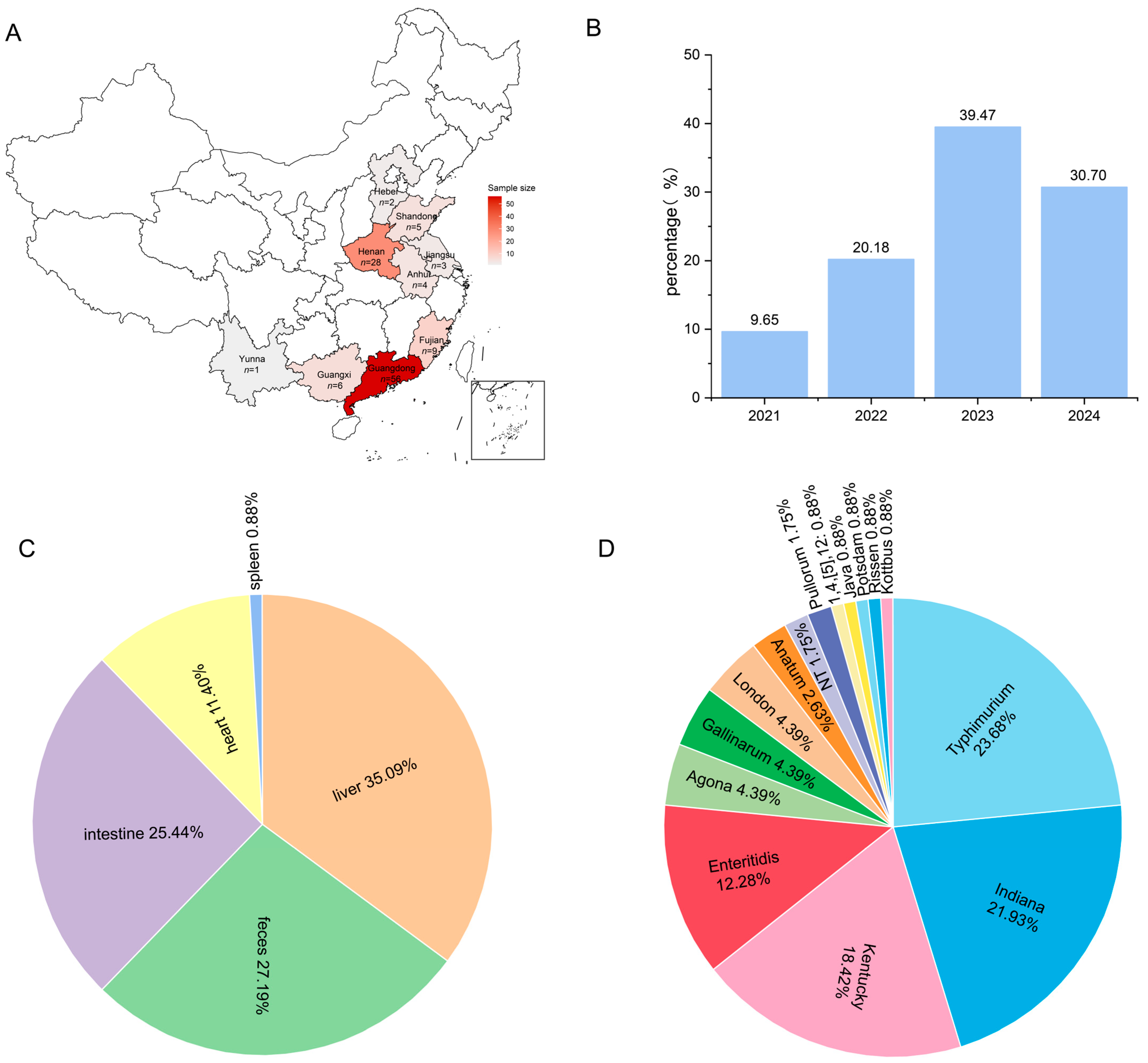
Figure 1. Epidemiological characteristics of 114 duck-derived Salmonella strains. (A) Distribution map of the duck-derived Salmonella in China. (B) Isolation time of the duck-derived Salmonella. (C) Isolation sites of the duck-derived Salmonella. (D) Results of serotyping of the duck-derived Salmonella.
Microbiolres 16 00184 g001
Figure 2. Distribution of the biofilm-forming ability of 114 duck-derived Salmonella strains. NT, no biofilm forming ability.
Microbiolres 16 00184 g002
Figure 3. Analysis of Salmonella drug resistance. (A) Bar graph of antibiotic resistance rates of 114 Salmonella isolates. (B) Distribution of multidrug resistance of 114 Salmonella isolates. (C) Difference in antibiotic resistance among dominant serotypes. ** p > 0.05, **** p > 0.0001, ns, not significant. Azithromycin (AZM); Cefotaxime sodium (CTX); Ceftriaxone (CRO); Meropenem (MER); Aztreonam (ATM); Ceftazidime (CAZ); Lomefloxacin (LOM); Norfloxacin (NOR); Gatifloxacin (GAT); Ciprofloxacin (CIP); Nalidixic acid (NAL); Enrofloxacin (ENR); Amikacin (AMK); Apramycin (APR); Gentamicin (GEN); Sulfamethoxazole (SUL); Florfenicol (FFC); Tetracycline (TET); and Doxycycline (DOX).
Microbiolres 16 00184 g003
Figure 4. Distribution of ARGs in 114 duck-derived Salmonella isolates. Different colors represent different antibiotic classifications.
Microbiolres 16 00184 g004
Figure 5. Correlation between dominant serotypes and drug resistance genes. The color depth and number of the colored blocks indicate the proportion of different drug-resistant genes in the serotype. The bar graph on the right shows the proportion of the 4 dominant serotypes carrying drug-resistant genes among the 114 strains of Salmonella.
Microbiolres 16 00184 g005
Figure 6. Distribution of virulence genes in 114 duck-derived salmonella isolates. Different colors represent different virulence gene classifications.
Microbiolres 16 00184 g006
Figure 7. Correlation between dominant serotypes and virulence genes. The color depth and numbers of the colored blocks indicate the proportion of different virulence genes in the serotype. The bar graph on the right shows the proportion of virulence genes carried by the four dominant serotypes among the 114 strains of Salmonella.
Microbiolres 16 00184 g007
Disclaimer/Publisher’s Note: The statements, opinions and data contained in all publications are solely those of the individual author(s) and contributor(s) and not of MDPI and/or the editor(s). MDPI and/or the editor(s) disclaim responsibility for any injury to people or property resulting from any ideas, methods, instructions or products referred to in the content.