Article

Assessing the Efficacy of Chemical and Green-Synthesized CuO Nanoparticles in Combatting Clinical Candida Species: A Comparative Study

1
Laboratory of Molecular and Cellular Screening Processes, Centre of Biotechnology of Sfax, P.O. Box 1177, Sfax 3018, Tunisia
2
Department of Microbiology, College of Veterinary Medicine, Tikrit University, Tikrit 34001, Iraq
3
Laboratory of Materials for Energy and Environment, and Modeling, Faculty of Sciences, University of Sfax, P.O. Box 1177, Sfax 3000, Tunisia
*
Author to whom correspondence should be addressed.
Microbiol. Res. 2025, 16(8), 178; https://doi.org/10.3390/microbiolres16080178 (registering DOI)
Submission received: 30 May 2025 / Revised: 28 July 2025 / Accepted: 29 July 2025 / Published: 1 August 2025

Abstract

The most prevalent growth of Candida cells is based on biofilm development, which causes the intensification of antifungal resistance against a large range of chemicals. Nanoparticles can be synthesized using green methods via various biological extracts and reducing agents to control Candida biofilms. This study aims to compare copper oxide nanoparticles (CuONPs) synthesized through chemical methods and those synthesized using Cinnamomum verum-based green methods against Candida infections and their biofilms isolated from Iraqi patients, with the potential to improve treatment outcomes. The physical and chemical properties of these nanoparticles were characterized using Fourier-transform infrared spectroscopy (FT-IR,) scanning electron microscopy (SEM), transmission electron microscopy (TEM), atomic force microscopy (AFM) and X-ray diffraction (XRD). Four strains of Candida were isolated and characterized from Iraqi patients in Tikrit Hospital and selected based on their ability to form biofilm on polystyrene microplates. The activity of green-synthesized CuONPs using cinnamon extract was compared with both undoped and doped (Fe, Sn) chemically synthesized CuONPs. Four pathogenic Candida strains (Candida glabrata, Candida lusitaniae, Candida albicans, and Candida tropicalis) were isolated from Iraqi patients, demonstrating high biofilm formation capabilities. Chemically and green-synthesized CuONPs from Cinnamomum verum showed comparable significant antiplanktonic and antibiofilm activities against all strains. Doped CuONPs with iron or tin demonstrated lower minimum inhibitory concentration (MIC) values, indicating stronger antibacterial activity, but exhibited weaker anti-adhesive properties compared to other nanoparticles. The antiadhesive activity revealed that C. albicans strain seems to produce the most resistant biofilms while C. glabrata strain seems to be more resistant towards the doped CuONPs. Moreover, C. tropicalis was the most sensitive to all the CuONPs. Remarkably, at a concentration of 100 µg/mL, all CuONPs were effective in eradicating preformed biofilms by 47–66%. The findings suggest that CuONPs could be effective in controlling biofilm formation by Candida species resistant to treatment in healthcare settings.

1. Introduction

C. albicans is the primary Candida species responsible for invasive candidiasis/candidemia in Australia, Japan, Korea, Hong Kong, Malaysia, Singapore, and Thailand, while Candida tropicalis is the most commonly identified Candida species in Pakistan and India [1]. The origin of Candida infection is more likely endogenous since these fungal yeasts belong to the human microflora of the gastrointestinal tract with a net dominance of Candida spp. [2]. Invasive candidiasis (IC), albeit less common than staphylococcal infections, is characterized by higher fatality rates, greater associated expenditures, and prolonged patient hospitalization [3,4].
The predominant antifungal agents employed for candidiasis treatment exhibit significant adverse effects, including the development of resistant Candida strains, which pose serious health risks [4]. As an example, in the Asia–Pacific region, azole antifungal resistance was reported for fluconazole to be 6.8–15%, Itraconazole 3.9–10% concerning isolates with the non-wild-type phenotype susceptibility against voriconazole 5–17.8%, and echinocandin 2.1–2.2% concerning invasive C. glabrata complex isolates. Moreover, in China, C. tropicalis has shown resistance to fluconazole 5.7–11.6% and to voriconazole 5.7–9.6%. Simultaneously, in the same area, invasive isolates of C. albicans, C. parapsilosis complex, and C. tropicalis exhibit a high susceptibility to fluconazole, exceeding 90% [1].
In Africa, a Tunisian study dealing with susceptibility of Candida spp. to antifungals found a higher resistance rate to fluconazole compared to caspofungin and micafungin. However, no resistance was revealed against voriconazole, amphotericin B, and 5-flucytosine [5].
The most prevalent growth of Candida cells is based on biofilm development, which causes the intensification of antifungal resistance against a large range of chemicals [6]. The initial stage of biofilm formation in any bacteria is adhesion, facilitated by cell wall proteins [7]. During this adherence, C. albicans produces a polysaccharide matrix, containing a combination of polysaccharides, proteins, DNA, and lipids [8]. This matrix serves as a barrier to the diffusion of antifungals inside the biofilm [9]. Interestingly, C. glabrata does not produce an exopolymeric matrix, but it shows an intrinsic antifungal drug resistance or rapidly becomes resistant when challenged with antifungals. C. glabrata adopts other antifungal resistance mechanisms involving the expression of a large number of adhesin-encoding genes. The adhesins play a very important role in biofilm formation in different substrates [10,11].
Therefore, the search for new materials with antifungal properties, particularly those effective against Candida biofilm, is required. Metal nanoparticles (NPs), as an alternative antifungal agent, are currently undergoing investigation in recent studies to develop effective therapeutic interventions for candidiasis, demonstrating encouraging outcomes [12]. Nanocomposites are heterogeneous/hybrid materials with improved flexibility, physical and chemical properties seem to have more potent antibiofilm activity [13].
Green synthesis refers to the eco-friendly production of nanoparticles using biological sources (plants, fungi, bacteria, algae, etc.) or natural reagents instead of toxic chemicals. For CuONPs, this involves reducing copper salts (e.g., CuSO4) with plant extracts or microbial enzymes, which act as both reducing and stabilizing agents [14].
Biological compounds can serve as effective reducing and stabilizing agents in the process of nanoparticle synthesis, offering a cost-effective and environmentally friendly approach that enhances the size uniformity and stability of the nanoparticles [15]. In particular, spices and condiments, as vital plant-derived products, are rich in bioactive compounds that hold potential for enhancing the green synthesis of nanomaterials [16].
The green synthesis of CuONPs enables better control over nanoparticle size and higher specific surface area (SSA) compared to conventional chemical methods thanks to natural bioactive compounds that act as stabilizers and prevent aggregation [17].
Synthesized highly stable CuONPs were obtained by using the extract of cinnamon bark due to its rich content in terpenoids, mainly eugenol, which was described as responsible for the biogenic reduction of Cu ions to NPs [18].
Therefore, this study aimed to target Candida spp. isolated from human clinical samples by utilizing synthesized CuONPs produced either chemically or through a green method using cinnamon extract. Additionally, the study incorporated metal doping, which has been previously used for its antibacterial and antibiofilm activities against some bacteria. This approach seeks to enhance the efficacy of the CuONPs against Candida infections, potentially improving treatment outcomes.

2. Material and Methods

2.1. Material

Four strains of Candida were isolated and characterized from Iraqi patients in Tikrit Hospital. The strains had been characterized and selected based on their ability to form biofilm on polystyrene microplates. Non-doped and doped (Fe, Sn), chemically synthesized CuONPs, previously described [19,20], were used.

2.2. Methods

2.2.1. Biosynthesis of CuONPs

First, 50 g of cinnamon powder was solubilized in 250 mL of distilled water. The solution was filtered through Whatman paper and 50 mL was mixed in a 1:6 ratio with a 1 M concentrated anhydrous copper sulphate solution. The CuSO4 solution was carefully added from the burette to the freshly prepared cinnamon extract while stirring at room temperature for three hours. The synthesis of CuONPs was confirmed by the loss of the solution’s brown coloration. Following 24 h of incubation, the mixture was centrifuged at 10,000 rpm for 10 min to provide a pellet, which was subsequently purified by washing three to five times with double-distilled water. The final residue was calcined in a muffle furnace at 400 °C to eliminate the adhering organic materials from the powdered cinnamon and subsequently stored for future use. Chemically synthesized doped and non-doped CuONPs have been previously documented [19].

2.2.2. X-Ray Diffraction Analysis

To check the crystallinity and crystal phases of the prepared powder, the XRD pattern was performed (Philips X-ray diffraction spectrometer, PW 1540, Eindhoven, The Netherlands). Notably, the XRD patterns of the CuONPs feature multiple diffraction peaks consistent with those found in the ICCD card no. 0029-110-96 for CuO [21]. It belongs to the monoclinic system with the space group C2/c.
From XRD pattern, the lattice parameters were calculated by using the Bragg’s equation:
2 d h k l sin ( θ )   =   n λ
where θ is the Bragg’s angle, λ is the X-ray wavelength; dhkl is the interplanar spacing and n is the diffraction order.
For CuO (monoclinic structure):
1 d 2 h k l   =   1 sin 2 ( β ) h 2 a 2 +   k 2 sin 2 ( β ) b 2     + l 2 c 2   2 h l cos   ( β ) ac
Moreover, the unit cell volume V, for the monoclinic CuO, is given by:
V   =   abc   sin ( β )
The average crystallite size and micro-strain (ε) of the synthesized oxides are determined from the observed FWHM of the XRD peaks utilizing the Williamson–Hall (W-H) model [22]:
β h k l cos θ   = k λ D   +   4 ε   sin θ
λ represents the wavelength of X-ray radiation, β h k l denotes the full width at half maximum (FWHM) of the diffraction peak, θ signifies Bragg’s diffraction angle, and k is the shape factor, which is equal to 0.9. The specific surface area (SSA) is pivotal in the antibacterial efficacy of nanoparticles, as it determines the interface with surrounding molecules. The SSA can be estimated using the following equations [23]:
SSA     =     6 D   ×   ρ
  ρ =   Z × M N × V
ρ   is the particle density [ g . cm 3 ], M is the molar mass of the substance (M(CuO) = 79.545 g.mol−1), Z represents the number of formula units in the unit cell (Z = 4 for CuO), V signifies the volume of the unit cell, and N is Avogadro’s number (N = 6.02214 × 10²³ mol−1).

2.2.3. CuO Characterization Methods

Morphological characterization was conducted utilizing a scanning electron microscope (JEOL JSM7100F, JEOL, Nieuw-Vennep, The Netherlands) in conjunction with an energy-dispersive spectrometer (EDS) for elemental composition measurement. X-ray diffraction (XRD) patterns were acquired with a Bruker D8 Advance diffractometer employing monochromated CuKα radiation (λ = 1.5406 Å). Infrared spectra were obtained within the 4000–400 cm−1 range utilizing a Perkin Elmer FT-IR spectrometer. Optical absorption was assessed using a Perkin Elmer UV-Vis Lambda 365 spectrophotometer within the 190–1100 nm wavelength range. Additionally, detailed microstructure analysis was conducted using a transmission electron microscope (TEM) and scanning electron microscopy (SEM, Ziess, Oberkochen, Germany), providing further insights into the size and distribution of particles at the nanoscale. Atomic force microscopy (AFM, Nanosurf, Liestal, Switzerland) was also employed to analyze the surface topography and roughness at the nanometer scale.
CuONPs were then imaged by TEM, SEM and AFM and were analyzed by X-Ray Diffraction in BPC Analysis Centre (Baghdad, Iraq). Descriptive statistics were calculated using ImageJ software (version 1.54g) from the microscopy image. FT-IR spectrum of CuONPs was performed with a Shimadzu instrument (FTIR-8400S, Shimadzu, Tokyo, Japan) in the Chemical Department, Faculty of Sciences, Tikrit University.

2.2.4. Biofilm Forming Assay

The capacity for biofilm generation in yeast isolates was assessed using a 96-well microtiter plate test employing the crystal violet staining technique. Each well of a 96-well flat-bottomed sterile polystyrene microplate, containing 199 μL of Sabouraud Dextrose Broth supplemented with 1% glucose, was infected with 20 μL of a yeast suspension at a concentration of 0.5–0.7 McFarland (1 × 10⁸ cfu/mL).
Microplates are incubated for 24 h at 37 degrees Celsius. The liquid medium was removed, and the adhering cells were rinsed twice with phosphate-buffered saline (PBS), followed by drying the wells at 60 °C for one hour. Subsequently, it was treated with 150 μL of 2% crystal violet for 15 min. The crystal violet-stained microplate wells were rinsed twice with PBS to eliminate excess stain. After air drying, the dye associated with the biofilm that coated the microplate walls was re-solubilized using 150 μL of 95% ethanol. Following 5–10 min, the microplate is analyzed spectrophotometrically at 570 nm using a microplate reader (Labtech International, Rotherham, UK). The experiment was conducted a minimum of three times with new samples on each occasion. Biofilm production was interpreted using the criteria of Stepanović et al. [24].

2.2.5. Effect of Nanoparticles on Planktonic Candida Viability

Well Diffusion Method
The well diffusion assay was conducted on Mueller–Hinton agar using an inoculum prepared from a 24 h Sabouraud dextrose agar culture, adjusted to a 0.5 McFarland turbidity (1.5 × 10⁸ CFU/mL) standard. Once the medium had solidified, the inoculum was plated on the surface and wells, each with a diameter of 6 mm, were created in the agar. Subsequently, 20 μL of the antifungal agent were placed into each well. The plates were then incubated at 35–37 °C for 24 h.
Microdilution Method
The MIC was measured according to [25]. The isolates were initially cultivated in the nutrient broth overnight at 37 °C. Subsequently, colonies cultivated overnight were suspended in sterile distilled water and well mixed to obtain a suspension with a turbidity of 0.5 McFarland (1.5 × 10⁸ CFU/mL). Next, 100 microliters of Mueller–Hinton broth were added into each well; 100 microliters of the nanoparticle solutions were also added into each well using the microdilution (7 dilutions for each solution). Subsequently, 10 microliters of Candida inoculum were added. The plates were incubated at a temperature of 35–37 °C for 24 h. The MIC test results were determined using an ELISA reader (Labtech International, England) by comparing the absorbance readings of the treated samples to those of the control. Additionally, the presence or absence of turbidity (indicative of growth) was assessed using an illumination box, as higher turbidity correlates with increased absorbance. The MIC corresponds to the lowest concentration that inhibits microbial growth. Yeast inoculum + medium only was used as a negative control (without antifungals).

2.2.6. Antibiofilm Activity

Then, 96-well polystyrene microtiter plates were used for the evaluation of the antibiofilm activity of nanoparticles. The first step is the development of yeast biofilms, which commenced with the initial attachment of yeast to the well surfaces. Overnight cultures in Nutrient broth at 37 °C were used to prepare an inoculum of 0.5 McFarland turbidity yeast suspension.
Antiadhesive Activity
Approximately 100 μL of the standardized yeast inoculum was introduced into sterile flat-bottomed 96-well plates, followed by the addition of 100 μL of the material of interest, and incubated for 24 h at 37 °C without agitation. Following a 24 h incubation, a crystal violet staining (CVS) experiment was conducted to quantify the biofilm biomass as follows: The wells were meticulously emptied and subsequently washed three times with sterile phosphate-buffered saline (PBS) to eliminate unattached yeast–planktonic cells. Plates were dried in an oven at 60 °C for 45 min, after which 150 μL of 1% crystal violet solution was added to each well and incubated for 20 min at room temperature. Excess discoloration was removed by washing the plates a minimum of five times with distilled water.
The crystal violet stain bound to the adherent cells was resolubilized with 150 μL of 95% ethanol to destain the wells where the biofilm attached. Controls and blank: Negative control (Yeast inoculum + SDB). Positive control [Yeast inoculum + SDB + antibiotics (fluconazole)]. Sample control (sample + SDB). Antibiotic control (antibiotic + SDB). Media control (SDB only) served as blank.
The optical density (OD) of each well stained with crystal violet is assessed at 570 nm using an ELISA microtiter plate reader. The issue with conventional OD readers is that they assess the OD just at a single spot in the center of the well. Therefore, if the biofilm thickness at that location markedly deviates from the remainder of the well, the measurement will be inaccurate. The recommended approach facilitates homogenous resolubilization of CV bound to the biofilm layer, allowing for indirect yet accurate monitoring of biofilm generation.
The absorbance of the plates was measured at 570 nm with an ELISA microplate reader. The mean absorbance (OD590 nm) of the sample was calculated, and the findings were represented as percentage inhibition (%) using the equation below [26].
inhibition     percent   % = OD Negative   control   -   OD Experimental OD Negative     control × 100
Biofilm Eradication Assay
The biofilm formation of yeast isolates was allowed by the use of 100 μL of the respective standardized yeast inoculum and performed in sterile flat-bottomed 96 well plates and incubated for an additional 24 h. Next, 100 μL of NPs were added and incubated for additional 24 H. Violet staining assay was performed as described previously to quantify the biofilm biomass.

3. Results

3.1. Biosynthesis of CuONPs

Green synthesis of CuONPs was performed using cinnamon extract, as described in the materials and methods section. The formation of CuO nanoparticles was visually confirmed by a distinct color change of the reaction mixture from blue to dark, indicating successful reduction and nanoparticle formation.
The purified NPs were the subject of various characterization methods.

3.2. CuONPs Characterization

3.2.1. Energy-Dispersive Spectroscopy (EDS) Analysis

To examine the chemical elements and assess the purity of the synthesized powders, we analyzed the obtained samples using the energy-dispersive spectroscopy (EDS) technique.
The variation in intensity [cps/eV] as a function of energy [keV] (Figure 1), allowed us to identify the chemical elements present within the synthesized samples.
The EDS analysis of the compounds, green CuO and chemical CuO, reveals the presence of oxygen, copper, and sulfur in similar intensities, indicating their relative abundances. The energy peaks at 0.5 keV and at 0.8 keV correspond, respectively to oxygen and copper, while sulfur is present in lesser amounts at 2.3 keV. Additionally, the peaks at higher energies (8 keV and 8.9 keV) for copper suggest deeper electronic transitions, although their intensity is relatively low.
The elemental composition is presented as a table in the inset of each figure. The shift from the predicted values is due to defects such as oxygen vacancies and/or Cu interstitials.

3.2.2. Scanning Electron Microscopy

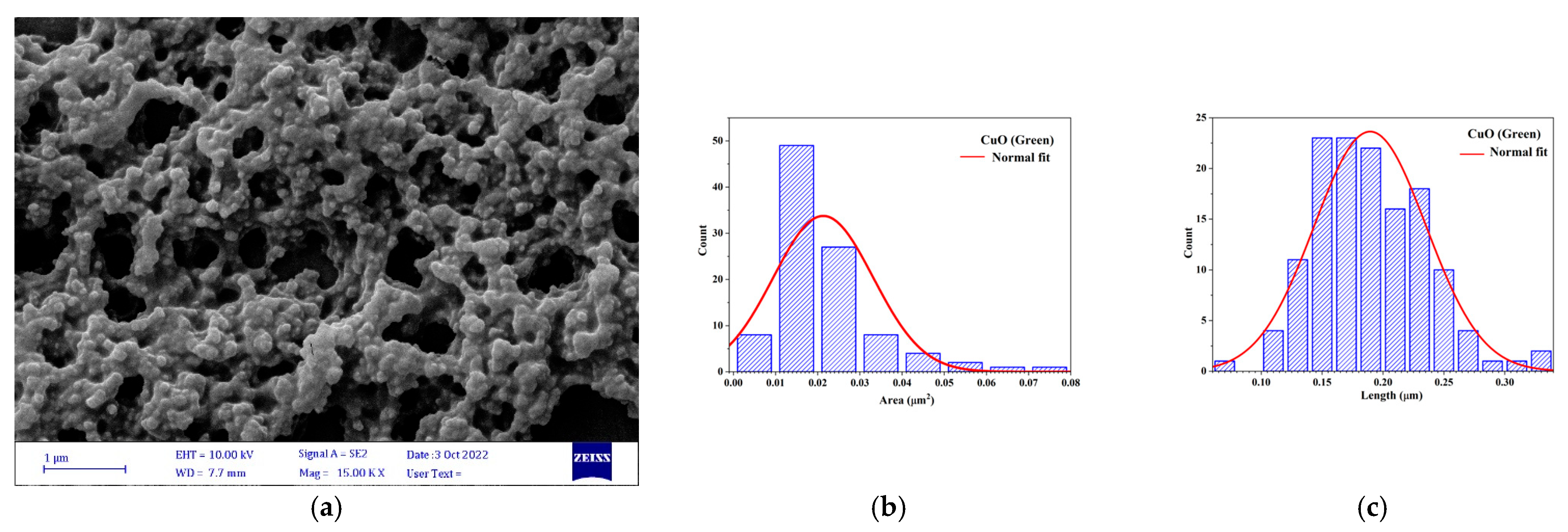
The surface morphology of the green-synthesized CuONPs has been studied through FE-SEM images at different magnifications (Figure 2).
The FE-SEM micrographs confirmed that the particles have sizes between 0.079 and 0.333 μm as calculated by the ImageJ program, which again confirmed the nanostructured nature of the copper oxide presented in Figure 2. The NPs produced via green synthesis are generally smaller, with reduced grain size and surface area compared to those obtained by chemical methods (Table 1).
Comparative analyses reveal that green-synthesized CuO exhibits significantly smaller (0.194 µm) and more homogeneous grains compared to chemically synthesized CuO (1.012 µm). This morphological difference, attributed to the controlled growth mediated by bio-reducing agents, results in a lower area for the green NPs (Table 1). In contrast, chemical synthesis yields coarser and more heterogeneous particles, characteristic of less controlled growth. These findings highlight the advantage of green processes for producing well-defined nanostructures, particularly suited for catalytic applications.

3.2.3. TEM

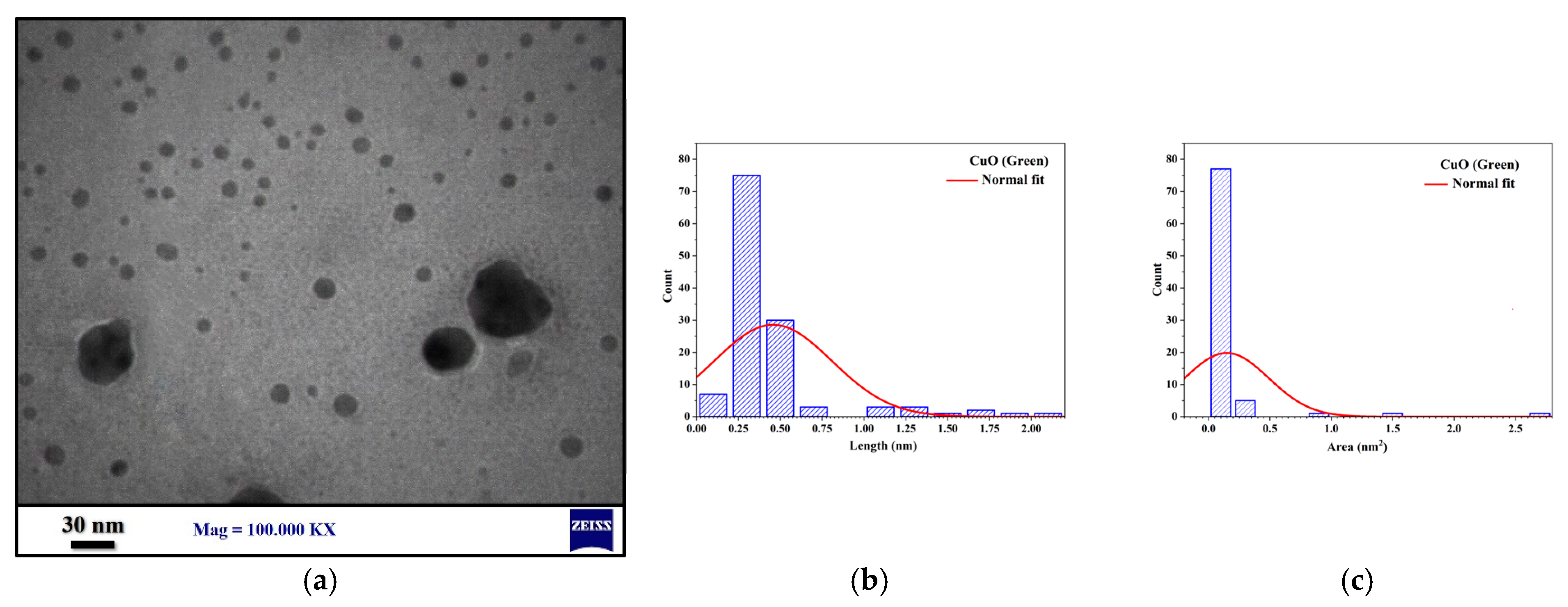
TEM analysis showed that the synthesized CuONPs have a spherical morphology and are uniformly distributed and homogeneous (Figure 3). The average particle size and area, determined using ImageJ software, are 0.457 nm and 0.147 nm2, respectively. These results confirm the findings from field-emission scanning electron microscopy (FE-SEM), which also reveal spherical and homogeneous nanostructures. This uniformity and precise size are crucial for consistent performance in various applications. Thus, both techniques confirm that the synthesis process is effective and reproducible, optimizing the properties of CuONPs.

3.2.4. AFM Analysis

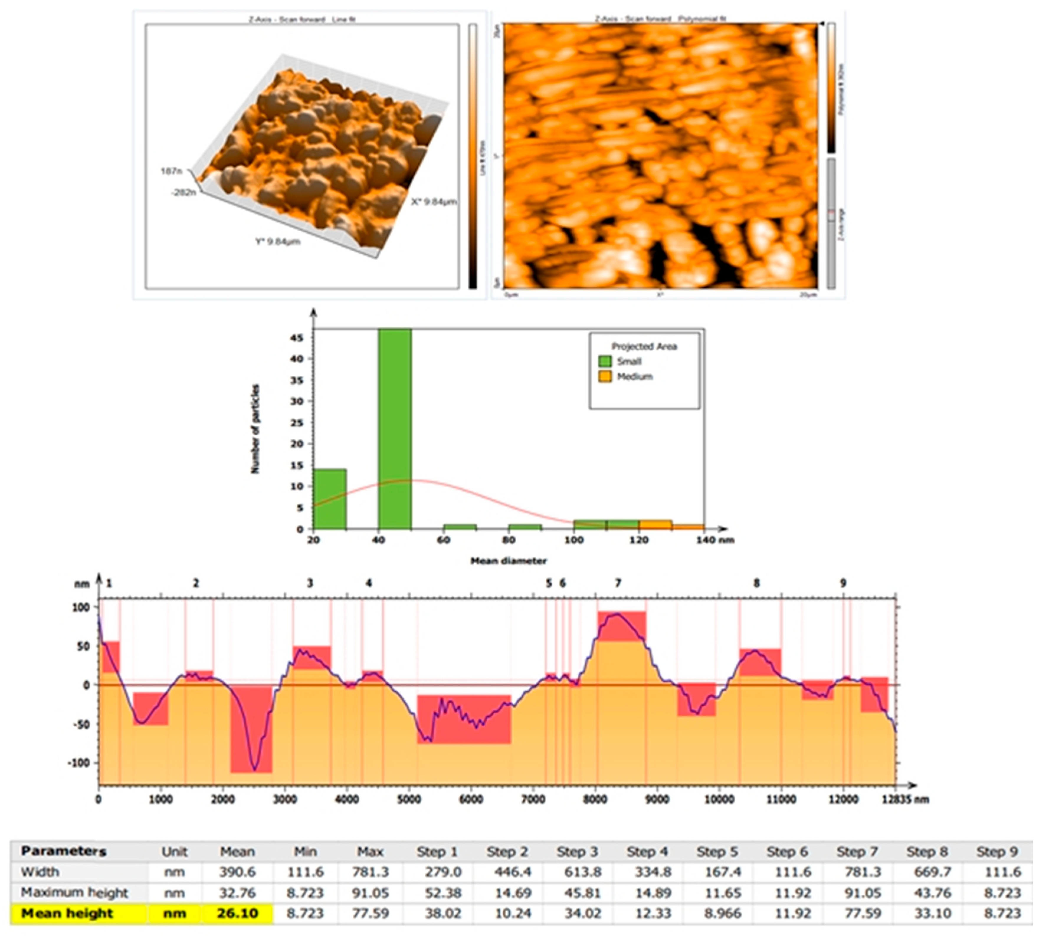
Figure 4 shows AFM analysis of the morphological structure of green-synthesized copper oxide nanoparticles. It shows images measured with a size = 20 × 20 µm. Two and three-dimensional AFM images explain the structural shape of grains. The average roughness was found to be (29.22 nm), the root mean square (RMS) is (37.89 nm), and the average diameter is (44.81 nm).
The width histogram reveals a broad distribution, ranging from 111.6 nm to 781.3 nm, indicating substantial heterogeneity in particle size (Figure 4).
The height graph (Figure 5) shows that while the maximum heights vary considerably (8.723 nm to 91.05 nm), the average heights are more moderate (8.723 nm to 38.02 nm).
This uneven distribution of dimensions suggests non-uniform particle growth, potentially impacting the material’s properties.

3.2.5. X Ray Diffraction Study

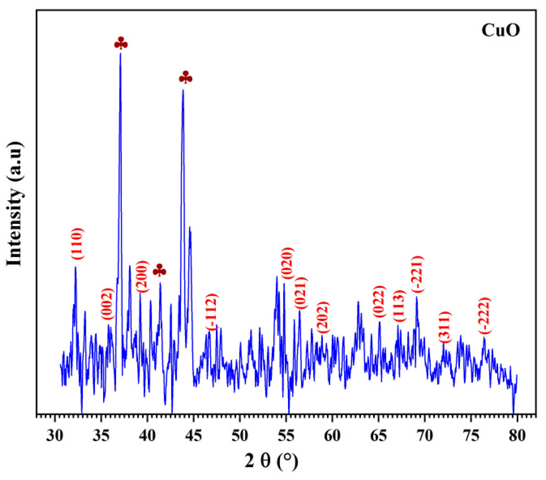
The XRD patterns of CuONPs is represented in Figure 6. Notably, the XRD patterns of the sample exhibit multiple diffraction peaks consistent with ICCD card no.: 0029-110-96. These diffraction patterns can easily be associated with the monoclinic crystal structure.
In addition to the primary CuO phase, the XRD data reveal the presence of additional reflections (marked with plum symbols) associated with impurities.
The structure of CuONPs, characterized by lattice parameters (a = 4.66 Å, b = 3.39 Å, c = 5.11 Å, β = 99.49°, and V = 79.59 Å3).a = 4.66 Å, b = 3.39 Å, c = 5.11 Å, β = 99.49°, and V = 79.59 Å3.
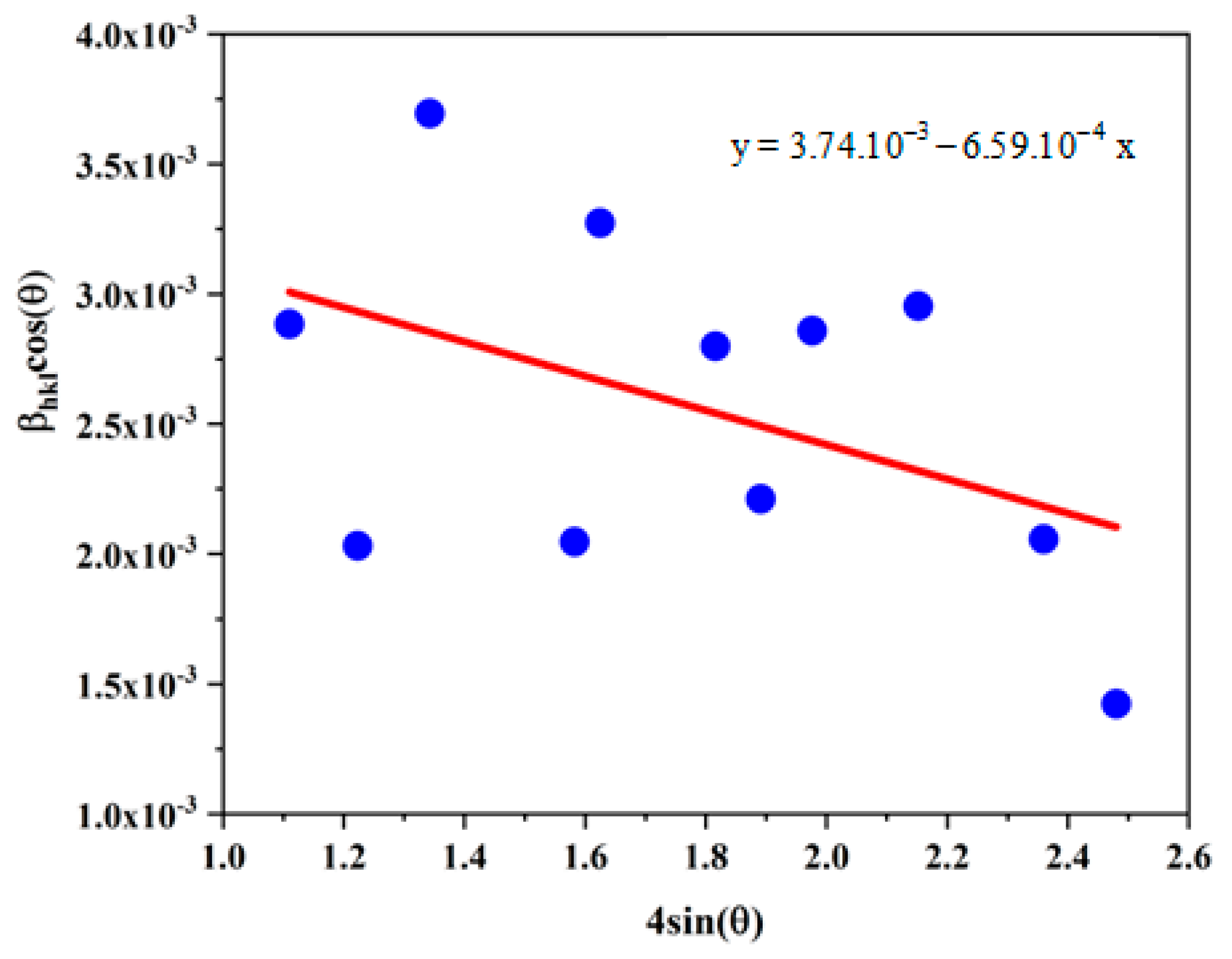
The W-H plot (βhklcos (θ) vs. 4 sin (θ)) (Figure 7) facilitates the assessment of micro-strain through the slope of the fit and crystallite size via the y-axis intercept.
Table 2 displays the values of the crystallite size (D), the strain (ε) and the SSA of the CuONPs.
The negative micro-strain suggests that the nanoparticles are subjected to slight internal compression and the density is close to the theoretical density of CuO (6.31 g.cm−3), which indicates a relatively high purity of the synthesized particles.
CuONPs synthesized via green synthesis with cinnamon are smaller (37.07 nm) and have a higher specific surface area (24.38 m2. g−1) compared to those obtained through chemical synthesis (51.93 nm and 17.20 m2.g−1), enhancing their effectiveness in biological applications by increasing reactivity and cellular interaction. The absolute value of dislocation density is lower for green synthesis (−6.59.10−4) compared to chemical synthesis (5.85.10−4), suggesting better stability and lower toxicity.

3.2.6. FT-IR Spectrum of CuONPs

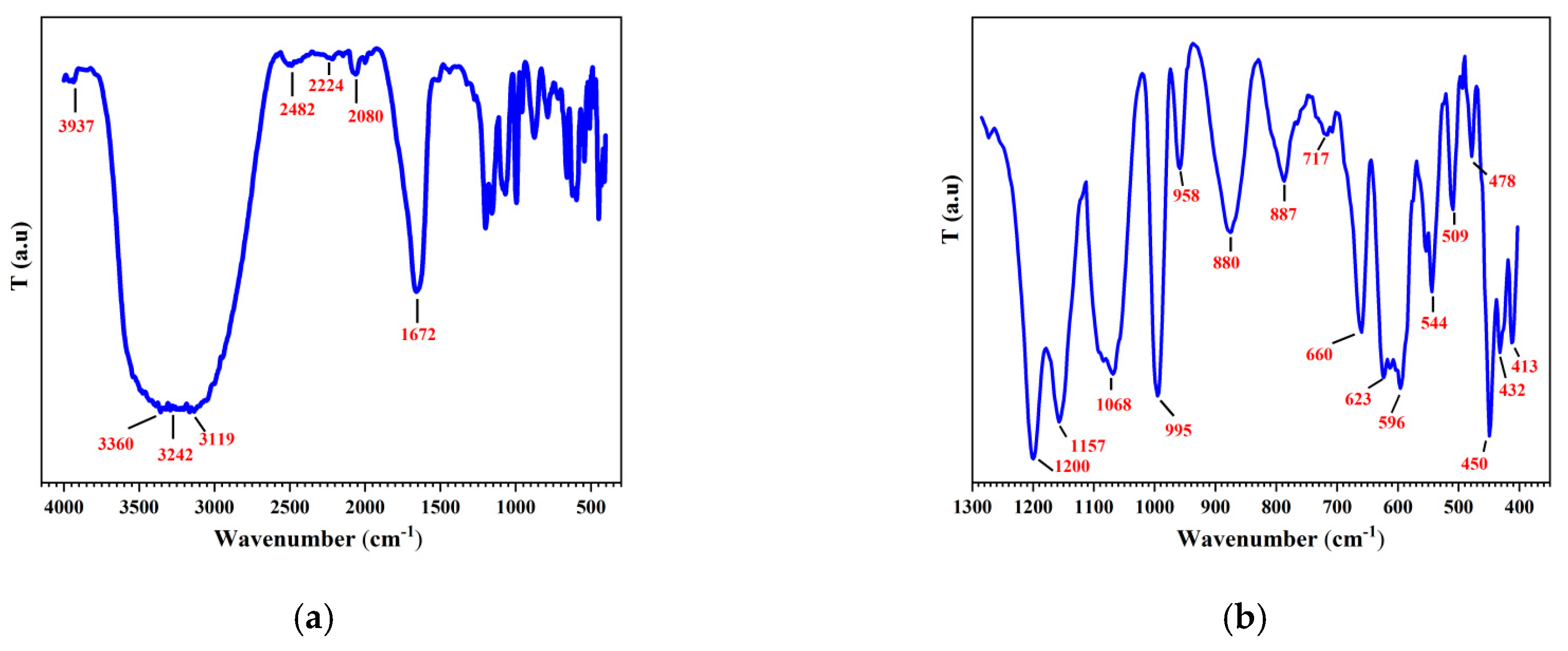
The FT-IR spectrum of the CuONPs (Figure 8) showed different bands. The IR spectra within the 400–700 cm−1 range exhibit vibrational bands linked to the stretching mode of the Cu-O bonds [27]. The bands detected in the frequency range of 400 to 800 cm−1 arise from the stretching vibrations of the Cu-O and O-Cu-O bonds. The existence of bands near 1000 cm−1 signifies several kinds of bending vibrations of the Cu–O bonds [28]. Furthermore, the broad bands at 3484 cm−1 and 3572 cm−1 correspond to the deformation and elongation vibrations of the O-H bonds in adsorbed water.
The presence of these functional groups within the structure of polyphenolics suggests that the spectrum can demonstrate the presence of phenolics in the plant leaf extract. The phenolics in the extract may be responsible for the reduction of metal ions and the formation of NPs [29].
The FTIR spectra of CuO prepared by green synthesis and co-precipitation revealed significant differences. The CuO synthesized via the green route displays a more complex spectrum, with numerous bands in the 413 to 3967 cm−1 regions, indicating the presence of Cu-O vibrations as well as organic residues, likely due to the biological precursors used. In comparison, the CuO obtained by co-precipitation shows fewer bands, primarily in the 480 to 3574 cm−1 regions, suggesting a purer structure with fewer organic contaminants. The O-H bands, present in both methods, are more intense and complex in the CuO synthesized via the green route, indicating greater interaction with water or hydroxyl groups. Thus, CuO prepared by green synthesis is more heterogeneous and richer in organic residues, while co-precipitation produces a purer and chemically simpler CuO.

3.3. Effect of NPs on Planktonic Candida Viability

The test was performed on different Candida strains belonging to C. albicans, C. glabrata, C. tropicalis and C. luistaniae.

3.3.1. Well Diffusion Method

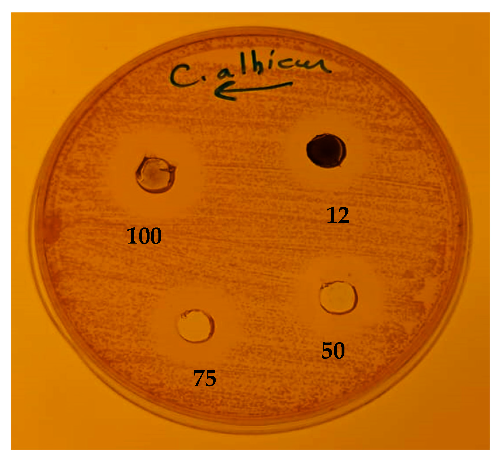
The CuO chemically synthesized NPs were tested against C. albicans cells grown on Mueller–Hinton agar plates (Figure 9). The NPs in varied concentrations were able to inhibit the growth of C. albicans strains. The nanoparticles concentration ranged from 12.5 to 100 mg/mL, which is followed by the appearance of clear zones (halos) ranging between 15 and 20 mm.

3.3.2. Microdilution Method

Non-doped and doped CuONPs were used in this study. The activities of nanoparticles were assessed against Candida strains by means of the microdilution method.
The present results show that all CuONPs can inhibit the growth of Candida cells with MIC values ranging between 12.5 and 125 μg/mL (Table 3). The doped CuONPs display the weakest values of MIC except C. glabrata, which was not inhibited by nano3 (the double-doped CuONPs). Flucanozole is still the best antifungal drug with MIC values of 8–32 μg/mL. MIC of CuONPs was estimated to 35.5 μg/mL for C. albicans against 25 μg/mL for flucanozole MIC [30].

3.4. Antibiofilm Assay

The capacity for biofilm generation in the four Candida strains was followed spectrophotometrically using a 96-well microtiter plate test employing the crystal violet staining technique. The four strains have the criterion 2 × ODc < OD ≤ 4 × ODc (where OD corresponds to the average optic density value and ODc the cut-off value of OD that separates biofilm-producing from non-biofilm-producing strains) [24] which shows a moderate biofilm producer (++). The four biofilm-forming Candida strains were used to evaluate the anti-biofilm activities of nanoparticles.

3.4.1. Antibiofilm Activity of Nanoparticles

The adopted concentration for inhibiting Biofilm was equal to the MIC value for each type of NPs, except C. glabrarata, for which we were unable to determine the MIC and we have used a concentration of 100 μg/mL, while fluconazole was used at 50 μg/mL.
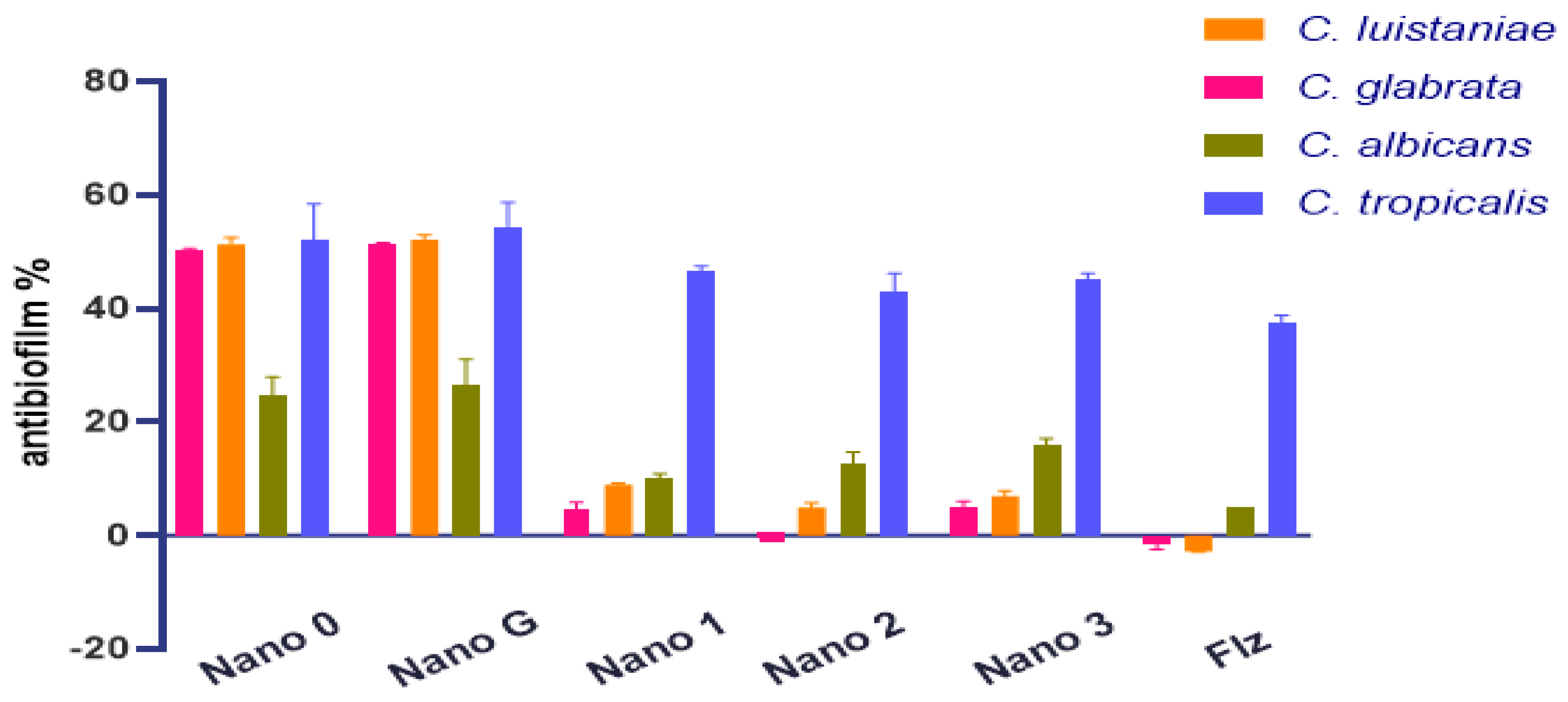
Both chemically and green-synthesized non-doped CuONPs exhibit a similar inhibition rate against all tested Candida biofilms, although they are less effective against C. albicans biofilms. This observation highlights the resistance of C. albicans, as its biofilm susceptibility to current therapeutic agents remains low [31]. Conversely, doped CuONPs show reduced activity against the same Candida strains but retain their antibiofilm efficacy against C. tropicalis. Biofilms of C. glabrata appear to be the most resistant to doped NPs, followed by those of C. lusitaniae. Notably, the antibiofilm activity against C. tropicalis is also exhibited by fluconazole (Table 4, Figure 10).

3.4.2. Biofilm Eradication Assay

The preformed biofilms of the four strains of Candida are treated with 100 μg/mL of all the types of NPs and fluconazole. The activities of the different NPs were similar (Table 5).
In fact, at 100 μg/mL, all the used compounds were able to remove the preformed biofilms at approximately 47–66%.

4. Discussion

The study detailed the synthesis and characterization of green CuONPs using
Cinnamomum verum extract. It investigated the antifungal activity of these nanoparticles against both planktonic and biofilm-forming Candida strains. The efficacy of the green-synthesized CuNPs was compared to that of previously synthesized non-doped and doped CuONPs [19].
The synthesized CuONPs exhibit a well-defined, uniform structure, with detailed morphological characteristics revealed through transmission electron microscopy and atomic force microscopy.
The smaller size and uniform distribution of CuONPs synthesized through green methods can lead to enhanced reactivity and performance in catalytic applications. Additionally, the non-uniform particle growth observed through AFM may suggest the need for optimization in synthesis parameters to achieve desired properties for specific applications.
The XRD study indicated that the particles first exist as collides, thereafter growing and reacting with environmental O2; this is corroborated by SEM data displaying particle aggregates, consistent with findings by Taran et al. [32].
The green synthesis of CuO has been described before for other plant extracts, such as [33]. The surface morphology of the synthesized CuONPs, studied through FE-SEM, shows overlapped nanostructures, resulting in an almost spherical shape justified on the surface of CuONPs [22].
This difference in CuONPs size is attributed to the less aggressive nature of green synthesis, leading to less agglomerated and better-dispersed particles. These findings are supported by various studies, such as the comparative study by Keabadile et al. on CuO synthesis methods [28].
Our CuONPs were able to inhibit the growth of C. albicans by creating an inhibition zone on Petri dishes at concentrations of 12 to 100 mg/mL (Figure 1). It has been reported that 100 μL of CuONP suspension was able to inhibit significant fungal growth, showing a good inhibition zone against C. glabrata 75%, Aspergillus flavus 68%, T. longifusus 60% [34]. In another study, C. albicans stopped growing at a concentration of 44.4 μg/mL of CuONPs. CuONPs at a dose of 35.5 μg/mL exhibited antifungal activity that was similar to that of conventional antifungals. However, fluconazole produced greater zones of inhibition at the same concentration [35].
In this study, we explored the growth inhibition of various Candida species isolated from clinical samples using different types of CuONPs. High MIC values were obtained, ranging between 12.5 and 125 µg/mL. Notably, doped CuONPs with iron (Fe) exhibited the most potent antifungal activity, with minimum inhibitory concentration (MIC) values between 12.5 and 50 µg/mL. Both doped and non-doped CuONPs demonstrated significant antibacterial activity, with MIC values ranging from 0.039 to 1.25 mg/mL [19].
The antibiofilm activity of CuO nanoparticles arises from multiple complex mechanisms. This is attributed both to the intrinsic physicochemical properties of the nanoparticles and to the composition of the biofilm matrix, which includes biological and environmental factors. Specifically, CuO nanoparticles can directly interact with bacterial membranes, generating reactive oxygen species (ROS) that induce oxidative stress. Additionally, the release of copper ions (Cu2+) plays a critical role in their antimicrobial toxicity. Furthermore, modifications to the support surface and its properties can influence nanoparticle–biofilm interactions and efficacy [19].
Moreover, doping nanoparticles can alter their morphology and size, while the non-uniform distribution of dopants on the surface can directly affect their interaction properties. For instance, CuO nanoparticles doped with iron at higher concentrations demonstrated enhanced anti-adhesive activity compared to undoped CuO. This finding highlights the potential of transition metal-doped CuO nanoparticles as promising candidates for biomedical applications, owing to the modified physicochemical properties imparted by doping [20].
The in vitro antifungal activity of CuONPs incorporated into polycaprolactone (PCL) fibers was evident. At an initial CuONP concentration of 25 mM in the fibers, growth inhibition of C. albicans and C. glabrata reached 53% under experimental conditions (equivalent to 200 mM of free CuONPs in solution). C. tropicalis exhibited even greater inhibition (up to 59%) under the same conditions [36].
A nanocomposite based on nanostarch, nanochitosan, and CuONPs exhibited promising antifungal activity against C. glabrata MTMA 19, C. glabrata MTMA 21 and C. tropicalis MTMA 24 strains that were selected as the most resistant toward commercial antifungal drugs [36]. CuONPs produced at pH levels of 6, 8, 10, and 12 showed the capacity to prevent biofilm formation by over 70% at the minimum fungicidal concentration (MFC). Notably, at this concentration, CuONPs produced at pH levels of 10 and 12 exhibited over 86% biofilm suppression in vitro [37].
Recently Malinaric NM et al. [38] compared the effect of ZnO and CuO against
Staphylococcus aureus and C. ssp. and demonstrated that ZnO added to PMMA (Polyméthacrylate de Méthyle) is considered a promising denture material showing antimicrobial properties against both strains.
Compared to previous investigations, which were limited to C. albicans [31], our study was conducted with a large number of Candida species to target their biofilm with a pool of CuONPs. The doped CuONPs were described as active as non-doped NPs against bacterial biofilms [19]. In another study, compared to undoped CuO, Fe and Ni-doped oxides show an improved activity when used at high concentrations [20]. Meanwhile, in the present antifungal investigation, doping reduced the antibiofilm activity of CuONPs, which could be explained by the lower minimum inhibitory concentration (MIC) values of the doped nanoparticles, as the antiadhesive activity was assessed at MIC concentrations. Moreover, it was shown that the addition of Sn, or Fe/Sn co-doped to the CuO network, causes an increase in grain size compared to the undoped sample [19], which could affect the penetrance of NPs within Candida cells and reduce the antiadhesive activity.
Moreover, our CuONPs effectively removed preformed biofilms, achieving 47–66% eradication across tested Candida species. Notably, in C. albicans, over 79% of the preformed biofilm was eliminated upon exposure to CuONPs, consistent with the antifungal and antibiofilm effects of biosynthesized CuONPs reported by Sasarom et al. [34]. While the biofilm eradication efficacy of CuONPs at the minimum fungicidal concentration (MFC) was below 50% for all pH levels tested, at 2× MFC, CuONPs synthesized at pH 10 and 12 exceeded 50% eradication (53.88 ± 5.79% and 52.52 ± 8.63%, respectively). At 4× MFC, CuONPs produced at pH 6, 8, 10, and 12 all achieved > 50% biofilm reduction. Similar trends were observed for C. tropicalis, underscoring the broad-spectrum potential of CuONPs against Candida biofilms [37].
The biofilm of C. tropicalis seems to be the most susceptible to the effect of CuONPs. Biofilm characteristics vary widely and are specific to each bacterial or yeast strain. This diversity is attributed to the complex genetic regulatory systems that govern the resistant state of these microorganisms, as well as the vastly differing environmental conditions they encounter. This could explain the difference obtained with the antibiofilm activities for the various strains and with the different nanoparticles tested [39].
Our study highlights the promising potential of nanoparticles (NPs) to effectively target some Candida species, paving the way for the development of innovative therapeutic tools, particularly utilizing CuONPs and other types of NPs. By varying the synthesis conditions and the choice of doping metals, we can enhance their antifungal efficacy and tailor these nanoparticles for improved treatment strategies.

5. Conclusions

Both CuONPs produced through Cinnamomum-based green synthesis and those synthesized chemically exhibit activity against Candida strains isolated from Iraqi patients, as well as their biofilms. Despite their physical differences, no significant variations were observed in the antifungal activities between the green and chemically synthesized CuONPs. Doped CuONPs (containing iron, tin, or both) demonstrated greater efficacy against planktonic Candida cells but were less effective against biofilms. While our in vitro results demonstrate promising anti-biofilm potential, translational applications require caution. Recent studies highlight challenges in transposing nanoparticle efficacy to in vivo settings, including host toxicity, biofilm microenvironment complexity, and immune interactions. Nevertheless, this study underscores the potential of CuONPs as a scaffold for developing innovative antifungal tools, provided that future work addresses these translational gaps.

Author Contributions

H.Y.K. performing experiments, F.B.N., design and interpreting physics data, B.S.N., design of sampling work, S.M., design and interpretation of antibiofilm activity, S.A., design, supervision and writing of the manuscript. All authors have read and agreed to the published version of the manuscript.

Funding

This research received no external funding.

Institutional Review Board Statement

Not applicable.

Informed Consent Statement

Not applicable.

Data Availability Statement

No new data were created or analyzed in this study. Data sharing is not applicable to this article.

Acknowledgments

This work was supported by the Tunisian and the Iraqi Ministries of Higher Education and Scientific Research. The authors thank the patients for giving their samples and allowing the present work.

Conflicts of Interest

The authors declare no conflict of interest.

References

  1. Cornely, O.A.; Sprute, R.; Bassetti, M.; Chen, S.C.-A.; Groll, A.H.; Kurzai, O.; Lass-Flörl, C.; Ostrosky-Zeichner, L.; Rautemaa-Richardson, R.; Revathi, G.; et al. Global guideline for the diagnosis and management of candidiasis: An initiative of the ECMM in cooperation with ISHAM and ASM. Lancet Infect. Dis. 2025, 25, e280–e293, Erratum in: Lancet Infect. Dis. 2025, 25, e203. Erratum in: Lancet Infect. Dis. 2025, 25, e261. [Google Scholar] [CrossRef] [PubMed]
  2. Kreulen, I.A.M.; de Jonge, W.J.; van den Wijngaard, R.M.; van Thiel, I.A.M. Candida spp. in Human Intestinal Health and Disease: More than a Gut Feeling. Mycopathologia 2023, 188, 845–862. [Google Scholar] [CrossRef]
  3. Bays, D.J.; Jenkins, E.N.; Lyman, M.; Chiller, T.; Strong, N.; Ostrosky-Zeichner, L.; Hoenigl, M.; Pappas, P.G.; Thompson, G. Epidemiology of Invasive Candidiasis. Clin. Epidemiol. 2024, 16, 549–566. [Google Scholar] [CrossRef] [PubMed] [PubMed Central]
  4. Re, V.L.; Carbonari, D.M.; Lewis, J.D.; Forde, K.A.; Goldberg, D.S.; Reddy, K.R.; Haynes, K.; Roy, J.A.; Sha, D.; Marks, A.R.; et al. Oral Azole Antifungal Medications and Risk of Acute Liver Injury, Overall and by Chronic Liver Disease Status. Am. J. Med. 2015, 129, 283–291.e5. [Google Scholar] [CrossRef] [PubMed]
  5. Ben-Rhouma, K.; Ben-Salah, S.F.; Boulehmi, N.; Bouratbine, A. Phenotypic and Genotypic Characterization of Intestinal Candida spp. in Tunisia. Jundishapur J. Microbiol. 2021, 14, e113800. [Google Scholar] [CrossRef]
  6. Malinovská, Z.; Čonková, E.; Váczi, P. Biofilm Formation in Medically Important Candida Species. J. Fungi 2023, 9, 955. [Google Scholar] [CrossRef]
  7. Sundstrom, P. Adhesion in Candida spp. Cell Microbiol. 2002, 4, 461–469. [Google Scholar] [CrossRef]
  8. Nett, J.E.; Andes, D.R. Contributions of the Biofilm Matrix to Candida Pathogenesis. J. Fungi 2020, 6, 21. [Google Scholar] [CrossRef]
  9. Baillie, G.S.; Douglas, L.J. Matrix polymers of Candida biofilms and their possible role in biofilm resistance to antifungal agents. J. Antimicrob. Chemother. 2000, 46, 397–403. [Google Scholar] [CrossRef]
  10. Droney, M.; Reed, E.; Sarwar, S.; Coe, K.; Tran, N. Fluconazole step-down therapy versus echinocandins for the treatment of Candida glabrata invasive candidiasis with candidaemia. J. Antimicrob. Chemother. 2025, 80, 996–1000. [Google Scholar] [CrossRef] [PubMed] [PubMed Central]
  11. Xia, C.; Liu, R.; Zhang, S.; Shen, J.; Wang, Z. Fluconazole-induced changes in azole resistance and biofilm production in Candida glabrata in vitro. Diagn. Microbiol. Infect. Dis. 2025, 111, 116683. [Google Scholar] [CrossRef] [PubMed]
  12. Li, B.; Pan, L.; Zhang, H.; Xie, L.; Wang, X.; Shou, J.; Qi, Y.; Yan, X. Recent Developments on Using Nanomaterials to Combat Candida albicans. Front. Chem. 2021, 9, 813973. [Google Scholar] [CrossRef]
  13. Varma, A.; Warghane, A.; Dhiman, N.K.; Paserkar, N.; Upadhye, V.; Modi, A.; Saini, R. The role of nanocomposites against biofilm infections in humans. Front. Cell. Infect. Microbiol. 2023, 13, 1104615. [Google Scholar] [CrossRef]
  14. Karunakaran, G.; Sudha, K.G.; Ali, S.; Cho, E.-B. Biosynthesis of Nanoparticles from Various Biological Sources and Its Biomedical Applications. Molecules 2023, 28, 4527. [Google Scholar] [CrossRef]
  15. Singh, H.; Desimone, M.F.; Pandya, S.; Jasani, S.; George, N.; Adnan, M.; Aldarhami, A.; Bazaid, A.S.; A Alderhami, S. Revisiting the Green Synthesis of Nanoparticles: Uncovering Influences of Plant Extracts as Reducing Agents for Enhanced Synthesis Efficiency and Its Biomedical Applications. Int. J. Nanomed. 2023, 18, 4727–4750. [Google Scholar] [CrossRef]
  16. D’souza, S.P.; Chavannavar, S.V.; Kanchanashri, B.; Niveditha, S.B. Pharmaceutical Perspectives of Spices and Condiments as Alternative Antimicrobial Remedy. J. Evid. Based Complement. Altern. Med. 2017, 22, 1002–1010. [Google Scholar] [CrossRef]
  17. Modan, E.M.; Schiopu, A.-G.; Moga, S.G.; Negrea, D.A.; Istrate, D.; Ciuca, I.; Oproescu, M. Advanced Copper Oxide Chemical and Green Synthesis: Characterization and Antibacterial Evaluation. Crystals 2024, 15, 7. [Google Scholar] [CrossRef]
  18. Islam, S.U.; Bairagi, S.; Kamali, M.R. Review on green biomass-synthesized metallic nanoparticles and composites and their photocatalytic water purification applications: Progress and perspectives. Chem. Eng. J. Adv. 2023, 14, 100460. [Google Scholar] [CrossRef]
  19. Ben Nasr, F.; Mnif, S.; Guermazi, H.; Duponchel, B.; Leroy, G.; Aifa, S.; Guermazi, S. Synthesis, Characterization of (Fe, Sn) Doped and Co-Doped Copper Oxide Nanoparticles and Evaluation of their Antibacterial Activities. J. Clust. Sci. 2024, 35, 1827–1843. [Google Scholar] [CrossRef]
  20. Khlifi, N.; Mnif, S.; Ben Nasr, F.; Fourati, N.; Zerrouki, C.; Chehimi, M.M.; Guermazi, H.; Aifa, S.; Guermazi, S. Non-doped and transition metal-doped CuO nano-powders: Structure-physical properties and anti-adhesion activity relationship. RSC Adv. 2022, 12, 23527–23543. [Google Scholar] [CrossRef] [PubMed]
  21. Tunell, G.; Posnjak, E.; Ksanda, C.J. Geometrical and optical properties, and crystal structure of tenorite. Z. Krist. Krist. Krist. 1935, 90, 120–142. [Google Scholar] [CrossRef]
  22. Ku, B.K.; Kulkarni, P. Comparison of diffusion charging and mobility-based methods for measurement of aerosol agglomerate surface area. J. Aerosol Sci. 2012, 47, 100–110. [Google Scholar] [CrossRef] [PubMed]
  23. Williamson, G.K.; Hall, W.H. X-ray line broadening from filed aluminium and wolfram. Acta Metall. 1953, 1, 22–31. [Google Scholar] [CrossRef]
  24. Stepanović, S.; Vuković, D.; Hola, V.; Bonaventura, G.D.; Djukić, S.; Ćirković, I.; Ruzicka, F. Quantification of biofilm in microtiter plates: Overview of testing conditions and practical recommendations for assessment of biofilm production by staphylococci. APMIS 2007, 115, 891–899. [Google Scholar] [CrossRef] [PubMed]
  25. Rodríguez-Tudela, J.; Barchiesi, F.; Bille, J.; Chryssanthou, E.; Cuenca-Estrella, M.; Denning, D.; Donnelly, J.; Dupont, B.; Fegeler, W.; Moore, C.; et al. Method for the determination of minimum inhibitory concentration (MIC) by broth dilution of fermentative yeasts. Clin. Microbiol. Infect. 2003, 9, i–viii. [Google Scholar] [CrossRef]
  26. Olawuwo, O.S.; Famuyide, I.M.; McGaw, L.J. Antibacterial and Antibiofilm Activity of Selected Medicinal Plant Leaf Extracts Against Pathogens Implicated in Poultry Diseases. Front. Veter. Sci. 2022, 9, 820304. [Google Scholar] [CrossRef]
  27. Raul, P.K.; Senapati, S.; Sahoo, A.K.; Umlong, I.M.; Devi, R.R.; Thakur, A.J.; Veer, V. CuO nanorods: A potential and efficient adsorbent in water purification. RSC Adv. 2014, 4, 40580–40587. [Google Scholar] [CrossRef]
  28. Keabadile, O.P.; Aremu, A.O.; Elugoke, S.E.; Fayemi, O.E. Green and Traditional Synthesis of Copper Oxide Nanoparticles—Comparative Study. Nanomaterials 2020, 10, 2502. [Google Scholar] [CrossRef]
  29. Alqarni, L.S.; Alghamdi, M.D.; Alshahrani, A.A.; Nassar, A.M.; Singh, A.K. Green Nanotechnology: Recent Research on Bioresource-Based Nanoparticle Synthesis and Applications. J. Chem. 2022, 2022, 1–31. [Google Scholar] [CrossRef]
  30. Garcia-Marin, L.E.; Juarez-Moreno, K.; Vilchis-Nestor, A.R.; Castro-Longoria, E. Highly Antifungal Activity of Biosynthesized Copper Oxide Nanoparticles against Candida albicans. Nanomaterials 2022, 12, 3856. [Google Scholar] [CrossRef]
  31. Pereira, R.; Fontenelle, R.O.d.S.; Brito, E.; Morais, S. Biofilm of Candida albicans: Formation, regulation and resistance. J. Appl. Microbiol. 2021, 131, 11–22. [Google Scholar] [CrossRef]
  32. Taran, M.; Rad, M.; Alavi, M. Antibacterial activity of copper oxide (CuO) nanoparticles biosynthesized by Bacillus sp. FU4: Optimization of experiment design. Pharm. Sci. 2017, 23, 198–206. [Google Scholar] [CrossRef]
  33. Vidovix, T.B.; Quesada, H.B.; Januário, E.F.D.; Bergamasco, R.; Vieira, A.M.S. Green synthesis of copper oxide nanoparticles using Punica granatum leaf extract applied to the removal of methylene blue. Mater. Lett. 2019, 257, 126685. [Google Scholar] [CrossRef]
  34. Latif, S.; Abdulaziz, F.; Alanazi, A.M.; Alsehli, A.H.; Alsowayigh, M.M.; Alanazi, A.A. Effect of H2O2 @CuONPs in the UV Light-Induced Removal of Organic Pollutant Congo Red Dye: Investigation into Mechanism with Additional Biomedical Study. Molecules 2023, 28, 410. [Google Scholar] [CrossRef]
  35. Saied, M.; Hasanin, M.; Abdelghany, T.M.; Amin, B.H.; Hashem, A.H. Anticandidal activity of nanocomposite based on nanochitosan, nanostarch and mycosynthesized copper oxide nanoparticles against multidrug-resistant Candida. Int. J. Biol. Macromol. 2023, 242 Pt 1, 124709. [Google Scholar] [CrossRef]
  36. Muñoz-Escobar, A.; Reyes-López, S.Y.; Mukherjee, A. Antifungal susceptibility of Candida species to copper oxide nanoparticles on polycaprolactone fibers (PCL-CuONPs). PLoS ONE 2020, 15, e0228864. [Google Scholar] [CrossRef] [PubMed]
  37. Sasarom, M.; Wanachantararak, P.; Chaijareenont, P.; Okonogi, S. Biosynthesis of copper oxide nanoparticles using Caesalpinia sappan extract: In vitro evaluation of antifungal and antibiofilm activities against Candida albicans. Drug Discov. Ther. 2023, 17, 238–247. [Google Scholar] [CrossRef] [PubMed]
  38. Mlinarić, N.M.; Zore, A.; Veselinovic, V.; Trtić, N.; Dolić, O.; Štukelj, R.; Abram, A.; Učakar, A.; Adamović, T.; Vidrih, R.; et al. Antimicrobial Activity of Poly(methyl methacrylate) Doped with CuO and ZnO Nanoparticles. ACS Omega 2025, 10, 13060–13072. [Google Scholar] [CrossRef]
  39. Wang, D.; Zeng, N.; Li, C.; Li, Z.; Zhang, N.; Li, B. Fungal biofilm formation and its regulatory mechanism. Heliyon 2024, 10, e32766. [Google Scholar] [CrossRef] [PubMed] [PubMed Central]
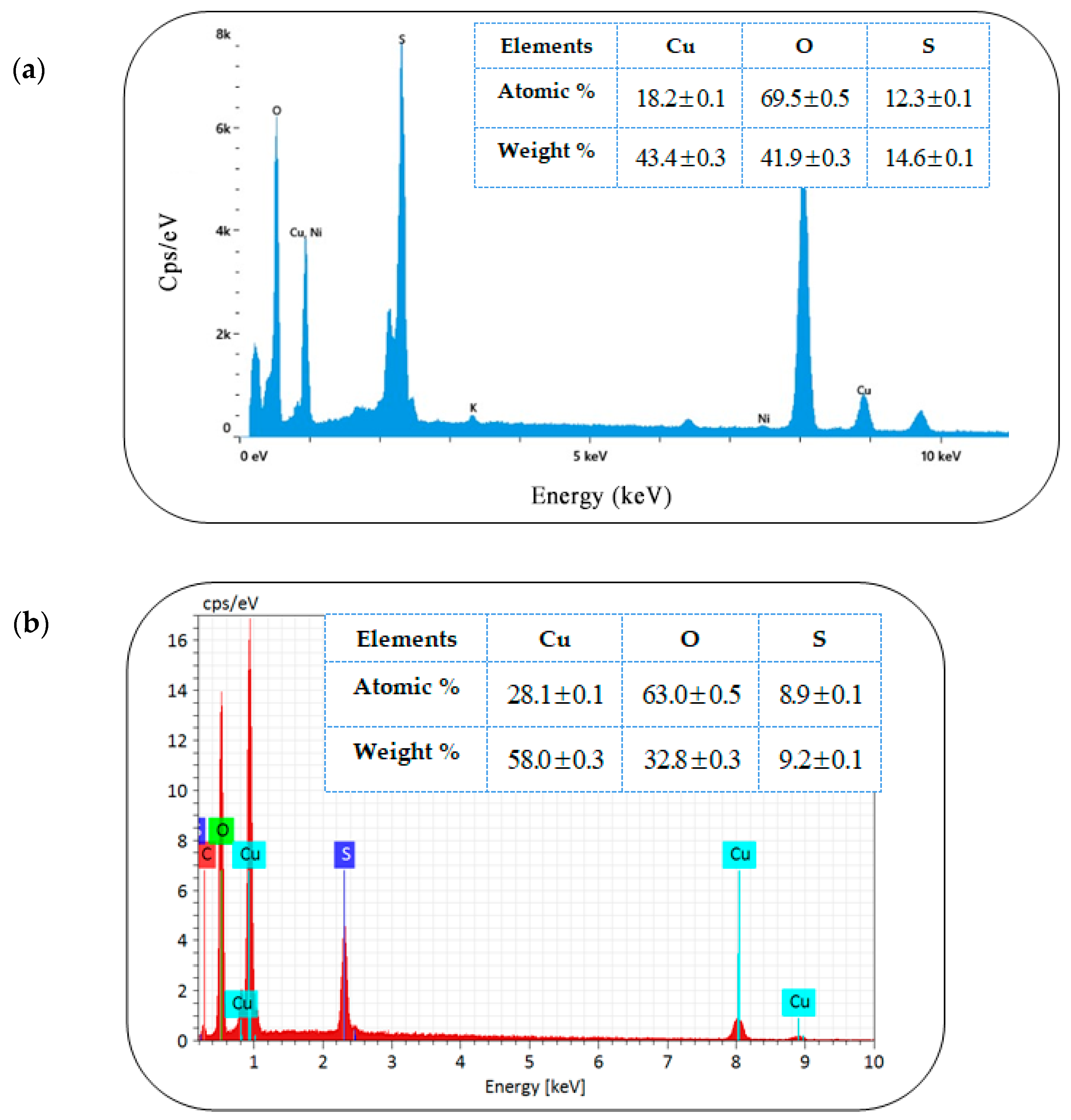
Figure 1. EDS spectra of CuONPs: (a) green synthesis; (b) chemical synthesis [19].
Microbiolres 16 00178 g001
Figure 2. (a) FE-SEM image; (b) the particle size; (c) area histograms of the green-synthesized CuONPs.
Microbiolres 16 00178 g002
Figure 3. TEM image of the green-synthesized CuONPs.
Microbiolres 16 00178 g003
Figure 4. AFM analysis of the synthesized CuONPs.
Microbiolres 16 00178 g004
Figure 5. Dimensional variability of CuONPs: width and height distributions.
Microbiolres 16 00178 g005
Figure 6. XRD patterns of CuONPs.
Microbiolres 16 00178 g006
Figure 7. Williamson–Hall (W–H) plot for CuONPs.
Microbiolres 16 00178 g007
Figure 8. FTIR spectra of CuONPs in the (a) 400–4000 cm−1 and (b) 400–1300 cm−1 range.
Microbiolres 16 00178 g008
Figure 9. Example of inhibition of C. albicans by green-synthesized CuONPs through well diffusion. The numbers 12, 50, 75 and 100 correspond to the concentrations CuONPs expressed in μg/mL. Then, 20 μL of each concentration was placed into each well after plating the C. albicans cells. The diffusion of CuONPs inhibits the growth of yeast cells to generate a halo of inhibition.
Microbiolres 16 00178 g009
Figure 10. Antiadhesive activity of different CuONPs on biofilm formation of the four selected Candia strains.
Microbiolres 16 00178 g010
Table 1. Mean value length and area of different CuO NPs.
SampleGrain Size (µm)Area (µm2)
CuONPs (green synthesis) 0.194 ± 0.012 0.021 ± 0.01
CuONPs (chemical synthesis) [19] 0.448 ± 0.027 0.106 ± 0.026
Table 2. Micro structural parameters of CuONPs.
SampleD (nm)ε (10−4)ρ (g.cm−3)SSA (m2. g−1)
CuO (green synthesis)37.07−6.596.6424.38
CuO (Chemical synthesis) [19]51.935.856.7117.20
Table 3. MIC concentration of non-doped and doped CuONPs.
CompoundConcentration of MIC (µg/mL) for Candida spp.
C. glabrataC. tropicalisC. luistaniaeC. albicans
Nano 0125125125125
Nano G12512512562.5
Nano 112.52512.550
Nano 2502550100
Nano 3n.a505025
Fluconazole832816
n.a = no activity until 100 µg/mL, Nano 0: chemically synthesized CuO, Nano G: green-synthesized CuO, Nano 1: doped CuO by 2%Fe, Nano 2: doped CuO by 2%Sn, Nano 3: doped CuO by 1%Fe/1%Sn.
Table 4. Biofilm inhibition by CuONPs: a MIC dose was adopted for all the strains except for C. glabrata, a concentration of 100μg/mL was used since the MIC was not determined.
Nanoparticle TypeAntibiofilm Activity %
C. glabrataC. luistaniaeC. albicansC. tropicalis
Nano 050.251.324.752.2
Nano G51.352.126.554.4
Nano 1 4.691046.5
Nano 2−0.44.912.743
Nano 35715.945
Fluconazole−1.5−2.8537.6
Table 5. Biofilm eradication.
MaterialsBiofilm Eradication Percentage %
Concentration (100 μg/mL)
C. glabrataC. lusitaniaeC. albicansC.tropicalis
Nano 066.6647.0859.8766.29
Nano G50.8157.6768.5161.32
Nano 167.2147.5158.0265.19
Nano 265.5760.7755.5562.98
Nano 368.3062.9867.9067.95
Fluconazole63.3866.1366.6666.85
Disclaimer/Publisher’s Note: The statements, opinions and data contained in all publications are solely those of the individual author(s) and contributor(s) and not of MDPI and/or the editor(s). MDPI and/or the editor(s) disclaim responsibility for any injury to people or property resulting from any ideas, methods, instructions or products referred to in the content.