Article

Proteomics-Based Prediction of Candidate Effectors in the Interaction Secretome of Trichoderma harzianum and Pseudocercospora fijiensis

by
Jewel Nicole Anna Todd
1,
Karla Gisel Carreón-Anguiano
2,
Gabriel Iturriaga
3,
Roberto Vázquez-Euán
4,
Ignacio Islas-Flores
5,
Miguel Tzec-Simá
5,
Miguel Ángel Canseco-Pérez
6,
César De Los Santos-Briones
1 and
Blondy Canto-Canché
1,*
1
Unidad de Biotecnología, Centro de Investigación Científica de Yucatán, A.C., Calle 43 No. 130, Colonia Chuburná de Hidalgo, Mérida C.P. 97205, Yucatán, Mexico
2
Facultad de Medicina, Universidad Autónoma de Durango Unidad Laguna, Cto. Industrial Durango #5001 ex. Ejido Emiliano Zapata, Gómez Palacio C.P. 35140, Durango, Mexico
3
Tecnológico Nacional de México, Campus Roque, Celaya C.P. 38110, Guanajuato, Mexico
4
Secihti-Facultad de Medicina, Universidad Autónoma de Yucatán, Avenida Itzáes # 498 x 59 y 59A, Col. Centro, Mérida C.P. 97000, Yucatán, Mexico
5
Unidad de Biología Integrativa, Centro de Investigación Científica de Yucatán, A.C., Calle 43 No. 130, Colonia Chuburná de Hidalgo, Mérida C.P. 97205, Yucatán, Mexico
6
Ingeniería en Alimentos, Universidad Politécnica de Chiapas, Portillo Zaragoza, Carretera Tuxtla Gutiérrez Km 21+500, Las Brisas, Suchiate C.P. 29150, Chiapas, Mexico
*
Author to whom correspondence should be addressed.
Microbiol. Res. 2025, 16(8), 175; https://doi.org/10.3390/microbiolres16080175
Submission received: 9 June 2025 / Revised: 3 July 2025 / Accepted: 4 July 2025 / Published: 1 August 2025

Abstract

Microbe–microbe interactions have been explored at the molecular level to a lesser degree than plant–pathogen interactions, primarily due to the economic impact of crop losses caused by pathogenic microorganisms. Effector proteins are well known for their role in disease development in many plant–pathogen pleinteractions, but there is increasing evidence showing their involvement in other types of interaction, including microbe–microbe interactions. Through the use of LC-MS/MS sequencing, effector candidates were identified in the in vitro interaction between a banana pathogen, Pseudocercospora fijiensis and a biological control agent, Trichoderma harzianum. The diverse interaction secretome revealed various glycoside hydrolase families, proteases and oxidoreductases. T. harzianum secreted more proteins in the microbial interaction compared to P. fijiensis, but its presence induced the secretion of more P. fijiensis proteins that were exclusive to the interaction secretome. The interaction secretome, containing 256 proteins, was screened for effector candidates using the algorithms EffHunter and WideEffHunter. Candidates with common fungal effector motifs and domains such as LysM, Cerato-platanin, NPP1 and CFEM, among others, were identified. Homologs of true effectors and virulence factors were found in the interaction secretome of T. harzianum and P. fijiensis. Further characterization revealed a potential novel effector of T. harzianum.

1. Introduction

Pseudocercospora fijiensis is the causal agent of the black Sigatoka disease, which affects species of the genus Musa, notably bananas, one of the world’s most consumed fruits [1]. The hemi-biotrophic filamentous fungus, P. fijiensis, devastates acreages of farmland in tropical and sub-tropical countries, which can result in a 35–100% loss in crop yield [2,3]. The most effective control method used is chemical control in the form of contact fungicides like Mancozeb and systemic fungicides like strobilurins and azoles (DMIs). In many tropical countries, these synthetic chemicals are alternately applied in an intensive manner, often more than 30 times per year [1,4]. The exhaustive use of these pesticides poses human health and environmental risks and also lowers the efficiency of their active ingredients due to fungicide resistance [5,6]. Alternatively, conventional breeding for resistance is challenging in Musa spp. due to the polyploid nature of cultivars, making conventional breeding costly and time-consuming [7]. Biological control is another alternative for disease management that can replace or supplement chemical control but has variable in-field efficacy. Key considerations for the use of microbial inoculants include environmental conditions, the type of formulations used and the method of application [8]. Application quantity and frequency must also be considered as the rhizosphere is a complex environment with many competitors [9]. Consequently, investigating the molecules used by biological control agents may uncover new and more effective tools for disease management compared to the use of the biological control agents themselves.
Trichoderma harzianum is a filamentous, saprophytic fungus and biocontrol agent used for the control of plant-pathogenic fungi [10,11]. More than 60% of the registered bio-fungicides on the commercial market originate from species of the Trichoderma genus [12,13], with T. harzianum being among the most popular biocontrol species. Fungal pathogens responsible for plant diseases of economic importance such as Fusarium spp. [14], Rhizoctonia solani [15] and Botrytis cinerea [16], among others, can be controlled by Trichoderma spp. due to its ability to compete for nutrients, release toxic metabolites, secrete lytic enzymes and induce systemic resistance in plant hosts [17]. Regarding the banana black Sigatoka pathogen, P. fijiensis, T. harzianum has shown promising results in vitro, in part because it grows faster and is able to better compete for space and nutrients [18]. It is likely that other unknown molecules play a role in the antagonism of P. fijiensis. T. harzianum can limit fungal growth of P. fijiensis by more than 60% in vitro [19], lending support to the investigation of T. harzianum and the molecules associated with its biocontrol capacity.
In the search for better alternatives for the control of this pathogen, the investigation of molecular targets called effectors is a viable field of study. Effectors are defined as small molecules that manipulate the structure and function of the host cell, establishing infection (virulence factors) and/or triggering a defense response (avirulence factors) in the host [20]. Thus, effectors play a vital role in plant defense where they can induce susceptibility (effector-triggered susceptibility, ETS) to an infection or induce resistance through complex and coordinated responses known as effector-triggered immunity (ETI). The induction of ETI can lead to a type of programmed cell death at the site of infection to restrict the pathogen’s advancement in the host, termed the hypersensitive response (HR); this phenomenon in the host is also triggered by the recognition of conserved pathogen or microorganism-associated molecular patterns (PAMPs or MAMPs) and the resulting immune response is termed PAMP-triggered immunity (PTI).
A pathogen’s effectorome can be considered its arsenal of biological weapons, largely involved in the suppression of the host’s immune responses [21,22]. More recently, effectors have been defined as molecules of diverse functionality and molecular nature that influence organisms’ interactions with each other, usually to the benefit of the producer organism [23]. The effector narrative has expanded from pathogens to include mutualists [24] and biological control agents [25,26] but the majority of what we know about effectors today comes from the literature on plant–pathogen interactions. An interesting subset of effectors in plant–pathogen interactions also function in microbe–microbe interactions. In the Zymoseptoria triticiTriticum aestivum interaction, the effector Zt6 is a RNase with phytotoxic activity in the host plant, but also presents activity against the microorganisms E. coli, S. cerevisiae and P. pastoris [27]. In Verticillium dahliae, the effector VdAve1 plays a role in both plant host and microbial interactions; the effector was found to inhibit the in vitro growth of certain Gram-positive bacteria and modify bacterial communities in planta [28]. Regarding Trichoderma spp., the T. atroviride effector Tal6 binds to cell-wall chitin and prevents the recognition of its chitin oligomers by the plant host [29]. Tal6 is expressed during microbial interactions and is involved in Trichoderma’s mycoparasitism, most likely protecting the cell wall against microbial chitinases. Epl1 is a cerato-platanin found in various Trichoderma species that induces the expression of plant defense-related genes [30,31,32]; in a microbial interaction between T. harzianum and B. cinerea, Epl1 was found to downregulate the expression of certain B. cinerea virulence-associated genes [33] and also affects hyphal coiling during the mycoparasitism of R. solani and Sclerotinia sclerotiorum [31].
Most effectors are secreted to the extracellular space since they are generally proteins that execute their functions in the host–whether plant, animal or microorganismple. The sum of all the proteins secreted to the extracellular space by a cell, tissue, organ or organism is referred to as the secretome [34], though the secretome can encompass other secreted molecules such as lipids and free nucleic acids. The protein-based secretome is a valuable reservoir containing many virulence-related proteins, including effector proteins, that can be mined with bioinformatics programs. In the predicted secretome of T. harzianum, 108 effector candidates were first identified [35], followed by 113 candidates [36]. With the improvement in the functionality of fungal effector algorithms, the number of possible effector candidates for T. harzianum has greatly expanded; 307 candidates were identified by EffHunter [37], and a considerable 2693 effector candidates in the complete effectorome were predicted by WideEffHunter [38]. Currently, the functions of only three P. fijiensis effectors (Avr4, Ecp6 and Ecp2) in the plant host are known [39].
Secretome studies have been reported between Trichoderma spp. and plant hosts such as Arabidopsis thaliana [40], maize [41,42] and banana [43], as well as different plant substrates such as cellulose and xylans [44]. The effectors that have been characterized in Trichoderma spp–plant interactions include hydrophobins, glycoside hydrolases and cerato-platanins [45]. Most of these molecules have been described as elicitors or inducers of plant defense pathways [46,47]. Secretome studies of Trichoderma spp. in the presence of fungal cell walls [48] or inactivated mycelium [49] have revealed candidate effectors that play a role in biological control, although effector identification in Trichoderma spp., particularly in microbial interactions, is an understudied theme of investigation. Tal6 and Epl1 are some of the few effectors whose characterization has begun in Trichoderma microbial interactions.
The first in silico-predicted secretomes of P. fijiensis were reported to have between 105 and 172 effector candidates [5,50]. Regarding experimental secretome studies, Escobar-Tovar et al. (2015), using SDS-PAGE and 2D-PAGE, observed 436 spots for a virulent strain of P. fijiensis growing in vitro, while they observed 101 spots for the virulent strain in interactions with the host plant [51]. The protein spots were sequenced by LC-MS and, in total, 90 proteins including virulence-associated proteins were identified. Chuc-Uc et al. 2011 using SDS-PAGE and PAGE characterized the P. fijiensis in vitro secretome which causes necrosis in resistant and susceptible banana cultivars, as well as the non-host Carica papaya [52]; a 30 kDa thermostable protease was also identified as a possible effector in the P. fijiensisMusa interaction. Burgos-Canul et al. 2019 [53] identified the cell wall proteins of two isolates of P. fijiensis using nano-HPLC–MS/MS. Of the 2686 proteins obtained, 24 shared pathogenicity factors were identified, including Avr4 and Ecp2.
Effector identification can be challenging since effector proteins possess different structural characteristics. The most common effector characteristics that have been identified include their small molecular weight, rich cysteine content, the presence of a secretory signal peptide and the absence of transmembrane domains [54]. Many bioinformatic tools are available for the in silico prediction of effector candidates such as Signal P [55], TMHMM [56] and LOCALIZER [57], which identify individual effector structural characteristics, while others combine various individual programs [37], use in-house pipelines [58] or use machine-learning programs [59] for effector identification. WideEffHunter [38] is one of the more recent effector algorithms capable of identifying effectors with the commonly observed structural characteristics (canonical effectors) as well as those that defy one or more of these characteristics; these effectors termed “non-canonical” were previously elusive to other effector identification pipelines that are based on canonical effector characteristics. The identification of effectors is a necessary step for their experimental characterization; the advent of improved effector identification algorithms facilitates high-throughput effector prediction from omics data. Here, we used the WideEffHunter and EffHunter algorithms to analyze the in vitro interaction secretome of T. harzianum and P. fijiensis, following the isolation and identification of interaction proteins by LC-MS/MS. The goal of this study was to identify, for the first time, effectors in the in vitro interaction secretome of T. harzianum and P. fijiensis.

2. Materials and Methods

2.1. Subsection Fungal Strains and Cultivation

Fungal isolates P. fijiensis C1233 and T. harzianum B13-1 were obtained from Musa acuminata cv. Grand Nain leaves from a plantation in Yucatan, Mexico [53], and decomposing banana from a plantation in Tabasco, Mexico [60], respectively. Fungi were cultured on Potato Dextrose Agar (PDA) (DIBICO, Cuautitlan Izcalli, Mexico) and kept at room temperature.

2.2. Fungal Interaction Assay

For the fungal interaction assay, the two fungi were individually inoculated (1 mL mycelial suspension) in 50 mL Potato Dextrose Broth (PDB) (DIBICO, Cuautitlan Izcalli, Mexico). Due to the differing growth rates of the two fungi, the period of cultivation was ten and five days for P. fijiensis and T. harzianum, respectively. During this period, fungi were maintained in shake culture at 25 °C and 100 rpm (LAB-LINE Instruments, Melrose Park, IL, USA). Fungal biomass was obtained by centrifugation (Centrifuge 5810R; Eppendorf, Hamburg, Germany) at 1699× g for 15 min, washed twice with sterile distilled water, and then transferred to 50 mL minimal media (yeast nitrogen base without amino acids and ammonium sulfate) (SigmaAldrich, St. Louis, MO, USA) supplemented with 10 g/L dextrose (Sigma). Conical flasks of minimal media were inoculated with 3 g of each fungus for the interaction, and 6 g of each fungus was used for the control flasks (monocultures of each fungus).

2.3. Isolation of the Interaction Secretome

Culture media on the 5th day of the interaction was collected in 50 mL Falcon tubes and lyophilized. A modified TCA-acetone protocol [61] was used for the precipitation of the proteins present in the interaction and monoculture samples. Briefly, lyophilized material was resuspended in Tris-HCl (Sigma-Aldrich, St. Louis, MO, USA) (pH 7.0) with 0.1 mM phenylmethylsulfonyl fluoride (PMSF, Sigma-Aldrich, USA) and protease inhibitor cocktail (Roche Complete™, EDTA-free Protease Inhibitor Cocktail). Cold 10% w/v TCA (Sigma-Aldrich, USA) in acetone (Sigma-Aldrich, St. Louis, MO, USA) was added per volume of sample and incubated overnight at −20 °C. After incubation, samples were centrifuged at 17,000× g at 4 °C for 45 min. The supernatant was discarded and protein precipitates (pellets) were washed three times with cold 90% acetone. Pellets were left to dry at room temperature for an hour and subsequently resuspended in 500 µL of Tris-HCl (pH 7.0). Proteins were quantified using the Linearized Bradford method [62] measuring the absorbance of the samples at 595 nm and 450 nm. Measurements of absorbance were interpolated on a standard curve based on bovine serum albumin (BSA, Sigma-Aldrich, USA) to calculate the concentration of protein in the samples. Samples were taken in triplicate and pooled for each condition: (a) T. harzianum monoculture, (b) P. fijiensis monoculture, and (c) the interaction. Protein pools were sent for LC/MS-MS analysis (Functional Genomics Center Zurich, Winterthurerstrasse, Zurich, Switzerland).

2.4. LC-MS/MS Analysis of Peptides

Sample processing was performed in duplicate for each sample (T. harzianum monoculture, P. fijiensis monoculture and interaction) according to the manufacturer’s protocol at the Functional Genomics Center Zurich. Briefly, proteins (100 mL) were precipitated with 100 mL of 20% TCA overnight and centrifuged at 17,000× g, and pellets were washed twice with cold acetone and dried at room temperature for 30 min. Pellets were dissolved in 45 mL 10 mM Tris/2 mM CaCl2 (pH 8.2) and then microwaved for 30 min. Samples were then reduced with 10 mM dithiothreitol (DTT) at 25 °C for 1 h, and protein alkylation was carried out by adding 15 mM iodoacetamide and incubating for 1 h at 25° C in the dark. Protein digestion was conducted using 5 μL trypsin (100 ng/mL in 10 mM HCl) and incubating overnight at 60 °C in the dark. Samples were desalted on C18 ZipTips, eluted with 150 mL 0.1% TFA and 35% acetonitrile, dried, and then dissolved in 20 μL 0.1% formic acid (1 mL was checked for the presence of peptides by MaldiTOF). Positive samples were transferred to autosampler vials for LC/MS/MS; 2 μL of each peptide solution was analyzed using an LTQ-Orbitrap VELOS mass spectrometer (Thermo Fischer Scientific, Bremen, Germany) coupled to an Eksigent-NanoLC-Ultra 1D plus HPLC system (Eksigent Technologies, Dublin, CA, USA). The solvent composition at the two channels was as follows: 0.2% formic acid, 1% acetonitrile for channel A, and 0.2% formic acid, 100% acetonitrile for channel B. Peptides were loaded on a column (75 μm × 80 mm) packed with reverse phase C18 material (AQ, 3 μm 200 Å, Bischoff GmbH, Leonberg, Germany) and eluted with a flow rate of 250 nL/min with an increasing gradient of 0.8% to 4.8% of B in 2 min, 35% at 57 min, 48% at 60 min and 97% at 65 min. The effluent from the HPLC was directly electrosprayed into the mass spectrometer by applying 2.0 kV through a platinum-based liquid junction. Full-scan MS spectra (300–2000 m/z) were acquired in the Orbitrap with a resolution of 30,000 at 400 m/z after accumulation to a target value of 1,000,000. Database searches with the raw data were performed using the Mascot software program version 2.4.1 [63] (SwissProt, all species; Trembl, fungi). Retrieved results were summarized as protein IDs in Scaffold files. The scaffold settings applied were as follows: 1% protein FDR, presence of a minimum of 2 peptides per protein, and 0.1% for peptide FDR. Fixed post-translational modifications: carbamidomethylation on cysteines; variable modifications: acetylation at protein N-terminal and oxidation on methionine.

2.5. Bioinformatic Analysis

Scaffold 4.1 was used to view the sequences identified using the UniProt TREMBl fungal database [64].
For functional annotation of the secreted proteins, sequences were annotated with GO terms using the program OmicsBox version 3.4 (Blast2GO, version 6.0), https://www.blast2go.com/ accessed on 30 April 2025 [65] and annotations were graphed using the online platform WEGO, https://wego.genomics.cn/, accessed on 30 April 2025 [66] for the comparison of control and interaction subset gene enrichment. The sequenced proteins were annotated with information from the InterProScan database version 105.0 (https://www.ebi.ac.uk/interpro/) accessed on 30 April 2025 in the Standalone mode [67].
Fasta files with the protein sequences were used as input for the effector identification algorithms.
EffHunter [37] and WideEffHunter [38] were used for the identification of canonical and non-canonical effectors, respectively. For EffHunter, the user-established parameters were amino acid length ≤400 amino acids and ≥4 cysteines (Perl scripts, Linux). Canonical proteins were identified that had 400 amino acids or less with at least 4 cysteines, zero transmembrane domains identified by TMHMM [56] and a signal peptide identified by SignalP 5.0 [55]. The output files contained candidate effector proteins in Fasta format. Subsequently, the original secretome datasets were analyzed with WideEffhunter. The program identified candidates based on the presence of effector motifs ([Li]xAR, PDI, CRN, LysM, EAR, ToxA, RXLR and YFWxC) using associated Regex in its database and homology to the 314 known effectors in its positive database [38]. For the in silico characterization of the proteins, the ProtParam tool, standalone version, which was accessed during 30 January 2025, https://web.expasy.org/protparam/ [68], was used to determine the characteristics of the protein sequences (length and amino acid composition), while the presence of signal peptides was determined by SignalP 5.0 [55], transmembrane domains by TMHMM [56] and subcellular localization using Localizer [57]. Additionally, all candidates were screened for functional domains using the Conserved Domains and Protein Classification in the NCBI CD-Search Tool version v3.21 (https://www.ncbi.nlm.nih.gov/Structure/bwrpsb/bwrpsb.cgi, accessed on 30 January 2025) [69], executing the analysis with default values (threshold value of 0.1 and 500 as the maximum number of hits).
To determine if the identified candidates were unique to the T. harzianumP. fijiensis interaction and to validate their effector potential, we compared these candidates with those found in previous P. fijiensis–banana and T. harzianum microbial interactions. For P. fijiensis, proteins were compared to those previously reported in the P. fijiensis in planta secretome [51] and the P. fijiensisMusa interaction transcriptome [70].
T. harzianum candidates identified in this study were also searched for in microbial interactions between Trichoderma spp. and S. sclerotiorum [71], Fusarium oxysporum [48], Guignardia citricarpa [49], R. solani [25], Alternaria alternata and Fusarium graminearum [72], as well as Trichoderma spp.–plant interactions [41,42,73].
Concurrently, all effector candidates identified in the interaction secretome were analyzed against the Pathogen–Host Interactions database (PHI-base v4.17) (http://www.phi-base.org/), accessed on 30 April 2025. PHI-base is a compendium of characterized virulence and pathogenicity factors which classifies the phenotypes of the pathogen’s gene mutants in interactions with plants (reduced virulence, hypervirulence, unaffected pathogenicity and effector plant avirulence determinant) [74].

2.6. Sequence Alignment and Phylogenetic Analysis

Homologs of ThCFEM1 (A0A0F9X6Z0) were searched for using BLASTP in the non-redundant database at GenBank with default settings. Eight hits were chosen to execute an alignment. Subsequently, a multiple alignment was performed using CLUSTALW integrated in the MEGA11 software program, version 11.0.13. The alignment was visualized and analyzed for amino acid residue conservation in the Jalview software version 2.11.4.1. Conserved amino acids were highlighted based on percent sequence identity.
For the phylogenetic analysis of ThCFEM1, an initial BLAST 1.4.0 using default settings identified 100 hits for the class Sordariomycetes. Subsequently, a different approach was taken to identify existing homologs in related and distantly related fungi to allow for a more extensive phylogenetic analysis. A BLAST was performed in different fungal taxonomic families using default settings. All Trichoderma spp. hits were retrieved followed by the top one to five resulting hits in each family analyzed. In total, 42 sequences were used for tree construction; the sequences were aligned using MUSCLE in the MEGA 11 software program. MEGA 11 was used for the construction of the phylogenetic tree using the maximum likelihood method with bootstrap resampling (1000).

2.7. Gene Cloning

The effector candidate (ThCFEM1) without its native signal peptide was cloned using the Gateway® cloning system (Thermo Fisher Scientific, San Francisco, CA, USA). Using attB-adapted primers, the effector candidate was amplified from cDNA derived from the T-harzianumP. fijiensis interaction using Phusion Green Hot Start II High Fidelity DNA Polymerase (Thermo Scientific). PCR products were purified using High Pure PCR Product Purification Kit (Roche) and recombined into pDONR™ 221 vector using the BP Clonase II enzyme mix (Thermo Fisher Scientific) in a 10 μL reaction following the manufacturer’s protocol. The product of the BP reaction was transformed into chemically competent E. coli DH5α cells. Selected entry clones grown on LB-kanamycin plates were sequenced using M13 primers to confirm the identity and correct reading frame orientation of effector candidates.
The effector candidate gene cassette flanked by the M13 primer binding sites was amplified from the entry vector for subsequent recombination. The PCR product was purified and then recombined with the destination vector, pEAQ-EGgw [75], using LR Clonase II enzyme mix (Thermo Fisher Scientific) in a 10 μL reaction following the manufacturer’s protocol. Expression clones were produced by transforming electrocompetent E. coli DH5α cells (Bio-Rad Gene Pulser Xcell electroporator, Hercules, CA, USA) with the LR reaction product.
The preliminary confirmation of transformed bacteria was performed by colony PCR using gene specific primers to confirm the presence of the transgene in the entry and expression clones. The insert was sequenced (Macrogen, Inc., Seoul, Republic of Korea) and the resulting data was aligned with the gene of interest to confirm that it was positioned in the correct open reading frame. Plasmid DNA was isolated from the selected positive colonies using the QIAprep® Spin Miniprep Gene Kit (Qiagen, Germantown, MD, USA) following the manufacturer’s protocol. The final expression construct was purified and dialyzed.

2.8. Transformation of Agrobacterium tumefaciens

For each transformation, 1 μL of dialyzed plasmid DNA (50 ng/μL) was used to transform 40 μL of A. tumefaciens GV3101. A. tumefaciens was transformed using a Bio-Rad Gene Pulser Xcell electroporator following the manufacturer’s default protocol for A. tumefaciens (Voltage 2.4 kV, Capacitance 25 mF, Resistance 200 W). Transformed bacteria were plated on LB agar supplemented with kanamycin (50 mg/L) and incubated at 27 °C for 24–48 h. Positive clones carrying the effector gene of interest were confirmed by colony PCR using gene-specific primers.

2.9. Agroinfiltration Assay in N. benthamiana

Individual colonies of transformed A. tumefaciens were cultivated in 4 mL LB with 50 mg/L kanamycin overnight under shaking incubation (200 rpm) at 27 °C. Five hundred microliters (0.5 mL) of primary inoculum were used to initiate a 20 mL culture of LB with kanamycin (50 mg/L), 10 mM MES (pH 5.6) and 200 μM acetosyringone for 18 h (200 rpm, 27 °C). Eighteen-hour cultures were then centrifuged (2800× g, 20 °C, 10 min) and pellets were resuspended in freshly made infiltration medium (5 g/L MS salts, 20 g/L sucrose, 10 mM MES, 200 μM acetosyringone) to an OD600 of 0.4. Cultures were then incubated in the dark at room temperature for 2–3 h before agroinfiltration.
For the agroinfiltration assay, recombinant bacterial suspensions were loaded into 3 mL needleless syringes and infiltrated into the abaxial surface of young leaves of 5-to-6-week-old plants. Plants were monitored for seven days post-agroinfiltration to evaluate HR development. Four plants were used per infiltration assay, and the experiment was executed three times.

2.10. Cell Death Suppression Assay

Recombinant A. tumefaciens was cultivated as previously mentioned for the agroinfiltration assay, except that bacteria were resuspended to a final OD600 of 0.3 in the infiltration buffer (5 g/L MS salts, 20 g/L sucrose, 10 mM MES, 200 μM acetosyringone). The abaxial sides of leaves were infiltrated with A. tumefaciens expressing the effector candidate or the GFP control, then 24 h later with A. tumefaciens expressing the Phytophthora sojae cell-death inducer, XEG1 [76] was infiltrated at the same site. Lesion development was assessed for 5 days post-infiltration.

3. Results

3.1. Expansions and Contractions in the T. harzianum–P. fijiensis Interaction Secretome

The interpretation of the MS spectra by the Mascot Program revealed more than 300 proteins that belonged to the three experimental conditions: the T. harzianumP. fijiensis interaction and each fungus grown separately in minimal media as the controls (Figure 1).
Two hundred and fifty-six proteins (256) were identified in the interaction between P. fijiensis and T. harzianum, of which sixty-two (62) proteins were found to be exclusive to the interaction (Figure 1) as they were absent in the T. harzianum or P. fijiensis control secretomes. A larger quantity of P. fijiensis proteins were identified in the total interaction secretome; 84 proteins were found in its control and 123 P. fijiensis proteins in the interaction. Meanwhile, for T. harzianum there was a decrease in proteins from the control secretome (202 proteins) to that of the interaction (133 proteins) and most sequences (122 sequences) identified from T. harzianum in the interaction were also found in its large control secretome. Protein sequences for each organism were analyzed for GO annotations with Blast2GO to ascertain which proteins were important to the interaction and their possible functions. Figure 2 shows a comparison of the sequence distribution for the most enriched GO terms between the two fungi in the control and interaction conditions generated using the web-based tool WEGO.
The contraction observed in the T. harzianum interaction secretome was in part attributed to a reduction in proteins related to the GO terms “binding” and “catalytic activity” (Figure 2). Within the “catalytic activity” category, there was a reduction in proteins associated with hydrolase activity in the interaction subset (84) compared to the control (129); however, this difference was not statistically significant. The term “binding” was also more enriched in the control subset compared to the interaction (50 in the control vs. 24 in the interaction) (Figure 2). Moreover, there was a pronounced difference in the number of proteins associated with the term “metabolic process,” favoring the control subset—particularly for “primary metabolic process” (94 vs. 63) and “organic substance metabolic process” (97 vs. 64). In total, there were 101 proteins in the control subset and 68 in the interaction subset associated with “metabolic process”. However, because Figure 2 shows percentages rather than raw values, this difference is not readily apparent due to the larger overall size of the T. harzianum control subset compared to its interaction subset. With respect to the P. fijiensis sequence distribution, there was a notable increase in the number of proteins in the interaction secretome related to “metabolic process” (50 in the control vs. 80 in the interaction) and “Hydrolase activity” (51 in the control vs. 83 in the interaction) which contributed to the expansion of its interaction secretome. Conversely, terms such as “Transporter Activity”, “Localization”, “Small molecule metabolic process” and “Binding” were more enriched in the control secretome. For both T. harzianum and P. fijiensis, enriched Cellular Component terms included “membrane”, “cell” and “cell part”. The percentage of proteins with the annotation “extracellular region” was similar in both conditions for P. fijiensis but higher for the proteins in the T. harzianum control secretome (Figure 2).

3.2. The Diverse Nature of the Interaction Secretome

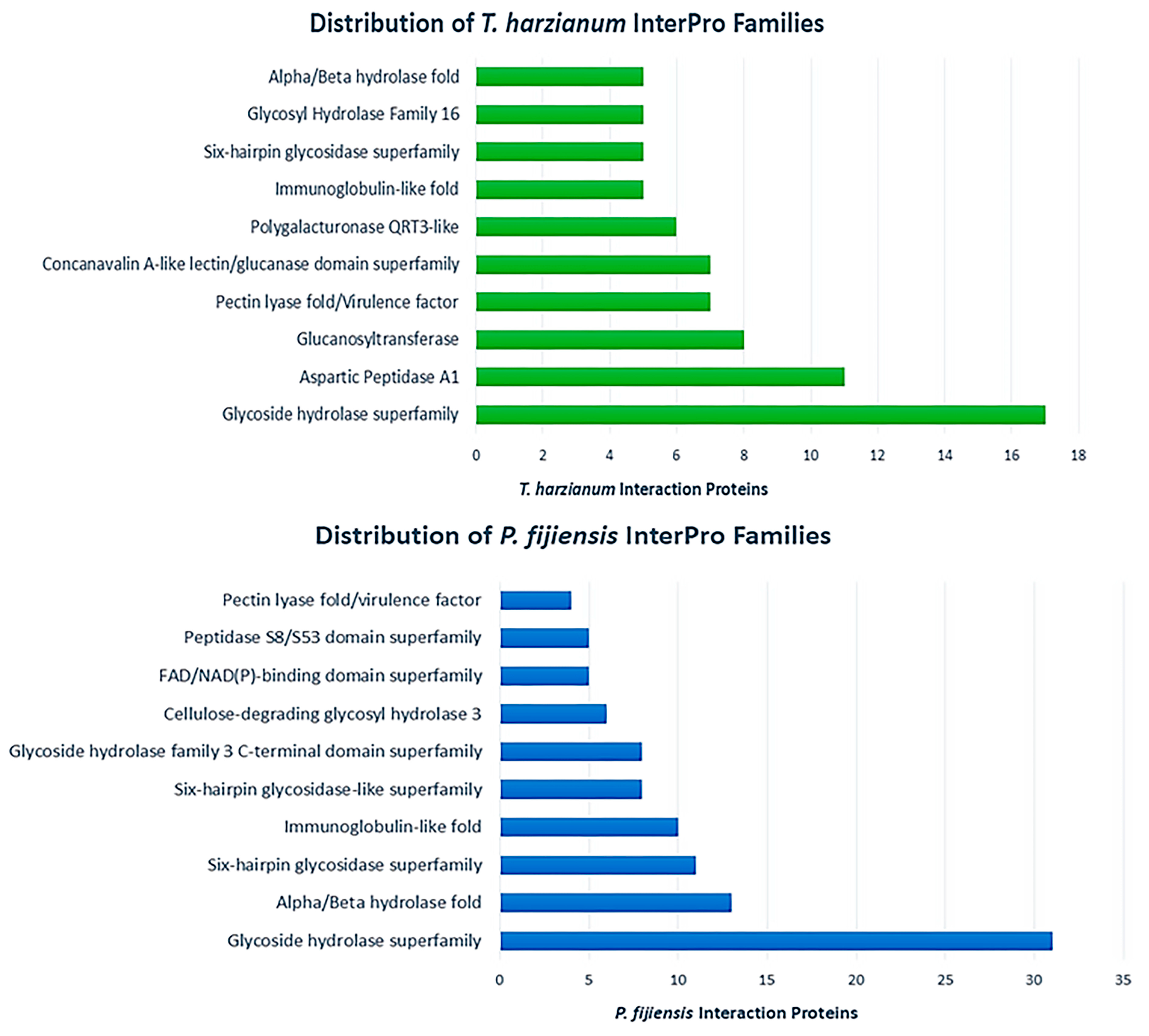
In order to identify the most abundant protein families secreted by each fungus during the interaction, each protein was annotated using InterProScan. The top 10 most abundant InterPro families for each fungus in the interaction secretome are presented in Figure 3. The glycoside hydrolase (GH) superfamily was the most prominent term observed for both fungi with 17 proteins for T. harzianum and 31 for P. fijiensis. For T. harzianum, GH family 16 (five sequences) and GH families 71 and 43 (two sequences each) were the most abundant among the GH families. GH family proteins were more abundant in the P. fijiensis interaction subset with GH families 3 (eight sequences) and 17 (four sequences), GH 43 (four sequences) and GH 92 (three sequences) being the most numerous GH proteins in the family. After the glycoside hydrolase superfamily, the alpha/beta hydrolase family was the second most abundant family for P. fijiensis (13 sequences) followed by the six-hairpin glycosidase superfamily (11 sequences). With respect to T. harzianum, after the GH superfamily, the most abundant families were aspartic peptidase A1 (11 sequences), glucanosyltransferases (8 sequences) and pectin lyases (7 sequences). With respect to proteases, interesting differences in protease families were observed between the two fungi. Proteases from the aspartic peptidase A1 family were most abundant in T. harzianum (11 sequences) followed by the subtilisin-like serine proteases with 3 sequences. Conversely, the peptidase S8/S53 family serine peptidases were most abundant in P. fijiensis (five sequences) followed by S10 serine peptidases (four sequences).
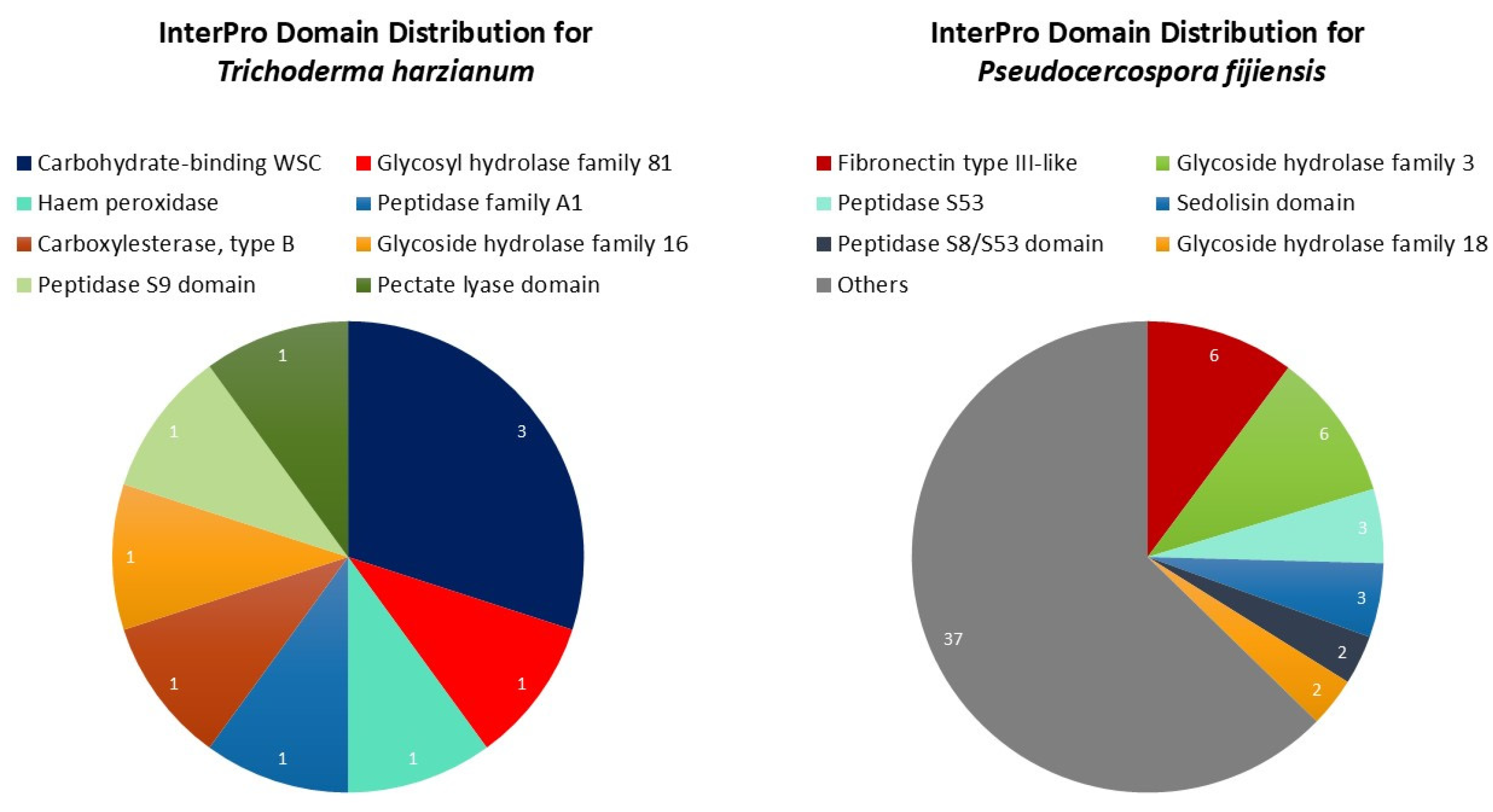
A closer analysis of the proteins unique to the interaction then followed for the 11 and 58 proteins found for T. harzianum and P. fijiensis, respectively. Regarding the InterPro domain distribution for P. fijiensis, the highest number of proteins was found in the Fibronectin type III-like classification (six sequences) and the glycoside hydrolase family 3 (six sequences), followed by peptidase S53 (three), sedolisin domain (three), peptidase S8/S53 (two) and glycoside hydrolase family 18 (two) (Figure 4). Finally, thirty-seven (37) other domains were identified among the P. fijiensis interaction-specific subset, each with one protein. With respect to T. harzianum, the InterPro domain annotations for its small subset of interaction-specific proteins were carbohydrate binding, WSC (four sequences), glycoside hydrolase families 81 (one sequence) and 16 (one sequence), haem peroxidase (one sequence), peptidase A1 (one), peptidase S9 (one), pectate lyase (one) and a carboxylesterase type B domain (one) (Figure 4).

3.3. The Interaction Secretome Contains Candidate Effector Proteins

Effector identification in the interaction secretome consisted of screening all proteins with the EffHunter and WideEffHunter algorithms. EffHunter was used for the identification of canonical effector candidates, while WideEffHunter focused primarily on non-canonical candidates. In total, 46 and 54 effector candidates were identified for T. harzianum and P. fijiensis, respectively. For P. fijiensis, EffHunter identified 17 canonical candidates and WideEffHunter identified 37 non-canonical candidates. For T. harzianum, 14 canonical candidates and 32 non-canonical candidates were identified by EffHunter and WideEffhunter, respectively. Of the P. fijiensis candidates, 21 were specific to the interaction secretome, while 33 were found in both interaction and control conditions. Among the T. harzianum proteins, 45 candidates were found in both interaction and control conditions while one candidate was found in the interaction-specific subset.
To obtain a better understanding of the effectors that each fungus secreted, the proteins were analyzed to determine sequence commonalities and differences, as well as the presence of effector-associated motifs and domains. All effector characterization data can be found in Supplementary Table S1. The in silico characterization of the effector candidates revealed that their protein lengths ranged from 136 to 1011 for P. fijiensis and 118 to 1117 for T. harzianum, with almost 60% of candidates surpassing 400 amino acids in both fungi. Most candidates (45 for P. fijiensis and 37 for T. harzianum) presented with effector motifs and domains as this is a key criterion used for effector identification with WideEffHunter. The most frequently occurring motifs for P. fijiensis in the interaction were LysM (14 candidates), [LI]xAR (10 candidates), and EAR (9 candidates), while LysM (11 candidates), RXLR (9 candidates) and EAR (6 candidates) were the most observed for T. harzianum. Regarding the abundance of domains, 48 (89%) P. fijiensis candidates and 40 (87%) T. harzianum candidates had identifiable domains with the NCBI CDD tool. Among the effector candidates with annotated domains, Bglx (Beta-glucosidase), Abhydrolase superfamily, Peptidase S53, PnbA and Glyco_hydro superfamily domains were among the most frequently occurring in P. fijiensis, while a wider variety of domains was found among individual T. harzianum candidates including different GH family proteins (13, 47, 61), proteases (Peptidase_M19, Peptidases_S8_5) and various DUF domain proteins (1996, 4965, 6595, 6736). The domains that were unique to the P. fijiensis candidates included Kre9_KNH, S1-P1_nuclease, Nis1 and SurE while those that were unique to the T. harzianum effector candidates included RNase_T2_euk, NPP1 and the LanC_like superfamily.
Regarding homology to known effectors in the positive dataset of WideEffHunter, both fungi had five proteins that were homologous to known effectors. Notable homologs include a protein (A0A139HLB6) which shares 43% identity with ChNis1, an effector of Colletotrichum higginsianum that suppresses PAMP-triggered immunity (PTI), another (M3A9Y6) which shares 30% identity with Bec1040, a haustorium-development related effector in Blumeria graminis, and one (A0A139H3Z9) with 52% identity to the effector Pr1 of Metarhizium anisopliae, a cuticle-degrading protease.
Using PHIB-BLAST v4.17 (http://phi-blast.phi-base.org/) with an e-value cutoff of 1 × 10⁻⁵, the interaction proteins were compared with the proteins in the PHI-base database version 4.17. Table 1 presents the top hits from the PHIB-BLAST. Notably, three T. harzianum candidates were similar to effector hits in the PHI-base with the annotation “effector plant avirulence (avr) determinant”: A0A0F9X4L9 shares 32.4% identity with effector MoPtep1 of Magnaporthe oryzae and candidates A0A0G0ARJ7 and A0A0F9ZFB8 share 46.6% and 45.5% identity, respectively, with the effector Crh1 of B. cinerea. One P. fijiensis candidate, M3A9Y6, shares 32.9% identity with the Avr effector Cep2 of M. oryzae. Many of the candidates that had hits in the PHI-base corresponded to proteins with the annotation “reduced virulence”; 23 and 20 candidate effectors of P. fijiensis and T. harzianum, respectively, share identity with bona fide virulence factors in the database (Table 1).

3.4. The Interaction Secretome Contains Broad-Host Range Effectors

To determine if the identified candidates were unique to the T. harzianumP. fijiensis interaction and to validate their effector potential, we compared these candidates with those found in previous P. fijiensis–banana and T. harzianum microbial interaction reports (Table 2A). Three P. fijiensis effector candidates from the T. harzianumP. fijiensis interaction were identified which were upregulated in planta during the late phase of infection in the banana host; a beta glucosidase (M3B352) and serine peptidase (M3B2Z2) which were secreted only in the presence of T. harzianum, and an uncharacterized protein (M3A4I0) which was found in both control and interaction secretomes. These three candidates were all non-canonical effectors. Conversely, an uncharacterized canonical effector (M2ZY96) was found in similar quantities in the control and interaction secretomes by both virulent and avirulent P. fijiensis isolates in another study but was secreted only in the presence of T. harzianum in this study (Table 2A). It is worth mentioning that this candidate shares identity with two effectors, BEC1040 (30.5%) and CEP2 (32.9%) (Table 1). The presence of these effectors in both banana and T. harzianum interactions suggests that microbial interactions can serve as a valuable tool for identifying virulence and pathogenicity factors beyond traditional pathosystems. It also highlights the importance of these effectors to the attack and/or defense strategies of P. fijiensis against different hosts.
T. harzianum candidates identified in this study were also searched for in microbial interactions between Trichoderma spp. and other fungi (Table 2B). T. harzianum effector candidates identified in this study were identified in other microbe–microbe interactions between T. harzianum and S. sclerotiorum and F. solani; two T. harzianum proteins were upregulated in the presence of S. sclerotiorum cell walls: an uncharacterized protein (A0A2N1LN60) and a beta-glucosidase (A0A0F9XHX6). Seven proteins were identified in T. harzianum interactions with F. solani cell walls; one of the proteins (A0A0F9XM39) was also identified in at least two other studies including an interaction with a plant host (Phaselous vulgaris). This is a known cerato-platanin effector of Trichoderma sp. that is constitutively expressed, though its role in plant interactions appears to be a positive one as a plant defense elicitor. Interestingly, a T. harzianum non-canonical effector candidate (A0A0F9XF89) with a beta-glucanase domain is also up-accumulated in an interaction with a plant host, Phaseolus vulgaris. Lastly, a CFEM domain-containing protein in the interaction, A0A0F9X6Z0, was also identified in a plant interaction where it was downregulated compared to the control while it was upregulated in the presence of F. solani cell walls. The effectors pinpointed in various interactions are high-priority candidates for further characterization and as such, further in silico and experimental analysis was performed for the T. harzianum candidate, A0A0F9X6Z0.

3.5. Characterization of ThCFEM1 (A0A0F9X6Z0), a Novel Broad Host-Range Effector from T. harzianum

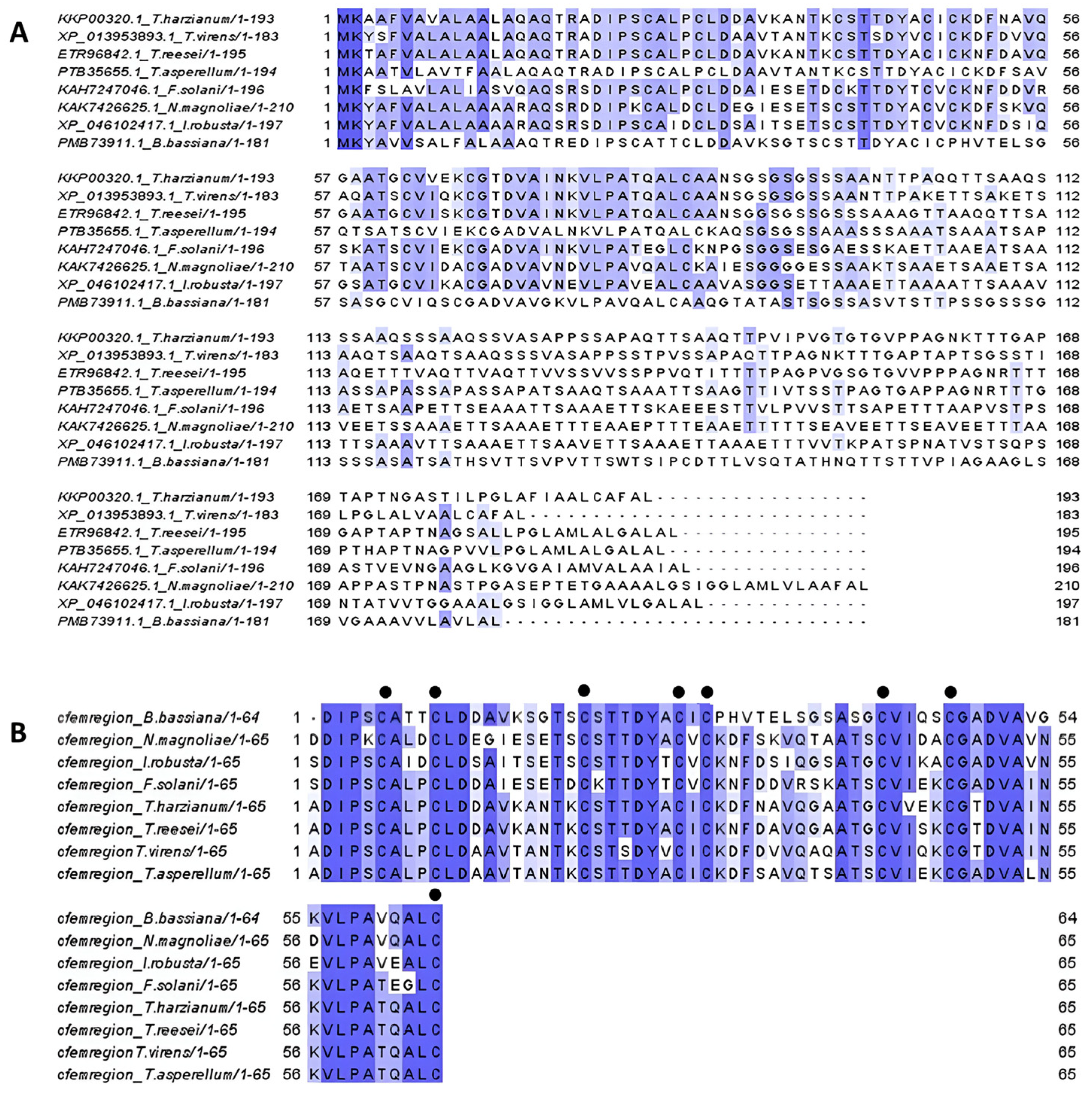
The T. harzianum candidate A0A0F9X6Z0 was chosen for further characterization as one of the candidates with antecedents in various interactions (T. harzianumP. fijiensis, T. harzianumF. solani, T. virensmaize). The protein was identified in the interaction secretome and also in the control secretome of T. harzianum in monoculture; it is a predicted effector according to EffHunter, WideEffHunter as well as the machine-learning classifier, EffectorP. Further in silico characterization with WideEffHunter revealed that the candidate is an extracellular cysteine-rich canonical effector with a YFWxC effector motif (Table 3). The protein also has a GPI anchor as predicted by PredGPI version 1.0 (http://busca.biocomp.unibo.it/predgpi/), accessed on 30 April 2025. A CFEM domain was identified in the sequence by InterProScan and NCBI CDD (amino acids 20–84) and the candidate was thereafter named ThCFEM1. ThCFEM1 also shares 51% identity with a virulence factor reported in the PHI-base, FGSG_02077. To determine its taxonomic distribution, a BLAST search (e value 1 × 10⁻⁵) revealed that the ThCFEM1 sequence is highly conserved among related species of the order Hypocreales and the class Sordariomycetes (Figure 5). This was also observed in the phylogenetic tree constructed for the homologs of ThCFEM1 where the largest observed clade consisted of Sordariomycetes (Figure 6). Among the Sordariomycetes, homologs were predominantly found in different Trichoderma sp. (mycoparasites and saprophytes) and Fusarium sp. (plant pathogens) and to a lesser extent in Metarrhizum sp. (entomopathogens), Ilyonectria sp. (root endophytes and opportunistic fungi) and the nematophagous fungi Pochonia chlamydosporia and Purpureocillium sp. A sequence alignment was performed with ThCFEM1 and seven related species: three from the genus Trichoderma and four others of the same taxonomic class. The alignment in Figure 5 shows a high degree of conservation in the amino acid region 1 to 96 among the sequences. This region encompasses the CFEM domain which can be found between positions 20 and 90 in the amino acid sequences for most of the homologs.
To determine the role of ThCFEM1, we characterized this effector candidate in the non-host model, N. benthamiana. The transient expression of ThCFEM1 through agroinfiltration of N. benthamiana did not exhibit typical HR-related cell death; rather, a slightly chlorotic phenotype was observed when individual infiltration was performed (Figure 7B). The cell death suppression assay followed, to determine whether ThCFEM1 was a suppressor instead of an inducer of host immunity. The agroinfiltration of ThCFEM1 at the same position 24 h before the cell-death inducer XEG1 led to smaller lesions than those induced by the agroinfiltration of GFP followed by XEG1 (Figure 7A). Taken together, these results show ThCFEM1’s role as a possible suppressor of the plant’s immune response.

4. Discussion

The secretome is a rich and diverse source of virulence-related molecular targets that can be exploited. More recently, secretomes of various microorganisms have been investigated with emphasis on the discovery of effectors and virulence-related proteins [78,79]. Comparably, we established an interaction between P. fijiensis and a biological control agent, T. harzianum, then isolated and identified the proteins in the interaction secretome. Subsequently, these proteins were screened with effector prediction algorithms. The previous literature has shown that nutrient-poor media can induce the expression of virulence-related proteins [80], and as such, minimal media (MM) was used for the T. harzianumP. fijiensis interaction and collected after five days of interaction in a shake culture. Interaction studies of Trichoderma spp. and plant pathogens have shown maximum enzymatic activity of certain chitinases, beta-glucanases and proteases of Trichoderma spp. between days 3 to 5 of the interaction [81,82]. Consequently, the day 5 secretome was selected as an adequate representation of both T. harzianum and P. fijiensis interaction proteins and was subjected to LC-MS/MS sequencing.
The composition of the proteins in the secretomes of the two fungi revealed domains of carbohydrate-active enzymes, proteases, alpha-beta hydrolases, oxidoreductases and proteins with no known domains. Among the GO-annotated proteins in the interaction secretome, more enzymes with hydrolase activity were identified for P. fijiensis than T. harzianum. Certain families such as GH 43, GH17 and GH16 that have been previously identified in P. fijiensis interactions with the host plant Musa [51,70] were also identified in the T. harzianumP. fijiensis microbial interaction. Interestingly, the P. fijiensis secretome expanded in response to T. harzianum, and many of its proteins were unique to the interaction secretome including GH 18, 20, 31 and 43 proteins, S10 and S53 proteases and hypothetical uncharacterized proteins. Extracellular proteases, specifically the S10 group, have been upregulated in in planta interactions [70], and along with GH proteins, can have functions in cell wall degradation and remodeling, enzyme degradation, detoxification and nutrient metabolism [83,84]. As such, these proteins usually constitute a large proportion of the fungal secretome. P. fijiensis also secreted more proteins related to metabolic processes than T. harzianum in the interaction. Such proteins are involved in the synthesis, catabolism and modification of macromolecules, oxidation-reduction processes and the regulation of metabolic processes. The contraction of the T. harzianum interaction secretome, compared to the control, was largely due to a reduction in proteins associated with metabolic processes, particularly primary and organic substance metabolism. A similar reduction in these proteins was seen in the T. harzianum interaction with the citrus pathogen, G. citricarpa [49], and the presence of many of the same hydrolytic enzymes in control and interaction conditions can be attributed to their dual roles in cell wall metabolism, as well as mycoparasitism. Within the small proportion of uniquely secreted proteins in the interaction by T. harzianum, GH 55, GH 16 and GH 81 family proteins, aspartic protease (A1 domain), carboxylesterase and WSC-domain-containing proteins were identified. It could be inferred that these proteins are particularly important to the interaction and are involved in T. harzianum mycoparasitism. This is a phenomenon which is marked by the production of hydrolytic enzymes and secondary metabolites and the creation of antagonistic environments where nutrients and space are competed for.
The most abundant protein families secreted by T. harzianum were the glycoside hydrolase superfamily, aspartic A1 domain-containing peptidase, glycosyltransferase, pectin lyase and concanavalin A-like lectin/glucanase families. Trichoderma sp. have large arsenals of proteolytic enzymes and extracellular aspartic proteases are common in Trichoderma sp. mycoparasitic interactions [85,86] where they may be involved in cell-wall degradation [87] and enzyme inactivation [88]. Other cell-wall-degrading enzymes secreted by T. harzianum included endo-glucosidases and GH 16 family proteins. Endo-1,3- beta- glucosidases degrade cellulose and beta-1,3-glucan are commonly upregulated during T. harzianum mycoparasitism where they likely attack the host’s cell wall [89,90]. Proteins associated with oxidative stress were found in both fungi but were more abundant in the T. harzianum secretome. These proteins consisted of peroxidases, thioredoxins and superoxide dismutases, which are commonly associated with scavenging reactive oxygen species [91], cell signaling and gene expression [92] and virulence [93].
Effector screening using EffHunter and WideEffHunter uncovered 100 candidates in the T. harizanumP. fijiensis interaction. WideEffHunter was used to identify non-canonical candidate effectors, while EffHunter, which is strictly based on traditional effector features was used to identify canonical effectors in the secretome. As expected, WideEffHunter widened the scope for effector retrieval; almost 70% of the identified effectors were non-canonical for both fungi. WideEffHunter’s algorithm identifies effector motifs and domains to predict if a protein is an effector. The most common effector motifs identified were LysM, RXLR and EAR. LysM domains facilitate binding to chitin and peptidoglycan compounds and have been frequently reported in other fungal effectors associated with the suppression of chitin-triggered immunity [94,95]. EAR motifs were also common to both fungi and are associated with transcriptional repression [96]. The RXLR motif is a predominantly oomycete-related N-terminal domain associated with cytoplasmic effectors. Many RXLR effectors induce host susceptibility to infection [97,98] and the domain may serve as a proteolytic cleavage site for unconventional effector secretion [99]. The domain was recently found in various fungi by WideEffHunter. [LI]xAR was a common domain found in the P. fijiensis candidates but not as common in T. harzianum; the motif is associated with effectors of the hemibiotroph, M. oryzae, but its role is unknown.
Regarding the effectors that share identity with other effectors compiled in the positive database of WideEffHunter, five T. harzianum effectors shared identify with Acp (C. albicans acid protease), CutA (B. cinerea cutinase) and Sap1, Sap2 and Sap3 (C. albicans aspartic proteinases). Similarly, five P. fijiensis candidates shared identity with the effectors Bec1040 (B. graminis haustorium-development effector), Acp, Sap1, Sap2, Sap3, ChNis1 (C. higginsianum necrosis-inducing protein and inhibitor of PTI) and Pr1 (M. anisopliae serine protease) according to WideEffHunter.
Additionally, PHIB-BLAST was used to identify characterized orthologs of the effector candidates in other organisms. The terms “effector protein” and “proteinaceous elicitor” were searched for in the PHI-base hits under gene function or gene name. Four T. harzianum candidates shared identity with effectors in the Pathogen–Host Interaction database: the effectors were Sm1 of T. virens (92.4%), Crh1 of B. cinerea (46.6% and 45.5%) and MoPtep1 of M. oryzae (32.4%). Crh1 and Sm1 are plant defense elicitors while MoPtep1 is a virulence factor of M. oryzae that suppresses PTI. Sm1 is a proteinaceous elicitor of host resistance in Trichoderma–plant interactions with a cerato-platanin domain. Certain cerato-platanins, e.g., BcSpl1 [100] and MgSM1 [101] can induce plant cell death. Other homologs of plant cell-death inducers were also found among the interaction proteins of T. harzianum (NPP1 and Crh1). The roles of cell-death inducers in plant hosts are difficult to infer in microbe–microbe interactions, and they likely have dual functions or roles outside of their cell-death-inducing activity, for example, NPP1 proteins are also expressed during vegetative growth and influence conidial germination and production [102,103], while cerato-platanins are also involved in spore maturation, hyphal growth and cell wall remodeling [104,105]. Crh1 triggers cell death in tobacco and tomato hosts but is also an elicitor of PTI and induces host resistance against B. cinerea; the protein is a highly conserved transglycolase (GH16 family) involved in fungal cell wall biogenesis. MoPtep1 is an effector with a cupredoxin domain which localizes to the plant peroxisome and prevents PTI-related cell death [106]. The homolog of MoPtep1 in T. harzianum is a non-canonical effector and was identified among the interaction-specific proteins of T. harzianum. A P. fijiensis candidate (M3AY96) shared identity with an effector in the PHI-base, Bec1040 (30.5%) of B. graminis, which is linked to haustorium development. The same candidate also shares identity with the M. oryzae effector, MoCEP2, that causes HR-related cell death in N. benthamiana and is vital for fungal pathogenicity [107]. The identification of candidates in this interaction which share identity with Bonafide effectors suggest their possible functions in the interaction as they are yet to be characterized.
BLASTp identified the functional domains present in the effectors and the majority were hydrolytic enzymes with GH family domains (e.g., GH16, 18, 20) and protease/peptidase domains (e.g., S8, S53) as was observed in the characterization of the global secretome proteins. In the interaction, the GH effectors are likely involved in sensing/recognition of the microbial counterpart, nutrient acquisition, cell wall remodeling and protection and possible detoxification of antimicrobial compounds [108]. The dependence on these types of effectors is understandable as the extracellular space represents the frontline of the battle between T. harzianum and P. fijiensis. Protease effectors were also common among both fungi; proteolytic degradation of host proteins can result in the disruption of cell signaling, defense, and physiological processes in the host [109]. These effectors can also affect the integrity of the cytoskeleton [110] and the cell wall [48,71,77]. WideEffHunter confirmed that both fungi secreted protease type effectors similar to the known effectors Acp and Sap1, Sap3 and Sap3.
A review of interaction-based literature uncovered several T. harzianumP. fijiensis effector candidates involved in other microbe–microbe interactions as well as microbe–plant interactions. Five P. fijiensis candidates identified in the interaction were also identified in P. fijiensis–banana interactions: a S53 peptidase, a betaglucosidase, a GH31 family protein and two candidates with unknown domains. One of the uncharacterized proteins was also upregulated in leaf tissue and WideEffHunter identified a LysM domain. The uncharacterized LysM protein identified by WideEffHunter and upregulated in banana tissue is a high priority effector candidate for further experimental validation. As expected, effector candidates of T. harzianum also play a role in other microbe–microbe interactions. Among the interaction effectors, CFEM, GH47, Bglc, DUF4965, NPP1 superfamily, Peptidase_S8_5, Glyco_hydro superfamily and cerato-platanin domain-containing proteins were identified in other Trichoderma microbial interactions. Regarding Trichoderma–plant interactions, candidates with cerato-platanin, superoxide dismutase, CFEM and GH55_beta13glucanase-like domains were found. One of the broad host range candidates containing a CFEM domain, ThCFEM1, was further characterized in N. benthamiana.
The CFEM (“Common in Fungal Extracellular Membranes”) domain is associated with extracellular membrane proteins of fungi, linked to fungal pathogenicity and virulence. Previously, CFEM effectors have been shown to both induce the hypersensitive response (HR) [111,112] and activate host resistance against pathogens [113], while others are suppressors of plant immunity and the HR, which encourage host colonization [114,115,116]. The domain has been found in various effectors with diverse roles. These proteins can be involved in nutrient (iron) acquisition [117], fungal pathogenicity/virulence [114,115], conidiogenesis [118], appressorium differentiation [117], biofilm formation [119,120], cell wall stability [121] and stress tolerance [118,122].
Many CFEM proteins of pathogens are also actively involved in subverting plant defense, contributing to virulence [123,124]. They can regulate plant immunity by suppressing host defense responses, for example, programmed cell death induced by PAMPs or effectors. ThCFEM1 was shown to interact with the non-host, N. benthamiana, in a similar manner, suppressing cell death induced by XEG1, a protein considered a PAMP and effector in Phytophthora spp. [76]. In this study, the protein was identified in both interaction and control subsets. In the presence of F. solani cell walls, the protein was upregulated [77], while it was downregulated in T. atroviride in the presence of maize roots compared to the control [41]. In interactions with plant hosts, the protein may aid in the initial suppression of host immunity to foster Trichoderma sp. colonization. In microbial interactions, ThCFEM1 could be involved in iron acquisition or sequestration [125,126] and adhesion of its hyphae to the microbial counterpart [127]. The protein may also have a protective function for the T. harzianum cell wall, maintaining cell wall integrity against oxidative and other stresses [121]. Further characterization of ThCFEM1 will shed light on its role in microbial interactions, particularly its contribution to T. harzianum mycoparasitism.
The number of effectors found coincides with the global protein landscape of the interaction secretome; the response of T. harzianum to its counterpart P. fijiensis involves many of the same effectors that it secretes on its own in minimal media. Interestingly, its presence induces P. fijiensis’ secretion of many interaction-specific proteins, including effectors, some of which have been identified in host (Musa spp.) interactions. The T. harzianum response did not involve the secretion of many new proteins in interactions with P. fijiensis compared with its vegetative growth. This is possibly indicative of a “less is more” way of life that has allowed to it to become the renown biological control agent it is today. It is likely able to dominate multiple types of prey because of the low metabolic cost associated with its mycoparasitism. On the other hand, its superior ability may be due to the efficiency of a select few of its effectors with broad-host range activity such as ThCFEM1.
Using a gel-free proteomics approach, effector candidates were uncovered in a microbial interaction where the pathogen, P. fijiensis, was challenged with a biological control agent, T. harzianum. This microbial interaction highlighted possible effector candidates involved in biological control. Further characterization of T. harzianum effectors will reveal robust biocontrol molecules for the control of P. fijiensis, the causal agent of the black Sigatoka disease. Furthermore, interactions between banana and P. fijiensis can be a challenge to establish if the right environmental conditions of high humidity and warm temperatures are not met. The added time taken to propagate plants and for them to reach a suitable infection age (at least 5–8 months) followed by disease progression (first visible symptoms at 2–3 weeks in field conditions and may take longer in vitro) must also be considered. Devising a simpler strategy based on a microbial interaction has proven effective for effector identification in a pathosystem that is relatively difficult to study.
WideEffHunter estimates that 5179 and 2693 effectors comprise the effectoromes of P. fijiensis and T. harzianum, respectively. With more extensive effectoromes being identified, the bottleneck has shifted from effector identification to effector prioritization for further characterization. Microbial interactions can provide a means to prioritize certain candidates for functional characterization in pathosystems where virulence factors have already been identified. Finally, screening for effectors in biological control agents like T. harzianum can reveal both interaction-specific and broad-spectrum effectors, valuable for innovative disease management strategies. Effectors can be employed as molecular markers to select superior biocontrol species or to engineer organisms with improved biological control activity. Alternatively, the production of recombinant proteins may prove viable in the field for the candidates displaying antimicrobial and/or elicitor activity.

5. Conclusions

The T. harzianumP. fijiensis interaction secretome consists of 256 proteins, of which 100 were identified as candidate effectors. The identified candidates, both canonical and non-canonical, represent a broad range of molecular targets for combating the banana pathogen P. fijiensis, the causal agent of black Sigatoka disease. Those candidates present in other interactions are possible broad-host range candidates while coming investigations will determine which candidates remain specific to this T. harzianumP. fijiensis interaction. Establishing an in vitro microbial interaction is a relatively rapid and efficient method for the identification of effector candidates for their characterization and future use in the development of disease control strategies.

Supplementary Materials

The following supporting information can be downloaded at: https://www.mdpi.com/article/10.3390/microbiolres16080175/s1, Table S1: In silico characterization data of effectors in the T. harzianumP. fijiensis interaction secretome.

Author Contributions

Conceptualization, B.C.-C. and J.N.A.T.; methodology, J.N.A.T., M.Á.C.-P., K.G.C.-A., I.I.-F. and M.T.-S.; software, K.G.C.-A.; validation, B.C.-C. and J.N.A.T.; formal analysis, J.N.A.T. and K.G.C.-A.; investigation, J.N.A.T.; resources, B.C.-C.; data curation, B.C.-C. and K.G.C.-A.; writing—original draft preparation, J.N.A.T. and B.C.-C.; writing—review and editing, J.N.A.T., K.G.C.-A., G.I., R.V.-E., I.I.-F., M.T.-S., M.Á.C.-P., C.D.L.S.-B. and B.C.-C.; supervision, B.C.-C., G.I., R.V.-E. and I.I.-F.; project administration, B.C.-C. and C.D.L.S.-B.; funding acquisition, B.C.-C. All authors have read and agreed to the published version of the manuscript.

Funding

This research received funding from the SECIHTI-Mexico projects CB-2013-01-220957 and FOP16-2021-01 No. 320993, and Fellowships No. 863239 for Todd J.N.A.

Institutional Review Board Statement

Not applicable.

Informed Consent Statement

Not applicable.

Data Availability Statement

Data are contained within the article.

Conflicts of Interest

The authors declare no conflicts of interest.

References

  1. Yonow, T.; Ramirez-Villegas, J.; Abadie, C.; Darnell, R.E.; Ota, N.; Kriticos, D.J. Black Sigatoka in Bananas: Ecoclimatic Suitability and Disease Pressure Assessments. PLoS ONE 2019, 14, e0220601. [Google Scholar] [CrossRef] [PubMed]
  2. Alakonya, A.E.; Kimunye, J.; Mahuku, G.; Amah, D.; Uwimana, B.; Brown, A.; Swennen, R. Progress in Understanding Pseudocercospora Banana Pathogens and the Development of Resistant Musa Germplasm. Plant Pathol. 2018, 67, 759–770. [Google Scholar] [CrossRef]
  3. Sardos, J.; Cenci, A.; Martin, G.; Breton, C.; Guignon, V.; Van Den Houwe, I.; Mendez, Y.; Sachter-Smith, G.L.; Chase, R.; Ruas, M.; et al. Painting the Diversity of a World’s Favorite Fruit: A next Generation Catalog of Cultivated Bananas. Plants People Planet 2025, 7, 263–283. [Google Scholar] [CrossRef]
  4. Gutierrez-Monsalve, J.A.; Mosquera, S.; González-Jaramillo, L.M.; Mira, J.J.; Villegas-Escobar, V. Effective Control of Black Sigatoka Disease Using a Microbial Fungicide Based on Bacillus subtilis EA-CB0015 Culture. Biol. Control 2015, 87, 39–46. [Google Scholar] [CrossRef]
  5. Arango Isaza, R.E.; Diaz-Trujillo, C.; Dhillon, B.; Aerts, A.; Carlier, J.; Crane, C.F.; de Jong, T.V.; de Vries, I.; Dietrich, R.; Farmer, A.D.; et al. Combating a Global Threat to a Clonal Crop: Banana Black Sigatoka Pathogen Pseudocercospora fijiensis (Synonym Mycosphaerella fijiensis) Genomes Reveal Clues for Disease Control. PLoS Genet. 2016, 12, e1005876. [Google Scholar] [CrossRef]
  6. Strobl, E.; Mohan, P. Climate and the Global Spread and Impact of Bananas’ Black Leaf Sigatoka Disease. Atmosphere 2020, 11, 947. [Google Scholar] [CrossRef]
  7. Mbo Nkoulou, L.F.; Ngalle, H.B.; Cros, D.; Adje, C.O.A.; Fassinou, N.V.H.; Bell, J.; Achigan-Dako, E.G. Perspective for Genomic-Enabled Prediction against Black Sigatoka Disease and Drought Stress in Polyploid Species. Front. Plant Sci. 2022, 13, 953133. [Google Scholar] [CrossRef]
  8. O’Callaghan, M.; Ballard, R.A.; Wright, D. Soil Microbial Inoculants for Sustainable Agriculture: Limitations and Opportunities. Soil Use Manag. 2022, 38, 1340–1369. [Google Scholar] [CrossRef]
  9. Lahlali, R.; Ezrari, S.; Radouane, N.; Kenfaoui, J.; Esmaeel, Q.; El Hamss, H.; Belabess, Z.; Barka, E.A. Biological Control of Plant Pathogens: A Global Perspective. Microorganisms 2022, 10, 596. [Google Scholar] [CrossRef]
  10. Grano-Maldonado, M.I.; Ramos-Payan, R.; Rivera-Chaparro, F.; Aguilar-Medina, M.; Romero-Quintana, J.G.; Rodríguez-Santiago, A.; Nieves-Soto, M. First Molecular Characterization of Colletotrichum sp. and Fusarium sp. Isolated from Mangrove in Mexico and the Antagonist Effect of Trichoderma harzianum as an Effective Biocontrol Agent. Plant Pathol. J. 2021, 37, 465–475. [Google Scholar] [CrossRef]
  11. Rahman, M.; Borah, S.M.; Borah, P.K.; Bora, P.; Sarmah, B.K.; Lal, M.K.; Tiwari, R.K.; Kumar, R. Deciphering the Antimicrobial Activity of Multifaceted Rhizospheric Biocontrol Agents of Solanaceous Crops Viz., Trichoderma harzianum MC2, and Trichoderma Harzianum NBG. Front. Plant Sci. 2023, 14, 1141506. [Google Scholar] [CrossRef] [PubMed]
  12. Verma, M.; Brar, S.K.; Tyagi, R.D.; Surampalli, R.Y.; Valéro, J.R. Antagonistic Fungi, Trichoderma spp.: Panoply of Biological Control. Biochem. Eng. J. 2007, 37, 1–20. [Google Scholar] [CrossRef]
  13. Rush, T.A.; Shrestha, H.K.; Gopalakrishnan Meena, M.; Spangler, M.K.; Ellis, J.C.; Labbé, J.L.; Abraham, P.E. Bioprospecting Trichoderma: A Systematic Roadmap to Screen Genomes and Natural Products for Biocontrol Applications. Front. Fungal Biol. 2021, 2, 716511. [Google Scholar] [CrossRef] [PubMed]
  14. Saravanakumar, K.; Li, Y.; Yu, C.; Wang, Q.; Wang, M.; Sun, J.; Gao, J.; Chen, J. Effect of Trichoderma harzianum on Maize Rhizosphere Microbiome and Biocontrol of Fusarium Stalk Rot. Sci. Rep. 2017, 7, 1771. [Google Scholar] [CrossRef]
  15. Huang, X.; Chen, L.; Ran, W.; Shen, Q.; Yang, X. Trichoderma harzianum Strain SQR-T37 and Its Bio-Organic Fertilizer Could Control Rhizoctonia solani Damping-off Disease in Cucumber Seedlings Mainly by the Mycoparasitism. Appl. Microbiol. Biotechnol. 2011, 91, 741–755. [Google Scholar] [CrossRef]
  16. You, J.; Zhang, J.; Wu, M.; Yang, L.; Chen, W.; Li, G. Multiple Criteria-Based Screening of Trichoderma Isolates for Biological Control of Botrytis cinerea on Tomato. Biol. Control 2016, 101, 31–38. [Google Scholar] [CrossRef]
  17. Guzmán-Guzmán, P.; Kumar, A.; de los Santos-Villalobos, S.; Parra-Cota, F.I.; Orozco-Mosqueda, M.d.C.; Fadiji, A.E.; Hyder, S.; Babalola, O.O.; Santoyo, G. Trichoderma Species: Our Best Fungal Allies in the Biocontrol of Plant Diseases—A Review. Plants 2023, 12, 432. [Google Scholar] [CrossRef]
  18. Acosta-Suárez, M.; Pichardo, T.; Roque, B.; Cruz-Martín, M.; Mena, E.; Leiva-Mora, M.; Castro, R.; Alvarado-Capó, Y. Antagonismo in vitro de Trichoderma harzianum Rifai contra Mycosphaerella fijiensis Morelet. Biotecnol. Veg. 2013, 13, 98–104. Available online: https://www.redalyc.org/articulo.oa?id=61224203 (accessed on 9 June 2025).
  19. Galarza, L.; Akagi, Y.; Takao, K.; Kim, C.S.; Maekawa, N.; Itai, A.; Peralta, E.; Santos, E.; Kodama, M. Characterization of Trichoderma Species Isolated in Ecuador and Their Antagonistic Activities against Phytopathogenic Fungi from Ecuador and Japan. J. Gen. Plant Pathol. 2015, 81, 201–210. [Google Scholar] [CrossRef]
  20. Selin, C.; de Kievit, T.R.; Belmonte, M.F.; Fernando, W.G.D. Elucidating the Role of Effectors in Plant-Fungal Interactions: Progress and Challenges. Front. Microbiol. 2016, 7, 600. [Google Scholar] [CrossRef]
  21. Zhang, S.; Li, C.; Si, J.; Han, Z.; Chen, D. Action Mechanisms of Effectors in Plant-Pathogen Interaction. Int. J. Mol. Sci. 2022, 23, 6758. [Google Scholar] [CrossRef] [PubMed]
  22. Thynne, E.; Ali, H.; Seong, K.; Abukhalaf, M.; Guerreiro, M.A.; Flores-Nunez, V.M.; Hansen, R.; Bergues, A.; Salman, M.J.; Rudd, J.J.; et al. An Array of Zymoseptoria tritici Effectors Suppress Plant Immune Responses. Mol. Plant Pathol. 2024, 25, e13500. [Google Scholar] [CrossRef] [PubMed]
  23. Todd, J.N.A.; Carreón-Anguiano, K.G.; Islas-Flores, I.; Canto-Canché, B. Fungal Effectoromics: A World in Constant Evolution. Int. J. Mol. Sci. 2022, 23, 13433. [Google Scholar] [CrossRef] [PubMed]
  24. Plett, J.M.; Daguerre, Y.; Wittulsky, S.; Vayssières, A.; Deveau, A.; Melton, S.J.; Kohler, A.; Morrell-Falvey, J.L.; Brun, A.; Veneault-Fourrey, C.; et al. Effector MiSSP7 of the Mutualistic Fungus Laccaria bicolor Stabilizes the Populus JAZ6 Protein and Represses Jasmonic Acid (JA) Responsive Genes. Proc. Natl. Acad. Sci. USA 2014, 111, 8299–8304. [Google Scholar] [CrossRef]
  25. Guzmán-Guzmán, P.; Alemán-Duarte, M.I.; Delaye, L.; Herrera-Estrella, A.; Olmedo-Monfil, V. Identification of Effector-like Proteins in Trichoderma spp. and Role of a Hydrophobin in the Plant-Fungus Interaction and Mycoparasitism. BMC Genet. 2017, 18, 16. [Google Scholar] [CrossRef]
  26. Laur, J.; Ramakrishnan, G.B.; Labbé, C.; Lefebvre, F.; Spanu, P.D.; Bélanger, R.R. Effectors Involved in Fungal–Fungal Interaction Lead to a Rare Phenomenon of Hyperbiotrophy in the Tritrophic System Biocontrol Agent–Powdery Mildew–Plant. New Phytol. 2018, 217, 713–725. [Google Scholar] [CrossRef]
  27. Kettles, G.J.; Bayon, C.; Sparks, C.A.; Canning, G.; Kanyuka, K.; Rudd, J.J. Characterization of an Antimicrobial and Phytotoxic Ribonuclease Secreted by the Fungal Wheat Pathogen Zymoseptoria tritici. New Phytol. 2018, 217, 320–331. [Google Scholar] [CrossRef]
  28. Snelders, N.C.; Rovenich, H.; Petti, G.C.; Rocafort, M.; van den Berg, G.C.M.; Vorholt, J.A.; Mesters, J.R.; Seidl, M.F.; Nijland, R.; Thomma, B.P.H.J. Microbiome Manipulation by a Soil-Borne Fungal Plant Pathogen Using Effector Proteins. Nat. Plants 2020, 6, 1365–1374. [Google Scholar] [CrossRef]
  29. Romero-Contreras, Y.J.; Ramírez-Valdespino, C.A.; Guzmán-Guzmán, P.; Macías-Segoviano, J.I.; Villagómez-Castro, J.C.; Olmedo-Monfil, V. Tal6 From Trichoderma atroviride Is a LysM Effector Involved in Mycoparasitism and Plant Association. Front. Microbiol. 2019, 10, 2231. [Google Scholar] [CrossRef]
  30. Salas-Marina, M.A.; Isordia-Jasso, M.I.; Islas-Osuna, M.A.; Delgado-Sánchez, P.; Jiménez-Bremont, J.F.; Rodríguez-Kessler, M.; Rosales-Saavedra, M.T.; Herrera-Estrella, A.; Casas-Flores, S. The Epl1 and Sm1 Proteins from Trichoderma atroviride and Trichoderma virens Differentially Modulate Systemic Disease Resistance against Different Life Style Pathogens in Solanum lycopersicum. Front. Plant Sci. 2015, 6, 77. [Google Scholar] [CrossRef]
  31. Gomes, E.V.; Costa, M.d.N.; de Paula, R.G.; Ricci de Azevedo, R.; da Silva, F.L.; Noronha, E.F.; José Ulhoa, C.; Neves Monteiro, V.; Elena Cardoza, R.; Gutiérrez, S.; et al. The Cerato-Platanin Protein Epl-1 from Trichoderma harzianum Is Involved in Mycoparasitism, Plant Resistance Induction and Self Cell Wall Protection. Sci. Rep. 2015, 5, 17998. [Google Scholar] [CrossRef] [PubMed]
  32. Cheng, C.-H.; Shen, B.-N.; Shang, Q.-W.; Liu, L.-Y.D.; Peng, K.-C.; Chen, Y.-H.; Chen, F.-F.; Hu, S.-F.; Wang, Y.-T.; Wang, H.-C.; et al. Gene-to-Gene Network Analysis of the Mediation of Plant Innate Immunity by the Eliciting Plant Response-Like 1 (Epl1) Elicitor of Trichoderma formosa. Mol. Plant-Microbe Interact. 2018, 31, 683–691. [Google Scholar] [CrossRef] [PubMed]
  33. Gomes, E.V.; Ulhoa, C.J.; Cardoza, R.E.; Silva, R.N.; Gutiérrez, S. Involvement of Trichoderma harzianum Epl-1 Protein in the Regulation of Botrytis Virulence- and Tomato Defense-Related Genes. Front. Plant Sci. 2017, 8, 880. [Google Scholar] [CrossRef]
  34. Agrawal, G.K.; Jwa, N.-S.; Lebrun, M.-H.; Job, D.; Rakwal, R. Plant Secretome: Unlocking Secrets of the Secreted Proteins. Proteomics 2010, 10, 799–827. [Google Scholar] [CrossRef]
  35. Mendoza-Mendoza, A.; Zaid, R.; Lawry, R.; Hermosa, R.; Monte, E.; Horwitz, B.A.; Mukherjee, P.K. Molecular Dialogues between Trichoderma and Roots: Role of the Fungal Secretome. Fungal Biol. Rev. 2018, 32, 62–85. [Google Scholar] [CrossRef]
  36. Kubicek, C.P.; Steindorff, A.S.; Chenthamara, K.; Manganiello, G.; Henrissat, B.; Zhang, J.; Cai, F.; Kopchinskiy, A.G.; Kubicek, E.M.; Kuo, A.; et al. Evolution and Comparative Genomics of the Most Common Trichoderma Species. BMC Genom. 2019, 20, 485. [Google Scholar] [CrossRef]
  37. Carreón-Anguiano, K.G.; Islas-Flores, I.; Vega-Arreguín, J.; Sáenz-Carbonell, L.; Canto-Canché, B. EffHunter: A Tool for Prediction of Effector Protein Candidates in Fungal Proteomic Databases. Biomolecules 2020, 10, 712. [Google Scholar] [CrossRef]
  38. Carreón-Anguiano, K.G.; Todd, J.N.A.; Chi-Manzanero, B.H.; Couoh-Dzul, O.J.; Islas-Flores, I.; Canto-Canché, B. WideEffHunter: An Algorithm to Predict Canonical and Non-Canonical Effectors in Fungi and Oomycetes. Int. J. Mol. Sci. 2022, 23, 13567. [Google Scholar] [CrossRef]
  39. Stergiopoulos, I.; van den Burg, H.A.; Okmen, B.; Beenen, H.G.; van Liere, S.; Kema, G.H.J.; de Wit, P.J.G.M. Tomato Cf Resistance Proteins Mediate Recognition of Cognate Homologous Effectors from Fungi Pathogenic on Dicots and Monocots. Proc. Natl. Acad. Sci. USA 2010, 107, 7610–7615. [Google Scholar] [CrossRef]
  40. González-López, M.D.C.; Jijón-Moreno, S.; Dautt-Castro, M.; Ovando-Vázquez, C.; Ziv, T.; Horwitz, B.A.; Casas-Flores, S. Secretome Analysis of Arabidopsis-Trichoderma atroviride Interaction Unveils New Roles for the Plant Glutamate:Glyoxylate Aminotransferase GGAT1 in Plant Growth Induced by the Fungus and Resistance against Botrytis cinerea. Int. J. Mol. Sci. 2021, 22, 6804. [Google Scholar] [CrossRef]
  41. Lamdan, N.-L.; Shalaby, S.; Ziv, T.; Kenerley, C.M.; Horwitz, B.A. Secretome of Trichoderma Interacting with Maize Roots: Role in Induced Systemic Resistance. Mol. Cell. Proteom. MCP 2015, 14, 1054–1063. [Google Scholar] [CrossRef] [PubMed]
  42. Nogueira-Lopez, G.; Greenwood, D.R.; Middleditch, M.; Winefield, C.; Eaton, C.; Steyaert, J.M.; Mendoza-Mendoza, A. The Apoplastic Secretome of Trichoderma virens During Interaction with Maize Roots Shows an Inhibition of Plant Defence and Scavenging Oxidative Stress Secreted Proteins. Front. Plant Sci. 2018, 9, 409. [Google Scholar] [CrossRef] [PubMed]
  43. Muthukathan, G.; Mukherjee, P.; Salaskar, D.; Pachauri, S.; Tak, H.; Ganapathi, T.R.; Mukherjee, P.K. Secretome of Trichoderma virens Induced by Banana Roots—Identification of Novel Fungal Proteins for Enhancing Plant Defence. Physiol. Mol. Plant Pathol. 2020, 110, 101476. [Google Scholar] [CrossRef]
  44. Gómez-Mendoza, D.P.; Junqueira, M.; do Vale, L.H.F.; Domont, G.B.; Ferreira Filho, E.X.; de Sousa, M.V.; Ricart, C.A.O. Secretomic Survey of Trichoderma harzianum Grown on Plant Biomass Substrates. J. Proteome Res. 2014, 13, 1810–1822. [Google Scholar] [CrossRef]
  45. Ramírez-Valdespino, C.A.; Casas-Flores, S.; Olmedo-Monfil, V. Trichoderma as a Model to Study Effector-Like Molecules. Front. Microbiol. 2019, 10, 1030. [Google Scholar] [CrossRef]
  46. Huang, Y.; Mijiti, G.; Wang, Z.; Yu, W.; Fan, H.; Zhang, R.; Liu, Z. Functional Analysis of the Class II Hydrophobin Gene HFB2-6 from the Biocontrol Agent Trichoderma asperellum ACCC30536. Microbiol. Res. 2015, 171, 8–20. [Google Scholar] [CrossRef]
  47. Wang, X.; Chen, W.; Zhang, J.; Hu, W.; Li, M.; Zhao, P.; Ren, A. Effector Cpe1 Secreted by Trichoderma longibrachiatum Induces Plant Disease Resistance. Biol. Control 2025, 202, 105726. [Google Scholar] [CrossRef]
  48. Ramada, M.H.S.; Steindorff, A.S.; Bloch, C.; Ulhoa, C.J. Secretome Analysis of the Mycoparasitic Fungus Trichoderma harzianum ALL 42 Cultivated in Different Media Supplemented with Fusarium solani Cell Wall or Glucose. Proteomics 2016, 16, 477–490. [Google Scholar] [CrossRef]
  49. Blauth de Lima, F.; Félix, C.; Osório, N.; Alves, A.; Vitorino, R.; Domingues, P.; da Silva Ribeiro, R.T.; Esteves, A.C. Trichoderma harzianum T1A Constitutively Secretes Proteins Involved in the Biological Control of Guignardia citricarpa. Biol. Control 2017, 106, 99–109. [Google Scholar] [CrossRef]
  50. Chang, T.-C.; Salvucci, A.; Crous, P.W.; Stergiopoulos, I. Comparative Genomics of the Sigatoka Disease Complex on Banana Suggests a Link between Parallel Evolutionary Changes in Pseudocercospora fijiensis and Pseudocercospora eumusae and Increased Virulence on the Banana Host. PLoS Genet. 2016, 12, e1005904. [Google Scholar] [CrossRef]
  51. Escobar-Tovar, L.; Guzmán-Quesada, M.; Sandoval-Fernández, J.A.; Gómez-Lim, M.A. Comparative Analysis of the in Vitro and in Planta Secretomes from Mycosphaerella fijiensis Isolates. Fungal Biol. 2015, 119, 447–470. [Google Scholar] [CrossRef] [PubMed]
  52. Chuc-Uc, J.; Brito-Argáez, L.; Canto-Canché, B.; Tzec-Simá, M.; Rodríguez-García, C.; Peraza-Echeverría, L.; Peraza-Echeverría, S.; James-Kay, A.; Cruz-Cruz, C.A.; Peña-Rodríguez, L.M.; et al. The in Vitro Secretome of Mycosphaerella fijiensis Induces Cell Death in Banana Leaves. Plant Physiol. Biochem. 2011, 49, 572–578. [Google Scholar] [CrossRef] [PubMed]
  53. Burgos-Canul, Y.Y.; Canto-Canché, B.; Berezovski, M.V.; Mironov, G.; Loyola-Vargas, V.M.; Barba de Rosa, A.P.; Tzec-Simá, M.; Brito-Argáez, L.; Carrillo-Pech, M.; Grijalva-Arango, R.; et al. The Cell Wall Proteome from Two Strains of Pseudocercospora fijiensis with Differences in Virulence. World J. Microbiol. Biotechnol. 2019, 35, 105. [Google Scholar] [CrossRef] [PubMed]
  54. Sonah, H.; Deshmukh, R.K.; Bélanger, R.R. Computational Prediction of Effector Proteins in Fungi: Opportunities and Challenges. Front. Plant Sci. 2016, 7, 126. [Google Scholar] [CrossRef]
  55. Almagro Armenteros, J.J.; Tsirigos, K.D.; Sønderby, C.K.; Petersen, T.N.; Winther, O.; Brunak, S.; von Heijne, G.; Nielsen, H. SignalP 5.0 Improves Signal Peptide Predictions Using Deep Neural Networks. Nat. Biotechnol. 2019, 37, 420–423. [Google Scholar] [CrossRef]
  56. Krogh, A.; Larsson, B.; von Heijne, G.; Sonnhammer, E.L. Predicting Transmembrane Protein Topology with a Hidden Markov Model: Application to Complete Genomes. J. Mol. Biol. 2001, 305, 567–580. [Google Scholar] [CrossRef]
  57. Sperschneider, J.; Catanzariti, A.-M.; DeBoer, K.; Petre, B.; Gardiner, D.M.; Singh, K.B.; Dodds, P.N.; Taylor, J.M. LOCALIZER: Subcellular Localization Prediction of Both Plant and Effector Proteins in the Plant Cell. Sci. Rep. 2017, 7, 44598. [Google Scholar] [CrossRef]
  58. Guyon, K.; Balagué, C.; Roby, D.; Raffaele, S. Secretome Analysis Reveals Effector Candidates Associated with Broad Host Range Necrotrophy in the Fungal Plant Pathogen Sclerotinia sclerotiorum. BMC Genom. 2014, 15, 336. [Google Scholar] [CrossRef]
  59. Sperschneider, J.; Dodds, P.N.; Singh, K.B.; Taylor, J.M. ApoplastP: Prediction of Effectors and Plant Proteins in the Apoplast Using Machine Learning. New Phytol. 2018, 217, 1764–1778. [Google Scholar] [CrossRef]
  60. Canseco-Pérez, M.A.; Castillo-Avila, G.M.; Chi-Manzanero, B.; Islas-Flores, I.; Apolinar-Hernández, M.M.; Rivera-Muñoz, G.; Gamboa-Angulo, M.; Sanchez-Teyer, F.; Couoh-Uicab, Y.; Canto-Canché, B. Fungal Screening on Olive Oil for Extracellular Triacylglycerol Lipases: Selection of a Trichoderma harzianum Strain and Genome Wide Search for the Genes. Genes 2018, 9, 62. [Google Scholar] [CrossRef]
  61. Wartenberg, D.; Lapp, K.; Jacobsen, I.D.; Dahse, H.-M.; Kniemeyer, O.; Heinekamp, T.; Brakhage, A.A. Secretome Analysis of Aspergillus fumigatus Reveals Asp-Hemolysin as a Major Secreted Protein. Int. J. Med. Microbiol. IJMM 2011, 301, 602–611. [Google Scholar] [CrossRef] [PubMed]
  62. Ernst, O.; Zor, T. Linearization of the Bradford Protein Assay. J. Vis. Exp. JoVE 2010, 38, 1918. [Google Scholar] [CrossRef]
  63. Perkins, D.N.; Pappin, D.J.; Creasy, D.M.; Cottrell, J.S. Probability-Based Protein Identification by Searching Sequence Databases Using Mass Spectrometry Data. Electrophoresis 1999, 20, 3551–3567. [Google Scholar] [CrossRef]
  64. The UniProt Consortium UniProt: The Universal Protein Knowledgebase in 2023. Nucleic Acids Res. 2023, 51, D523–D531. [CrossRef]
  65. Conesa, A.; Götz, S. Blast2GO: A Comprehensive Suite for Functional Analysis in Plant Genomics. Int. J. Plant Genom. 2008, 2008, 619832. [Google Scholar] [CrossRef]
  66. Ye, J.; Zhang, Y.; Cui, H.; Liu, J.; Wu, Y.; Cheng, Y.; Xu, H.; Huang, X.; Li, S.; Zhou, A.; et al. WEGO 2.0: A Web Tool for Analyzing and Plotting GO Annotations, 2018 Update. Nucleic Acids Res. 2018, 46, W71–W75. [Google Scholar] [CrossRef]
  67. Blum, M.; Andreeva, A.; Florentino, L.C.; Chuguransky, S.R.; Grego, T.; Hobbs, E.; Pinto, B.L.; Orr, A.; Paysan-Lafosse, T.; Ponamareva, I.; et al. InterPro: The Protein Sequence Classification Resource in 2025. Nucleic Acids Res. 2025, 53, D444–D456. [Google Scholar] [CrossRef]
  68. Duvaud, S.; Gabella, C.; Lisacek, F.; Stockinger, H.; Ioannidis, V.; Durinx, C. Expasy, the Swiss Bioinformatics Resource Portal, as Designed by Its Users. Nucleic Acids Res. 2021, 49, W216–W227. [Google Scholar] [CrossRef]
  69. Wang, J.; Chitsaz, F.; Derbyshire, M.K.; Gonzales, N.R.; Gwadz, M.; Lu, S.; Marchler, G.H.; Song, J.S.; Thanki, N.; Yamashita, R.A.; et al. The Conserved Domain Database in 2023. Nucleic Acids Res. 2023, 51, D384–D388. [Google Scholar] [CrossRef]
  70. Noar, R.D.; Daub, M.E. Transcriptome Sequencing of Mycosphaerella fijiensis during Association with Musa acuminata Reveals Candidate Pathogenicity Genes. BMC Genom. 2016, 17, 690. [Google Scholar] [CrossRef]
  71. Steindorff, A.S.; Ramada, M.H.S.; Coelho, A.S.G.; Miller, R.N.G.; Pappas, G.J.; Ulhoa, C.J.; Noronha, E.F. Identification of Mycoparasitism-Related Genes against the Phytopathogen Sclerotinia sclerotiorum through Transcriptome and Expression Profile Analysis in Trichoderma harzianum. BMC Genom. 2014, 15, 204. [Google Scholar] [CrossRef] [PubMed]
  72. Stange, P.; Kersting, J.; Sivaprakasam Padmanaban, P.B.; Schnitzler, J.-P.; Rosenkranz, M.; Karl, T.; Benz, J.P. The Decision for or against Mycoparasitic Attack by Trichoderma spp. Is Taken Already at a Distance in a Prey-Specific Manner and Benefits Plant-Beneficial Interactions. Fungal Biol. Biotechnol. 2024, 11, 14. [Google Scholar] [CrossRef] [PubMed]
  73. Lopes da Silva, F.; Aquino, E.N.; Costa da Cunha, D.; Vieira Hamann, P.R.; Magalhães, T.B.; Steindorff, A.S.; Ulhoa, C.J.; Noronha, E.F. Analysis of Trichoderma harzianum TR 274 Secretome to Assign Candidate Proteins Involved in Symbiotic Interactions with Phaseolus vulgaris. Biocatal. Agric. Biotechnol. 2022, 43, 102380. [Google Scholar] [CrossRef]
  74. Urban, M.; Cuzick, A.; Seager, J.; Nonavinakere, N.; Sahoo, J.; Sahu, P.; Iyer, V.L.; Khamari, L.; Martinez, M.C.; Hammond-Kosack, K.E. PHI-Base—The Multi-Species Pathogen–Host Interaction Database in 2025. Nucleic Acids Res. 2025, 53, D826–D838. [Google Scholar] [CrossRef] [PubMed]
  75. Berthold, F.; Roujol, D.; Hemmer, C.; Jamet, E.; Ritzenthaler, C.; Hoffmann, L.; Schmitt-Keichinger, C. Inside or Outside? A New Collection of Gateway Vectors Allowing Plant Protein Subcellular Localization or over-Expression. Plasmid 2019, 105, 102436. [Google Scholar] [CrossRef]
  76. Ma, Z.; Song, T.; Zhu, L.; Ye, W.; Wang, Y.; Shao, Y.; Dong, S.; Zhang, Z.; Dou, D.; Zheng, X.; et al. A Phytophthora sojae Glycoside Hydrolase 12 Protein Is a Major Virulence Factor during Soybean Infection and Is Recognized as a PAMP. Plant Cell 2015, 27, 2057–2072. [Google Scholar] [CrossRef]
  77. Steindorff, A.S.; Silva, R.d.N.; Coelho, A.S.G.; Nagata, T.; Noronha, E.F.; Ulhoa, C.J. Trichoderma harzianum Expressed Sequence Tags for Identification of Genes with Putative Roles in Mycoparasitism against Fusarium solani. Biol. Control 2012, 61, 134–140. [Google Scholar] [CrossRef]
  78. Dourado, M.N.; Pierry, P.M.; Feitosa-Junior, O.R.; Uceda-Campos, G.; Barbosa, D.; Zaini, P.A.; Dandekar, A.M.; da Silva, A.M.; Araújo, W.L. Transcriptome and Secretome Analyses of Endophyte Methylobacterium mesophilicum and Pathogen Xylella fastidiosa Interacting Show Nutrient Competition. Microorganisms 2023, 11, 2755. [Google Scholar] [CrossRef]
  79. Wu, J.; Wang, P.; Wang, W.; Hu, H.; Wei, Q.; Bao, C.; Yan, Y. Comprehensive Genomic and Proteomic Analysis Identifies Effectors of Fusarium oxysporum f. sp. melongenae. J. Fungi 2024, 10, 828. [Google Scholar] [CrossRef]
  80. Oh, Y.; Robertson, S.L.; Parker, J.; Muddiman, D.C.; Dean, R.A. Comparative Proteomic Analysis between Nitrogen Supplemented and Starved Conditions in Magnaporthe oryzae. Proteome Sci. 2017, 15, 20. [Google Scholar] [CrossRef]
  81. Tseng, S.-C.; Liu, S.-Y.; Yang, H.-H.; Lo, C.-T.; Peng, K.-C. Proteomic Study of Biocontrol Mechanisms of Trichoderma harzianum ETS 323 in Response to Rhizoctonia solani. J. Agric. Food Chem. 2008, 56, 6914–6922. [Google Scholar] [CrossRef] [PubMed]
  82. Parmar, H.J.; Bodar, N.P.; Lakhani, H.N.; Patel, S.V.; Umrania, V.V.; Hassan, M.M. Production of Lytic Enzymes by Trichoderma Strains during in Vitro Antagonism with Sclerotium rolfsii, the Causal Agent of Stem Rot of Groundnut. Afr. J. Microbiol. Res. 2015, 9, 365–372. [Google Scholar] [CrossRef]
  83. Gruber, S.; Seidl-Seiboth, V. Self versus Non-Self: Fungal Cell Wall Degradation in Trichoderma. Microbiol. Read. Engl. 2012, 158, 26–34. [Google Scholar] [CrossRef] [PubMed]
  84. Chen, L.; Champramary, S.; Sahu, N.; Indic, B.; Szűcs, A.; Nagy, G.; Maróti, G.; Pap, B.; Languar, O.; Vágvölgyi, C.; et al. Dual RNA-Seq Profiling Unveils Mycoparasitic Activities of Trichoderma atroviride against Haploid Armillaria ostoyae in Antagonistic Interaction Assays. Microbiol. Spectr. 2023, 11, e04626-22. [Google Scholar] [CrossRef]
  85. Suárez, M.B.; Sanz, L.; Chamorro, M.I.; Rey, M.; González, F.J.; Llobell, A.; Monte, E. Proteomic Analysis of Secreted Proteins from Trichoderma harzianum: Identification of a Fungal Cell Wall-Induced Aspartic Protease. Fungal Genet. Biol. 2005, 42, 924–934. [Google Scholar] [CrossRef]
  86. Sharma, V.; Salwan, R.; Sharma, P.N. Differential Response of Extracellular Proteases of Trichoderma harzianum Against Fungal Phytopathogens. Curr. Microbiol. 2016, 73, 419–425. [Google Scholar] [CrossRef]
  87. Deng, J.-J.; Huang, W.-Q.; Li, Z.-W.; Lu, D.-L.; Zhang, Y.; Luo, X. Biocontrol Activity of Recombinant Aspartic Protease from Trichoderma harzianum against Pathogenic Fungi. Enzyme Microb. Technol. 2018, 112, 35–42. [Google Scholar] [CrossRef]
  88. Huang, A.; Lu, M.; Ling, E.; Li, P.; Wang, C. A M35 Family Metalloprotease Is Required for Fungal Virulence against Insects by Inactivating Host Prophenoloxidases and Beyond. Virulence 2020, 11, 222–237. [Google Scholar] [CrossRef]
  89. Wang, Y.; Wang, J.; Zhu, X.; Wang, W. Genome and Transcriptome Sequencing of Trichoderma harzianum T4, an Important Biocontrol Fungus of Rhizoctonia solani, Reveals Genes Related to Mycoparasitism. Can. J. Microbiol. 2024, 70, 86–101. [Google Scholar] [CrossRef]
  90. Wang, Y.; Zhu, X.; Wang, J.; Shen, C.; Wang, W. Identification of Mycoparasitism-Related Genes against the Phytopathogen Botrytis cinerea via Transcriptome Analysis of Trichoderma harzianum T4. J. Fungi 2023, 9, 324. [Google Scholar] [CrossRef]
  91. Zandi, P.; Schnug, E. Reactive Oxygen Species, Antioxidant Responses and Implications from a Microbial Modulation Perspective. Biology 2022, 11, 155. [Google Scholar] [CrossRef] [PubMed]
  92. Lennicke, C.; Cochemé, H.M. Redox Metabolism: ROS as Specific Molecular Regulators of Cell Signaling and Function. Mol. Cell 2021, 81, 3691–3707. [Google Scholar] [CrossRef] [PubMed]
  93. Yao, S.-H.; Guo, Y.; Wang, Y.-Z.; Zhang, D.; Xu, L.; Tang, W.-H. A Cytoplasmic Cu-Zn Superoxide Dismutase SOD1 Contributes to Hyphal Growth and Virulence of Fusarium graminearum. Fungal Genet. Biol. 2016, 91, 32–42. [Google Scholar] [CrossRef]
  94. Zeng, T.; Rodriguez-Moreno, L.; Mansurkhodzaev, A.; Wang, P.; van den Berg, W.; Gasciolli, V.; Cottaz, S.; Fort, S.; Thomma, B.P.H.J.; Bono, J.-J.; et al. A Lysin Motif Effector Subverts Chitin-Triggered Immunity to Facilitate Arbuscular Mycorrhizal Symbiosis. New Phytol. 2020, 225, 448–460. [Google Scholar] [CrossRef]
  95. Zhang, H.; Wen, S.; Li, P.; Lu, L.; Yang, X.; Zhang, C.; Guo, L.; Wang, D.; Zhu, X. LysM Protein BdLM1 of Botryosphaeria dothidea Plays an Important Role in Full Virulence and Inhibits Plant Immunity by Binding Chitin and Protecting Hyphae from Hydrolysis. Front. Plant Sci. 2024, 14, 1320980. [Google Scholar] [CrossRef]
  96. Chow, V.; Kirzinger, M.W.; Kagale, S. Lend Me Your EARs: A Systematic Review of the Broad Functions of EAR Motif-Containing Transcriptional Repressors in Plants. Genes 2023, 14, 270. [Google Scholar] [CrossRef]
  97. Deb, D.; Anderson, R.G.; How-Yew-Kin, T.; Tyler, B.M.; McDowell, J.M. Conserved RxLR Effectors from Oomycetes Hyaloperonospora arabidopsidis and Phytophthora sojae Suppress PAMP- and Effector-Triggered Immunity in Diverse Plants. Mol. Plant-Microbe Interact. 2018, 31, 374–385. [Google Scholar] [CrossRef]
  98. Bogino, M.F.; Lapegna Senz, J.M.; Kourdova, L.T.; Tamagnone, N.; Romanowski, A.; Wirthmueller, L.; Fabro, G. Downy Mildew Effector HaRxL106 Interacts with the Transcription Factor BIM1 Altering Plant Growth, BR Signaling and Susceptibility to Pathogens. Plant J. 2025, 121, e17159. [Google Scholar] [CrossRef]
  99. Xu, L.; Wang, S.; Wang, W.; Wang, H.; Welsh, L.; Boevink, P.C.; Whisson, S.C.; Birch, P.R.J. Proteolytic Processing of Both RXLR and EER Motifs in Oomycete Effectors. New Phytol. 2025, 245, 1640–1654. [Google Scholar] [CrossRef]
  100. Frías, M.; González, C.; Brito, N. BcSpl1, a Cerato-Platanin Family Protein, Contributes to Botrytis cinerea Virulence and Elicits the Hypersensitive Response in the Host. New Phytol. 2011, 192, 483–495. [Google Scholar] [CrossRef]
  101. Yang, Y.; Zhang, H.; Li, G.; Li, W.; Wang, X.; Song, F. Ectopic Expression of MgSM1, a Cerato-Platanin Family Protein from Magnaporthe grisea, Confers Broad-Spectrum Disease Resistance in Arabidopsis. Plant Biotechnol. J. 2009, 7, 763–777. [Google Scholar] [CrossRef] [PubMed]
  102. Yang, G.; Yang, J.; Zhang, Q.; Wang, W.; Feng, L.; Zhao, L.; An, B.; Wang, Q.; He, C.; Luo, H. The Effector Protein CgNLP1 of Colletotrichum gloeosporioides Affects Invasion and Disrupts Nuclear Localization of Necrosis-Induced Transcription Factor HbMYB8-Like to Suppress Plant Defense Signaling. Front. Microbiol. 2022, 13, 911479. [Google Scholar] [CrossRef] [PubMed]
  103. Lian, J.; Han, H.; Chen, X.; Chen, Q.; Zhao, J.; Li, C. Stemphylium lycopersici Nep1-like Protein (NLP) Is a Key Virulence Factor in Tomato Gray Leaf Spot Disease. J. Fungi 2022, 8, 518. [Google Scholar] [CrossRef]
  104. Luti, S.; Sella, L.; Quarantin, A.; Pazzagli, L.; Baccelli, I. Twenty Years of Research on Cerato-Platanin Family Proteins: Clues, Conclusions, and Unsolved Issues. Fungal Biol. Rev. 2020, 34, 13–24. [Google Scholar] [CrossRef]
  105. Silva, A.C.; Oshiquiri, L.H.; de Morais Costa de Jesus, L.F.; Maués, D.B.; Silva, R.d.N. The Cerato-Platanin EPL2 from Trichoderma reesei Is Not Directly Involved in Cellulase Formation but in Cell Wall Remodeling. Microorganisms 2023, 11, 1965. [Google Scholar] [CrossRef]
  106. Ning, N.; Xie, X.; Yu, H.; Mei, J.; Li, Q.; Zuo, S.; Wu, H.; Liu, W.; Li, Z. Plant Peroxisome-Targeting Effector MoPtep1 Is Required for the Virulence of Magnaporthe oryzae. Int. J. Mol. Sci. 2022, 23, 2515. [Google Scholar] [CrossRef]
  107. Liu, D.; Lun, Z.; Liu, N.; Yuan, G.; Wang, X.; Li, S.; Peng, Y.-L.; Lu, X. Identification and Characterization of Novel Candidate Effector Proteins from Magnaporthe oryzae. J. Fungi 2023, 9, 574. [Google Scholar] [CrossRef]
  108. Bradley, E.L.; Ökmen, B.; Doehlemann, G.; Henrissat, B.; Bradshaw, R.E.; Mesarich, C.H. Secreted Glycoside Hydrolase Proteins as Effectors and Invasion Patterns of Plant-Associated Fungi and Oomycetes. Front. Plant Sci. 2022, 13, 853106. [Google Scholar] [CrossRef]
  109. Rocafort, M.; Fudal, I.; Mesarich, C.H. Apoplastic Effector Proteins of Plant-Associated Fungi and Oomycetes. Curr. Opin. Plant Biol. 2020, 56, 9–19. [Google Scholar] [CrossRef]
  110. Viana, F.; Peringathara, S.S.; Rizvi, A.; Schroeder, G.N. Host Manipulation by Bacterial Type III and Type IV Secretion System Effector Proteases. Cell. Microbiol. 2021, 23, e13384. [Google Scholar] [CrossRef]
  111. Qiu, C.; Halterman, D.; Zhang, H.; Liu, Z. Multifunctionality of AsCFEM6 and AsCFEM12 Effectors from the Potato Early Blight Pathogen Alternaria solani. Int. J. Biol. Macromol. 2024, 257, 128575. [Google Scholar] [CrossRef] [PubMed]
  112. Cai, N.; Liu, R.; Yan, D.; Zhang, N.; Zhu, K.; Zhang, D.; Nong, X.; Tu, X.; Zhang, Z.; Wang, G. Bioinformatics Analysis and Functional Characterization of the CFEM Proteins of Metarhizium anisopliae. J. Fungi 2022, 8, 661. [Google Scholar] [CrossRef] [PubMed]
  113. Cai, N.; Nong, X.; Liu, R.; McNeill, M.R.; Wang, G.; Zhang, Z.; Tu, X. The Conserved Cysteine-Rich Secretory Protein MaCFEM85 Interacts with MsWAK16 to Activate Plant Defenses. Int. J. Mol. Sci. 2023, 24, 4037. [Google Scholar] [CrossRef] [PubMed]
  114. Qian, Y.; Zheng, X.; Wang, X.; Yang, J.; Zheng, X.; Zeng, Q.; Li, J.; Zhuge, Q.; Xiong, Q. Systematic Identification and Functional Characterization of the CFEM Proteins in Poplar Fungus Marssonina brunnea. Front. Cell. Infect. Microbiol. 2022, 12, 1045615. [Google Scholar] [CrossRef]
  115. Shang, S.; Liu, G.; Zhang, S.; Liang, X.; Zhang, R.; Sun, G. A Fungal CFEM-Containing Effector Targets NPR1 Regulator NIMIN2 to Suppress Plant Immunity. Plant Biotechnol. J. 2024, 22, 82–97. [Google Scholar] [CrossRef]
  116. Hong, T.; Wang, S.; Luo, Z.; Ren, Q.; Wu, D.; Wang, L.; Bao, Y.; Yao, W.; Zhang, M.; Hu, Q. Fusarium sacchari CFEM Proteins Suppress Host Immunity and Differentially Contribute to Virulence. Int. J. Mol. Sci. 2024, 25, 12805. [Google Scholar] [CrossRef]
  117. Liu, S.; Bu, Z.; Zhang, X.; Chen, Y.; Sun, Q.; Wu, F.; Guo, S.; Zhu, Y.; Tan, X. The New CFEM Protein CgCsa Required for Fe3+ Homeostasis Regulates the Growth, Development, and Pathogenicity of Colletotrichum gloeosporioides. Int. J. Biol. Macromol. 2024, 274, 133216. [Google Scholar] [CrossRef]
  118. Zhu, W.; Wei, W.; Wu, Y.; Zhou, Y.; Peng, F.; Zhang, S.; Chen, P.; Xu, X. BcCFEM1, a CFEM Domain-Containing Protein with Putative GPI-Anchored Site, Is Involved in Pathogenicity, Conidial Production, and Stress Tolerance in Botrytis cinerea. Front. Microbiol. 2017, 8, 1807. [Google Scholar] [CrossRef]
  119. Srikantha, T.; Daniels, K.J.; Pujol, C.; Kim, E.; Soll, D.R. Identification of Genes Upregulated by the Transcription Factor Bcr1 That Are Involved in Impermeability, Impenetrability, and Drug Resistance of Candida albicans a/α Biofilms. Eukaryot. Cell 2013, 12, 875–888. [Google Scholar] [CrossRef]
  120. Okamoto-Shibayama, K.; Kikuchi, Y.; Kokubu, E.; Ishihara, K. Possible Involvement of Surface Antigen Protein 2 in the Morphological Transition and Biofilm Formation of Candida albicans. Med. Mycol. J. 2017, 58, E139–E143. [Google Scholar] [CrossRef]
  121. Vaknin, Y.; Shadkchan, Y.; Levdansky, E.; Morozov, M.; Romano, J.; Osherov, N. The Three Aspergillus fumigatus CFEM-Domain GPI-Anchored Proteins (CfmA-C) Affect Cell-Wall Stability but Do Not Play a Role in Fungal Virulence. Fungal Genet. Biol. 2014, 63, 55–64. [Google Scholar] [CrossRef] [PubMed]
  122. Feng, L.; Dong, M.; Huang, Z.; Wang, Q.; An, B.; He, C.; Wang, Q.; Luo, H. CgCFEM1 Is Required for the Full Virulence of Colletotrichum gloeosporioides. Int. J. Mol. Sci. 2024, 25, 2937. [Google Scholar] [CrossRef] [PubMed]
  123. Wang, D.; Zhang, D.-D.; Song, J.; Li, J.-J.; Wang, J.; Li, R.; Klosterman, S.J.; Kong, Z.-Q.; Lin, F.-Z.; Dai, X.-F.; et al. Verticillium dahliae CFEM Proteins Manipulate Host Immunity and Differentially Contribute to Virulence. BMC Biol. 2022, 20, 55. [Google Scholar] [CrossRef]
  124. Huang, Z.; Zhou, Y.; Li, H.; Bao, Y.; Duan, Z.; Wang, C.; Powell, C.A.; Wang, K.; Hu, Q.; Chen, B.; et al. Identification of Common Fungal Extracellular Membrane (CFEM) Proteins in Fusarium sacchari That Inhibit Plant Immunity and Contribute to Virulence. Microbiol. Spectr. 2023, 11, e01452-23. [Google Scholar] [CrossRef]
  125. Peng, Y.-J.; Hou, J.; Zhang, H.; Lei, J.-H.; Lin, H.-Y.; Ding, J.-L.; Feng, M.-G.; Ying, S.-H. Systematic Contributions of CFEM Domain-Containing Proteins to Iron Acquisition Are Essential for Interspecies Interaction of the Filamentous Pathogenic Fungus Beauveria bassiana. Environ. Microbiol. 2022, 24, 3693–3704. [Google Scholar] [CrossRef]
  126. Zhang, X.; Wang, G.; Chen, B.; Peng, Y. The Virulence Contribution of the CFEM Family Genes of Beauveria bassiana Is Closely Influenced by the External Iron Environment. Microbiol. Spectr. 2025, 13, e03096-24. [Google Scholar] [CrossRef]
  127. Pérez, A.; Pedrós, B.; Murgui, A.; Casanova, M.; López-Ribot, J.L.; Martínez, J.P. Biofilm Formation by Candida albicans Mutants for Genes Coding Fungal Proteins Exhibiting the Eight-Cysteine-Containing CFEM Domain. FEMS Yeast Res. 2006, 6, 1074–1084. [Google Scholar] [CrossRef]
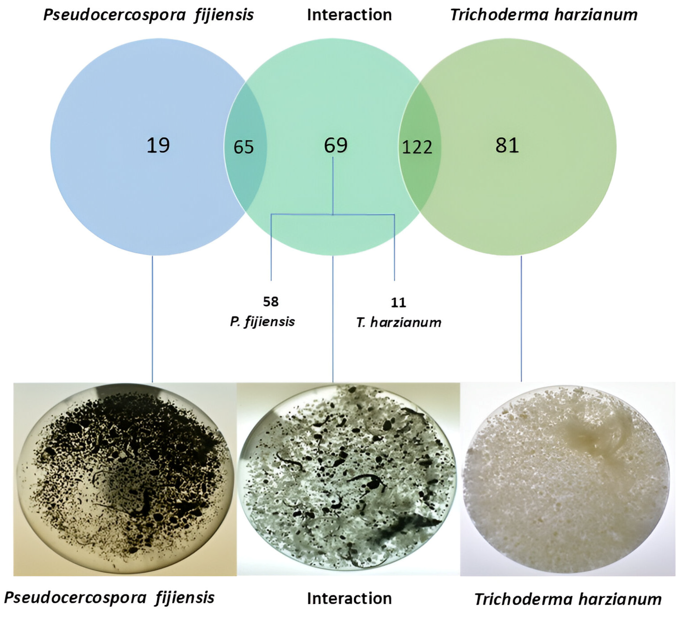
Figure 1. Venn diagram of secretome proteins of Pseudocercospora fijiensis and Trichoderma harzianum under monoculture (control) and dual-culture (interaction) conditions.
Microbiolres 16 00175 g001
Figure 2. Gene Ontology-based classification of sequences identified in the control and interaction secretomes from (A) Pseudocercospora fijiensis and (B) Trichoderma harzianum. Graphs show annotated sequences for the categories “Cellular Component”, “Molecular Function” and “Biological process”.
Microbiolres 16 00175 g002
Figure 3. Comparison of the most represented InterPro families belonging to Trichoderma harzianum and Pseudocercospora fijiensis in the interaction secretome.
Microbiolres 16 00175 g003
Figure 4. Top InterPro domains enriched in the interaction-specific proteins of Trichoderma harzianum and Pseudocercospora fijiensis.
Microbiolres 16 00175 g004
Figure 5. Multiple sequence alignment of THCFEM1. (A) Global alignment of the ThCFEM1 protein with hits from related microorganisms of the Sordariomycetes class. Aligned sequences are from Trichoderma harzianum, T. virens, T. reesei, T. asperellum, Fusarium solani, Neonectria magnoliae, Ilyonectria robusta and Beauveria bassiana. Visualization of the alignment was obtained with Jalview 2.11.4.1. The conserved amino acids are highlighted in indigo, and the intensity of the color is based on the percent identity of the amino acid at each position in the alignment. (B) The second alignment is based on the CFEM domain of the related homologs. The eight conserved cysteine residues that are characteristic of the CFEM domain are marked with black dots.
Microbiolres 16 00175 g005
Figure 6. Phylogenetic analysis of THCFEM1. Phylogenetic tree of ThCfem1 homologous proteins present in related and unrelated organisms: Sordariomycetes (Trichoderma sp., Fusarium sp., Geosmithia morbida, Neonectria magnoliae, Mycena capillaripes, Pochonia chlamydosporia, Metarhizium acridum and Thelonectria olida), Agaricomycetes (Panaeolus papilionaceus, Suillus decipiens, Gloeopeniophorella convolvens, Cantharellus anzutake), Pezizomycetes (Tuber sp., Trichophaea hybrida, Wilcoxina mikolae), Basidiomycetes (Mycena capillaripes), Pucciniomycetes (Austropuccinia psidii, Puccinia coronata f. sp. avenae, Puccinia sorghi, Cronartium quercuum f. sp. fusiforme, Morchella snyderi) Microbotryomycetes (Microbotryum saponariae) and Neolectomycetes (Neolecta irregularis). Sordariomycetes can be observed in green while all other classes are in black. The node numbers signify the percentage of their occurrence in 1000 bootstrap replicates. The tree was constructed in MEGA 11 with 42 sequences using the maximum likelihood method.
Microbiolres 16 00175 g006
Figure 7. Transient expression of ThCFEM1 in Nicotiana benthamiana. Infiltration zones are marked with dashed circles. (A) Suppression of XEG1-induced cell death by ThCFEM1. Infiltrations of A. tumefaciens GV3101 expressing GFP and the effector ThCFEM1 were performed on the left and right sides of the leaf, respectively. Twenty-four hours later, the proven cell-death-inducing effector XEG1 of Phytophthora sojae was agroinfiltrated at the same sites to determine ThCFEM1’s ability to suppress the HR caused by XEG1. The leaves were photographed 5 days after infiltration of XEG1. The experiment was performed 3 times and leaves presented a similar phenotype. (B) ThCFEM1 does not induce a cell death phenotype in N. benthamiana. Isolated infiltrations of the ThCFEM1, GFP and XEG1 were performed at the three different sites on the leaf. Leaves were evaluated daily and photographs were taken at 5 days post infiltration. Only at the XEG1 site were necrotic lesions observed. The experiment was repeated three times with similar results.
Microbiolres 16 00175 g007
Table 1. Effector candidates in the Trichoderma harzianumPseudocercospora fijiensis secretome, which shares identity with known effectors and other virulence-associated proteins.
(A) Trichoderma harzianum effector candidates
ConditionUniprot IDSize
(aa)
Domain
(CDD NCBI)
PHI-Base Virulence and
Avirulence Factors *
Top Hit
% Identity of Effector Homologs
(WideEffHunter)
Int onlyA0A0F9X4L9220Cupredoxin domain-containing proteinMoPtep1 of Magnaporthe oryzae
(32.4%)
-
Int+ ControlA0A0G0ARJ7395Glycoside hydrolase family 16 proteinCrh1 of Botrytis cinerea
(Plant avr determinant) (46.7%)
-
Int+ ControlA0A0F9ZFB8458Glycoside hydrolase family 16 proteinCrh1 of Botrytis cinerea
(Plant avr determinant) (45.6%)
-
Int+ ControlA0A1T3C5B5454Aspergillopepsin-like proteaseBbepnL-1 of Beauveria bassiana (35.6%)Acp (28.9%) of Candida tropicalis, Sap2 (28.1%), Sap3 (27.1%) and Sap1 (25.8%2) of Candida albicans
Int+ ControlA0A0F9XKY7298CutinaseSsCut1 of Sclerotinia sclerotiorum (30.1%)CutA (26.5%) of
Fusarium solani f. sp. cucurbitae
Int+ Control A0A0F9XAH1515Pepsin-like aspartic protease
(SAP-like)
N/AAcp (25.3%) of Candida tropicalis,
Sap2 (24.3%), Sap1 (23.7%) and Sap3 (23.7%) of Candida albicans
Int+ ControlA0A0F9WZ69530Pepsin-like aspartic protease
(SAP-like)
N/ASap1 (34.4%), Acp (34.1%), Sap2 (33.3%) and Sap3 (31.8%) of Candida albicans
Int+ ControlA0A0F9XAQ4624Pepsin-like aspartic protease (SAP-like)SAP1 of Candida albicans (35.9%)Sap1 (35.9%), Sap2 (35.3%) and Sap3 (34.5%) of Candida albicans and Acp (33.2%) of Candida tropicalis
Int+ ControlA0A1T3CKS4154Copper/zinc superoxide dismutaseSOD1 of Fusarium graminearum (87.6%)-
Int+ ControlA0A0F9ZKW4552Bifunctional metallophosphatase/5′-nucleotidase,
UshA Superfamily
AdsA of Staphylococcus aureus (26.2%)-
Int+ ControlA0A0F9ZHK4324Transaldolase BTalA of Francisella tularensis (52.1%)-
Int+ ControlA0A0F9XJM3372Pepsin/retropepsin-like aspartic protease family proteinACP of Candida tropicalis (26.13%)-
-
Int+ ControlA0A0F9XK41429Lytic polysaccharide monooxygenase (LMPO) auxiliary activity family 9 proteinPsFP1 of Phytophthora sojae (36.2%)-
-
Int+ ControlA0A0F9XJ17595Histidine phosphatase family protein Aph1 of Cryptococcus neoformans (34.4%)-
Int+ ControlA0A0F9ZZN6541Glyco_hydro superfamilyGas1 of Fusarium oxysporum (60.15%)-
Int+ ControlA0A0F9ZXC91117WSC domain-containing protein;
GO-like_E_set
GLX of Fusarium graminearum (44.5%)-
Int+ ControlA0A0G0AHN3617DUF1996 superfamilyGLX of Fusarium graminearum (37.6%)-
Int+ ControlA0A0F9X6Z0193CFEM domain-containing proteinFGSG_02077 of Fusarium graminearum (51.22%)
Int+ ControlA0A0F9XM39138Cerato-platanin superfamilySm1 of Trichoderma virens (91.3%)-
Int+ ControlA0A0F9ZCK3463Alpha amylase
(AmyAc_euk_AmyA domain)
MalS of Salmonella enterica (26.82%)-
(B) Pseudocercospora fijiensis effector candidates
ConditionUniprot IDSize
(aa)
Domain
(CDD NCBI)
PHI-Base Virulence and
Avirulence Factors *
Top Hit
% Identity of Effector Homologs
(WideEffHunter)
Int onlyA0A139HLB6147Nis1 family; Necrosis-inducing secreted protein 1-ChNIS1 (43.3%) of Colletotrichum gloeosporioides
Int+ ControlM3AIE7518Pepsin/retropepsin-like aspartic protease family proteinN/ASap1 (29.7%),
Sap3 (28.2%) and
Sap2 (28.1%) of Candida albicans and
Acp (26.4%) of Candida tropicalis
Int+ ControlA0A139H3Z9408S9 Family peptidaseN/APr1 (52.3%) of Metarhizium anisopliae
Int onlyM2ZK33423Alpha/beta hydrolase family proteinFGSG_03846 of Fusarium graminearum
(56.7%)
-
Int+ ControlM3A2P3354Aspergillopepsin-likeBbepnL-1 of Beauveria bassiana (36.4%)Acp (27%) of Candida tropicalis and
Sap1 (28.2%),
Sap2 (26.6%) and
Sap3 (25.4%) of Candida albicans
Int+ ControlM2ZEM9590GMC family oxidoreductase GOX of Aspergillus carbonarius (30.28%)-
Int+ ControlM3AVS4608GMC family oxidoreductaseBCIN_03g01540 of Botrytis cinerea (48.19%)-
Int+ ControlM3A5F8581GMC family oxidoreductaseBab2_0277 of Brucella abortus (25.5%)-
Int onlyM3A9Y6196-BEC1040 of Blumeria graminis (30.5%)
CEP2 avr effector of Magnaporthe oryzae (32.9%)
Bec1040 (30.5%) of Blumeria graminis
Int+ ControlN1QC30308Glycoside hydrolase family 17 protein (Scw11 domain)BGL2 of Candida albicans (40.6%)-
Int+ ControlM2ZG05548Carboxylesterase/lipase family protein (PnbA domain)FGSG_03243 of Fusarium graminearum (39%)-
Int+ ControlM3B3X0573Carboxylesterase/lipase family protein (PnbA domain)FGSG_03243 of Fusarium graminearum (42.05%)-
Int onlyM3ASU3434PeroxidaseMoHPX1 of Magnaporthe oryzae (34.51%)-
Int onlyA0A139IQP5545Peptidase S10 superfamily proteinRs-scp-1 of Radopholus similis (32.2%)-
Int+ ControlM3AEN1328Alpha/beta hydrolase family esterase (LpqC domain)FAED1 of Valsa mali (34.5%)-
Int+ ControlM3BB201011Glycosyl hydrolase family 31BcBGL5 of Botrytis cinerea (57.7%)-
Int+ ControlM3AMA8461Glycoside hydrolase family proteinGEL2 of Aspergillus fumigatus (54.7%)-
Int onlyM3AE521002Glycosyl hydrolase family 31BcBGL5 of Botrytis cinerea (35.3%)-
Int onlyM2YTH3569Beta-N-acetylhexosaminidaseHEX1 of Candida albicans (30%)-
Int onlyM3B352820Beta-glucosidase (Bglx)BcBGL1 of Botrytis cinerea (57.6%)-
Int+ ControlN1QB78878Beta-glucosidase (Bglx)BcBGL3 of Botrytis cinerea (62.3%)-
Int onlyM2ZWF9761Beta-glucosidase (Bglx)BcBGL1 of Botrytis cinerea (44.8%)-
Int+ ControlM2ZLK3553GMC family oxidoreductase (BetA)Bab2_0277 of Brucella abortus (23.9%)-
PHI-base virulence and avirulence factors *: Only top hits with a “reduced virulence” mutant phenotype were considered for virulence factors. N/A: Top hit is neither a virulence nor avirulence factor. Int: Interaction.
Table 2. Effector candidates of Pseudocercospora fijiensis and Trichoderma harzianum involved in other interactions (plant–pathogen, microbe–microbe).
(A) Pseudocercospora fijiensis effector candidates identified in other interactions
ConditionUniprot IDSize
(aa)
Domain
(CDD NCBI)
EffHunterWideEffHunterInteractionNotes
Int onlyM3B352820BglXnoyesP. fijiensisMusa acuminata
[70]
Upregulated in leaf tissue
Int onlyM3B2Z2681Peptidases_S53noyesP. fijiensisMusa acuminata
[70]
Upregulated in leaf tissue
Control +
Int
M3A4I0175-noyesP. fijiensisM. acuminata
[70]
Upregulated in leaf tissue
Control +
Int
M3BB201011Glyco_hydro_31noyesP. fijiensisM. acuminata
[70]
Down regulated in
leaf tissue vs. mycelium
Int onlyM2ZY96195-yesnoP. fijiensisM. acuminata
[51]
Secreted in vitro and in planta by both avirulent and virulent P. fijiensis isolates
(B) Trichoderma harzianum effector candidates identified in other interactions
ConditionUniprot IDSize
(aa)
Domain
(CDD NCBI)
EffHunterWideEffHunterInteractionNotes
Control + IntA0A0F9XHX6429BglC superfamilynoyesT. harzianum TR274–S. sclerotiorum
[71]
Upregulated in presence of
S. sclerotiorum cell walls vs. glucose (12, 24, 36 h)
Control + IntA0A2N1LN60118-yesnoT. harzianum TR274–S. sclerotiorum
[71]
Upregulated in presence of
S. sclerotiorum cell walls vs. glucose at 24 and 36 h
Control +
Int
A0A0F9WYR7515Glyco_hydro_47noyesT. harzianum ALL42–F. solani
[48]
Secreted in culture media with F. solani cell walls
Control +
Int
A0A0F9X6H8689DUF4965noyesT. harzianum ALL42–F. solani
[48]
Secreted in culture media with F. solani cell walls
Control +
Int
A0A0F9XM39138Cerato-platanin superfamilyyesnoT. harzianum ALL42–F. solani
[48]
T. virensZea mays
[41]
T. harzianumPhaseolus vulgaris
[31]
Secreted in culture media with F. solani cell walls and culture media with only glucose
Secreted in similar proportions in interactions with maize and when alone; most abundantly secreted protein
Control +
Int
A0A0F9ZTU1281NPP1 superfamilyyesnoT. harzianum ALL42–F. solani
[48]
Secreted in both culture media with F. solani cell walls
Control +
Int
A0A0G0A4H5878Peptidases_S8_5noyesT. harzianum ALL42–F. solani
[48]
Secreted in both culture media with F. solani cell walls
Control +
Int
A0A0F9ZZN6541Glyco_hydro superfamilynoyesT. harzianum ALL42–F. solani
[48]
Secreted in minimal medium with F. solani cell walls
Control +
Int
A0A0F9X6Z0197CFEMyesyesT. virensZea mays
[41]
T. harzianumF. solani
[77]
Up-accumulated in T. virens control secretome vs. T. virens–maize interaction secretome
Upregulated in the presence of F. solani cell walls
Control +
Int
A0A0F9XF89776GH55_beta13glucanase-likenoyesT. harzianum TR274–Phaseolus vulgaris
[73]
Up-accumulated in interactions with Phaseolus vulgaris
Control +
Int
A0A1T3CKS4154Sod_CunoyesT.virensZea mays
[42]
Secreted by T. virens after 5 days in contact with maize roots
Table 3. Characteristics of ThCFEM1.
Uniprot IDSize
(aa)
Peptide SignalLocalizationMolecular FunctionDomainPHI-BaseEffector MotifTaxonomic Distribution
A0A0F9X6Z0193YesExtracellular,
Cell membrane
Metal ion bindingCFEMFGSG_02077 of
F. graminearum (51.22%)
YFWxCRelated species in the order Hypocreales
Disclaimer/Publisher’s Note: The statements, opinions and data contained in all publications are solely those of the individual author(s) and contributor(s) and not of MDPI and/or the editor(s). MDPI and/or the editor(s) disclaim responsibility for any injury to people or property resulting from any ideas, methods, instructions or products referred to in the content.