Article

Microbial Communities’ Composition of Supralittoral and Intertidal Sediments in Two East African Beaches (Djibouti Republic)

1
Department of Biology, University of Florence, Via Madonna del Piano 6, Sesto Fiorentino, 50019 Florence, Italy
2
Department of Chemistry “Ugo Schiff”, University of Florence, Via della Lastruccia, 3, Sesto Fiorentino, 50019 Florence, Italy
3
Campus de Balbala, Faculté des Sciences, Université de Djibouti, Croisement RN2-RN5 B.P., Djibouti 1904, Djibouti
4
Sezione di Zoologia “La Specola”, Museo di Storia Naturale, University of Florence, 50125 Firenze, Italy
5
Consorzio per il Centro Interuniversitario di Biologia Marina ed Ecologia Applicata (CIBM), 57128 Livorno, Italy
*
Authors to whom correspondence should be addressed.
These authors contributed equally to this work.
Microbiol. Res. 2025, 16(8), 173; https://doi.org/10.3390/microbiolres16080173
Submission received: 15 June 2025 / Revised: 17 July 2025 / Accepted: 21 July 2025 / Published: 1 August 2025

Abstract

Tropical sandy beaches are dynamic ecosystems where microbial communities play crucial roles in biogeochemical processes and tracking human impact. Despite their importance, these habitats remain underexplored. Here, using amplicon-based sequencing of bacterial (V3-V4 16S rRNA) and fungal (ITS2) markers, we first describe microbial communities inhabiting supralittoral–intertidal sediments of two contrasting sandy beaches in the Tadjoura Gulf (Djibouti Republic): Sagallou-Kalaf (SK, rural, siliceous sand) and Siesta Plage (SP, urban, calcareous sand). Sand samples were collected at low tide along 10 m transects perpendicular to the shoreline. Bacterial communities differed significantly between sites and along the sea-to-land gradient, suggesting an influence from both anthropogenic activity and sediment granulometry. SK was dominated by Escherichia-Shigella, Staphylococcus, and Bifidobacterium, associated with human and agricultural sources. SP showed higher richness, with enriched marine-associated genera such as Hoeflea, Xanthomarina, and Marinobacter, also linked to hydrocarbon degradation. Fungal diversity was less variable, but showed significant shifts along transects. SK communities were dominated by Kluyveromyces and Candida, while SP hosted a broader fungal assemblage, including Pichia, Rhodotorula, and Aureobasidium. The higher richness at SP suggests that calcium-rich sands, possibly due to their buffering capacity and greater moisture retention, offer more favorable conditions for microbial colonization.

Graphical Abstract

1. Introduction

Sandy beach ecosystems represent ecologically important ecotonal zones between terrestrial and marine environments, exchanging materials from both sea and land with important effects on trophic networks [1,2]. These peculiar environments are characterized by highly variable biotic and abiotic factors. These variations produce unique dynamic physical properties, such as moisture levels and nutrient availability [3]. Bacterial communities’ diversity is known to be strongly habitat-driven, with higher bacterial abundance in sediments than in the overlying water column. These communities are shaped by abiotic factors, such as sediment grain size and the desiccation gradient along the sea–land axis [4,5], as well as by anthropogenic activities. A large body of research focuses on bacterial communities in the supralittoral and intertidal zones of sandy marine beaches at temperate latitudes. Many of these concern the presence and monitoring of pathogens potentially harmful to human health [6,7,8,9], while others examine the possibility of bioremediation of beaches contaminated by oil spills and the effects on functional and taxonomic diversities [10,11,12,13,14,15,16]. In contrast, studies on microbial diversity and composition in marine sandy beaches and the influencing chemical and physical factors are much fewer [17], often targeting unique environments such as coastal sand dunes [18,19] or mangrove intertidal belts [11,20]. Due to their environmental and sanitary importance, anthropogenic impact has also been investigated at tropical and sub-tropical latitudes, mainly focusing on microbial indicators of marine recreational water quality, including fecal coliforms and pathogens, which tend to be more abundant in heavily frequented areas [21,22,23,24]. Despite their ubiquity in marine systems and ecological relevance [25], fungal communities in sandy beach sediments remain poorly studied. Marine fungi have been mainly studied in association with plants or marine animals [26,27] and historically overlooked by microbiologists as inactive in marine environments [28,29]. However, high-throughput sequencing has renewed interest in the study of marine fungi by revealing both specialized marine species and ubiquitous osmo-/halotolerant taxa [30]. Recent findings suggest that sediment characteristics, such as mineral composition and buffering capacity, may influence fungal assemblages in beach environments [31]. To our knowledge, no studies have addressed microbial communities inhabiting the sandy beaches of the Horn of Africa. The coast from Djibouti to southern Somalia is sparsely developed, with few ports [32], limited tourist infrastructure, and scarce perennial waterways, factors likely shaping microbial presence and distribution. Nonetheless, increasing maritime traffic, due to the Horn of Africa’s position near the Indian Ocean–Red Sea transition, may influence microbial composition, potentially favoring hydrocarbon-degrading taxa (e.g., see [33]). Here, we evaluated bacterial and fungal diversity in two sandy beaches of the Republic of Djibouti, one rural and one urban, exposed to different anthropogenic pressures. This study provides a first insight into microbial assemblages on Djibouti beaches and offers a foundation for future research on tropical coastal microbiomes and their responses to environmental and anthropogenic pressures.

2. Materials and Methods
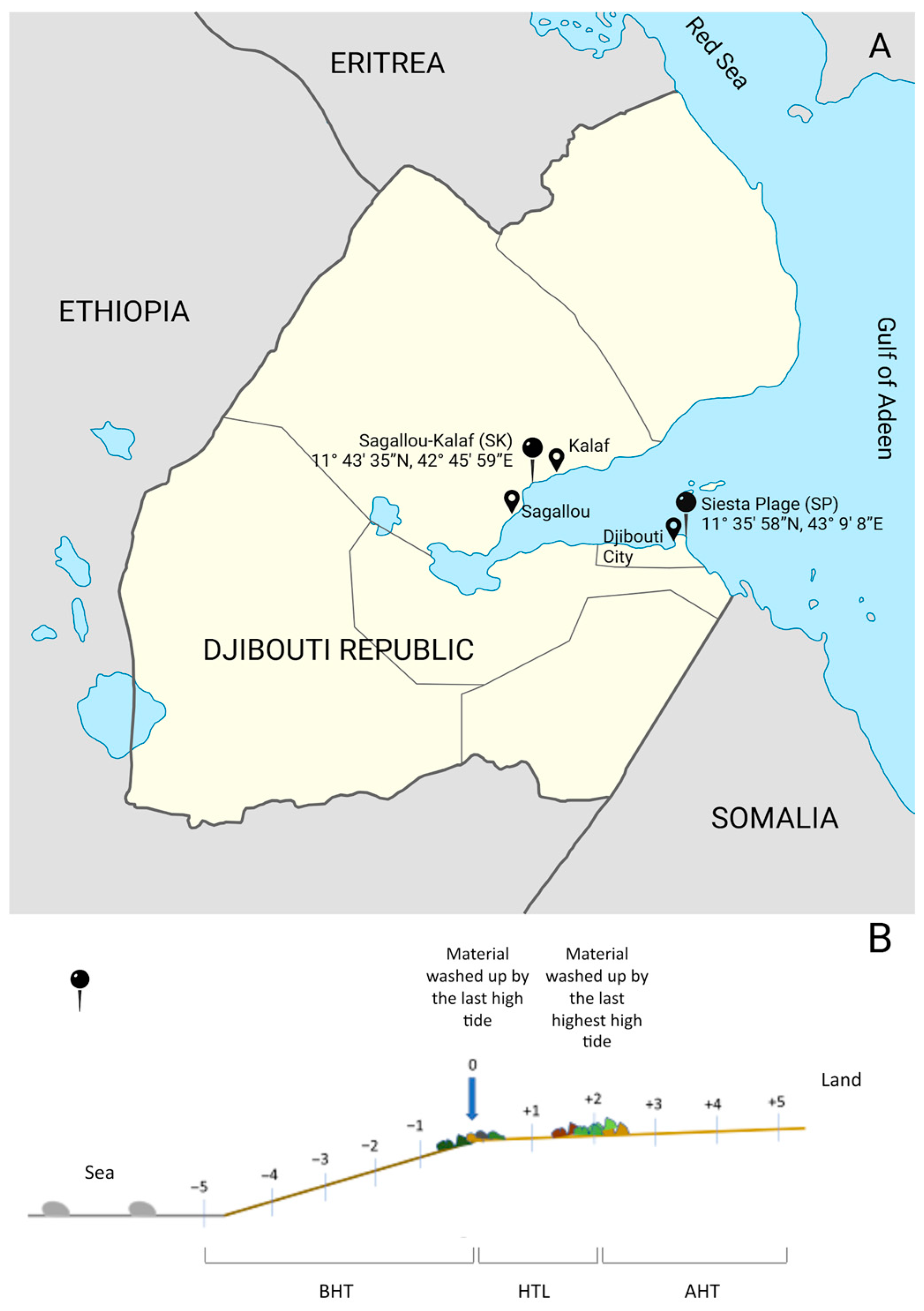
2.1. Sampling

In February 2022, we collected 22 aliquots of sandy sediment from two distinct locations within the territory of the Republic of Djibouti. Eleven samples were obtained at Siesta Plage (11°35′58″ N, 43°9′8″ E, SP from now on), an urban coastal area subject to substantial anthropogenic pressure. The other 11 samples were gathered at a point located between Sagallou and Kalaf (11°43′35″ N, 42°45′59″ E, SK from now on), an area less affected by human disturbance and located far from major urban settlements. Sampling took place during the diurnal low tide phase, employing linear transects oriented perpendicular to the shoreline. At each site, sampling points were systematically labeled (Figure 1). Point 0 was established at the location of the last high tide deposit. From this central reference point, samples were collected at one-meter intervals (approximately 1 cm depth) both seaward (points −1 to −5) and landward (points +1 to +5), yielding a total of 11 sampling points per site. Given the influence of tidal dynamics on coastal environments, the samples were categorized into three tidal zones with respect to the high tide line for microbial community analysis: Below High Tide (BHT), High Tide Line (HTL), and Above High Tide (AHT)). By grouping the samples according to their tidal exposure, we enhance the ecological resolution of our microbiological analyses and allow for a more accurate interpretation of the observed patterns in microbial diversity, structure, and function across environmental gradients. We stored each sediment sample in sterile 50 mL tubes, clearly labeled either “SP” or “SK” to indicate the sampling site, and assigned a numerical identifier from −5 to +5 corresponding to the sampling point.

2.2. Sediment Characterization: Granulometry and Mineralogical Composition

Particle size profiles were determined by sieving dry sediments (60 °C, 24 h) through a column of sieves with different mesh sizes according to the Udden–Wentworth classification (<63 μm pelitic fraction, 63–125 μm very fine sand, 125–250 μm fine sand, 250–500 μm medium sand, 500–1000 μm coarse sand, 1000–2000 μm very coarse sand, >2000 μm gravel) [34]. All the conventional sediment parameters were expressed as percentages of the total dry sample weight. The elemental composition of the sand samples was analyzed using a Shimadzu (Kyoto, Japan) EDX-7000 x-ray fluorescence (XRF) energy dispersive spectrometer equipped with a W X Ray source and an SDD detector with a resolution less than 140 eV. The X-ray source was set for the element Al-U at the 50 kV 144 uA, and for the element Na-Sc, the X-ray source was set at 15 kV 999 uA. The collimator was set at 10 mm for all the samples. The samples were dried to remove excess moisture and scanned on 10 mm of surface area. The data collected were analyzed with the built-in PCEDX Navi Software using a semi-quantitative pre-calibrated method.

2.3. Sediment-Associated Microbial Community Analyses

2.3.1. Genomic DNA Extraction and Microbial Taxonomic Marker Sequencing

For each sample point, sand was collected by a commercially available sterile collection tube, then stored at −20 °C in order to preserve nucleic acids. Total genomic DNA was extracted from 250 mg (wet weight) of each sample using the DNeasy PowerSoil Pro Kit (Qiagen, Hilden, Germany) according to the manufacturer’s instructions. The Qubit 4 Fluorometer (Thermo Fisher Scientific, Waltham, Massachusetts, USA) and the 1x dsDNA High Sensitivity kit were used to assess the DNA concentration before the downstream analyses. For each DNA sample, the 16S rRNA gene was amplified using a specific primer set for the V3-V4 region (341f: 5′-CCTACGGGNGGCWGCAG-3′ and 805r: 5′GACTACNVGGGTWTCTAATCC--3′) [35], while for the fungal internal transcribed spacer (ITS), a primer set specific for the ITS2 region was used (ITS3f 5′-GCATCGATGAAGAACGCAGC-3′ and ITS4r 5′-TCCTCCGCTTATTGATATGC-3′) [36] with overhang Illumina adapters. The PCR thermal cycling parameters were as follows: for 16S, 95 °C for 2 min, 30 cycles of 95° C for 25 s, 58° C for 30 s, 72° C for 30 s, followed by a final extension at 72° C for 5 min. For ITS2, 95° C for 3 min, 35 cycles of 95 °C for 20 s, 50° C for 45 s, 72° C for 90 s, followed by a final extension at 72° C for 10 min. PCRs for each primer set were carried out in a 25 μL reaction volume containing 0.5 U PCRBIO Taq DNA Polymerase (PCRBIOSYSTEMS, London, UK) in 5× PCRBIO Reaction Buffer, 10 pmol of forward primer, 10 pmol of reverse primer, and 12.5 ng of genomic DNA template (as per Illumina recommendations). Library preparation was performed according to Illumina Protocol 16S Metagenomic Sequencing Library Preparation (Part #15044223 Rev.B), with appropriate changes for the fungal marker [37]. Sequencing was performed on the Illumina MiSeq platform (Illumina, San Diego, CA, USA) using the V3 chemistry 600-cycle paired-end reads (2 × 300) protocol at the Department of Biology, University of Florence, Italy.

2.3.2. Sequence Processing, Taxonomic Assignment, and Community Analysis

Primer sequences were removed using Cutadapt v1.18 [38], and raw paired-end reads were processed using the DADA2 pipeline v1.26 [39] in R v4.3.2. This included quality filtering, denoising, merging, chimera removal, and inference of amplicon sequence variants (ASVs). For the 16S dataset, 1,841,536 raw reads (mean per sample: 83,706) were reduced to 1,224,098 high-quality reads (66.5% of the initial) after processing. The ITS dataset yielded 2,212,637 raw reads (mean per sample: 100,574), with 1,450,153 reads retained post-processing (65.5%). Four 16S rRNA samples (SP + 1, SP + 5, SK-4, and SK-3) and one ITS2 (SK-3) sample that failed to produce a sufficient number of reads during sequencing were excluded from downstream analyses. Taxonomic assignment was performed using DECIPHER [40], aligning 16S ASVs to the SILVA v138.1 database [41] and ITS ASVs to the UNITE v10.05.2024 database [42]. Downstream microbial community analyses were conducted using the phyloseq [43], microbiome [44], and vegan [45] packages in R. We assessed alpha diversity using observed ASVs and the inverse Simpson index, and compared group differences using the Wilcoxon rank-sum test. For beta diversity, we calculated Bray–Curtis dissimilarity, visualized through NMDS (metaMDS function), and tested it with PERMANOVA (adonis2 function). Differential abundance was evaluated using ANCOM-BC v2.4.0 [46], with a significance threshold of FDR-adjusted p < 0.05. All visualizations were generated using ggplot2 v3.4.2 [47].

3. Results

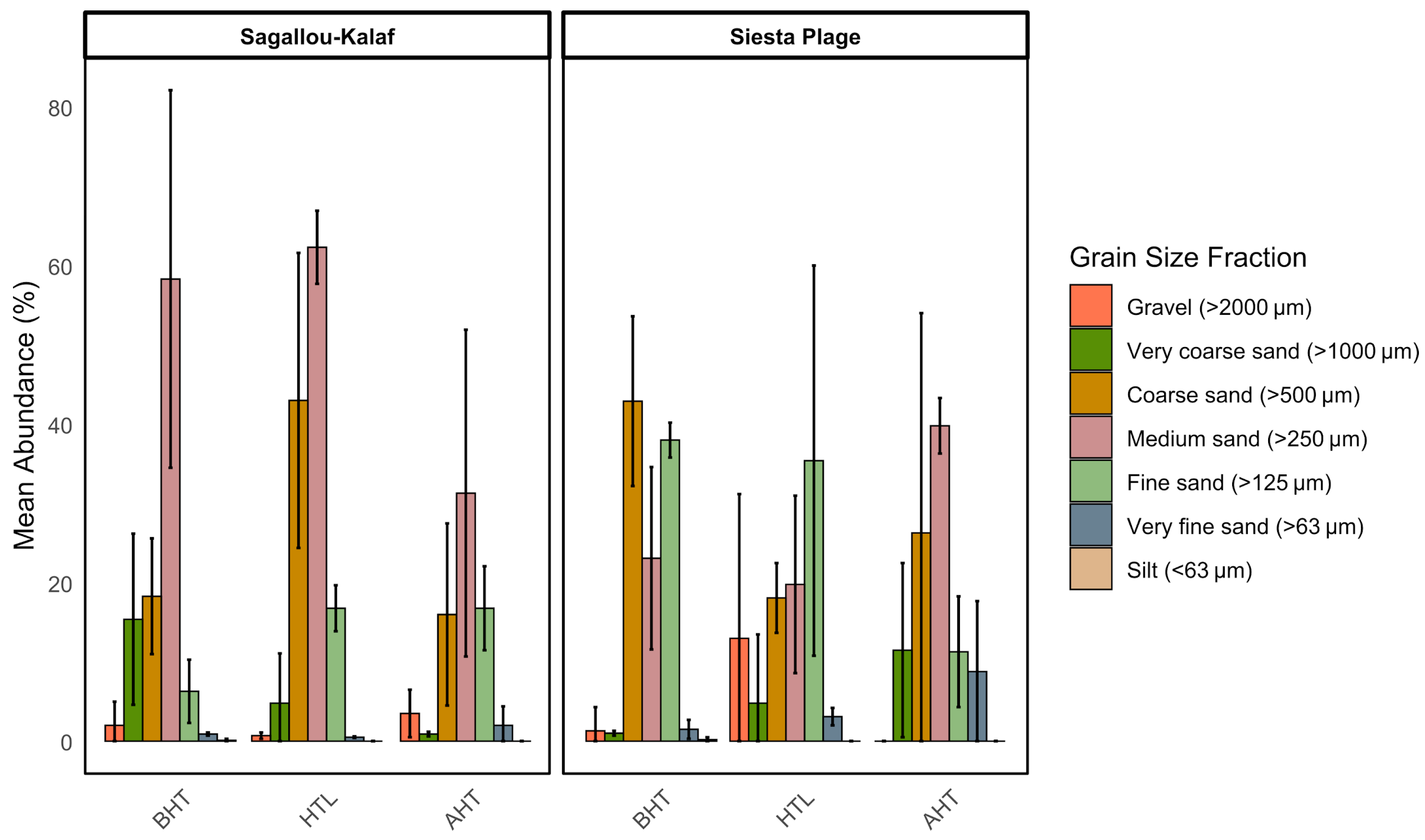
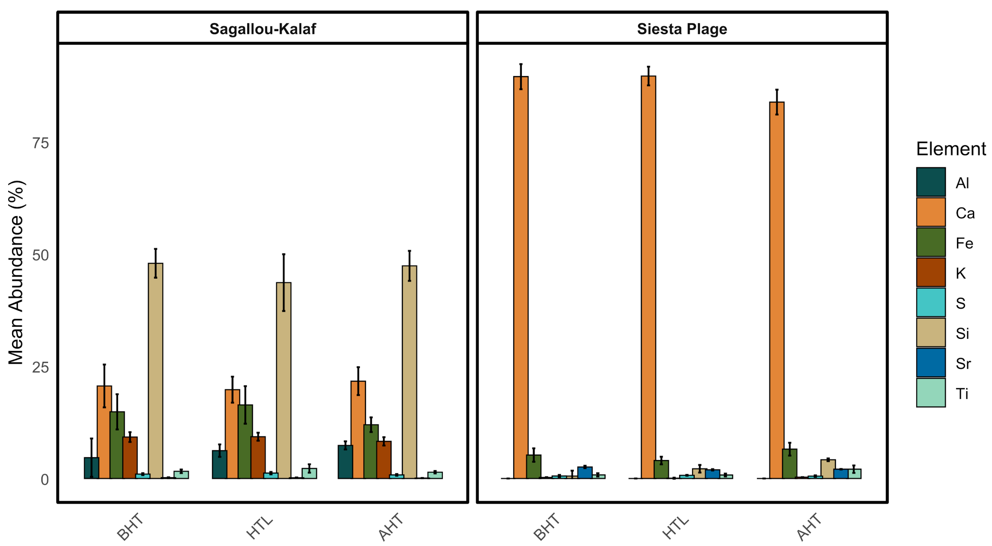
3.1. Sediment Analysis

Grain size analysis shows that on both beaches, medium and coarse sand fractions are consistently well represented along the entire transects (Figure 2). However, in SK, there is a prevalence of medium sand compared to SP, where fine sand prevails. In SP, a certain quantity of gravel and very coarse sand is observed at sampling point 0, while in SK, very coarse sand is found at both terminal sampling points of the transect. Very fine sand and silt are present in low quantities, mainly in the last two points of withdrawal towards the sea in both SP and SK.
Mineralogical composition analysis reveals that SP is clearly a limestone beach, in which other elements are present in modest quantities at each sampling point (Figure 3): silicon (Si) is present mainly towards the land side, while iron (Fe) and strontium (Sr) are evenly distributed. Conversely, SK is predominantly a siliceous beach, but with an evident presence of uniformly distributed calcium (Ca). Iron (Fe) and potassium (K) are consistently detected in fair concentrations across all sampling points. Aluminum (Al) appears to be missing from the last two collection points towards the sea. Other elements are present in very small quantities.

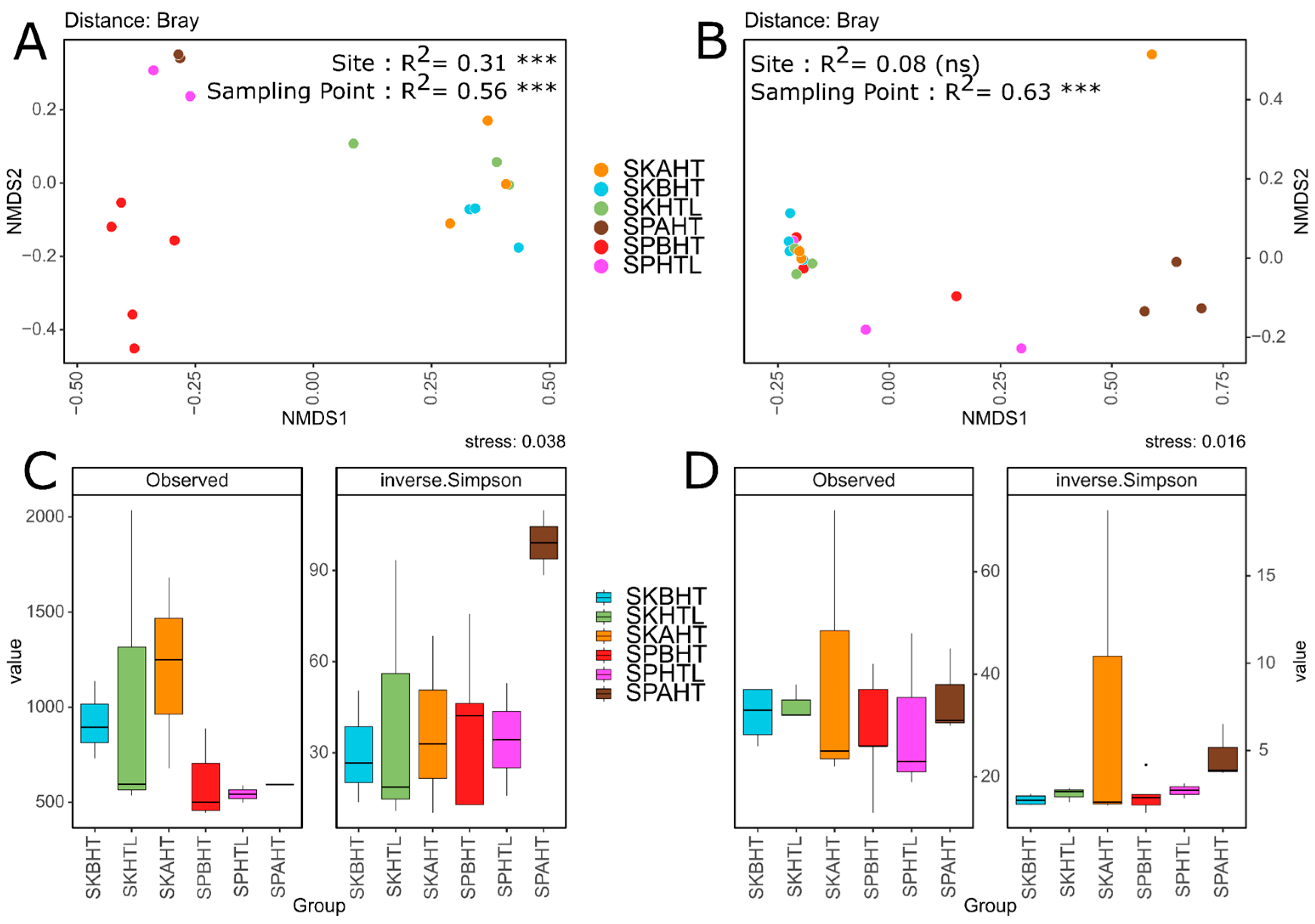
3.2. Microbial Community Structure and Diversity Patterns

Microbial community diversity was assessed through both alpha and beta diversity analyses on bacterial and fungal datasets. Alpha diversity measured as observed ASVs and inverse Simpson indices did not reveal statistically significant differences between sites (SK and SP) or along the transect. Nevertheless, in SP, we noted a general reduction in the number of bacterial ASVs, suggesting lower bacterial richness (Figure 4C). Fungal communities, in contrast, displayed a more heterogeneous richness pattern across the samples, with inverse Simpson index denoting an increased diversity in SK above the AHT. Beta diversity, analyzed using non-metric multidimensional scaling analysis (NMDS) based on Bray–Curtis distance, showed different trends for bacteria and fungi. In the bacterial dataset, the samples clustered according to both geographical site (SK and SP) and sampling point along the transect, particularly in SP (Figure 4A). These trends were supported by PERMANOVA (R2 in Figure 4A), which identified the factor “sampling point” as the main source of variance (56%), followed by “geographical site” (31%). Conversely, fungal communities exhibited a weaker spatial separation (Figure 4B), although PERMANOVA still indicated a statistically significant effect of the sampling point variable (R2 in Figure 4B), explaining 63% of variance between the samples, while the contribution of geographical sites accounted only for 8% (ns).
To complement diversity analyses, the composition of the dominant phylum (Figures S1 and S2) and genera (Figure 5) were evaluated. At both sites, Proteobacteria dominate the bacterial communities, followed by Firmicutes, which show relatively stable abundance across zones and locations. Bacteroidota and Actinobacteriota are also consistently represented, with Actinobacteriota slightly more abundant at SP. Minor phyla such as Verrucomicrobiota, Planctomycetota, and Acidobacteriota display site- and zone-specific variations, particularly at SP, suggesting localized environmental influences. In terms of genera, at SK, bacterial communities were relatively stable across the transect, dominated by Escherichia-Shigella, Staphylococcus, and Bifidobacterium, which collectively accounted for approximately 40% of the community. Interestingly, the genus Alcanivorax was particularly abundant in SK-BHT. Conversely, the SP site seemed to present a more heterogeneous community, with Xantomarina dominating the SP-BHT group, followed by Marinobacter, Cobetia, and Halobacillus in the SP-HTL group, while Halobacillus, Rhodopirellula, and Salimicrobium were abundant in the SP-AHT group. Regarding the fungal dataset (Figure 5B), community composition appeared more homogeneous across both geographical sites and transect points, with Ascomycota dominating the community in nearly all groups. At the genus level, Kluyveromyces represents over 45% of the overall community composition. Exceptions were found in SP-AHT, where a strong abundance of Corollospora, Varicosporina, and Lindra was notable, and in SK-AHT, which showed a more even distribution among different fungal genera. Overall, bacterial communities exhibited clearer spatial structuring and site-specific differentiation, while fungal communities were more compositionally consistent but locally variable in specific transect segments.

3.3. Influence of the Distance from the High Tide Line on the Distribution of Bacterial Taxa

We conducted differential abundance analysis separately for the two areas (SP and SK) across three defined areas of tidal exposure (BHT, HTL, AHT). Based on significantly different ASVs abundance distributions (q-value < 0,05), the bacterial dataset (Figure 6) revealed distinct clustering by geographical sites, with SP and SK forming two separate groups. Within SP, the samples also separated further according to their position relative to the last high tide line, while the SK samples did not show any clear stratification along the transect. Several ASVs showed increased abundance in SP-HTL and SP-AHT, particularly those assigned to the genera Halomonas, Halobacillus, Erytrhobacter, Actibacter, Schleiferia, Sediminibacterium, and Pontibacillus. In contrast, wet area samples (SP-BHT) were enriched in ASVs associated with Membranicola, Hyphomonas, Muricauda, Candidatus Thiobio, Actibacter, Hoeflea, and Xanthomarina. In SK, we did not observe any clear clustering by transect point. However, we detected several ASVs corresponding to bacterial genera commonly associated with human and livestock microbiota, such as Staphylococcus, Streptococcus, Bifidobacterium, Lactobacillus, and Escherichia-Shigella [48,49,50], suggesting a potential anthropogenic influence at this site. Regarding the fungal dataset (Figure S3), differential abundance analysis did not reveal clear clustering patterns among the samples. However, consistent positive selection of Kluyveromyces was observed in wet zone samples (BHT) of both SP and SK, indicating possible environmental selection for this genus in wetter sediment conditions. We report the log fold change (logFC) values and q-values for both datasets in Supplementary Tables S1 and S2.

4. Discussion

4.1. Site-Specific Microbial Signatures: Rural vs. Urban Beaches

Sandy beaches are dynamic microbial habitats capable of hosting diverse communities, including potential human and animal pathogens [9,51]. The two investigated sites, Sagallou-Kalaf (SK) and Siesta Plage (SP), differ in their exposure to anthropogenic influence, which appears to shape their respective microbial communities. Djibouti City, characterized by intense marine traffic and port-related industrial activities, is among the most impacted coastal areas in the region. A recent study [52] has documented significant anthropogenic pollution in this area, in contrast to the relatively undisturbed coastal zone farther from the city. SP, situated within the urban complex of Djibouti City and frequented for recreational activities near a commercial port, displays higher bacterial diversity and is dominated by halotolerant and marine-associated genera, including Xanthomarina, Marinobacter, Cobetia, and Halobacillus. In contrast, SK, located in a rural setting with limited urban development but active animal presence (e.g., sheep, goats, and dromedaries), harbors bacterial communities rich in genera commonly associated with human and animal microbiota, such as Escherichia-Shigella, Staphylococcus, and Bifidobacterium. Differential abundance analysis (DESeq2) confirms site-specific clustering of bacterial communities, with SP exhibiting a clear structuring along the tidal transect. This spatial organization is absent in SK, likely due to its more homogeneous sediment environment, which may reduce spatial variability in microbial communities [53,54]. These patterns are further supported by lower alpha and beta diversity values in SK, aligning with previous studies highlighting the role of anthropogenic activity and land use in coastal microbial ecology [4]. Regarding the fungal dataset, differential abundance analysis did not reveal clear clustering patterns, suggesting stable and homogeneous communities between the two areas. This likely reflects ecological and dispersal traits: bacteria respond rapidly to local environmental changes and differing anthropogenic activities, whereas fungi, often with broader niches and high spore dispersal, tend to form more homogeneous communities across spatial scales.

4.2. Microbial Distribution Alongside the Transect

The intertidal gradient may play a role in shaping the microbial community structure along the transects. At both beaches, microbial analyses suggest potential variations above and below the last high-tide line. Bacterial beta diversity (as shown by NMDS and PERMANOVA) appears to be influenced by both site and transect position, which could reflect sensitivity to environmental gradients such as moisture, salinity, and UV exposure. Fungal communities, however, appear more strongly structured by local abiotic factors along the transect rather than by geographical location. In SP, bacterial communities display a marked shift along the transect, with samples below the high-tide line (SP-BHT) enriched in marine- and pollutant-associated genera such as Membranicola, Hyphomonas, Muricauda, Candidatus Thiobios, Hoeflea, and Xanthomarina, taxa often linked to sediment and seawater environments [55,56]. Membranicola, in particular, has also been reported in polluted sites such as landfills and wastewater systems [57]. These genera often possess metabolic versatility, including the ability to degrade recalcitrant pollutants like hydrocarbons [58,59], pointing to environmental filtering along the tidal gradient. Fungal communities, while more stable across sites, do show transect-level variation, especially at the AHT point, suggesting responses to tidal exposure or sediment-associated microclimates. Genera such as Kluyveromyces, Corollospora, Hortaea, and Varicosporina, many of which are known from marine and coastal habitats [60,61,62], were observed. Ubiquitous fungi such as Candida and Malassezia, associated with human and animal activity [63,64], were present at both sites. Their occurrence aligns with the recreational use and grazing pressures documented in these areas, indicating that anthropogenic factors also contribute to shaping fungal community composition on a local scale.

4.3. Sediment Composition and Microbial Communities

Sediment composition emerged as a key environmental driver influencing microbial diversity and distribution. SP, characterized by heterogeneous carbonate-rich sands, supports a more spatially variable and taxonomically diverse microbial community. In contrast, the siliceous and compositionally uniform sediments at SK are associated with a more homogeneous bacterial community structure and reduced diversity. These findings align with previous research indicating that sediment grain size and mineralogy influence parameters such as porosity, pH, moisture retention, and organic matter availability [65,66]. The presence of alkaliphilic bacterial genera at SP, such as Cobetia, Thiomicrospira, and Halobacillus, corresponds with the buffering capacity of carbonate sediments that can maintain higher pH levels [67,68]. Conversely, SK’s more inert siliceous sands may contribute to more limited microbial niches and reduced physicochemical variability. This geological contrast between sites may explain the observed differences in microbial spatial structuring and ecological diversity. Fungal community composition appears less tightly linked to sediment mineralogy, although some effects may be indirect, mediated by physicochemical factors such as salinity, moisture, and pH [69,70,71]. Fungi involved in organic matter decomposition, such as Cladosporium, Lindra, Corollospora, and Xylodon, were found at both sites, underlining their ecological role in the recycling of lignin and cellulose in beach environments [72,73,74].

5. Conclusions

This study provides the first comparative analysis of microbial communities inhabiting sandy beaches of the Tadjoura Gulf (Djibouti Republic), highlighting how environmental gradients, sediment composition, and anthropogenic activity influence microbial diversity. Combining granulometric, mineralogical, and metabarcoding analyses, we propose that tropical sandy beaches act as dynamic microbial habitats that are sensitive to both natural and human-driven factors. The two study sites, Sagallou-Kalaf, a rural, siliceous beach, and Siesta Plage, an urban, calcareous beach, revealed distinct microbial signatures, with a positive selection of bacterial genera reflecting local human activities. Overall, bacterial communities exhibit greater spatial and compositional variability than fungal ones, although the latter also show notable shifts along the tidal transects. In fact, several taxa related to coastal environments, organic matter decomposition, and human or animal presence were detected, emphasizing the ecological adaptability of beach fungi. Sediment composition emerged as a potential key driver influencing microbial structure: carbonate-rich sands at Siesta Plage may enhance diversity via moisture retention and pH buffering, while the more homogeneous, siliceous sands at Sagallou-Kalaf may limit variability. However, fungal communities seemed more resilient to variations in sediment type and mineralogy. In summary, these findings point to the ecological significance of coastal sediments as reservoirs of microbial diversity, shaped by both physicochemical features and anthropogenic pressures. However, since sampling was restricted to a single coastal region and conducted only during the winter of 2022, the generalizability of the findings is limited despite the inclusion of sites with markedly different levels of anthropic disturbance. Future studies should include multi-seasonal sampling across multiple coastal zones to better evaluate microbial patterns at a broader spatial scale and assess their temporal consistency. It is also important to note that environmental variables such as porewater salinity could not be measured at the time of sampling, leaving their direct influence on microbial community structure unquantified. These parameters should be integrated into future surveys to better resolve the impact of environmental drivers on the microbial communities of tropical sandy beaches. Nonetheless, we propose these first insights as baseline data for coastal microbial ecology in tropical regions, supporting the use of beach microbiomes as potential bioindicators for environmental monitoring.

Supplementary Materials

The following supporting information can be downloaded at https://www.mdpi.com/article/10.3390/microbiolres16080173/s1: Figure S1: Dominant bacterial phyla; Figure S2: Dominant fungal phyla; Figure S3: Heatmaps and cluster analysis of differentially abundant ASVs in the fungal dataset; Table S1: Differential abundance results from ANCOM-BC analysis of bacterial ASVs; Table S2: Differential abundance results from ANCOM-BC analysis of fungal ASVs.

Author Contributions

Conceptualization, A.U. and D.C.; Methodology, S.R., A.R., A.U., A.D., C.P. and S.C.; Sampling, A.U. and A.N.; Software, A.D.; Formal Analysis, S.R. and A.R.; Data Curation, S.R., A.R. and A.U.; Writing—Original Draft Preparation, S.R. and A.R.; Writing—Review and Editing, S.R., A.R., A.D., S.C., A.N., C.P., A.U., D.C. and S.C.S.; Visualization, S.R. and A.R.; Supervision, A.U. and D.C.; Funding Acquisition, A.U. and D.C. All authors have read and agreed to the published version of the manuscript.

Funding

This research was supported by the Consorzio Interuniversitario per le Biotecnologie (CIB) through funding provided by the Italian Ministry of University and Research (MUR) under Decree No. 809 dated 7 July 2023. In 2023, support was granted under the initiative “Innovation in Biotechnology in the Era of Precision Medicine, Climate Change, and Circular Economy”, and in 2024 under the theme “Innovative Models for Advanced Omics Science Applications”. Funding was awarded to Duccio Cavalieri. Additional support was provided by the University of Florence through local RICATEN funds, awarded to Alberto Ugolini.

Institutional Review Board Statement

Not applicable.

Informed Consent Statement

Not applicable.

Data Availability Statement

Raw sequence data are deposited in the European Nucleotide Archive (ENA) under accession code PRJEB90295 (ERP173311). The dataset available in the EBI-ENA database includes V3–V4 and ITS2 raw sequences from the sediments of both geographical sites. Further information can be provided upon request to the corresponding authors.

Acknowledgments

A.U. and A.N. would like to thank Miriam Martinelli and Carlo Astini for their hospitality and Gianni Rizzo, Honorary Consul General of Italy in Djibouti, for his assistance during their stay in Djibouti.

Conflicts of Interest

The authors declare no conflicts of interest.

Abbreviations

The following abbreviations are used in this manuscript:
SKSagallou-Kalaf
SPSiesta Plage
BHTBelow High Tide
HTLHigh Tide Line
AHTAbove High Tide

References

  1. McLachlan, A.; Brown, A.C. The Ecology of Sandy Shores, 2nd ed.; Academic Press: Cambridge, MA, USA, 2006. [Google Scholar] [CrossRef]
  2. Vassallo, P.; Paoli, C.; Fabiano, M. Ecosystem level analysis of sandy beaches using thermodynamic and network analyses: A study case in the NW Mediterranean Sea. Ecol. Indic. 2012, 15, 10–17. [Google Scholar] [CrossRef]
  3. Defeo, O.; McLachlan, A.; Schoeman, D.S.; Schlacher, T.A.; Dugan, J.; Jones, A.; Lastra, M.; Scapini, F. Threats to sandy beach ecosystems: A review. Estuar. Coast. Shelf Sci. 2009, 81, 1–12. [Google Scholar] [CrossRef]
  4. Santmire, J.A.; Leff, L.G. The effect of sediment grain size on bacterial communities in streams. J. N. Am. Benthol. Soc. 2007, 26, 601–610. [Google Scholar] [CrossRef]
  5. Arboleda-Baena, C.; Freilich, M.; Pareja, C.B.; Logares, R.; De la Iglesia, R.; Navarrete, S.A. Microbial community and network responses across strong environmental gradients: How do they compare with macroorganisms? FEMS Microbiol. Ecol. 2024, 100, fiae017. [Google Scholar] [CrossRef] [PubMed]
  6. Feng, F.; Goto, D.; Yan, T. Effects of autochthonous microbial community on the die-off of fecal indicators in tropical beach sand. FEMS Microbiol. Ecol. 2010, 74, 214–225. [Google Scholar] [CrossRef] [PubMed]
  7. Whitman, R.L.; Harwood, V.J.; Edge, T.A.; Nevers, M.B.; Byappanahalli, M.N.; Vijayavel, K.; Brandão, J.; Sadowsky, M.J.; Alm, E.W.; Crowe, A.; et al. Microbes in Beach Sands: Integrating Environment, Ecology and Public Health. Rev. Environ. Sci. Bio. Technol. 2014, 13, 329–368. [Google Scholar] [CrossRef] [PubMed]
  8. Sabino, R.; Rodrigues, R.; Costa, I.; Carneiro, C.; Cunha, M.; Duarte, A.; Faria, N.; Ferreira, F.; Gargaté, M.; Júlio, C.; et al. Routine screening of harmful microorganisms in beach sands: Implications to public health. Sci. Total. Environ. 2014, 472, 1062–1069. [Google Scholar] [CrossRef] [PubMed]
  9. Solo-Gabriele, H.M.; Harwood, V.J.; Kay, D.; Fujioka, R.S.; Sadowsky, M.J.; Whitman, R.L.; Wither, A.; Caniça, M.; da Fonseca, R.C.; Duarte, A.; et al. Beach sand and the potential for infectious disease transmission: Observations and recommendations. J. Mar. Biol. Assoc. UK 2016, 96, 101–120. [Google Scholar] [CrossRef]
  10. Horel, A.; Mortazavi, B.; Sobecky, P.A. Responses of microbial community from northern Gulf of Mexico sandy sediments following exposure to deepwater horizon crude oil. Environ. Toxicol. Chem. 2012, 31, 1004–1011. [Google Scholar] [CrossRef] [PubMed]
  11. Almeida, R.; Mucha, A.P.; Teixeira, C.; Bordalo, A.A.; Almeida, C.M.R. Biodegradation of petroleum hydrocarbons in estuarine sediments: Metal influence. Biodegradation 2013, 24, 111–123. [Google Scholar] [CrossRef] [PubMed]
  12. Newton, R.J.; Huse, S.M.; Morrison, H.G.; Peake, C.S.; Sogin, M.L.; McLellan, S.L.; Gilbert, J.A. Shifts in the microbial community composition of Gulf Coast beaches following beach oiling. PLoS ONE 2013, 8, e74265. [Google Scholar] [CrossRef] [PubMed]
  13. Engel, A.S.; Gupta, A.A.; Kellogg, C.A. Regime shift in sandy beach microbial communities following Deepwater Horizon oil spill remediation efforts. PLoS ONE 2014, 9, e102934. [Google Scholar] [CrossRef] [PubMed]
  14. Gouveia, V.; Almeida, C.; Almeida, T.; Teixeira, C.; Mucha, A.P. Indigenous microbial communities along the NW Portuguese Coast: Potential for hydrocarbons degradation and relation with sediment contamination. Mar. Pollut. Bull. 2018, 131 Pt A, 620–632. [Google Scholar] [CrossRef] [PubMed]
  15. Rodriguez-R, L.M.; Overholt, W.A.; Hagan, C.; Huettel, M.; Kostka, J.E.; Konstantinidis, K.T. Microbial community successional patterns in beach sands impacted by the Deepwater Horizon oil spill. ISME J. 2015, 9, 1928–1940. [Google Scholar] [CrossRef] [PubMed]
  16. Heritier-Robbins, P.; Karthikeyan, S.; Hatt, J.K.; Kim, M.; Huettel, M.; Kostka, J.E.; Konstantinidis, K.T.; Rodriguez-R, L.M. Beach sand oil spills select for generalist microbial populations. ISME J. 2021, 15, 3418–3422. [Google Scholar] [CrossRef] [PubMed]
  17. Nair, S.; LokaBharathi, P.A. Heterotrophic Bacterial Populations in Tropical Sandy Beaches. 1980. Available online: https://drs.nio.res.in/drs/handle/2264/6833 (accessed on 3 June 2025).
  18. Shet, S.A.; Garg, S. Prokaryotic diversity of tropical coastal sand dunes ecosystem using metagenomics. 3 Biotech 2021, 11, 252. [Google Scholar] [CrossRef] [PubMed]
  19. Nayak, S.; Behera, S.; Dash, P.K. Potential of Microbial Diversity of Coastal Sand Dunes: Need for Exploration in Odisha Coast of India. Sci. World J. 2019, 2019, 2758501. [Google Scholar] [CrossRef] [PubMed]
  20. Palit, K.; Rath, S.; Chatterjee, S.; Das, S. Microbial diversity and ecological interactions of microorganisms in the mangrove ecosystem: Threats, vulnerability, and adaptations. Environ. Sci. Pollut. Res. 2022, 29, 32467–32512. [Google Scholar] [CrossRef] [PubMed]
  21. Shibata, T.; Solo-Gabriele, H.M.; Fleming, L.E.; Elmir, S. Monitoring marine recreational water quality using multiple microbial indicators in an urban tropical environment. Water Res. 2004, 38, 3119–3131. [Google Scholar] [CrossRef] [PubMed]
  22. Piggot, A.M.; Klaus, J.S.; Johnson, S.; Phillips, M.C.; Solo-Gabriele, H.M. Relationship between Enterococcal Levels and Sediment Biofilms at Recreational Beaches in South Florida. Appl. Environ. Microbiol. 2012, 78, 5973–5982. [Google Scholar] [CrossRef] [PubMed]
  23. Yamahara, K.M.; Walters, S.P.; Boehm, A.B. Growth of Enterococci in Unaltered, Unseeded Beach Sands Subjected to Tidal Wetting. Appl. Environ. Microbiol. 2009, 75, 1517–1524. [Google Scholar] [CrossRef]
  24. Samarasekera, K.; Abeygunawardena, S.I. Microbiology of Seawater and Sand in a Selected Bathing Site of Sri Lanka—A Study Towards Microbial Quality Assessment. Front. Environ. Microbiol. 2017, 3, 1. [Google Scholar] [CrossRef]
  25. Jones, E.G. Are there more marine fungi to be described? Bot. Mar. 2011, 54, 343–354. [Google Scholar] [CrossRef]
  26. Yarden, O. Fungal association with sessile marine invertebrates. Front. Microbiol. 2014, 5, 228. [Google Scholar] [CrossRef] [PubMed]
  27. Godinho, V.M.; de Paula, M.T.R.; Silva, D.A.S.; Paresque, K.; Martins, A.P.; Colepicolo, P.; Rosa, C.A.; Rosa, L.H. Diversity and distribution of hidden cultivable fungi associated with marine animals of Antarctica. Fungal Biol. 2019, 123, 507–516. [Google Scholar] [CrossRef] [PubMed]
  28. Amend, A.; Burgaud, G.; Cunliffe, M.; Edgcomb, V.P.; Ettinger, C.L.; Gutiérrez, M.H.; Heitman, J.; Hom, E.F.Y.; Ianiri, G.; Jones, A.C.; et al. Fungi in the Marine Environment: Open Questions and Unsolved Problems. mBio 2019, 10, e01189-18. [Google Scholar] [CrossRef] [PubMed]
  29. Stal, L.J.; Cretoiu, M.S. The Marine Microbiome. In The Microbiomes of Humans, Animals, Plants, and the Environment; Springer Nature: Dordrecht, The Netherlands, 2022; Volume 3. [Google Scholar]
  30. Overy, D.P.; Rämä, T.; Oosterhuis, R.; Walker, A.K.; Pang, K.-L. The Neglected Marine Fungi, Sensu stricto, and Their Isolation for Natural Products’ Discovery. Mar. Drugs 2019, 17, 42. [Google Scholar] [CrossRef] [PubMed]
  31. Lin, C.Y.; Bradbury, H.J.; Antler, G.; Burdige, D.J.; Bennett, T.D.; Li, S.; Turchyn, A.V. Sediment mineralogy influences the rate of microbial sulfate reduction in marine sediments. Earth Planet. Sci. Lett. 2022, 598, 117841. [Google Scholar] [CrossRef]
  32. de Boer, W.; Mao, Y.; Hagenaars, G.; de Vries, S.; Slinger, J.; Vellinga, T. Mapping the Sandy Beach Evolution Around Seaports at the Scale of the African Continent. J. Mar. Sci. Eng. 2019, 7, 151. [Google Scholar] [CrossRef]
  33. Curren, E.; Leong, S.C.Y. Profiles of bacterial assemblages from microplastics of tropical coastal environments. Sci. Total. Environ. 2019, 655, 313–320. [Google Scholar] [CrossRef] [PubMed]
  34. Wentworth, C.K. A Scale of Grade and Class Terms for Clastic Sediments. J. Geol. 1922, 30, 377–392. [Google Scholar] [CrossRef]
  35. Takahashi, S.; Tomita, J.; Nishioka, K.; Hisada, T.; Nishijima, M.; Bourtzis, K. Development of a prokaryotic universal primer for simultaneous analysis of Bacteria and Archaea using next-generation sequencing. PLoS ONE 2014, 9, e105592. [Google Scholar] [CrossRef] [PubMed]
  36. White, T.J.; Bruns, T.; Lee, S.; Taylor, J. Amplification and direct sequencing of fungal ribosomal RNA genes for phylogenetics. In PCR Protocols: A Guide to Methods and Applications; Innis, M.A., Gelfand, D.H., Sninsky, J.J., White, T.J., Eds.; Academic Press: New York, NY, USA, 1990; Volume 18, pp. 315–322. [Google Scholar] [CrossRef]
  37. Renzi, S.; Nenciarini, S.; Bacci, G.; Cavalieri, D. Yeast metagenomics: Analytical challenges in the analysis of the eukaryotic microbiome. Microbiome Res. Rep. 2024, 3, 2. [Google Scholar] [CrossRef] [PubMed]
  38. Martin, M. Cutadapt removes adapter sequences from high-throughput sequencing reads. EMBnet. J. 2011, 17, 10–12. [Google Scholar] [CrossRef]
  39. Callahan, B.J.; Mcmurdie, P.J.; Rosen, M.J.; Han, A.W.; Johnson, A.J.A.; Holmes, S.P. DADA2: High-resolution sample inference from Illumina amplicon data. Nat. Methods 2016, 13, 581–583. [Google Scholar] [CrossRef] [PubMed]
  40. Erik, S.W. Using DECIPHER v2.0 to Analyze Big Biological Sequence Data in R. R J. 2016, 8, 352. [Google Scholar] [CrossRef]
  41. Yilmaz, P.; Parfrey, L.W.; Yarza, P.; Gerken, J.; Pruesse, E.; Quast, C.; Schweer, T.; Peplies, J.; Ludwig, W.; Glöckner, F.O. The SILVA and “All-species Living Tree Project (LTP)” taxonomic frameworks. Nucleic Acids Res. 2014, 42, D643–D648. [Google Scholar] [CrossRef] [PubMed]
  42. Abarenkov, K.; Nilsson, R.H.; Larsson, K.-H.; Taylor, A.F.S.; May, T.W.; Frøslev, T.G.; Pawlowska, J.; Lindahl, B.; Põldmaa, K.; Truong, C.; et al. The UNITE database for molecular identification and taxonomic communication of fungi and other eukaryotes: Sequences, taxa and classifications reconsidered. Nucleic Acids Res. 2024, 52, D791–D797. [Google Scholar] [CrossRef] [PubMed]
  43. McMurdie, P.J.; Holmes, S. phyloseq: An R package for reproducible interactive analysis and graphics of microbiome census data. PLoS ONE 2013, 8, e61217. [Google Scholar] [CrossRef] [PubMed]
  44. Lahti, L.; Shetty, S. Microbiome R Package, version 3.21; Bioconductor: Boston, MA, USA, 2017. [Google Scholar] [CrossRef]
  45. Oksanen, J.; Simpson, G.L.; Blanchet, F.G.; Kindt, R.; Legendre, P.; Minchin, P.R.; O’HAra, R.; Solymos, P.; Stevens, M.H.H.; Szoecs, E.; et al. Vegan: Community Ecology Package_. R Package Version 2.6–4. 2022. Available online: https://cir.nii.ac.jp/crid/1370584340724217483 (accessed on 3 June 2025).
  46. Lin, H.; Das Peddada, S. Analysis of compositions of microbiomes with bias correction. Nat. Commun. 2020, 11, 3514. [Google Scholar] [CrossRef] [PubMed]
  47. Villanueva, R.A.M.; Chen, Z.J. ggplot2: Elegant Graphics for Data Analysis (2nd ed.). Meas. Interdiscip. Res. Perspect. 2019, 17, 160–167. [Google Scholar] [CrossRef]
  48. Huuki, H.; Vilkki, J.; Vanhatalo, A.; Tapio, I. Fecal microbiota colonization dynamics in dairy heifers associated with early-life rumen microbiota modulation and gut health. Front. Microbiol. 2024, 15, 1353874. [Google Scholar] [CrossRef] [PubMed]
  49. Amin, N.; Schwarzkopf, S.; Kinoshita, A.; Tröscher-Mußotter, J.; Dänicke, S.; Camarinha-Silva, A.; Huber, K.; Frahm, J.; Seifert, J. Evolution of rumen and oral microbiota in calves is influenced by age and time of weaning. Anim. Microbiome 2021, 3, 31. [Google Scholar] [CrossRef] [PubMed]
  50. Goodwin, K.D.; McNay, M.; Cao, Y.; Ebentier, D.; Madison, M.; Griffith, J.F. A multi-beach study of Staphylococcus aureus, MRSA, and enterococci in seawater and beach sand. Water Res. 2012, 46, 4195–4207. [Google Scholar] [CrossRef] [PubMed]
  51. Sibanda, T.; Ramganesh, S. Taxonomic and functional analyses reveal existence of virulence and antibiotic resistance genes in beach sand bacterial populations. Arch. Microbiol. 2021, 203, 1753–1766. [Google Scholar] [CrossRef] [PubMed]
  52. Ahmed, N.; Benmessaoud, Y.; Hamdail, B.R.; Ezziyyani, M. Intelligent System for Forecasting Climate Deterioration and Assessing the Potential Impact of Pollutants. In International Conference on Advanced Intelligent Systems for Sustainable Development (AI2SD’2023); Ezziyyani, M., Kacprzyk, J., Balas, V.E., Eds.; AI2SD 2023. Lecture Notes in Networks and Systems; Springer: Cham, Switzerland, 2024; Volume 930. [Google Scholar] [CrossRef]
  53. Liu, L.; Zou, L.; Gao, D.M. Spatiotemporal Distribution of Microorganism in Mudflats Environment of the Yellow River Estuary Intertidal Area. Adv. Mater. Res. 2014, 1030–1032, 603–608. [Google Scholar] [CrossRef]
  54. Wang, Z.; Fuad, T.I.; Liu, J.; Lin, K.; Liu, L.; Gao, C.; Wang, W.; Liu, X. Spatial Patterns of Microbial Communities in Intertidal Sediments of the Yellow River Estuary, China. Microb. Ecol. 2025, 87, 173. [Google Scholar] [CrossRef] [PubMed]
  55. Wang, T.; Lin, Z.; Kuang, B.; Ni, Z.; Chen, X.; Guo, B.; Zhu, G.; Bai, S. Electroactive algae-bacteria wetlands for the treatment of micro-polluted aquaculture wastewater: Pilot-scale verification. Biochem. Eng. J. 2022, 184, 108471. [Google Scholar] [CrossRef]
  56. Rinke, C.; Schmitz-Esser, S.; Stoecker, K.; Nussbaumer, A.D.; Molnár, D.A.; Vanura, K.; Wagner, M.; Horn, M.; Ott, J.A.; Bright, M. “Candidatus Thiobios zoothamnicoli,” an Ectosymbiotic Bacterium Covering the Giant Marine Ciliate Zoothamnium niveum. Appl. Environ. Microbiol. 2006, 72, 2014–2021. [Google Scholar] [CrossRef] [PubMed]
  57. Liang, Y.; Yin, Q.; Jiang, Z.; Yan, H.; Nian, Y. Pollution characteristics and microbial community succession of a rural informal landfill in an arid climate. Ecotoxicol. Environ. Saf. 2023, 262, 115295. [Google Scholar] [CrossRef] [PubMed]
  58. Huang, Y.; Li, L.; Yin, X.; Zhang, T. Polycyclic aromatic hydrocarbon (PAH) biodegradation capacity revealed by a genome-function relationship approach. Environ. Microbiome 2023, 18, 39. [Google Scholar] [CrossRef] [PubMed]
  59. Rahul, K.; Azmatunnisa, M.; Sasikala, C.; Ramana, C.V. Hoeflea olei sp. nov., a diesel-oil-degrading, anoxygenic, phototrophic bacterium isolated from backwaters and emended description of the genus Hoeflea. Int. J. Syst. Evol. Microbiol. 2015, 65, 2403–2409. [Google Scholar] [CrossRef] [PubMed]
  60. Atalla, M.M.; Zainab, H.K.; Eman, R.H.; Youssry, A.; Aty, A.A.E. Screening of some marine-derived fungal isolates for lignin degrading enzymes (LDEs) production. Agric. Biol. J. North Am. 2010, 1, 591–599. [Google Scholar]
  61. Hodhod, M.S.E.-D.; Gaafar, A.-R.Z.; Alshameri, A.; Qahtan, A.A.; Noor, A.; Abdel-Wahab, M. Molecular characterization and bioactive potential of newly identified strains of the extremophilic black yeast Hortaea werneckii isolated from Red Sea mangrove. Biotechnol. Biotechnol. Equip. 2020, 34, 1288–1298. [Google Scholar] [CrossRef]
  62. Poli, A.; Varese, G.C.; Garzoli, L.; Prigione, V. Seagrasses, seaweeds and plant debris: An extraordinary reservoir of fungal diversity in the Mediterranean Sea. Fungal Ecol. 2022, 60, 101156. [Google Scholar] [CrossRef]
  63. Ianiri, G.; LeibundGut-Landmann, S.; Dawson, T.L. Malassezia: A Commensal, Pathogen, and Mutualist of Human and Animal Skin. Annu. Rev. Microbiol. 2022, 76, 757–782. [Google Scholar] [CrossRef] [PubMed]
  64. Nenciarini, S.; Renzi, S.; di Paola, M.; Meriggi, N.; Cavalieri, D. The yeast–human coevolution: Fungal transition from passengers, colonizers, and invaders. WIREs Mech. Dis. 2024, 16, e1639. [Google Scholar] [CrossRef] [PubMed]
  65. Saxton, K.E.; Rawls, W.J. Soil Water Characteristic Estimates by Texture and Organic Matter for Hydrologic Solutions. Soil Sci. Soc. Am. J. 2006, 70, 1569–1578. [Google Scholar] [CrossRef]
  66. Ning, Z.; Yu, K.; Wang, Y.; Huang, X.; Han, M.; Zhang, J. Carbon and nutrient dynamics of permeable carbonate and silicate sands adjacent to coral reefs around Weizhou Island in the northern South China Sea Estuarine Coast. Shelf Sci. 2019, 225, 106229. [Google Scholar] [CrossRef]
  67. Boyadzhieva, I.; Berberov, K.; Atanasova, N.; Krumov, N.; Kabaivanova, L. Isolation, Purification and In Vitro Characterization of a Newly Isolated Alkalophilic Phytase Produced by the Halophile Cobetia marina Strain 439 for Use as Animal Food Supplement. Fermentation 2025, 11, 39. [Google Scholar] [CrossRef]
  68. Romano, I.; Finore, I.; Nicolaus, G.; Huertas, F.J.; Lama, L.; Nicolaus, B.; Poli, A. Halobacillus alkaliphilus sp. nov., a halophilic bacterium isolated from a salt lake in Fuente de Piedra, southern Spain. Int. J. Syst. Evol. Microbiol. 2008, 58 Pt 4, 886–890. [Google Scholar] [CrossRef] [PubMed]
  69. D’entremont, T.W.; López-Gutiérrez, J.C.; Walker, A.K.; Taylor, I.E. Inoculating rhizome-propagated Sporobolus pumilus with a native mycorrhizal fungus increases salt marsh plant growth and survival. FACETS 2021, 6, 1134–1145. [Google Scholar] [CrossRef]
  70. Lin, W.; Liu, X.; Gong, L.; Liu, R.; Ling, M.; Guo, C.; Meng, H.; Luo, Z.; Du, X.; Guo, Y.; et al. Impact of environmental factors on diversity of fungi in sediments from the Shenzhen River Estuary. Arch. Microbiol. 2023, 205, 96. [Google Scholar] [CrossRef] [PubMed]
  71. Lobo, L.Q.; Izabel-Shen, D.; Albertsson, J.; Raymond, C.; Gunnarsson, J.S.; Broman, E.; Nascimento, F.J.A. Salinity and resource availability as drivers of Baltic benthic fungal diversity. Environ. DNA 2024, 6, e526. [Google Scholar] [CrossRef]
  72. Floudas, D.; Binder, M.; Riley, R.; Barry, K.; Blanchette, R.A.; Henrissat, B.; Martínez, A.T.; Otillar, R.; Spatafora, J.W.; Yadav, J.S.; et al. The Paleozoic origin of enzymatic lignin decomposition reconstructed from 31 fungal genomes. Science 2012, 336, 1715–1719. [Google Scholar] [CrossRef] [PubMed]
  73. Vrijmoed, L.L.P.; Hodgkiss, I.J. Wood DegradIng Activity of some Lignicolous Marine Fungi. In Biodeterioration 7; Houghton, D.R.J., Smith, R.N., Eggins, H.O.W., Eds.; Springer Netherlands: Dordrecht, The Netherlands, 1988; pp. 768–773. [Google Scholar] [CrossRef]
  74. Olguin, J.; Velez, P.; Solís-Weiss, V.; Barrios, A.; Walker, A.K.; Ponce-Vélez, G.; González, M.C.; Figueroa, M.; Botello, A. An overview of fungal taxonomic, functional, and genetic diversity in coastal and oceanic biomes in megadiverse Mexico. Bot. Mar. 2023, 66, 471–490. [Google Scholar] [CrossRef]
Figure 1. (A) Map of Djibouti Republic with administrative divisions highlighting sampling sites; (B) graphical representation of sampling design. Here, 0 represents the point of the last high tide, detected by stranded material. Each point of the transect is spaced 1 m apart. The points are grouped according to their relative position with respect to the material washed up by the last high tide and by the last highest tide. These groups were used for the microbial sequence analysis. BHT, Below High Tide; HTL, High Tide Line; AHT, Above High Tide. Map were adapted from “Djibouti location map” by user “NordNordWest”, via Wikimedia Commons (https://commons.wikimedia.org/wiki/File:Djibouti_location_map.svg, accessed on 15 June 2025), CC BY-SA 3.0.
Microbiolres 16 00173 g001
Figure 2. Granulometric composition of sediment samples collected along two transects. Each bar cluster represents the mean relative abundance (%) of grain size fractions in the three tidal zones. Sediment fractions include gravel (>2000 µm), very coarse sand (>1000 µm), coarse sand (>500 µm), medium sand (>250 µm), fine sand (>125 µm), very fine sand (>63 µm), and silt (<63 µm). Sampling points are reported according to the following legend: BHT, Below High Tide; HTL, High Tide Line; AHT, Above High Tide.
Microbiolres 16 00173 g002
Figure 3. Comparison of elemental concentrations between site SP and site SK. Mean relative abundance of selected elements measured in three tidal zones. Sampling points are reported according to the following legend: BHT, Below High Tide; HTL, High Tide Line; AHT, Above High Tide.
Microbiolres 16 00173 g003
Figure 4. Microbial diversity analysis. (A,B) Non-metric multidimensional scaling (NMDS) analysis based on Bray–Curtis dissimilarity for (A) bacterial and (B) fungal datasets. R-squared values and p-values from PERMANOVA (adonis2) testing the effects of site and sampling point variables are shown in each panel. Significant pairwise comparisons are indicated by asterisks (ns = not significant; ***, p  <  0.001). (C,D) Boxplots of alpha diversity metrics (observed ASVs and inverse Simpson index) by tidal zone for (C) bacterial and (D) fungal datasets. Sampling points are reported according to the following legend: SK, Sagallou-Kalaf; SP, Siesta Plage; BHT, Below High Tide; HTL, High Tide Line; AHT, Above High Tide.
Microbiolres 16 00173 g004
Figure 5. Taxonomic composition at the genus level. Stacked barplots showing the relative abundances of the 30 most dominant microbial genera in the two different sampling areas (SK and SP) across the three tidal zones. (A) 16S rRNA amplicon data for bacteria and (B) ITS2 amplicon data for fungi. Sampling points are reported according to the following legend: BHT, Below High Tide; HTL, High Tide Line; AHT, Above High Tide.
Microbiolres 16 00173 g005
Figure 6. Heatmaps and cluster analysis of differentially abundant ASVs in the bacterial dataset. The heatmap shows scaled and centered relative abundance of ASVs, from lower value (blue scale) to higher value (red scale). The dendrogram on top shows the samples’ cluster analysis results (color indicates the sampling point). The dendrogram on the right shows the results of ASV cluster analysis, with color indicating the different clusters found. Sampling points are reported according to the following legend: SK, Sagallou-Kalaf; SP, Siesta Plage; BHT, Below High Tide; HTL, High Tide Line; AHT, Above High Tide.
Microbiolres 16 00173 g006
Disclaimer/Publisher’s Note: The statements, opinions and data contained in all publications are solely those of the individual author(s) and contributor(s) and not of MDPI and/or the editor(s). MDPI and/or the editor(s) disclaim responsibility for any injury to people or property resulting from any ideas, methods, instructions or products referred to in the content.

