Article

AiiA Lactonase Suppresses ETEC Pathogenicity Through 3OC12-HSL Quenching in a Murine Model

1
College of Veterinary Medicine, Yangzhou University, Yangzhou 225009, China
2
Jiangsu Co-Innovation Center for Important Animal Infectious Diseases and Zoonoses, Joint Laboratory of International Cooperation on Prevention and Control Technology of Important Animal Diseases and Zoonoses of Jiangsu, Yangzhou 225009, China
*
Authors to whom correspondence should be addressed.
These authors contributed equally to this work.
Microbiol. Res. 2025, 16(8), 166; https://doi.org/10.3390/microbiolres16080166
Submission received: 12 July 2025 / Revised: 28 July 2025 / Accepted: 31 July 2025 / Published: 31 July 2025

Abstract

This study elucidates how the quorum-sensing (QS) signal 3OC12-HSL exacerbates enterotoxigenic E. coli (ETEC) pathogenicity and intestinal barrier dysfunction. In vitro, 3OC12-HSL enhanced ETEC C83902 growth (66.7% CFU increase at 8 h) and dysregulated stress/growth genes (e.g., eight-fold rmf upregulation under static conditions). In synthetic gut microbiota, 3OC12-HSL selectively augmented E. coli colonization (37.6% 16S rDNA increase at 12 h). Murine studies revealed 3OC12-HSL reduced jejunal villus height (381.5 μm vs. 543.2 μm in controls), elevated serum LPS, D-lactate, and DAO, and altered microbial composition (Firmicutes/Bacteroidetes imbalance). The lactonase AiiA reversed these effects by degrading 3OC12-HSL. It abrogated bacterial growth stimulation (in vitro CFU restored to baseline), normalized microbiota diversity (Shannon index recovered to control levels), suppressed pro-inflammatory cytokines (IL-6/TNF-α reduction), and restored intestinal integrity (villus length: 472.5 μm, 20.5% increase vs. ETEC-infected mice). Our findings establish AiiA as a potent quorum-quenching agent that counteracts ETEC virulence via targeted signal inactivation, highlighting its translational value.

1. Introduction

Enterotoxigenic Escherichia coli (ETEC) infections inflict substantial morbidity and economic losses in human and veterinary medicine, with F4ac-fimbriated strains like C83902 driving diarrheal disease in neonatal and weaned animals through intestinal colonization and enterotoxin production [1,2,3]. Conventional antibiotic therapies are increasingly compromised by antimicrobial resistance, necessitating novel strategies targeting non-lethal virulence pathways [4,5,6]. Quorum sensing (QS), a bacterial communication system orchestrating collective behaviors via diffusible signals such as acyl-homoserine lactones (AHLs), offers a promising anti-infective target [7,8]. Of these, 3-oxo-C12-homoserine lactone (3OC12-HSL) serves as a key interspecies QS mediator in Gram-negative pathogens yet remains inadequately characterized in ETEC pathogenesis. Critically, despite evidence that ETEC “eavesdrops” on exogenous AHLs to modulate virulence [9,10], the mechanisms through which 3OC12-HSL influences ETEC-mediated intestinal barrier dysfunction remain unresolved. This knowledge gap spans microbial dysbiosis, structural damage, and immune hyperactivation, impeding targeted therapeutic development.
The intestinal barrier constitutes a multicomponent defense system integrating microbiological, mechanical, and immunological elements [11,12,13]. While 3OC12-HSL is known to directly disrupt epithelial integrity [14], its capacity to amplify ETEC pathogenicity and consequently sabotage barrier function remains poorly defined. ETEC lacks AHL synthases but exploits environmental AHLs via receptors like SdiA, potentially enhancing colonization, stress adaptation, and immune evasion [15,16,17]. Such eavesdropping may trigger cascading effects: altered gut microbiota composition, pro-inflammatory cytokine release, and villus atrophy. Specific studies in piglet models reveal that ETEC infection reduces microbial diversity, lowers the Bacteroidetes-to-Firmicutes ratio, and increases opportunistic pathogens like Lactococcus, with fecal microbiota transplantation from infected animals inducing diarrhea in healthy recipients, highlighting dysbiosis as a key disease contributor [18,19]. Despite preliminary associations between AHLs and enteric pathogenesis, no integrated analysis has elucidated whether 3OC12-HSL’s synergy with ETEC exacerbates barrier failure or if quenching this signal can restore homeostasis.
Central to this knowledge gap is 3OC12-HSL’s role in potentiating ETEC virulence. As a ubiquitous gut signal, 3OC12-HSL may reprogram ETEC transcriptionally, accelerate growth, and bias competitive dynamics in complex microbiota. Collectively, these effects foster dysbiosis and barrier breach [20,21]. Key unanswered questions persist: Does 3OC12-HSL selectively enhance ETEC fitness in simulated gut ecosystems? How does it modulate microbial community structure in vivo? More critically, does its synergy with ETEC amplify epithelial injury and inflammation beyond direct AHL toxicity? Resolving these questions would validate QS signals as a therapeutic target for ETEC infections.
Quorum-quenching enzymes like AiiA lactonase [22,23], which hydrolyze AHLs, present a compelling strategy to disarm QS-dependent virulence without selecting for resistance. This approach is strongly supported by evidence across diverse pathogens: In Pseudomonas aeruginosa, AiiA and other lactonases (e.g., SsoPox and PvdQ) significantly reduce virulence factor production (e.g., pyocyanin and proteases), biofilm formation, and mortality in rodent and invertebrate infection models [5,6]. Similarly, in Enterobacter cloacae, AiiA expression enhances proteolytic activity and disrupts early biofilm development on industrial surfaces [22]. Although AiiA protects against direct AHL-induced epithelial damage, its efficacy in blunting ETEC pathogenesis potentiated by 3OC12-HSL remains unexplored. This is particularly true for restoring multifaceted barrier integrity. We thus hypothesized that AiiA-mediated degradation of 3OC12-HSL would reverse ETEC virulence activation, microbiota dysbiosis, and barrier dysfunction, offering a synergistic therapeutic advantage.
Here, we demonstrate that 3OC12-HSL exacerbates ETEC pathogenicity by accelerating growth, dysregulating stress-response genes, distorting gut microbiota, and impairing intestinal integrity in mice. Critically, AiiA lactonase counteracts these effects by degrading 3OC12-HSL: it normalizes bacterial proliferation, restores microbial diversity, suppresses inflammation, and synergistically repairs barrier damage. Our work establishes AiiA as a potent quorum-quenching agent against ETEC infections and mechanistically links AHL eavesdropping to barrier failure, highlighting translational avenues for anti-virulence therapy.

2. Materials and Methods

2.1. Bacterial Strains and Culture Conditions

The F4ac+ enterotoxigenic Escherichia coli (ETEC) strain C83902 was maintained in our laboratory at –80 °C in 20% glycerol stocks [24]. Bacteroides fragilis (ATCC 255285), Bifidobacterium bifidum (GDM1.169), and Enterococcus faecium (ATCC 700221) were obtained from the Guangdong Microbial Culture Collection Center (GDMCC). All strains were cultured aerobically in LB medium (LB; Haibo Biotechnology, Qingdao, China) at 37 °C unless otherwise specified. For AHL supplementation, 3-oxo-C12-homoserine lactone (3OC12-HSL; Macklin Biochemical, Shanghai, China, ≥98% purity) was dissolved in dimethyl sulfoxide (DMSO; Solarbio, Beijing, China) and added to media at a final concentration of 200 μM [14]. AiiA-based QSI (1000 U/g, Beijing Challenge Agricultural Technology Co., Beijing, China) was added to the feed at 0.5% (w/w) in animal experiments.

2.2. Growth Kinetics and Gene Expression Analysis

ETEC C83902 was grown statically in LB or LB + 200 μM 3OC12-HSL. Growth curves were generated by measuring optical density at 600 nm (OD600) hourly for 12 h (n = 3) [25]. For RNA isolation [26], cultures were harvested at mid-log phase (OD600 = 1.0) under three conditions: shaking (200 rpm), static, or anaerobic (using 2.5 L anaerobic jars with gas packs; Oxoid, Basingstoke, Hampshire, UK). Total RNA was extracted using FastPure Plasmid Mini Kit (Vazyme, Nanjing, China) and reverse-transcribed with the PrimeScript RT Reagent Kit (Takara Bio, Dalian, China) [27]. Quantitative RT-PCR (qPCR) was performed using AceQ Universal SYBR qPCR Master Mix (Vazyme) on a LightCycler 96 System (Roche Diagnostics, Basel, Switzerland) [27]. Target genes (ftsZ, sulA, recA, rpoS, rmf, nrdA, ftsY, and 16S rRNA) were amplified using primers listed in Table 1. The 2−ΔΔCt method was employed for relative quantification.

2.3. In Vitro Polymicrobial Culture Model

B. fragilis and B. bifidum were cultured anaerobically on blood agar plates (Kaiheng Biotechnology, Nantong, China) in 2.5 L anaerobic jars with gas packs (Oxoid). E. faecium was cultured in MRS broth (HaiBo), and ETEC C83902 in LB broth. Bacterial suspensions (OD600 = 1.0) were centrifuged (780× g, 10 min; Eppendorf Centrifuge 5430 R, Eppendorf SE, Hamburg, Germany), washed with PBS, and resuspended in DMEM (Gibco™, Thermo Fisher Scientific, Waltham, MA, USA) or DMEM + 200 μM 3OC12-HSL. Final inocula were adjusted to 109 CFU/mL for Bifidobacterium and Bacteroides, and 106 CFU/mL for ETEC and Enterococcus. Co-cultures were incubated anaerobically at 37 °C [28]. At 6 h intervals, DNA was extracted using TIANamp Bacterial DNA Kit (Tiangen, Beijing, China) and species-specific qPCR was performed with primers targeting 16S rRNA genes (Table 1).

2.4. Mouse Experiments

The animal study protocol was approved by the Animal Welfare and Ethics Committee of Yangzhou University (Approval Number: 202202106; Approval Date: 23 February 2022) and was conducted in compliance with the ethical standards of animal welfare and the Jiangsu Provincial Animal Management Committee. Five-week-old male ICR mice (purchased from Yangzhou University Comparative Medicine Center, Experimental Animal Production License Number: SCXK (Jiangsu) 2017-0007) were housed in the Yangzhou University Experimental Animal Center (Experimental Animal Use License Number: SYXK (Jiangsu) 2121-0026). For microbiota studies, mice (n = 5/group) received either sterile water (control) or 200 μM of 3OC12-HSL in drinking water ad libitum for 72 h. Fecal DNA was extracted using QIAamp DNA Stool Kit (Qiagen, Beijing, China) and analyzed by 16S rRNA gene sequencing (Novogene, Beijing, China) on the Illumina MiSeq platform (Illumina, Inc., San Diego, CA, USA) [29]. Paired-end sequencing of the V3-V4 hypervariable regions was performed, with an average depth of 50,000 high-quality reads per sample. Raw sequences were processed using DADA2 for ASV inference, followed by taxonomic assignment against the SILVA 138 database. For α-diversity (Shannon), data were normalized via DESeq2 and compared by the Kruskal–Wallis test. β-diversity was assessed using Bray–Curtis and weighted UniFrac distances calculated from CSS-normalized counts, with PERMANOVA testing for group differences.
For infection models, mice were fasted for 12 h and divided into 4 groups (n = 5/group): Control: PBS oral gavage + sterile water; ETEC: 1010 CFU ETEC C83902 gavage + water; ETEC+3OC12-HSL: ETEC gavage + 200 μM of 3OC12-HSL in water; ETEC+3OC12-HSL+AiiA: ETEC gavage + 200 μM of 3OC12-HSL + 0.5% (w/w) AiiA enzyme in feed [14].

2.5. Histopathology and Apoptosis Assays

Jejunal tissues were collected from a standardized anatomical site, a 1 cm segment centered 3 cm distal to the ligament of Treitz. Tissues were immediately fixed in 4% paraformaldehyde for 24 h at 4 °C. Jejunal tissues were fixed in 4% paraformaldehyde, embedded in paraffin, and sectioned at 5 μm thickness using a fully automated rotary microtome (Leica RM2255, Leica Biosystems, Wetzlar, Germany) [30]. All sections were prepared under standardized conditions: slice speed 120 mm/s, sample retraction 20 μm, and ambient temperature 22 °C. To ensure spatial representation, three non-consecutive sections per animal were selected at 200 μm intervals along the jejunum longitudinal axis. Villus height quantification was performed by two independent observers blinded to experimental groups. For each section, the 10 longest, vertically oriented, and intact villi (excluding branched or damaged structures) were selected from well-oriented jejunal regions. Measurements were conducted at 200× magnification [14]. Villus height was defined as the linear distance from the crypt-villus junction to the villus tip. Each villus was measured three times, and the average value was recorded. A total of 30 villi per animal (10 villi × 3 sections) were analyzed. TUNEL assays were performed per manufacturer’s protocol: Sections were digested with 1 μg/mL proteinase K (Solarbio) in 10 mM Tris-HCl (pH 8.0), incubated with TdT enzyme and digoxigenin-dUTP (Beyotime, Shanghai, China), and visualized with peroxidase-conjugated anti-digoxigenin and DAB substrate [14,31]. Images were captured using a Nikon Eclipse Ni microscope (Nikon Corporation, Tokyo, Japan). Three non-adjacent sections per animal were selected from jejunal segments spaced at 200-μm intervals. For each section, five fields (100 × 100 μm2/field) were systematically captured from well-oriented villus-crypt regions. Based on the above data, a qualitative analysis of the apoptosis trends in each group was conducted.

2.6. ELISA and Barrier Function Markers

Serum was analyzed for cytokines (IL-1β, IL-6, IL-10, TNF-α, and TGF-β) and barrier integrity markers (DAO, D-lactate, LPS) using mouse ELISA kits (Hengyuan Bio, Shanghai, China), following the manufacturer’s instructions [14,32]. Plates were read at 450 nm on a BioTek Synergy H1 microplate reader (Agilent Technologies Inc., Santa Clara, CA, USA).

2.7. Statistical Analysis

Data are expressed as mean ± SD (n = 3 biological replicates). Significance was determined using a Student’s t-test (two groups) or one-way ANOVA with Tukey’s post hoc test (≥3 groups) using GraphPad Prism 9.0 (p < 0.05 deemed significant).

3. Results

3.1. AiiA Reverses Quorum-Sensing Induced Growth Stimulation in ETEC

To determine the antagonistic efficacy of AiiA protein against the QS signal 3OC12-HSL, we examined ETEC strain C83902 growth dynamics using growth curve analyses and plate counting. In growth kinetics experiments, cultures were inoculated at 1:100 into three media formulations—LB (control), LB supplemented with 200 μmol/L 3OC12-HSL (stimulated), or LB with both 3OC12-HSL and AiiA (intervened)—and statically incubated at 37 °C. Hourly OD600 readings (mean of triplicate biological replicates) indicated complete overlap in growth profiles between the intervened and control groups from 6 to 12 h, whereas the stimulated group exhibited significant growth enhancement (Figure 1A). Parallel plate counting at 8, 10, and 12 h, after dilution to 10−6, revealed that 3OC12-HSL supplementation markedly increased relative CFUs by 66.7% (8 h), 31.6% (10 h), and 45.9% (12 h) compared to the control group (p < 0.001) (Figure 1B,C). These results demonstrate that exogenous 3OC12-HSL enhances ETEC growth kinetics, while AiiA effectively counteracts this stimulation via enzymatic signal degradation.

3.2. 3OC12-HSL Modulates Key Gene Transcription in Diverse Growth Conditions

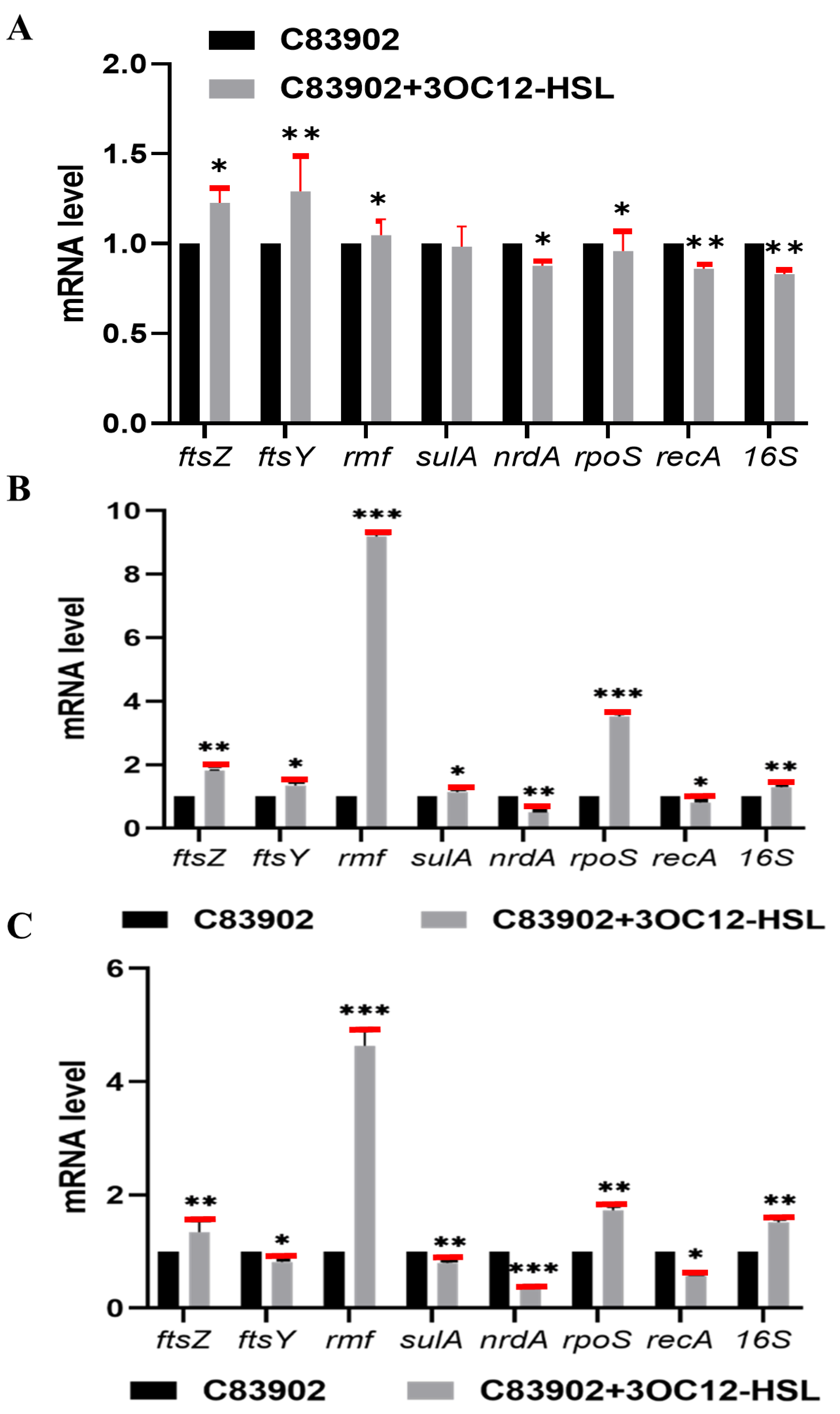
To evaluate the transcriptional effects of exogenous 3OC12-HSL on ETEC strain C83902, we quantified mRNA levels via real-time RT-PCR in LB medium containing or lacking 200 μmol/L 3OC12-HSL under three distinct conditions: aerobic agitation, aerobic static, and anaerobic static. In aerobic agitated cultures (Figure 2A), 3OC12-HSL exposure significantly upregulated ftsZ (23%) and ftsY (29%) expression, while downregulating nrdA (14%), recA (13%), and 16S (18%). Under aerobic static incubation (Figure 2B), treatment induced significant upregulation of ftsZ (82%), rmf (8-fold), and rpoS (~2.5-fold), with nrdA expression declining nearly 1-fold. For anaerobic static settings (Figure 2C), 3OC12-HSL elevated ftsZ expression (34%), rmf (~four-fold), rpoS (~one-fold), and 16S (~one-fold), whereas nrdA and recA expression decreased ~one-fold, with all changes statistically significant. Collectively, these results establish that 3OC12-HSL robustly regulates the transcription of essential genes, with effects contingent on oxygen and agitation dynamics.

3.3. 3OC12-HSL Specifically Enhances E. coli in a Simulated Gut Microbiota Model

To determine the selective impact of 3OC12-HSL on E. coli growth within a simulated gut microbiota, we constructed an anaerobic polymicrobial community featuring Bacteroides fragilis (~109 CFU/mL), Bifidobacterium (~109 CFU/mL), Enterococcus faecium (~106 CFU/mL), and E. coli C83902 (~106 CFU/mL). Following incubation in DMEM containing 200 μmol/L 3OC12-HSL or control medium under anaerobic conditions, genomic DNA was harvested every 6 h and quantified via E. coli-specific real-time qPCR. The data revealed statistically significant elevations in E. coli 16S rDNA copy numbers for the treated group relative to the control: 20.5% at 6 h, 37.6% at 12 h, 27.5% at 18 h, and 33.9% at 24 h (all p < 0.05) (Table 2). These findings confirm that 3OC12-HSL specifically augments E. coli colonization in this simulated gut microcosm model.

3.4. AiiA Enzyme Reverses 3OC12-HSL-Induced Gut Microbiota Dysbiosis in Mice

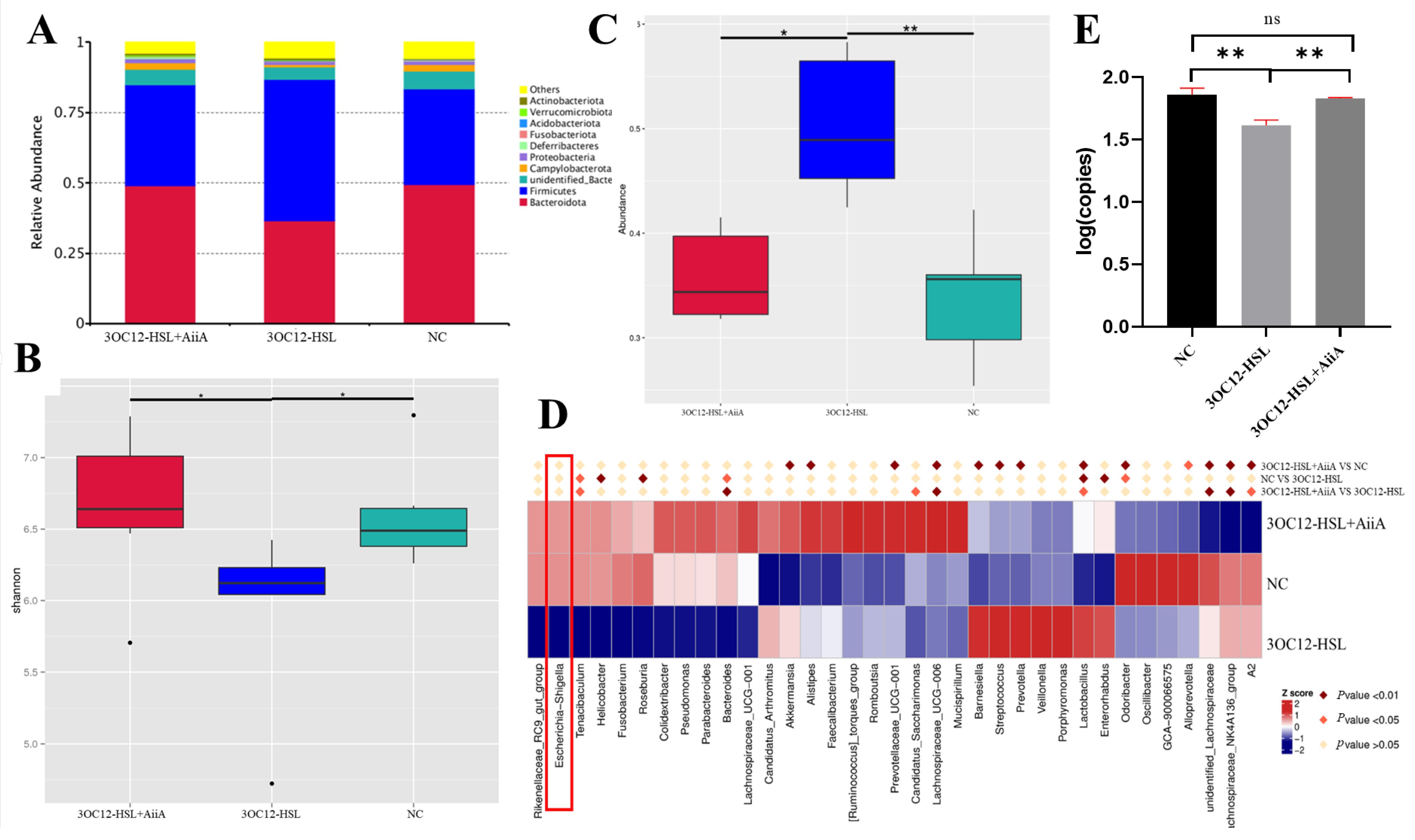
To investigate AiiA-mediated regulation of 3OC12-HSL-induced gut dysbiosis, 5-week-old ICR mice were randomized into three cohorts: negative control (NC, sterile water), 3OC12-HSL-exposed (200 μmol/L in water), and 3OC12-HSL+AiiA-supplemented (3OC12-HSL water + 0.5% AiiA-feed). Analysis of day 3 fecal samples via Illumina MiSeq sequencing revealed no significant differences in phylum-level composition between the NC and 3OC12-HSL+AiiA groups (Figure 3A). However, relative to the 3OC12-HSL group, the 3OC12-HSL+AiiA cohort showed significantly reduced Firmicutes (p < 0.01) and elevated Bacteroidetes abundance (Figure 3A,C), demonstrating effective AHL signal quenching.
The 3OC12-HSL+AiiA group exhibited significantly higher Shannon α-diversity than the 3OC12-HSL group (* p < 0.05), recovering to NC-equivalent levels (Figure 3B). Heatmap analysis identified a marked enhancement of Escherichia-Shigella solely in the 3OC12-HSL group, contrasting with consistently low abundance in both 3OC12-HSL+AiiA and NC cohorts (Figure 3D). Species-resolved quantification confirmed that AiiA restored Escherichia coli abundance to 0.28%, equivalent to NC (0.28%) and significantly exceeding the 3OC12-HSL group (0.02%, p < 0.01) (Table 3). Critical validation by qPCR demonstrated a 54.1% increase in viable E. coli in the 3OC12-HSL+AiiA group relative to 3OC12-HSL (p < 0.01), achieving parity with NC (Figure 3E). These multimodal data establish AiiA’s efficacy in rescuing 3OC12-HSL-driven dysbiosis through AHL-inactivation, restoring microbial diversity, and reversing pathogen suppression.

3.5. AiiA Modulates 3OC12-HSL-Induced Serum Cytokine Profiles in Mice

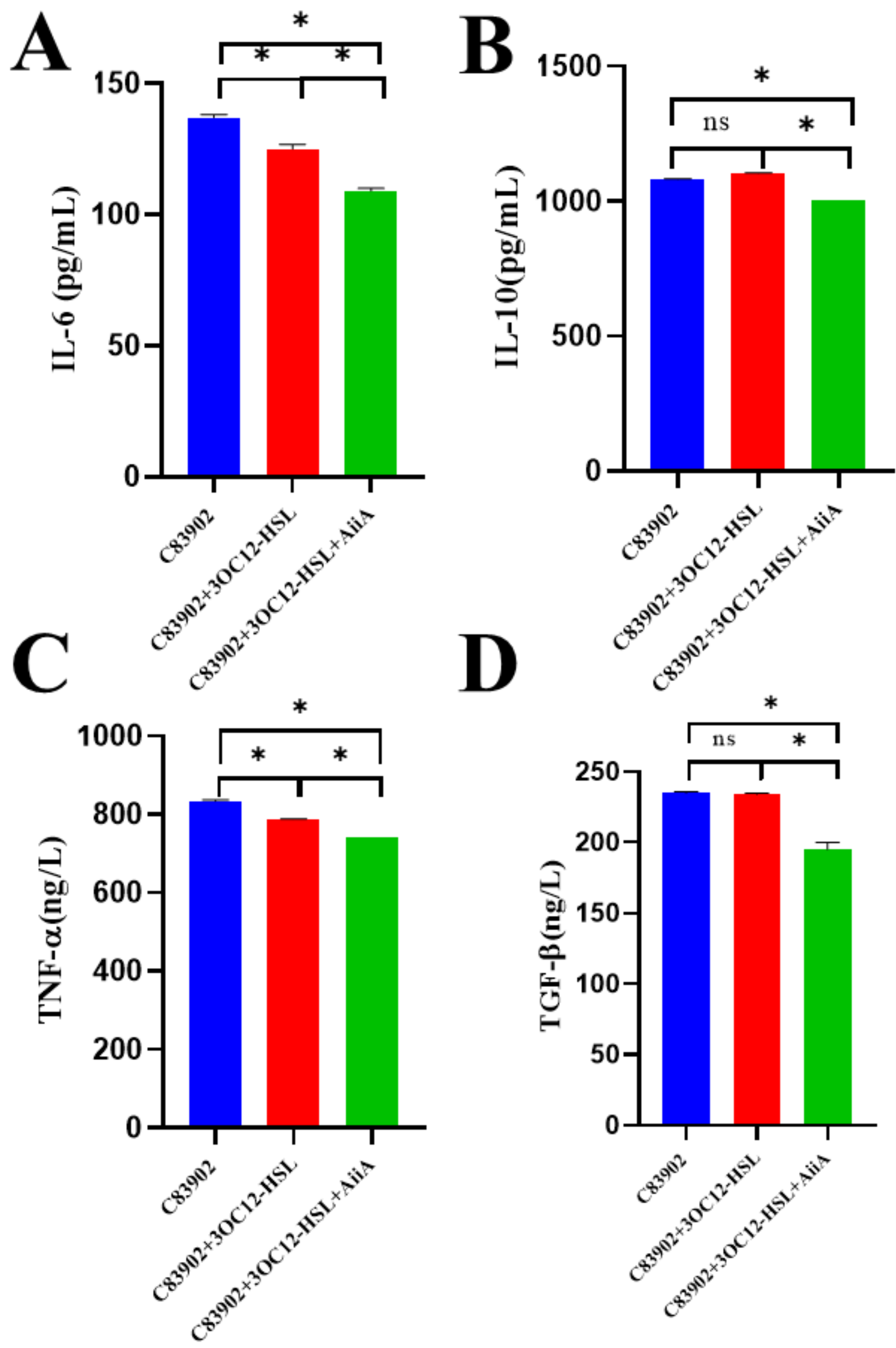
To investigate the impact of AiiA on 3OC12-HSL-mediated cytokine alterations in murine serum, we conducted enzyme-linked immunosorbent assay (ELISA) analyses across three experimental cohorts: C83902 (ETEC-challenged), C83902 + 3OC12-HSL, and C83902 + 3OC12-HSL + AiiA. As evidenced by Figure 4A, IL-6 concentrations were significantly diminished in the AiiA-supplemented group compared to the 3OC12-HSL-exposed group, revealing that AiiA mitigates pro-inflammatory augmentation induced by 3OC12-HSL. For IL-10 (Figure 4B), ELISA data demonstrated a statistically significant decrease (p < 0.01) with AiiA treatment relative to 3OC12-HSL alone, indicating suppression of the cytokine’s upregulation and associated inflammatory responses. TNF-α quantification (Figure 4C) exhibited a marked reduction (p < 0.01) in the AiiA-co-administered cohort versus the 3OC12-HSL cohort, affirming that AiiA attenuates inflammatory amplification. Regarding TGF-β (Figure 4D), serum levels were significantly lower with AiiA supplementation compared to 3OC12-HSL exposure; however, no significant difference emerged between the 3OC12-HSL and C83902 groups, suggesting that AiiA selectively reverses the increase in TGF-β elicited by 3OC12-HSL, implicating a regulatory role in immune dynamics.

3.6. Synergistic Enhancement of Intestinal Recovery by AiiA and 3OC12-HSL in ETEC-Infected Mice

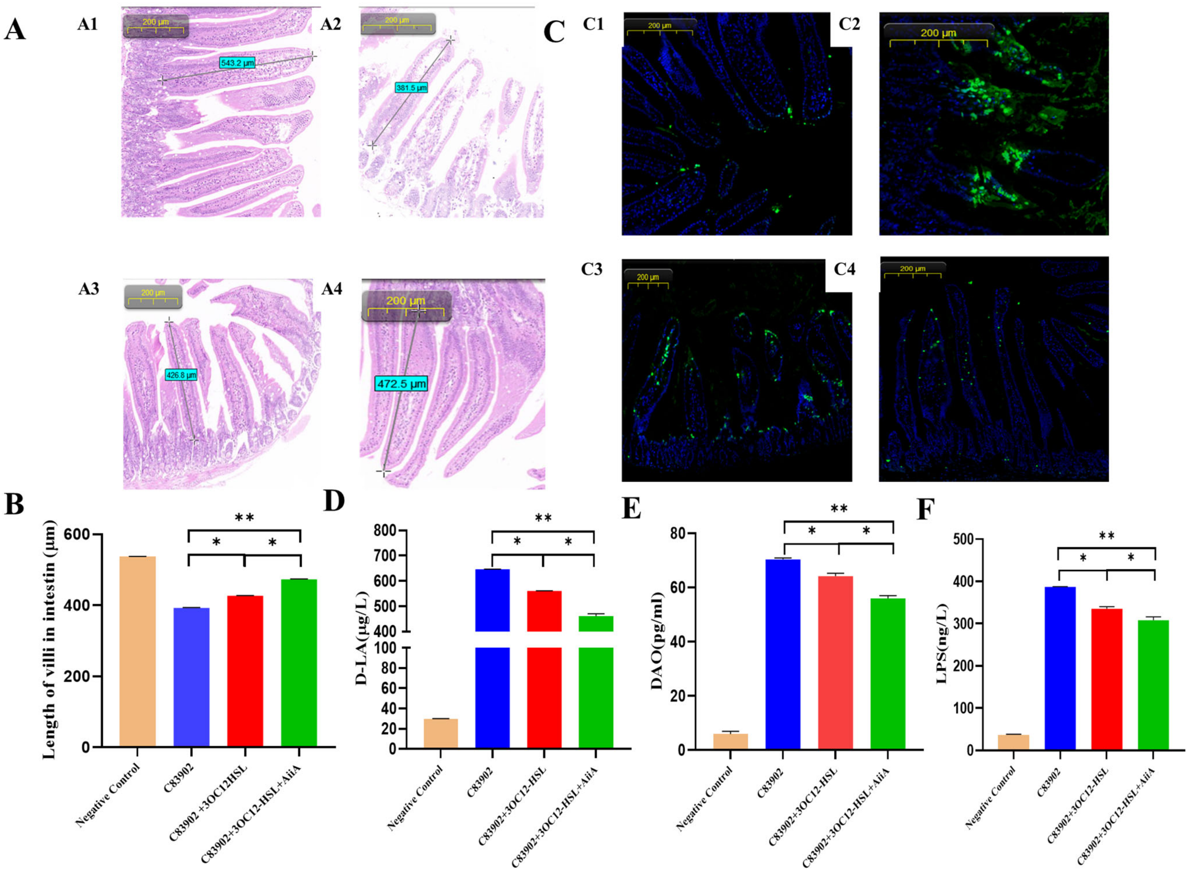
To investigate the impact of therapeutic interventions on jejunal integrity in ETEC-infected mice, we employed a stratified group design: negative control, ETEC C83902 challenge, C83902+3OC12-HSL supplementation, and C83902+3OC12-HSL+AiiA co-administration. Morphometric analysis using HE staining (200 μm scale) revealed severe villus atrophy in the C83902 group (381.5 μm), contrasting with intact negative control villi (543.2 μm). 3OC12-HSL partially ameliorated shortening to 426.8 μm (8.6% increase over C83902, p < 0.05), while AiiA co-delivery achieved maximal recovery to 472.5 μm (20.5% and 10.9% gains relative to C83902 and C83902+3OC12-HSL, respectively; p < 0.01), demonstrating synergistic mucosal restoration. Subsequently, qualitative assessment of TUNEL assays indicated an apparent reduction in apoptotic signals within the C83902 group relative to controls. This trend was attenuated by AiiA treatment, as evidenced by diminished TUNEL staining intensity in representative images (Figure 5C). For barrier compromise, ELISA quantification showed significant elevation of serum D-lactate, diamine oxidase (DAO), and lipopolysaccharide (LPS) levels in ETEC-infected animals, with C83902+3OC12-HSL exhibiting minor reductions. Crucially, AiiA administration significantly attenuated all three biomarkers compared to both C83902 and C83902+3OC12-HSL groups (p < 0.05 for D-LA/DAO, p < 0.01 for LPS), underscoring its synergistic role with 3OC12-HSL in preserving intestinal barrier function.

4. Discussion

This study demonstrates that AiiA lactonase effectively counteracts the exacerbation of ETEC virulence mediated by the QS signal 3OC12-HSL. AiiA restored baseline bacterial proliferation through enzymatic degradation of the signal molecule, rescued microbiome diversity, suppressed pro-inflammatory cytokine responses, and synergistically restored intestinal barrier integrity. A key observation was the context-dependent impact of 3OC12-HSL on ETEC growth: significant stimulation occurred under static in vitro conditions (66.7% CFU increase at 8 h) and within a synthetic gut microbiota model (37.6% increase in E. coli 16S rDNA at 12 h), yet suppression occurred in the murine gut environment (13-fold decrease in abundance vs control). This apparent paradox highlights the complexity of AHL signaling within the gastrointestinal niche, where microbial competition, host responses, and environmental constraints likely dictate the functional outcome.
The robust upregulation of the stress-response genes rpoS [33,34,35] (~2.5-fold) and rmf [36,37,38] (8-fold) in ETEC exposed to 3OC12-HSL specifically under aerobic static and anaerobic conditions (Figure 2B,C), provides a potential mechanistic link. Static and anoxic environments likely generate nutrient scarcity and metabolic stress, approximating challenges within the fluctuating intestinal lumen. Under such conditions, 3OC12-HSL may prime ETEC for persistence by activating the RpoS-mediated general stress response and Rmf-induced ribosome hibernation [39], potentially conferring a fitness advantage. This aligns with the observed growth stimulation in controlled, nutrient-poor static in vitro systems and underscores how AHL eavesdropping might optimize bacterial survival strategies under adversity.
Importantly, the divergent ETEC growth phenotypes (stimulation vs. suppression) consistently demonstrate 3OC12-HSL’s potent role as a disruptor of ETEC population dynamics. Regardless of direction, this modulation significantly perturbs the intestinal microbial barrier [40]. The in vivo suppression suggests that within the complex murine gut ecosystem, 3OC12-HSL might also stimulate competing AHL-responsive bacteria, shifting community balance against ETEC, or potentially trigger host factors that indirectly restrict ETEC proliferation. This discrepancy reveals a fundamental limitation: the inherent difficulty in modeling the intricate multi-species QS interactions governing the gut environment. The precise mechanisms underlying the observed growth reversal in vivo require further elucidation.
ETEC infection induced severe barrier dysfunction and inflammation, characterized by jejunal villus atrophy (381.5 μm vs. control 543.2 μm), elevated serum barrier damage markers (LPS, D-lactate, DAO; p < 0.01), and increased pro-inflammatory cytokines (IL-6, TNF-α). Intriguingly, mice receiving both ETEC and 3OC12-HSL (ETEC+3OC12-HSL) exhibited moderately reduced barrier damage and inflammation compared to ETEC alone, despite the concurrent AHL-mediated suppression of E. coli abundance. This observation might reflect a dual effect: 3OC12-HSL suppressed ETEC proliferation while simultaneously initiating host or microbial compensatory responses that partially mitigated damage. Interestingly, IL-10 reduction by AiiA warrants nuanced interpretation. While IL-10 is canonically anti-inflammatory and promotes mucosal repair, pathogens can exploit it to evade immune clearance. Here, IL-10 elevation in the ETEC+3OC12-HSL group may reflect a host attempt to mitigate inflammation, yet concurrently facilitate ETEC persistence by suppressing antibacterial responses. AiiA’s ability to recalibrate IL-10 toward homeostasis likely contributes to its therapeutic synergy. The critical therapeutic outcome emerged upon addition of AiiA (ETEC+3OC12-HSL+AiiA). AiiA significantly outperformed both the ETEC and ETEC+3OC12-HSL groups, restoring villus height to 472.5 μm (p < 0.01 vs. ETEC), suppressing cytokines like IL-6 and TNF-α, and reducing barrier biomarkers. This robust recovery likely stems from AiiA’s dual action: (1) eliminating direct 3OC12-HSL-induced epithelial toxicity, and (2) abrogating the QS signal’s disruptive effects on the complex microbial community, including its apparent suppression of ETEC abundance in vivo.
AiiA’s efficacy validates the therapeutic potential of targeting AHL signaling. Its capacity to restore microbial diversity (Shannon index recovered to control levels, Figure 3B), correct dysbiosis (Firmicutes/Bacteroidetes ratio normalized, Figure 3C), and rescue structural integrity demonstrates the centrality of QS eavesdropping in ETEC pathogenesis. The suppression of critical inflammatory mediators (IL-6, TNF-α) further confirms that quenching 3OC12-HSL effectively dampens immune hyperactivation driven by ETEC and the AHL signal.
Several limitations warrant consideration. First, the molecular pathways linking 3OC12-HSL perception (potentially via SdiA) to the activation of rpoS and rmf under specific environmental conditions and their contribution to virulence modulation remain incompletely resolved. Second, our models could not fully recapitulate the complex network of microbial interactions responsible for the opposing in vitro versus in vivo growth responses to AHL. Third, while highly effective, AiiA treatment might leave residual low-level AHL activity due to incomplete degradation. The high cost of purified AiiA enzyme limited our ability to investigate its dose-response relationship in vivo. Future studies exploring varying concentrations are essential to determine the threshold of AHL degradation required for complete blockade of its effects on stress gene induction and barrier restoration. While the 3OC12-HSL working solutions were prepared by high dilution of DMSO stocks to ensure a final DMSO concentration below 0.5% (v/v). A threshold aligned with established biological safety standards where concentrations of 0.1–1% are documented to avoid cytotoxicity and functional interference. The omission of dedicated DMSO control groups marginally compromises methodological rigor. We acknowledge this oversight, though residual DMSO at ≤1% is unlikely to substantially impact the reported data. Future studies will incorporate solvent-only controls to eliminate potential confounders. Additionally, the functional significance of IL-10 dynamics in AiiA-mediated protection requires deeper mechanistic dissection. Future work will delineate whether IL-10 reduction directly disrupts pathogen immune evasion or indirectly enhances barrier repair via crosstalk. Finally, although levels as high as 300–600 μM of AHL have been reported in biofilms [41], our use of 200 μM of AHL may exceed physiologically relevant concentrations. Future studies employing lower concentrations (e.g., ≤10 μM) could better elucidate AHL/AiiA interaction.
Therefore, our future research would focus on the following: (1) Employing targeted mutagenesis (e.g., sdiA knockout ETEC) to definitively link AHL receptors to the observed stress gene activation under specific conditions and its virulence consequences. (2) Developing advanced in vitro consortia models incorporating diverse AHL-producing and AHL-responding species to dissect the ecological interplay leading to ETEC suppression in vivo. (3) Conducting detailed dose-response studies with AiiA to establish optimal therapeutic thresholds for complete AHL inactivation.
In summary, this work establishes that 3OC12-HSL significantly modulates ETEC pathogenicity and disrupts intestinal barrier function through mechanisms involving context-dependent growth effects, dysregulation of stress adaptation pathways (rpoS, rmf), and induction of microbial dysbiosis. Critically, AiiA lactonase effectively mitigates these detrimental effects via targeted degradation of 3OC12-HSL, restoring microbial balance, attenuating inflammation, and synergistically repairing intestinal damage. These findings strongly support AiiA-mediated quorum quenching as a promising anti-virulence strategy against ETEC infections.

5. Conclusions

This study establishes AiiA lactonase as a potent quorum-quenching agent that mitigates ETEC virulence through degradation of 3OC12-HSL, restoring microbial homeostasis and intestinal barrier function. The results underscore AiiA’s therapeutic promise. Future research will prioritize elucidating molecular mechanisms via targeted SdiA mutagenesis, advancing in vitro polymicrobial models to decode ecological dynamics, and defining optimal dosing regimens for clinical application. Such endeavors will accelerate the development of precision anti-infective therapies against ETEC.

Author Contributions

Conceptualization, Y.Y. and G.Z.; methodology, Y.Y., J.S. and Z.H.; validation, J.S., J.L. and Q.F.; formal analysis, Z.H.; investigation, Y.Y.; data curation, J.S. and Z.H.; writing—original draft preparation, J.S.; writing—review and editing, Y.Y.; supervision, Y.Y. and G.Z.; funding acquisition, Y.Y. and G.Z. All authors have read and agreed to the published version of the manuscript.

Funding

This research was funded by the Chinese National Science Foundation Grant (No. 31972708), the Project of Yangzhou Municipal Program for International Science and Technology Cooperation (YZ2023261), Yangzhou Municipal Modern Agriculture Project (YZ2023040), and the Priority Academic Program Development of Jiangsu Higher Education Institutions (PAPD).

Institutional Review Board Statement

The animal study protocol was approved by the Animal Welfare and Ethics Committee of Yangzhou University (Approval Number: 202202106; Approval Date: 20220223), and was conducted in compliance with the ethical standards of animal welfare and the Jiangsu Provincial Animal Management Committee. Five-week-old male ICR mice (purchased from Yangzhou University Comparative Medicine Center, Experimental Animal Production License Number: SCXK (Jiangsu) 2017-0007) were housed in the Yangzhou University Experimental Animal Center (Experimental Animal Use License Number: SYXK (Jiangsu) 2121-0026).

Informed Consent Statement

Not applicable.

Data Availability Statement

No datasets were generated or analysed during the current study.

Acknowledgments

We give special thanks to Philip Hardwidge for generous support and help.

Conflicts of Interest

The authors declare that they have no conflicts of interest.

References

  1. Khalil, I.A.; Troeger, C.; Blacker, B.F.; Rao, P.C.; Brown, A.; Atherly, D.E.; Brewer, T.G.; Engmann, C.M.; Houpt, E.R.; Kang, G.; et al. Morbidity and mortality due to shigella and enterotoxigenic escherichia coli diarrhoea: The global burden of disease study 1990–2016. Lancet Infect. Dis. 2018, 18, 1229. [Google Scholar] [CrossRef]
  2. Khalil, I.; Anderson, J.D.; Bagamian, K.H.; Baqar, S.; Giersing, B.; Hausdorff, W.P.; Marshall, C.; Porter, C.K.; Walker, R.I.; Bourgeois, A.L. Vaccine value profile for enterotoxigenic escherichia coli (ETEC). Vaccine 2023, 41, S95. [Google Scholar] [CrossRef]
  3. Lee, C.Y.; Kim, S.J.; Park, B.C.; Han, J.H. Effects of dietary supplementation of bacteriophages against enterotoxigenic escherichia coli (ETEC) k88 on clinical symptoms of post-weaning pigs challenged with the ETEC pathogen. J. Anim. Physiol. Anim. Nutr. 2017, 101, 88. [Google Scholar] [CrossRef]
  4. Kondakindi, V.R.; Nalam, R.S.S.; Pabbati, R.; Maddela, N.R. Quorum quenching a sustainable biofilm mitigation strategy. Res. J. Biotechnol. 2022, 17, 200. [Google Scholar] [CrossRef]
  5. Sikdar, R.; Elias, M. Quorum quenching enzymes and their effects on virulence, biofilm, and microbiomes: A review of recent advances. Expert Rev. Anti-Infect. Ther. 2020, 18, 1221. [Google Scholar] [CrossRef] [PubMed]
  6. Hemmati, F.; Salehi, R.; Ghotaslou, R.; Samadi, K.H.; Hasani, A.; Gholizadeh, P.; Nouri, R.; Ahangarzadeh, R.M. Quorum quenching: A potential target for antipseudomonal therapy. Infect. Drug Resist. 2020, 13, 2989. [Google Scholar] [CrossRef] [PubMed]
  7. Guo, J.; Yoshida, K.; Ikegame, M.; Okamura, H. Quorum sensing molecule n-(3-oxododecanoyl)-l-homoserine lactone: An all-rounder in mammalian cell modification. J. Oral Biosci. 2020, 62, 16. [Google Scholar] [CrossRef]
  8. Haque, S.; Yadav, D.K.; Bisht, S.C.; Yadav, N.; Singh, V.; Dubey, K.K.; Jawed, A.; Wahid, M.; Dar, S.A. Quorum sensing pathways in gram-positive and -negative bacteria: Potential of their interruption in abating drug resistance. J. Chemother. 2019, 31, 161. [Google Scholar] [CrossRef]
  9. Smith, J.L.; Fratamico, P.M.; Yan, X. Eavesdropping by bacteria: The role of SdiA in escherichia coli and salmonella enterica serovar typhimurium quorum sensing. Foodborne Pathog. Dis. 2011, 8, 169. [Google Scholar] [CrossRef]
  10. Schwieters, A.; Ahmer, B. Role of the LuxR solo, SdiA, in eavesdropping on foreign bacteria. FEMS Microbiol. Rev. 2025, 49, fuaf015. [Google Scholar] [CrossRef]
  11. Peterson, L.W.; Artis, D. Intestinal epithelial cells: Regulators of barrier function and immune homeostasis. Nat. Rev. Immunol. 2014, 14, 141. [Google Scholar] [CrossRef] [PubMed]
  12. Turner, J.R. Intestinal mucosal barrier function in health and disease. Nat. Rev. Immunol. 2009, 9, 799. [Google Scholar] [CrossRef] [PubMed]
  13. Song, C.; Chai, Z.; Chen, S.; Zhang, H.; Zhang, X.; Zhou, Y. Intestinal mucus components and secretion mechanisms: What we do and do not know. Exp. Mol. Med. 2023, 55, 681. [Google Scholar] [CrossRef] [PubMed]
  14. Yang, Y.; Zhang, X.; Yang, J.; Wu, Z.; Li, J.; Song, R.; Meng, C.; Zhu, G. N-(3-oxododecanoyl)-homoserine lactone induces intestinal barrier damage in piglets via the lipid raft-mediated apoptosis pathway. Vet. Sci. 2025, 12, 233. [Google Scholar] [CrossRef]
  15. Ma, X.; Zhang, S.; Xu, Z.; Li, H.; Xiao, Q.; Qiu, F.; Zhang, W.; Long, Y.; Zheng, D.; Huang, B.; et al. SdiA improves the acid tolerance of e. Coli by regulating GadW and GadY expression. Front. Microbiol. 2020, 11, 1078. [Google Scholar] [CrossRef]
  16. Nguyen, Y.; Nguyen, N.X.; Rogers, J.L.; Liao, J.; MacMillan, J.B.; Jiang, Y.; Sperandio, V. Structural and mechanistic roles of novel chemical ligands on the SdiA quorum-sensing transcription regulator. mBio 2015, 6. [Google Scholar] [CrossRef]
  17. Cao, Y.; Ren, J.; Zhang, Y.; Xie, Y.; Xiao, X.; Zhang, Z.; Lou, W.; Liu, F. Transcriptomics analysis of the role of SdiA in desiccation tolerance of cronobacter sakazakii in powdered infant formula. Int. J. Food Microbiol. 2025, 426, 110916. [Google Scholar] [CrossRef]
  18. Bin, P.; Tang, Z.; Liu, S.; Chen, S.; Xia, Y.; Liu, J.; Wu, H.; Zhu, G. Intestinal microbiota mediates enterotoxigenic escherichia coli-induced diarrhea in piglets. BMC Vet. Res. 2018, 14, 385. [Google Scholar] [CrossRef]
  19. Ren, W.; Yin, J.; Xiao, H.; Chen, S.; Liu, G.; Tan, B.; Li, N.; Peng, Y.; Li, T.; Zeng, B.; et al. Intestinal microbiota-derived GABA mediates interleukin-17 expression during enterotoxigenic escherichia coli infection. Front. Immunol. 2017, 7, 685. [Google Scholar] [CrossRef]
  20. Sturbelle, R.T.; de Avila, L.F.D.C.; Roos, T.B.; Borchardt, J.L.; Da Conceicao, R.D.C.D.; Dellagostin, O.A.; Leite, F.P.L. The role of quorum sensing in escherichia coli (ETEC) virulence factors. Vet. Microbiol. 2015, 180, 245. [Google Scholar] [CrossRef]
  21. Shi, Y.; Liu, J.; Zhou, H.; Wu, Z.; Qiu, Y.; Ye, C. Dihydromyricetin alleviates ETEC k88-induced intestinal inflammatory injury by inhibiting quorum sensing-related virulence factors. BMC Microbiol. 2025, 25, 201. [Google Scholar] [CrossRef]
  22. Ponce, A.D.R.; Martins, M.L.; de Araujo, E.F.; Mantovani, H.C.; Vanetti, M.C.D. AiiA quorum-sensing quenching controls proteolytic activity and biofilm formation by enterobacter cloacae. Curr. Microbiol. 2012, 65, 758. [Google Scholar]
  23. Wang, P.; Wang, Q.; Zhao, Y.; Ou, J. Targeting n-acyl-homoserine lactones (AHLs) on the quorum quenching AiiA protein to eradicate biofilm formation in vibrio parahaemolyticus. CyTA-J. Food 2024, 22, 2383763. [Google Scholar] [CrossRef]
  24. Yang, Y.; Shao, J.; Zhou, M.; Duan, Q.; Zhang, X.; Zhu, G. Effects of quorum sensing AHL signaling on the biological characteristics of porcine derived f4ac+ enterotoxigenic escherichia coli. Pak. J. Zool. 2022, 54, 701. [Google Scholar] [CrossRef]
  25. Duan, Q.; Zhou, M.; Zhu, X.; Yang, Y.; Zhu, J.; Bao, W.; Wu, S.; Ruan, X.; Zhang, W.; Zhu, G. Flagella from f18+escherichia coli play a role in adhesion to pig epithelial cell lines. Microb. Pathog. 2013, 55, 32. [Google Scholar] [CrossRef]
  26. Zhou, M.; Duan, Q.; Li, Y.; Yang, Y.; Hardwidge, P.R.; Zhu, G. Membrane cholesterol plays an important role in enteropathogen adhesion and the activation of innate immunity via flagellin-TLR5 signaling. Arch. Microbiol. 2015, 197, 797–803. [Google Scholar] [CrossRef]
  27. Meng, X.; He, M.; Chen, B.; Xia, P.; Wang, J.; Zhu, C.; Wang, H.; Zhu, G. RyhB paralogs downregulate the expressions of multiple survival-associated genes and attenuate the survival of salmonella enteritidis in the chicken macrophage HD11. Microorganisms 2023, 11, 214. [Google Scholar] [CrossRef]
  28. Hassall, J.; Cheng, J.K.J.; Unnikrishnan, M. Erratum for Hassall et al., “Dissecting individual interactions between pathogenic and commensal bacteria within a multispecies gut microbial community”. mSphere 2024, 9, e0089324. [Google Scholar] [CrossRef]
  29. Videnska, P.; Smerkova, K.; Zwinsova, B.; Popovici, V.; Micenkova, L.; Sedlar, K.; Budinska, E. Stool sampling and DNA isolation kits affect DNA quality and bacterial composition following 16s rRNA gene sequencing using MiSeq illumina platform. Sci. Rep. 2019, 9, 13814. [Google Scholar] [CrossRef]
  30. Rieger, J.; Pelckmann, L.; Drewes, B. Preservation and Processing of Intestinal Tissue for the Assessment of Histopathology; Nagamoto-Combs, K., Ed.; Springer: New York, NY, USA, 2021; p. 267. [Google Scholar]
  31. Patankar, J.V.; Becker, C. Cell death in the gut epithelium and implications for chronic inflammation. Nat. Rev. Gastroenterol. Hepatol. 2020, 17, 543. [Google Scholar] [CrossRef]
  32. Vasilev, G.; Manolova, I.; Ivanova, M.; Stanilov, I.; Miteva, L.; Stanilova, S. The role of IL-18 in addition to th17 cytokines in rheumatoid arthritis development and treatment in women. Sci. Rep. 2021, 11, 15391. [Google Scholar] [CrossRef]
  33. Bouillet, S.; Hamdallah, I.; Majdalani, N.; Tripathi, A.; Gottesman, S. A negative feedback loop is critical for recovery of RpoS after stress in escherichia coli. PLoS Genet. 2024, 20, e1011059. [Google Scholar] [CrossRef]
  34. Schellhorn, H.E. Function, evolution, and composition of the RpoS regulon in escherichia coli. Front. Microbiol. 2020, 11, 560099. [Google Scholar] [CrossRef] [PubMed]
  35. Zhang, D.; Li, S.H.; King, C.G.; Wingreen, N.S.; Gitai, Z.; Li, Z. Global and gene-specific translational regulation in escherichia coli across different conditions. PLoS Comput. Biol. 2022, 18, e1010641. [Google Scholar] [CrossRef] [PubMed]
  36. Izutsu, K.; Wada, A.; Wada, C. Expression of ribosome modulation factor (RMF) in escherichia coli requires ppgpp. Genes Cells 2001, 6, 665. [Google Scholar] [CrossRef] [PubMed]
  37. Usachev, K.S.; Yusupov, M.M.; Validov, S.Z. Hibernation as a stage of ribosome functioning. Biochem.-Mosc. 2020, 85, 1434. [Google Scholar] [CrossRef]
  38. Polikanov, Y.S.; Blaha, G.M.; Steitz, T.A. How hibernation factors RMF, HPF, and YfiA turn off protein synthesis. Science 2012, 336, 915. [Google Scholar] [CrossRef]
  39. Khaova, E.A.; Kashevarova, N.M.; Tkachenko, A.G. Ribosome hibernation: Molecular strategy of bacterial survival (review). Appl. Biochem. Microbiol. 2022, 58, 213. [Google Scholar] [CrossRef]
  40. Alhasan, M.M.; Holsken, O.; Duerr, C.; Helfrich, S.; Branzk, N.; Philipp, A.; Leitz, D.; Duerr, J.; Almousa, Y.; Barrientos, G.; et al. Antibiotic use during pregnancy is linked to offspring gut microbial dysbiosis, barrier disruption, and altered immunity along the gut-lung axis. Eur. J. Immunol. 2023, 53, e2350394. [Google Scholar] [CrossRef]
  41. Charlton, T.S.; De Nys, R.; Netting, A.; Kumar, N.; Hentzer, M.; Givskov, M.; Kjelleberg, S. A novel and sensitive method for the quantification of n-3-oxoacyl homoserine lactones using gas chromatography-mass spectrometry: Application to a model bacterial biofilm. Environ. Microbiol. 2000, 2, 530. [Google Scholar] [CrossRef]
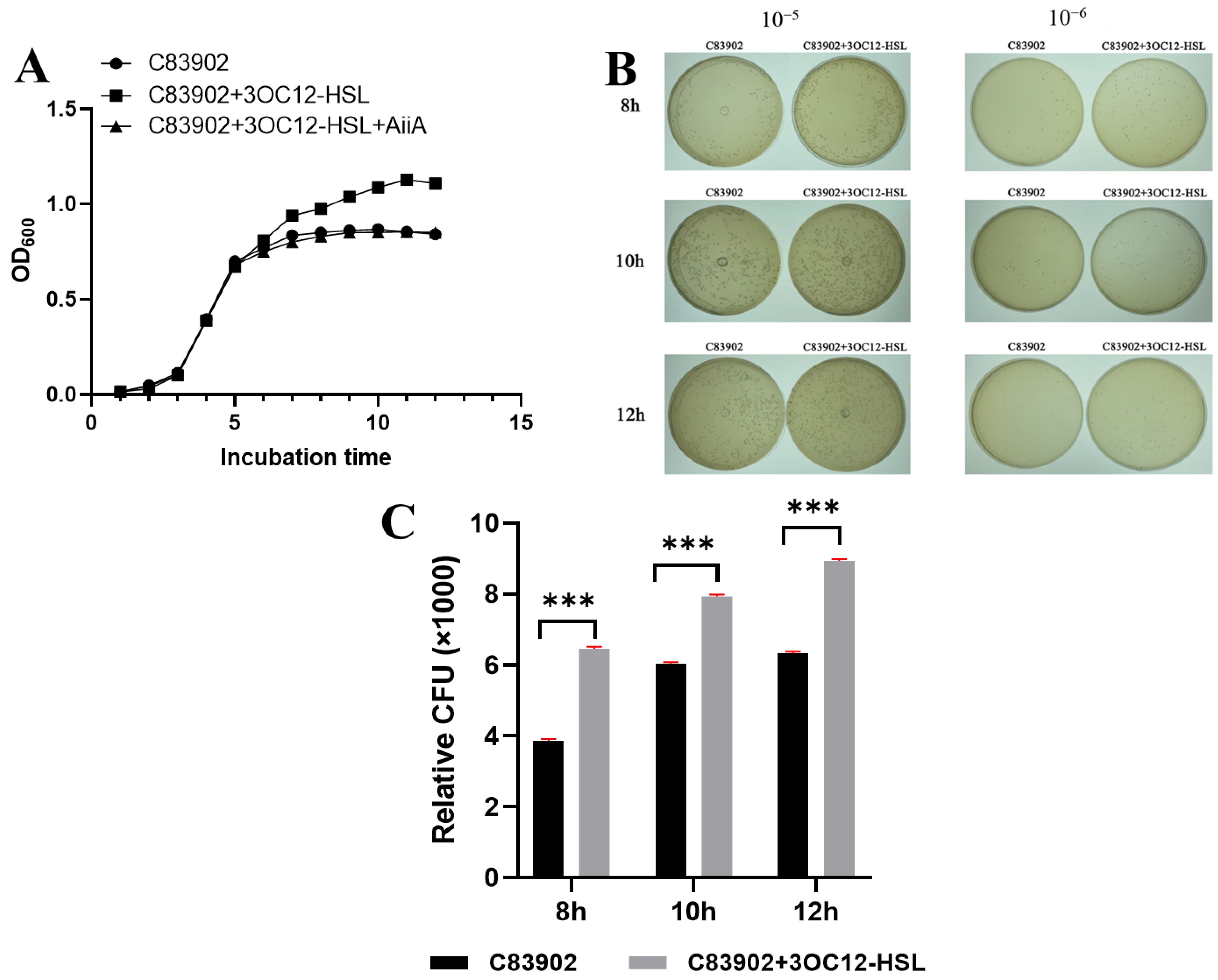
Figure 1. Enzymatic inactivation of 3OC12-HSL by AiiA protein abrogates growth enhancement in ETEC C83902. (A) Growth curves of ETEC strain C83902 cultivated statically at 37 °C in unsupplemented LB (control, squares); LB containing 200 μmol/L 3OC12-HSL (circles); LB with 200 μmol/L 3OC12-HSL plus AiiA protein (triangles). Cultures were initiated by 1:100 dilution of overnight pre-cultures with hourly OD600 monitoring (mean ± SD; n = 3 biological replicates). (B) Bacterial colonies observed on LB agar plates after 10-fold serial dilution (10−5 and 10−6) and 24-h incubation at 37 °C following sampling at specified time points. (C) Quantified colony-forming units (CFUs) per mL at 10−6 dilution for control and 3OC12-HSL-treated cultures. Statistical significance relative to control was determined using a two-way ANOVA (*** p < 0.001).
Microbiolres 16 00166 g001
Figure 2. Transcriptional Responses of Critical Genes to 3OC12-HSL Treatment Under Variable Culture Regimes. (A) Standard LB culture: quantitative RT-PCR analysis of mRNA expression for target genes (ftsZ, ftsY, rmf, sulA, nrdA, rpoS, recA, 16S rRNA) in C83902 wild-type strain cultured in LB medium with or without 200 μmol/L 3OC12-HSL. (B) Aerobic static culture: real-time RT-PCR assessment of gene expression in C83902 grown under static aerobic conditions ± 200 μmol/L 3OC12-HSL. (C) Anaerobic static culture: mRNA quantification via real-time RT-PCR for genes in C83902 cultivated anaerobically to early-log phase (OD600 = 1.00) in LB medium containing 200 μmol/L 3OC12-HSL or vehicle control. Statistical annotation: * p < 0.05; ** p < 0.01; *** p < 0.001.
Microbiolres 16 00166 g002
Figure 3. Modulation of 3OC12-HSL-induced gut microbiota alterations by AiiA supplementation. (A) Phylum-level community profiling of fecal microbiota via Illumina MiSeq sequencing (n = 10 per cohort). Fecal samples were collected on day 3 from: negative control (NC) mice maintained on sterile water; 3OC12-HSL-exposed group receiving aqueous 200 μmol/L 3OC12-HSL; and 3OC12-HSL+AiiA group administered identical aqueous 3OC12-HSL plus 0.5% (w/w) AiiA-supplemented feed. (B) α-Diversity analysis using the Shannon index. Statistical significance (* p < 0.05) was assessed via the Kruskal–Wallis test followed by Dunn’s multiple comparisons. (C) Comparative abundance of Firmicutes (log10-transformed). p < 0.01 denotes significant divergence as determined by the method described in (B). (D) Genus-resolved heatmap visualization with relative abundances standardized by Z-score. Color gradient from blue (low) to red (high) indicates taxon-specific enrichment. (E) Escherichia coli quantification through real-time qPCR targeting the 16S rDNA locus. Values represent log10-transformed gene copies per gram fecal material. ** p < 0.01 indicates significant difference relative to the 3OC12-HSL group (one-way ANOVA with Tukey’s post hoc analysis).
Microbiolres 16 00166 g003
Figure 4. ELISA-based quantification of serum cytokine levels across experimental cohorts. (A) Serum IL-6 concentrations. Serum IL-6 levels measured by ELISA in C83902 (ETEC-challenged); C83902+3OC12-HSL (3OC12-HSL-exposed); C83902+3OC12-HSL+AiiA (AiiA-supplemented). (B) IL-10 level modulation. IL-10 concentrations quantified via ELISA across C83902, C83902+3OC12-HSL, and C83902+3OC12-HSL+AiiA. (C) TNF-α serum profiles. TNF-α concentrations determined by ELISA in C83902 (ETEC-challenged), C83902+3OC12-HSL (3OC12-HSL-exposed), and C83902+3OC12-HSL+AiiA (AiiA-supplemented). (D) TGF-β1 dynamics with AiiA intervention. TGF-β1 levels analyzed using ELISA across C83902, C83902+3OC12-HSL, and C83902+3OC12-HSL+AiiA. Statistical annotation: * p < 0.05; ns: no significant difference.
Microbiolres 16 00166 g004
Figure 5. Assessment of intestinal alterations induced by ETEC infection and therapeutic interventions. (A) Histomorphological evaluation of jejunal villi. Representative H&E-stained sections (scale bar: 200 µm) with corresponding villus length measurements. Panel designations: (A1): Negative control; (A2): C83902 group; (A3): C83902+3OC12-HSL group; (A4): C83902+3OC12-HSL+AiiA group. (B) Quantitative analysis of jejunal villus length across experimental cohorts. (C) Apoptosis detection by TUNEL assay in jejunal tissues. Green fluorescence labels fragmented DNA. Panel designations: (C1): Negative control; (C2): C83902+3OC12-HSL; (C3): C83902; (C4): C83902+3OC12-HSL+AiiA. (DF) Serum biomarkers of intestinal permeability quantified using ELISA kits: (D) D-lactate (D-LA), (E) diamine oxidase (DAO), (F) lipopolysaccharide (LPS). Data presented as mean ± SEM (* p < 0.05; ** p < 0.05).
Microbiolres 16 00166 g005
Table 1. Primers used in this study.
PrimersSequences (5′-3′)
ftsZ-RT-FCAGTCGTCGCTGAAGTGG
ftsZ-RT-RTTTGTCGTTCGGGATAGTG
ftsY-RT-FACGAAACTGGACGGCAC
ftsY-RT-RGACGCAAATCCTCAATACG
rmF-RT-FCTGGAACGGGCACATCAAC
rmF-RT-RTTACTACCCTGTCCGCCATG
sulA-RT-FCTTCGTCGTTCTCAT CCG
sulA-RT-RTGCTGACCGAGTTGCTGT
nrdA-RT-FGATGGACACCTTTATCGA
nrdA-RT-RGTACCAGATATTTGCCTTC
rpoS-RT-FACGATATGAAGCAGAGCATCGT
rpoS-RT-RAGGCCAATTTCACGACCTACAT
16S-RT-FCGTGCTACAATGGACAATACAAA
16S-RT-RATCTACGATTACTAGCGATTCCA
recA-RT-FTGGCGGGTAACCTGAAGCA
recA-RT-RCGAGACGAACAGAG GCGTAGAAT
16s-BA-FTCGCGTCCGGTGTGAAAG
16s-BA-RCCACATCCAGCATCCAC
16s-Efm-FCCCTTATTGTTAGTTGCCATCATT
16s-Efm-RACTCGTTGTACTTCCCATTGT
16s-BF-FGAAAGCATTAAGTATTCCACCTG
16s-BF-RCGGTGATTGGTCACTGACA
16s-E.coli-FGTTAATACCTTTGCTCATTGA
16s-E.coli-RACCAGGGTATCTAATCCTGTT
Table 2. The result of 16S rDNA qPCR in simulated gut microbiota model (log(copies/mL)).
Intestinal Bacteria6 h12 h18 h24 h
No AHLAHLNo AHLAHLNo AHLAHLNo AHLAHL
E. coli4.294.394.424.634.524.664.684.86
Enterococcus faecium3.743.413.773.453.923.583.933.78
Bacteroides fragilis7.247.547.397.697.587.737.667.81
Bifidobacterium5.946.286.096.356.146.456.306.59
Table 3. Proportion of top 10 bacteria at species level.
TaxonomyNC3OC12-HSL3OC12-HSL+AiiA
Lactobacillus_murinus4.13%21.38%12.53%
Bacteroides_gallinaceum1.16%2.65%5.26%
Lactobacillus_reuteri0.46%1.57%1.34%
Clostridium_sp_Culture-271.45%0.91%0.11%
Helicobacter_typhlonius0.82%0.33%0.46%
Bacteroides_acidifaciens0.21%0.38%0.72%
Helicobacter_hepaticus0.67%0.24%0.31%
Romboutsia_ilealis0.11%0.21%0.52%
Escherichia_coli0.28%0.02%0.28%
Akkermansia_muciniphila0.00%0.18%0.27%
Others90.72%70.32%78.21%
Disclaimer/Publisher’s Note: The statements, opinions and data contained in all publications are solely those of the individual author(s) and contributor(s) and not of MDPI and/or the editor(s). MDPI and/or the editor(s) disclaim responsibility for any injury to people or property resulting from any ideas, methods, instructions or products referred to in the content.