Article

Comprehensive Analysis of Etiological Agents and Drug Resistance Patterns in Ventilator-Associated Pneumonia

1
Department of Biotechnology, School of Bioengineering and Biosciences, Lovely Professional University, Phagwara 144411, India
2
Department of Microbiology & Molecular Biology, Max Super Speciality Hospital, Saket, New Delhi 110017, India
3
Establishment & Strengthening NCDC Branches, New Admn Building, National Centre for Disease Control Dte.GHS, Ministry of Health & Family Welfare, Government of India, Delhi 110054, India
4
Department of Internal Medicine, Max Super Speciality Hospital, New Delhi 110017, India
5
Indian Council of Medical Research Headquarters, New Delhi 110029, India
6
Devansh Lab Werks, Homewood, AL 35209, USA
7
Microgen Health Inc., Chantilly, VA 20151, USA
*
Authors to whom correspondence should be addressed.
Microbiol. Res. 2025, 16(7), 152; https://doi.org/10.3390/microbiolres16070152
Submission received: 19 May 2025 / Revised: 26 June 2025 / Accepted: 2 July 2025 / Published: 4 July 2025

Abstract

Ventilator-associated pneumonia (VAP) develops in patients who stay on mechanical ventilation for more than 48 h. In the presence of causative pathogens, the patient develops clinical signs such as purulent tracheal discharge, fever, and respiratory distress. A prospective observational study was carried out in the Intensive Care Unit (ICU) of Max Healthcare Centre, New Delhi, from 2020 to 2023. The study comprised 70 samples from patients diagnosed with VAP. This study thoroughly examined VAP-associated microorganisms and resistance in the hospital ICU. Klebsiella pneumoniae, Acinetobacter baumannii, and Pseudomonas aeruginosa were the most commonly reported pathogens. Significant drug resistance was seen in P. aeruginosa, K. pneumoniae, A. baumannii and Staphylococcus aureus. The heatmap also supported the antibiotic resistance data patterns obtained from conventional and automated systems of determination. Notably, Serratia marcescens, Escherichia coli, Klebsiella pneumoniae, Ralstonia insidiosa, and Ralstonia mannitolilytica, showed 60 to 100% of resistance to a number of antibiotics. Among all VAP patients, 31.42% early-onset and 68.57% late-onset VAP cases were detected. Out of 70 patients, 43 patients died (mortality rate 61.4%); majority of them suffered from late-onset VAP. The study goal was to describe the antibiotic resistance patterns and microbial ecology of the pathogens that were isolated from VAP patients. According to the heatmap analysis, a varied VAP microbiome with high prevalences of MDR in A. baumannii, P. aeruginosa, K. pneumoniae, and S. aureus was identified. To address the increasing prevalence of MDR VAP, the study highlights the critical need for improved VAP monitoring, strong infection control, and appropriate antibiotic usage.

1. Introduction

Ventilator-associated pneumonia (VAP) is defined as a disease where the patient is on mechanical ventilation for more than 48 h on the date of event, with day of ventilator placement being day 1, and the ventilator was in place on the date of event or the day before. If the ventilator was in place prior to inpatient admission, the ventilator day count begins with the admission date to the first inpatient location [1]. VAP usually occurs when the breathing machine, which uses a tube that is passed through the patient’s mouth and windpipe for two or more consecutive days, carries harmful pathogens that infect the lungs [2]. VAP is a serious healthcare concern that affects ICUs all around the world. VAP is a significant challenge to healthcare professionals due to its elevated risk of mortality, morbidity, and extended hospital stays, along with hefty healthcare expenses [3]. Researchers have demonstrated that VAP is related with mortality rates of more than 24–50% and up to 76% in certain instances or when caused by high-risk pathogens, depending on the population studied and the underlying diseases [4,5].
Patients with VAP often have symptoms like fever, respiratory secretion, as well as radiological infiltrates. Despite of that VAP is difficult to diagnose due to the lack of distinct clinical signs and symptoms, and it frequently requires the thorough finding of clinical, radiological, and culture data [6]. Bacteria, viruses, and fungi are the etiological agents responsible for VAP. Usually, bacteria are the most common etiological agents, whereas viruses and fungi are implicated in a smaller number of cases [7,8]. Staphylococcus aureus (particularly methicillin-resistant S. aureus, or MRSA), Streptococcus pneumoniae, Haemophilus influenzae, K. pneumoniae, Pseudomonas aeruginosa, Acinetobacter spp., as well as E. coli are common bacterial infections that cause VAP [5,7]. However, the incidence of these etiological agents varies widely. This heterogeneity is determined by factors such as healthcare settings, patient demographics, and geographical regions. Understanding these local variances through surveillance data is crucial for guiding effective empirical treatment strategies, highlighting the dynamic nature of microbial epidemiology in VAP [5,7,9,10,11]. One of the most serious barriers to treating a VAP infection is the increasing rates of antibiotic resistance in the etiological agents. VAP primarily appears in the intensive care unit, which is supposed to be an ideal environment for the emergence of drug-resistant microorganisms. When clinicians encounter bacteria that are resistant to common antibiotics, it can be difficult to choose an empirical antimicrobial therapy because antimicrobial resistance (AMR) is a global health concern, whereas appropriate administration of empirical antimicrobial therapy is critically important for the treatment of VAP patients. The delayed administration of appropriate empirical antibiotics can lead to a high mortality rate of 75%, with the survival rate decreasing by 8% for each hour of delay [7,12]. So, the rapid diagnostic techniques are important to diagnose VAP infection as soon as possible.
The emergence of drug-resistant pathogens has recently made VAP management more difficult. The increasing prevalence of extensive-drug-resistant (XDR) and multidrug-resistant (MDR) bacteria has raised the therapeutic complexity of VAP [13]. Previous antibiotic treatments, poor infection control practices, and irrational antibiotic use may cause pathogens to become antibiotic-resistant [14,15].
Several variables increased the incidence of MDR VAP as compared to non-MDR VAP: recent intravenous antibiotic usage (within 90 days), a hospital stay of five or more days before VAP onset, septic shock at the time of VAP diagnosis, ARDS prior to VAP, and the necessity for acute renal replacement therapy before VAP [16]. Thus, determining the etiological agents causing VAP and related patterns of therapeutic resistance with underlying diseases has become essential for effective clinical therapy and infection management.
Due to the development of multidrug-resistant (MDR) and extensive-drug-resistant (XDR) strains of bacteria in patients, antibiotic treatment has become ineffective [2,17]. XDR bacteria are resistant to nearly all the major classes of antibiotic drugs available in the market, whereas MDR bacteria are resistant to more than three classes of drugs [17]. The prevalence of these resistant bacteria not only affects patient outcomes but also calls for the use of last-line antibiotics, which have been linked to higher toxicity and decreased efficacy.
There may be significant differences in the relative abundance of VAP pathogens between hospitals, as well as among different critical care units within the same hospital. Consequently, when choosing antibiotic therapies, the most recent European and American guidelines for addressing VAP recommend somewhat different strategies based on risk factors. While both guidelines employ strong methodology and are supported by current clinical studies, their practical implementation can be challenging.
The overuse of broad-spectrum antibiotics may occur in environments with a high frequency of risk factors and a lack of local epidemiological data, particularly in environments with a moderate or low prevalence of MDR pathogens [18]. Importantly, regardless of the time of intubation, the local ICU ecology is still a major risk factor for acquiring MDR infections and may contribute to the global increase in MDR pathogens [19].
The emergence of raising global antibiotic resistance in VAP pathogens poses a serious threat to the clinical practices used for the diagnosis and treatment. The current study focuses on the etiological agents responsible for VAP and their drug-resistance pattern evaluation by implying the robust combination of conventional and automated diagnostic systemic approach. The approach will significantly address the critical gaps in identification and drug resistance of the VAP pathogens. The study will also bring about the important insights to physicians, researchers, and policymakers to enhance the management and prevention of this lethal infection.

2. Materials and Methods

2.1. Study Design

A prospective observational study was conducted in the intensive care unit (ICU) of Max Healthcare Centre, New Delhi, from 2020 to 2023. This study comprised 70 samples from patients who had been diagnosed with VAP.

2.2. Inclusion and Exclusion Criteria

Inclusion criteria were as follows:
  • Participant older than 18 yrs
  • VAP-diagnosed participants, as per CDC guidelines [8].
Exclusion criteria were as follows:
  • Patients diagnosed with pneumonia before initiation of mechanical ventilation;
  • Patients without sufficient data/without a complete medical record;
  • Patients without informed consent (or whose guardians did not provide consent if the patient was unable to do so).

2.3. Patient Selection and Sample Collection

The inclusion and exclusion criteria listed above were used to choose the patients from three centres (a unit of Devki Devi Foundation, Max Healthcare Institute Limited, and Gujarmal Modi Hospital and Research Centre for Medical Sciences) of Max Healthcare Institutes, Saket, Delhi, India. The current study assessed hospital infections and quality control at an academic hospital that provides tertiary care with ethics committee approval. Patients over the age of 18 years who were placed on a mechanical ventilator for more than 48 h and had a Clinical Pulmonary Infection Score of more than 6 were included in the research. Also, the patients having symptoms (fever, tracheal secretions, leukocytosis, and fresh or progressive infiltrate on chest radiograph) and microbiological evidence from respiratory samples were investigated to confirm the diagnosis of VAP according to CDC guidelines [8]. Trained medical personnel aseptically collected respiratory samples, bronchoalveolar lavage (BAL) fluid, and endotracheal aspirates (ET). Samples were processed immediately after being transferred to the microbiological lab.

2.4. Laboratory Methods

2.4.1. Identification of Bacterial and Fungal Pathogens

Bacterial Culture Identification
The semi-quantitative culture methods were performed by culturing the suspected bacterial pathogenic samples of Bronchoalveolar lavage (BAL) and endotracheal aspirate (ETA) in blood agar, MacConkey agar, and chocolate agar plates at 35 °C for a maximum period of 5 days. The organisms were identified using Vitek 2 automated system (Vitek 2 automated system, bioMérieux, Marcy-l’ Etoile, France) and MALDI-TOF (Bruker Daltonics, Bremen, Germany) [20,21]. The bacteria were identified using colony morphology and Gram staining [20].
Fungal Identification
For the identification of filamentous fungal species, suspected samples were examined microscopically using wet mount and KOH (10–20%, w/v). Decontaminated samples were then inoculated to 1 Sabouraud Dextrose Agar (SDA) media plate and 4 SDA (supplemented with chloramphenicol/cycloheximide) slant tube. Inoculated 1 SDA media plate and one set of 2 SDA (supplemented with chloramphenicol/cycloheximide) slant tubes were placed into a 25 °C incubator and another set of 2 SDA (supplemented with chloramphenicol/cycloheximide) slants tube were placed into a 35 °C incubator for 21 days. Species of yeasts were identified using an automated system of VITEK 2 and MALDI-TOF MS. However, filamentous fungal species were also identified using the MALDI-TOF MS identification system.
Antibiotic Sensitivity
Gram-negative bacteria were examined with imipenem, ceftazidime, netilmicin, ticarcillin/clavulanic acid, piperacillin–tazobactam, amikacin, gentamicin, ciprofloxacin and levofloxacin. Furthermore, vancomycin, erythromycin, ciprofloxacin, clindamycin, amoxiclav, tetracycline, doxycycline, amoxicillin, and gentamicin were used to test for Gram’s positive bacteria. Vancomycin, linezolid, teicoplanin, and mupirocin were used to screen for Gram’s positive bacteria that were resistant to oxacillin. Gram-positive control organisms included Enterococcus faecalis ATCC29212 and Staphylococcus aureus ATCCBAA1026. For Gram-negative Escherichia coli ATCC 25922 and Pseudomonas aeruginosa ATCC 27853 were used [22].
Antifungal E-strips were used to determine the minimum inhibitory concentration (MIC) of several antifungal drugs against filamentous fungi on agar media. For yeast, the VITEK-2 identification system was used for antifungal susceptibility [23].
VITEK-2 System: Pure cultures were used to perform antibiotic susceptibility testing using the VITEK-2 system as per the instructions/guidelines given by manufacturer (bioMérieux, Marcy-l’ Etoile, France). Briefly, pure culture colonies were suspended in sterile saline to achieve the appropriate McFarland turbidity according to the ID card (yeast: 1.80–2.20, bacteria: 0.50–0.63). The suspension was loaded on to specific reagent cards (ASTN405, ASTN406, ASTN235, ASTN628, ASTST03, and ASTYS08 panels). Panels were inserted into the VITEK-2 cassette (up to ten tests) followed by manual placement in the incubator (34.5–36.5 °C) after being inoculated with microbial suspensions. Up to 60 cards were incubated on a carousel, while optical measurements were taken every 15 min by the system, which also automatically sealed the cards and took out the transfer tubes. Different ID (identification) cards were used for various organism group detection, and AST was performed using specific AST panels. Gram-positive cocci and non-spore-forming bacilli were tested using the ID-GP card; Gram-negative fermenting and nonfermenting bacilli were tested using the ID-GN card; yeasts and yeast-like organisms were tested using the YST card; and Neisseria and Haemophilus species were tested using the NH card. Anaerobic and corynebacterium species were represented by an ANC card, whereas endospore-forming organisms from the Bacillaceae family were represented by BCL. The VITEK-2 system analyzed the data to identify the organism and determine its antibiotic sensitivity, automatically recording and printing the results via the VITEK-2 Compact Software version 9.02.
The antibiotics (aztreonam—30 mcg, doripenem—10 mcg, levofloxacin—5 mcg, levonadifloxacin—10 mcg, nitrofurantoin—300 mcg, piperacillin/tazobactam—100/10 mcg cefepime tazobactam—30/10 mcg, ticarcillin/clavulanic acid—75/10 mcg, ampicillin/sulbactam—10/10 mcg, chloramphenicol—30 mcg, ceftazidime/avibactam—50 mcg, nitrofurantoin—300 mcg, netilmicin—30 mcg, minocycline—30 mcg and doxycycline—30 mcg) that were not associated with the panel of antibiotics in the VITEK 2 system were used to determine the sensitivity/resistance of the pathogens using the Kirby–Bauer disc-diffusion method [21,24]. Briefly, a standardized bacterial saline solution (0.85%, w/v) was prepared within an adjusted turbidity of 0.5 MacFarland. The suspension was spread uniformly over the Mueller–Hinton agar plate. The inoculated plates were then loaded with different antibiotics disc with specific concentrations. The antibiotic-loaded culture plates were incubated at 37 °C for 24–48 h. After the incubation, plates were observed for the zone of inhibition against bacterial growth. The sensitivity/resistance of the pathogens was evaluated by measuring the diameter of the inhibition zone [21,24].

2.4.2. Polymerase Chain Reaction

The BIOFIRE® FILMARRAY® Pneumonia plus Panel was used for quick and accurate automated testing of bacteria and viruses that cause pneumonia and lower respiratory tract infections (LRTI), as well as the detection of seven genetic markers for drug resistance. The hands-on time was only 2 min, with no pipetting necessary and a 1 h turnaround time for detecting 34 targets at once. A multiplex PCR technique was used by the PNplus Panel (BioFire Diagnostics, Salt Lake City, UT, USA) to identify a variety of respiratory pathogens and resistance genes. It was able to identify seven antibiotic resistance genes (CTX-M, KPC, OXA-48-like, NDM, VIM, IMP, and mecA/C/MREJ), eight viruses, three atypical bacteria, and fifteen typical bacteria (including species such as Acinetobacter calcoaceticus-baumannii complex, Escherichia coli, Klebsiella pneumoniae, Pseudomonas aeruginosa, and Staphylococcus aureus). This approach showed a sensitivity of 96.3% and a specificity of 97.2% when tested with sputum samples [25].
Statistical Analysis
Origin 2024b and GraphPad Prism 8.0.1 were used for data analysis and graphical visualization, while a Microsoft Office 365 excel document was used for data input. The significance of qualitative data was assessed through the Chi-square test.

3. Result

The prospective observational study conducted in the intensive care unit (ICU) of three branches (a unit of Devki Devi Foundation, Max Healthcare Institute Limited, and Gujarmal Modi Hospital and Research Centre for Medical Sciences) of Max Healthcare Institutes, Saket, Delhi, India. A total of 70 patients were diagnosed with VAP, and their clinical and demographic data are shown in Table 1. Patients were categorized based on age, sex, residence, co-morbidities, and treatment received. The majority (n = 45) of patients were older than 60 yrs. In this study, the testing showed VAP among 28 diabetic patients. Patients with other co-morbidities, which are listed in the table, include renal failure, heart disease, sepsis, hypertension, cancer, thyroid disorders, and liver disease. A significant number of patients (n = 34) received steroids as part of their treatment regimen. A total of 66 bacterial, 3 fungal and 1 viral infections of VAP were identified. The study highlighted most bacterial isolates detected in VAP patients from individuals with diabetes, hypertension, thyroid, COPD (conic obstructive pulmonary disease), kidney failure, heart disease, and sepsis (Table 2). The most common isolates found in patient groups with diabetes, hypertension, thyroid disorders, COPD, kidney failure, cardiac disease, and sepsis were Pseudomonas aeruginosa, Acinetobacter baumannii, and Klebsiella pneumoniae. Average hospital stay of VAP patients was 32.7 days.
VAP associated pathogens (bacterial and fungal) detected with MALDI-TOF and VITEK were Acinetobacter baumannii, Pseudomonas aeruginosa, Klebsiella pneumoniae group, Escherichia coli, Burkholderia cepacia, Staphylococcus aureus, Serratia marcescens, Elizabethkingia meningoseptica, Candida tropicalis, Candida albicans, Ralstonia insidiosa, Ralstonia mannitolilytica, Stenotrophomonas maltophilia, Aspergillus, Streptococcus pneumoniae, Haemophilus influenzae, and Mycoplasma pneumoniae.
The pathogens from bacterial and viral groups, detected with BioFire® FilmArray® multiplex PCR panel, were Escherichia coli, Klebsiella pneumoniae group, Pseudomonas aeruginosa, Acinetobacter calcoaceticus-baumannii complex, Staphylococcus aureus, Serratia marcescens, and Haemophilus influenzae. Furthermore, viral pathogen, human rhinovirus/enterovirus, was also identified.
Based on the type of infection (bacterial, fungal, or viral) and the total infections, Table 1 showed the clinical and demographic characteristics of VAP patients. Both total VAP (p = 0.0499) and bacterial VAP (p = 0.0247) were significantly correlated with age. Similarly, certain VAP categories were significantly correlated with both sex and residential status. Steroid usage was significant for both bacterial and total cases, while co-morbidity conditions were highly significant for both bacterial and total VAP (p-value < 0.0001) indicating that their distributions were not random.

3.1. Pathogens Implicated to Both Late-Onset and Early-Onset VAP

Early-onset VAP was caused by many strains of the Enterobacteriaceae, Candida albicans, Acinetobacter baumannii, Burkholderia cepacia, Stenotrophomonas maltophilia, Serratia marcescens, Candida tropicalis, Ralstonia mannitolilytica, Streptococcus pneumoniae, Haemophilus influenzae, and Mycoplasma pneumoniae. Late-onset VAP was mainly linked with Acinetobacter baumannii, Pseudomonas aeruginosa, Klebsiella pneumoniae, and polymicrobial infections (Table 3).
Most pathogens responsible for VAP (both early- and late-onset) were Acinetobacter baumannii, Pseudomonas aeruginosa, and Klebsiella pneumoniae group. According to Table 3, late-onset VAP showed more frequently isolated pathogens than early-onset VAP, and the polymicrobial infection caused a higher number of late-onset VAP cases than monomicrobial infection. Overall number of pathogens responsible for early- and late-onset VAP showed significant p-values and chi sq. tests, however the Klebsiella pneumoniae group and polymicrobial infection showed significant p-values compared to other pathogens individually. Early-onset VAP affected 31.42% of the individuals, whereas late-onset VAP affected 68.57%. Early-onset VAP presented 19 cases of monomicrobial infection and 3 cases of polymicrobial infection whereas late-onset VAP showed 29 cases of monomicrobial infection and 19 cases of polymicrobial infection. Seventy clinical samples were identified as VAP, of which monomicrobial infections accounted for 68.58%, while polymicrobial infections contributed 31.42%. Out of 70 patients, 43 patients died; the majority of them suffered from late-onset VAP. This indicates that the mortality rate within the group was quite high—61.4%. In this study 31 patients died out of 48 cases of late-onset VAP, so the mortality rate of late-onset VAP was 64.5%.
The chi-square test revealed a statistically significant association between early- and late-onset VAP because of the Klebsiella pneumoniae group (Chi sq.—5.99, p-value < 0.0144) and polymicrobial infection (Chi sq.—10.16, p-value < 0.0014), indicating that their distributions were not random.

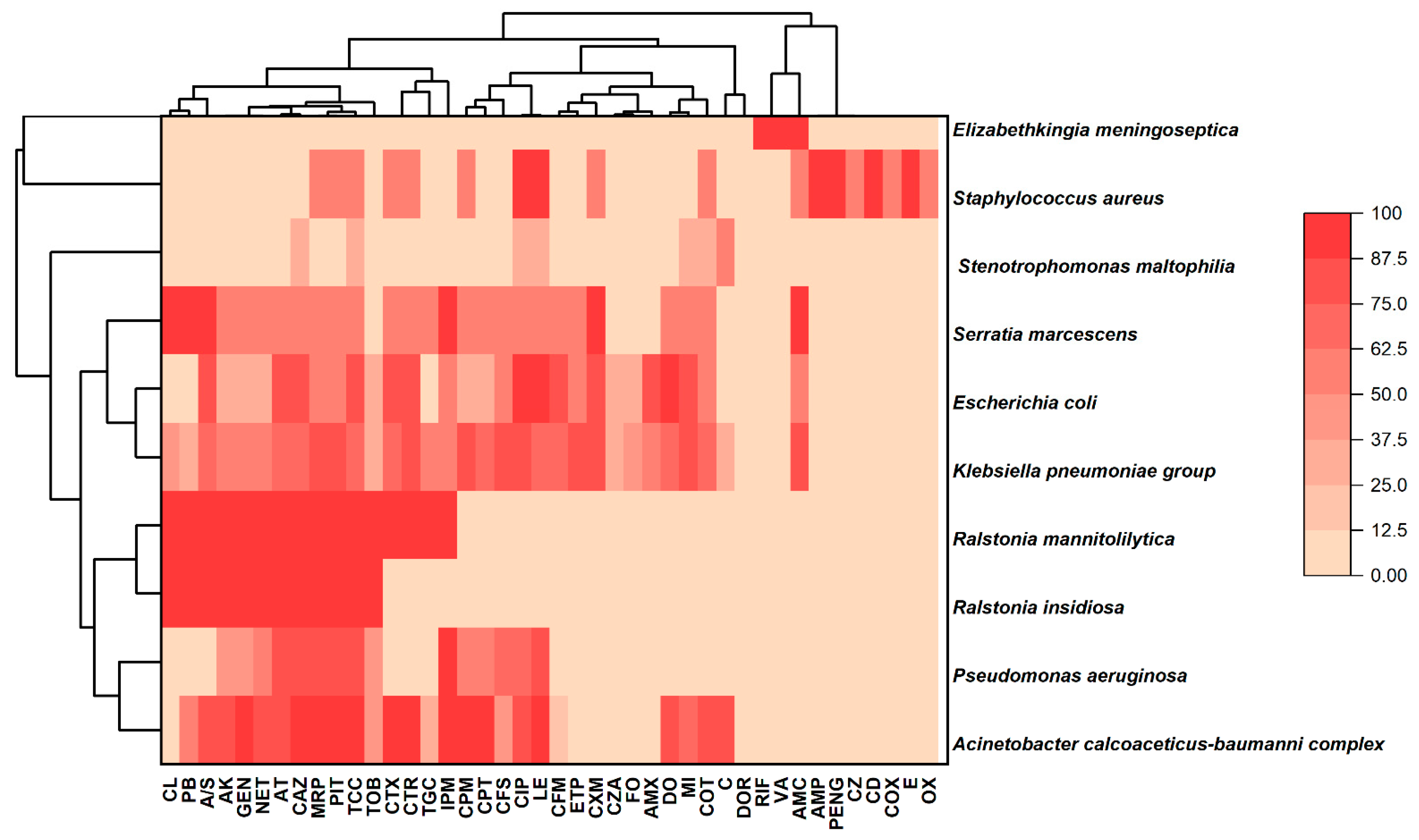
3.2. Pattern of Antibiotic Resistance of Isolated Pathogens Against Different Drugs

The commonly identified pathogens like Acinetobacter baumannii, Pseudomonas aeruginosa, and Klebsiella pneumoniae showed resistance to multiple antibiotics such as ceftazidime, piperacillin/tazobactam, meropenem, ticarcillin/clavulanic acid, levofloxacin, aztreonam, imipenem, ceftriaxone, ciprofloxacin, trimethoprim/sulfamethoxazole, cefotaxime, doxycycline. Resistance was observed in case of Staphylococcus aureus to ampicillin, benzylpenicillin, ciprofloxacin, clindamycin, erythromycin, and levofloxacin.
In this study the heat map analysis was carried out to check the resistance patterns of the pathogens detected in VAP patients (Figure 1). Gram-negative pathogens showed 100% resistance against some antibiotics, including polymyxin, ampicillin, third-generation cephalosporins, beta-lactamase, aminoglycoside, beta-lactam and carbapenem. Serratia marcescens and Klebsiella pneumoniae group showed 100% resistance against AMC- amoxicillin/clavulanic acid. Serratia marcescens, Escherichia. coli, Klebsiella pneumoniae group, Ralstonia insidiosa, and Ralstonia mannitolilytica were arising in a single cluster with a moderate to high resistance against colistion, polymixine B, ampicillin/sulbactam, amikacine, gentamicine, netilmicin, aztreonam, ceftazidime, meropenem, piperacillin/tazobactam and ticarcillin/clavulanic acid. However, a very low resistance percentage was observed in Serratia marcescens against tobramycin (TOB) and in E. coli against colistin, polymyxin B, amikacin, gentamicin and netilmicin, whereas in a single cluster of a Pseudomonas aeruginosa and Acinetobacter calcoaceticus-baumanni complex a high resistance was discerned in the heatmap.
A separate cluster was observed for Elizabethkingia meningoseptica (resistance against rifampicin, vancomycin, amoxicillin/clavulanic acid) and Staphylococcus aureus (resistance against amoxicillin/clavulanic acid, ampicillin, benzylpenicillin, cefazolin, clindamycin, cloxacillin, erythromycin and oxacillin). There was an absence of resistance reported in Stenotrophomonas maltophilia against the majority of antibiotics used in the current research investigation.

4. Discussion

This study included 70 samples from individuals who were diagnosed with VAP. The selected sample size was designed to create a robust and accurate dataset for precisely identifying prevalent diseases and monitoring resistance variations. The study evaluated resistance patterns in a wide variety of bacterial and fungal species according to the VITEK-2 system’s extensive database, which covers a broad range of antibiotics.
The distribution of patients by age group revealed that the majority (n = 45) were beyond the age of 60, indicating a greater risk of VAP in the senior population. Another study also revealed that patients between the ages of 51 to 75 had a high incidence of VAP [26].
Forty-five patients were males and twenty-five were women, indicating a slight male dominance. In another study with a higher prevalence of men the researchers reported that gender seems to influence the risk of VAP [26], while other studies showed a higher prevalence in women [27]. These discrepancies might have occurred due to variations among study populations and methodologies.
The study investigated the etiological agents and resistance patterns of VAP in ICU patients. Suspected patient’s samples were transferred to the lab and processed using the semiquantitative culture method for the identification of pathogens. Through these techniques positive growth was defined as reaching a threshold value of 104 colony-forming-unit (CFU)/mL when a BAL sample taken and >105 to 106 colony forming units for an ET sample. Another study also supported these threshold values of 104 colony-forming-unit (CFU)/mL for BAL and >105 to 106 CFU/mL for ET [28]. In this study the identification of pathogens was validated by the MALDI-TOF MS (Bruker Daltonics, Germany) and VITEK-2 systems (bioMérieux, France) for identification and susceptibility testing (ID/ST) [29,30]. Fungal isolation involved direct smears (KOH/lactophenol cotton blue) and cultures on Sabouraud dextrose agar. Candida was identified via Gram-positive pseudohyphae and growth on specific media, while filamentous fungi like Aspergillus were recognized by macroscopic and microscopic characteristics, with Aspergillus niger forming brown colonies with radiating conidial heads. Positive cultures at least 48 h after ventilation were used to diagnose bacterial/fungal superinfection in clinically suspected patients with previous negative findings [31].
Seventy clinical samples were identified as VAP, of which monomicrobial infections accounted for 68.58% of VAP cases, while polymicrobial infections contributed 31.42%. These results are comparable to another study which reported 72.2% and 27.8%, respectively [32].
The frequently isolated pathogens in late-onset VAP comprised Acinetobacter baumannii, Pseudomonas aeruginosa, Klebsiella pneumoniae, and Staphylococcus aureus. These findings were consistent with a previous study, apart from Staphylococcus aureus [33]. Development of late-onset VAP was mostly associated with drug resistant pathogens. In this study the mortality rate of late-onset VAP was high (64.5%) due to Acinetobacter baumannii, Pseudomonas aeruginosa, Klebsiella pneumoniae, Staphylococcus aureus, and polymicrobial infections. Previous studies also revealed that the mortality rate (11.11%) of VAP was greater in hospitals with previous K. pneumoniae infections [34]. Other retrospective observational study conducted during January 2010 to Jun 2015 also reported a high mortality rate as 39.8% [35]. Apart from Klebsiella pneumoniae, Acinetobacter baumannii, and Pseudomonas aeruginosa, Candida species and Aspergillus were also detected in VAP patients. The study is also consistent with the study by Meawed et al. (2021), which reported that the most frequent bacterial and fungal pathogens isolated were pan-drug-resistant (PDR) Klebsiella pneumoniae and multidrug-resistant (MDR) Acinetobacter baumannii, Candida species, and Aspergillus [31]. These findings of VAP-associated pathogens were also consistent with another previous study, which identified E. coli, K. pneumoniae, A. baumannii, P. aeruginosa, S. aureus, Candida albicans, and Candida tropicalis as common isolates, confirmed the consistent epidemiological trends of these key VAP pathogens across different settings using MALDI-TOF and VITEK systems [36].
Furthermore, the identification of human rhinovirus/enterovirus in our cohort emphasized the rising role of viral pathogens in VAP, which is supported by studies showing rhinovirus’s frequent occurrence in respiratory infections among critically ill patients. Other studies had implicated a broader range of viruses in severe pneumonia and VAP, including respiratory syncytial virus, influenza, adenoviruses, human metapneumovirus, herpes simplex virus (HSV), and cytomegalovirus (CMV) [37,38,39,40,41,42]. The findings of this study highlighted the importance of rapid multiplex PCR in revealing the polymicrobial nature of VAP and guiding comprehensive patient management.
The comprehensive identification of common Gram-negative and Gram-positive bacteria through Biofire FilmArray panel was consistent with the findings of various studies that evaluated this platform [43,44,45,46,47].
This study revealed that most prevalent underlying diseases to those with VAP were high blood pressure, diabetes, thyroid disorders, COPD, kidney failure, cardiac disease, and sepsis. These co-morbidities might have increased the chance of developing VAP. These findings are consistent with a previous study which identified COPD, high blood pressure and diabetes as the most common comorbidities [7]. In this study many patients (n = 28) had diabetes, emphasizing the link between diabetes and VAP risk. Meawed et al. (2021) also justified that the risk of fungal VAP was diabetes mellitus, chest disease, hypothyroidism and longer duration of mechanical ventilation [31]. Findings showed that polymicrobial infection in early-onset and late-onset VAP was 4.28% and 27.14%, respectively, whereas two grouped studies conducted in 2013 revealed that 50% of polymicrobial infections were identified in both late-onset and early-onset VAP [33].
More than 70% of the Gram-negative pathogen isolates in the current study were resistant to ampicillin and 3rd-generation cephalosporins. The combination of gentamicin with β lactam-β lactamase inhibitors (e.g., piperacillin–tazobactam) demonstrated about 50–75% resistance, while amikacin, imipenem, and meropenem showed 50–90% resistance. Other study of AMR among Gram-negative bacteria found that approximately 50–75% of isolates were resistant to gentamicin and combination of β-lactam–β-lactamase inhibitors, while over 75% became resistant to ampicillin and 3rd-generation cephalosporins. In decreasing order, levofloxacin, amikacin, imipenem, and meropenem showed the least resistance [7].
Individual Gram-negative bacteria had comparable levels of resistance, with the exception of Acinetobacter baumannii, which showed increased resistance to the highest-level drugs such as amikacin and carbapenems (imipenem and meropenem). Staphylococcus aureus was the only Gram-positive pathogen that showed 50% of methicillin resistant isolates. Also, higher levels of resistance to fluoroquinolones and aminoglycosides were observed. Another research found that resistance to anti-MRSA drugs such vancomycin, teicoplanin, and linezolid did not rise further in VAP patients, which is consistent with these investigations [7].
In this study, early-onset VAP accounted for 31.42% of all VAP cases (22/70), whereas late-onset VAP accounted for 68.57% (48/70). The majority of the pathogens that were isolated were K. pneumoniae, P. aeruginosa, and A. baumannii. On the other hand, 91.7% of patients in another study experienced early-onset VAP, and 8.3% experienced late-onset VAP, where S. aureus, Enterococcus species, Acinetobacter species, K. pneumoniae, and P. aeruginosa were among the organisms that were isolated [48].
A heat map analysis depicted a clear resistance pattern of the pathogens detected in VAP patients. In a study, a hierarchical cluster heat map analysis was carried out to analyze the differences in clustering patterns of common AMR profiles among pathogens [49]. Yudhanto et al. (2022) used hierarchical clustering to identify common AMR pattern and assist specific actions. This approach illustrates linkages by identifying groups of isolates with shared AMR pattern using tree-like arrangements. These clusters improve antimicrobial stewardship, enabling targeted surveillance and treatment methods [50].

5. Limitations of the Study

In the current study, we could not include the multiple underlying risk factors involved in the VAP patients due to a lack of patient metadata and ethical consent.

6. Conclusions

This study examines the pathogens and resistant patterns of antimicrobial agents linked with ventilator-associated pneumonia in the critical care unit. Findings from the study revealed a statistically significant association between early- and late-onset VAP, based on a chi-squared test, caused by the Klebsiella pneumoniae group (Chi sq.—5.99, p-value < 0.0144) and Polymicrobial infection (Chi sq.—10.16, p-value < 0.0014), indicating that their distributions were not random. Most of the underlying diseases associated with VAP pathogens were diabetes, kidney failure, heart disease, respiratory failure, and hypertension. The study also indicates the emergence of multidrug-resistant (MDR) bacteria, specifically Pseudomonas aeruginosa, Acinetobacter baumannii, Klebsiella pneumoniae, and Staphylococcus aureus. These pathogens showed high resistance to aminoglycosides, carbapenems, 2nd, 3rd, and 4th generations of cephalosporins, macrolides, penicillin, penicillin combinations with beta-lactamase inhibitors, sulfonamide/folate synthesis inhibitor combinations, and tetracyclines. The heatmap analysis revealed a complex microbial ecology of VAP, with different groups of pathogens exhibiting varying levels of antibiotic resistance. These findings emphasize the crucial need for vigilant VAP surveillance, the development of effective infection control measures, and careful administration of antibiotics to address the increasing risk of MDR organisms. Using local resistance patterns to guide targeted antimicrobial therapy is critical for improving outcomes of therapy and reducing the emergence of new resistance.

Author Contributions

Conceptualization, B.T. and A.B.; research and writing—original draft, H.K.T.; writing—review and editing, P.S., S.K., M.K.J., P.K., G.M. and A.K.O.; guidance, B.T. and M.K.J. All authors have read and agreed to the published version of the manuscript.

Funding

This research received no external funding.

Institutional Review Board Statement

Ethics approval was obtained on 5 August 2022 from the Institutional Ethics Committee, Devki Devi Foundation (Reference Number: BHR/TS/MSSH/DDF/SKT-2/IEC/MICRO/22-16), Max Healthcare Ethics Committee (BHR/TS/MSSH/MHIL/SKT-1/MHEC/MICRO/22-09), Max Healthcare Ethics Committee (BHR/TS/MSSSH/GMHRCMS/MHEC/MICRO/22-09).

Informed Consent Statement

Informed consent was obtained from all subjects involved in the study.

Data Availability Statement

The datasets generated and/or analyzed during the current study are available in the manuscript itself.

Acknowledgments

We are thankful to Kush K. Pandey, School of Veterinary Medicine and Biomedical Sciences, University of Nebraska-Lincoln, Lincoln, NE 68583, USA; Nebraska Center for Virology, University of Nebraska-Lincoln, Lincoln, NE 68583, USA, for editing and valuable inputs on the manuscript.

Conflicts of Interest

Authors Sudhakar Kancharla and Gowtham Mandadapu were employed by the company Devansh Lab Werks, and Prachetha Kolli was employed by the company Microgen Health Inc. The remaining authors declare that the research was conducted in the absence of any commercial or financial relationships that could be construed as a potential conflict of interest.

References

  1. Center for Disease Control and Prevention. Pneumonia (Ventilator-Associated [VAP] and Non-Ventilator-Associated Pneumonia [PNEU]) Event; National Healthcare safety network, Center for Disease Control and Prevention: Atlanta, GA, USA, 2023; pp. 1–19. [Google Scholar]
  2. Arthur, L.E.; Kizor, R.S.; Selim, A.G.; van Driel, M.L.; Seoane, L. Antibiotics for ventilator-associated pneumonia. Cochrane Database Syst. Rev. 2016, 10, CD004267. [Google Scholar] [CrossRef] [PubMed]
  3. Mastrogianni, M.; Katsoulas, T.; Galanis, P.; Korompeli, A.; Myrianthefs, P. The Impact of Care Bundles on Ventilator-Associated Pneumonia (VAP) Prevention in Adult ICUs: A Systematic Review. Antibiotics 2023, 12, 227. [Google Scholar] [CrossRef] [PubMed]
  4. Amin, A. Clinical and economic consequences of ventilator-associated pneumonia. Clin. Infect. Dis. 2009, 49 (Suppl. S1), S36–S43. [Google Scholar] [CrossRef] [PubMed]
  5. Chastre, J.; Fagon, J.Y. Ventilator-associated pneumonia. Am. J. Respir. Crit. Care Med. 2002, 165, 867–903. [Google Scholar] [CrossRef]
  6. Rea-Neto, A.; Youssef, N.C.; Tuche, F.; Brunkhorst, F.; Ranieri, V.M.; Reinhart, K.; Sakr, Y. Diagnosis of ventilator-associated pneumonia: A systematic review of the literature. Crit. Care 2008, 12, R56. [Google Scholar] [CrossRef]
  7. Rao, S.V.; Thilakchand, K.R.; Boloor, R.; Suresh, S.; George, T.; Pais, M.L.; Jakribettu, R.P.; Baliga, M.S. Antimicrobial resistance pattern in aerobic bacteria isolated from endotracheal aspirate in ventilator-associated pneumonia: Ten years observation from a tertiary care hospital. J. Anaesthesiol. Clin. Pharmacol. 2024, 40, 324–329. [Google Scholar] [CrossRef]
  8. Kohbodi, G.N.A.; Rajasurya, V.; Noor, A. Ventilator-Associated Pneumonia. In StatPearls [Internet]; 4 September 2023; StatPearls Publishing: Treasure Island, FL, USA, 2024. Available online: https://www.ncbi.nlm.nih.gov/books/NBK507711/ (accessed on 20 October 2024).
  9. Dey, A.; Bairy, I. Incidence of multidrug-resistant organisms causing ventilator-associated pneumonia in a tertiary care hospital: A nine months’ prospective study. Ann. Thorac. Med. 2007, 2, 52–57. [Google Scholar] [CrossRef]
  10. Dennesen, P.J.; van der Ven, A.J.; Kessels, A.G.; Ramsay, G.; Bonten, M.J. Resolution of infectious parameters after antimicrobial therapy in patients with ventilator-associated pneumonia. Am. J. Respir. Crit. Care Med. 2001, 163, 1371–1375. [Google Scholar] [CrossRef]
  11. Edis, E.C.; Hatipoglu, O.N.; Tansel, O.; Sut, N. Acinetobacter pneumonia: Is the outcome different from the pneumonias caused by other agents. Ann. Thorac. Med. 2010, 5, 92–96. [Google Scholar] [CrossRef]
  12. Kumar, A.; Roberts, D.; Wood, K.E.; Light, B.; Parrillo, J.E.; Sharma, S.; Suppes, R.; Feinstein, D.; Zanotti, S.; Taiberg, L.; et al. Duration of hypotension before initiation of effective antimicrobial therapy is the critical determinant of survival in human septic shock. Crit. Care Med. 2006, 34, 1589–1596. [Google Scholar] [CrossRef]
  13. Thakur, H.K.; Tarai, B.; Bhargava, A.; Soni, P.; Rath, P.K.; Mishra, B.P.; Jena, M.K. Pathogenesis, Diagnosis and Therapeutic Strategies for Ventilator-associated Pneumonia. J. Pure Appl. Microbiol. 2024, 18, 772–796. [Google Scholar] [CrossRef]
  14. Farag, A.M.; Tawfick, M.M.; Abozeed, M.Y.; Shaban, E.A.; Abo-Shadi, M.A. Microbiological profile of ventilator-associated pneumonia among intensive care unit patients in tertiary Egyptian hospitals. J. Infect. Dev. Ctries. 2020, 14, 153–161. [Google Scholar] [CrossRef] [PubMed]
  15. Garland, J.S. Strategies to prevent ventilator-associated pneumonia in neonates. Clin. Perinatol. 2010, 37, 629–643. [Google Scholar] [CrossRef] [PubMed]
  16. Kalil, A.C.; Metersky, M.L.; Klompas, M.; Muscedere, J.; Sweeney, D.A.; Palmer, L.B.; Napolitano, L.M.; O’Grady, N.P.; Bartlett, J.G.; Carratalà, J.; et al. Management of Adults With Hospital-acquired and Ventilator-associated Pneumonia: 2016 Clinical Practice Guidelines by the Infectious Diseases Society of America and the American Thoracic Society. Clin. Infect. Dis. 2016, 63, e61–e111, Erratum in Clin. Infect. Dis. 2017, 64, 1298. https://doi.org/10.1093/cid/ciw799; Erratum in Clin. Infect. Dis. 2017, 65, 1435. https://doi.org/10.1093/cid/cix587; Erratum in Clin. Infect. Dis. 2017, 65, 2161. https://doi.org/10.1093/cid/cix759. [Google Scholar] [CrossRef]
  17. Tanwar, J.; Das, S.; Fatima, Z.; Hameed, S. Multidrug resistance: An emerging crisis. Interdiscip. Perspect. Infect. Dis. 2014, 2014, 541340. [Google Scholar] [CrossRef]
  18. Dominedò, C.; Ceccato, A.; Torres, A. Ventilator-associated pneumonia: New principles guiding empiric antibiotic therapy. Curr. Opin. Infect. Dis. 2020, 33, 182–188. [Google Scholar] [CrossRef] [PubMed]
  19. Alnimr, A. Antimicrobial Resistance in Ventilator-Associated Pneumonia: Predictive Microbiology and Evidence-Based Therapy. Infect. Dis. Ther. 2023, 12, 1527–1552. [Google Scholar] [CrossRef]
  20. Fang, X.; Mei, Q.; Fan, X.; Zhu, C.; Yang, T.; Zhang, L.; Geng, S.; Pan, A. Diagnostic Value of Metagenomic Next-Generation Sequencing for the Detection of Pathogens in Bronchoalveolar Lavage Fluid in Ventilator-Associated Pneumonia Patients. Front. Microbiol. 2020, 11, 599756. [Google Scholar] [CrossRef]
  21. Peiffer-Smadja, N.; Bouadma, L.; Mathy, V.; Allouche, K.; Patrier, J.; Reboul, M.; Montravers, P.; Timsit, J.-F.; Armand-Lefevre, L. Performance and impact of a multiplex PCR in ICU patients with ventilator-associated pneumonia or ventilated hospital-acquired pneumonia. Crit. Care 2020, 24, 366. [Google Scholar] [CrossRef]
  22. Tamma, P.D.; Cosgrove, S.E.; Maragakis, L.L. Combination therapy for treatment of infections with gram-negative bacteria. Clin. Microbiol. Rev. 2012, 25, 450–470. [Google Scholar] [CrossRef]
  23. CLSI M-38 A Document (Reference Method for Broth Dilution Antifungal Susceptibility Testing of Filamentous Fungi), 3rd ed.; Clinical and Laboratory Standards Institute: Wayne, PA, USA, 2017.
  24. Hudzicki, J. Kirby-Bauer Disk Diffusion Susceptibility Test Protocol. American Society for Microbiology. 8 December 2009. Available online: https://asm.org/getattachment/2594ce26-bd44-47f6-8287-0657aa9185ad/Kirby-Bauer-Disk-Diffusion-Susceptibility-Test-Protocol-pdf.pdf (accessed on 17 March 2025).
  25. BIOFIRE® FILMARRAY® Pneumonia Plus Panel. 2021. Available online: https://www.biomerieux.com/corp/en/our-offer/clinical-products/biofire-filmarray-pneumonia-panels.html (accessed on 22 March 2025).
  26. Wałaszek, M.; Kosiarska, A.; Gniadek, A.; Kołpa, M.; Wolak, Z.; Dobroś, W.; Siadek, J. The risk factors for hospital-acquired pneumonia in the Intensive Care Unit. Przegl Epidemiol. 2016, 70, 15–110. [Google Scholar]
  27. Ibn Saied, W.; Merceron, S.; Schwebel, C.; Le Monnier, A.; Oziel, J.; Garrouste-Orgeas, M.; Marcotte, G.; Ruckly, S.; Souweine, B.; Darmon, M.; et al. Ventilator-associated pneumonia due to Stenotrophomonas maltophilia: Risk factors and outcome. J. Infect. 2020, 80, 279–285. [Google Scholar] [CrossRef] [PubMed]
  28. Ioanas, M.; Ferrer, R.; Angrill, J.; Ferrer, M.; Torres, A. Microbial investigation in ventilator-associated pneumonia. Eur. Respir. J. 2001, 17, 791–801. [Google Scholar] [CrossRef]
  29. Croxatto, A.; Prod’hom, G.; Greub, G. Applications of MALDI-TOF mass spectrometry in clinical diagnostic microbiology. FEMS Microbiol. Rev. 2012, 36, 380–407. [Google Scholar] [CrossRef] [PubMed]
  30. Wallet, F.; Loïez, C.; Renaux, E.; Lemaitre, N.; Courcol, R.J. Performances of VITEK 2 colorimetric cards for identification of gram-positive and gram-negative bacteria. J. Clin. Microbiol. 2005, 43, 4402–4406. [Google Scholar] [CrossRef] [PubMed]
  31. Meawed, T.E.; Ahmed, S.M.; Mowafy, S.M.S.; Samir, G.M.; Anis, R.H. Bacterial and fungal ventilator associated pneumonia in critically ill COVID-19 patients during the second wave. J. Infect. Public Health 2021, 14, 1375–1380. [Google Scholar] [CrossRef]
  32. Charles, M.P.; Easow, J.M.; Joseph, N.M.; Ravishankar, M.; Kumar, S.; Sivaraman, U. Aetiological agents of ventilator-associated pneumonia and its resistance pattern—A teat for treatment. Australas. Med. J. 2013, 6, 430–434. [Google Scholar] [CrossRef]
  33. Restrepo, M.I.; Peterson, J.; Fernandez, J.F.; Qin, Z.; Fisher, A.C.; Nicholson, S.C. Comparison of the bacterial etiology of early-onset and late-onset ventilator-associated pneumonia in subjects enrolled in 2 large clinical studies. Respir. Care 2013, 58, 1220–1225. [Google Scholar] [CrossRef]
  34. Ding, X.; Ma, X.; Gao, S.; Su, L.; Shan, G.; Hu, Y.; Chen, J.; Ma, D.; Zhang, F.; Zhu, W.; et al. Effect of ICU quality control indicators on VAP incidence rate and mortality: A retrospective study of 1267 hospitals in China. Crit. Care 2022, 26, 405. [Google Scholar] [CrossRef]
  35. But, A.; Yetkin, M.A.; Kanyilmaz, D.; Aslaner, H.; Baştuğ, A.; Aypak, A.; Öngürü, P.; Akinci, E.; Mutlu, N.M.; Bodur, H. Analysis of epidemiology and risk factors for mortality in ventilator-associated pneumonia attacks in intensive care unit patients. Turk. J. Med. Sci. 2017, 47, 812–816. [Google Scholar] [CrossRef]
  36. Garza-González, E.; Camacho-Ortíz, A.; Rodríguez-Noriega, E.; Esparza-Ahumada, S.; Flores-Trevino, S.; Bocanegra-Ibarias, P.; Tijerina-Rodriguez, L.; Morfin-Otero, R. Comparison of Matrix-assisted Laser Desorption Ionization Time-of-flight Mass Spectrometry (MALDI-TOF MS) and the Vitek 2 System for Routine Identification of Clinically Relevant Bacteria and Yeast. Ann. Clin. Lab. Sci. 2020, 50, 119–127. [Google Scholar] [PubMed]
  37. Hong, H.L.; Hong, S.B.; Ko, G.B.; Huh, J.W.; Sung, H.; Do, K.-H.; Kim, S.-H.; Lee, S.-O.; Kim, M.-N.; Jeong, J.-Y.; et al. Viral infection is not uncommon in adult patients with severe hospital-acquired pneumonia. PLoS ONE 2014, 9, e95865. [Google Scholar] [CrossRef] [PubMed]
  38. Loubet, P.; Voiriot, G.; Houhou-Fidouh, N.; Neuville, M.; Bouadma, L.; Lescure, F.-X.; Descamps, D.; Timsit, J.-F.; Yazdanpanah, Y.; Visseaux, B. Impact of respiratory viruses in hospital-acquired pneumonia in the intensive care unit: A single-center retrospective study. J. Clin. Virol. 2017, 91, 52–57. [Google Scholar] [CrossRef] [PubMed]
  39. van Someren Gréve, F.; Juffermans, N.P.; Bos, L.D.J.; Binnekade, J.M.; Braber, A.; Cremer, O.L.; de Jonge, E.; Molenkamp, R.; Ong, D.S.Y.; Rebers, S.P.H.; et al. Respiratory Viruses in Invasively Ventilated Critically Ill Patients-A Prospective Multicenter Observational Study. Crit. Care Med. 2018, 46, 29–36. [Google Scholar] [CrossRef]
  40. Luyt, C.E.; Combes, A.; Deback, C.; Aubriot-Lorton, M.-H.; Nieszkowska, A.; Trouillet, J.-L.; Capron, F.; Agut, H.; Gibert, C.; Chastre, J. Herpes simplex virus lung infection in patients undergoing prolonged mechanical ventilation. Am. J. Respir. Crit. Care Med. 2007, 175, 935–942. [Google Scholar] [CrossRef]
  41. Limaye, A.P.; Kirby, K.A.; Rubenfeld, G.D.; Leisenring, W.M.; Bulger, E.M.; Neff, M.J.; Gibran, N.S.; Huang, M.-L.; Hayes, T.K.S.; Corey, L.; et al. Cytomegalovirus reactivation in critically ill immunocompetent patients. JAMA 2008, 300, 413–422. [Google Scholar] [CrossRef]
  42. Papazian, L.; Fraisse, A.; Garbe, L.; Zandotti, C.; Thomas, P.; Saux, P.; Perrin, G.; Gouin, F. Cytomegalovirus. An unexpected cause of ventilator-associated pneumonia. Anesthesiology 1996, 84, 280–287. [Google Scholar] [CrossRef]
  43. Buchan, B.W.; Windham, S.; Balada-Llasat, J.M.; Leber, A.; Harrington, A.; Relich, R.; Murphy, C.; Bard, J.D.; Naccache, S.; Ronen, S.; et al. Practical Comparison of the BioFire FilmArray Pneumonia Panel to Routine Diagnostic Methods and Potential Impact on Antimicrobial Stewardship in Adult Hospitalized Patients with Lower Respiratory Tract Infections. J. Clin. Microbiol. 2020, 58, e00135-20. [Google Scholar] [CrossRef]
  44. Foschi, C.; Zignoli, A.; Gaibani, P.; Vocale, C.; Rossini, G.; Lafratta, S.; Liberatore, A.; Turello, G.; Lazzarotto, T.; Ambretti, S. Respiratory bacterial co-infections in intensive care unit-hospitalized COVID-19 patients: Conventional culture vs BioFire FilmArray pneumonia Plus panel. J. Microbiol. Methods 2021, 186, 106259. [Google Scholar] [CrossRef]
  45. Ginocchio, C.C.; Garcia-Mondragon, C.; Mauerhofer, B.; Rindlisbacher, C.; The EME Evaluation Program Collaborative. Multinational evaluation of the BioFire® FilmArray® Pneumonia plus Panel as compared to standard of care testing. Eur. J. Clin. Microbiol. Infect. Dis. 2021, 40, 1609–1622. [Google Scholar] [CrossRef]
  46. Molina, F.J.; Botero, L.E.; Isaza, J.P.; Cano, L.E.; López, L.; Tamayo, L.; Torres, A. Diagnostic concordance between BioFire® FilmArray® Pneumonia Panel and culture in patients with COVID-19 pneumonia admitted to intensive care units: The experience of the third wave in eight hospitals in Colombia. Crit. Care 2022, 26, 130. [Google Scholar] [CrossRef] [PubMed]
  47. Søgaard, K.K.; Hinic, V.; Goldenberger, D.; Gensch, A.; Schweitzer, M.; Bättig, V.; Siegemund, M.; Bassetti, S.; Bingisser, R.; Tamm, M.; et al. Evaluation of the clinical relevance of the Biofire© FilmArray pneumonia panel among hospitalized patients. Infection 2024, 52, 173–181. [Google Scholar] [CrossRef] [PubMed]
  48. Rezia, A.; Vijendra, R. A clinical study on the pattern of antimicrobial drug use and drug resistance in patients with ventilator-associated pneumonia in a tertiary care hospital. Indian J. Pharmacol. 2023, 55, 89–96. [Google Scholar] [CrossRef] [PubMed]
  49. Das, M.; Ojha, A.K.; Dolma, K.G.; Majumdar, T.; Sarmah, P.; Hazarika, S.; Modi, D.; Gogoi, D.; Das, S.; Ramamurthy, T. Monitoring the potential dissemination of antimicrobial resistance in foods, environment, and clinical samples: A one health prospective. Food Sci. Biotechnol. 2024, 34, 803–813. [Google Scholar] [CrossRef]
  50. Yudhanto, S.; Hung, C.C.; Maddox, C.W.; Varga, C. Antimicrobial Resistance in Bacteria Isolated From Canine Urine Samples Submitted to a Veterinary Diagnostic Laboratory, Illinois, United States. Front. Vet. Sci. 2022, 9, 867784. [Google Scholar] [CrossRef]
Figure 1. Cluster heatmap of VAP pathogens with antibiotic resistance pattern. Rows and columns of cluster heatmap plot represent pathogens and antibiotics, respectively. CL colistin, PB polymyxin B, A/S ampicillin/sulbactam, AK amikacin, GEN gentamicin, NET netilmicin, AT aztreonam, CAZ ceftazidime, MRP meropenem, PIT piperacillin/tazobactam, TCC ticarcillin/clavulanic acid, TOB tobramycin, CTX cefotaxime, CTR ceftriaxone, TGC tigecycline, IMP imipenem, CPM cefepime, CPT cefepime-tazobactam, CFS cefoperazone/sulbactam, CIP ciprofloxacin, LE levofloxacin, CFM cefixime, ETP ertapenem, CXM cefuroxime, CZA ceftazidime/avibactam, FO fosfomycin, AMX amoxicillin, DO doxycycline, MI minocycline, COT trimethoprim/sulfamethoxazole, C chloramphenicol, DOR doripenem, RIF rifampicin, VA vancomycin, AMC amoxicillin/clavulanic acid, AMP ampicillin, PENG benzylpenicillin, CZ cefazolin, CD clindamycin, COX cloxacillin, E erythromycin, OX oxacillin.
Microbiolres 16 00152 g001
Table 1. Demographic profile of patients and underlying diseases associated with the pathogens detected.
Demographic Profile of Patients with VAP
DemographyBacterialFungalViralTotal
Age
<60 yrs223025
>60 yrs440145
Total663170
p value0.02470.08650.3190.0499
Sex
Male431145
Female232025
Total663170
p value0.04140.56780.3190.0499
Residential
Urban602163
Rural6107
Total663170
p value<0.00010.56780.319<0.0001
Co-morbidity condition
Patients with single co-morbidities120012
Patients with ≥2 co-morbidities *443047
Patients with missing data/no co-morbidities 100111
Total663170
p value<0.00010.05410.371<0.0001
Treatment given
Intake of steroids343037
Intake of antibiotics673070
p value0.0115>0.9999 0.0154
* Co-morbid condition: immunocompromised, post-operative, lung disease, DM, COPD, kidney failure, accidental, heart disease, sepsis, hypertension, cancer, thyroid, liver, missing/no underlying disease.
Table 2. Number of patients infected with VAP causing pathogens with underlying disease; ND, not detected; HTN, hypertension; DM, diabetic mellitus; COPD, conic obstructive pulmonary disease. Values in the table signify the number of patients infected with pathogens with underlying co-morbid conditions.
PathogensDMKidney FailureAccidentalHeart DiseaseSepsisRespiratory FailureHTNImmuno-Compromised Cancer ThyroidMotor Neuron DiseaseCOPDSurgeryLiver
Acinetobacter baumannii117184814ND35121ND
Pseudomonas aeruginosa105342410ND2413ND1
Klebsiella pneumoniae group12816710171ND3ND321
Escherichia coli61ND2114NDND1NDNDNDND
Burkholderia cepacia1NDND1NDND1NDND1NDNDNDND
Staphylococcus aureus521ND125ND11NDNDNDND
Serratia marcescens2NDNDNDND11ND12NDNDNDND
Elizabethkingia meningosepticaNDNDNDND1NDNDNDNDND1NDNDND
Candida tropicalis3312213NDNDNDNDNDNDND
Candida albicansNDNDNDNDND11ND1NDNDNDNDND
Ralstonia insidiosa1NDNDNDNDNDNDND1NDNDNDNDND
Stenotrophomonas maltophiliaNDNDND1ND11NDNDNDNDNDNDND
Ralstonia mannitolilyticaNDNDND1NDNDNDNDNDNDNDNDNDND
Aspergillus11ND1111NDNDNDNDNDNDND
Streptococcus pneumoniaeNDNDNDNDNDNDNDNDNDNDNDNDNDND
Table 3. The pathogens causing both early- and late-onset VAP.
PathogensEarly-Onset-VAP, n (%)Late-Onset-VAP, n (%)Chi sq.p Value
Acinetobacter baumannii4 (5.71)5 (7.14)0.10440.7466
Pseudomonas aeruginosa4 (5.71)7 (10)0.75890.3837
Klebsiella pneumoniae group1 (1.42)9 (12.85)5.990.0144
Escherichia coli2 (2.85)01.9720.1602
Burkholderia cepacia01 (1.42)0.9930.319
Staphylococcus aureus01 (1.42)0.9930.319
Serratia marcescens1 (1.42)00.9930.319
Elizabethkingia meningoseptica01 (1.42)0.9930.319
Candida tropicalis2 (2.85)01.9720.1602
Candida albicans01 (1.42)0.9930.319
Ralstonia insidiosa01 (1.42)0.9930.319
Stenotrophomonas maltophilia01 (1.42)0.9930.319
Ralstonia mannitolilytica1 (1.42)00.9930.319
Aspergillus01 (1.42)0.9930.319
Streptococcus pneumoniae1 (1.42)00.9930.319
Polymicrobial infection3 (4.28)19 (27.14)10.160.0014
Haemophilus influenzae1 (1.42)1 (1.42)0>0.9999
Human Rhino/Enterovirus1 (1.42)00.9930.319
Mycoplasma pneumoniae1 (1.42)00.9930.319
Total22 (31.42)48 (68.57)6.5380.0106
Disclaimer/Publisher’s Note: The statements, opinions and data contained in all publications are solely those of the individual author(s) and contributor(s) and not of MDPI and/or the editor(s). MDPI and/or the editor(s) disclaim responsibility for any injury to people or property resulting from any ideas, methods, instructions or products referred to in the content.

