Review

Azo Dyes and the Microbial World: Synthesis, Breakdown, and Bioactivity

by
Valery M. Dembitsky
* and
Alexander O. Terent’ev
N.D. Zelinsky Institute of Organic Chemistry, Russian Academy of Sciences, Leninsky Prospect, 47, 119334 Moscow, Russia
*
Author to whom correspondence should be addressed.
Microbiol. Res. 2025, 16(5), 100; https://doi.org/10.3390/microbiolres16050100
Submission received: 16 April 2025 / Revised: 10 May 2025 / Accepted: 14 May 2025 / Published: 16 May 2025

Abstract

:
This review discusses natural and synthetic azo compounds found in bacteria, fungal endophytes, fungi, plants, and invertebrates. More than 100 of these compounds have demonstrated significant pharmacological activity, including antitumor, antimicrobial, and antibacterial effects. Using mathematical algorithms and the PASS program, researchers predict new potential applications based on their structure–activity relationships. This review emphasizes the importance of natural azo compounds as promising drug prototypes and key players in drug discovery. It also explores the synthesis and degradation of azo dyes and their potential uses in medicine, food, cosmetics, and related fields. Additionally, the role of microorganisms in producing natural azo compounds and their synthetic counterparts is examined, showcasing their potential in drug development and human health advancements.

1. Introduction

Azo compounds, whether natural or synthetic, contain one or more azo groups (-N=N-), a functional group where two nitrogen atoms are joined by a double bond. Their general formula is R-N=N-R’, with R and R’ being either aryl or alkyl groups. The azo group plays a key role in determining their chemical properties, which are also influenced by other attached groups [1,2,3]. Azo ligands significantly affect the color of these compounds, creating bright and intense shades. These colors may change depending on pH, which may cause changes in the properties of the molecules, probably due to changes in the protonation state, which may lead to changes in electron density [4,5,6].
Azoxy metabolites, in contrast, feature an azoxy functional group, where two nitrogen atoms are linked by a double bond, with one also bonded to an oxygen atom. Their general formula is R-N(O)=N-R’, where R and R’ represent organic groups [7,8,9].
Some bacteria might be capable of producing azo or azoxy compounds, either as part of their metabolism or as secondary metabolites. If so, this ability would be specific to certain strains. Bacteria can also transform or break down synthetic compounds, including azoxy compounds, which may contribute to environmental cleanup processes like bioremediation [1,2,10,11,12,13].
Bacteria are often genetically engineered in biotechnology to produce useful chemical compounds. It is possible that bacteria could also be modified to synthesize azo or azoxy compounds for industrial purposes [14,15,16].
This review explores the structures and biological activities of natural azo and azoxy compounds, particularly those produced by microorganisms, as potential sources for drug discovery. It also discusses how microorganisms degrade azo dyes. The presented data allow us to understand the role of microorganisms in the biosynthesis and degradation of these unusual natural metabolites. The findings on azo compounds are particularly relevant to pharmacologists, chemists, and biologists.

2. Microorganisms as Producers of Azo and Azoxy Compounds

A well-known example of a natural azoxy antibiotic is valanimicin (1), which is derived from the fermentation broth of Streptomyces viridifaciens MG456-hF10. Recent research has identified the genes responsible for valanimicin biosynthesis. The VlmF protein, part of the major facilitator superfamily, provides resistance to valanimicin. Meanwhile, VlmD, VlmH, and VlmR convert L-valine into isobutyl-hydroxylamine, and VlmL facilitates the formation of L-seryl-tRNA from L-serine (see Figure 1) [17,18].
Valanimicin is effective against both Gram-positive and Gram-negative bacteria, particularly E. coli BE1121, a mutant strain of E. coli K12 that is deficient in DNA repair. It has also shown toxicity against L1210, P388/S (doxorubicin-sensitive) and P388/ADR (doxorubicin-resistant) murine leukemia cells, with IC50 values of 0.8, 2.7, and 1.4 pg/mL, respectively. In animal studies, valanimicin extended the survival of mice with Ehrlich carcinoma or L1210 leukemia cells [19].
Several valinomycin derivatives have been discovered from different sources. A valanimicin derivative (2, see structure in Figure 2) was found in the culture broth of Streptomyces viridifaciens MG456-hF10 during valanimicin biosynthesis [20], with its structure confirmed using a more stable ammonia adduct (3) [21]. Table 1 summarizes the biological activities of compounds (127). The antibiotic LL-BH872α (4), which contains an α,β-unsaturated azoxy group, was isolated from Streptomyces hinnulinis [22]. More recently, LL-BH872a, 2z-OH (5), produced by Actinomadura sp., was found in apricot (Prunus armeniaca) roots [21], along with LL-BH872a, 2z-OH, 4′z-OH (6), produced by Streptomyces misionensis FERM 10459 [23].
Several microorganisms produce antifungal antibiotics. For example, manivamycins A (7) and B (8), isolated from Streptomyces prasinopilosus, showed broad antifungal and antibacterial activity against multiple Candida and Trichophyton species, Cryptococcus neoformans, and Staphylococcus aureus [24]. Another antifungal agent, azoxybacillin (9), was isolated from Bacillus cereus NR2991 and was particularly effective against Aspergillus fumigatus and Trichophyton mentagrophytes [25,26]. Azoxyalkene (10), an unstable azoxy compound, was discovered in Actinomadura sp. (strain A7), which grows in apricot roots, though it only displayed weak antifungal activity against Rhodotorula sp. [21].
Elaiomycin (11), an azoxy antibiotic isolated from Streptomyces hepaticus, was found to strongly inhibit Mycobacterium tuberculosis growth [27,28,29,30]. Elaomycins D–G (1215), produced by Streptomyces sp. HKI0708, exhibit antimicrobial, anti-mycobacterial, and cytotoxic activities [31,32]. Additionally, elaomycins K (16), L (17), and elaomycin K amide (18), azoxy-type antibiotics found in Streptomyces sp. Tü 6399, showed weak antibacterial effects against Bacillus subtilis, Staphylococcus lentus, and Xanthomonas campestris, a plant pathogen [33].
Antiparasitic antibiotics, particularly those targeting nematodes, are rare. Jietacins A (19) and B (20), isolated from Streptomyces sp. [34,35], showed ten-times the activity of avermectin Bla, a veterinary nematicidal agent, against the pine wood nematode Bursaphelenchus hylocomi [36,37]. Additionally, geralcin C (21), D, and E, isolated from Streptomyces sp. LMA-545, exhibited anticancer properties, with geralcin C showing an IC50 of 0.8 μM against KB and HCT116 cell lines. It also inhibited E. coli DnaG primase, a Gram-negative antimicrobial target, with an IC50 of 0.7 μM [38]. Lastly, propanosine (K-76, 22), identified in Micromonospora chalcea 671-AV2 extracts, inhibited Valsa ceratosperma growth [39].
Lyophylline (23) is an antibiotic obtained from the mushroom Lyophyllum shimeji or its fungal endophytes. It has been found to prevent forebrain blister formation in the cranial mesenchyme at a concentration of 50 μg/mL [40].
The antitumor antibiotics DC1881A (24) and DC1881B (25) are produced by Streptomyces sp. DO-118 [41]. Additionally, the azoxy compounds KA-7367A (26) and KA-7367B (27, activity see in Table 2), which have antifungal properties, were discovered in Streptomyces sp. (KC-7367, FERM BP-1277). KA-7367A (26) showed activity against Aspergillus fumigatus, Candida albicans, Cryptococcus neoformans, Trichophyton mentagrophytes, and T. rubrum [42].
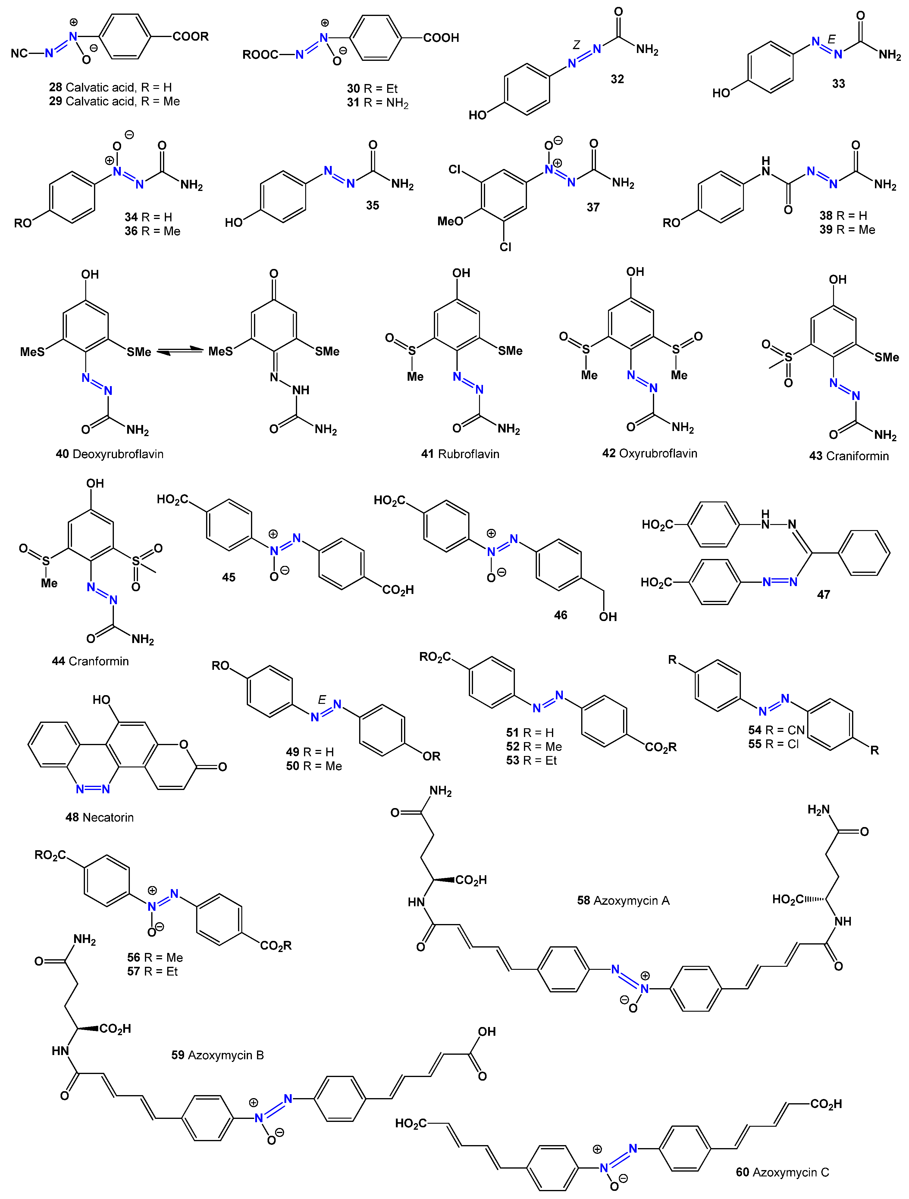
Calvacic acid (also called alvacic acid or calvatinic acid, 28) and its methyl derivatives (29, see structure in Figure 3, and Table 3) belong to a group of antitumor antibiotics with a diazene N-oxide structure. These compounds are produced by the fungi Calvatia craniformis (Agaricaceae) [43], Calvatia lilacina [44], and Lycoperdon pyriforme [45]. Another antibiotic, clavulanic acid, inhibits the growth of Gram-positive and Gram-negative bacteria at concentrations of 3–6 μg/mL [44]. It has cytotoxic effects, preventing the growth of Yoshida sarcoma cells [43], and has shown carcinostatic activity against hepatoma and leukemia K562 cells [46]. Clavulanic acid is also effective against Helicobacter pylori, a Gram-negative bacterium associated with gastric infections [47].
Amoxicillin–clavulanic acid is frequently used to treat many of the World Health Organization’s Priority Infectious Syndromes in adults and children. However, in some cases, a delayed antibiotic prescription or amoxicillin alone could be sufficient, reducing unnecessary use [48,49]. Additionally, clavulanic acid is being investigated for its potential in preventing and treating scopolamine-induced Alzheimer’s disease [50]. Some of its analogues (30 and 31) have been found to exhibit anti-microtubule activity [51].
Azoformamide (32), its (E)-form (33 and 35), and its azoxy derivatives (34 and 36) have been isolated from the puffball fungus Lycoperdon pyriforme [52,53] and have also been found in Calvatia craniformis [54]. A detection method for azoformamide in food additives has been developed [55].
Table 1. Biological activity of azo dyes produced by microorganisms (113) [13,24,34].
No.Dominated ActivityRankAdditional ActivityRank
1AntineoplasticStrongAntibioticStrong
2AntineoplasticStrongHepatic disorders treatmentStrong
3AntineoplasticStrong
4AntineoplasticModerate
5Antiviral (Arbovirus)ModerateAntineoplasticModerate
6AntineoplasticModerateAntifungalWeak
7AntineoplasticModerateAntifungalModerate
8Mucositis treatmentModerateAntiviral (Arbovirus)Moderate
9AntifungalModerateAntiviral (Arbovirus)Moderate
10Antiviral (Arbovirus)ModerateAntifungalModerate
11VasodilatorModerateAntifungalModerate
12AntifungalModerateAntiviral (Arbovirus)Moderate
13AntifungalModerateNatural killer cell stimulantWeak
Extracts from the fungus Lycoperdon pyriforme yielded two compounds, 4-methoxy-benzene-1-azoformamide (33) and 4-methoxybenzene-1-OH N-azoxyformamide (34), both of which were found to be effective against the parasitic nematode Meloidogyne incognita. A related compound, 3,5-dichloro- 4-methoxybenzene-1-OH-azoxyformamide (37), showed weaker nematicidal effects but higher cytotoxicity than compounds (33) and (34) [56].
Azoxyformamides (34 and 36) and two azoformamide derivatives (38 and 39) were isolated from the fruiting bodies of Calvatia craniiformis and Lycoperdon hiemale, respectively. Compounds (34) and (39) inhibited rootlet growth in lettuce seedlings, suggesting that their azoxy structure contributes to this effect. Additionally, compounds (34, 36, and 39) also inhibited the growth of pearl millet seedlings [57,58].
Fungi and their endophytes are known to produce colored pigments that contain azo groups. One such pigment, the red minor compound deoxyrubroflavin (40), was isolated from the lump fungus Calvatia rubro-flava [59,60]. Another pigment, the orange-colored rubroflavin (41), was found in the dried fruiting bodies of Calvatia rubro-flava and Calvatia craniformis [60,61]. Additional pigments, including oxyrubroflavin (42), craniformin (43), and cranformin (44, activity see in Table 4), were also identified in Calvatia rubro-flava [60,61,62].
The insect-pathogenic fungus Entomophthora virulenta produces a mixture of 4,4′-azoxybenzenedicarboxylic acid (45) and 4,4′-hydroxymethylazoxybenzene carboxylic acid (46), both of which have insecticidal properties [63,64]. A formazan derivative (47) was isolated from Agaricus silvicola [64]. Another notable fungal compound, necatorin (48), was discovered in Lactarius necator. This mutagenic alkaloid contains an azo group within a polycyclic aromatic structure that includes a 1-benzopyran moiety and a ketone group at carbon C2 [65,66,67,68].
Table 2. Biological activity of azo dyes produced by microorganisms (1427) [13,36,37,41].
No.Dominated ActivityRankAdditional ActivityRank
14AntifungalModerateNatural killer cell stimulantWeak
15Vasodilator, peripheralWeakAntineoplasticWeak
16Antiviral (Arbovirus)StrongMucositis treatmentModerate
17Pre-neoplasticStrongMucositis treatmentModerate
18Mucositis treatmentModerateNatural killer cell stimulantWeak
19Hepatic disorders treatmentStrongAntifungalWeak
20Hepatic disorders treatmentStrongAntifungalWeak
21AntineoplasticStrongAntibioticModerate
22AntineoplasticStrongAntiviral (Picornavirus)Moderate
23AntineoplasticStrongAntiviral (Arbovirus)Weak
24AntineoplasticStrongAntibacterialModerate
25AntineoplasticStrongAntibacterialModerate
26Hepatic disorders treatmentModerateAntifungalModerate
27Hepatic disorders treatmentStrongAntibacterialModerate
The azo dyes 4,4′-dihydroxyazobenzene (49) and its methyl derivative (50) were detected in fresh sporophores of Agaricus xanthodermus and exhibited antimicrobial activity against Staphylococcus species [69,70,71]. Several fungi, including Beauveria bassiana, Beauveria brongniartii, Metarhizium anisopliae, and Verticillium lecanii, produce azo dye toxins (5157) [72,73,74,75,76].
Additionally, three aromatic azoxy compounds—azoxymycins A (58), B (59), and C (60)—were discovered in Streptomyces chattanoogensis L10 and Streptomyces sp. strain P8-A2 [77,78,79]. Azoxymycin B demonstrated cytotoxicity against human leukemia HL-60 cells, with an IC50 value of 2.2 μM [78].
Asterionellins A (61), B (62), and C (63) are eight-membered compounds with an azoxy-like structure, discovered in the microalga Asterionella sp. [80,81,82]. In the fruiting bodies of Agaricus xanthoderma, researchers identified an unstable agaritine derivative (64) and a metabolite (65) [83]. Another compound, glutamylase phenol (62), was also found in Agaricus sp. [84]. Among these, compound (65) displayed strong antibiotic and anticancer properties [84,85].
Table 3. Biological activity of azo dyes produced by microorganisms (2841) [13,44,47].
No.Dominated ActivityRankAdditional ActivityRank
28Anti-Helicobacter pyloriStrong
29Anti-Helicobacter pyloriStrong
30Anti-Helicobacter pyloriStrongFibrinolyticWeak
31Anti-Helicobacter pyloriStrongPre-neoplastic conditions treatmentWeak
32Anti-inflammatoryStrongHemostaticWeak
33Anti-inflammatoryStrongHemostaticWeak
34Anti-Helicobacter pyloriStrong
35Anti-Helicobacter pyloriStrongPre-neoplastic conditions treatmentWeak
36Anti-Helicobacter pyloriStrong
37Anti-Helicobacter pyloriStrongPre-neoplastic conditions treatmentWeak
38Apoptosis agonistStrongAntineoplasticStrong
39Apoptosis agonistStrongAntineoplasticStrong
40Anti-inflammatoryStrongAntineoplasticStrong
41HemostaticStrongAnti-inflammatoryStrong
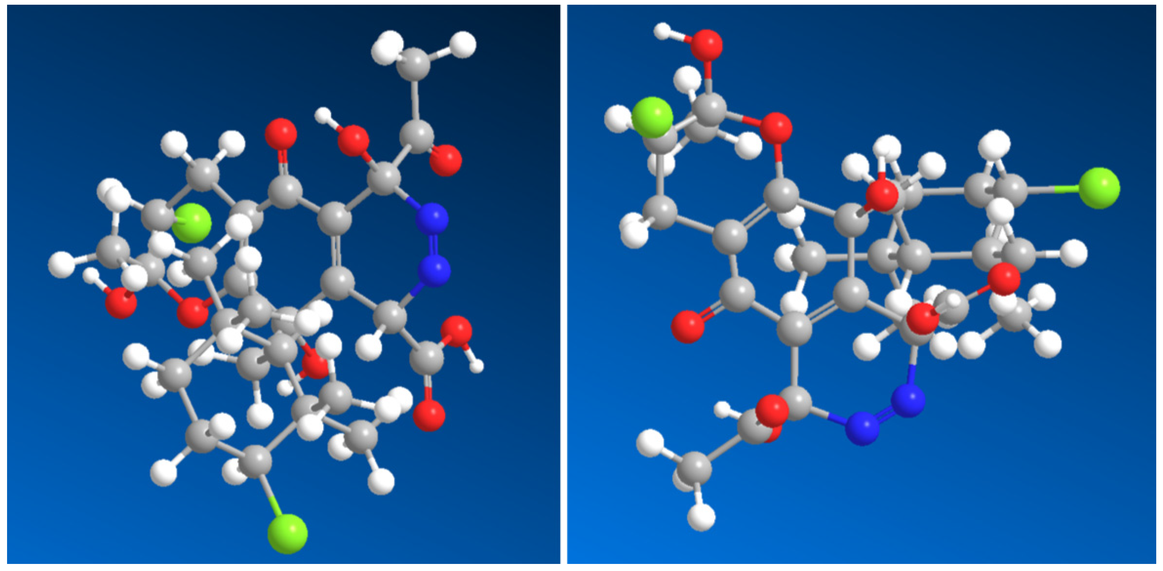
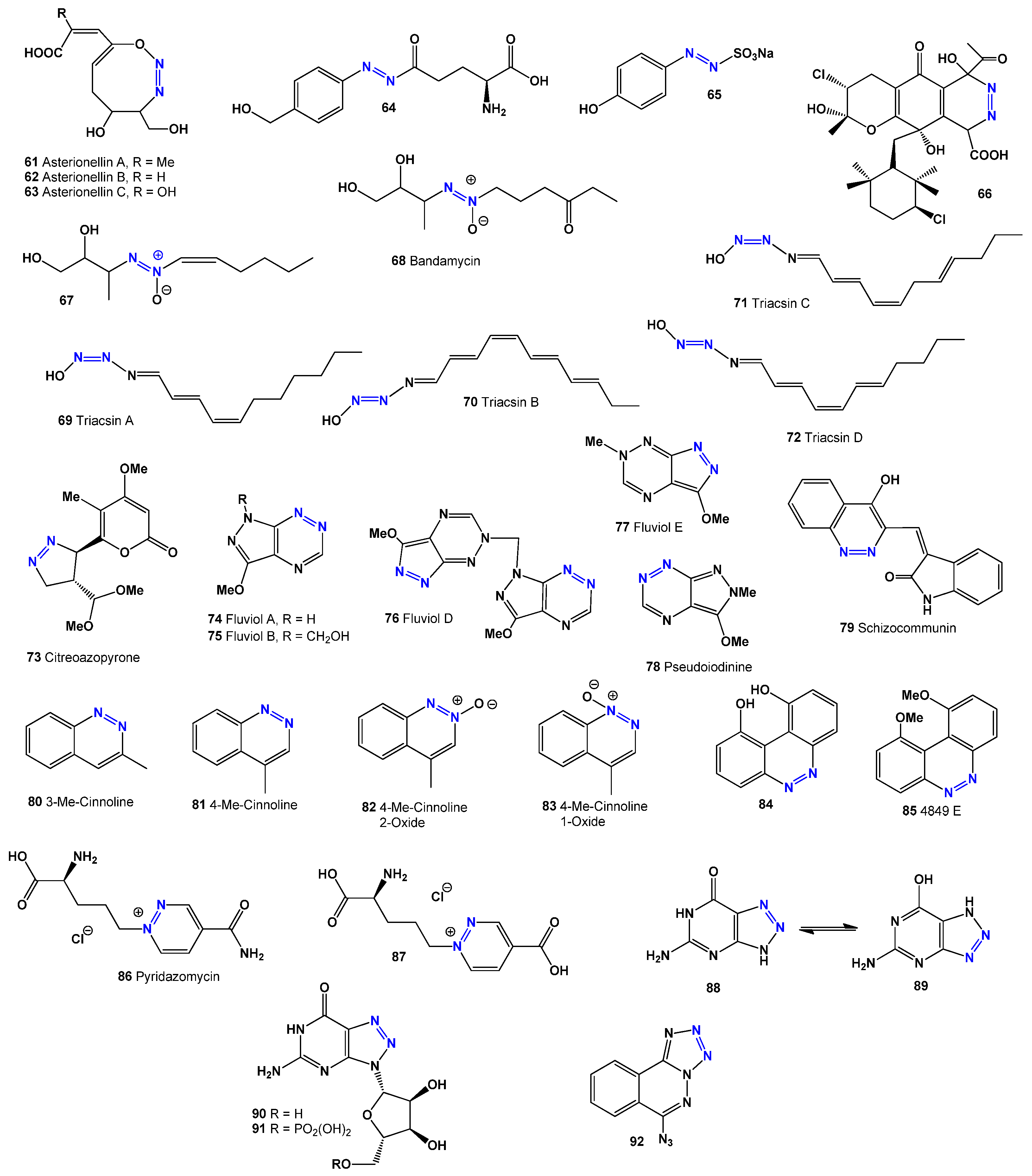
Azamerone, an anti-infective meroterpenoid obtained from marine Streptomyces sp. grown in salt culture, features a unique chloropyranphthalazinone core with a distinctive side chain. It showed mild cytotoxic effects on mouse T-cell splenocytes and macrophages in laboratory tests. Additionally, the biosynthetic precursor of its azo-containing derivative (66, 3D structure see in Figure 4) was also detected in the same Streptomyces sp. strain [86,87]. The Gram-positive bacterium Streptomyces sp. Ank75 produces azoxy antibiotics 67 and 68, which have antifungal properties against Candida albicans and Mucor miehei [88].
Researchers identified two vasodilators, WS-1228 A (triaxin C, 69) and WS-1228 B (triaxin D, 70), in the culture filtrate of Streptomyces aureofaciens [89,90]. A few years later, Omura and colleagues [91] discovered triaxins A (69) and B (70) in Streptomyces sp. SK-1894 and found that they function as acyl-CoA synthetase inhibitors. These compounds, along with WS-1228 A and B, share a unique N-hydroxytriazene structure and are known for their vasodilatory effects. Additionally, they inhibit acyl-CoA synthetase, with IC50 values of 5.5 μg/mL for triaxin A and 3.6 μg/mL for WS-1228 A. The triaxins (A, B, C, and D) differ in their inhibition levels, with triaxin C (71) being the most potent, followed by triaxin A, triaxin D (72), and triaxin B [92,93].
A rare compound, citreoazopyrone (73), was isolated from a hybrid strain of Penicillium citreo-viride and was found to inhibit lettuce seedling growth [94]. The fluviol groups of antibiotics (7477) and pseudoiodine (78), produced by Pseudomonas fluorescens and Nostoc spongiaeforme, exhibit antimicrobial, antiviral, and antitumor effects [95,96,97,98]. The activities of compounds 7898 are detailed in Table 5. Additionally, schizocommunin (79), isolated from the fungus Schizophyllum commune, showed strong cytotoxic effects against mouse lymphoma cells [99].
Table 4. Biological activity of azo dyes produced by microorganisms (42–60) [13,72,73,74,75,76,77,78].
No.Dominated ActivityRankAdditional ActivityRank
42Anti-inflammatoryStrongHemostaticStrong
43Anti-inflammatoryStrongHemostaticStrong
44Anti-inflammatoryStrongHemostaticStrong
45Kidney function stimulantWeakPre-neoplastic conditionsWeak
46ImmunosuppressantWeakFibrinolyticWeak
47Pre-neoplastic conditionsWeakKidney function stimulantWeak
48Anti-mutagenicStrongSpasmolytic, urinaryWeak
49Anti-seborrheicStrong
50CarminativeStrongAnti-microbialModerate
51Phobic disorders treatmentStrongKidney function stimulant (0.786)Moderate
52FibrinolyticModeratePre-neoplastic conditionsModerate
53AcaricideStrongAnti-seborrheicStrong
54Alopecia treatmentModerateAnti-inflammatory, intestinalWeak
55Anti-seborrheicStrongAcaricideModerate
56Pre-neoplastic conditions treatmentWeakImmunosuppressantWeak
57FibrinolyticModerateAcaricideWeak
58Mucositis treatmentStrongImmunosuppressantWeak
59Mucositis treatmentModerateImmunosuppressantWeak
60Antiviral(Arbovirus)WeakImmunosuppressantWeak
Researchers identified 3- and 4-methylcinnolines (80 and 81) in the volatile components of Hibiscus esculentus pods [100], while azoxy compounds (82 and 83) were found in yeast extract [101]. Two cinnoline derivatives, compound 84 and 4849F (85), were isolated from Streptomyces sp. 4849.171. Compound 84 functions as an IL-4 receptor inhibitor, while 4849F (85) shows antibacterial properties [102].
Pyridazomycin (86), an antifungal antibiotic from Streptomyces violaceoniger sp. griseofuscus, inhibits Mucor hiemalis [103,104]. Both pyridazomycin (86) and its chloride salt analogue (87) exhibit antimicrobial activity [105]. The compound 5-amino-1,4-dihydro-7H-1,2,3-triazolo [4,5-d]-pyrimidin-7-one (88 or 89), also called 8-azaguanine, is produced from guanine by Streptomyces albus [106]. Its cytotoxic effects on various cancers, including carcinoma and sarcoma, were first reported over 70 years ago [107].
Another form of 8-azaguanine, known as pathocidin (89), is an antifungal antibiotic obtained from Actinomycetes [108,109,110], which effectively inhibits fungal growth, including Penicillium chrysogenum. Two related natural metabolites, 8-azaguanine-3N-β-D-ribofuranosyl (90) and its 5′-phosphate version (91, see structure in Figure 5), have demonstrated anticancer effects against lymphoid leukemia and adenocarcinoma [111].
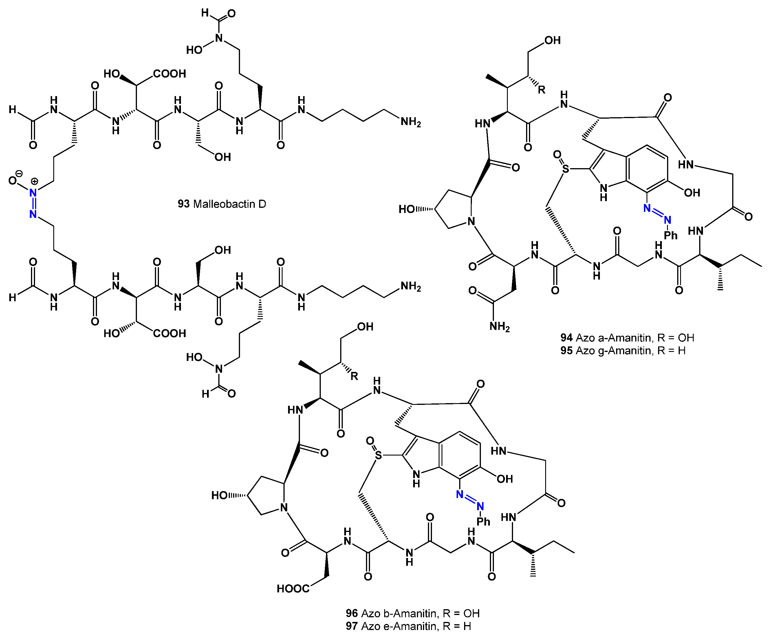
The red dinoflagellate Gymnodinium breve (syn. Ptychodiscus brevis) produces the antibiotic 6-azidotetrazolo [5,1-a]phthalazine (92) [112]. Additionally, the pathogenic Gram-negative Burkholderia species secretes extracellular enzymes involved in various biological processes, as well as toxins, antibiotics, and siderophores [113]. A rare dimeric siderophore, malleobactin D (93, see structure in Figure 6, and activity shown in Table 6), was identified in Burkholderia pseudomallei (formerly Pseudomonas pseudomallei) [114].
Amatoxins are toxic bicyclic octapeptides found in several fungi from the Agaricales family, including Amanita phalloides, A. ocreata, A. verna, A. bisporigera, Conocybe filaris, and Galerina marginata (syn. G. autumnalis), among others. These toxins are known for their harmful effects, including heat-induced toxicity, gastrointestinal issues, and the ability to inhibit RNA polymerase II [115,116,117,118]. Azoamanitins (9497), which are semi-synthetic versions of amatoxins, have been studied for their potential in antiviral, antimicrobial, and anticancer treatments [119,120,121].

3. Azo Dyes Derived from Terrestrial and Marine Sources

Azo dyes have been found in both terrestrial and marine environments, but it remains unclear whether bacteria or fungal endophytes played a role in their production, as these microorganisms often act as symbionts in plants and invertebrates [1,2,3,4,8,13,79]. Some key compounds isolated from terrestrial and marine sources include the following:
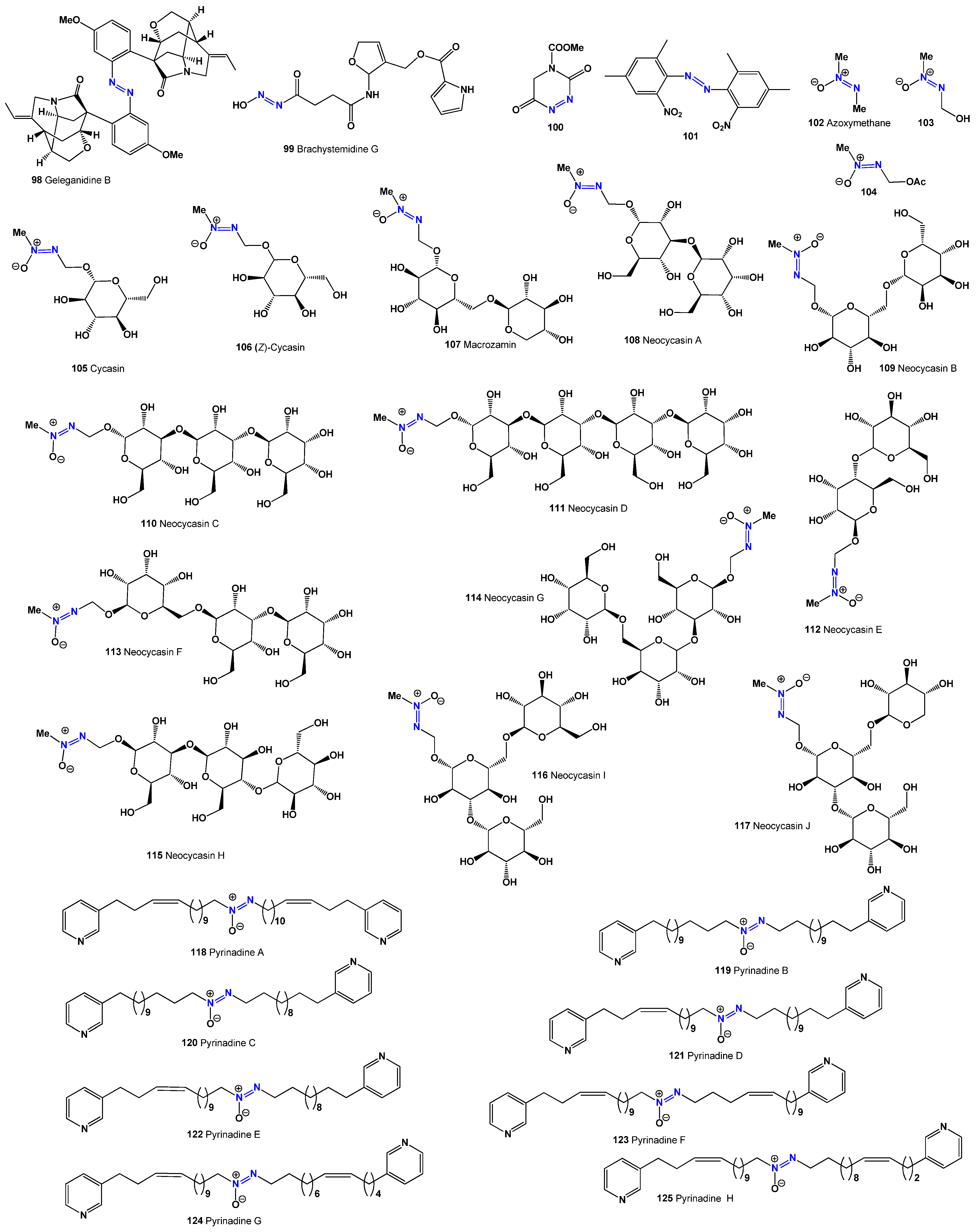
Geleganidin D (98, see structure in Figure 7 and activity in Table 7): A dimeric monoterpenoid indole alkaloid from Gelsemium elegans with moderate cytotoxic activity against MCF-7 and PC-12 cells [122]. G. elegans is used in clinical applications due to its indole alkaloids, which have antitumor, anti-inflammatory, analgesic, and immune-modulatory effects, though its therapeutic dose is close to toxic levels [122].
Brachystemidin G (99): An alkaloid from Brachystemma calycinum with potent immunosuppressant properties, inhibiting murine T- and B-lymphocyte proliferation (IC50 = 5.6 µg/mL) [123].
1,2,4-Triazine derivative (100): Extracted from Butea monosperma (family Caryophyllaceae) [124]. This plant’s odor repels mosquitoes, its flowers are used as a dye, and its gum, known as kamarkas in Hindi, is used in traditional cooking [125].
bis-(2,4-Dimethyl-6-nitrophenyl)-diazene (101): Isolated from the leaves of Aconitum sungpanense [126]. Azoxyglycosides, which contain the aglycone methylazoxymethanol (MAM), have been found in Cycadaceae plants, with all known glycosides having β-glycosidic linkages [127]. Metabolites, such as methylazoxymethane (102), MAM (103), methylazoxymethanol acetate (104), and cycasins (105,106), have been extracted from Cycadaceae, Stangeriaceae, and Zamiaceae, which are coniferous trees found in tropical and subtropical regions [127,128,129,130,131,132]. MAM (103) has been linked to liver and kidney carcinomas [133], while cycasins (105,106) and macrosamine (107) are highly toxic azoxyglycosides. Various insects, including caterpillars and weevils, feed on cycads’ roots, stems, leaves, and reproductive parts [127,132].
Azoxyglycosides are believed to function as natural herbivore deterrents [134,135,136]. Cycasin, the primary azoxyglycoside found in cycads, has been shown to cause effects similar to those seen in carcinogenicity, mutagenicity, and neurotoxicity tests. The first isolation of neocycasin A (108) was recorded in 1959 [137], and, more recently, neocycasins B–J (109117) have been identified from different plant species [138,139,140,141,142,143,144,145,146].
The first cytotoxic bis-3-alkylpyridine alkaloid with an azoxy moiety, pyrinadine A (118), was discovered in the Okinawan marine sponge Cribrochalina sp. [147,148]. Further cytotoxic bis-3-alkylpyridine alkaloids, pyrinadines B–H (118125), were later extracted from the same sponge, demonstrating cytotoxicity against P388 murine leukemia cells [147,148,149]. The activities of these compounds (118125) are summarized in Table 7.
Table 7. Biological activity of azo dyes produced by microorganisms (98125) [13,122,137,144].
No.Dominated ActivityRankAdditional ActivityRank
98AntineoplasticModerateAntiprotozoal (Plasmodium)Weak
99β-1,3-Galactosyl-O-glycosyl-glycoproteinStrongβ-1,6-N-acetylglucosaminyl
transferase inhibitor
Strong
100Neurodegenerative diseases treatmentStrong
101AntiparkinsonianStrongAnxiolyticStrong
102AcaricideModerateAntiviral (Arbovirus)Weak
103AntineoplasticStrong
104AntineoplasticModerate
105AntineoplasticModerate
106Anti-infertility, femaleStrongAntineoplasticStrong
107CarcinogenicStrongEmbryotoxicStrong
108CarcinogenicStrongEmbryotoxicStrong
109AntineoplasticStrong
110CarcinogenicStrongEmbryotoxicStrong
111AntineoplasticStrong
112AntineoplasticStrongAnti-infectiveStrong
113AntineoplasticStrongAnti-infectiveStrong
114AntineoplasticStrongAnti-infectiveStrong
115AntineoplasticStrongAnti-infectiveStrong
116AntineoplasticStrongAnti-infectiveStrong
117AntineoplasticStrongAnti-infectiveStrong
118CytotoxicModerateAnti-inflammatoryWeak
119CytotoxicModerateAnti-inflammatoryWeak
120CytotoxicModerateAnti-inflammatoryWeak
121CytotoxicModerateAnti-inflammatoryWeak
122CytotoxicModerateAnti-inflammatoryWeak
123CytotoxicModerateAnti-inflammatoryWeak
124CytotoxicModerateAnti-inflammatoryWeak
125CytotoxicModerateAnti-inflammatoryWeak

4. Microbial Degradation of Textile Azo Dyes

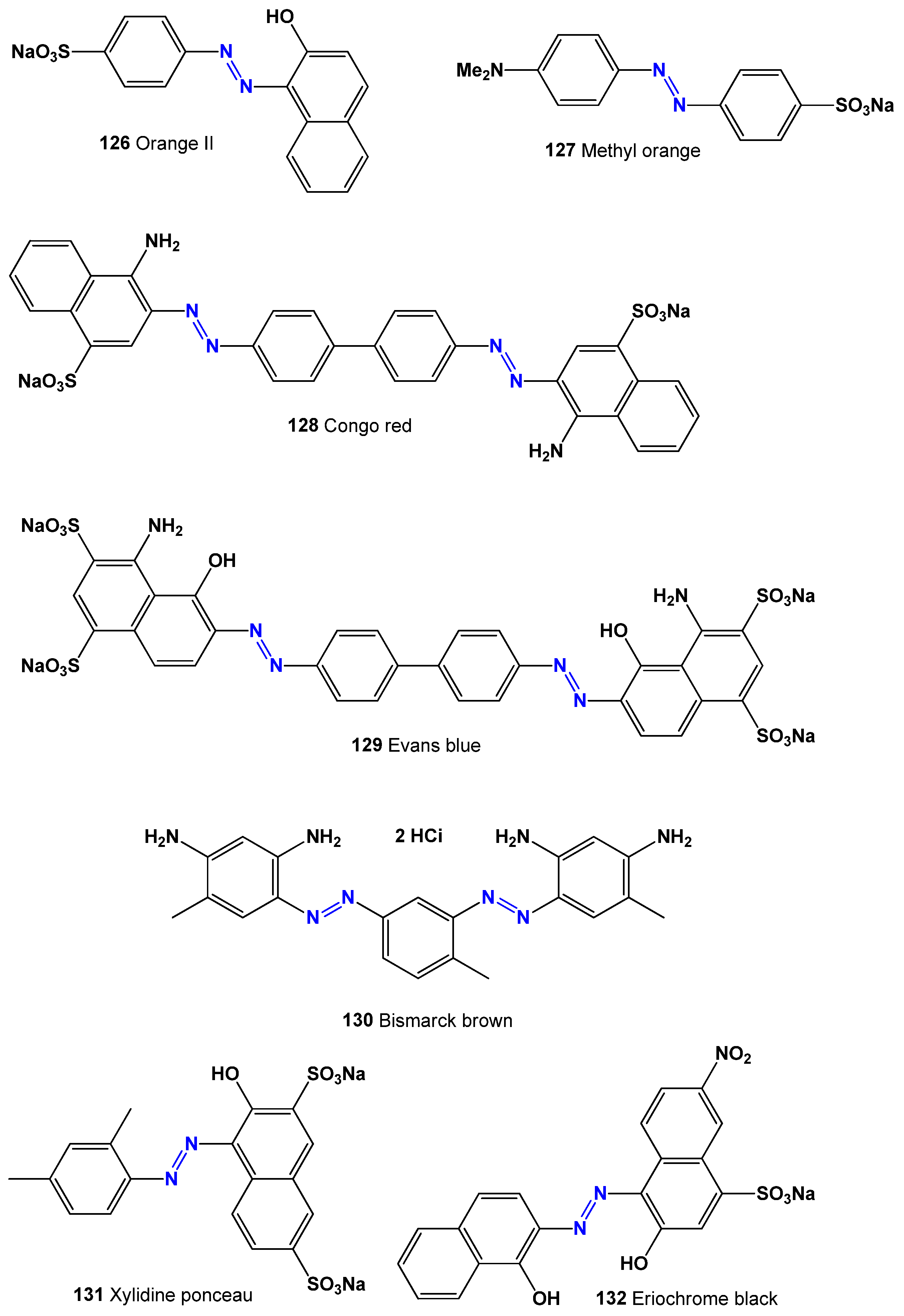
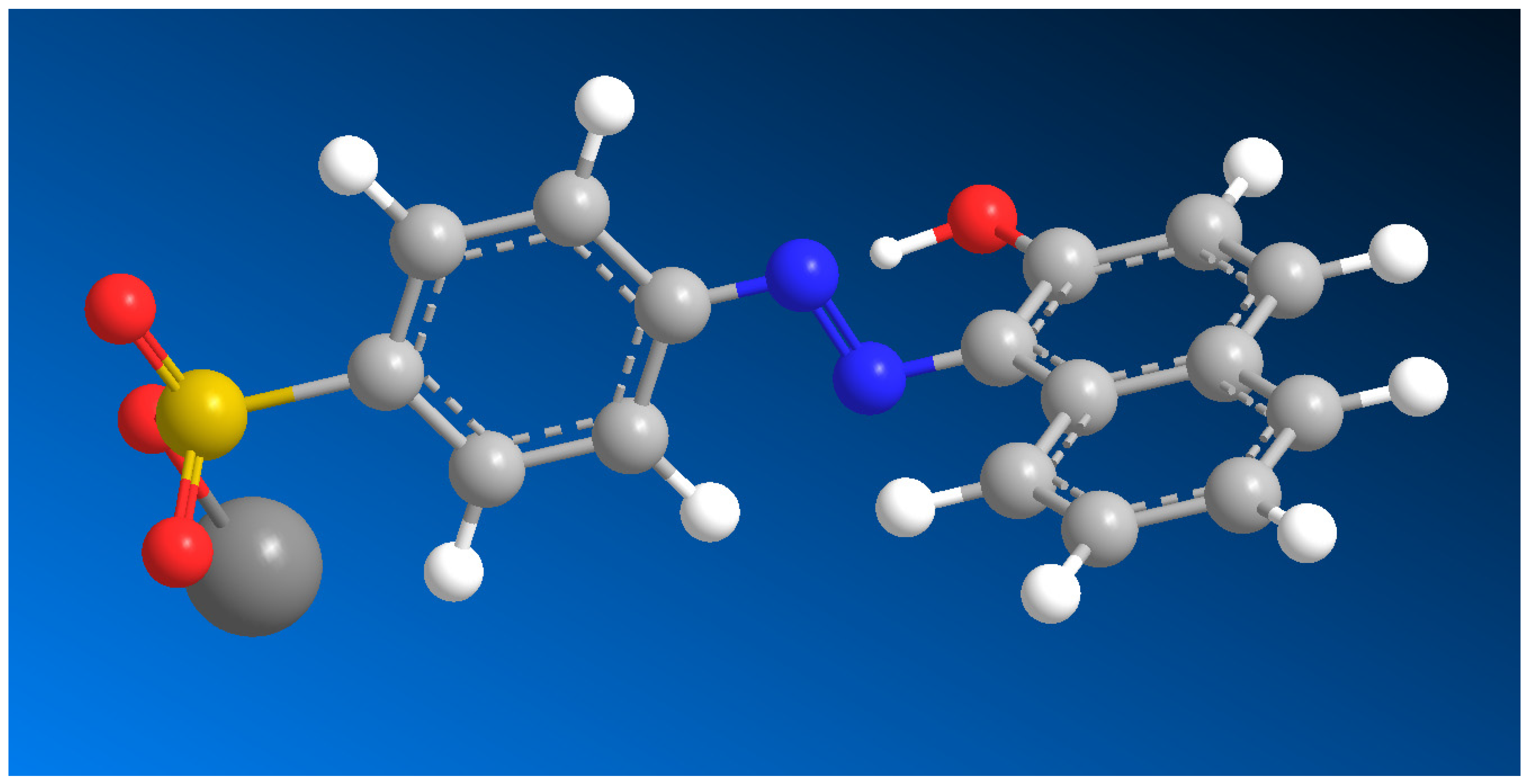
The textile industry relies heavily on both natural and synthetic azo dyes [150,151,152]. Natural dyes have been used since ancient times and vary in color and chemical structure. Among synthetic azo dyes, commonly used examples include Orange II (126, see 3D structure in Figure 8 and activity in Table 8), Methyl Orange (127), Congo Red (128), Evans Blue (129), Bismarck Brown (130), Xylidine Ponceau (131), and Eriochrome Black (132). These dyes are widely applied in the textile and leather industries but contribute to environmental pollution, particularly through wastewater discharge. This contamination affects ecosystems, microbial communities in the soil, and the natural balance of certain species.
Azo dyes are aromatic compounds containing azo (-N=N-) and sulfonic (-SO₃-) groups. They are produced through the diazotization of aromatic amines, followed by a coupling reaction, creating a range of colors from yellow to red and orange [150,151,152].
Every year, inefficient dyeing processes result in the release of hundreds of thousands of tons of textile dyes into wastewater. This improper disposal has severe environmental consequences and poses health risks to humans and animals. Many azo dyes are known to be carcinogenic and mutagenic [153,154,155,156]. While various wastewater treatment methods exist, one promising approach is the use of microorganisms capable of degrading azo dyes naturally [157,158,159,160].
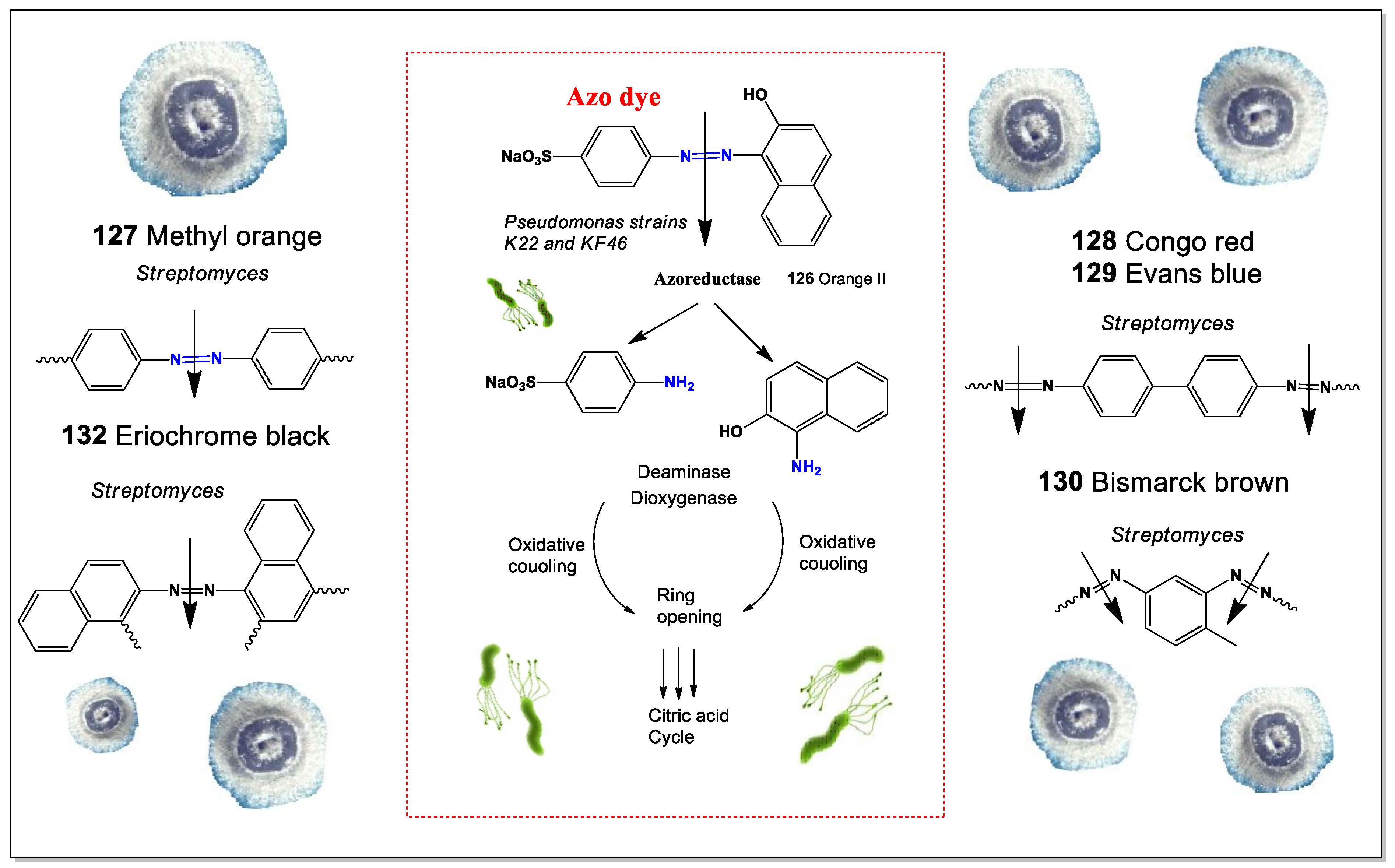
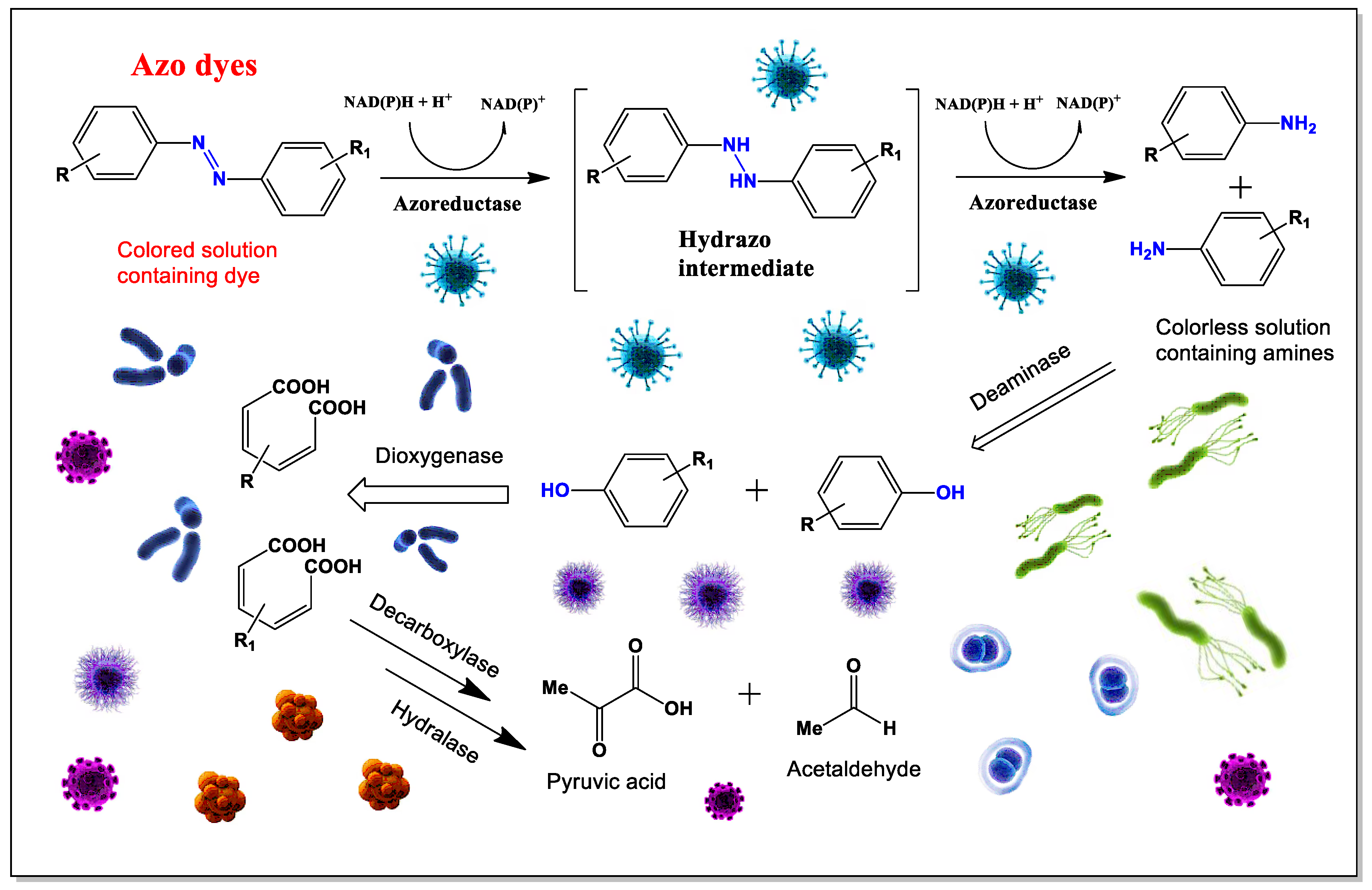
Extensive research has been conducted in this field, with numerous studies published annually. However, in this discussion, we will focus on how Actinomyces and other microbes contribute to azo dye degradation [161,162,163]. Several species of Streptomyces have been found to completely break down azo pigments (128–133) (see structures in Figure 9). These include S. chromofuscus, S. coelicolor, S. viridosporus, S. rochei, S. psammoticus, S. diastaticus, S. ipomoeae, S. sviceus, S. ardesiacus, and S. cyaneus [164,165,166,167]. In addition, a mixed culture of two Pseudomonas strains has shown effective degradation of azo dyes. Microbial enzymes known as azoreductases play a crucial role in this process by cleaving azo bonds (-N=N-) under mild conditions (see Figure 10). These enzymes are valuable for developing environmentally friendly dye removal methods [168,169].
Table 8. Biological activity of textile azo dyes [164,165,166,167,168,169].
No.Dominated ActivityRankAdditional ActivityRank
126EmbryotoxicStrong
127CarcinogenicStrongMutagenicStrong
128CarcinogenicStrongMutagenicStrong
129AntineoplasticStrong
130AntiviralStrong
131AntibacterialModerateAntifungalModerate
132AntimicrobialModerateAntibacterialModerate

5. Microbial Degradation of Food Azo Dyes

Azo dyes are widely used in the food industry as color additives in products, such as soft drinks, jams, candies, and pickles (azo dye coloring in Figure 11) [170]. Their popularity comes from their ability to produce bright, vibrant colors while being inexpensive, stable, and free from unwanted tastes or odors.
However, while azo dyes make food visually appealing, they may also pose health risks. Consuming them within the acceptable daily intake is generally considered safe, but higher doses—especially in children—have been linked to various health issues. These include allergies, ADHD, asthma, anxiety, and even potential cytotoxic and genotoxic effects, which may contribute to cancer development. Additionally, azo dyes are highly stable and do not break down easily under aerobic conditions [171,172,173].
Several synthetic azo dyes, such as Sunset Yellow (133, see structure in Figure 12, and activity see in Table 9), Carmoisine (134), Amaranth (135), Ponceau 4R (136), Brown HT (137), and Allura Red (138), are commonly added to food [170]. They enhance color but offer no nutritional benefits, preservation properties, or health advantages. Because they are cost-effective, chemically stable, and provide intense coloration without altering taste, the food industry often prefers them over natural dyes.
Although food dyes are regularly tested for safety, their health effects remain controversial. Some studies suggest that the breakdown of azo dyes can produce harmful byproducts, raising concerns about their long-term safety [174,175].
Food azo dyes may have potential health risks, primarily due to their breakdown by intestinal bacteria after ingestion. When these dyes are reduced by gut microflora, their azo bonds are cleaved, producing aromatic amines. These amines can then undergo further chemical modifications, such as N-hydroxylation or N-acetylation. In mammals, azo dye reduction mainly takes place in the anaerobic environment of the lower gastrointestinal tract, where bacterial azoreductase enzymes are responsible for the process. While mammalian enzymes in the liver and kidney also contribute to azo dye metabolism, their exact role is not yet fully understood [176,177].
Many anaerobic bacteria found in the cecal contents of both humans and experimental animals have been shown to break down azo dyes into aromatic amines. This reaction is facilitated by azoreductases, which require flavins for optimal activity and are highly sensitive to oxygen [172,173,174,175,176,177].
Aromatic amines and their derivatives are a major class of chemicals formed when azo dyes are broken down by azoreductases, which cleave the azo bond. Different microorganisms produce azoreductases with varying kinetic properties and substrate specificities. Research shows that flavin-containing azoreductases follow a Ping-Pong bi-bi reaction mechanism, where the enzyme’s activity depends on the substrate concentration. Many azoreductases show a higher affinity for NADPH over NADH, although they can utilize either as an electron source, with or without flavin [178,179,180].
Not all bacteria produce azoreductases, but several known species do. These include strains from both animals and humans, such as Aquiflexum sp., Bacillus sp., Clostridium perfringens, Escherichia coli, Enterococcus faecalis, Pseudomonas aeruginosa, Rhodobacter sphaeroides, Shewanella oneidensis, Staphylococcus aureus, and Xenophilus azovorans KF46F [178,181]. Several azo dyes are commonly used in food production:
Sunset Yellow (133) is an artificial dye used to enhance food appearance and shelf life. It has antioxidant properties, which increase with concentration, but also exhibits phytotoxicity [182,183,184].
Carmoisine (134) is a red azo dye used in foods that undergo heat treatment after fermentation. While azo dyes have been linked to carcinogenicity, especially bladder cancer, carmoisine itself has not been proven carcinogenic. Interestingly, its copper complex has demonstrated anticancer activity against colon cancer cells (HT-29), making it a potential candidate for future treatments [185,186,187].
Amaranth (135) is a red food dye used worldwide, although its use is banned in India due to concerns about its safety. Despite this, illegal production continues, with India contributing 2% of global output. Some studies suggest that amaranth may be carcinogenic under certain conditions [188,189].
Ponceau 4R (136) is widely used in pharmaceuticals and food products. It is stable in light, heat, and acidic conditions but breaks down when exposed to ascorbic acid. While approved in Europe, Asia, and Australia, the US FDA has not approved it due to concerns about its potential to form non-sulfone aromatic amines, which may be carcinogenic [190,191].
Brown HT (137), also called “food brown 3” or “chocolate brown HT”, is a pigment with antioxidant properties. It is found in small amounts in coal tar [192,193,194].
Allura Red (138) is a widely used food dye, commonly found in sweets, beverages, and processed foods. It dissolves in water but poorly in 50% ethanol. Long-term consumption may cause liver and kidney toxicity [195,196,197]. Degradation of azo dyes and the cleavage of the (-N=N-) bond are shown in Figure 13.

6. Conclusions

Natural and/or synthetic azo compounds represent a valuable class of bioactive molecules with diverse pharmacological applications. Their confirmed antitumor, antimicrobial, and antibacterial properties, along with predicted novel activities, highlight their potential as drug prototypes and leads in pharmaceutical development. The role of microorganisms in the biosynthesis of natural azo compounds and their synthetic analogs further expands opportunities for drug discovery and therapeutic advancements. Additionally, the synthesis and degradation pathways of azo dyes present promising applications in medicine, food, cosmetics, and related industries. Continued research into the structure–activity relationships and biological mechanisms of azo compounds will be essential for unlocking their full potential in improving human health.

Author Contributions

Conceptualization, V.M.D.; methodology, V.M.D.; software, A.O.T.; investigation, V.M.D.; resources, V.M.D.; writing—original draft preparation, A.O.T. and V.M.D.; writing—review and editing, A.O.T. and V.M.D. All authors have read and agreed to the published version of the manuscript.

Funding

This research received no funding.

Conflicts of Interest

The authors declare no conflicts of interest.

References

  1. Bafana, A.; Devi, S.S.; Chakrabarti, T. Azo dyes: Past, present and the future. Environ. Rev. 2011, 19, 350–371. [Google Scholar] [CrossRef]
  2. Goswami, D.; Mukherjee, J.; Mondal, C.; Bhunia, B. Bioremediation of azo dye: A review on strategies, toxicity assessment, mechanisms, bottlenecks and prospects. Sci. Total Environ. 2024, 954, 176426. [Google Scholar] [CrossRef]
  3. Kolya, H.; Kang, C.W. Recent advances in polymer nanocomposites for the adsorptive removal of toxic azo dyes from water. Discov. Water 2025, 5, 28. [Google Scholar] [CrossRef]
  4. Aljamali, N.M. Review in azo compounds and its biological activity. Biochem. Anal. Biochem. 2015, 4, 169. [Google Scholar] [CrossRef]
  5. Benkhaya, S.; M’rabet, S.; El Harfi, A. Classifications, properties, recent synthesis and applications of azo dyes. Heliyon 2020, 6, e03271. [Google Scholar] [CrossRef]
  6. Eltaboni, F.; Bader, N.; El-Kailany, R.; Elsharif, N.; Ahmida, A. Dyes: A comprehensive review. J. Chem. Rev. 2022, 4, 313–330. [Google Scholar]
  7. Tyagi, S.; Kapoor, R.T.; Singh, R.; Shah, M.P. Insights on microbial enzymes mediated biodegradation of azo dyes: A sustainable strategy for environment clean up. Bioremediat. J. 2025, 1–43. [Google Scholar] [CrossRef]
  8. Shindy, A. Problems and solutions in colors, dyes and pigments chemistry: A review. Chem. Int. 2017, 3, 97–105. [Google Scholar]
  9. Yandovskii, V.N.; Gidaspov, B.V.; Tselinskii, V.E. The formation of the azoxy-group in reactions involving anions with nitrogen–nitrogen bonds. Russ. Chem. Rev. 1980, 49, 237. [Google Scholar] [CrossRef]
  10. Blair, L.M.; Sperry, J. Natural products containing a nitrogen-nitrogen bond. J. Nat. Prod. 2013, 76, 794–812. [Google Scholar] [CrossRef]
  11. Khan, A.; Ju, P.; Han, Z.; Ni, C. A comprehensive review on adsorptive removal of azo dyes using functional materials. AQUA—Water Infrastruct. Ecosyst. Soc. 2024, 73, 266–285. [Google Scholar] [CrossRef]
  12. Katsuyama, Y.; Matsuda, K. Recent advance in the biosynthesis of nitrogen–nitrogen bond–containing natural products. Curr. Opin. Chem. Biol. 2020, 59, 62–68. [Google Scholar] [CrossRef]
  13. Dembitsky, V.M.; Gloriozova, T.A.; Poroikov, V.V. Pharmacological and predicted activities of natural azo compounds. Nat. Prod. Bioprospect. 2017, 7, 151–169. [Google Scholar] [CrossRef]
  14. He, H.Y.; Niikura, H.; Du, Y.L.; Ryan, K.S. Synthetic and biosynthetic routes to nitrogen–nitrogen bonds. Chem. Soc. Rev. 2022, 51, 2991–3046. [Google Scholar] [CrossRef]
  15. Howard, J.B.; Rees, D.C. Structural basis of biological nitrogen fixation. Chem. Rev. 1996, 96, 2965–2982. [Google Scholar] [CrossRef] [PubMed]
  16. Khan, M.I.; Lee, J.; Park, J. Microbial degradation and toxicity of hexahydro-1,3,5-trinitro-1,3,5-triazine. J. Microbiol. Biotechnol. 2012, 22, 1311–1323. [Google Scholar] [CrossRef]
  17. Nawrat, C.C.; Moody, C.J. Natural products containing a diazo group. Nat. Prod. Rep. 2011, 28, 1426–1444. [Google Scholar] [CrossRef]
  18. Vey, J.L.; Al-Mestarihi, A.; Hu, Y.; Funk, M.A.; Bachmann, B.O.; Iverson, T.M. Structure and mechanism of ORF36, an amino sugar oxidizing enzyme in everninomicin biosynthesis. Biochemistry 2010, 49, 9306–9317. [Google Scholar] [CrossRef] [PubMed]
  19. Yamato, M.; Iinuma, H.; Naganawa, H.; Yamagishi, Y.; Hamada, M.; Masuda, T.; Umezawa, H.; Abe, V.; Hori, M. Isolation and properties of valanimycin, a new azoxy antibiotic. J. Antibiot. 1986, 39, 184–191. [Google Scholar] [CrossRef]
  20. Ma, Y.; Parry, R.J. A novel valanimycin-resistance determinant (vlmF) from Streptomyces viridifaciens MG456-hF10. Microbiology 2000, 146, 345–352. [Google Scholar] [CrossRef]
  21. Bianchi, G.; Dallavalle, S.; Merlini, L.; Nasini, G.; Quaroni, S. A new azoxyalkene from a strain of an actinomadura-like fungus. Planta Med. 2003, 69, 574–576. [Google Scholar] [PubMed]
  22. McGahren, W.J.; Kunstmann, M.P. Novel .alpha.,.beta.-unsaturated azoxy-containing antibiotic. J. Am. Chem. Soc. 1969, 91, 2808–2810. [Google Scholar] [CrossRef] [PubMed]
  23. McGahren, W.J.; Kunstmann, M.P. Circular dichroism studies on the azoxy chromophore of the antibiotics LL-BH872.alpha. and elaiomycin. J. Am. Chem. Soc. 1970, 92, 1587–1590. [Google Scholar] [CrossRef]
  24. Nakayama, M.; Takahashi, Y.; Itoh, H.; Kamiya, K.; Shiratsuchi, M.; Otani, G. Novel antifungal antibiotics maniwamycins A and B. I. Taxonomy of the producing organism, fermentation, isolation, physico-chemical properties and biological properties. J. Antibiot. 1989, 42, 1535–1540. [Google Scholar] [CrossRef]
  25. Fujiu, M.; Sawairi, S.; Shimada, H.; Takaya, H.; Aoki, Y.; Okuda, T.; Yokose, K. Azoxybacilin, a novel antifungal agent produced by Bacillus cereus NR2991. Production, isolation and structure elucidation. J. Antibiot. 1994, 47, 833. [Google Scholar]
  26. Aoki, Y.; Kondoh, M.; Nakamura, M.; Fujii, T.; Yamazaki, T.; Shimada, H.; Arisawa, M. A new methionine antagonist that has antifungal activity: Mode of action. J. Antibiot. 1994, 47, 909–916. [Google Scholar] [CrossRef]
  27. Ehrlich, J.; Anderson, L.E.; Coffey, G.L.; Feldman, W.H. Elaiomycin, a new tuberculostatic antibiotic; biologic studies. Antibiot. Chemother. 1954, 4, 338–342. [Google Scholar]
  28. Anderson, L.E.; Ehrlich, J.; Sun, S.H.; Burkholder, P.R. Strains of Streptomyces, the sources of azaserine, elaiomycin, griseoviridin, and viridogrisein. Antibiot. Chemother. 1956, 6, 100–115. [Google Scholar]
  29. Haskell, T.H.; Ryder, A.; Bartz, Q.R. Elaiomycin, a new tuberculostatic antibiotic; isolation and chemical characterization. Antibiot. Chemother. 1954, 4, 141. [Google Scholar]
  30. Ohkuma, K.; Nakamura, G.; Yamashita, S. An antibiotic produced by Streptomyces, strain no. 1252, identical with elaiomycin. J. Antibiot. 1957, 10, 224–225. [Google Scholar]
  31. Manderscheid, N.; Helaly, S.E.; Kulik, A.; Wiese, J. Elaiomycins K and L, new azoxy antibiotics from Streptomyces sp. Tü 6399. J. Antibiot. 2013, 66, 85–88. [Google Scholar] [CrossRef]
  32. Ding, L.; Ndejouong, B.S.; Maier, A.; Fiebig, H.H.; Hertweck, C. Elaiomycins D-F, antimicrobial and cytotoxic azoxides from Streptomyces sp. strain HKI0708. J. Nat. Prod. 2012, 75, 1729–1734. [Google Scholar] [CrossRef]
  33. Omura, S.; Otoguro, K.; Imamura, N.; Kuga, H. Jietacins A and B, new nematocidal antibiotics from a Streptomyces sp. Taxonomy, isolation, and physico-chemical and biological properties. J. Antibiot. 1987, 40, 623–629. [Google Scholar] [CrossRef] [PubMed]
  34. Imamura, N.; Kuga, H.; Otoguro, K.; Tanaka, H.; Omura, S. Structures of jietacines: Unique α, β-unsaturated azoxy antibiotics. J. Antibiot. 1989, 42, 156. [Google Scholar] [CrossRef] [PubMed]
  35. Tsuzuki, K.; Yan, F.S.; Otoguro, K.; Omura, S. Synthesis and nematocidal activities of jietacin A and its analogs. J. Antibiot. 1991, 44, 774–784. [Google Scholar] [CrossRef] [PubMed]
  36. Egerton, J.R.; Ostlind, D.A.; Blair, L.S.; Eary, C.H. Avermectins, new family of potent anthelmintic agents: Efficacy of the B1a component. Antimicrob. Agents Chemother. 1979, 15, 372–378. [Google Scholar] [CrossRef]
  37. Sugawara, A.; Kubo, M.; Nakashima, T.; Hirose, T. Jietacins with potent nematocidal activity; efficient isolation of novel analogs and divergent total synthesis of jietacin A, B, C, and D. Tetrahedron 2015, 71, 2149–2157. [Google Scholar] [CrossRef]
  38. Le Goff, G.; Martin, M.T.; Iorga, B.I.; Adelin, E. Isolation and characterization of unusual hydrazides from Streptomyces sp. impact of the cultivation support and extraction procedure. J. Nat. Prod. 2013, 76, 142–149. [Google Scholar] [CrossRef]
  39. Abe, Y.; Kadokura, J.I.; Shimazu, A.; Seto, H.; Otake, N. Propanosine (K-76), a new antibiotic active against Valsa ceratosperma, the pathogen of apple canker disease. Agric. Biol. Chem. 1983, 47, 2703–2705. [Google Scholar] [CrossRef]
  40. Chan, W.Y.; Ng, T.B.; Lam, J.S.; Wong, J.H. The mushroom ribosome-inactivating protein lyophyllin exerts deleterious effects on mouse embryonic development in vitro. Appl. Microbiol. Biotechnol. 2010, 85, 985–993. [Google Scholar] [CrossRef]
  41. Nakano, H.; Hara, M.; Katsuyama, T.; Uozaki, Y.; Gomi, K. Bioactive azo compounds. Chem. Abstr. 1993, 119, 158353. [Google Scholar]
  42. Nakaynma, M.; Ito, H.; Watanabe, I.; Shiratsuchi, M. Novel azoxy compounds and process for production thereof. U.S. Patent 4,981,954, 1 January 1991. [Google Scholar]
  43. Umezawa, H.; Takeuchi, T.; Iinuma, H.; Ito, M.; Ishizuka, M. A new antibiotic, calvatic acid. J. Antibiot. 1975, 28, 87–90. [Google Scholar] [CrossRef]
  44. Calvino, R.; Fruttero, R.; Gasco, A.; Miglietta, A.; Gabriel, L. Chemical and biological studies on calvatic acid and its analogs. J. Antibiot. 1986, 39, 864–868. [Google Scholar] [CrossRef] [PubMed]
  45. Gasco, A.; Serafino, A.; Mortarinin, V.; Menziani, E. An antibacterial and antifungal compound from Calvatia lilacina. Tetrahedron Lett. 1974, 38, 3431. [Google Scholar] [CrossRef]
  46. Brossa, O.; Gadoni, E.; Olivero, A.; Seccia, M.; Miglietta, A.; Gabriel, L.; Gravela, E. Effect of calvatic acid and its analogs on ornithine decarboxylase activity in tumour cells. Res. Commun. Chem. Pathol. Pharmacol. 1990, 70, 143–153. [Google Scholar]
  47. Boschi, D.; Cena, C.; Fruttero, R.; Brenciaglia, M.I. Activity of calvatic acid and its analogs against Helicobacter pylori. Pharm. 2001, 56, 670–672. [Google Scholar]
  48. Huttner, A.; Bielicki, J.; Clements, M.N.; Frimodt-Møller, N.; Muller, A.E.; Paccaud, J.P.; Mouton, J.W. Oral amoxicillin and amoxicillin–clavulanic acid: Properties, indications and usage. Clin. Microbiol. Infect. 2020, 26, 871–879. [Google Scholar] [CrossRef] [PubMed]
  49. Mancabelli, L.; Mancino, W.; Lugli, G.A.; Argentini, C.; Longhi, G.; Milani, C.; Turroni, F. Amoxicillin-clavulanic acid resistance in the genus Bifidobacterium. Appl. Environ. Microbial. 2021, 87, e03137-20. [Google Scholar] [CrossRef]
  50. Anoush, M.; Pourmansouri, Z.; Javadi, R.; GhorbanPour, B.; Sharafi, A.; Mohamadpour, H.; Andalib, S. Clavulanic acid: A novel potential agent in prevention and treatment of scopolamine-induced Alzheimer’s disease. ACS Omega 2022, 7, 13861–13869. [Google Scholar] [CrossRef]
  51. Miglietta, A.; Gadoni, E.; Buffa, M.; Olivero, A.; Gabriel, L. Interactions between calvatic acid and related compounds with rat liver microsomes. Eur. J. Drug Metab. Pharm. 1995, 20, 249–254. [Google Scholar] [CrossRef]
  52. Okuda, T.; Nakayama, N.; Fujiwara, A. Calvatic acid production by the lycoperdaceae 1. calvatic acid and related compounds produced by Lycoperdon pyriforme. Trans. Mycol. Soc. Jpn. 1982, 23, 225–234. [Google Scholar]
  53. Okuda, T.; Fujiwara, A. Calvatic acid production by the Lycoperdaceae, 2: Distribution among the Gasteromycetes. Nippon. Kingakkai Kaiho 1984, 23, 235–239. [Google Scholar]
  54. Takaishi, Y.; Murakami, Y.; Uda, M.; Ohashi, T.; Hamamura, N.; Kidota, M.; Kadota, S. Hydroxyphenyl-azoformamide derivatives from Calvatia craniformis. Phytochem 1997, 45, 997–1001. [Google Scholar] [CrossRef]
  55. Wang, J.; Liu, L.; Li, Y.; Liu, S. Detection of azoformamide in flour based on Raman hyperspectral imaging. Electr. Measurem. Technol. 2024, 45, 97–102. [Google Scholar]
  56. Köpcke, B.; Mayer, A.A.H.; Sterner, O. Bioactive azo- and azoxyformamides from Lycoperdon pyriforme (Schaeff. Ex Pers.). Nat. Prod. Lett. 1999, 13, 41–46. [Google Scholar] [CrossRef]
  57. La Rue, T.A. Naturally occurring compounds containing a nitrogen-nitrogen bond. Lloydia 1977, 40, 307–321. [Google Scholar]
  58. Kamo, T.; Kashiwabara, M.; Tanaka, K.; Ando, S.; Shibata, H.; Hirota, M. Plant growth inhibitory activity of azo-and azoxyformamides from Calvatia craniiformis and Lycoperdon hiemale. Nat. Prod. Res. 2006, 20, 507–510. [Google Scholar] [CrossRef]
  59. Gill, M. Pigments of fungi (Macromycetes). Nat. Prod. Rep. 2003, 20, 615–639. [Google Scholar] [CrossRef]
  60. Coetze, J.C.; Van Wyk, A.E. The genus Calvatia (‘Gasteromycetes’, Lycoperdaceae): A review of its ethnomycology and biotechnological potential. Afr. J. Biotechnol. 2009, 8, 6007–6015. [Google Scholar]
  61. Wu, J.S.; Zhu, F. Chemical constituents pre-analysis and antioxidant activity of the Puffball Calvatia candida from Foshan. J. Org. Chem. Res. 2016, 4, 69–76. [Google Scholar] [CrossRef]
  62. Fugmann, B.; Arnold, S.; Steglich, W.; Fleischhauer, J.; Repges, C.; Koslowski, A.; Raabe, G. Pigments from the Puffball Calvatia rubro-flava—Isolation, structural elucidation and synthesis. Eur. J. Org. Chem. 2001, 16, 3097–3104. [Google Scholar] [CrossRef]
  63. Claydon, N. Insecticidal secondary metabolites from entomogenous fungi: Entomophthora virulenta. J. Inverteb. Pathol. 1978, 32, 319–324. [Google Scholar] [CrossRef]
  64. Roberts, D.W. Toxins of entomopathogenic fungi. In Microbia Control of Pests and Plant Diseases 1970–1980; Burges, H.D., Ed.; Academic Press: New York, NY, USA, 1981; pp. 441–464. [Google Scholar]
  65. Tkach, V.V.; Storoshchuk, N.M.; Gaivão, I.; Ivanushko, Y.G.; Yagodynets, P.I.; Morozova, T.V.; Garcia, J.R. Electrochemical sensor for sucralose and its genotoxic ester. A mathematical description. Span. J. Environ. Mutagen. Genom. 2024, 28, 45. [Google Scholar]
  66. Amini, M.; Yousefi-Massumabad, H.; Younesi, H.; Abyar, H.; Bahramifar, N. Production of the polyhydroxyalkanoate biopolymer by Cupriavidus necator using beer brewery wastewater containing maltose as a primary carbon source. J. Environ. Chem. Engineer. 2020, 8, 103588. [Google Scholar] [CrossRef]
  67. Suortti, T. Stability of necatorin, a highly mutagenic compound from Lactarius necator mushroom. Food Chem. Toxicol. 1984, 22, 579–581. [Google Scholar] [CrossRef] [PubMed]
  68. Suortti, T.; von Wright, A. Isolation of a mutagenic fraction from aqueous extracts of the wild edible mushroom Lactarius necator (a preliminary note). J. Chromatogr. 1983, 255, 529–532. [Google Scholar] [CrossRef]
  69. Gill, M.; Strauch, R.J. Constituents of Agaricus xanthodermus Genevier: The first naturally endogenous azo compound and toxic phenolic metabolites. Z. Naturforsch. C 1984, 39, 1027–1029. [Google Scholar] [CrossRef]
  70. Airinei, A.; Isac, D.L.; Fifere, N.; Maftei, D.; Rusu, E. Computational and experimental investigation of photoresponsive behavior of 4,4′-dihydroxyazobenzene diglycidyl ether. Res. Chem. 2023, 5, 100709. [Google Scholar] [CrossRef]
  71. Pérez-Aranda, M.; Pajuelo, E.; Navarro-Torre, S.; Pérez-Palacios, P.; Begines, B.; Rodríguez-Llorente, I.D.; Alcudia, A. Antimicrobial and antibiofilm effect of 4,4′-dihydroxy-azobenzene against clinically resistant Staphylococci. Antibiot 2022, 11, 1800. [Google Scholar] [CrossRef]
  72. Gillespie, A.T.; Claydon, N. The use of entomogenous fungi for pest control and the role of toxins in pathogenesis. Pestic. Sci. 1989, 27, 203–215. [Google Scholar] [CrossRef]
  73. Bidochka, M.J.; Khachatourians, G.G. The implication of metabolic acids produced by Beauveria bassiana in pathogenesis of the migratory grasshopper, Melanoplus sanguinipes. J. Insect. Pathol. 1991, 58, 106. [Google Scholar] [CrossRef]
  74. Khachatourians, G.G. Biochemistry and molecular biology of entomopathogenic fungi. In The Mycota VI. Human and Animal Relationships; Howard, D.H., Miller, J.D., Eds.; Springer: Berlin, Germany, 1996; pp. 331–363. [Google Scholar]
  75. Pegler, D.N. Mushrooms and Toadstools; Mitchell Beazley: London, UK, 1983. [Google Scholar]
  76. Dickinson, C.; Lucas, J. The Encyclopedia of Mushrooms; G.P. Putnam’s Sons: New York, NY, USA, 1979. [Google Scholar]
  77. Guo, Y.Y.; Li, H.; Zhou, Z.X.; Mao, X.M. Identification and biosynthetic characterization of natural aromatic azoxy products from Streptomyces chattanoogensis L10. Org. Lett. 2015, 17, 6114. [Google Scholar] [CrossRef]
  78. Wibowo, M.; Gotfredsen, C.H.; Sassetti, E.; Melchiorsen, J.; Clausen, M.H.; Gram, L.; Ding, L. Azodyrecins A–C: Azoxides from a soil-derived Streptomyces species. J. Nat. Prod. 2020, 83, 3519–3525. [Google Scholar] [CrossRef]
  79. Wibowo, M.; Ding, L. Chemistry and biology of natural azoxy compounds. J. Nat. Prod. 2020, 83, 3482–3491. [Google Scholar] [CrossRef] [PubMed]
  80. Kim, J.H.; Kim, Y.H.; Lee, I.K. Dynamics of phytoplankton community in Lake Chungju. Algae 1998, 13, 339–354. [Google Scholar]
  81. Wang, R. The investigation of biologically active secondary metabolites produced by diatoms. Ph.D. Dissertation, University of Rhode Island, Kingston, RI, USA, 1992. [Google Scholar]
  82. Shimizu, Y. Microalgal metabolites. Chem. Rev. 1993, 93, 1685–1698. [Google Scholar] [CrossRef]
  83. Levenberg, B. Isolation and structure of agaritine, a γ-glutamyl-substituted arylhydrazine derivative from Agaricaceae. J. Biol. Chem. 1964, 239, 2267–2274. [Google Scholar] [CrossRef]
  84. Hilbig, S.; Andries, T.; Steglich, W.; Anke, T. Zur Chemie und antibiotischen Aktivität des Carbolegerlings (Agaricus xanthoderma). Angew. Chem. 1985, 97, 1063. [Google Scholar] [CrossRef]
  85. Dornberger, K.; Ihn, W.; Schade, W.; Tresselt, D.; Zureck, A.; Radics, L. Antibiotics from Basidiomycetes evidence for the occurrence of the 4-hydroxy-benzenediazonium ion in the extracts of Agaricus xanthodermus Genevier (Agaricales). Tetrahedron Lett. 1986, 27, 559–566. [Google Scholar] [CrossRef]
  86. Cho, J.Y.; Kwon, H.C.; Williams, P.G.; Jensen, P.R.; Fenical, W. Azamerone, a terpenoid phthalazinone from a marine-derived bacterium related to the genus Streptomyces (Actinomycetales). Org. Lett. 2006, 8, 2471–2474. [Google Scholar] [CrossRef]
  87. Chen, L.; Deng, Z.; Zhao, C. Nitrogen–nitrogen bond formation reactions involved in natural product biosynthesis. ACS Chem. Biol. 2021, 16, 559–570. [Google Scholar] [CrossRef]
  88. Bahi, M. Bandamycin as New Antifungal Agent and Further Secondary Metabolites from Terrestrial and Marine Microorganisms. Ph.D. Dissertation, Georg-August-Universität Göttingen, Göttingen, Germany, 2012. [Google Scholar]
  89. Yoshida, K.; Okamoto, M.; Umehara, K.; Iwami, M. Studies on new vasodilators, WS-1228 A and B. I. Discovery, taxonomy, isolation and characterization. J. Antibiot. 1982, 35, 151–156. [Google Scholar] [CrossRef]
  90. Tanaka, H.; Yoshida, K.; Itoh, Y.; Imanaka, H. Studies on new vasodilators, WS-1228 A and B. II. Structure and synthesis. J. Antibiot. 1982, 35, 157–163. [Google Scholar] [CrossRef]
  91. Omura, S.; Tomoda, H.; Xu, Q.M.; Takahashi, Y.; Iwai, Y. Triacsins, new inhibitors of acyl-CoA synthetase produced by Streptomyces sp. J. Antibiot. 1986, 39, 1211–1218. [Google Scholar] [CrossRef] [PubMed]
  92. Tomoda, H.; Igarashi, K.; Omura, S. Inhibition of acyl-CoA synthetase by triacsins. Biochim. Biophys. Acta 1987, 921, 595–598. [Google Scholar] [PubMed]
  93. Ui, H.; Ishiyama, A.; Sekiguchi, H.; Namatame, M.; Nishihara, A. Selective and potent in vitro antimalarial activities found in four microbial metabolites. J. Antibiot. 2007, 60, 220–222. [Google Scholar] [CrossRef] [PubMed]
  94. Kosemura, S.; Yamamura, S. Citreoazopyrone, a novel metabolite of a hybrid strain KO 0011 derived from Penicillium citreo-viride B. IFO 6200 and 4692. Tetrahedron Lett. 1997, 38, 3025–3026. [Google Scholar] [CrossRef]
  95. Smirnov, V.V.; Kiprianova, E.A.; Garagulya, A.D.; Esipov, S.E.; Dovjenko, S.A. Fluviols, bicyclic nitrogen-rich antibiotics produced by Pseudomonas fluorescens. FEMS Microbiol. Lett. 1997, 153, 357–361. [Google Scholar] [CrossRef]
  96. Lindner, H.J.; Schaden, G. Pyrazolo [4.3e] as triazin, ein neues heterocyclisches system aus Pseudomonas fluorescens var. pseudoiodinum. Chem. Ber. 1972, 105, 1949–1955. [Google Scholar] [CrossRef]
  97. Hirata, K.; Nakagami, H.; Takashina, J.; Mahmud, T.; Kobayashi, M. Novel violet pigment, nostocine A, an extracellular metabolite from cyanobacterium Nostoc spongiaeforme. Heterocycles 1996, 43, 1513–1519. [Google Scholar]
  98. Mojzych, M. Antiviral activity evaluation of pyrazolo [4,3-e][1,2,4] triazines. J. Chem. Soc. Pak. 2011, 33, 698–702. [Google Scholar]
  99. Hosoe, T.; Nozawa, K.; Kawahara, N.; Fukushima, K. Isolation of a new potent cytotoxic pigment along with indigotin from the pathogenic basidiomycetous fungus Schizophyllum commune. Mycopathologia 1999, 146, 9–12. [Google Scholar] [CrossRef] [PubMed]
  100. Ames, J.M.; MacLeod, G. Volatile components of okra. Phytochemistry 1990, 29, 1201–1207. [Google Scholar] [CrossRef]
  101. Brian, P.W. Antibiotics produced by fungi. Bot. Rev. 1951, 17, 357–430. [Google Scholar] [CrossRef]
  102. Wang, K.; Guo, L.; Zou, Y.; Li, Y.; Wu, J. 4849F, a new metabolite produced by the Streptomyces sp. 4849 as an inhibitor of IL-4 receptor. J. Antibiot. 2007, 60, 325–327. [Google Scholar] [CrossRef]
  103. Grote, R.; Chen, Y.; Zeeck, A. Metabolic products of microorganisms. 243. Pyridazomycin, a new antifungal antibiotic produced by Streptomyces violaceoniger. J. Antibiot. 1988, 41, 595–601. [Google Scholar]
  104. Wermuth, C.G. Are pyridazines privileged structures? MedChemComm 2011, 2, 935–941. [Google Scholar] [CrossRef]
  105. Bockholt, H.; Beale, J.M.; Rohr, J. Biosynthesestudien an Pyridazomycin. Angew. Chem. 1994, 106, 1733–1735. [Google Scholar] [CrossRef]
  106. Hirasawa, K.; Isono, K. Formation of 8-azaguanine from guanine by Streptomyces albus. J. Antibiot. 1978, 31, 628. [Google Scholar] [CrossRef]
  107. Susugiura, K.; Hitchings, G.H.; Cavalieri, L.F.; Stock, C.C. The effect of 8-azaguanine on the growth of carcinoma, sarcoma, osteogenic sarcoma, lympho-sarcoma and melanoma in Animals. Cancer Res. 1950, 10, 178–185. [Google Scholar]
  108. Anzai, K.; Nagatsu, J.; Suzuki, S. Pathocidin, a new antifungal antibiotic. I isolation, physical and chemical properties, and biological activities. J. Antibiot. 1961, 14, 340–342. [Google Scholar]
  109. Anzai, K.; Suzuki, S. Chemical structure of pathocidin. J. Antibiot. 1961, 14, 253. [Google Scholar]
  110. Nagatsu, J.; Anzai, K.; Suzuki, S. Pathocidin, a new antifungal antibiotic. II taxonomic studies on the pathocidin-producing organism Streptomyces albus var. pathocidicus. J. Antibiot. 1962, 15, 103–106. [Google Scholar]
  111. Stachelska-Wierzchowska, A.; Wierzchowski, J.; Bzowska, A.; Wielgus-Kutrowska, B. Site-selective ribosylation of fluorescent nucleobase analogs using purine-nucleoside phosphorylase as a catalyst: Effects of point mutations. Molecules 2016, 21, 44. [Google Scholar] [CrossRef]
  112. Hossain, M.B.; van der Helm, D. Structure of 6-azidotetrazolo [5,1-a] phthalazine, C8H4N8, isolated from the toxic dinoflaggelate Gymnodinium breve. Acta Crystallogr. 1985, 41C, 1199–1202. [Google Scholar] [CrossRef]
  113. Ludovic, V.; Groleau, M.C.; Dekimpe, V.; Deziel, E. Burkholderia diversity and versatility: An inventory of the extracellular. Prod. J. Microbiol. Biotechnol. 2007, 17, 1407–1429. [Google Scholar]
  114. Franke, J.; Ishida, K.; Ishida-Ito, M.; Hertweck, C. Nitro versus hydroxamate in siderophores of pathogenic bacteria: Effect of missing hydroxylamine protection in malleobactin biosynthesis. Angew. Chem. Int. Ed. 2013, 52, 8271. [Google Scholar] [CrossRef]
  115. Wieland, T.; Faulstich, H. Amatoxins, phallotoxins, phallolysin, and antamanide: The biologically active components of poisonous Amanita mushrooms. CRC Crit. Rev. Biochem. 1978, 5, 185–260. [Google Scholar] [CrossRef]
  116. Enjalbert, F.; Rapiorm, S.; Nouguier-Soulém, J.; Guillon, S.; Amouroux, N.; Cabot, C. Treatment of amatoxin poisoning: 20-year retrospective analysis. J. Toxicol. Clin. Toxicol. 2002, 40, 715–757. [Google Scholar] [CrossRef]
  117. Karlson-Stiber, C.; Persson, H. Cytotoxic fungi—An overview. Toxicon 2003, 42, 339–349. [Google Scholar] [CrossRef]
  118. Garcia, J.; Costa, V.M.; Carvalho, A.; Baptista, P.; de Pinho, P.G.; de Lourdes Bastos, M.; Carvalho, F. Amanita phalloides poisoning: Mechanisms of toxicity and treatment. Food Chem. Toxicol. 2015, 86, 41–55. [Google Scholar] [CrossRef] [PubMed]
  119. Falck-Pedersen, E.; Morris, P.W.; Venton, D.L. Regiospecific arylazo substitution into α-amanitin with retention of inhibitory properties against eukaryotic Class II RNA polymerase. Int. J. Pept. Protein Res. 1983, 21, 431–439. [Google Scholar] [CrossRef]
  120. Falck-Pedersen, E.S. Synthesis and characterization of 7′-azo-amatoxins. Ph.D. Dissertation, University of Illinois Urbana, Ann Arbor, MI, USA, 1981. [Google Scholar]
  121. Wieland, T. Peptides of Poisonous Amanita Mushrooms; Springer: New York, NY, USA; Berlin/Heidelberg, Germany; London, UK; Paris, France; Tokyo, Japan, 1986. [Google Scholar]
  122. Zhang, W.; Huang, X.J.; Zhang, S.-Y.; Zhang, D.-M.; Jiang, R.-W. Geleganidines A–C, unusual mono-terpenoid indole alkaloids from Gelsemium elegans. J. Nat. Prod. 2015, 78, 2036–2044. [Google Scholar] [CrossRef]
  123. Lu, Q.; Zhang, L.; He, G.-R.; Liang, H.-X.; Du, G.-H.; Cheng, Y.-X. Two new alkaloids from Brachystemma calycinum and their inhibitory effects on lymphocyte proliferation. Chem. Biodiver 2007, 4, 2948–2952. [Google Scholar] [CrossRef] [PubMed]
  124. Porwal, M.; Mehta, B.K.; Gupta, D.N. Isolation of a new triazine and antifertility activity of Butea monosperma (Lam.) Kuntze seed coat. Nat. Acad. Sci. Lett. 1988, 2, 81–84. [Google Scholar]
  125. Rana, F.; Avijit, M. Review on Butea monosperma. Int. J. Res. Pham. Chem. 2012, 2, 1035–1039. [Google Scholar]
  126. Wang, X.; Lib, Z.; Yanga, B. Trans-2,2′,4,4′-tetramethyl-6,6′-dinitro-azobenzene from Aconitum sungpanense. Fitoterapia 2004, 75, 789–791. [Google Scholar] [CrossRef] [PubMed]
  127. Spencer, P.; Fry, R.C.; Kisby, G.E. Unraveling 50-year-old clues linking neurodegeneration and cancer to cycad toxins: Are microRNAs common mediators? Front. Genet. 2012, 3, 192–194. [Google Scholar] [CrossRef]
  128. Laqueur, G.L.; Spatz, M. Toxicology of cycasin. Cancer Res. 1968, 28, 2262–2267. [Google Scholar] [CrossRef]
  129. Morgan, R.W.; Hoffmann, G.R. Cycasin and its mutagenic metabolites. Mutat. Res. 1983, 114, 19–58. [Google Scholar] [CrossRef]
  130. Schneider, D.; Wink, M.; Sporer, F.; Lounibos, P. Cycads: Their evolution, toxins, herbivores and insect pollinators. Naturwis-Senschaften 2002, 89, 281–294. [Google Scholar] [CrossRef] [PubMed]
  131. Prado, A.; Ledezma, J.; Cubilla-Rios, L.; Bede, J.C.; Windsor, D.M. Two genera of Aulacoscelinae beetles reflexively bleed azoxyglycosides found in their host cycads. J. Chem. Ecol. 2011, 37, 736–740. [Google Scholar] [CrossRef]
  132. Bode, A.M.; Dong, Z. Toxic phytochemicals and their potential risks for human cancer. Cancer Prev. Res. 2015, 8, 1–8. [Google Scholar] [CrossRef]
  133. Tanaka, T.; Kohno, H.; Murakami, M.; Shimada, R.; Kagami, S. Colitis-related rat colon carcinogenesis induced by 1-hydroxy-anthraquinone and methylazoxymethanol acetate. Oncol. Rep. 2000, 7, 501–508. [Google Scholar] [CrossRef] [PubMed]
  134. Yagi, F. Azoxyglycoside content and β-glycosidase activities in leaves of various cycads. Phytochemistry 2004, 65, 3243–3247. [Google Scholar] [CrossRef] [PubMed]
  135. Riggs, N.V. Decomposition and carcinogenic activity of azoxyglycosides. Nature 1965, 207, 632. [Google Scholar] [CrossRef]
  136. Hemeryck, L.Y.; Vanhaecke, L. Diet-related DNA adduct formation in relation to carcinogenesis. Nutr. Rev. 2016, 74, 475–489. [Google Scholar] [CrossRef]
  137. Nishida, K.; Kobayashi, A.; Nagahama, T.; Numata, T. Studies on some new azoxy glycosides of Cycas revoluta Thunb. Bull. Agric. Chem. Soc. Jpn. 1959, 23, 460–464. [Google Scholar] [CrossRef]
  138. Nagahama, T.; Numata, T.; Nishida, K. Neocycasin B and macrozamin. Bull. Agric. Chem. Soc. Jpn. 1959, 23, 556–559. [Google Scholar]
  139. Wells, W.W.; Yang, M.G.; Bolzer, W.; Mickelsen, O. Gas-liquid chromatographic analysis of cycasin in cycad flour. Analyt. Biochem. 1968, 25, 325–329. [Google Scholar] [CrossRef]
  140. Moretti, A.; Sabato, S.; Siniscalco, G. Distribution of macrozamin in australasian cycads. Phytochemistry 1981, 20, 1415–1416. [Google Scholar] [CrossRef]
  141. Lindblad, P.; Tadera, K.; Yagi, F. Occurrence of azoxyglycosides in Macrozamia riedlei and their effects on the free-living isolated Nostoc PCC 73102. Environ. Exp. Bot. 1990, 30, 429–434. [Google Scholar] [CrossRef]
  142. Poulton, J.E. Enzymology of cyanogenesis in rosaceous stone fruits ß-glucosidases. Chapter 12. ACS Symp. Ser. 1993, 533, 170–190. [Google Scholar]
  143. Yagi, F.; Tadera, K. Substrate specificity and trans glucosylation catalyzed by cycad β-glucosidase. Biochim. Biophys. Acta 1996, 1289, 315–321. [Google Scholar] [CrossRef]
  144. Chang, S.S.; Chan, Y.L.; Wu, M.L.; Deng, J.F.; Chiu, T. Acute Cycas seed poisoning in Taiwan. J. Toxicol. Clin. Toxicol. 2004, 42, 49–54. [Google Scholar] [CrossRef]
  145. Hwang, D.-F.; Chen, T.-Y. Toxins in food: Naturally occurring. Ref. Modul. Food Sci. Encyclop. Food Health 2016, 4, 326–330. [Google Scholar]
  146. Prakash, V.; Kaur, H.; Kumari, A.; Kumar, M.; Bala, R.; Gupta, S. Phytochemicals and biological studies on Cycas revoluta Thunb.: A review. Adv. Tradit. Med. 2021, 21, 389–404. [Google Scholar] [CrossRef]
  147. Kariya, Y.; Kubota, T.; Fromont, J.; Kobayashi, J. Pyrinadine A, a novel pyridine alkaloid with an azoxy moiety from sponge Cribrochalina sp. Tetrahedron Lett. 2005, 47, 997–998. [Google Scholar] [CrossRef]
  148. Kariya, Y.; Kubota, T.; Fromont, J.; Kobayashi, J. Pyrinadines B–G, new bis-pyridine alkaloids with an azoxy moiety from sponge Cribrochalina sp. Bioorg. Med. Chem. 2006, 14, 8415–8419. [Google Scholar] [CrossRef]
  149. Kubota, T.; Kura, K.; Fromont, J.; Kobayashi, J. Pyrinodemins G–I, new bis-3-alkylpyridine alkaloids from a marine sponge Amphimedon sp. Tetrahedron 2013, 69, 96–100. [Google Scholar] [CrossRef]
  150. de Campos Ventura-Camargo, B.; Marin-Morales, M.A. Azo dyes: Characterization and toxicity—A review. Textil. Light Ind. Sci. Technol. 2013, 2, 85–103. [Google Scholar]
  151. Chung, K.T. Azo dyes and human health: A review. J. Environ. Sci. Health 2016, 34C, 233–261. [Google Scholar] [CrossRef] [PubMed]
  152. Sreedharan, V.; Bhaskara Rao, K.V. Biodegradation of textile azo dyes. Nanosci. Biotechnol. Environ. Appl. 2019, 2, 115–139. [Google Scholar]
  153. Singh, P.K.; Singh, R.L. Bio-removal of azo dyes: A review. Int. J. Appl. Sci. Biotechnol. 2017, 5, 108–126. [Google Scholar] [CrossRef]
  154. Sarkar, S.; Banerjee, A.; Halder, U.; Biswas, R.; Bandopadhyay, R. Degradation of synthetic azo dyes of textile industry: A sustainable approach using microbial enzymes. Water Conserv. Sci. Eng. 2017, 2, 121–131. [Google Scholar] [CrossRef]
  155. Riu, J.; Schönsee, I.; Barcelo, D.; Rafols, C. Determination of sulphonated azo dyes in water and wastewater. TrAC Trends Anal. Chem. 1997, 16, 405–419. [Google Scholar] [CrossRef]
  156. Shi, Y.; Yang, Z.; Xing, L.; Zhang, X.; Li, X.; Zhang, D. Recent advances in the biodegradation of azo dyes. World J. Microbiol. Biotechnol. 2021, 37, 137. [Google Scholar] [CrossRef]
  157. Zafar, S.; Bukhari, D.A.; Rehman, A. Azo dyes degradation by microorganisms—An efficient and sustainable approach. Saudi J. Biol. Sci. 2022, 29, 103437. [Google Scholar] [CrossRef]
  158. Stolz, A. Basic and applied aspects in the microbial degradation of azo dyes. Appl. Microbiol. Biotechnol. 2001, 56, 69–80. [Google Scholar] [CrossRef]
  159. Ngo, A.C.R.; Tischler, D. Microbial degradation of azo dyes: Approaches and prospects for a hazard-free conversion by microorganisms. Int. J. Environ. Res. Public Health 2022, 19, 4740. [Google Scholar] [CrossRef]
  160. Solís, M.; Solís, A.; Pérez, H.I.; Manjarrez, N.; Flores, M. Microbial decolouration of azo dyes: A review. Process Biochem. 2012, 47, 1723–1748. [Google Scholar] [CrossRef]
  161. Bautista-Pinzón, F.; Fonseca-Ordoñez, J.; Falla-Obando, M.; Gonzales-Tuta, J.; Diaz-Barrera, L. Streptomyces as a novel biotool for azo pigments remediation in contaminated scenarios. Front. Biosci. Elite 2024, 16, 29. [Google Scholar] [CrossRef]
  162. Chengalroyen, M.D.; Dabbs, E.R. The microbial degradation of azo dyes: Minireview. World J. Microbiol. Biotechnol. 2013, 29, 389–399. [Google Scholar] [CrossRef]
  163. Saratale, R.G.; Saratale, G.D.; Chang, J.S.; Govindwar, S.P. Bacterial decolorization and degradation of azo dyes: A review. J. Taiwan Inst. Chem. Eng. 2011, 42, 138–157. [Google Scholar] [CrossRef]
  164. Wadhawan, G.; Kalra, A.; Gupta, A. Potential of halophiles and alkaliphiles in bioremediation of azo dyes-laden textile wastewater: A review. Biotech 2024, 14, 194. [Google Scholar] [CrossRef] [PubMed]
  165. Paszczynski, A.; Pasti, M.B.; Goszczynski, S.; Crawford, D.L.; Crawford, R.L. New approach to improve degradation of recalcitrant azo dyes by Streptomyces spp. and Phanerochaete chrysosporium. Enzym. Microb. Technol. 1991, 13, 378–384. [Google Scholar] [CrossRef]
  166. Pasti-Grigsby, M.B.; Paszczynski, A.; Goszczynski, S.; Crawford, D.L.; Crawford, R.L. Influence of aromatic substitution patterns on azo dye degradability by Streptomyces spp. and Phanerochaete chrysosporium. Appl. Environ. Microbiol. 1992, 58, 3605–3613. [Google Scholar] [CrossRef]
  167. Chakravarthi, B.; Mathkala, V.; Palempalli, U.M.D. Degradation and detoxification of congo red azo dye by immobilized laccase of Streptomyces sviceus. J. Pure Appl. Microbiol. 2021, 15, 198. [Google Scholar] [CrossRef]
  168. Pandey, A.; Singh, P.; Iyengar, L. Bacterial decolorization and degradation of azo dyes. Int. Biodeterior. Biodegrad. 2007, 59, 73–84. [Google Scholar] [CrossRef]
  169. Puvaneswari, N.; Muthukrishnan, J.; Gunasekaran, P. Toxicity assessment and microbial degradation of azo dyes. Indian J. Exp. Biol. 2006, 44, 618–626. [Google Scholar]
  170. Barciela, P.; Perez-Vazquez, A.; Prieto, M.A. Azo dyes in the food industry: Features, classification, toxicity, alternatives, and regulation. Food Chem. Toxicol. 2023, 178, 113935. [Google Scholar] [CrossRef] [PubMed]
  171. Yamjala, K.; Nainar, M.S.; Ramisetti, N.R. Methods for the analysis of azo dyes employed in food industry—A review. Food Chem. 2016, 192, 813–824. [Google Scholar] [CrossRef] [PubMed]
  172. Lipskikh, O.I.; Korotkova, E.I.; Khristunova, Y.P.; Barek, J.; Kratochvil, B. Sensors for voltammetric determination of food azo dyes-A critical review. Electrochim. Acta 2018, 260, 974–985. [Google Scholar] [CrossRef]
  173. Ramos-Souza, C.; Bandoni, D.H.; Bragotto, A.P.A.; De Rosso, V.V. Risk assessment of azo dyes as food additives: Revision and discussion of data gaps toward their improvement. Comp. Rev. Food Sci. Food Saf. 2023, 22, 380–407. [Google Scholar] [CrossRef]
  174. Chung, K.T.; Stevens, S.E.; Cerniglia, C.E. The reduction of azo dyes by the intestinal microflora. Crit. Rev. Microbiol. 1992, 18, 175–190. [Google Scholar] [CrossRef] [PubMed]
  175. Levine, W.G. Metabolism of azo dyes: Implication for detoxication and activation. Drug Metabol. Rev. 1991, 23, 253–309. [Google Scholar] [CrossRef]
  176. Feng, J.; Cerniglia, C.E.; Chen, H. Toxicological significance of azo dye metabolism by human intestinal microbiota. Front. Biosci. (Elite Ed.) 2012, 4, 568. [Google Scholar]
  177. Jain, A.; Gupta, Y.; Jain, S.K. Azo chemistry and its potential for colonic delivery. Crit. Rev. Ther. Drug Carr. Syst. 2006, 23, 5. [Google Scholar] [CrossRef]
  178. Misal, S.A.; Gawai, K.R. Azoreductase: A key player of xenobiotic metabolism. Bioresour. Bioproces. 2018, 5, 17. [Google Scholar] [CrossRef]
  179. Sandhya, S. Biodegradation of azo dyes under anaerobic condition: Role of azoreductase. In Biodegradation of Azo Dyes; Springer: Berlin/Heidelberg, Germany, 2010; pp. 39–57. [Google Scholar]
  180. Mahmood, S.; Khalid, A.; Arshad, M.; Mahmood, T.; Crowley, D.E. Detoxification of azo dyes by bacterial oxidoreductase enzymes. Crit. Rev. Biotechnol. 2016, 36, 639–651. [Google Scholar] [CrossRef] [PubMed]
  181. Ariffin, F.; Latif, M.A.S. Biological treatment of azo dye in textile wastewater: A comprehensive review. Nutr. Color. Compd. Wastewater 2025, 11, 495–514. [Google Scholar]
  182. Leulescu, M.; Pălărie, I.; Rotaru, A.; Moanţă, A.; Cioateră, N.; Popescu, M.; Rotaru, P. Sunset Yellow: Physical, thermal and bioactive properties of the widely employed food, pharmaceutical and cosmetic orange azo-dye material. J. Therm. Anal. Calorim. 2023, 148, 1265–1287. [Google Scholar] [CrossRef]
  183. Akpomie, K.G.; Ghosh, S.; Gryzenhout, M.; Conradie, J. One-pot synthesis of zinc oxide nanoparticles via chemical precipitation for bromophenol blue adsorption and the antifungal activity against filamentous fungi. Sci. Rep. 2021, 11, 8305. [Google Scholar] [CrossRef]
  184. Hassanien, R.; Abed-Elmageed, A.A.; Husein, D.Z. Eco-friendly approach to synthesize selenium nanoparticles: Photocatalytic degradation of sunset yellow azo dye and anticancer activity. Chem. Select. 2019, 4, 9018–9026. [Google Scholar] [CrossRef]
  185. McLean, P.; Reid, E.; Gurney, M.W. Effect of azo-dye carcinogenesis on enzymes concerned with urea synthesis in rat liver. Biochem. J. 1964, 91, 464–473. [Google Scholar] [CrossRef]
  186. Ford, G.P.; Stevenson, B.I.; Evans, J.G. Long-term toxicity study of carmoisine in rats using animals exposed in utero. Food Chem. Toxicol. 1987, 25, 919–925. [Google Scholar] [CrossRef]
  187. Shahabadi, N.; Akbari, A.; Jamshidbeigi, M.; Khodarahmi, R. Synthesis, characterization, molecular modeling, and DNA interaction studies of copper complex containing food additive carmoisine dye. Nucleosides Nucleotides Nucleic Acids 2016, 35, 315–333. [Google Scholar] [CrossRef] [PubMed]
  188. Das, A.; Mukherjee, A. Genotoxicity testing of the food colours amaranth and tartrazine. Int. J. Hum. Genet. 2004, 4, 277–280. [Google Scholar] [CrossRef]
  189. Chaves, O.A.; Loureiro, R.J.; Costa-Tuna, A.; Almeida, Z.L.; Pina, J.; Brito, R.M.; Serpa, C. Interaction of two commercial azobenzene food dyes, amaranth and new coccine, with human serum albumin: Biophysical characterization. ACS Food Sci. Technol. 2023, 3, 955–968. [Google Scholar] [CrossRef]
  190. Ghalkhani, M.; Zare, N.; Karimi, F.; Karaman, C.; Alizadeh, M. Recent advances in Ponceau dyes monitoring as food colorant substances by electrochemical sensors and developed procedures for their removal from real samples. Food Chem. Toxicol. 2022, 161, 112830. [Google Scholar] [CrossRef]
  191. Yeroshenko, G.A.; Kinash, O.V.; Lisachenko, O.D.; Hryhorenko, A.S. Effect of ponceau 4R food dye on humans and animals: The literature review. Vest. Prob. Biol. Med. 2022, 1, 29. [Google Scholar] [CrossRef]
  192. Soutelino, M.E.M.; de Paiva Vieira, G.; Goulart, M.B.; Miranda, K.C.; da Conceição, R.P. Natural food dyes on dairy products: A critical approach between 2012-2023 literature regarding the technological and functional aspects, health benefits and future trends. Trends Food Sci. Technol. 2024, 11, 104370. [Google Scholar] [CrossRef]
  193. Oplatowska-Stachowiak, M.; Elliott, C.T. Food colors: Existing and emerging food safety concerns. Crit. Rev. Food Sci. Nutr. 2017, 57, 524–548. [Google Scholar] [CrossRef]
  194. Leulescu, M.; Pălărie, I.; Moanţă, A.; Cioateră, N.; Popescu, M.; Morîntale, E.; Rotaru, P. Brown HT: Physical, thermal and biophysical properties of the food azo dye. J. Therm. Anal. Calorim. 2019, 136, 1249–1268. [Google Scholar] [CrossRef]
  195. Rovina, K.; Siddiquee, S.; Shaarani, S.M. Extraction, analytical and advanced methods for detection of allura red AC (E129) in food and beverages products. Front. Microbiol. 2016, 7, 798. [Google Scholar] [CrossRef] [PubMed]
  196. Hofseth, L.J.; Hebert, J.R.; Murphy, E.A.; Trauner, E.; Vikas, A.; Harris, Q.; Chumanevich, A.A. Allura Red AC is a xenobiotic. Is it also a carcinogen? Carcinogenesis 2024, 45, 711–720. [Google Scholar] [CrossRef]
  197. Alshehrei, F. Role of microorganisms in biodegradation of food additive Azo dyes: A review. Afr. J. Biotechnol. 2020, 19, 799–805. [Google Scholar]
Figure 1. Common biosynthetic pathway for valanimycin by Streptomyces species, showing structures of the primary precursors l-valine and l-serine and structures of known intermediates. The pathway proceeds from L-valine and L-serine through the intermediacy of isobutylhydroxylamine.
Microbiolres 16 00100 g001
Figure 2. Bioactive azo and azoxy compounds derived from microorganisms. The presented natural metabolites demonstrate strong or moderate antineoplastic, antiviral, or antifungal activities.
Microbiolres 16 00100 g002
Figure 3. Bioactive azo compounds are produced by fungi and fungal endophytes. The presented natural metabolites demonstrate strong or moderate antineoplastic, anti-inflammatory, anti-Helicobacter pylori or antifungal activities.
Microbiolres 16 00100 g003
Figure 4. 3D Chemical structure of azo-containing derivative (66) produced by Streptomyces sp. The (-N=N-) bond is shown in blue, oxygen is shown in red, and chlorine atoms are shown in green. The chemical formula is shown from different directions to better view the structure.
Microbiolres 16 00100 g004
Figure 5. Bioactive azo compounds are produced by bacteria, fungi, and fungal endophytes. The azo compounds shown exhibit strong or moderate antineurotic, anti-allergic, antifungal, antineurotic or analgesic activity, and are also DNA synthesis inhibitor, and Lysase stimulant.
Microbiolres 16 00100 g005
Figure 6. Bioactive azo dyes produced by the bacterium Pseudomonas pseudomallei. The presented natural metabolites demonstrate strong or moderate antiviral, antineoplastic, anti-ischemic, anxiolytic or anti-Helicobacter activities, and also demonstrate properties for the prevention of guanyl-specific ribonuclease T1 inhibitor, hepatic disorders treatment, gout treatment.
Microbiolres 16 00100 g006
Figure 7. Bioactive azo compounds isolated from various sources. The presented natural metabolites demonstrate moderate or strong cytotoxic, antineoplastic, anti-parkinsonian activity, and also demonstrate strong properties for neurodegenerative diseases treatment.
Microbiolres 16 00100 g007
Figure 8. Azo dyes used in the textile industry. The presented synthesized compounds demonstrate strong carcinogenic activity and are dangerous to human health. Degradation using various microorganisms is important. Details are described in the text.
Microbiolres 16 00100 g008
Figure 9. 3D structure of bioactive synthetic azo dye Orange II (126). Orange II or other azo dyes from the food industry are ingested via digestion and are metabolized to aromatic amines by intestinal microorganisms. Reductive enzymes in the liver can also catalyze the reductive cleavage of the azo bond to produce aromatic amines. However, evidence suggests that intestinal microbial azo reductase may be more important than liver enzymes in azo reduction. Azoreductase activity in various intestinal preparations was dependent on various dietary factors such as cellulose, proteins, fibers, antibiotics, or the addition of live Lactobacilli cultures.
Microbiolres 16 00100 g009
Figure 10. Summarizing the articles describing the degradation of azo dyes by various bacteria Pseudomonas strains or Streptomyces strains, we can conclude that bacteria adsorb azo dyes and with the help of azoreductase enzyme the (-N=N-) bond is cleaved. Aromatic hydrolysis products undergo oxidative coupling with subsequent opening of the aromatic ring. And at the final stage, the oxidation products of the aromatic ring are included in the citric acid cycle. The figure shows images Pseudomonas and Streptomyces strains that may be involved in the degradation of azo dyes and the cleavage of the azo bond.
Microbiolres 16 00100 g010
Figure 11. Food azo dyes coloring: Sunset Yellow (1), Carmoisine (2), Amaranth (3), Ponceau 4R (4), Brown HT (5), and Allura Red (6). The azo dyes mentioned above are added to foods to impart color, but they have no nutritional, food preservation, or health benefits. Because of their availability, accessibility, stability, and low cost, and because they provide intense color to the product without introducing undesirable flavors, the food industry often prefers to use synthetic azo dyes instead of natural dyes.
Microbiolres 16 00100 g011
Figure 12. Azo dyes, as an important part of food additives, can not only improve the sensory properties of food products, but also increase appetite. They cannot provide the human body with any nutrients and even have toxicity, carcinogenic or mutagenic effects, which can cause serious harm to the health of consumers.
Microbiolres 16 00100 g012
Figure 13. General scheme degradation of azo dyes which occurs in several stages when using microorganisms. In the first stage, azoreductases cleave the azo bond using NADH or NADPH, forming an intermediate product, hydrazo intermediate, which, when cleaved, forms aromatic amines. Microorganisms producing azoreductases participate in this process. In the second stage, aromatic amines are converted into the corresponding phenols and their derivatives, under the action of deaminase. Dioxygenase breaks down aromatic phenols into the corresponding dicarboxylic acids and their derivatives. And in the last stage, the enzymes decarboxylase and then hydrolase convert dicarboxylic acids into pyruvic acid and acetaldehyde, and the latter metabolites can be converted into water and carbon dioxide. The figure shows images of various bacteria that may be involved in the degradation and utilization of azo dyes.
Microbiolres 16 00100 g013
Table 5. Biological activity of azo dyes produced by microorganisms (6178) [13,89,90,95].
No.Dominated ActivityRankAdditional ActivityRank
61Genital warts treatmentModerateAntineoplasticWeak
62Genital warts treatmentModerateAntineoplasticWeak
63Genital warts treatmentModerateAntineoplasticWeak
64Mucositis treatmentStrongAntiviral (Arbovirus)Moderate
65Alopecia treatmentWeakVascular (peripheral) disease treatmentWeak
66AntineoplasticStrongAntibacterialWeak
67Hepatic disorders treatmentStrongHepatoprotectantStrong
68Hepatic disorders treatmentStrongAntineoplasticModerate
69VasodilatorStrongVasodilator, peripheralModerate
70VasodilatorStrongVasodilator, peripheralModerate
71VasodilatorStrongSpasmolyticModerate
72VasodilatorStrongSpasmolyticModerate
73Anti-ischemic, cerebralModerateAntimicrobialModerate
74Antineoplastic (solid tumors)ModerateAntineoplastic (renal cancer)Moderate
75Gout treatmentStrong
76Guanyl-specific ribonuclease T1 inhibitorModerate
77AnxiolyticStrongPsychotropicModerate
78Atherosclerosis treatmentStrong
Table 6. Biological activity of azo dyes produced by microorganisms (7997) [13,102,105].
No.Dominated ActivityRankAdditional ActivityRank
79Endothelial growth factor antagonistStrong
80Phobic disorders treatmentModerateAntineuroticModerate
81AntineuroticModeratePhobic disorders treatmentWeak
82Lysase stimulantStrong
83Lysase stimulantStrong
84Interleukin 4 antagonistModerate
85AntineuroticStrongPhobic disorders treatmentModerate
86AntifungalStrongAntimicrobialModerate
87AntifungalModerateAntimicrobialModerate
88Pterin deaminase inhibitorStrong
89Anti-allergicModerateCytostaticModerate
90DNA synthesis inhibitorStrongCytostaticModerate
91DNA synthesis inhibitorStrongNeuroprotectorStrong
92Antineoplastic (sarcoma)Strong
93AntineoplasticStrongAntibacterialModerate
94AnalgesicModerateGlycopeptide-like antibioticWeak
95Glycopeptide-like antibioticModerateAnalgesicWeak
96Glycopeptide-like antibioticModerateAnalgesicWeak
97Glycopeptide-like antibioticModerateAnalgesicWeak
Table 9. Biological activity of food azo dyes.
No.Dominated ActivityRankAdditional ActivityRank
133EmbryotoxicStrong
134CarcinogenicStrongMutagenicStrong
135CarcinogenicStrongMutagenicStrong
136AntineoplasticStrong
137AntiviralStrong
138AntibacterialModerateAntifungalModerate
Disclaimer/Publisher’s Note: The statements, opinions and data contained in all publications are solely those of the individual author(s) and contributor(s) and not of MDPI and/or the editor(s). MDPI and/or the editor(s) disclaim responsibility for any injury to people or property resulting from any ideas, methods, instructions or products referred to in the content.