Article

Influence of Milking Process and Production System on Raw Goat Milk Bacteriome

by
Ezquibel Montesinos Rivera
1,
Estela Garza Brenner
2,
Pascuala Ambriz Morales
1,
Williams Arellano Vera
1,
Rogelio de J. Treviño-Rangel
3 and
Ana María Sifuentes Rincón
1,*
1
Laboratorio de Biotecnología Animal, Centro de Biotecnología Genómica, Instituto Politécnico Nacional, Reynosa 88710, Tamaulipas, Mexico
2
Facultad de Agronomía, Posgrado Conjunto, Universidad Autónoma de Nuevo León, General Escobedo 66050, Nuevo León, Mexico
3
Departamento de Microbiología, Facultad de Medicina, Universidad Autónoma de Nuevo León, Monterrey 64460, Nuevo León, Mexico
*
Author to whom correspondence should be addressed.
Microbiol. Res. 2025, 16(10), 218; https://doi.org/10.3390/microbiolres16100218
Submission received: 16 August 2025 / Revised: 1 October 2025 / Accepted: 2 October 2025 / Published: 4 October 2025

Abstract

The aim of this study was to compare, during milking, the bacteriomes of goat milk from farms in Mexico representing traditional and semi-intensive production systems. Metagenomic DNA was isolated from pooled milk samples collected at different milking stages, and following 16S rRNA-targeted sequencing, alpha (Shannon H’ and Simpson D) and beta (Bray–Curtis) diversity indices were calculated. Within the semi-intensive system, fore-stripping showed lower diversity (H’ = 1.5 vs. H’ = 4.0) but greater evenness (D = 0.5 vs. D = 0.8) than the milking stage. In contrast, no differences between stages in the traditional system were observed. The Bray–Curtis index revealed that the use of the semi-intensive system explained 99.4% of the variability, while the traditional system accounted for only 0.5%. In the semi-intensive system, fore-stripping was dominated by Mesoplasma (51.9%) and Staphylococcus (42.1%), whereas Enterococcus (27.2%) and Lactococcus (18.5%) prevailed during milking. Meanwhile, in the traditional system, Pseudomonas (46.9% and 22.7) and Lactococcus (22.7% and 29.2%) predominated in both stages. Management practices strongly influence the microbiological profile of milk, leading to changes in not only the diversity and abundance of pathogenic bacteria but also in the presence of beneficial lactic acid bacteria and, hence, the overall expected milk quality.

1. Introduction

Goat milk and dairy products are a growing industry, and studies have demonstrated their nutritional and functional properties [1]. Due to its lower concentration of lactose and αS1-casein, goat’s milk exhibits higher digestibility than cow’s milk and a reduced allergenic potential [2]. It also has been proposed as a better option for producing infant formulas [3]. Moreover, its lipid profile and oligosaccharide composition promote prebiotic effects that modulate the gut microbiota and contribute to intestinal anti-inflammatory and antimicrobial activity [4].
Goat milk’s nutritional composition makes it an ideal medium for the growth and development of diverse microbial communities, which can exert either beneficial or detrimental effects in either animals or the milk’s final consumers.
The diversity and abundance of these microorganisms in goat milk are largely influenced by factors such as the facility type, milking hygiene, and the production system. In Mexico, the latter is classified as traditional, semi-intensive, or intensive [5,6,7]. Depending on the production system, the milk is subjected to different extraction methods and specific practices such as fore-stripping, which involves discarding the first streams of milk at the beginning of milking [8]. It has been reported that implementing fore-stripping in the milking process is beneficial as it enables the early detection of diseases such as mastitis and contributes to reducing the microbial load in the teat canal, as well as the somatic cell count [9]. As a result, the milk can maintain a higher quality and productive yield in the cooling tanks, representing a significant advantage for producers [9]. All these differences have been linked to variations in the microbiological quality of milk, favoring the presence of specific microorganisms [10,11], while other factors such as diet, breed, and lactation stage can also influence the goat milk microbiota [10,11,12].
Metagenomics is a major application of high-throughput sequencing that allows for the analysis of microbial communities, including microorganisms that are difficult to detect using traditional culture-based methods [13]. Metagenomic studies expand the knowledge of the influence of microorganisms on milk traits [14] and identify species with biotechnological potential, as well as the impact of pathogenic bacteria, among other factors [10]. In terms of bacteria, lactic acid bacteria (LABs) are industrially important organisms with key roles in relation to milk end products, enhancing their organoleptic attributes, probiotic potential, and preservation properties [15]. On the other hand, pathogenic bacteria are considered a potential food safety risk [16]. Although these microorganisms may be part of the natural microbiota, their proliferation beyond permissible limits compromises product quality and may cause foodborne illnesses in humans and compromise animal health and productive performance [17]. Studying milk metagenomics not only ensures the quality and safety of the product but also contributes to public health by preventing the transmission of pathogenic bacteria through dairy products [17].
Nowadays, most reported studies on the goat milk bacteriome have been achieved using samples obtained exclusively during milking, without considering the impact of the practices and management that defines the semi-intensive and traditional production systems, including the fore-stripping stage, and there is therefore a gap in the understanding of how these factors influence milk microbial diversity. The aim of the present study was to compare the goat milk bacteriome through two milking process stages between semi-intensive and traditional production systems. The results we found are novel and significant, not only for their contribution to the global description of the goat milk microbiome but also in terms of their application in practical management strategies.

2. Materials and Methods

2.1. Origin of Biological Material

Milk sample mixtures were obtained from two goat herds during the fore-stripping and milking stages. In the semi-intensive production system, the milk sample mixes represented a group of 160 lactating Saanen, Alpine, Nubian, and Toggenburg goats located in Linares, Nuevo León. This herd’s diet consisted exclusively of ground corn stover and protein concentrate, and they were supplemented daily with a four-hour free grazing period. The facilities in this system consisted of outdoor areas with fencing and metal roofing and without bedding—the animals remained directly on the ground—and there was no physical separation between the resting area and the milking area, although both were delineated. Milking was performed in a semi-mechanized manner, following hygiene practices that included handwashing, udder disinfection before milking, and post-milking teat sealing, with a daily milk production of 250 L. For the traditional system, the milk sample mixes represented 60 lactating creole goats located in Jiménez, Michoacán. Their feeding regimen was based on browsing, without any additional supplementation, and the facilities were rustic and without bedding. Milking was performed manually, and neither prior disinfection nor reproductive control was reported.
At the time of sampling in both production systems, lactating goats were in the late stage of lactation. Milk samples were collected in 15 mL tubes and screw-cap containers, transported under cold-chain conditions (4–7 °C) in under 24 h, and finally stored at −20 °C until analysis.

2.2. Bacteriome Sequencing

The milk samples gradually thawed at 4 °C; after this period, they were vortexed to ensure homogenization. Metagenomic DNA was isolated from the whole raw goat milk mixes following the protocol of the Quick DNA TM Fecal/Soil Microbe MiniPrep Kit (ZYMO RESEARCH cat D6010, Irvine, CA, USA), DNA integrity was verified by electrophoresis on a 1.2% agarose gel stained with SYBR Gold™, and DNA quantification was performed with a NanoDrop 2000 (Thermo Scientific Inc., Waltham, MA, USA).
Targeted 16S bacteriome analysis was conducted at the Laboratorio Nacional de Nutrigenómica y Microbiómica Digestiva Animal, IPN (LAMNDA-IPN). To amplify the 16S rRNA gene, the universal primers 27F (5′ AGA GTT TGA TCM TGG CTC AG 3′) and 1492R (5′ GGT TAC CTT GTT ACG ACT T 3′) were used [18]. Polymerase chain reaction was performed with 15 µL samples in a Thermocycler DNA Engine TETRAD 2 Peltier thermal cycler (MJ Research, Inc., Waltham, MA, USA), with the reaction mixtures containing 50 ng of metagenomic DNA, 2.5 mM MgCl2, 0.2 mM dNTPs, 0.1 µM of each primer, and 2.5 U of GoTaq polymerase (Promega Corporation, Madison, WI, USA). A touchdown method was used, and the amplification profile included an initial denaturation step of 95 °C for 10 min, five three-step cycles of 45 s at 95 °C, an annealing step for 45 s that started at 68 °C and decreased by 2 °C during each cycle, 45 s at 72 °C, and 25 three-step cycles of 45 s each at 95 °C, 55 °C, and 72 °C. Sequencing was performed using the Illumina MiniSeq sequencer (Illumina Inc., San Diego, CA, USA) following the Illumina reference guide [19]. Briefly, the libraries were prepared using tagmentation, an enzymatic reaction, to fragment DNA and add adapter sequences; after a post-tagmentation cleanup, libraries of tagmented DNA were prepared, adding index adapters by PCR, and then pooled to sequencing. The raw sequence reads were submitted to the National Center for Biotechnology Information (NCBI access numbers: SRR35229169, SRR35229170, SRR35229167, SRR35229168).
For sequence analysis, low-quality sequences were removed and the selected reads were indexed and sorted at the 5′ and 3′ ends according to the primer sequences. Megahit [20] was used for assembly; a minimum of 5 grouped sequences was the baseline for a consensus sequence. The taxonomic classification of bacterial sequences was conducted using the Illumina 16S Metagenomics application, which assigns 16S rRNA amplicon reads based on a curated taxonomic database. This workflow ensured a high-performance implementation of the Ribosomal Database Project (RDP) classifier [21].

2.3. Diversity Analysis

The alpha diversity indices, Shannon (H’) and Simpson (D), were calculated using the “qiime diversity alpha” command in QIIME 2 [22,23,24].
Taxonomic bar plots were generated to visualize the overall bacterial diversity at the phylum, genus, and species levels for each milk sample corresponding to the studied stages using the “qiime taxa barplot” command in QIIME 2 [22], and beta diversity was calculated using the Bray–Curtis dissimilarity index (Bray–Curtis) [22,25].

3. Results

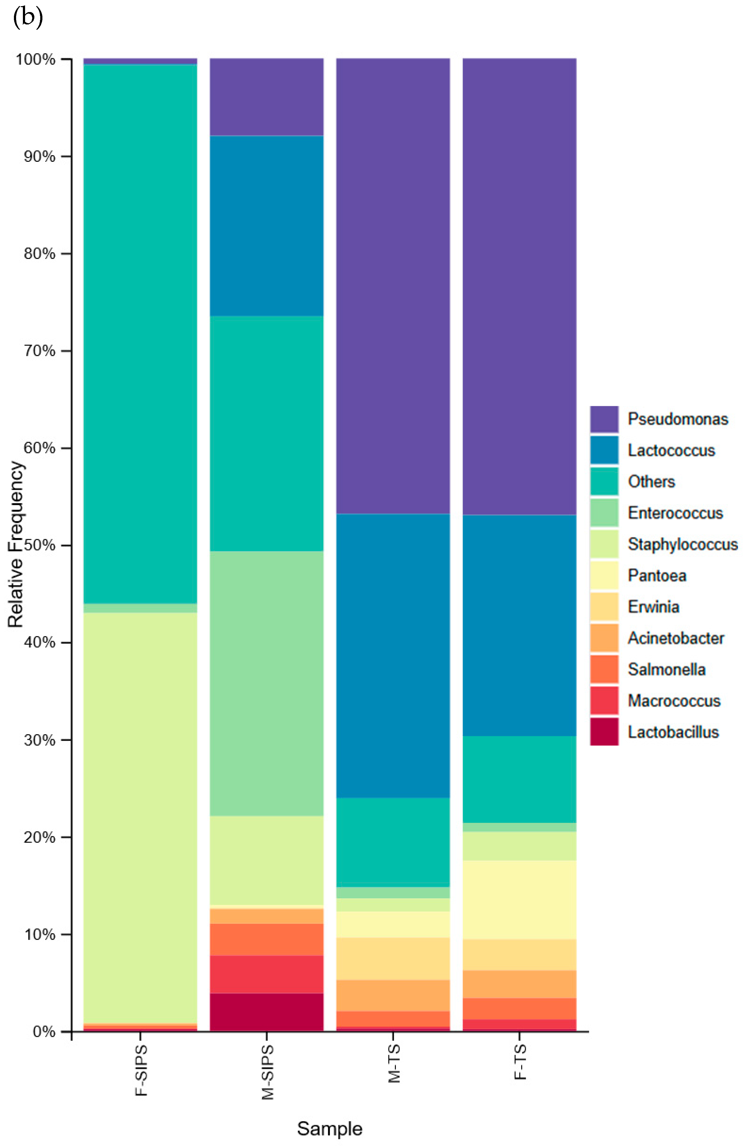
The sequence analysis allowed us to characterize bacteriomes. In the semi-intensive system, a more heterogeneous bacterial distribution was observed (Figure 1a). During the fore-stripping stage, the dominant phyla were Tenericutes (51.1%) and Firmicutes (44.5%), while during the milking stage, we saw an increase in the abundance of Firmicutes (56.6%) and Proteobacteria (38.7%), with the abundance of Tenericutes decreasing to 2.0%.
In both evaluated stages of the traditional system, Proteobacteria (72.0% and 67.6%) and Firmicutes (27.2% and 31.7%) were the most abundant phyla (Figure 1a).
At the genus level, the fore-stripping stage in the semi-intensive system was characterized by a higher abundance of the genera Mesoplasma (51.9%) and Staphylococcus (42.1%), which decreased to 2.4% and 9.1% during the milking stage. The levels of the genera Enterococcus (27.2%), Lactococcus (18.5%), Staphylococcus (9.1%), Pseudomonas (7.9%), Macrococcus (3.9%), Lactobacillus (3.8%), and Salmonella (3.2%) were higher in the milking stage (Figure 1b).
At the genus level, Pseudomonas (46.9%), Lactococcus (22.7%), and Pantoea (8.0%) were the predominant genera within the traditional system. During the milking stage, Pseudomonas and Pantoea showed a decrease of 0.1% and 5.3%, respectively, compared to during fore-stripping, but they remained among the most abundant genera. Increases of 6.5% and 0.3% in Lactococcus and Acinetobacter, respectively, were also observed (Figure 1b).
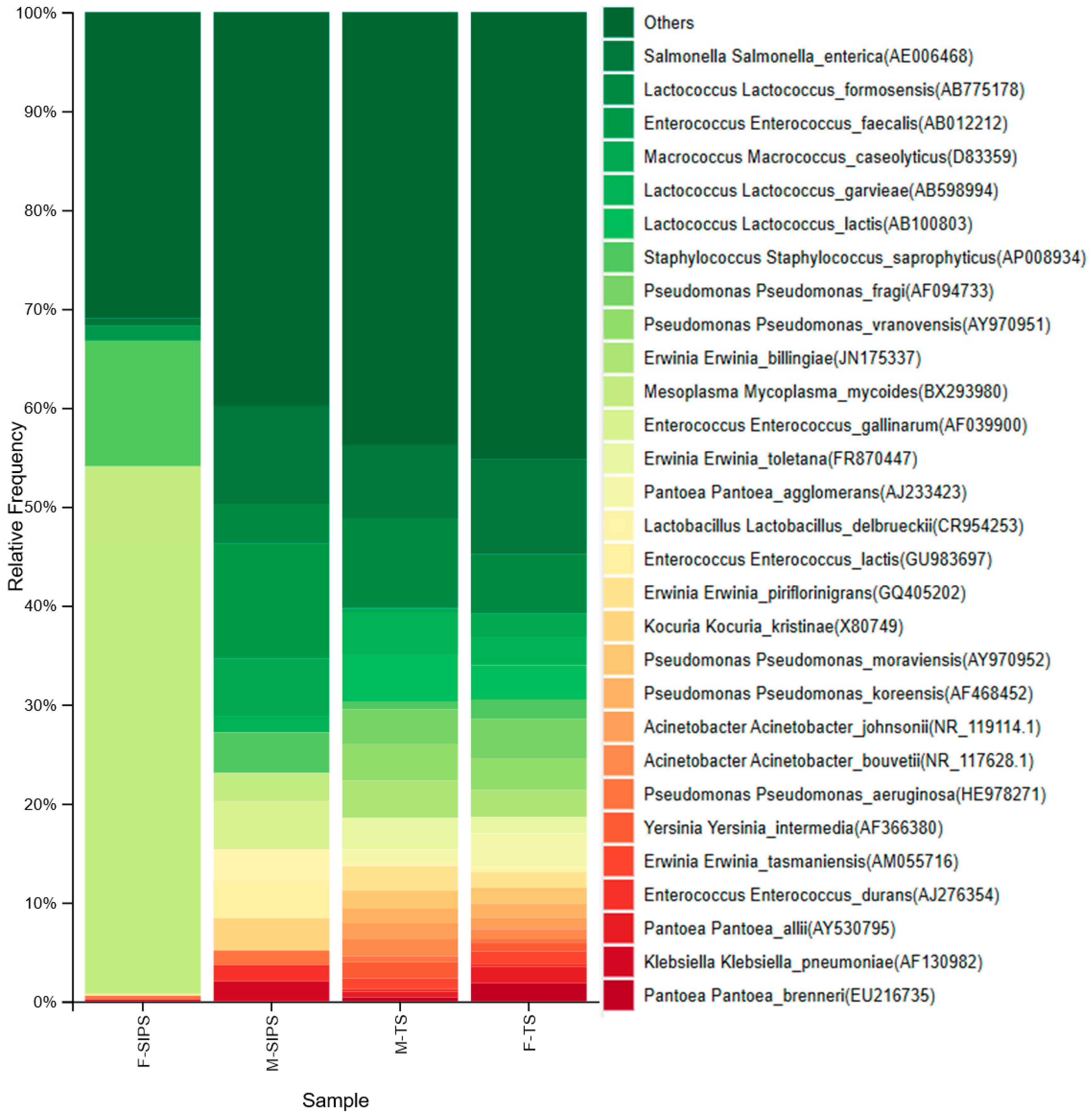
In both systems, differences were also observed during the milking process in terms of species abundance and diversity (Figure 2). In the semi-intensive system, species such as Mycoplasma mycoides (BX293980) (53.3%), Mycoplasma mycoides (U26037) (19.7%), and Staphylococcus saprophyticus (12.7%) were the most abundant in the fore-stripping stage and decreased in the milking stage. This pattern was also observed in the traditional system, where species such as Salmonella enterica, Pseudomonas fragi, and Pantoea agglomerans showed higher abundance in fore-stripping than during the milking stage.
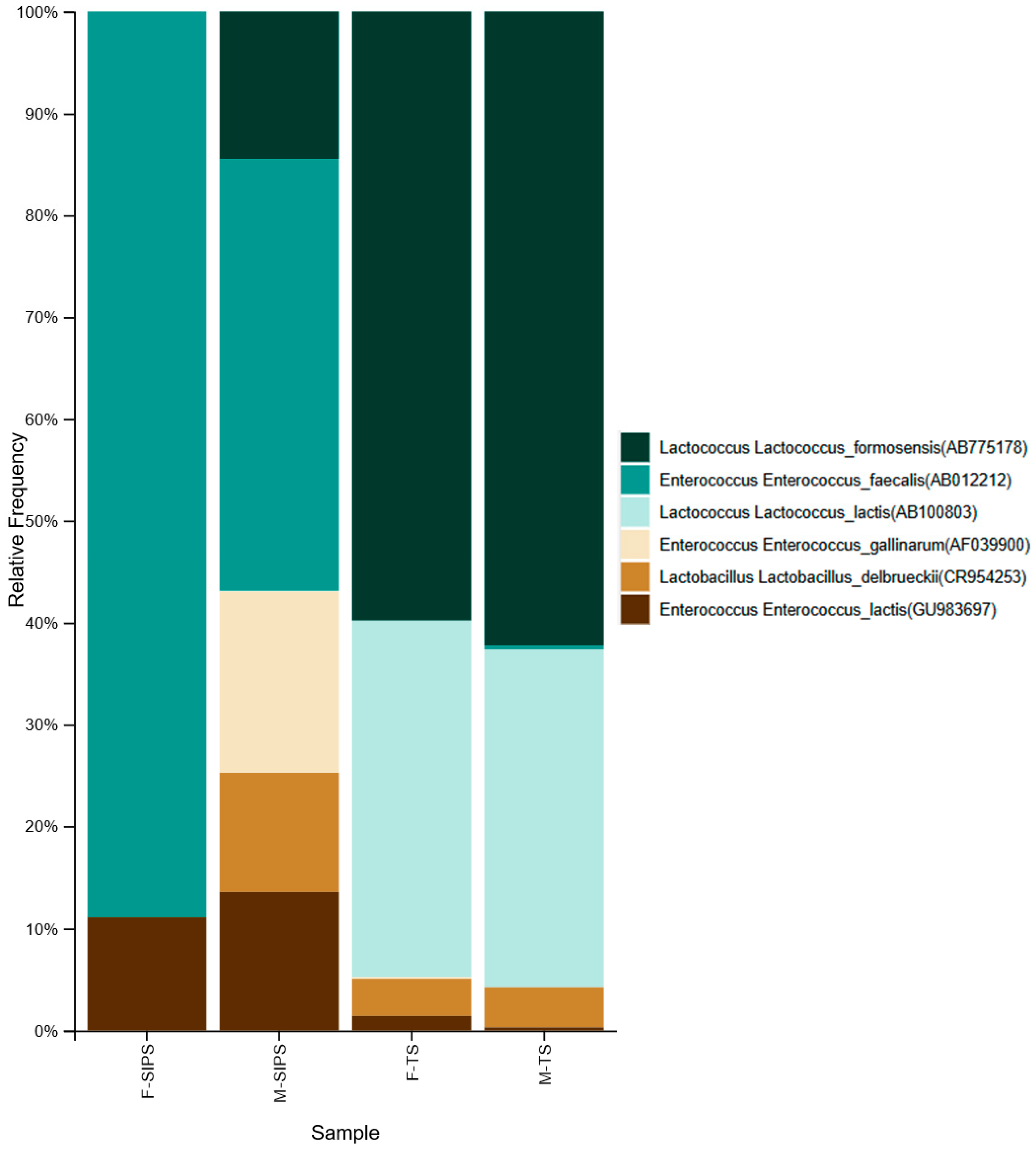
Regarding LAB species identification, in both production systems, the proportion of LABs was higher during milking compared with the fore-stripping stage. However, in the semi-intensive system, a higher LAB diversity was observed, with species such as Enterococcus faecalis, Enterococcus gallinarum, Lactococcus formosensis, Enterococcus lactis, and Lactobacillus delbrueckii were identified in both stages at different proportions (Figure 3).
According to the α-diversity values, the semi-intensive system exhibited low bacterial diversity during the fore-stripping stage, with a Shannon index of 1.5 and a Simpson index of 0.5, indicating moderate species dominance. In contrast, the milking stage showed higher diversity, with a Shannon and Simpson index of 4.0 and 0.8, respectively, corresponding to high species dominance and low evenness [26,27].
In the traditional system, both stages exhibited moderate diversity, with fore-stripping showing a Shannon index of 2.9 and a Simpson index of 0.7 and the milking stage presenting similar values of 2.7 and 0.6, respectively, reflecting moderate diversity and dominance of few species [26,27].
The dissimilarity distance matrix revealed particularly clear differences between production systems, highlighting the dissimilarities between the fore-stripping and milking stages within the semi-intensive system. The bacterial composition of the semi-intensive system accounted for 99.4% variability compared to the 0.5% of traditional system. The fore-stripping stage of the semi-intensive system showed the greatest variability in bacterial composition (70.9%), while the milking stage accounted for 28.5%, and in contrast, the traditional system showed less variation between the fore-stripping and milking stages trends, as was also observed in the alpha diversity values.
Based on these reference values, the semi-intensive system displayed greater variability in bacterial diversity between stages, whereas the traditional system maintained a more stable diversity profile throughout the milking process.

4. Discussion

While several studies have evaluated the bacterial diversity in goat milk [13,28,29,30,31,32], our study contributes to the global literature on the goat milk bacteriome by considering novel environmental factors that affect its composition.
Zhang et al. [28] analyzed the microbial population of raw goat milk from two breeds—Saanen and Guanzhong—with the most abundant genera identified in both breeds being Enterobacter, Acinetobacter, Pseudomonas, Staphylococcus, and Stenotrophomonas, and the predominant LAB genera being Lactococcus, Lactobacillus, Bifidobacterium, and Enterococcus. On the other hand, McInnis et al. [12] studied the bacterial diversity in the Alpine, Toggenburg, Saanen, and La Mancha breeds, identifying the genera Pseudomonas, Micrococcus, Rhodococcus, Stenotrophomonas, Phyllobacterium, Streptococcus, and Agrobacterium. Among these, Pseudomonas was also identified in our study as the most abundant genus detected. Additionally, Lauková et al. [33] studied the microbiome of raw Slovak goat milk, identifying Curtobacterium, Staphylococcus, Bacteroidetes, Bifidobacterium, and Streptococcus as the most predominant genera.
Here we reported evidence to support the idea that differences in bacterial diversity and abundance are dependent on milking process and hygienic practices, which are consistently related to the production system, as with removal of the first streams of milk (fore-stripping) and udder cleaning, which act as bacterial filters that reduce microbial load. However, practices carried out during and after milking may play a critical role in the transfer of certain bacterial species to goat milk [10,11].
In our study, milking during the fore-stripping stage was performed manually in both systems. However, in the semi-intensive system, the goats’ udders were disinfected prior to milk extraction, with these hygiene practices associated with lower bacterial diversity and abundance [34,35]. Previous studies have indicated that the skin of the udder harbors a greater number of microorganisms, sometimes even more than the milk itself [36], which may explain the higher bacterial abundance observed during the fore-stripping stage in the traditional system.
Additionally, milk collected during the milking stage was extracted semi-automatically in the semi-intensive system, while manual milking was used in the traditional system. The semi-intensive system exhibited higher bacterial abundance. Variations in both the increase and decrease in bacterial communities in the two production systems may be attributed to factors such as good or poor hygiene practices, the number of animals sampled, transportation, milk storage temperatures, and environmental conditions (e.g., temperature and humidity) [34].
The most abundant species in the semi-intensive system during the fore-stripping stage were Staphylococcus saprophyticus and Mycoplasma sp. In contrast, during the milking stage, the dominant species included Enterococcus sp., Salmonella enterica, Macrococcus caseolyticus, Staphylococcus saprophyticus, Lactococcus formosensis, Kocuria kristinae, and Lactobacillus delbrueckii.
In the traditional system, the fore-stripping stage was dominated by the species Salmonella enterica, Lactococcus formosensis, Pseudomonas vranovensis, and Lactococcus lactis, and during the milking stage, Lactococcus sp., Salmonella enterica, Erwinia billingiae, Pseudomonas sp., and Erwinia toletana were the most prevalent. It is noteworthy that the main bacterial genera identified in goat milk include pathogenic species. According to Wei et al. [37], the nutritional composition of goat milk may lead to the predominance of certain bacteria, including pathogenic bacteria, in comparison to cow’s milk.
The presence of potentially pathogenic bacteria in goat milk represents a significant food safety risk. The detection of S. saprophyticus reinforces these health concerns, as it is a frequent contaminant in raw milk. Its ability to persist in animal skin and its association with mastitis cases link S. saprophyticus to poor hygiene practices during milking [38,39]. Its presence suggests inadequate management conditions, and the consumption of untreated milk could represent a public health risk [39,40].
M. mycoides is a highly relevant pathogen, not only due to its impact on animal health, but also because of the economic losses it causes in the livestock industry [41,42].
Likewise, the detection of S. enterica constitutes a direct threat to public health and compromises the microbiological quality of products [43]. Pseudomonas spp., due to their psychotropic capacity, significantly contribute to the spoilage of milk and its derivatives under refrigeration conditions, affecting both shelf life and sensory properties [44]. The detection of these undesirable bacteria in milk influences the implementation of food safety practices, such as those described in the Good Agricultural Practices (GAP), Good Veterinary Practices (GVP), and Good Hygiene Practices (GHP), which are fundamental in primary production [45]. Also, the pasteurization process is an essential step for ensuring the safety of milk for consumption, and in cases where more rigorous control is required, the Hazard Analysis and Critical Control Points (HACCP) system may be implemented [46]. During the milking stage, E. billingae and E. toletana were identified, though their presence is likely associated with environmental contamination as these species are commonly found in vegetation [47,48,49].
On the other hand, genera belonging to LABs were detected in both production systems at both milking stages. It is important to highlight that in the semi-intensive system, the fore-stripping stage showed the lowest abundance of the genera Enterococcus, Lactococcus, and Lactobacillus, while in contrast, during the milking stage, these genera reached higher abundances.
The traditional system maintained a consistent presence of the genera Lactococcus, Enterococcus, and Lactobacillus. This system was characterized by the absence of hygiene practices, which may be related to the greater stability of these genera in goat milk.
The nutritional composition of goat milk is influenced by the breed’s genetics, which also affects bacterial communities [28]. In Saanen goats, for example, a higher proportion of lactose and certain amino acids may promote the growth of genera such as Lactococcus and Lactobacillus, contributing to their higher abundance in the milk [28]. In our study, the abundance of the Lactococcus genus was higher in milk from the traditional system taken from creole goats than milk from the semi-intensive system, mainly comprising Saanen goats.
We found that the phylum Firmicutes, which includes LABs, was among the most abundant phyla. Moreover, certain LAB genera and species were present in higher proportions compared to some non-lactic acid bacteria in both production systems; however, their overall abundance was lower than that of the total non-lactic acid bacterial population. These findings are consistent with those reported by McInnis et al. [12], who noted that the phylum Firmicutes, including LABs, represents only a small proportion of the milk bacteriome.
In addition, it has been shown that the lactation period has an impact on microbial diversity, particularly on the diversity of LABs. McInnis et al. [12] reported that the phylum Firmicutes had higher abundance during the late lactation stage in Alpine, Toggenburg, Saanen, and La Mancha goats, likely due to increased concentrations of nutrients such as fats, proteins, and lactose. This may explain the predominance of Firmicutes observed in the production systems analyzed in our study, as the samples were also collected during the late lactation stage.
Some bacteria from the genus Enterococcus, although not banned in food production, are regarded as potential pathogens [50,51]; however, the species E. faecalis, E. gallinarum, E. lactis, Lactococcus formosensis, and Lactobacillus delbrueckii have demonstrated significance in both the food and health industries, and as they are part of human and animal digestive systems, most of them are considered safe. Here, we found that goat milk could be a source of these bacteria, which also have probiotic properties that contribute to reducing the symptoms of lactose intolerance, strengthening host immune response, and supporting the normal development of intestinal mucosa. Their enzymatic machinery also plays a key role in the generation of pleasant aromas and flavors in food products [52,53].
In this study, bacterial sequences were obtained through full-length 16S rRNA gene sequencing. However, it is acknowledged that even the complete 16S gene has intrinsic limitations for reliable species-level identification [54]. Therefore, the species assignments presented here should be interpreted with caution and considered putative until their validation, especially for critical species such as Salmonella enterica and Mycoplasma mycoides.
The diversity parameters provide clearer evidence that the goat milk bacteriome is influenced both by the production system and milking process, which was also reflected in the taxonomic analysis [24]. The milking stage of the semi-intensive system was found to have a higher bacterial diversity (H’ = 4.0) than the fore-stripping stage, and even more so than both stages of the traditional system. The Simpson index values of the analyzed bacteriomes indicated low diversity, dominated by three phyla, ten genera, and six species, and the analysis of beta diversity showed differences in bacterial population between the production systems (semi-intensive and traditional), as well as between the fore-stripping and milking stages within the semi-intensive system, but not between these stages in the traditional system.

5. Conclusions

We found novel evidence showing that the breeding and management practices defining goat production systems and milking processes are important factors influencing the diversity and abundance of bacteria in goat milk. These practices are closely related to milk microbiological profile and quality, and their evaluation and optimization will allow for the identification of the most effective strategies for reducing pathogen prevalence and promoting the presence of favorable bacteria, such as LABs, which are important across different applications, including food safety and health.

Author Contributions

Conceptualization, A.M.S.R.; methodology and formal analysis, E.M.R., A.M.S.R., P.A.M., W.A.V. and R.d.J.T.-R.; investigation E.M.R. and A.M.S.R., writing—original draft preparation, E.M.R. and A.M.S.R.; writing—review and editing, E.M.R., A.M.S.R., E.G.B. and R.d.J.T.-R.; supervision, project administration, and funding acquisition, A.M.S.R. All authors have read and agreed to the published version of the manuscript.

Funding

The authors want to thank the Instituto Politécnico Nacional via project SIP 20241271 and 20250980.

Institutional Review Board Statement

Not applicable.

Informed Consent Statement

Not applicable.

Data Availability Statement

The raw sequencing data have been deposited in the NCBI Sequence Read Archive (SRA) under accession number PRJNA1314283.

Acknowledgments

The authors want to thank the Secretaría de Ciencia, Humanidades, Tecnología e Innovación for support through a master’s degree program scholarship (E.M.R.) and the Instituto Politécnico Nacional via SIP projects 20241271 and 20250980 (A.M.S.-R.). The authors also acknowledge producers from Jimenez, Michoacan, and Ing. Wilfrido Du Solier Espinosa for their collaboration on the agreement IPN-RANCHO CABRIO, S.P.R. DE R.L.

Conflicts of Interest

The authors declare that they have no known competing financial interests or personal relationships that could have appeared to influence the work reported in this paper.

References

  1. Kumar, H.; Yadav, D.; Kumar, N.; Seth, R.; Goyal, A.K. Nutritional and nutraceutical properties of goat milk—A review. Indian J. Dairy Sci. 2016, 69, 513–518. Available online: https://www.researchgate.net/publication/301693090 (accessed on 16 February 2025).
  2. Nayik, G.A.; Jagdale, Y.D.; Gaikwad, S.A.; Devkatte, A.N.; Dar, A.H.; Dezmirean, D.S.; Bobis, O.; Ranjha, M.M.A.N.; Ansari, M.J.; Hemeg, H.A.; et al. Recent insights into processing approaches and potential health benefits of goat milk and its products: A review. Front. Nutr. 2021, 8, 789117. [Google Scholar] [CrossRef] [PubMed]
  3. Prosser, C.G. Compositional and functional characteristics of goat milk and relevance as a base for infant formula. J. Food Sci. 2021, 86, 257–265. [Google Scholar] [CrossRef]
  4. Nayik, G.A.; Jagdale, Y.D.; Gaikwad, S.A.; Devkatte, A.N.; Dar, A.H.; Ansari, M.J. Nutritional profile, processing and potential products: A comparative review of goat milk. Dairy 2022, 3, 622–647. [Google Scholar] [CrossRef]
  5. Gioffredo, J.; Petryna, A. Caprinos: Generalidades, Nutrición, Reproducción e Instalaciones; Dpto. de Producción Animal, Facultad de Agronomía y Veterinaria, Universidad Nacional de Río Cuarto: Río Cuarto, Argentina, 2010. [Google Scholar]
  6. Hernández, Z.J.S. La caprinocultura en el marco de la ganadería poblana (México): Contribución de la especie caprina y sistemas de producción. Arch. Zootec. 2000, 49, 341–352. [Google Scholar]
  7. Meneses, R. Manual de Producción Caprina; Instituto de Desarrollo Agropecuario (INDAP) e Instituto de Investigaciones Agropecuarias (INIA): Santiago, Chile, 2017. [Google Scholar]
  8. Dos Santos, M.A.G.; Castelani, L.; Mitsunaga, T.M.; Soares, W.V.B.; Giglioti, R.; Júnior, L.C.R. Evaluation of fore-stripping milk and its effects on milk quality. Acta Vet. Bras. 2022, 16, 47–52. [Google Scholar] [CrossRef]
  9. Wagner, A.M.; Ruegg, P.L. The effect of manual fore-stripping on milking performance of Holstein dairy cows. J. Dairy Sci. 2002, 85, 804–809. [Google Scholar] [CrossRef]
  10. Quigley, L.; O’Sullivan, O.; Stanton, C.; Beresford, T.P.; Ross, R.P.; Fitzgerald, G.F.; Cotter, P.D. The complex microbiota of raw milk. FEMS Microbiol. Rev. 2013, 37, 664–698. [Google Scholar] [CrossRef] [PubMed]
  11. Verdier-Metz, I.; Michel, V.; Delbes, C.; Montel, M.C. Do milking practices influence the bacterial diversity of raw milk? Food Microbiol. 2009, 37, 664–698. [Google Scholar] [CrossRef]
  12. McInnis, E.A.; Kalanetra, K.M.; Mills, D.A.; Maga, E.A. Analysis of raw goat milk microbiota: Impact of stage of lactation and lysozyme on microbial diversity. Food Microbiol. 2015, 46, 121–131. [Google Scholar] [CrossRef]
  13. Prayogo, F.A.; Budiharjo, A.; Kusumaningrum, H.P.; Wijanarka, W.; Suprihadi, A.; Nurhayati, N. Metagenomic applications in exploration and development of novel enzymes from nature: A review. J. Genet. Eng. Biotechnol. 2020, 18, 39. [Google Scholar] [CrossRef]
  14. You, L.; Yang, C.; Jin, H.; Kwok, L.Y.; Sun, Z.; Zhang, H. Metagenomic features of traditional fermented milk products. LWT 2022, 155, 112945. [Google Scholar] [CrossRef]
  15. Karssa, T.H.; Kussaga, J.B.; Semedo-Lemsaddek, T.; Mugula, J.K. Perspectivas sobre la microbiología de los productos lácteos fermentados etíopes: Una revisión. Cienc. Aliment. Nutr. 2024, 12, 6990–7003. [Google Scholar] [CrossRef]
  16. Langer, A.J.; Ayers, T.; Grass, J.; Lynch, M.; Angulo, F.J.; Mahon, B.E. Nonpasteurized dairy products, disease outbreaks, and state laws—United States. 1993–2006. Emerg. Infect. Dis. 2012, 18, 385. [Google Scholar] [CrossRef] [PubMed]
  17. Oliver, S.P.; Boor, K.J.; Murphy, S.C.; Murinda, S.E. Food safety hazards associated with consumption of raw milk. Foodborne Pathog. Dis. 2009, 6, 793–806. [Google Scholar] [CrossRef]
  18. James, G. Universal Bacterial Identification by PCR and DNA Sequencing of 16S rRNA Gene. In PCR for Clinical Microbiology; Schuller, M., Sloots, T., James, G., Halliday, C., Carter, I., Eds.; Springer: Dordrecht, The Netherlands, 2010. [Google Scholar] [CrossRef]
  19. ILLUMINA. Illumina DNA Prep (Reference Guide). Illumina. Available online: https://support-docs.illumina.com/LP/IlluminaDNAPrep/Content/LP/Illumina_DNA/DNA-Prep/Protocol_IDP.htm (accessed on 3 October 2025).
  20. Li, D.; Liu, C.M.; Luo, R.; Sadakane, K.; Lam, T.W. MEGAHIT: An ultra-fast single-node solution for large and complex metagenomics assembly via succinct de Bruijn graph. Bioinformatics 2015, 31, 1674–1676. [Google Scholar] [CrossRef]
  21. Wang, Q.; Garrity, G.M.; Tiedje, J.M.; Cole, J.R. Naïve Bayesian Classifier for Rapid Assignment of rRNA Sequences into the New Bacterial Taxonomy. Appl. Environ. Microbiol. 2007, 73, 5261–5267. [Google Scholar] [CrossRef]
  22. Bolyen, E.; Rideout, J.R.; Dillon, M.R.; Bokulich, N.A.; Abnet, C.C.; Al-Ghalith, G.A.; Alexander, H.; Alm, E.J.; Arumugam, M.; Asnicar, F.; et al. Reproducible, interactive, scalable and extensible microbiome data science using QIIME 2. Nat. Biotechnol. 2019, 37, 852–857. [Google Scholar] [CrossRef]
  23. Shannon, C.E. A mathematical theory of communication. Bell Syst. Tech. J. 1948, 27, 379–423. [Google Scholar] [CrossRef]
  24. Simpson, E.H. Measurement of diversity. Nature 1949, 163, 668. [Google Scholar] [CrossRef]
  25. Xia, Y.; Sun, J. Beta diversity metrics and ordination. In Bioinformatic and Statistical Analysis of Microbiome Data: From Raw Sequences to Advanced Modeling with QIIME 2 and R; Springer International Publishing: Cham, Switzerland, 2023; pp. 335–395. [Google Scholar] [CrossRef]
  26. Medrano Meraz, M.D.J.; Hernández, F.J.; Corral Rivas, S.; Nájera Luna, J.A. Diversidad arbórea a diferentes niveles de altitud en la región de El Salto, Durango. Rev. Mex. Cienc. For. 2017, 8, 57–68. Available online: http://www.scielo.org.mx/scielo.php?script=sci_arttext&pid=S2007-11322017000200057&lng=es&tlng=es (accessed on 17 April 2025).
  27. Kim, B.R.; Shin, J.; Guevarra, R.B.; Lee, J.H.; Kim, D.W.; Seol, K.H.; Isaacson, R.E. Deciphering diversity indices for a better understanding of microbial communities. J. Microbiol. Biotechnol. 2017, 27, 2089–2093. [Google Scholar] [CrossRef]
  28. Zhang, F.; Wang, Z.; Lei, F.; Wang, B.; Jiang, S.; Peng, Q.; Shao, Y. Bacterial diversity in goat milk from the Guanzhong area of China. J. Dairy Sci. 2017, 100, 7812–7824. [Google Scholar] [CrossRef]
  29. Tormo, H.; Lekhal, D.A.H.; Roques, C. Phenotypic and genotypic characterization of lactic acid bacteria isolated from raw goat milk and effect of farming practices on the dominant species of lactic acid bacteria. Int. J. Food Microbiol. 2015, 210, 9–15. [Google Scholar] [CrossRef]
  30. Kamilari, E.; Anagnostopoulos, D.A.; Papademas, P.; Efthymiou, M.; Tretiak, S.; Tsaltas, D. Snapshot of Cyprus raw goat milk bacterial diversity via 16S rDNA high-throughput sequencing; impact of cold storage conditions. Fermentation 2020, 6, 100. [Google Scholar] [CrossRef]
  31. Callon, C.; Duthoit, F.; Delbès, C.; Ferrand, M.; Le Frileux, Y.; De Crémoux, R.; Montel, M.C. Stability of microbial communities in goat milk during a lactation year: Molecular approaches. Syst. Appl. Microbiol. 2007, 30, 547–560. [Google Scholar] [CrossRef] [PubMed]
  32. NNiyazbekova, Z.; Yao, X.-T.; Liu, M.-J.; Bold, N.; Tong, J.-Z.; Chang, J.-J.; Wen, Y.; Li, L.; Wang, Y.; Chen, D.-K.; et al. Compositional and functional comparisons of the microbiota in the colostrum and mature milk of dairy goats. Animals 2020, 10, 1955. [Google Scholar] [CrossRef]
  33. Lauková, A.; Micenková, L.; Grešáková, Ľ.; Maďarová, M.; Simonová, M.P.; Focková, V.; Ščerbová, J. Microbiome Associated with Slovak Raw Goat Milk, Trace Minerals, and Vitamin E ContentE. Int. J. Food Sci. 2022, 2022, 4595473. [Google Scholar] [CrossRef]
  34. Delgado-Pertiñez, M.; Alcalde, M.J.; Guzmán-Guerrero, J.L.; Castel, J.M.; Mena, Y.; Caravaca, F. Effect of hygiene-sanitary management on goat milk quality in semi-extensive systems in Spain. Small Rumin. Res. 2003, 47, 51–61. [Google Scholar] [CrossRef]
  35. Montel, M.C.; Buchin, S.; Mallet, A.; Delbes-Paus, C.; Vuitton, D.A.; Desmasures, N.; Berthier, F. Traditional cheeses: Rich and diverse microbiota with associated benefits. Int. J. Food Microbiol. 2014, 177, 136–154. [Google Scholar] [CrossRef] [PubMed]
  36. Derakhshani, H.; Plaizier, J.C.; De Buck, J.; Barkema, H.W.; Khafipour, E. Composition of the teat canal and intramammary microbiota of dairy cows subjected to antimicrobial dry cow therapy and internal teat sealant. J. Dairy Sci. 2018, 101, 10191–10205. [Google Scholar] [CrossRef]
  37. Wei, W.; Hu, X.; Hou, Z.; Wang, Y.; Zhu, L. Microbial community structure and diversity in different types of non-bovine milk. Curr. Opin. Food Sci. 2021, 40, 51–57. [Google Scholar] [CrossRef]
  38. Júnior, A.E.S.; Vasconcelos, P.C.; Saraiva, M.M.; Santos Filho, L.; Silva, N.M.; Givisiez, P.E.; Oliveira, C.J. Antimicrobial susceptibility profiles of Staphylococcus spp. contaminating raw goat milk. Vet. World 2021, 14, 1074–1079. [Google Scholar] [CrossRef] [PubMed]
  39. Organji, S.R.; Abulreesh, H.H.; Elbanna, K.; Osman, G.E.; Almalki, M.H. Diversity and characterization of Staphylococcus spp. in food and dairy products: A foodstuff safety assessment. J. Microbiol. Biotechnol. Food Sci. 2018, 7, 586–593. [Google Scholar] [CrossRef]
  40. Lawal, O.U.; Barata, M.; Fraqueza, M.J.; Worning, P.; Bartels, M.D.; Goncalves, L.; Miragaia, M. Staphylococcus saprophyticus from clinical and environmental origins have distinct biofilm composition. Front. Microbiol. 2021, 12, 663768. [Google Scholar] [CrossRef]
  41. Nicholas, R.A.J. Improvements in the diagnosis and control of diseases of small ruminants caused by mycoplasmas. Small Rumin. Res. 2002, 45, 145–149. [Google Scholar] [CrossRef]
  42. Zhang, Z.; Jiang, J.; He, M.; Li, H.; Cheng, Y.; An, Q.; Wang, F. First Report and Comparative Genomic Analysis of a Mycoplasma mycoides Subspecies capri HN-A in Hainan Island. Microorganisms 2022, 10, 1908. [Google Scholar] [CrossRef] [PubMed]
  43. Gorski, L.; Parker, C.T.; Liang, A.; Cooley, M.B.; Jay-Russell, M.T.; Gordus, A.G.; Mandrell, R.E. Prevalence, distribution, and diversity of Salmonella enterica in a major produce region of California. Appl. Environ. Microbiol. 2011, 77, 2734–2748. [Google Scholar] [CrossRef]
  44. Ercolini, D.; Casaburi, A.; Nasi, A.; Ferrocino, I.; Di Monaco, R.; Ferranti, P.; Villani, F. Different molecular types of Pseudomonas fragi have the same overall behaviour as meat spoilers. Int. J. Food Microbiol. 2010, 142, 120–131. [Google Scholar] [CrossRef]
  45. Haldar, L.; Raghu, H.V.; Ray, P.R. Milk and milk product safety and quality assurance for achieving better public health outcomes. In Agriculture, Livestock Production and Aquaculture: Advances for Smallholder Farming Systems Volume 1; Springer: Cham, Switzerland, 2022; pp. 217–259. [Google Scholar] [CrossRef]
  46. Kumar, S.; Lodh, J.; Kumar, S.; Mishra, D. Risk analysis, assessment, practices, and quality management in milk hygiene. In The Microbiology, Pathogenesis and Zoonosis of Milk Borne Diseases; Academic Press: Cambridge, MA, USA, 2024; pp. 387–405. [Google Scholar] [CrossRef]
  47. Brady, C.; Kaur, S.; Crampton, B.; Maddock, D.; Arnold, D.; Denman, S. Transfer of Erwinia toletana and Erwinia iniecta to a novel genus Winslowiella gen. nov. as Winslowiella toletana comb. nov. and Winslowiella iniecta comb. nov. and description of Winslowiella arboricola sp. nov., isolated from bleeding cankers on broadleaf hosts. Front. Microbiol. 2022, 13, 1063107. [Google Scholar] [CrossRef]
  48. Buonaurio, R.; Moretti, C.; da Silva, D.P.; Cortese, C.; Ramos, C.; Venturi, V. The olive knot disease as a model to study the role of interspecies bacterial communities in plant disease. Front. Plant Sci. 2015, 6, 434. [Google Scholar] [CrossRef]
  49. Prod’homme, M.; Micol, L.A.; Weitsch, S.; Gassend, J.L.; Martinet, O.; Bellini, C. Cutaneous infection and bactaeremia caused by Erwinia billingiae: A case report. New Microbes New Infect. 2017, 19, 134–136. [Google Scholar] [CrossRef]
  50. Monticelli, J.; Knezevich, A.; Luzzati, R.; Di Bella, S. Clinical management of non-faecium non-faecalis vancomycin-resistant enterococci infection. Focus on Enterococcus gallinarum and Enterococcus casseliflavus/flavescens. J. Infect. Chemother. 2018, 24, 237–246. [Google Scholar] [CrossRef]
  51. Belloso Daza, M.V.; Almeida-Santos, A.C.; Novais, C.; Read, A.; Alves, V.; Cocconcelli, P.S.; Peixe, L. Distinction between Enterococcus faecium and Enterococcus lactis by a gluP PCR-based assay for accurate identification and diagnostics. Microbiol. Spectr. 2022, 10, e03268-22. [Google Scholar] [CrossRef] [PubMed]
  52. Germond, J.E.; Lapierre, L.; Delley, M.; Mollet, B.; Felis, G.E.; Dellaglio, F. Evolution of the bacterial species Lactobacillus delbrueckii: A partial genomic study with reflections on prokaryotic species concept. Mol. Biol. Evol. 2003, 20, 93–104. [Google Scholar] [CrossRef]
  53. Guglielmotti, D.M.; Marcó, M.B.; Golowczyc, M.; Reinheimer, J.A.; Quiberoni, A.D.L. Probiotic potential of Lactobacillus delbrueckii strains and their phage resistant mutants. Int. Dairy J. 2007, 17, 916–925. [Google Scholar] [CrossRef]
  54. Bartoš, O.; Chmel, M.; Swierczková, I. The overlooked evolutionary dynamics of 16S rRNA revises its role as the “gold standard” for bacterial species identification. Sci. Rep. 2024, 14, 9067. [Google Scholar] [CrossRef] [PubMed]
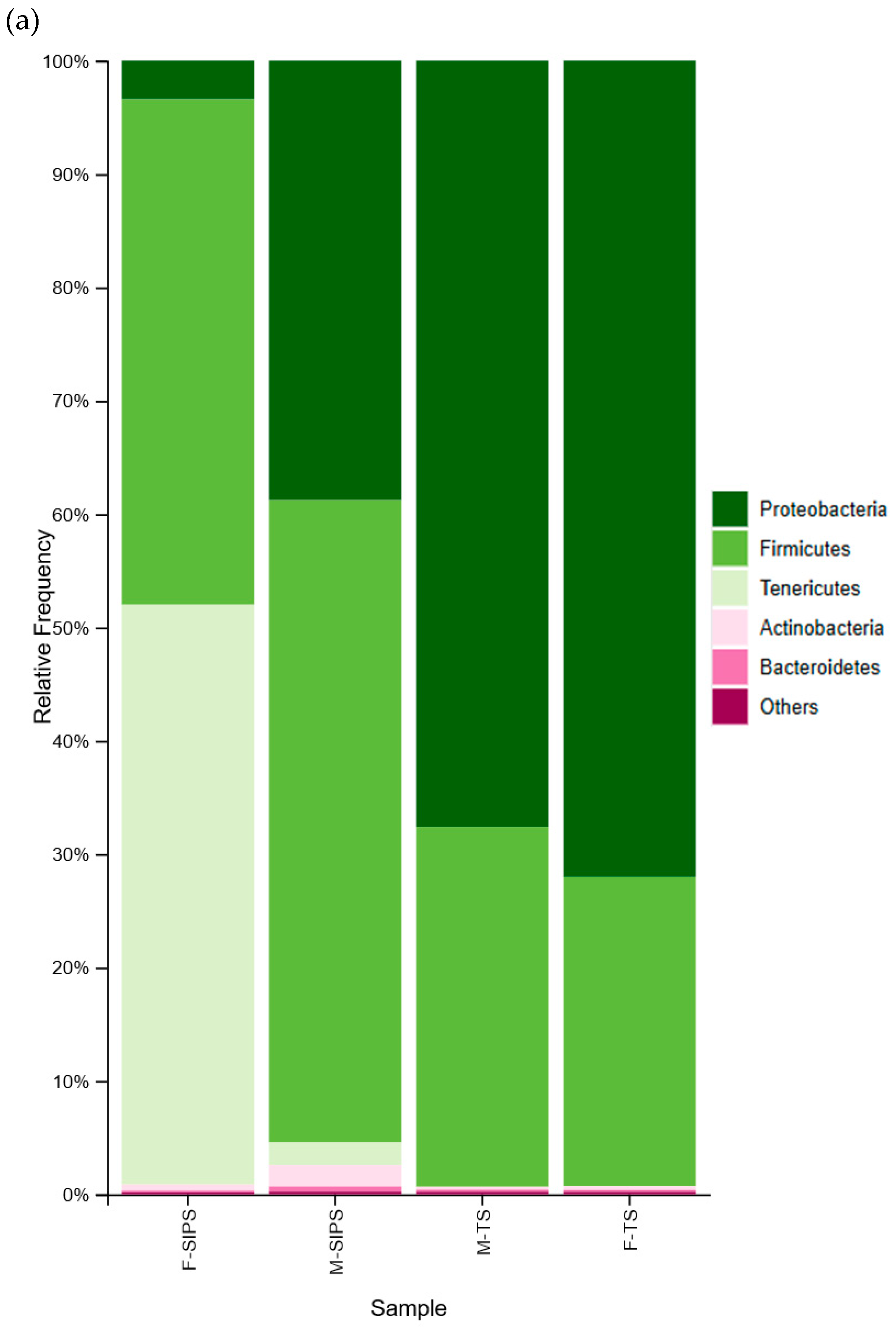
Figure 1. The relative abundance identified in the goat milk bacteriomes. (a) The abundance of bacterial phyla and (b) bacterial genera identified in raw goat milk. Semi-intensive system: F-SIP—fore-stripping stage; M-SIPS—milking stage. Traditional system: F-TS—fore-stripping stage; M-TS—milking stage.
Microbiolres 16 00218 g001aMicrobiolres 16 00218 g001b
Figure 2. Relative abundance of bacterial species identified in goat milk bacteriomes. Semi-intensive system: F-SIPS—fore-stripping stage; M-SIPS—milking stage. Traditional system: F-TS—fore-stripping stage; M-TS—milking stage, Michoacán.
Microbiolres 16 00218 g002
Figure 3. Relative abundance of genera and species of LABs in goat milk. Semi-intensive system: F-SIPS—fore-stripping stage; M-SIPS—milking stage. Traditional system: F-TS—fore-stripping stage; F-TS—milking stage.
Microbiolres 16 00218 g003
Disclaimer/Publisher’s Note: The statements, opinions and data contained in all publications are solely those of the individual author(s) and contributor(s) and not of MDPI and/or the editor(s). MDPI and/or the editor(s) disclaim responsibility for any injury to people or property resulting from any ideas, methods, instructions or products referred to in the content.