Article

Research on the Influence of Enterobius vermicularis on the Composition and Quality of the Intestinal Microbiota, and the Susceptibility to Co-Infections

1
Department of Parasitology and Tropical Medicine, National Centre of Infectious and Parasitic Diseases, 26 Yanko Sakazov Blvd., 1504 Sofia, Bulgaria
2
Department of Microbiology, National Centre of Infectious and Parasitic Diseases, 26 Yanko Sakazov Blvd., 1504 Sofia, Bulgaria
3
Medical Diagnostic Laboratory “Cibalab”, 83 “Gyueshevo” Str., 1379 Sofia, Bulgaria
4
Department of Virology, National Centre of Infectious and Parasitic Diseases, 44A Gen. Stoletov Blvd., 1233 Sofia, Bulgaria
*
Author to whom correspondence should be addressed.
Microbiol. Res. 2025, 16(10), 215; https://doi.org/10.3390/microbiolres16100215
Submission received: 27 August 2025 / Revised: 23 September 2025 / Accepted: 26 September 2025 / Published: 30 September 2025

Abstract

The present study examined the presence of concomitant intestinal parasites and bacteria in individuals infected with Enterobius vermicularis in Bulgaria, and analyzed its effects on the intestinal microbiome and the risk of co-infection. Fecal samples from people with and without (control group) enterobiasis were tested for the presence of concomitant bacterial infection. The results were compared to find out about the intestinal microbiome in these groups. The microbiological examination of the control group showed that 8.3% had only conditionally pathogenic flora. However, in individuals with enterobiasis, 56% had normal intestinal bacterial flora, but 46% had disturbed microbiota: 7% were carriers of pathogenic intestinal bacteria and 24% had opportunistic pathogens. The most prevalent were Klebsiella pneumoniae (49%), followed by Enterobacter spp. (22%), and Proteus mirabilis (12.2%). Our study is the first in the country to investigate the impact of E. vermicularis infection on the composition and quality of the gut microbiome, as well as the potential for co-infections with pathogenic gut bacteria. Although our findings are preliminary, they suggest that this nematode may significantly contribute to a predisposition for dysbiosis or the onset of secondary bacterial infections.

1. Introduction

Enterobius vermicularis (E. vermicularis) is a widespread intestinal parasite. This helminth is commonly found in public institutions, such as kindergartens, schools, orphanages and psychiatric clinics, where large numbers of people gather. It is also prevalent in families with a low socioeconomic status where hygiene standards are inadequate [1]. It is estimated that around 200 million people worldwide are infected with E. vermicularis, with children being the most commonly affected group [1]. This intestinal nematode is transmitted through ingestion of eggs from contaminated sources, and less commonly through the inhalation of eggs from the air [2].
The life cycle of E. vermicularis is simple and direct, taking place entirely within the digestive system. Infection begins with the ingestion of infectious eggs, which then release larvae that moult twice and grow [3]. Large numbers of these can be found in the large intestine, to which they migrate. Female parasites actively reach the anal canal at night and deposit their eggs onto perianal skin [4]. Therefore, microscopy of material taken from the perianal area with a Scotch tape (Graham’s test) is the main method for diagnosing the disease, as this can detect the characteristic pinworm eggs [5].
Approximately 40% of affected individuals exhibit no symptoms, and enterobiasis tends to be self-limiting due to the brief lifespan of adult parasites, unless autoinfection occurs [6]. The main symptom is prolonged itching around the anus, often worse at night, which can disrupt sleep and impair concentration during the day [4,7]. Scratching may lead to ulcerations and predispose the area to bacterial superinfection, resulting in complications like anal dermatitis or rectal abscesses [5]. In rare cases, about 1%, other organs may also be affected [8]. In girls, pinworms can migrate to the vaginal area, potentially causing vulvovaginitis or urinary tract infections from bacteria like Escherichia coli [9]. There is also a link between E. vermicularis infection and some cases of acute appendicitis [10].
The human gut microbiota comprises around 1014 microorganisms and over 1000 bacterial species [11]. This complex ecosystem significantly influences human health, aiding digestion, producing essential nutrients and vitamins, and regulating the immune system to protect against pathogens [12]. Dysbiosis is marked by changes in microbial diversity, such as decreased beneficial bacteria and increased harmful bacteria. Although not a disease itself, dysbiosis can contribute to the development of various health issues [13].
Recent studies show that E. vermicularis has a significant impact on the intestinal microbiome, altering its composition and consequently affecting the intestinal epithelium, metabolism and immune responses in the host [14]. It has been reported that E. vermicularis parasitism in children leads to the suppression of non-specific immunity and secondary immunodeficiency. It also causes hypoacidity, reducing the bactericidal action of gastric juice and contributing to the development of various somatic, infectious and other diseases [15]. Co-infections with other parasites are also frequently observed [16].
Studies on the intestinal microflora of individuals infected with helminths are limited, with most research having been conducted on animal models [17]. The relationship between enterobiasis and the presence of pathogenic intestinal flora and other parasitic pathogens has been examined in only a few studies, and there are none for Bulgaria. Due to this, our objective was to investigate the presence of concomitant intestinal parasitic and bacterial pathogens in individuals diagnosed with enterobiasis in Bulgaria through laboratory testing, and to assess the impact of E. vermicularis infection on the composition and quality of the intestinal microbiome, as well as its potential role in elevating the risk of coinfections with other intestinal pathogens.

2. Materials and Methods

2.1. Study Period and Design

This retrospective study covers the period from January 2022 to May 2025. It includes 229 individuals from different regions of the country who were examined by the Department of Parasitology and Tropical Medicine (DPTM) at the National Centre of Infectious and Parasitic Diseases (NCIPD), Sofia, for enterobiasis, based on clinical, epidemiological or prophylactic grounds. The same team performed laboratory diagnostics over time, using the same diagnostic methods and kits to ensure data comparability between years. Using the laboratory results, the individuals were divided into two groups: those with laboratory-confirmed enterobiasis (n = 169) and a control group with no laboratory data for protozoan and/or helminth infection (n = 60).

2.1.1. Individuals Infected with E. vermicularis

This group consists of 76 males and 93 females. Among the 169 individuals, 162 were children and adolescents (aged 1 to 18, average age 6), while 7 were adults (aged 37 to 54, average age 44). The age distribution of the children was as follows: 25 aged 1–3, 77 aged 4–6, 36 aged 7–9, 20 aged 10–12, and 4 aged 13–18. Diagnosis was made based on the presence of E. vermicularis eggs in perianal impressions.

2.1.2. Control Group of Individuals with No Data on Enterobiasis or Other Parasitic Infection

The group consisted of 47 children (21 boys and 26 girls) and 13 adults (three men and 10 women). Their ages ranged from 1 to 68 years (mean age 12 years). Among children and adolescents (aged 1 to 18 years), the average age was 3.4 years, while among adults (aged 22 to 68 years), the average age was 42 years. The presence of E. vermicularis and other intestinal protozoa and helminths in these individuals was ruled out after microscopic examination of perianal impressions and fecal samples.

2.2. Parasitological Diagnosis

A perianal tape sample (tape test) is used to diagnose E. vermicularis. Typical eggs found in the samples are examined under a light microscope at 100x and 400x magnification.
Samples are taken from the perianal region after waking (Graham’s test) and before toilet use. Three perianal prints are obtained from all suspected individuals, on three different days. The presence of single eggs in the perianal prints indicates a positive result. Adult parasites are also found in some patients.
Fecal samples are examined microscopically for other intestinal parasites and microbiologically for bacterial flora. Information on age, place of residence and clinical symptoms is obtained from all individuals.

2.3. Microbiological Diagnosis

Fecal samples stored at −20 °C were examined for intestinal pathogens (Salmonella, Shigella, Yersinia enterocolitica, pathogenic E. coli, Campylobacter and enterotoxigenic Klebsiella pneumoniae) at the National Reference Laboratory for Intestinal Infections, Pathogenic Cocci and Diphtheria at the NCIPD. Culture methods were used to detect viable bacterial pathogens, and molecular methods were used to detect enterotoxin-producing genes in the culture of the isolated bacterial pathogen for pathogenic E. coli and enterotoxigenic K. pneumoniae. The tests were performed within 24–48 h of receiving the samples. Serotyping of the isolated bacterial pathogens, such as Salmonella and diarrheagenic E. coli, took an additional 24–48 h. Patient samples were cultured on MacConkey agar to distinguish lactose-positive from lactose-negative colonies, as well as on selective media for Salmonella and Shigella (apocholate deoxycitrate agar), and fecal samples were enriched in selenite broth and/or Rapaport-Vasiliadis for 16–18 h at 37 °C. Identification of the bacterial isolates was performed using an automated MALDI-TOF system, alongside serological diagnosis for E. coli, Salmonella, Shigella and Y. enterocolitica, using the relevant typing sera (Sifin diagnostics GmbH Berlin, Germany and SSI, Copenhagen, Denmark).
Microbiological testing for Campylobacter sp., which is difficult to culture, was performed by inoculating fecal samples onto blood agar containing 5% sheep blood using the membrane filtration technique with 0.45 µm nitrocellulose filters. The samples were then cultivated at 42 °C for 48–72 h under microaerophilic conditions, which were obtained using gas-generating packets. Identification was performed using the MALDI-TOF system.

2.4. Statistical Analysis

Statistical analysis was performed using statistical package for the social sciences (SPSS) software (version 19.0). The Chi-squared test was performed to evaluate the associations between E. vermicularis infection and variables such as gender and age. A value less than 0.05 was considered statistically significant.

3. Results

3.1. Individuals Infected with E. vermicularis

3.1.1. Clinical Characteristics, Stage of the Parasite (Eggs/Adults) Found in Clinical Samples and Parasite Burden Grade

Of infected people, 87 were asymptomatic. The remaining 82 reported symptoms, including perianal itching (37), diarrhea (12), abdominal pain (21), and bruxism (7). Three patients found adult parasites in their feces, and one was diagnosed with eosinophilia. In 77% of the cases, only eggs of the parasite were found in the tested samples, while in the remaining 23%, adult forms were also present. Most examined persons (66.9%) had a high number of parasite eggs, while the remaining 33.1% had a single or small numbers of eggs in the perianal imprints examined (Table 1).
Microscopic examination of individuals infected with E. vermicularis for the presence of other intestinal parasites revealed Giardia intestinalis in one infected child (a four-year-old girl examined prior to admission to kindergarten) and Blastocystis spp. in an 11-year-old boy presenting with abdominal pain.

3.1.2. Bacterial Co-Infections

Microbiological analysis revealed the presence of pathogenic intestinal flora and co-infection in 12 cases (Table 2).
We identified a prevalence of opportunistic bacterial pathogens in 41 individuals, accounting for 24.3% of the group studied (Table 3).
Of the individuals in whom a monoculture of supramucosal Klebsiella pneumoniae was isolated, two had K. pneumoniae and E. cloacae present in the primary culture and after enrichment, with no other bacterial flora. The following distribution of bacterial species has been established among individuals with Enterobacter isolates: E. cloacae was isolated as monoculture in mass from six subjects, E. aerogenes from one, and E. ludwigii from two subjects. Five individuals had Proteus mirabilis as the predominant bacteria: a three-year-old girl attending kindergarten; a six-year-old boy with diarrhea; a five-year-old boy examined prophylactically; an eight-year-old girl with perianal itching; and an eight-year-old girl examined prophylactically. Citrobacter freundii was found in: one 6-year-old girl with contact; one 6-year-old boy with perianal itching; one 6-year-old girl examined prophylactically; and one 7-year-old boy examined prophylactically. We found the presence of other opportunistic bacterial pathogens in three of the individuals: Pseudomonas aeruginosa (one case), Enterococcus fecalis (one case) and Morganella morganii (one case). Candida spp. were detected in six patients (3.6%). In 19 cases (11.2%), samples showed a lack of normal flora, indicating dysbiosis. Normal intestinal flora was demonstrated by 91 individuals (53.8%), and no bacterial intestinal pathogens were found in them (Figure 1).

3.1.3. Statistical Analysis

No statistically significant differences were found between males and females with regard to the presence of pathogenic and conditionally pathogenic bacterial flora in individuals with enterobiasis. Age distribution also showed no statistically significant differences (p = 0.273, Chi-Square Test). However, the number of individuals over 18 years of age with enterobiasis is too small to be considered significant.
We found statistically significant differences in the presence of pathogenic bacterial flora between the different age groups of the child population. Among individuals with established pathogenic bacterial flora, the age groups most affected are 4–6 years and 10–12 years. Conditionally pathogenic flora were again found predominantly in the 4–6 age group. The highest relative proportion of children with no commensal microorganisms was found in the 7–9 age group. No correlation was found between reporting certain clinical symptoms and the presence of microorganisms that could cause intestinal diseases (Table 4).
Of the 12 cases with isolated bacterial pathogens, symptoms of the gastrointestinal tract were reported in seven (58.3%), while no such symptoms were recorded in five (41.7%) (Table 5).
Out of the cases with isolated opportunistic pathogens, 46% exhibited symptoms (Table 4). Diarrhea was reported as a symptom in 12 patients, of whom two had isolated pathogenic flora, four had conditionally pathogenic flora and one had dysbacteriosis. A total of 22 patients reported abdominal pain. Of these, one had pathogenic flora, three had conditionally pathogenic flora and five had dysbacteriosis. Perianal itching was reported by 37 individuals with enterobiasis, 10 of whom (27%) also tested positive for opportunistic pathogens.
The data show that, in individuals in whom only the presence of parasite eggs is detected, opportunistic pathogens are mainly isolated, while dysbacteriosis is mainly observed in patients with adult forms present in clinical materials. Statistical processing shows a correlation between the two (Table 4).
In individuals with enterobiasis, the degree of parasitic burden correlates with the detection of abnormalities in the normal microflora. Of the 56 patients with low-intensity parasitic burden, 66% had normal commensal flora, while 34% had deviations. By contrast, among individuals with high-grade parasitic burden 48% had normal flora and (52% had deviations, and the difference between these two groups was statistically significant (Table 4).

3.2. Control Group

A microbiological examination of fecal samples obtained from individuals with no history of parasitic infection revealed the presence of a monoculture of hypermucoviscous Klebsiella pneumoniae in a three-year-old girl. A monoculture of Proteus mirabilis was isolated in two individuals (a 56-year-old woman and a one-year-old girl), and a monoculture of Citrobacter freundii was isolated in two individuals (a two-year-old girl and a 49-year-old woman).

4. Discussion

4.1. Epidemiology

Enterobiasis is of great importance to public health due to its widespread prevalence, particularly among pre-school and school-age children [16,18]. The disease has been reported at all social levels [19].
Our research indicates that 55% of females are infected with E. vermicularis, compared to 45% of males. While there is no direct link between gender and enterobiasis, some studies report a higher prevalence of infection among boys, which they attribute to the way they play [20,21]. Other authors have found that girls have a significantly higher prevalence of this helminthiasis than boys [22]. We did not find any statistically significant differences in the presence of pathogenic and conditionally pathogenic bacterial flora in individuals with enterobiasis according to gender.

4.2. Co-Infections

A review of the literature reveals that helminthiases disrupt the balance of intestinal microflora [23]. The symptoms of intestinal parasitic diseases are highly diverse and not always pathognomonic. Microscopic detection of parasites in feces, especially helminths, which in most cases cause mild or asymptomatic forms of the disease, cannot always be linked to gastrointestinal symptoms. In such cases, a correct diagnosis requires the exclusion of other intestinal agents [24]. No correlation was found between the presence of enterobiasis and the presence of other intestinal parasitic pathogens in the individuals with enterobiasis included in our study. Only two of those with enterobiasis were found to be co-infected with the protozoa Giardia intestinalis and Blastocystis spp. In this case, the two individuals with co-infection are both children. The child with an E. vermicularis and G. intestinalis co-infection was asymptomatic, whereas the child co-infected with Blastocystis spp. experienced abdominal pain. It is difficult to determine the leading cause of the clinical manifestation in this case. Protozoan diseases can manifest with various clinical symptoms, including fever, acute watery diarrhea, abdominal pain, vomiting and dehydration [25]. Two common intestinal protozoan parasites, Giardia intestinalis and Blastocystis spp., cause diarrhea, especially in children [26]. They are transmitted similarly and mostly via the fecal-oral route. The pathogenicity of Blastocystis is debated due to the high proportion of asymptomatic carriers. Some authors say infection only causes gastrointestinal symptoms in immunosuppressed individuals [27]. Giardiasis and blastocystosis are associated with low living standards, poor hygiene and large families [28], and are prevalent in certain age groups [26].

4.3. Microbiota and Immunity

Research into the effects of helminths on intestinal microbiota and host immunity is limited, and studies into their role in the development of autoimmune disorders and inflammatory bowel diseases are ongoing. A key issue in parasitology is understanding the intricate relationship between the host, microbiota, and parasites, which requires further targeted research [29]. It is well known that some common intestinal parasitic infections can lead to secondary immunodeficiency conditions, even in cases of mere carriage [30]. Pinworms have been proven to suppress the development of post-vaccination immunity against diphtheria and smallpox [31]. Marchenko and Stepanchenko (2020) found that recurrent enterobiasis is associated with a reduction in T-helper and T-suppressor immune cells and a significant decrease in the level of immunoregulatory cells with T-helper activity. According to the authors, this is an important factor in the development of stable immunocompromise, which may lead to reinfection [32]. People can be exposed to multiple pathogens simultaneously or sequentially, which can result in co-infections developing. This is because the microbes may have synergistic or antagonistic effects, affecting the host’s immune response and disease severity. In some cases, co-infections can alter the metabolic microenvironment of both the host and the pathogen, compromising the host’s immune status and affecting microbial pathogenicity and tissue colonization [33]. Parasitic-bacterial co-infections can affect hosts in many ways. In 2022, Akoolo et al. found that helminth immunomodulation often has an adverse effect on immune responses against co-infecting bacterial pathogens, increasing bacterial persistence and resulting in more severe disease symptoms [33].
In healthy individuals, it is normal to isolate the genera Klebsiella, Proteus, Pseudomonas, Citrobacter, Enterobacter and Morganella as single colonies, and a mass of non-pathogenic E. coli. However, when representatives of these genera are isolated in a mass of monoculture in primary bacterial culture and E. coli is absent, this indicates severe bacteriosis, which may be caused by an imbalance in the intestinal flora [34].
The microbiological examination we performed on the individuals in the control group showed that 3% of them had only conditionally pathogenic flora present. The situation is different when it comes to the microbiological testing in individuals with enterobiasis. The data show that approximately 56% of subjects have normal intestinal bacterial flora. However, we found some disturbance of the normal microbiota in 46% of the subjects infected with E. vermicularis: 7% were carriers of pathogenic intestinal bacteria (Table 5), or had a secondary bacterial infection acquired recently. In 24% of infected patients, we observed overgrowth of opportunistic pathogens (monoculture in mass), with Klebsiella pneumoniae being the most prevalent (Table 3).
According to our study’s data, there is a statistical correlation between microbial imbalance in the gastrointestinal tract and the age of children infected with E. vermicularis, with those aged 4–6 being the most affected. Enterobiasis is the most common helminthic infection in children, with those aged 4–11 being more frequently infected due to attending kindergarten or school. Close social contact, putting toys in the mouth and nail biting play an important role in the transmission of E. vermicularis [18]. Our results also show that the detection of adult pinworms and high-grade parasitism correlates with disturbances to the composition of the intestinal microbiota and the development of dysbacteriosis in infected patients. Contact-household transmission is the main route of infection with this helminth and transmission of E. vermicularis can be facilitated by factors such as poor hygiene, overcrowding in childcare facilities and educational institutions, and low health and general cultural awareness [35]. These factors can also lead to infection with other intestinal pathogens.

4.4. Clinical Implications

The widespread prevalence of E. vermicularis infection suggests that it may be involved in the development of various conditions. Although enterobiasis itself is not dangerous, prolonged and untreated infections can trigger other problems affecting the proper development of children. Like other parasites, E. vermicularis obtains the nutrients it needs to survive and perform vital functions from its host. Data on the immunosuppressive effect that the parasite has on the host organism, as well as the changes to the intestinal environment resulting from its activity, can affect the diversity and concentration of various intestinal microbiota members. This can cause certain Enterobacteriaceae family members to proliferate, which can lead to various intestinal and extraintestinal diseases under conditions of dysbiosis. For this reason, it is important to make timely parasitological and microbiological diagnoses and monitor patients, as this will aid their treatment and recovery and protect them from subsequent complications.
Our data clearly show that the cultural isolation of monocultures of the enteropathogens Klebsiella, Proteus and Citrobacter could be significant for both dysbacteriosis and an ongoing infectious process in the gastrointestinal tract. Many of these opportunistic pathogens produce a thermolabile enterotoxin, which is a virulence factor in enterotoxigenic E. coli and intestinal Klebsiella pneumoniae.

4.5. Limitations of the Study

Metagenomic sequencing enables the analysis of the entire genomic material in a complex sample, such as a microbial community, in order to understand its composition and functional potential. This method eliminates the need to isolate individual organisms. This method is highly beneficial for identifying different microbial species and their genes, thereby enriching our knowledge of ecosystems and the human microbiome and, in some cases, aiding clinical diagnoses [36]. With regard to this, it should be noted that our study has some limitations connected to the use of culture diagnostic methods for the identification of bacterial species. We are fully aware that this approach creates prerequisites for gaps in our understanding of the bacterial intestinal flora in both people with enterobiasis and uninfected individuals. As this study forms part of a larger project concerning the phylogenetic and genotypic analysis of E. vermicularis in the Bulgarian population, budgetary constraints prevented us from conducting metagenomic sequencing. Nevertheless, given that the microbiological analysis was performed at the National Reference Laboratory for Intestinal Infections, Pathogenic Cocci and Diphtheria by highly specialized and trained personnel, and covered the most common pathogenic and opportunistic bacteria among humans, we believe that the resulting picture is very close to reality, despite the possibility of some omissions.

4.6. Future Prospects

Despite scientific progress in all areas, some issues remain unresolved, such as the interplay between parasites and hosts. Our study, although initial and relatively limited in scope, is still the first for the country and one of the relatively small numbers of such studies internationally. We believe that it could serve as a basis for future larger-scale studies that would improve our knowledge of the mechanisms of these processes occurring during the interaction between parasites and hosts, and their impact on human health.

5. Conclusions

Our study is the first in the country to investigate the impact of E. vermicularis infection on the composition and quality of the gut microbiome, and to examine the potential for co-infection with pathogenic gut bacteria. Although our findings are preliminary, they suggest that this nematode may significantly contribute to a predisposition for dysbiosis or the onset of secondary bacterial infections. These data highlight the need for preventive and educational strategies in communities, including regular screening, prompt anti-parasitic treatment, and monitoring of the intestinal microbiota, to limit the spread of infection and prevent associated complications.

Author Contributions

Conceptualization, E.K. and R.H.; methodology, E.K.; software, R.H.; validation, D.V., I.A. and R.D.; formal analysis, R.B.; investigation, M.V. and N.T.; resources, E.K., D.V. and M.P.; data curation, A.I., D.V., M.P. and N.T.; writing—original draft preparation, E.K.; writing—review and editing, R.H.; visualization, R.H.; supervision, R.H.; project administration, E.K.; funding acquisition, E.K. All authors have read and agreed to the published version of the manuscript.

Funding

The Bulgarian National Science Fund funded this research (project no. KP-06-H53/4/11.11.2021) under the “Competition for financial support for basic research projects-2021”.

Institutional Review Board Statement

The study was conducted in accordance with the Declaration of Helsinki and approved by the Institutional Review Board (IRB 00006384) of the National Centre of Infectious and Parasitic Diseases (protocol #5/29 May 2024).

Informed Consent Statement

Informed consent was obtained from all subjects involved in the study.

Data Availability Statement

The original contributions presented in this study are included in the article. Further inquiries can be directed to the corresponding author.

Conflicts of Interest

The authors declare no conflicts of interest.

Abbreviations

The following abbreviations are used in this manuscript:
E. vermicularisEnterobius vermicularis
E. coliEscherichia coli
NCIPDNational Centre of Infectious and Parasitic Diseases
DPTMDepartment of Parasitology and Tropical Medicine

References

  1. Burkhart, C.; Burkhart, C. Assessment of frequency, transmission, and genitourinary complications of enterobiasis (pinworms). Int. J. Dermatol. 2005, 44, 837–840. [Google Scholar] [CrossRef]
  2. Kim, B.; Lee, B.; Chung, H.; Lee, K.; Chung, H.; Ock, M. Egg positive rate of Enterobius vermicularis of primary school children in Geoge Island. Korean J. Parasitol. 2003, 4, 75–77. [Google Scholar] [CrossRef]
  3. Paniker, C. Textbook of Medical Parasitology, 7th ed.; Jaypee Brothers Medical Publisher Ltd.: New Delhi, India, 2013; pp. 190–194. [Google Scholar]
  4. Cranston, I.; Potgieter, N.; Mathebula, S.; Ensink, J. Transmission of Enterobius vermicularis eggs through hands of school children in rural South Africa. Acta Trop. 2015, 150, 94–96. [Google Scholar] [CrossRef]
  5. Cook, G. Enterobius vermicularis infection. Gut 1994, 35, 1159–1162. [Google Scholar] [CrossRef]
  6. Ibarra, J. Threadworms: A starting point for family hygiene. Br. J. Community Nurs. 2001, 6, 414–420. [Google Scholar] [CrossRef] [PubMed]
  7. Otu-Bassey, I.; Ejezie, G.; Epoke, J.; Useh, M. Enterobiasis and its relationship with anal itching and enuresis among school-age children in Calabar, Nigeria. Ann. Trop. Med. Parasitol. 2005, 99, 611–616. [Google Scholar] [CrossRef]
  8. Tornieporth, N.; Disko, R.; Brandis, A.; Barutzki, D. Ectopic Enterobiasis: A case report and review. J. Infect. 1992, 24, 87–90. [Google Scholar] [CrossRef] [PubMed]
  9. Ok, U.; Ertan, P.; Limoncu, E.; Ece, A.; Ozbakkaloglu, B. Relationship between pinworm and urinary tract infections in young girls. APMIS 1999, 107, 474–476. [Google Scholar] [CrossRef] [PubMed]
  10. Taghipour, A.; Olfatifar, M.; Javanmard, E.; Norouzi, M.; Mirjalali, H.; Zali, M. The neglected role of Enterobius vermicularis in appendicitis: A systematic review and meta-analysis. PLoS ONE 2020, 15, e02322143. [Google Scholar] [CrossRef]
  11. Human Microbiome Project Consortium. Structure, function and diversity of the healthy human microbiome. Nature 2012, 486, 207–214. [Google Scholar] [CrossRef]
  12. Clemente, J.C.; Ursell, L.K.; Parfrey, L.W.; Knight, R. The impact of the gut microbiota on human health: An integrative view. Cell 2012, 148, 1258–1270. [Google Scholar] [CrossRef]
  13. Moreira de Gouveia, M.; Donadille, A.; Jubelin, G. Enterobacteriaceae in the human gut: Dinamics and ecological roles in health and disease. Biology 2024, 13, 142. [Google Scholar] [CrossRef]
  14. Kupritz, J.; Angelova, A.; Nutman, T.B.; Gazzinelli-Guimaraes, P.H. Helminth-Induced Human Gastrointestinal Dysbiosis: A Systematic Review and Meta-Analysis Reveals Insights into Altered Taxon Diversity and Microbial Gradient Collapse. mBio 2021, 12, e02890-21. [Google Scholar] [CrossRef] [PubMed]
  15. Chakarova, B. Enterobiasis. In Parasitology (Local and Tropical Parasitosis), 2nd ed.; Boeva-Bangyozova, V., Vutova, K., Eds.; Arso: Sofia, Bulgaria, 2008; pp. 244–249. (In Bulgarian) [Google Scholar]
  16. Dudlová, A.; Juriš, P.; Jarčuška, P.; Vasilková, Z.; Vargová, V.; Sumková, M.; Krčméry, V. The incidence of pinworm (Enterobius vermicularis) in pre-school and school aged children in the Eastern Slovakia. Helminthologia 2018, 55, 275–280. [Google Scholar] [CrossRef] [PubMed]
  17. Berrilli, F.; Di Cave, D.; Cavallero, S.; D’Amelio, S. Interactions between parasites and microbial communities in the human gut. Fron. Cell. Infect. Microbiol. 2012, 16, 141. [Google Scholar] [CrossRef] [PubMed]
  18. Chen, K.; Yen, C.; Hwang, K.; Wang, L. Enterobius vermicularis infection and its risk factors among pre-school children in Taipei, Taiwan. J. Microbiol. Immunol. Infect. 2018, 51, 559–564. [Google Scholar] [CrossRef]
  19. Dutto, M.; Montù, D.; Raineri, G. Enterobiasis in pediatric subjects in north-western Italy: A study of home remedies. Ann. Ig. 2012, 24, 81–84. (In Italian) [Google Scholar] [PubMed]
  20. Fan, C.K.; Chuang, T.W.; Huang, Y.C.; Yin, A.W.; Chou, C.M.; Hsu, Y.T.; Kios, R.; Hsu, S.L.; Wang, Y.T.; Wu, M.S.; et al. Enterobius vermicularis infection: Prevalence and risk factors among preschool children in kindergarten in the capital area, Republic of the Marshall Islands. BMC Infect. Dis. 2019, 19, 536. [Google Scholar] [CrossRef]
  21. Friesen, J.; Bergmann, C.; Neuber, R.; Fuhrmann, J.; Wenzel, T.; Durst, A.; Müller, M.; Ignatius, R. Detection of Enterobius vermicularis in greater Berlin, 2007–2017: Seasonality and increased frequency of detection. Eur. J. Clin. Microbiol. Infect. Dis. 2019, 38, 719–723. [Google Scholar] [CrossRef]
  22. Vargas-Arzola, J.; Segura-Salvador, A.; Hernández-Osorio, L.; Santos-Hernández, N.G.; Vidal-López, D.G.; Moreno-Rodríguez, A.; De Fuentes-Vicente, J.A. Enterobius vermicularis infection in a child population with evidence of vulvovaginitis and bacterial coinfection in girls in Oaxaca, Mexico. Adv. Public Health 2024, 2024, 8408028. [Google Scholar] [CrossRef]
  23. Lee, S.C.; Tang, M.S.; Lim, Y.A.; Choy, S.H.; Kurtz, Z.D.; Cox, L.M.; Gundra, U.M.; Cho, I.; Bonneau, R.; Blaser, M.J.; et al. Correction: Helminth colonization is associated with increased diversity of the gut microbiota. PLoS Negl. Trop. Dis. 2021, 15, e0009325. [Google Scholar] [CrossRef]
  24. Kurdova-Mincheva, R. Pathology of the gastrointestinal tract. In Clinical Parasitology and Tropical Medicine; Petrov, P., Kurdova-Mincheva, R., Eds.; East-West: Sofia, Bulgaria, 2016; pp. 79–85. (In Bulgarian) [Google Scholar]
  25. Korpe, P.S.; Ravdin, J.I.; Petri, W.A.; Bennett, J. Introduction to Protozoal Diseases. In Mandell, Douglas, and Bennett’s Principles and Practice of Infectious Diseases, 8th ed.; Bennett, J.E., Dolin, R., Blaser, M.J., Eds.; W.B. Saunders: Philadelphia, PA, USA, 2015; pp. 3045–3046. [Google Scholar] [CrossRef]
  26. Hamdy, D.; El Wahab, W.; Senosy, S.; Mabrouk, A. Blastocystis spp. and Giardia intestinalis co-infection profile in children suffering from acute diarrhea. J. Parasit. Dis. 2020, 44, 88–98. [Google Scholar] [CrossRef]
  27. Zali, M.R.; Mehr, A.J.; Rezaian, M.; Meamar, A.R.; Vaziri, S.; Mohraz, M. Prevalence of intestinal parasitic pathogens among HIV positive individuals in Iran. Jpn. J. Infect. Dis. 2004, 57, 268–270. [Google Scholar] [CrossRef] [PubMed]
  28. Azarinoosh, S.A.; Nahrevanian, H.; Assmar, M.; Esfandiary, B.; Amirkhani, A. Simultaneous prevalence of Blastocystis hominis in patients with giardiasis from Tonekabon city, Mazandaran province. Iran. J. Biol. Sci. 2010, 3, 1–7. [Google Scholar]
  29. Izvekova, G. Parasitic infections and intestinal microbiota: A review. Biol. Bull. Russ. Acad. Sci. 2022, 49, 323–332. [Google Scholar] [CrossRef]
  30. Palmas, C.; Gabriele, F.; Conchedda, M.; Bortoletti, G.; Ecca, A. Causality or coincidence: May the slow disappearance of helminths be responsible for the imbalances in immune control mechanisms? J. Helminthol. 2017, 77, 147–153. [Google Scholar] [CrossRef]
  31. Blagova, N.; Hudoyan, Z. Enterobioz; YaGMA: Yaroslavl, Russia, 2017; p. 31. (In Russian) [Google Scholar]
  32. Marchenko, V.; Stepanchenko, K. Condition of immune status in adults with enterobiasis invasion. World Med. Biol. 2020, 74, 93–97. [Google Scholar] [CrossRef]
  33. Akoolo, L.; Rocha, S.; Parveen, N. Protozoan co-infections and paraite influence on the efficacy of vaccines against bacterial and viral pathogens. Front. Microbiol. 2022, 13, 1020029. [Google Scholar] [CrossRef] [PubMed]
  34. Dahal, R.H.; Kim, S.; Kim, Y.K.; Kim, E.S.; Kim, J. Insight into gut dysbiosis of patients with inflammatory bowel disease and ischemic colitis. Front. Microbiol. 2023, 14, 1174832. [Google Scholar] [CrossRef]
  35. Song, H.J.; Cho, C.H.; Kim, J.S.; Choi, M.H.; Hong, S.T. Prevalence and risk factors for enterobiasis among preschool children in a metropolitan city in Korea. Parasitol. Res. 2003, 91, 46–50. [Google Scholar] [CrossRef] [PubMed]
  36. Morgan, J.L.; Darling, A.E.; Eisen, J.A. Metagenomic Sequencing of an In Vitro-Simulated Microbial Community. PLoS ONE 2010, 5, e10209. [Google Scholar] [CrossRef] [PubMed]
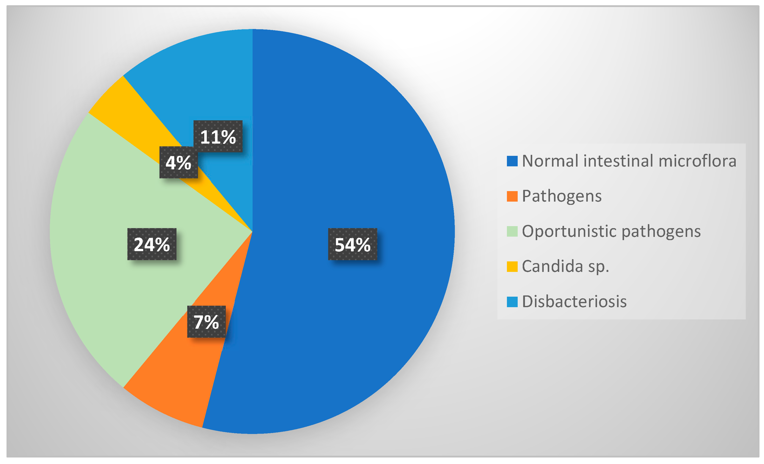
Figure 1. Summary data on isolated intestinal microorganisms in individuals with enterobiasis (n = 169).
Microbiolres 16 00215 g001
Table 1. Demographic and clinical characteristics of persons with enterobiasis, presence of eggs/adults forms in clinical samples and parasite burden grade.
CharacteristicsGenderAge
Male
Number (%)
Female
Number (%)
Children and Adolescents
Number (%)
Adults
Number (%)
asymptomatic
with symptoms
45 (59%)
31 (41%)
42 (45%)
51 (55%)
85(52%)
77 (48%)
2 (29%)
5 (71%)
no presence of adult forms in clinical samples
with the presence of adult forms in clinical samples
62 (82%)
14 (18%)
67 (73%)
25 (27%)
124 (77%)
37 (23%)
5 (71%)
2 (28%)
low parasite burden
high parasite burden
27 (36%)
49 (64%)
29 (31%)
64 (69%)
53 (33%)
109 (67%)
3 (43%)
4 (57%)
Table 2. Demographic and clinical characteristics of persons with enterobiasis and pathogenic bacterial co-infection.
Gender Age (Year)Clinical SymptomIsolated Intestinal Pathogen
male5bruxismE. coli 0139
female3asymptomaticE. coli 075
male4asymptomaticE. coli 0146
female18abdominal painE. coli 018
male11bruxismE. coli 018
female7asymptomaticE. coli 018
male10asymptomaticE. coli 0142
female10perianal itchingE. coli 0158
female6asymptomaticE. coli 0146
female6tenesmusE. coli 018
male13diarrheaSalmonella arizonae
female7diarrheaCitrobacter gillenii
Table 3. Opportunistic bacterial pathogens isolated from individuals with enterobiasis.
Distribution of Cases by Gender
(Number of Cases/%)
Species of Isolated Opportunistic Pathogen
Klebsiella pneumoniaeEnterobacter spp.Proteus mirabilisCitrobacter freundiiPseudomonas aeruginosa, Enterococcus fecalis, Morganella morganii
Males 9 (47.4%)5 (26.3%)2 (10.5%)2 (10.5%)1 (5.3%)
Females11 (50%)4 (18.2%)3 (13.6%)2 (9.1%)2 (9.1%)
Proportion of all cases with opportunistic bacterial pathogens 20 (48.8%)9 (22%)5 (12.2%)4 (9.8%)3 (7.3%)
Table 4. Findings from the statistical analysis of the link between the presence and type of pathogenic and conditionally pathogenic microflora, the demographic and clinical traits of the patients in the study and the presence of E. vermicularis eggs/adults in the clinical samples, as well as parasite load.
Normal Intestinal Flora
Number (%)
Pathogenic Flora
Number (%)
Opportunistic Pathogens
Number (%)
Candida spp.
Number (%)
Lack of Commensal Flora
Number (%)
Chi-Square Test
p Value
Gender
male (76)
female (93)

39 (23%)
52 (31%)

5 (3%)
7 (4%)

19 (11%)
22 (13%)

3 (2%)
3 (2%)

10 (6%)
9 (5%)
0.944
Children’s age groups
1–3 (n = 25)
4–6 (n = 77)
7–9 (n = 36)
10–12 (n = 20)
13–15 (n = 2)
16–18 (n = 2)

17 (10%)
42 (26%)
18 (11%)
8 (5%)
1 (1%)
0 (0%)

1 (1%)
4 (2%)
2 (1%)
3 (2%)
1 (1%)
1 (1%)

3 (2%)
25 (15%)
7 (4%)
6 (4%)
0 (0%)
0 (0%)

1 (1%)
0 (0%)
2 (1%)
2 (1%)
0 (0%)
0 (0%)

3 (2%)
6 (4%)
7 (4%)
1 (1%)
0 (0%)
1 (1%)
0.031
asymptomatic (n = 87)
with clinical symptoms (n = 82)
49 (29%)
42 (25%)
5 (3%)
7 (4%)
22 (13%)
19 (11%)
2 (1%)
4 (2%)
9 (5%)
10 (12%)
0.797
Only E. vermicularis eggs in the clinical samples (n = 129)
Presence of adult forms in clinical samples (n = 39)
76 (45%)
15 (9%)
8 (5%)
4 (2%)
31 (19%)
9 (5%)
5 (3%)
1 (1%)
9 (5%)
10 (6%)
0.015
Low parasitic burden (n = 56)
High parasitic burden (113)
37 (22%)
54 (32%)
4 (2%)
8 (5%)
10 (6%)
31 (18%)
1 (1%)
5 (3%)
4 (2%)
15 (9%)
0.220
Table 5. Distribution of individuals co-infected with enterobiasis and a bacterial pathogen, categorized by their demographic characteristics, the reason for their examination and the type of bacterial flora isolated.
GenderAgeReason for ExaminationIsolated Bacterial Species
male5bruxismE. coli 0139
female3prophylactic examinationE. coli 075
male4prophylactic examinationE. coli 0146
female18abdominal painE. coli 018
male11bruxismE. coli 018
female7prophylactic examinationE. coli 018
male10prophylactic examinationE. coli 0142
female10perianal itchingE. coli 0158
female6prophylactic examinationE. coli 0146
female6tenesmusE. coli 018
male13diarrheaSalmonella arizonae
female7diarrheaCitrobacter galineii
Disclaimer/Publisher’s Note: The statements, opinions and data contained in all publications are solely those of the individual author(s) and contributor(s) and not of MDPI and/or the editor(s). MDPI and/or the editor(s) disclaim responsibility for any injury to people or property resulting from any ideas, methods, instructions or products referred to in the content.