Article

Closing the Diagnostic Gap in Encephalitis and Acute Disseminated Encephalomyelitis through Digital Case Classification and Viral Metagenomics

1
Infectious Diseases & Vaccines, Vaccine Safety Initiative, D-10437 Berlin, Germany
2
Laboratoire Chrono-Environnement LCE, UMR CNRS 6249, Université Bourgogne Franche-Comté, F-25000 Besançon, France
3
National Reference Center for Influenza, Robert Koch-Institute, D-13353 Berlin, Germany
4
National Reference Laboratory for Adenoviruses, Hannover Medical School, D-30625 Hannover, Germany
*
Author to whom correspondence should be addressed.
Microbiol. Res. 2024, 15(2), 900-913; https://doi.org/10.3390/microbiolres15020059
Submission received: 28 March 2024 / Revised: 5 May 2024 / Accepted: 11 May 2024 / Published: 23 May 2024

Abstract

:
Encephalitis and acute disseminated encephalomyelitis (ADEM) are often caused or triggered by viruses—but the specific pathogen commonly remains unidentified in routine care. We explored the use of viral metagenomic next-generation sequencing (mNGS) in addition to PCR testing of non-invasive stool samples to see if unbiased testing could potentially increase diagnostic yield. To identify specific clinical cases at the point of care, we took advantage of a previously published digital app allowing instant clinical case classification based on consensus case criteria, the VACC-Tool. This hospital-based prospective digital surveillance program assessed 100 pediatric patients (mean age: 11 years, range: 0.15–17.85; 49% male) with case-confirmed encephalitis and/or ADEM. Analysis of case classification at the point of care revealed that in routine care, 96% of confirmed encephalitis/ADEM cases had been missed. Overall agreement of routine care diagnoses with digital encephalitis/ADEM case classification was <50%. Also in routine care, only 13% of cases held a virus-related diagnosis, i.e., herpesvirus (n = 8) and enterovirus infection (n = 5). Use of mNGS increased the yield of virus detection by 77% (n = 23 virus hits). Specifically, mNGS identified 10 additional virus species beyond herpes- and enteroviruses. Of the additional 23 virus hits detected with mNGS, PCR confirmation was possible post hoc in 14 cases (61%). Linking digital case classification, mNGS, and PCR testing may not be feasible in routine care at this point but may help to provide hints to the pathogenesis of encephalitis/ADEM in childhood, warranting further research and exploration.

1. Introduction

Encephalitis and acute disseminated encephalomyelitis (ADEM) are infectious/inflammatory diseases of the central nervous system (CNS). Both are potentially serious health conditions requiring early and accurate diagnosis [1]. However, neurological signs and symptoms can be subtle or atypical, especially in children, thus implicating diagnostic challenges [2,3,4].
To help healthcare professionals establish a reliable diagnosis with minimal ascertainment bias, health authorities advocate the use of consensus case criteria. But in routine care, they are hardly ever used [5]. To facilitate compliance with case definitions, the Vaccine Safety Initiative (VIVI, formerly Vienna Vaccine Safety Initiative), an international non-profit research organization, developed the VACC-Tool (Vaccine Safety Automated Case Classification Tool): a mobile precision medicine application comparing the patient’s clinical presentation to published case definitions in real-time. The VACC-Tool was prospectively validated in the context of a digital surveillance and quality improvement program where it proved to efficiently harmonize diagnoses compared to retroactive chart review and ICD (International Statistical Classification of Diseases and Related Health Problems) coding, which are prone to bias and rather serve for billing purposes [5,6]. A key benefit of the VACC-Tool was that capturing clinical data at the bedside allowed the “right questions to be asked at the right time”, overcoming another drawback of routine medical records, which are often incomplete [5,7].
Once a precision clinical diagnosis is established and ascertained using standardized case criteria, the causality assessment constitutes the next logical step to initiate adequate therapy and/or infection control measures. The majority of encephalitis and ADEM cases are associated with viral infection [8]. In routine care, however, the question of etiology remains unresolved in ≈50% of cases. Possible associations may remain undetected when only a limited number of specific pathogens, including viruses, are routinely tested with CNS serology and conventional PCR [3,9,10]. In addition, lumbar puncture is invasive and viruses triggering post-infectious ADEM may no longer be detectable in the CNS compartment at the time of symptom onset. This means that little progress has been made in the resolution of encephalitis and ADEM cases in routine care, leaving clinicians and patients/parents puzzled and unsatisfied.
On this note, the study team wished to explore whether unbiased/agnostic molecular pathogen detection techniques, such as metagenomic next-generation sequencing (mNGS) may enable the identification of any genomic sequence present in a well-defined set of tightly controlled clinically confirmed cases. In contrast to sequence-dependent conventional detection methods, mNGS asks an “open-ended question” thereby promising to diminish bias on the laboratory end [11]. Still, mNGS signals do not prove causality, and the technique requires specific expertise to adequately interpret mNGS results and a laboratory skilled at controlling for any contamination or misinterpretation. Therefore, mNGS has not yet entered the realm of routine diagnostics, also due to variations in sensitivity and methodology between laboratories [11,12].
However, one of the requirements for adequate interpretation of mNGS results lies in the precise delineation and differentiation of clinical cases (where mNGS samples are obtained) based on rigorous case definition and data standards. This appears to have been a major obstacle to advancing knowledge of the role of specific pathogens (or combinations thereof) in cases of CNS infection/inflammation.
A comprehensive diagnosis cannot usually be made based on clinical or laboratory findings alone, but rather by linking them. Particularly in the case of (post- or para-) infectious diseases, a diagnosis is most specific when it includes a syndromic label and a reference to the infectious trigger. The diagnostic gap in complex diseases such as encephalitis and ADEM can only be closed via methods that eliminate uncertainty and bias in both the clinical and laboratory domain.
In this study, we implemented a nested digital surveillance approach for the investigation of pediatric encephalitis and ADEM, combining two means of precision medicine:
(I)
Automated case classification using the VACC-Tool at the patient’s bedside;
(II)
Viral mNGS and conventional PCR testing of stool samples.
The goal is to close the ‘diagnostic gap’ in complex infectious/inflammatory CNS diseases by mitigating bias towards both the clinical and laboratory end.

2. Materials and Methods

We conducted the present study in the context of a prospective digital surveillance and quality improvement program for children with CNS infection/inflammation, which was performed independently from routine care at the Charité University Hospital in Berlin, Germany. The digital surveillance and quality improvement program was approved by the Charité Institutional Review Board (IRB number: EA2/161/11). All procedures were performed in compliance with the ethical standards of the Declaration of Helsinki and according to German laws. Written informed consent was waived for the purpose of quality improvement and infection control. Verbal informed consent was obtained from patients above 16 years of age or from parents or caretakers of underage patients [13].

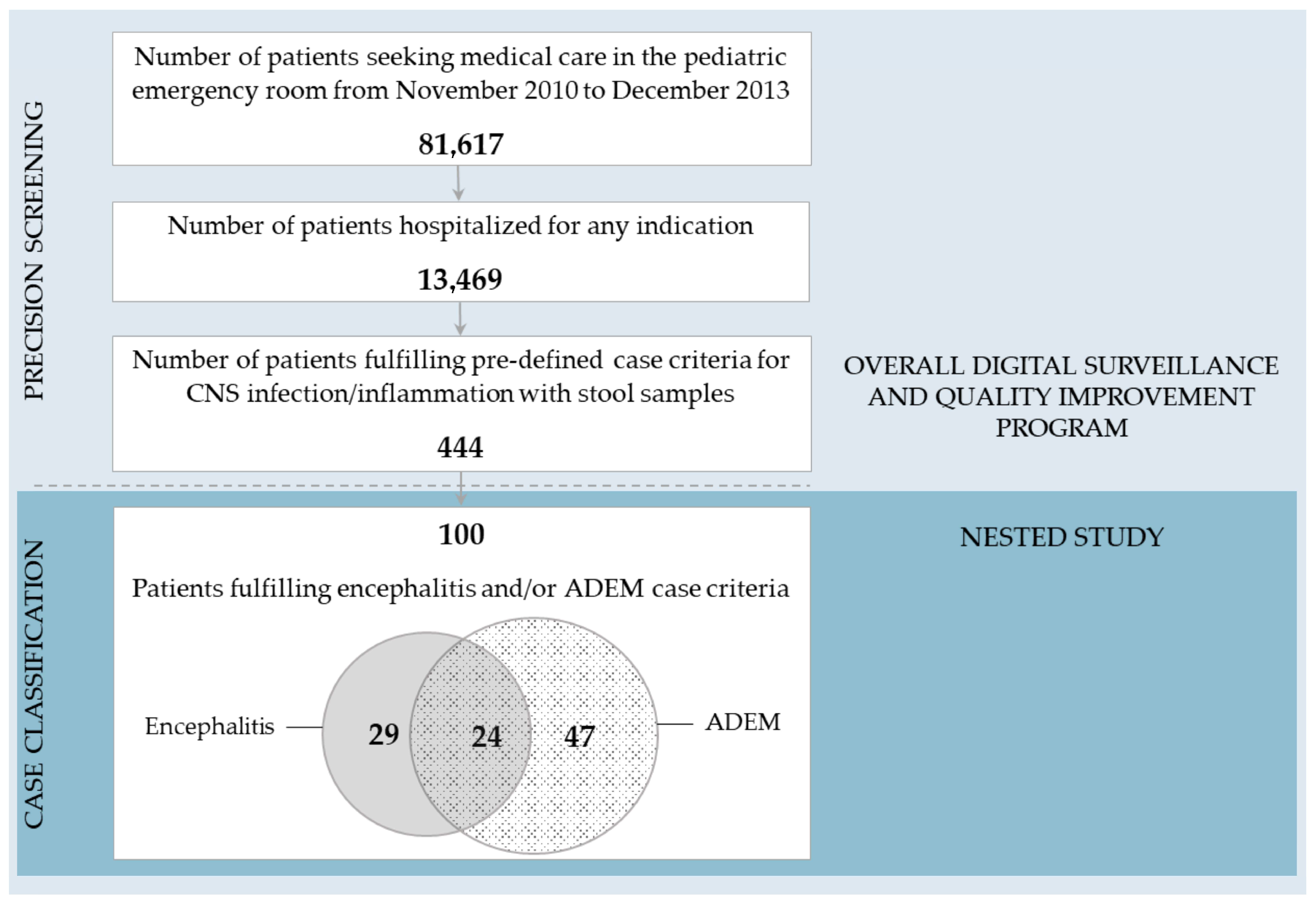
2.1. The Overall Digital Surveillance and Quality Improvement Program

From November 2010 to December 2013, 444 pediatric inpatients (mean age: 7 years, range: 0.03–18; 56% male) who met entry criteria of suspected CNS infection/inflammation (fever and specific CNS symptoms and/or lumbar puncture) [13] participated in the prospective digital surveillance and quality improvement program. Exclusion criteria were known seizure disorder, CNS lesion/tumor, intoxication, traumatic head injury, or acute diarrhea/dehydration [13].
All patients underwent standardized assessments using the VACC-Tool for automated case classification at the point of care performed by specifically trained staff [5,7,13,14]. The VACC-Tool 1.0 is a validated mobile application allowing clinicians to assess a patient while comparing the disease presentation with published case definitions for aseptic meningitis, encephalitis, myelitis, and ADEM [4,15]. Data entered into the VACC-Tool are fully compliant with international data standards issued by the Clinical Data Interchange Standards Consortium (CDISC) to enable interoperability of data across sites [5,7,16].
From all patients, stool samples were collected and transferred to the German National Reference Center for Poliomyelitis and Enteroviruses at the Robert Koch-Institute in Berlin, Germany for systematic blinded PCR testing for enteroviruses and parechoviruses as described earlier [13,14].

2.2. The Nested Study Cohort—100 Patients under Investigation

The present nested study was limited to 100 patients/patient samples for logistical and cost constraints. We selected all patients fulfilling encephalitis and/or ADEM case criteria within the overall digital surveillance and quality improvement program, arriving at 100 encephalitis/ADEM patients from November 2010 to December 2013 (Figure 1).
We searched routine care discharge letters of all 100 encephalitis/ADEM patients for encephalitis and/or ADEM and/or other ‘infectious disease’-related discharge diagnoses according to ICD-10 coding.

2.2.1. Viral mNGS—Sample Processing

Stool samples of all 100 encephalitis/ADEM patients underwent pooled viral mNGS. Stool suspensions were mixed with phosphate-buffered saline (0.75 mL) and zirconia beads (0.2 g), vortexed, and spun at 12,000 rounds per minute (rpm) in a tabletop microfuge for 10 min and supernatants were transferred into Eppendorf tubes. Supernatants (200 µL) were filtered through a 0.45 μm pore filter to exclude cells and large particles. The filtrates were then digested with a combination of DNAse and RNAse nucleases to reduce background of host and bacterial genetic material and enrich for viral nucleic acids protected from nuclease digestion within their capsids [17,18]. Nucleic acid extraction was performed using the Qiagen Viral RNA mini kit (Qiagen, Hilden, Germany) according to the manufacturer’s instructions. Viral cDNA synthesis was performed separately on each individual sample using viral nucleic acids (10 µL) with random hexamers (100 pmol) at 72 °C for 2 min. 200 U SuperScript III reverse transcriptase (Invitrogen, Waltham, MA, USA), 0.5 mM of each deoxynucloside triphosphate (dNTP), 10 mM dithiothreitol, and 1× first-strand extension buffer were added to the mixture and incubated at 25 °C for 10 min, followed by 50 °C incubation for 1 h and 70 °C for 15 min. Second-strand cDNA synthesis was performed via incubation of reversely transcribed products with 5 U of Klenow Fragment DNA polymerase (New England Biolabs, Ipswich, MA, USA) at 37 °C for 1 h followed by 75 °C for 20 min.
The resulting double-stranded cDNAs from groups of five stool samples were then pooled (20 pools total). The transposon-based Nextera XT DNA sample preparation kit (Illumina, San Diego, CA, USA) was used followed by PCR using unique pairs of index barcodes for each pool. Each library was quantified using Kapa Library Quant kit (Kapa Biosystems, Wilmington, MA, USA) following the manufacturer’s instructions, and the equimolar DNA quantities were pooled for sequencing using one flow cell on the Illumina HiSeq4000 instrument with 150 paired-end sequencing.

2.2.2. Viral mNGS—Bioinformatics Pipeline

Paired-end sequencing reads were debarcoded with Illumina vendor software v3.
Reads were considered duplicates if base positions 5 to 55 were identical. One random copy of duplicates was kept. Low sequencing quality tails were trimmed using Phred quality score 20. Adaptor and primer sequences were trimmed using VecScreen11 default parameters. Cleaned reads were de novo assembled using Ensemble Assembler [19]. Assembled contigs, along with the remaining singlets, were aligned to an in-house viral proteome database using BLASTx. Matches to virus proteins were aligned to an in-house non-virus–non-redundant (NVNR) universal proteome database using BLASTx.
To subtract human and bacterial reads, nucleotide databases were compiled as follows: Human reference genome sequences and mRNA sequences (hg38) were concatenated. Bacterial nucleotide sequences were extracted from NCBI nr fasta files based on NCBI taxonomy. Human and bacterial nucleotide sequences were compiled into Bowtie2 (version 2.2.4) databases [20] for cellular sequences subtraction. Two databases were constructed: (1) a virus BLASTx database using the NCBI virus reference proteome to which viral protein sequences from NCBI nr fasta files were added and (2) a NVNR database using sequences extracted from NCBI nr fasta file. Repeats and low-complexity regions were masked using segmasker from blast+ suite (version 2.2.7). Hits with lower adjusted E-value to NVNR than to viral proteins were removed.
To account for potential barcode index-switching between pools, which had been described earlier using the Illumina HiSeq4000 instrument [21], the following algorithm was established for reporting the presence of viruses: The pool with the highest percentage of viral reads for a given viral species (hottest pool) was considered true-positive. Other pools sharing an index barcode with viral reads >15% of the percentage of reads in that hottest pool were considered positive. If genetic analysis of the reads relative to the genome or contigs from the hottest pool showed the presence of a different genotype, that sample was also considered positive with reads <15% threshold. The pool with the second highest percentage of viral reads considered true-positive was then used to exclude pools with which it shared an index barcode according to the same rules. This process was repeated until no more pools with reads to that virus species could be excluded.

2.2.3. PCR Testing

Aliquots of pooled samples were reassessed using specific confirmatory PCR to narrow down pooled mNGS results to the individual patient level for human adeno-, boca-, rota-, saffold-, and sapovirus [22,23,24,25,26].
PCR testing of the same samples for enteroviruses and parechovirus A had been performed as per protocol of the overall digital surveillance and quality improvement program [13].

2.2.4. Statistics

Descriptive statistics were performed with SPSS 22.0 (IBM Corp., Armonk, NY, USA). To compare discharge diagnoses from routine care and VACC-Tool case classification as well as enterovirus PCR and the corresponding viral mNGS results among the 100 patients under investigation, we used cross tabulations and determined positive and negative percent agreement (PPA and NPA) and overall rates of agreement (ORA) as described previously (Appendix A) [5]. We calculated Cohen’s kappa coefficients (κ) to assess the coincidence of concordant/discordant results, i.e., inter-rater reliability, following the Food and Drug Administration guidelines and suggested terminologies for the reporting of results from studies evaluating diagnostic tests [5,27,28].

3. Results

Overall, 444 patients entered the digital surveillance and quality improvement program and had stool samples available for virological analysis; 100 of whom met standardized case criteria for encephalitis and/or ADEM according to VACC-Tool case classification at the bedside (mean age: 11 years, range: 0.15–17.85; 49% male; see Table 1 for detailed patient characteristics). Figure 1 illustrates the precision screening process and classification results.

3.1. Comparison of Encephalitis/ADEM VACC-Tool Case Classification and Discharge Diagnoses

Among the 100 patients who fulfilled encephalitis and/or ADEM case criteria, VACC-Tool case classification yielded 29 encephalitis cases, 47 ADEM cases, and 24 ‘overlap’ cases fulfilling both encephalitis and ADEM case criteria as per case definition [4]. In routine care discharge letters of the same patients, four cases were labeled as ‘encephalitis’ and there was no case of ADEM. That is, 96 of 100 patients were misdiagnosed in routine care. Accordingly, all measures of agreement between VACC-Tool case classification and discharge diagnoses were low (Table 2). Kappa scores <0.1 reflected low reliability of agreements [28].
The most common syndromic misdiagnosis in routine care was ‘meningitis’ (n = 13/100), which was not supported with consensus case criteria implemented in the VACC-Tool [5,15].
Of the 100 patients with confirmed encephalitis and/or ADEM, 13 held a virus-related diagnosis in their routine care discharge letter (Figure 2). Of those, eight were ‘herpesvirus infection of the CNS’ (four labeled as ‘encephalitis’ and four labeled as ‘zoster (oticus)’), five of whom tested positive via PCR from cerebrospinal fluid (CSF), two via antibody testing from CSF, and one via serology. Another five were ‘enterovirus infection’ (four labeled as ‘meningitis’ and one labeled as ‘unspecific enterovirus infection’), two of whom tested positive via PCR from CSF, one via PCR from saliva, one via PCR from serum, and one based on clinical suspicion only.
Another six cases were ‘other/unspecified (viral) infection’ and nine more cases ‘borrelia infection’. The remainder (72%) did not receive any virus/infectious disease diagnosis.

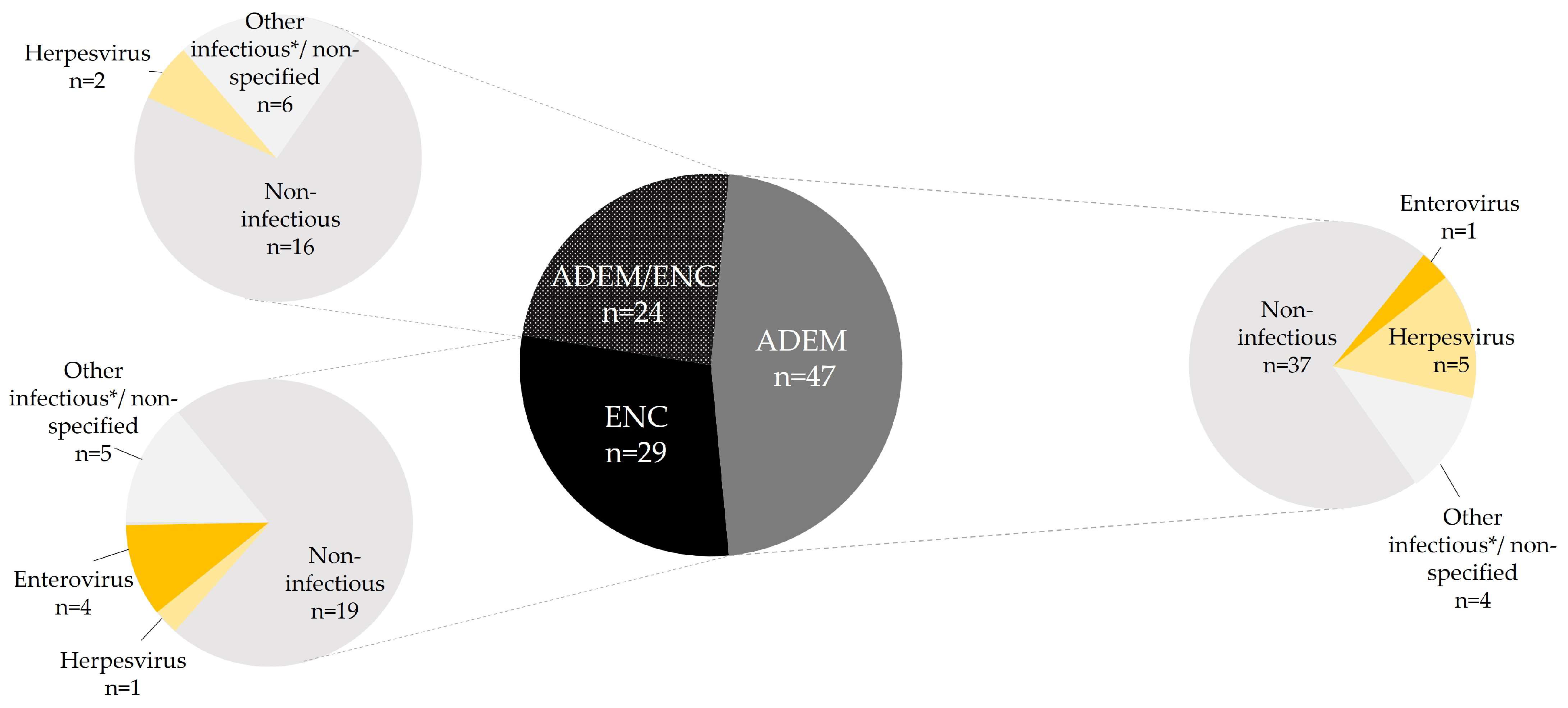
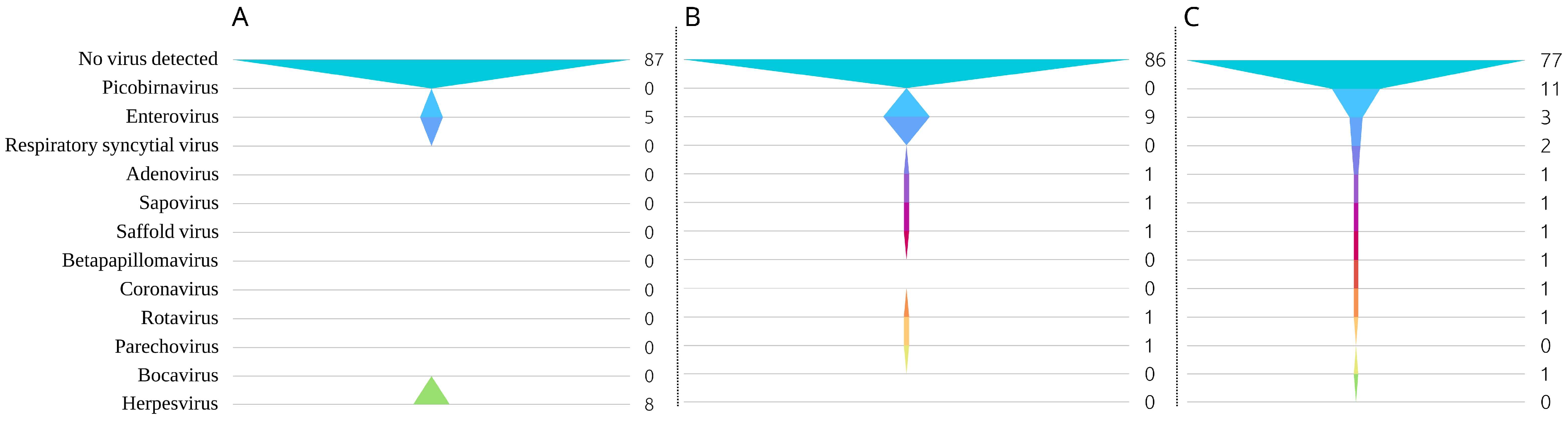
3.2. Viral mNGS Results

Viral mNGS yielded 16 positive pools, with a total of 10 distinct viruses: adenovirus C, betapapillomavirus, bocavirus-1, coronavirus-NL63, enterovirus B, picobirnavirus, respiratory syncytial virus, rotavirus A, saffold virus, and sapovirus.
Most frequently, reads were detected from picobirnaviruses (11/16 positive pools), followed by enterovirus (3/16 positive pools), and respiratory syncytial virus (2/16 positive pools). All other viruses were detected in only one pool, respectively.
Pathogen-specific read counts were highest for bocavirus (3086 of 4,764,228 reads in the respective sample; ≈0.06% reads), followed by sapovirus (1005 of 5,139,726 reads in the respective sample; ≈0.02% reads), enterovirus (1858 of 14,161,484 reads in the respective sample; ≈0.01% reads), respiratory syncytial virus (800 of 7,142,404 reads in the respective sample; ≈0.01% reads), and adenovirus (625 of 4,848,500 reads in the respective sample; ≈0.01% reads). The remainder yielded <0.003% reads of the respective sample.

3.2.1. Confirmatory PCR Testing of mNGS Hits

Confirmatory identification of individual cases via post hoc PCR testing was possible for sapovirus (≈0.02% reads, see above), rotavirus (7 of 7,068,164 reads in the respective sample; ≈0.0001% reads), adenovirus (≈0.01% reads, see above), and saffold virus (130 reads of 4,764,228 reads in the respective sample; ≈0.0027% reads). Detection of bocavirus reads (≈0.06% reads, see above) via viral mNGS could not be confirmed with PCR.

3.2.2. Systematic PCR Testing for Enteroviruses and Parechovirus as Compared to mNGS

As per protocol of the overall digital surveillance and quality improvement program, PCR testing for enteroviruses was not only performed in cases where mNGS had yielded a positive result, but systematically in all (the same) stool samples. In the nested study cohort, enterovirus PCR yielded a total of 9/100 individual positive results, while 3/20 pools tested positive for enterovirus B via mNGS. In these three pools, one sample tested positive for echoviruses 9 (1858 of 14,161,484 reads in the respective sample; ≈0.01% reads), 21 (92 of 4,300,010 reads in the respective sample; ≈0.002% reads), and 30 (42 of 4,519,164 reads in the respective sample; ≈0.0009% reads), respectively. Consequently, mNGS missed six individual enterovirus results, which were detected with specific PCR. Overall agreement between enterovirus PCR and enterovirus hits with mNGS was 94%, with a kappa score of 0.5 reflecting moderate reliability of agreement (Table 3) [28].
As per protocol of the overall digital surveillance and quality improvement program, PCR testing for parechovirus A was systematically performed as well. Via PCR, one sample tested positive for parechovirus A [14], but parechovirus A reads were not detected with mNGS.

3.3. Increased Diagnostic Yield through VACC-Tool Case Classification, mNGS, and PCR Testing

The combined use of automated case classification based on consensus case criteria applied at the bedside with mNGS and PCR testing provided standardized encephalitis and ADEM diagnoses and increased diagnostic yield. Cautiously assuming that a ‘positive pool’ of five samples corresponded to a single positive individual sample, we increased virus-specific diagnostic yield within the nested study by 8–77% compared to 13 virus-specific diagnoses from routine care in the same patients (Figure 2): mNGS alone yielded 23 hits (increase by 77%); 14 of which were confirmed or ruled-out with PCR testing (increase by 8%). Figure 3 provides an overview of increased virus-specific diagnostic yield through mNGS and PCR testing compared to routine care.
While herpesvirus detection from routine care (n = 8) could not be reproduced from stool samples in our nested study, we increased the diagnostic yield for enteroviruses by 80%: a total of five enterovirus infections were detected in routine care, only one of which could be reproduced via both mNGS and PCR (echovirus 30); but another eight stool samples tested positive for enteroviruses in our nested study.

3.4. Clinical Vignettes of Significant Cases

The linkage of VACC-Tool automated case classification in real-time with mNGS and PCR testing led to the identification of at least four significant and PCR-confirmed associations between encephalitis or ADEM and a virus that were missed in routine care.

3.4.1. Adenovirus in a 13-Year-Old with ADEM

A 13-year-old male presented with headache, vomiting, anosmia, decreased peripheral sensitivity and paresthesia, and transient sudden loss of consciousness witnessed by his parents. A somatosensory evoked-potential test showed focal interruption of the median nerve. Blood testing revealed moderate leukocytosis. The patient fully recovered without specific treatment and was discharged after four days.
ADEM case criteria, including focal neurological deficits, were met as confirmed with the VACC-Tool. The stool sample collected on hospital day 1 tested positive for adenovirus C2 [22] via viral mNGS and confirmatory PCR.

3.4.2. Rotavirus in a 2-Year-Old with ADEM

A 2-year-old female presented with an afebrile convulsive seizure and ataxia. No gastroenteritis or other symptoms were present. On hospital day 7, a maximum body temperature of 38.8 °C was measured. CSF analysis showed elevated total protein levels and normal white blood cell counts. In routine care, microbiology testing was all negative. The patient fully recovered without specific treatment and was discharged after nine days.
ADEM case criteria, including cerebellar dysfunction, were met and confirmed using the VACC-Tool. The stool sample tested positive for rotavirus A via viral mNGS as confirmed with real-time RT-PCR (crossing point value: 13.24).

3.4.3. Saffold Virus in a 4-Year-Old with ADEM

A 4-year-old male presented with acute afebrile seizures, behavioral changes, fatigue, motor weakness of the limbs, and bladder dysfunction in addition to mild respiratory symptoms and conjunctivitis. In routine care, human rhinovirus (crossing point value: 30) was detected in a nasopharyngeal sample with PCR. CSF analysis was normal, except for cytoalbuminologic dissociation. The patient fully recovered without specific treatment and was discharged after 9 days.
ADEM case criteria, including focal neurological deficits, were met and confirmed with the VACC-Tool. The stool sample collected on hospital day 2 tested positive for saffold virus via viral mNGS as confirmed with nested RT-PCR.

3.4.4. Sapovirus in a 2-Year-Old with Encephalitis

A 2-year-old female was hospitalized with fever, cough, a decreased level of consciousness, acute personality changes, phonophobia, and bilateral motor weakness of the limbs. Broad-spectrum antibiotics, oseltamivir, and intravenous immunoglobulins were administered upon admission.
Blood analysis showed elevated C-reactive protein and interleukin-6 levels and respiratory acidosis. CSF analysis was normal except for elevated glucose and protein levels, i.e., cytoalbuminologic dissociation. Routine care bacterial testing from CSF and blood were all negative. Routine care viral multiplex PCR testing from respiratory secretions was positive for RSV, enterovirus, rhinovirus, and Epstein-Barr virus on day 2. A stool sample obtained on day 19 tested positive for rotavirus via PCR in routine care. Cranial magnet resonance imaging revealed parenchymal signal enhancement.
The patient fully recovered and was discharged after 26 days.
Encephalitis case criteria were met, as confirmed with the VACC-Tool. The stool sample obtained within the quality improvement program on hospital day 2 tested positive for sapovirus via viral mNGS as confirmed with real-time RT-PCR (crossing point value: 23.5).

4. Discussion

With the present study, we introduced a nested precision medicine approach combining automated case classification for encephalitis and ADEM in real-time using mobile health technology with viral mNGS and PCR testing to diminish bias in the clinical and laboratory context. This study provided insight into three years of standardized digital infectious disease surveillance with an established total number of comparable cases.
Clinical diagnostic challenges of complex infectious/inflammatory CNS disease entities such as encephalitis and ADEM arise from varying patient signs and symptoms in addition to intrinsic inter-rater variability and observer bias. The use of standardized case criteria at the point of care enables harmonization of diagnoses [4]. And if case criteria are taken immediately while the patient is assessed, including digital time stamps and audit trails, all pertinent information is obtained with minimal bias, thereby avoiding missing data and post hoc assumptions [5,6,7]. In our study, we used mobile health technology at the patient’s bedside to implement a precision screening and automated case classification workflow resulting in immediate and unbiased, reproducible encephalitis and/or ADEM diagnosis in a total of 100 comparable cases.
Following the precision screening and diagnosis steps based on consensus case criteria incorporated into the VACC-Tool [5], we investigated whether subsequent, sequence-independent mNGS might help in closing the ‘diagnostic gap’ in children with encephalitis and/or ADEM, with a particular focus on viruses as their most common cause [3,4,10].
In line with previous studies, few encephalitis and ADEM cases in our study (13%) held a specific infectious diagnosis in routine care. Review of the patients’ medical records revealed inconsistent proceedings in the diagnostic work-up, including one diagnosis of ‘enterovirus infection’ based on clinical suspicion only. This exemplified the potential pitfalls of retrospective chart review of ICD codes and discharge summaries for scientific or surveillance purposes.
Oftentimes, the brevity of clinician’s interactions with patients and repeated (often conflicting) assessments due to shift work and high staff turnover add to the confusion. Last but not least, there are financial constraints that limit the options of diagnostics in routine care.
If the diagnostic work-up is left to the treating physician, it is usually confined to bacterial cultures and certain viruses, e.g., herpes or enteroviruses. Syndromic multiplex panels may help to test for a limited range of an additional, known 10–20 pathogens [12]; mNGS on the other hand, is an unbiased detection method which can be leveraged in “hard-to-diagnose cases” or outbreak scenarios [29]. In our study, mNGS identified 10 different additional viruses; 6 of which were tested orthogonally with PCR, yielding positive results for 5 viruses. That is, mNGS and “add-on” pathogen-specific PCR combined increased the number of viruses detected in our patient cohort.
While mNGS may help to raise hypotheses, it is not yet able to state a definite diagnosis or causality. In contrast to PCR, mNGS laboratory protocols are not yet standardized, and accuracy may vary. Our study underlines that complementary PCR testing can help to ascertain or fail to confirm an mNGS signal retroactively. In our study, complementary PCR did not confirm a bocavirus signal, but mNGS also failed to detect enterovirus and parechovirus signals that were detected with PCR. Evidently, the level of accuracy of mNGS may be improved in the future, as it depends on methods used to enrich viruses, to generate DNA libraries, and the depth of sequencing [9,29]. Also, massively parallel or multiplexed sequencing to reduce costs may lead to index-switching in pooled libraries and artifact results, requiring method-specific solutions at the (pre-)analytical level and/or in data curation [21,30,31,32]. Applying these considerations to our specific mNGS results, the false-positive bocavirus signal could have resulted from a barcode index-switch (e.g., carryover of nucleic acids from runs to runs) or contamination during library construction, for example. Vice versa, overly restrictive parameters in the data curation could cause false-negative mNGS results.
Viral mNGS and confirmatory PCR testing in our cohort identified rare or novel potential links between pathogens to specific clinical entities ascertained using the VACC-Tool: three cases of ADEM could thus be linked to (a) saffold virus, (b) rotavirus, and (c) adenovirus detection [22] and one case of encephalitis was linked to sapovirus detection. All of these viral pathogens are frequently detected in patients with gastrointestinal disease, mainly presenting with acute diarrhea [33,34,35]. Diarrhea, however, was an exclusion criterion in our prospective digital surveillance and quality improvement program [13], meaning that the 100 clinical cases assessed in this study presented with confirmed CNS signs and symptoms in the confirmed absence of gastroenteritis. As the findings in this study show, textbook knowledge of how specific viruses are “presumed to present clinically” may need to be updated based on scientific evidence, if confirmatory studies point in the same direction.
Saffold virus [36,37], rotavirus [37,38], and adenoviruses [39,40] have previously been presumed to be associated with CNS disease in children and adults. Our study is the first of its kind to confirm this link using standardized case criteria.
To our knowledge, sapovirus detection has not yet been associated with neurological disease [41]. Establishing a causal relationship between encephalitis and sapovirus detection was not possible in our case due to detection of four other viruses (entero-, rhino-, respiratory syncytial, and rotavirus) in a respiratory/saliva sample obtained in routine care. More systematic clinical and laboratory research is needed to prove/disprove any such associations.
The timing of sample collection and the selection of the specimen type play a decisive role for the plausibility of causal relationships. Enteric viruses, such as adenoviruses, enteroviruses, rotavirus, or sapovirus, are known to be shed in feces or saliva for weeks or months after initial infection [42,43]. This circumstance may help to identify pathogens responsible for post-infectious syndromes such as ADEM or post-infectious encephalitis, but it also increases the likelihood of simultaneous detection of multiple viruses. In our study, we used stool samples as a non-invasive screening tool and to provide an extended diagnostic window beyond the time of early viral clearance from the CSF compartment [9,14,39]. While many CNS pathogens can be readily detected in stool samples, for some viruses such as herpesviruses or West Nile virus, detection from stool samples is unlikely in humans [44,45]; this is confirmed in our study where positive herpesvirus detection in the CSF could not be reproduced in stool samples from the same patients. Similarly, of two enterovirus detections in the CSF in routine care, only one was also positive (via both, mNGS and PCR) in the stool. Therefore, more research with comparative studies of mNGS and PCR from different body compartments including blood, urine, saliva, or CSF are warranted [46].
Limitations to our study included the pooling of samples for mNGS, which was more cost-effective but likely impaired accuracy of viral mNGS. Also, future use of mNGS to ‘close the diagnostic gap’ in cases of encephalitis and ADEM will need to include parasites, bacteria, and fungi. In our study, the number of patients with unusual viral identifications such as adeno-, saffold-, sapo-, or rotavirus was low. Therefore, we did not perform statistical analysis of these cases. Further studies are needed to assess the true clinical significance of the detection of specific pathogens.
Another limitation of the hospital-based surveillance was the lack of a control group, e.g., involving healthy individuals to further investigate whether mNGS signals may constitute a bystander effect. Unfortunately, confirmatory PCR testing of picobirnavirus, respiratory syncytial virus, betapapillomavirus, and coronavirus could not be performed for a lack of sample material/for logistical reasons. Especially for picobirnaviruses, which were detected in the majority of mNGS pools in our study, the role as bystander versus pathogen remains unclear, warranting further research [47].
Future studies may also incorporate additional infectious/inflammatory CNS disease entities providing reliable and reproducible case classification. The VACC-Tool (in its 2.0 beta-version) allows for automated case classification of 14 different infectious/inflammatory CNS diseases, including facial nerve palsy, progressive multifocal leukoencephalopathy, or Guillain–Barré syndrome in full compliance with data standards across sites and data privacy [7]. Not only does the VACC-Tool help to ascertain diagnoses swiftly and with minimal bias, but it also has the potential to flag clinically suspicious cases, prompting further diagnostic work-up [5].
Key messages:
We combined real-time automated case classification for encephalitis and acute disseminated encephalomyelitis (ADEM) with viral metagenomic next-generation sequencing and confirmatory polymerase chain reaction;
We identified potential links between encephalitis and the detection of sapovirus, and between ADEM and saffold virus, rotavirus, and adenovirus;
The use of digital tools at the patient’s bedside and advanced molecular detection techniques helps to diminish bias and enhance diagnostic yield, including for rare, unusual, or novel pathogens.

5. Conclusions

We conclude that the combined use of the VACC-Tool for standardized digital case classification in real-time with viral mNGS and PCR testing helps to diminish bias and enhance diagnostic yield in pediatric encephalitis and ADEM.
In the long term, we expect that innovative mobile health technology and advanced laboratory methods will improve infectious disease surveillance and management by providing complementary, insightful data sets.
Application of viral mNGS remains under investigation and validation but could be particularly useful for unusual and novel pathogens.

Author Contributions

Conceptualization, B.A.R.; methodology, B.A.R.; formal analysis, P.E.O., B.A.R., X.M. and A.H.; investigation, P.E.O., B.A.R., X.M. and A.H.; resources, B.A.R. and A.H.; data curation, P.E.O., B.A.R., X.M. and A.H.; writing—original draft preparation, P.E.O.; writing—review and editing, P.E.O., B.A.R., X.M. and A.H.; visualization, P.E.O. and A.H.; supervision, B.A.R. and A.H.; project administration, B.A.R. and A.H. All authors have read and agreed to the published version of the manuscript.

Funding

This research received no external funding.

Institutional Review Board Statement

The prospective non-interventional surveillance program was approved by the Charité Institutional Review Board (IRB number: EA2/161/11). This study was performed in compliance with the ethical standards of the Declaration of Helsinki and according to German laws. Written informed consent was waived for the purpose of quality improvement and infection control given the observational nature of the study.

Informed Consent Statement

Verbal informed consent was obtained from patients above 16 years of age or parents or caretakers of underage patients [12].

Data Availability Statement

The datasets presented in this article are not readily available due to privacy and ethical restrictions. Requests to access the datasets should be directed to [email protected].

Acknowledgments

The authors thank the entire team for their thorough work and sensitive approach to sick children and adolescents and their families. The laboratory team deserve great thanks for the careful and timely processing of the virological samples.

Conflicts of Interest

The authors declare no conflicts of interest.

Appendix A

Table A1. Calculation of positive and negative percent agreement (PPA and NPA) and overall rates of agreement (ORA).
Discharge Diagnosis Positive/
mNGS Positive
Discharge Diagnosis Negative/
mNGS Negative
PPA: 100 × A ( A + C )
NPA: 100 × D ( B + D )
ORA: 100 × ( A + D ) ( A + B + C + D )
VACC-Tool positive/
PCR positive
AB
VACC-Tool negative/
PCR negative
CD

References

  1. GBD 2017 US Neurological Disorders Collaborators; Feigin, V.L.; Vos, T.; Alahdab, F.; Amit, A.M.L.; Barnighausen, T.W.; Beghi, E.; Beheshti, M.; Chavan, P.P.; Criqui, M.H.; et al. Burden of Neurological Disorders Across the US From 1990-2017: A Global Burden of Disease Study. JAMA Neurol. 2021, 78, 165–176. [Google Scholar] [CrossRef] [PubMed]
  2. Yong, H.Y.F.; Pastula, D.M.; Kapadia, R.K. Diagnosing viral encephalitis and emerging concepts. Curr. Opin. Neurol. 2023, 36, 175–184. [Google Scholar] [CrossRef] [PubMed]
  3. Messacar, K.; Fischer, M.; Dominguez, S.R.; Tyler, K.L.; Abzug, M.J. Encephalitis in US Children. Infect. Dis. Clin. N. Am. 2018, 32, 145–162. [Google Scholar] [CrossRef] [PubMed]
  4. Sejvar, J.J.; Kohl, K.S.; Bilynsky, R.; Blumberg, D.; Cvetkovich, T.; Galama, J.; Gidudu, J.; Katikaneni, L.; Khuri-Bulos, N.; Oleske, J.; et al. Encephalitis, myelitis, and acute disseminated encephalomyelitis (ADEM): Case definitions and guidelines for collection, analysis, and presentation of immunization safety data. Vaccine 2007, 25, 5771–5792. [Google Scholar] [CrossRef] [PubMed]
  5. Obermeier, P.; Muehlhans, S.; Hoppe, C.; Karsch, K.; Tief, F.; Seeber, L.; Chen, X.; Conrad, T.; Boettcher, S.; Diedrich, S.; et al. Enabling Precision Medicine with Digital Case Classification at the Point-of-Care. EBioMedicine 2016, 4, 191–196. [Google Scholar] [CrossRef] [PubMed]
  6. Rath, B.; Magnus, M.; Heininger, U. Evaluating the Brighton Collaboration case definitions, aseptic meningitis, encephalitis, myelitis, and acute disseminated encephalomyelitis, by systematic analysis of 255 clinical cases. Vaccine 2010, 28, 3488–3495. [Google Scholar] [CrossRef] [PubMed]
  7. Hoppe, C.; Obermeier, P.; Muehlhans, S.; Alchikh, M.; Seeber, L.; Tief, F.; Karsch, K.; Chen, X.; Boettcher, S.; Diedrich, S.; et al. Innovative Digital Tools and Surveillance Systems for the Timely Detection of Adverse Events at the Point of Care: A Proof-of-Concept Study. Drug Saf. 2016, 39, 977–988. [Google Scholar] [CrossRef] [PubMed]
  8. Said, S.; Kang, M. Viral Encephalitis; StatPearls: Treasure Island, FL, USA, 2024. [Google Scholar]
  9. Ramachandran, P.S.; Wilson, M.R. Metagenomics for neurological infections-expanding our imagination. Nat. Rev. Neurol. 2020, 16, 547–556. [Google Scholar] [CrossRef] [PubMed]
  10. Granerod, J.; Ambrose, H.E.; Davies, N.W.; Clewley, J.P.; Walsh, A.L.; Morgan, D.; Cunningham, R.; Zuckerman, M.; Mutton, K.J.; Solomon, T.; et al. Causes of encephalitis and differences in their clinical presentations in England: A multicentre, population-based prospective study. Lancet Infect. Dis. 2010, 10, 835–844. [Google Scholar] [CrossRef]
  11. Jia, X.; Hu, L.; Wu, M.; Ling, Y.; Wang, W.; Lu, H.; Yuan, Z.; Yi, Z.; Zhang, X. A streamlined clinical metagenomic sequencing protocol for rapid pathogen identification. Sci. Rep. 2021, 11, 4405. [Google Scholar] [CrossRef]
  12. Chiu, C.Y.; Miller, S.A. Clinical metagenomics. Nat. Rev. Genet. 2019, 20, 341–355. [Google Scholar] [CrossRef] [PubMed]
  13. Karsch, K.; Obermeier, P.; Seeber, L.; Chen, X.; Tief, F.; Muhlhans, S.; Hoppe, C.; Conrad, T.; Bottcher, S.; Diedrich, S.; et al. Human Parechovirus Infections Associated with Seizures and Rash in Infants and Toddlers. Pediatr. Infect. Dis. J. 2015, 34, 1049–1055. [Google Scholar] [CrossRef] [PubMed]
  14. Obermeier, P.E.; Karsch, K.; Hoppe, C.; Seeber, L.; Schneider, J.; Muhlhans, S.; Chen, X.; Tief, F.; Kaindl, A.M.; Weschke, B.; et al. Acute Disseminated Encephalomyelitis After Human Parechovirus Infection. Pediatr. Infect. Dis. J. 2016, 35, 35–38. [Google Scholar] [CrossRef] [PubMed]
  15. Tapiainen, T.; Prevots, R.; Izurieta, H.S.; Abramson, J.; Bilynsky, R.; Bonhoeffer, J.; Bonnet, M.C.; Center, K.; Galama, J.; Gillard, P.; et al. Aseptic meningitis: Case definition and guidelines for collection, analysis and presentation of immunization safety data. Vaccine 2007, 25, 5793–5802. [Google Scholar] [CrossRef]
  16. Souza, T.; Kush, R.; Evans, J.P. Global clinical data interchange standards are here! Drug Discov. Today 2007, 12, 174–181. [Google Scholar] [CrossRef]
  17. Allander, T.; Emerson, S.U.; Engle, R.E.; Purcell, R.H.; Bukh, J. A virus discovery method incorporating DNase treatment and its application to the identification of two bovine parvovirus species. Proc. Natl. Acad. Sci. USA 2001, 98, 11609–11614. [Google Scholar] [CrossRef]
  18. Victoria, J.G.; Kapoor, A.; Li, L.; Blinkova, O.; Slikas, B.; Wang, C.; Naeem, A.; Zaidi, S.; Delwart, E. Metagenomic analyses of viruses in stool samples from children with acute flaccid paralysis. J. Virol. 2009, 83, 4642–4651. [Google Scholar] [CrossRef]
  19. Deng, X.; Naccache, S.N.; Ng, T.; Federman, S.; Li, L.; Chiu, C.Y.; Delwart, E.L. An ensemble strategy that significantly improves de novo assembly of microbial genomes from metagenomic next-generation sequencing data. Nucleic Acids Res. 2015, 43, e46. [Google Scholar] [CrossRef] [PubMed]
  20. Langmead, B.; Salzberg, S.L. Fast gapped-read alignment with Bowtie 2. Nat. Methods 2012, 9, 357–359. [Google Scholar] [CrossRef]
  21. Sinha, R.; Stanley, G.; Gulati, G.S.; Ezran, C.; Travaglini, K.J.; Wei, E.; Chan, C.K.F.; Nabhan, A.N.; Su, T.; Morganti, R.M.; et al. Index switching causes “spreading-of-signal” among multiplexed samples in Illumina HiSeq 4000 DNA sequencing. bioRxiv 2017, 125724. [Google Scholar] [CrossRef]
  22. Dhingra, A.; Hage, E.; Ganzenmueller, T.; Bottcher, S.; Hofmann, J.; Hamprecht, K.; Obermeier, P.; Rath, B.; Hausmann, F.; Dobner, T.; et al. Molecular Evolution of Human Adenovirus (HAdV) Species C. Sci. Rep. 2019, 9, 1039. [Google Scholar] [CrossRef] [PubMed]
  23. Oka, T.; Katayama, K.; Hansman, G.S.; Kageyama, T.; Ogawa, S.; Wu, F.T.; White, P.A.; Takeda, N. Detection of human sapovirus by real-time reverse transcription-polymerase chain reaction. J. Med. Virol. 2006, 78, 1347–1353. [Google Scholar] [CrossRef] [PubMed]
  24. Adlhoch, C.; Kaiser, M.; Hoehne, M.; Mas Marques, A.; Stefas, I.; Veas, F.; Ellerbrok, H. Highly sensitive detection of the group A Rotavirus using Apolipoprotein H-coated ELISA plates compared to quantitative real-time PCR. Virol. J. 2011, 8, 63. [Google Scholar] [CrossRef] [PubMed]
  25. Ma, X.; Conrad, T.; Alchikh, M.; Reiche, J.; Schweiger, B.; Rath, B. Can we distinguish respiratory viral infections based on clinical features? A prospective pediatric cohort compared to systematic literature review. Rev. Med. Virol. 2018, 28, e1997. [Google Scholar] [CrossRef] [PubMed]
  26. Kapusinszky, B.; Minor, P.; Delwart, E. Nearly Constant Shedding of Diverse Enteric Viruses by Two Healthy Infants. J. Clin. Microbiol. 2012, 50, 3427–3434. [Google Scholar] [CrossRef] [PubMed]
  27. U.S. Food and Drug Administration (Center for Devices and Radiological Health). Statistical Guidance on Reporting Results from Studies Evaluating Diagnostic Tests-Guidance for Industry and FDA Staff. Available online: https://www.fda.gov/media/71147/download (accessed on 6 February 2024).
  28. Hallgren, K.A. Computing Inter-Rater Reliability for Observational Data: An Overview and Tutorial. Tutor. Quant. Methods Psychol. 2012, 8, 23–34. [Google Scholar] [CrossRef] [PubMed]
  29. Kufner, V.; Plate, A.; Schmutz, S.; Braun, D.L.; Gunthard, H.F.; Capaul, R.; Zbinden, A.; Mueller, N.J.; Trkola, A.; Huber, M. Two Years of Viral Metagenomics in a Tertiary Diagnostics Unit: Evaluation of the First 105 Cases. Genes 2019, 10, 661. [Google Scholar] [CrossRef] [PubMed]
  30. Costello, M.; Fleharty, M.; Abreu, J.; Farjoun, Y.; Ferriera, S.; Holmes, L.; Granger, B.; Green, L.; Howd, T.; Mason, T.; et al. Characterization and remediation of sample index swaps by non-redundant dual indexing on massively parallel sequencing platforms. BMC Genom. 2018, 19, 332. [Google Scholar] [CrossRef] [PubMed]
  31. Farouni, R.; Djambazian, H.; Ferri, L.E.; Ragoussis, J.; Najafabadi, H.S. Model-based analysis of sample index hopping reveals its widespread artifacts in multiplexed single-cell RNA-sequencing. Nat. Commun. 2020, 11, 2704. [Google Scholar] [CrossRef]
  32. Perlejewski, K.; Bukowska-Osko, I.; Rydzanicz, M.; Pawelczyk, A.; Caraballo Corts, K.; Osuch, S.; Paciorek, M.; Dzieciatkowski, T.; Radkowski, M.; Laskus, T. Next-generation sequencing in the diagnosis of viral encephalitis: Sensitivity and clinical limitations. Sci. Rep. 2020, 10, 16173. [Google Scholar] [CrossRef]
  33. Kumthip, K.; Khamrin, P.; Ushijima, H.; Maneekarn, N. Enteric and non-enteric adenoviruses associated with acute gastroenteritis in pediatric patients in Thailand, 2011 to 2017. PLoS ONE 2019, 14, e0220263. [Google Scholar] [CrossRef] [PubMed]
  34. Cao, Y.; Yang, J.; Li, N.; Zhang, R.; Jiang, L.; Zhou, X.; Xiang, Y.; Cun, J.; Qiao, E. Detection and complete genome sequence analysis of human adenovirus in children with acute diarrhea in Yunnan, China, 2015–2021. Arch. Virol. 2024, 169, 34. [Google Scholar] [CrossRef]
  35. Hogben, E.; Khamrin, P.; Kumthip, K.; Yodmeeklin, A.; Maneekarn, N. Distribution and molecular characterization of saffold virus and human cosavirus in children admitted to hospitals with acute gastroenteritis in Thailand, 2017–2022. J. Med. Virol. 2023, 95, e29159. [Google Scholar] [CrossRef] [PubMed]
  36. Ugai, S.; Iwaya, A.; Taneichi, H.; Hirokawa, C.; Aizawa, Y.; Hatakeyama, S.; Saitoh, A. Clinical Characteristics of Saffold Virus Infection in Children. Pediatr. Infect. Dis. J. 2019, 38, 781–785. [Google Scholar] [CrossRef]
  37. McGill, F.; Tokarz, R.; Thomson, E.C.; Filipe, A.; Sameroff, S.; Jain, K.; Bhuva, N.; Ashraf, S.; Lipkin, W.I.; Corless, C.; et al. Viral capture sequencing detects unexpected viruses in the cerebrospinal fluid of adults with meningitis. J. Infect. 2022, 84, 499–510. [Google Scholar] [CrossRef] [PubMed]
  38. Rath, B.A.; Gentsch, J.; Seckinger, J.; Ward, K.; Deputy, S. Rotavirus encephalitis with basal ganglia involvement in an 8-month-old infant. Clin. Pediatr. 2013, 52, 260–264. [Google Scholar] [CrossRef] [PubMed]
  39. Schwartz, K.L.; Richardson, S.E.; MacGregor, D.; Mahant, S.; Raghuram, K.; Bitnun, A. Adenovirus-Associated Central Nervous System Disease in Children. J. Pediatr. 2019, 205, 130–137. [Google Scholar] [CrossRef] [PubMed]
  40. Campbell, D.; Wong, G.S.; Park, H.; McLeod, G. An Adult Case of Adenovirus-Associated Acute Disseminated Encephalomyelitis. Case Rep. Infect. Dis. 2023, 2023, 5528198. [Google Scholar] [CrossRef] [PubMed]
  41. Rubio-Mora, E.; Carrascoso, G.R.; Rodriguez, J.G. Sapovirus infection as another cause of persistent viral diarrhea: Case series and review of the literature. Eur. J. Clin. Microbiol. Infect. Dis. 2024, 43, 55–59. [Google Scholar] [CrossRef] [PubMed]
  42. Oka, T.; Wang, Q.; Katayama, K.; Saif, L.J. Comprehensive review of human sapoviruses. Clin. Microbiol. Rev. 2015, 28, 32–53. [Google Scholar] [CrossRef]
  43. Mirabelli, C.; Wobus, C.E. Enteric viruses: A new route of infection via kissing? Cell Host Microbe 2022, 30, 1187–1189. [Google Scholar] [CrossRef] [PubMed]
  44. Costa, B.K.D.; Sato, D.K. Viral encephalitis: A practical review on diagnostic approach and treatment. J. Pediatr. 2020, 96 (Suppl. S1), 12–19. [Google Scholar] [CrossRef] [PubMed]
  45. Pan American Health Organization. Laboratory Diagnosis of West Nile Virus Encephalitis Virus Infection. Available online: https://www3.paho.org/hq/index.php?option=com_docman&view=document&alias=50988-laboratory-diagnosis-of-west-nile-virus-encephalitis-virus-infection&category_slug=technical-documents-8971&Itemid=270&lang=es (accessed on 8 February 2024).
  46. Edridge, A.W.D.; Deijs, M.; van Zeggeren, I.E.; Kinsella, C.M.; Jebbink, M.F.; Bakker, M.; van de Beek, D.; Brouwer, M.C.; van der Hoek, L. Viral Metagenomics on Cerebrospinal Fluid. Genes 2019, 10, 332. [Google Scholar] [CrossRef]
  47. Wang, D. The enigma of picobirnaviruses: Viruses of animals, fungi, or bacteria? Curr. Opin. Virol. 2022, 54, 101232. [Google Scholar] [CrossRef] [PubMed]
Figure 1. Flowchart showing how the nested study (n = 100) embeds into the overall digital surveillance and quality improvement program for pediatric patients with infection/inflammation of the central nervous system (CNS).
Microbiolres 15 00059 g001
Figure 2. Routine care infectious disease diagnoses among patients with confirmed encephalitis (ENC) and/or ADEM according to VACC-Tool automated case classification in real-time (n = 100). * including “other spirochetal infection”, i.e., International Statistical Classification of Diseases and Related Health Problems 10th Revision (ICD-10) code A69.x and “other infectious diseases”, i.e., ICD-10 code B99.
Microbiolres 15 00059 g002
Figure 3. Funnel chart of absolute numbers/percentage of virus detection results from (A) routine care discharge diagnoses, (B) combined mNGS and PCR, and (C) mNGS only among the same encephalitis/ADEM patients (n = 100). Note that for (B), PCR testing for picobirnavirus, respiratory syncytial virus, betapapillomavirus, and coronavirus could not be performed and, thus, case numbers were cautiously set to 0 for those viruses.
Microbiolres 15 00059 g003
Table 1. Patient characteristics.
Total (n = 444)Patients Selected for Metagenomic Testing (n = 100)Encephalitis Cases (n = 29)ADEM
Cases (n = 47)
Cases Meeting Both
Encephalitis and ADEM Case Criteria (n = 24)
Mean age in
years (range)
7 (0–18)11 (0.1–17.8)9 (0.7–17.3)11 (0.1–17.8)11 (0.9–17.3)
0–28 days11 (2%)0 (0%)0 (0%)0 (0%)0 (0%)
28–365 days91 (20%)4 (4%)1 (3%)2 (4%)1 (4%)
1–2 years51 (11%)6 (6%)5 (17%)0 (0%)1 (4%)
3–5 years70 (16%)10 (10%)5 (17%)3 (6%)2 (8%)
6–18 years221 (50%)80 (80%)18 (62%)42 (89%)20 (83%)
Gender
Male247 (56%)49 (49%)11 (38%)24 (51%)14 (58%)
Female197 (44%)51 (51%)18 (62%)23 (49%)10 (42%)
Table 2. Comparison of automated case classification according to the VACC-Tool and routine care discharge diagnoses based on overall rates of agreement (ORA), positive percent agreement (PPA), negative percent agreement (NPA), and kappa scores (κ) (n = 100). For this comparison, we added the number of ‘overlap’ cases (n = 24) to the number of cases that were exclusively classified as encephalitis (n = 29) or ADEM (n = 47), respectively.
VACC-Tool Positive/
Routine Care Positive (n)
VACC-Tool Positive/Routine Care Negative (n) VACC-Tool Negative/Routine Care Positive (n) VACC-Tool Negative/Routine Care Negative (n) ORA PPA NPA κ
Encephalitis (n = 53) 35014649%75%48%0.03 *
ADEM (n = 71) 07102929%-29%0 *
* kappa scores 0–0.2 reflect slight reliability of agreement.
Table 3. Comparison of enterovirus PCR test results with mNGS findings based on overall rates of agreement (ORA), positive percent agreement (PPA), negative percent agreement (NPA), and kappa scores (κ) (n = 100).
PCR
Positive/
mNGS
Positive (n)
PCR
Positive/
mNGS
Negative (n)
PCR
Negative/
mNGS
Positive (n)
PCR
Negative/
mNGS
Negative (n)
ORAPPANPAκ
Enterovirus (n = 9)3609194%100%94%0.5 **
** kappa scores 0.41–0.6 reflect moderate reliability of agreement.
Disclaimer/Publisher’s Note: The statements, opinions and data contained in all publications are solely those of the individual author(s) and contributor(s) and not of MDPI and/or the editor(s). MDPI and/or the editor(s) disclaim responsibility for any injury to people or property resulting from any ideas, methods, instructions or products referred to in the content.

