Review

Sage and Lavender Essential Oils as Potential Antimicrobial Agents for Foods

Department of Agriculture, Food, Natural Resources and Engineering (DAFNE), University of Foggia, Via Napoli 25, 71122 Foggia, Italy
*
Authors to whom correspondence should be addressed.
Microbiol. Res. 2023, 14(3), 1089-1113; https://doi.org/10.3390/microbiolres14030073
Submission received: 19 June 2023 / Revised: 25 July 2023 / Accepted: 4 August 2023 / Published: 7 August 2023

Abstract

:
Modern society is becoming more and more reluctant to use antibiotic or chemical compounds in food production and is demanding foods without what they perceive as artificial and harmful chemicals, including many used as antimicrobials and preservatives in food. Another big problem is the improper use of antibiotics, especially broad-spectrum ones, which has significantly contributed to increased antibiotic resistance in many microorganisms. As a consequence, the whole scientific world has recently concentrated numerous studies on the research of natural remedies capable of counteracting multidrug-resistant strains and fighting infections: the use of aromatic plants and their essential oils (EOs) as potential alternatives to conventional antimicrobials to extend shelf life and combat foodborne pathogens has heightened. Among EOs, sage and lavender have also been promoted for their potential antimicrobial capabilities. In this review, we summarize the latest research studies performed about sage and lavender EOs, focusing on their chemical composition and their biological and antimicrobial properties; the aim is to give an overview of the current knowledge about their major components, effectiveness, mechanisms of action, synergistic effects and use in foods to facilitate a widespread application in both food and pharmaceuticals industries.

1. Introduction

Essential oils (EOs) can be defined as volatile secondary metabolites produced by plants possessing significant antiseptic, antibacterial, antiviral, antioxidant, anti-parasitic, antifungal and insecticidal activities [1]. Nowadays, about 17,500 species (belonging to Lamiaceae, Rutaceae, Myrtaceae, Zingiberaceae, and Asteraceae) have been recognized as producers of EOs, with more than 9000 plants known for their biological properties and about 1500 species used for their aroma and flavor [2]. More than 250 types of EOs, at a value of 8.8 billion USD estimated in 2022, are traded annually on the international market, and a prevision indicates that this value will grow up to 15.3 billion USD by the year 2027 [3].
At present, most of EOs-based products are exploited for their cosmetic, perfuming and aromatherapy properties [4], but EOs could be also a powerful tool to control food spoilage and pathogenic bacteria. In fact, inhibiting the growth of microorganisms through natural preservatives is becoming a great issue because modern society is becoming more and more reluctant to use antibiotic or chemical compounds in food production [5]. Another big problem is the improper use of antibiotics, especially broad-spectrum ones, which has significantly contributed to the increased antibiotic resistance in many microorganisms [6,7]. As a consequence, the whole scientific world has recently concentrated numerous studies about the research of natural remedies capable of counteracting the multidrug-resistant (MDR) strains and fighting infections; the use of aromatic plants and their metabolites to contain this phenomenon has also been promoted by the World Health Organization (WHO) [8].
EOs are volatile, liquid, limpid, lipid-soluble, rarely colored and soluble in organic solvents. They are complex mixtures comprising about 20–60 components at different concentrations; in general, they are characterized by two or three major components, mainly terpenes, terpenoids, phenolic compounds and phenylpropanoids (at a level of 20–70%), and other compounds, such as fatty acids, oxides and sulfur derivatives, present in traces [9]. The capability of the main components to form complexes with enzymes by inhibiting them, to have toxic effects on microbial membrane structure and integrity and/or to quench free radicals, makes EOs able to exert biological and antimicrobial properties [1,10,11].
Among aromatic plants, the genus Salvia includes about 900 species, thus representing the largest genus of the Labiatae/Lamiaceae family [12]. It is diffuse both in the Old and New Worlds: over 500 species in Central and South America, more than 250 species in Central Asia and the Mediterranean areas and almost 90 species in East Asia [5]. In Italy, it is found spontaneously only in the Mediterranean area, from Central to Southern Italy, especially in marginal arid and stony areas. In Northern Italy, it is rarely found in the wild state, but it is cultivated in greenhouses for culinary use. Due to its aromatic properties, the aerial parts of the shrub find predominantly application in cookery, but an important use of this plant concerns traditional medicine thanks to its therapeutic properties in the treatment of numerous disorders and diseases [12] and its well-known antimicrobial activity [5]. In fact, many Salvia species are cultivated exclusively for their secondary metabolites that are employed in the production of essential oils, pharmaceuticals, colorants, dyes, cosmetics and biocides. The analysis of literature data evidenced that Salvia plants have a wide range of pharmacological activities, including anti-cancer, anti-inflammatory, antinociceptive, antioxidant, antimicrobial, hypoglycemic, hypolipidemic and memory-enhancing effects [12]. It is commonly used to treat mild dyspeptic complaints such as heartburn and bloating, excessive sweating, inflammations (mouth, throat and skin), gastrointestinal problems, colds, coughs and toothache but can also be used against rheumatism and sexual debility in neurological treatments [12].
Lavandula spp. is one of the most cultivated plants in the world on account of its EO properties; several studies have shown both antimicrobial and biological activities [13,14]. This genus comprises 39 species with about 400 registered cultivars but also several hybrids [15]; among these, three species (Lavandula angustifolia Mill., Lavandula x intermedia Emeric ex Loisel and Lavandula latifolia Medicus) are cultivated just for the production of EOs and their commercial importance (estimated at 50 million dollars) in the pharmaceutical, food and cosmetic industries [16]. Its EOs are widely used in the perfume manufacturing (e.g., soaps, colognes, fragrances, lotions and other cosmetics) but also in the food industry as natural flavoring for beverages, ice cream, candy, baked goods and chewing gum. Recently, lavender EOs have found application in ceramics, paint coatings, porcelain and other technical goods production [16]. L. angustifolia is the most important species of this genus; it is diffuse and cultivated mainly in Europe, primarily in France and Northern Italy [17]. In Italy, it covers an area of about 137 ha of marginal and abandoned lands due to its resistance to climatic changes and diseases, especially in Piedmont, Trentino Alto Adige, Lombardy, Veneto, Emilia Romagna and Tuscany.
Considering that EOs stand up as a potential alternative to synthetic compounds in foods, in this review, we summarize the latest research studies performed about sage and lavender EOs, focusing on their chemical composition and their biological and antimicrobial properties; the aim is to give an overview of the current knowledge about their major components, effectiveness, mechanisms of action, synergistic effects and use in foods to facilitate applications in both the food and pharmaceuticals industries.

2. Salvia officinalis Essential Oils

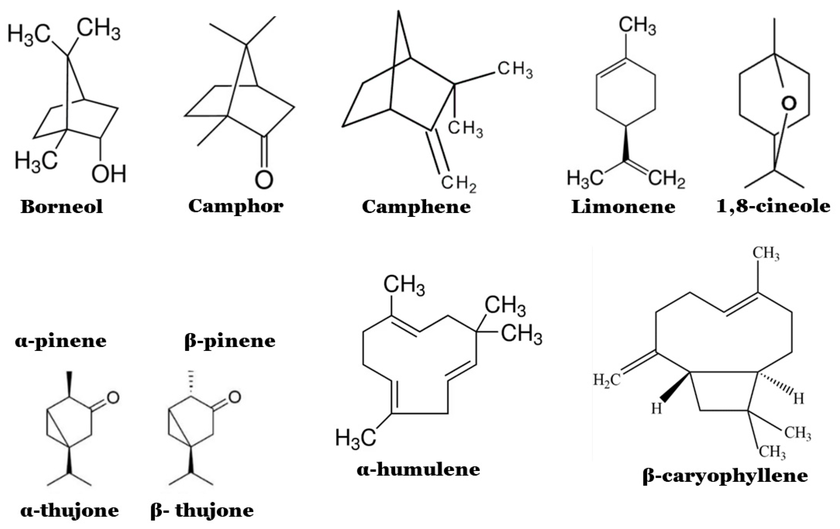
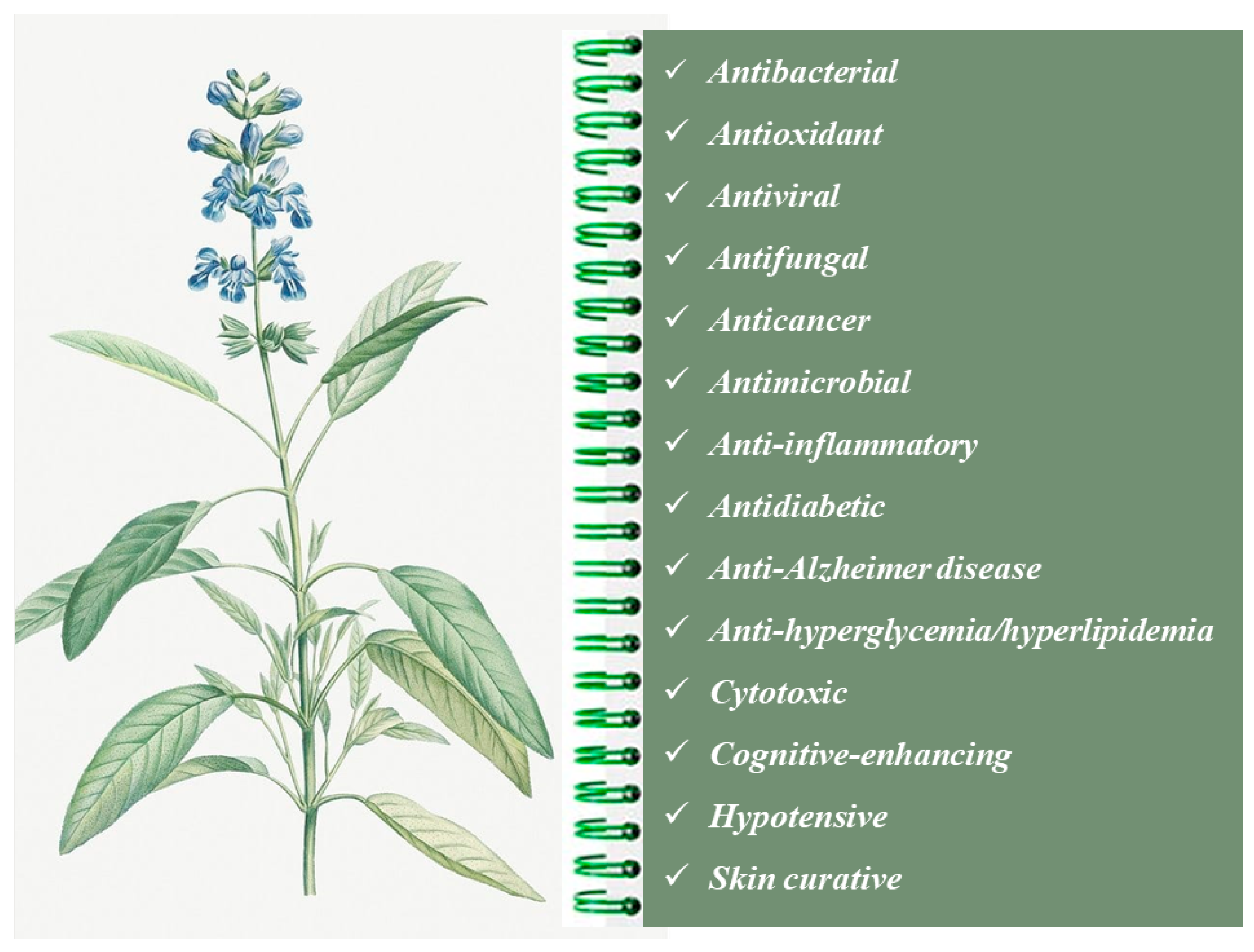
2.1. Chemical Composition and Biological Properties

Salvia officinalis EO (SOEO) has an intense and aromatic scent, with sweet and herbaceous notes: it is a clear yellowish-green liquid. Its yield ranges from 0.07 to 6%, depending on balsams and similar resinous plant exudations [18]. Over 120 constituents have been identified in SOEO; its main components are ketones and monoterpene hydrocarbons, such as borneol, camphor, 1.8-cineole, camphene, limonene, α-pinene, β-pinene, α-thujone, β-thujone, α-humulene sesquiterpene derivatives and β-caryophyllene [19,20,21]. However, α-humulene, ledene, viridiflorol, manool, and sclareol were also recovered. According to the International Organization for Standardization [22], a high-quality SOEO should contain 3–8.5% trans-thujone (α-thujone) and 18–43% cis-thujone (β-thujone), even if a high content of this compound should be avoided due to its neurotoxic effect. Phenolic compounds, monoterpenoids and triterpenoids are usually extracted by aerial parts, whereas diterpenoids are the main compounds of roots [23]. The structure of the major components of SOEO is shown in Figure 1.
Similar to the other EOs, the composition of SOEOs is strongly correlated to their origins, and numerous extrinsic and intrinsic factors, such as agricultural practices, season, light, plant growth stage, etc., may play a significant role in their compositional characteristics [5].
In a recent study, Yilar and co-authors [24] analyzed the composition of three different EOs extracted from S. officinalis L., S. cryptantha and S. tomentosa collected in Turkey (Tokat province). The compositions showed distinct similarities in species, but all EOs resulted especially rich in terpenoid compounds, flavonoids and phenolic compounds; quinonoids were also recovered to a lesser extent. In particular, in S. cryptantha EO 32 components were identified, predominantly eucalyptol (27.64%), camphor (29.87%), α-pinene (11.91%) and borneol (6.57%); an S. tomentosa EO contained 41 compounds, with the most abundant being β-thujene (40–69%), borneol (1.79–10.90%) and camphor (0.40–7.25%). Finally, in an S. officinalis EO, 31 components were identified, including 3-thujonene (31.95%), camphor (28.53%) and eucalyptol (7.35%). Similar results were also obtained for S. officinalis L. cultivated in Spain (Murcia Province) [25].
The genus name “Salvia” comes from Latin and means “to cure,” while the name “officinalis” (also from Latin) means “medicinal” [26]; this makes us realize how this officinal plant has important biological properties. In ancient Rome, this plant was considered “sacred” due to its use to treat various diseases in popular medicine [27,28]. Evidence of its use is also present in ancient Egypt, Greece and Anglo-Saxon countries [29].
Today, the European Medicines Agency has proposed S. officinalis EO for oral use due to its anticancer and anti-inflammatory capabilities and for the treatment of several diseases such as dyspepsia, pharyngitis, stomatitis and inflammation in the mouth or throat [30]. In a study performed by Russo et al. [26], the anticancer activity was demonstrated to be correlated to the presence of α- and β-thujone isomers and other compounds (such as camphor), acting together in a synergistic action; these components are able to reduce side effects affecting cancer patients by inducing apoptotic cancer cell death.
The richness in terpenoids, flavonoids, phenolic compounds and quinonoids confers to SOEOs antibacterial, antifeedant, antioxidant, antiviral, antifungal, cytotoxic and antimicrobial properties [24,31,32] (Figure 2). Anti-inflammatory, antidiabetic and antimutagenic capacities suggest its use for dementia treatment [25].
The essential oils, oleoresins (solvent-free) and natural extractives (including distillates) of S. officinalis, S. fruticosa, S. lavandulaefolia and S. sclarea are generally recognized as safe for the Food and Drug Administration (USA), as well as in Europe, although the European Medicines Agency affirms that sage leaf is safe when used in recommended dosages, i.e., 3.5 and 6.6 mg/day (LD50 value equal to 2.6 g/kg); this recommendation considers the neurotoxic effect of thujone when present in high doses and establish that the amount of this compound in preparations needs to be clearly specified to permit a case-by-case benefit/risk analysis [33,34]. However, it was observed that prolonged use or overdose (corresponding to more than 15 g of the leaves) could cause some unwanted effects such as vomiting, salivation, tachycardia, vertigo, hot flushes, allergic reactions, tongue swallowing, cyanosis and convulsions [12]. Camphor, thujone and terpene ketones may also induce toxic effects on the fetus and newborn; thus, consumption of S. officinalis is not recommended in pregnancy and lactation [12].
Some Salvia plants produce other toxic compounds, such as the psychotropic molecule salvinorin A (produced by S. divinorum) which has dissociative, hallucinogenic and amnesiac effects. Also, red sage (S. haematodes Wall.) could have anticonvulsant effects and depress the central nervous system [23].

2.2. Antimicrobial Properties

The antibacterial activity of SOEOs has been reported by several authors. Table 1 shows the most recent in vitro studies performed on antimicrobial activities of “sage” (colloquial definition of Salvia officinalis) in the last five years. A quick look underlines a broad variability among the research, depending on the sensitivity of microorganisms and the efficiency of the tested components. Sage EO has shown antibacterial activity against Escherichia coli, Bacillus subtilis, Salmonella spp., Listeria monocytogenes, Staphylococcus aureus, Staphylococcus epidermidis, Streptococcus mutans and Shigella sonnei, highlighting good efficacy against both Gram-positive and Gram-negative bacteria [5]. However, Gram-positive bacteria have been observed to be more sensitive to sage EOs than other bacteria [5,19]. Its antimicrobial activity is attributed mainly to the presence of camphor, thujone and 1,8-cineole [35].
In 2018, Haziri and co-authors [36] reported that S. officinalis EO was active against St. aureus, L. monocytogenes and E. coli; the more significant effect was recorded for the growth of St. aureus. Similar results were observed by Mohamed and Mustafa [37], who tested sage EOs obtained through different extraction methods. In the same year, Medeiros de Almeida et al. [38] published a study on the antimicrobial activity of S. officinalis against five Gram-positive bacterial strains and five yeast strains, finding strong activity against Streptococcus strains and moderate activity against Candida strains. In 2019, an interesting study was performed by de Oliveira et al. [39], who tested sage EOs on microbial species responsible of oral disease (see details in Table 1) and demonstrated that all the tested microorganisms were inhibited at a concentration of 50 mg/mL; but, by halving the concentration (25 mg/mL), the extract was effective on just over half of the target microorganisms (58.3%), suggesting a dose-dependent effect.
Despite the fact that several studies have been performed on the use of sage EO to inhibit microbial growth, there have been limited studies about its use in combination with other antimicrobial compounds. Recently, Mokhtari et al. [20] evaluated the antimicrobial activity of sage and thyme (Thymus vulgaris L.) and their mixture extracts. Sage exhibited the lowest antibacterial activity against Bacillus cereus, St. aureus and E. coli with inhibition zone diameters (i.z.d.) of 21.73, 19.12 and 16.76 mm, respectively. The antibacterial activity was improved when Salvia was combined with thyme: in this case, the i.z.d. were 31.25, 28.67 and 22.13 mm, respectively, probably due to the increased phenolic and flavonoid contents [20]. Sulaiman et al. [21] evaluated the antimicrobial activity of different sage EOs (commercial or extracted) and their synergistic effect with meropenem, an antibiotic with a broad spectrum of action and low toxicity for the treatment of severe and nosocomial infections [40]. The results highlighted that only the extracted EOs were effective against E. coli ATCC 25922, L. monocytogenes, St. aureus ATCC 6538, Streptococcus pyogenes and Pseudomonas aeruginosa but not against Klebsiella pneumoniae, with i.z.d. of 18 ± 0.4, 16 ± 0.8, 15 ± 0.6, 14 ± 0.4, and 10 ± 0.2 mm, respectively. Concerning the interaction with meropenem, the authors reported that when the compounds were tested as single drugs against Kl. pneumoniae and E. coli, the MIC (minimum inhibitory concentration) values were 500 and 320 mg/mL for EOs and meropenem, respectively, but these values were reduced to 100 and 40 mg/mL when used in combination.
Besides S. officinalis L., other Salvia species are also able to influence microbial growth; for example, Salvia sclarea is a medicinal herb of the Mediterranean countries, known for its antidiabetic, anti-inflammatory, antimicrobial, antioxidant and antitumor properties [41]. In 2015, Cui and co-workers [41] studied the antibacterial activity and the mode of action of S. sclarea EO against seven pathogens: E. coli, St. aureus, Bacillus pumilus, Kl. pneumoniae, B. subtilis, Salmonella Typhimurium and Ps. aeruginosa. The oil was effective against all tested strains, showing a MIC of 0.05 and a minimum bactericide concentration (MBC) of 0.1%. The antimicrobial activity was also tested on the growth of E. coli in phosphate-buffered saline (PBS) and meats (chicken, pork and beef). The pathogen population was reduced by approx. 99.99% in all the conditions tested. Concerning the mode of action, the authors speculated that the effect was probably due to damage to the cell membrane, with a consequent alteration of permeability and leakage of intracellular material such as ATP and DNA [41].
Salvia hispanica L. is also known as “chia”, a name acquired from indigenous South Americans; it originated between Mexico and Guatemala and was cultivated in Central America. Thanks to their high nutritional value and fiber content, chia seeds are widely used in medicine and in food preparations because the extracted oil is characterized by a high content of proteins, antioxidants and polyphenols; moreover, their antibacterial and antiviral activity against various microorganisms has also been demonstrated [42,43]. For example, Elshafie et al. [43] tested the antimicrobial activity of chia against 4 Gram-positive bacteria, 6 Gram-negative bacteria and 10 fungi (see details in Table 1). Chia EO was more effective against Gram-positive than Gram-negative bacteria, and the antimicrobial activity was dose-dependent; in fact, the most effective inhibition resulted at the maximum concentration of the oil. Regarding the antifungal activity, chia EOs strongly inhibited Aspergillus fumigatus, Penicillium expansum, Monilinia laxa and Monilia fructigena. The antimicrobial activity was probably due to the presence of caryophyllene, a sesquiterpene volatile fraction that was able to affect the permeability of the bacterial membrane; however, the exact mode of antimicrobial action is still unknown and under investigation [44].
Salvia miltiorrhiza, also known as red sage, is mainly composed of hydrophilic compounds (salvianolic acids) and lipophilic compounds (tanshinones); these compounds have shown antimicrobial activities against Gram-positive and Gram-negative bacteria and fungi [45]. Chen et al., 2021 [46] reported that crypto-tanshinone (CT) exhibited a bacteriostatic against three strains of St. aureus and one of B. subtilis. The MIC values were between 4 and 16 µg/mL; the MBC > 64 µg/mL for each microorganism, and the MBC/MIC ratios were > 4. The authors hypothesized an involvement of this compound (CT) in the respiratory chain as an inhibitory agent since it did not damage the bacterial membrane but rapidly dissipated the membrane potential; the mechanisms of its antibacterial activity need further investigation. In a recent study, Lim Ah Tock et al. [47] compared the antimicrobial activity of Salvia africana-lutea, Salvia lanceolata and Salvia chamelaeagnea (also known as golden sage, rusty sage and rough blue sage, respectively) against three Gram-positive bacteria (St. aureus, B. cereus and B. subtilis) and four Gram-negative bacteria (Acinetobacter baumannii, E. coli, Enterococcus faecium and Ps. aeruginosa). The authors observed that the best performances were achieved by S. chamelaeagnea extracts, probably due to the presence of carnosol and carnosic acid, two antimicrobial compounds not detected in S. africana-lutea and S. lanceolata [47,48].
Table 1. In vitro studies performed on antimicrobial activities of Salvia EOs in the last five years.
Salvia EOs OriginMicroorganisms TestedMethod UsedMain ResultsReference
Ethanol extract from Salvia officinalis (L.)Staphylococcus aureus, Listeria monocytogenes, Escherichia coliAgar disk diffusion test
All of the concentrations used (1, 3 and 5 mg/mL) were effective towards microbial targets.
At 5 mg/mL, S. officinalis extract showed a stronger antibacterial activity towards St. aureus with inhibition zone of 9 mm.
[36]
S. officinalis (leave and stem) extracted by maceration (with ethyl acetate), by hydro distillation (with anhydrous sodium sulphate) and by Soxhlet (with hexane)St. aureus, E. coliAgar disk diffusion test
Regarding the Soxhlet extraction method, the leaves extract showed higher antibacterial activity than the stem extract.
About the maceration and hydro distillation, both extracts possessed remarkable antibacterial activity, but the volatile extract from the leaves was more effective than the extract from the stem.
E. coli was less sensitive than St. aureus
[37]
Ethanol extract of leaves of S. officinalisStreptococcus mutans, Streptococcus mitis, Streptococcus oralis, Streptococcus salivarius, Streptococcus sanguis, Candida albicans, Candida glabrata, Candida guillermondii, Candida krusei, Candida tropicalisBroth microdilution
method
Ethanolic extract of S. officinalis L. (diluted in alcohol at 40%, at a concentration of 8 mg/mL) showed strong antibacterial activity towards Streptococcus strains (MIC ranged from 0.25 to 1 mg/mL) and moderate antifungal activity towards Candida strains (MIC = 1 mg/mL).[38]
S. officinalis9 clinical isolates from the oral cavity of tuberculosis patients, St. aureus, Staphylococcus epidermidis, Str. mutans, Ca. albicans, Ca. tropicalis, Ca. glabrataBroth microdilution
method
50.0 mg/mL of S. officinalis was effective against all microorganisms.
25.0 and 12.5 mg/mL were partially effective, on 58.3% and 8.3% of strains, respectively)
[39]
S. officinalis L. combined with thyme (Tymus vulgaris L.)E. coli, St. aureus, Bacillus cereus, Salmonella TyphimuriumAgar well diffusion methodThe thyme–sage mixture showed the highest antimicrobial activity against B. cereus (inhibition zone diameters, i.z.d. = 31.25), S. aureus (i.z.d. = 28.67), Salm. Typhimurium (i.z.d. = 23.65), E. coli (i.z.d. = 22.13)[20]
Extracted and purchased S. officinalis essential oils and synergistic effect with meropenem (antibiotic)St. aureus, L. monocytogenes, Streptococcus pyogenes, E. coli, Pseudomonas aeruginosa, Klebsiella pneumoniaeAgar well diffusion
method
The extracted oil inhibited the growth of all bacteria except the Kl. pneumoniae.
The highest activity was observed against E. coli (i.z.d. = 18 ± 0.4 mm; MIC = 6.25 ± 0.2 mg/mL), the lowest activity against Ps. aeruginosa (i.z.d. = 10 ± 0.2 mm; MIC = 25 mg/mL).
The purchased oil did not show any antibacterial activity
[21]
Ethanol extract of S. officinalis leavesSt. aureus (2 strains), E. coli (three strains), Ps. aeruginosa, Proteus mirabilis, Kl. pneumoniae, Klebsiella oxytoca, Acinetobacter baumannii, Enterobacter aerogenes, Helicobacter pyloriAgar diffusion methodThe highest antimicrobial potentials were observed with the extracts dried at ambient temperature and oven at 45 °C, which inhibited eight of the tested microorganisms: two strains of E. coli, Ps. aeruginosa, two strains of St. aureus, Pr. mirabilis. Ac. baumannii, H. pylori. Salvia extract obtained from oven dried plant at 60 °C exhibited the lowest antibacterial activity[49]
Steam distillate essential oil and corresponding hydrolate from S. officinalisIsolates from wound swabs of hospitalized patients:
St. aureus, Enterobacter cloacae, Ps. aeruginosa, Ca. albicans, Kl. oxytoca, E. coli
Isolates from blood cultures:
St. aureus, Kl. pneumoniae
Broth microdilution method
S. officinalis oil was more effective than hydrolates.
Kl. oxytoca was the most sensitive to the EOs (MIC) and MBC were 14.20 and 28.4 µL /mL, respectively).
MIC and MBC values of other bacteria ranged between 28.40 and 227.25 µL/mL while for Ca. albicans MIC/MFC ranged between 28.40/56.81 and 56.81/113.63 µL/mL.
Only for Ps. aeruginosa MIC and MBC values were higher than the highest used concentration in the test
[50]
Essential oils extracted from the stem and leaves of Salvia hispanica L. plantTwo strains of Bacillus megaterium, Bacillus mojavensis, Clavibacter michiganensis, Xanthomonas campestris, Xanthomonas vesicatoria, Pseudomonas syringae pv. phaseolicola, E. coli, Burkholderia gladioli pv. agaricicola, Monilinia laxa, Monilinia fructicola, Monilinia fructigena, Aspergillus flavus, Aspergillus niger, Aspergillus fumigatus, Penicillium digitatum, Penicillium expansum, Sclerotinia sclerotiorum, Fusarium oxysporum.Disk diffusion methodAt the highest concentration (50%) the EOs of S. hispanica showed greater antibacterial activity against Gram-positive bacteria than Gram-negative bacteria.
At the highest EOs concentration (40%), the highest inhibition of fungal mycelium growth was observed as follows:
High inhibition towards A. fumigatus, P. expansum, M. laxa, and M. fructigena.
Moderate inhibition towards M. fructicola and P. digitatum.
Slightly inhibition towards A. niger, A. flavus and F. oxysporum.
Any inhibition against Sc. sclerotiorum.
[43]
Methanol extracts from 81 Salvia samples (Salvia africana-lutea; Salvia lanceolata; Salvia chamelaeagnea)St. aureus, Ac. baumannii, B. cereus, E. coli, Enterococcus faecium, Ps. aeruginosa, Bacillus subtilisMicrodilution assayThe antibacterial activity of S. chamelaeagnea was superior to that of the other two species and was highly effective against all seven pathogens with average MICs 0.23–1.3 mg/mL; then S. africana-lutea (0.52–3.0 mg/mL) and S. lanceolata (0.46–4.2 mg/mL).[47]
Aqueous, ethanolic, and methanolic extracts of Salvia argenteaEnterococcus faecalis, St. aureus, L. monocytogenes, Methicillin-resistant St. aureus (MRSA), B. subtilis, B. cereus, Pr. mirabilis, Pasteurella multocida, Salm. Typhimurium, Campylobacter fetus, E. coli, Kl. pneumoniae, En. cloacae, Citrobacter freundii, Ps. aeruginosa, Salmonella enterica, Ca. albicans (3 strains), Saccharomyces cerevisiae.Agar diffusion and
micro dilution methods
The extracts were effective on the majority of microorganisms tested, with inhibition zones up to 23 ± 2.6 mm (in bacteria) and 24 ± 1.5 mm (in yeasts).
MIC results between 3.90 and 7.81 mg/mL were recorded for bacterial strains (Kl. pneumoniae, Pa. multocida, MRSA, Ci. freundii), and of 3.90 mg/mL for the yeast strain S. cerevisiae.
S. argentea exerted a bactericidal action against multi-resistant bacterial strains, a fungicidal action on the three strains of Ca. albicans and a fungistatic action against S. cerevisiae
[51]
EOs obtained from the dried flowering tops of the Salvia rosmarinus Spenn. and Salvia jordanii by hydrodistillationYersinia enterocolitica, L. monocytogenes, Enterococcus durans, Ec. faecalis, Ec. faecium, Ca. albicans, Ca. tropicalis, Ca. guilliermondii, Ca. krusei, Ca. parapsilosis, S. cerevisiaeAgar disk diffusion and broth microdilution methodsEOs presented a moderate antibacterial activity on the bacterial strains and were not active towards yeasts.[52]

3. Lavandula Essential Oils

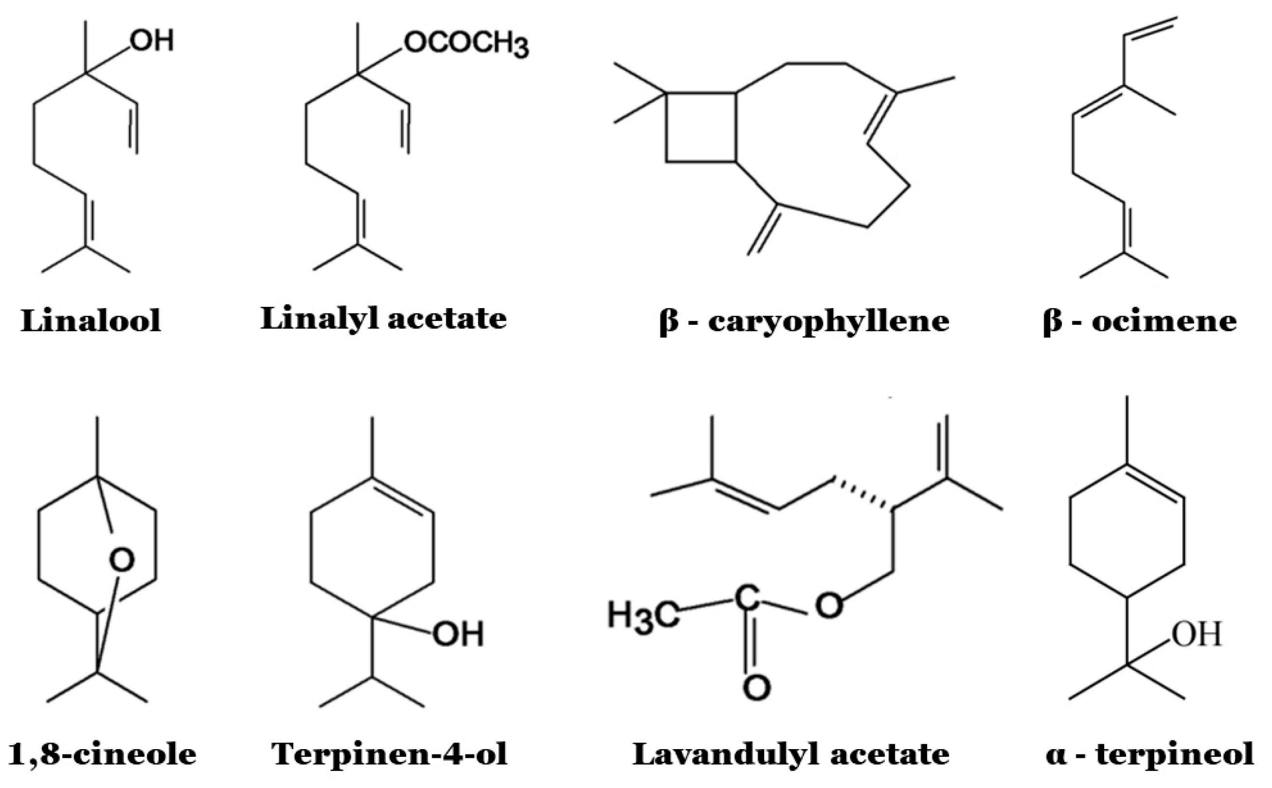
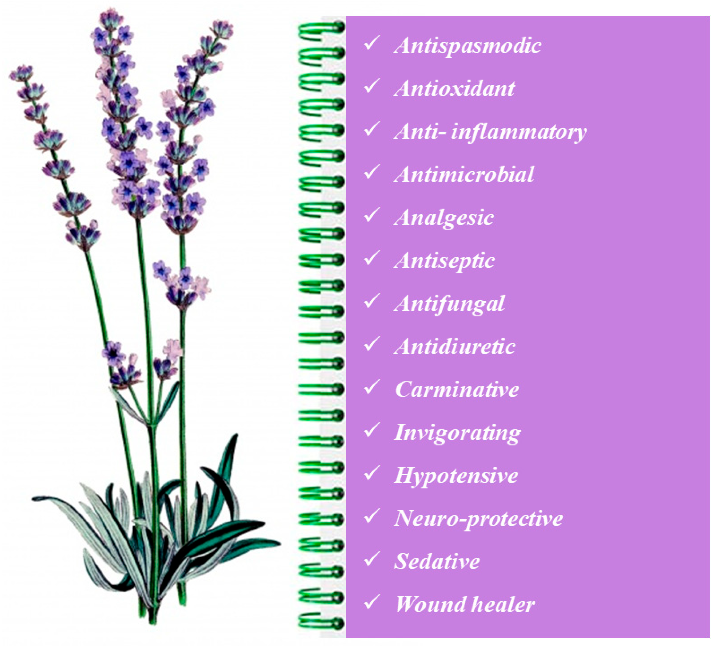
3.1. Chemical Composition and Biological Properties

The Lavandula EO (LEO) is yellow and has an intense floral scent; it is obtained mainly by steam or hydro-distillation (yield is about 3%), and its composition varies greatly among different species and within the same species too, depending on the genotype, seasonal and climatic conditions, location, extraction methods and drying [53]. Even if over hundreds of chemical compounds can be recovered, the major components of Lavandula are oxygenated monoterpenes, monoterpene hydrocarbons and sesquiterpenes, mainly linalool (27.3–42.2%), linalyl acetate (27.2–46.6%), (Z)-β-ocimene (0.2–11.6%), terpinen-4-ol (0.70–4.6%), lavandulyl acetate (0.50–4.8%), β-caryophyllene (1.8–5.1%), (E)-β-ocimene (0.30–3.8%), α-terpineol (0.30–2.0%) and 1,8-cineole (0.10–1.2%) [53,54] (Figure 3). The contents of linalool and linalyl acetate determines the quality of the oil; on the other hand, due to the very strong influence on the scent, the contents of ocymen, cineole, camphor or terpin-4-ol should be low [35].
The oxygenated monoterpenes are responsible for the precious scent of lavender, variable from the sweet, floral, herbaceous and refreshing odor of the true lavender EO to the warm, slightly fruity and camphorous fragrance of L. stoechas EO until the atypical, more floral with a warm, rosy note and not herbaceous aroma of L. heterophylla Goodwin Creek [54]. An interesting overview of the chemistry of LEO, exploiting the characteristic chemical constituents and chemotypes of seventeen Lavandula species, can be found in the work of Aprotosoaie et al. [54].
As mentioned above, many factors may influence the chemical composition of LEO. In a study performed on locally grown plants in western Romania (L. angustifolia and L. intermedia), it was observed that the EO of L. angustifolia Miller contained 22 components (99.9% of the total), the main ones being caryophyllene (24.1%), β-phellandrene (16%) and eucalyptol (15.6%), while the EO of L. intermedia contained 24 components (98.26% of the total), mainly camphor (32.7%) and eucalyptol (26.9%) [55]. With respect to what is observed in the literature, where the major components of Lavandula are linalool and linalyl acetate, in the composition of the EOs analyzed in this study, these compounds were not found, underlining the great impact of environmental conditions (location and season) on the EO composition [53]. Contrarily, Turgut and co-workers [16], in their study about L. angustifolia Mill. grown in Burdur Örtülü locality (Turkey), found that linalool, linalyl acetate and α-terpineol were the most representative compounds (42.22%, 23.12% and 4.91%, respectively). A similar composition was found by Blažeković and coauthors [56] for EO of L. angustifolia cultivated in Croatia.
In 2017, an Italian study evidenced the effect of the growing season of organic lavender (L. angustifolia) and lavandin (L. hybrida) under the pedo-climatic conditions of central Italy by highlighting significant variations in EO composition that affected all the classes of compounds, except for oxygenated monoterpenes [57].
Since ancient times, Lavandula spp. and its EOs have been used in popular medicine as a natural remedy [58]. Over the years, it has been studied for its high content of bioactive compounds able to exert beneficial effects on human health and well-being. In particular, the richness in phenolic compounds confers to lavender EOs a high antioxidant activity due to a protective effect against the oxidative damage caused by free radicals [16]. The production of free radicals is a physiological event normally occurring in our bodies: a certain number of free radicals is essential to protect us from infections by viruses and bacteria, but an excessive amount of ROS (oxygen free radicals) is harmful since it could cause damage to various macromolecules (such as DNA, proteins, carbohydrates, lipids and enzymes), establishing a condition of oxidative stress, that is a risk factor for human health leading to cellular aging, inflammatory pathologies and degenerative diseases [53].
LEOs possess numerous biological activities such as antispasmodic, carminative, analgesic, sedative, hypotensive, antiseptic, antimicrobial, antifungal, antidiuretic and general tonic action [56]. In addition, anticholinesterase, anti-epileptogenic, neuroprotective, anxiolytic, anti-depressive, hyaluronidase and lipoxygenase inhibitory activities were also reported [53]. Figure 4 synthesizes all recognized biological properties of LEOs that suggest their potential addition to food to improve consumers’ health.
Due to its action against lipid peroxidation, LEO is often suggested in the treatment of patients with rhinitis as potential natural medicine [59]. In a study carried out by Cardia and co-workers [60], the results highlighted that LEO was able to inhibit the release of important inflammatory mediators, particularly IL-1β and nitric oxide, and myeloperoxidase activity, showing significant anti-inflammatory activity. A wound-healing effect by increasing collagen synthesis and differentiation of fibroblasts through the up-regulation of transforming growth factor beta (TGF-β) was also demonstrated [61].
Toxicity from the use of lavender is uncommon, and LEO did not demonstrate cytotoxicity both in vitro and in vivo (mice); oral administration was considered safe with an LD50 of 13.5 g/kg [60]. However, some studies indicated that prolonged exposure to linalool (LEO main component) may result in allergic reactions: this is why the 7th Amendment of the European Cosmetics Legislation requires natural products, including linalool, to be labeled as potentially allergenic [60]. Contrarily, linalyl acetate may have aneugenic activity [62]. Genotoxicity in the proliferation of lymphocytes, damage to the cell membrane of human skin and contact dermatitis have been reported as adverse effects [60]. However, all these aspects are still under investigation, and efforts should be made in this direction to favor a widespread use, especially in clinical trials exploiting the use of LEO in improving the health of adult patients in various clinical settings and conditions.

3.2. Antimicrobial Properties

Table 2 shows the most recent in vitro studies performed on antimicrobial activities of LEOs (last five years). There is an extensive literature on the antimicrobial properties of LEO, but drawing unequivocal conclusions is difficult since there is no composition consistency, being influenced by variety, growing season and geographical location. In general, the literature shows that the chemical compound responsible for the antimicrobial and antifungal activity of LEO is linalool. LEOs have demonstrated antimicrobial activity against both Gram-positive and Gram-negative bacteria, yeasts and molds, with controversial results.
For example, a recent study performed in Saudi Arabia [63] tested the EO hydrodistilled from flowering aerial parts of L. pubescens Decne against thirteen strains of Gram-positive and Gram-negative bacteria using the agar diffusion assay. The oil showed good effectiveness, especially against Ac. baumannii, Salm. Typhimurium, Sh. sonnei, Enterococcus faecalis and St. epidermidis. The authors also tested carvacrol (the major constituent of lavender oil, 55.7%) by comparing it to some conventional antibiotics, i.e., vancomycin, amikacin and ciprofloxacin. The results revealed that Gram-negative strains were more susceptible than the Gram-positive ones. Contrarily, the Gram-positive bacteria turned out to be the most vulnerable to EOs recovered by the flower essential oil of lavender grown in Poland [64].
In 2017, Hossain et al. [65] tested the effectiveness of an EO extracted by flowers of L. angustifolia grown in Bulgaria against thirty-eight strains of turtle-borne pathogenic bacteria belonging to seven species: Aeromonas hydrophila, Aeromonas caviae, Aeromonas dhakensis, Citrobacter freundii, Proteus mirabilis, Salmonella enterica and Ps. aeruginosa. The results revealed that LEO was active against all tested bacteria except Ps. aeruginosa. Contrarily, a study performed on EO by L. angustifolia grown in Burdur Örtülü (Turkey) revealed significant effectiveness against this pathogen being able to eliminate contamination completely [16]. A good antimicrobial activity was also observed against Candida albicans, St. aureus and Aspergillus brasiliensis [16].
In 2021, significant inhibition of the growth of B. subtilis and E. coli was recorded by Caprari and co-workers [66] in their study about the use of EOs extracted from dried and fresh flowers of Lavandula angustifolia L. grown in central Italy. In addition, a bactericidal effect on E. coli was observed, particularly when oil from fresh flowers was used at the highest concentrations. However, apart from antimicrobial assays, this study is noteworthy because it reports the first evaluation of lavender EOs as a green method to control biodeterioration phenomena on a historical artistic wood painting (“Madonna con Bambino”, XIX century, located on the inner wall of the S. Maria del Lago church at Pesche in Isernia, Italy).
The effectiveness of LEOs could be improved if combined with other antimicrobial compounds. An interesting approach to the synergistic use of LEOs and other antimicrobials can be found in the study by Nafis et al. [67], who tested the efficacy of three EOs extracted from Moroccan lavender species (L. pedunculata, L. angustifolia, L. maroccana), combined with a conventional antibiotic (ciprofloxacin), against three pathogenic foodborne bacteria. EOs alone or combined showed remarkable antimicrobial activity against the tested bacteria with minimum inhibitory concentrations (MICs) ranging from 3.53 to 15.96 mg/mL, but when ciprofloxacin was added, the antibiotic MIC values were significantly lower (0.06 to 3.91 μg/mL). This synergism was more evident against Salmonella spp., suggesting that a mixture of lavender EOs is able to improve the efficacy of ciprofloxacin, which should be taken into consideration for a possible application in the pharmaceutical industries.
More recently, Aicha El Baabouaa et al. [68] evaluated the antibacterial activity of Lavandula stoechas L. EO (31.81% fenchone, 29.60% camphor, 13.14% terpineol, 8.96% menthone and 5.88% eucalyptol) against 11 multidrug-resistant strains of Campylobacter spp. Moreover, they studied its synergistic effect with two antibiotics (ampicillin and tetracycline) and examined the capability of Campylobacter strains to produce biofilm. The results showed that Campylobacter multidrug-resistant strains were much sensible to L. stoechas EO, with MIC values ranging from 0.063 to 0.25 µg/mL, depending on the strain. However, these values were significantly reduced during the combined use with antibiotics: MIC values ranged from 0.004 to 0.003 µg/mL with ampicillin and from 0.004 to 0.125 µg/mL with tetracycline, respectively. An impressive inhibitory impact of lavender oil on biofilm formation was also recovered.
Table 2. In vitro studies performed on antimicrobial activities of Lavandula essential oils (LEOs) in the last five years.
LEOs OriginMicroorganisms TestedMethod UsedMain ResultsReference
Croatian indigenous cultivar of:
Lavandin (L. x intermedia)
Lavender (L. angustifolia)
Bacillus cereus, Bacillus pumilus, Enterococcus faecalis, Escherichia coli, Klebsiella oxytoca, Klebsiella pneumoniae, Kocuria rhizophila, Listeria monocytogenes, Proteus mirabilis, Pseudomonas aeruginosa, Salmonella Enteritidis, Staphylococcus aureus, Streptococcus pyogenes, Yersinia enterocolitica, Candida albicans, Candida glabrata, Candida kefyr, Candida krusei, Candida tropicalis, Cryptococcus neoformans, Hansenula anomala, Saprochaete capitate, Microsporum canis, Microsporum gypseum, Trichophyton mentagrophytes, Trichophyton rubrum, Aspergillus fumigatus, Aspergillus niger, Fusarium oxysporum, Penicillium citrinumDisk diffusion assay
Determination of minimum inhibitory concentration and minimum bactericidal/fungicidal concentration
Lavandin EO showed a broad spectrum of antibacterial (MICs 0.25–2.5 mg/mL) and antifungal (MICs 0.1–2 mg/mL) activities, being generally more than or at least as effective as those from L. angustifolia.
Among the two dominant compounds (also tested), linalool was more effective than linalyl acetate.
[56]
Four cultivars of
Lavandula x intermedia
L. monocytogenes (24 strains)
Salmonella enterica (10 food strains)
Disk diffusion assay
Determination of minimum inhibitory concentration and minimum bactericidal/fungicidal concentration
The antibacterial activity was related to EOs composition.
Cultivars showing the highest antimicrobial activity were the richest in linalool (38.17–61.98%), camphor (8.97–10.30%) and 1,8-cineole (6.89–8.11%, respectively).
[69]
Lavandula stoechas grown in Extremadura (Spain)St. aureus, B. cereus, L. monocytogenes, Listeria innocua, Salmonella Choleraesuis, E. coli, Candida boidinii, Priceomyces carsonii, Kregervanrija fluxuum, Zygosacharomyces bailii, Aspergillus flavus (2 strains producing aflatoxins)Disk diffusion assay
Determination of inhibition zone diameters
Greater inhibition capacity against L. innocua
No inhibition against L. monocytogenes, St. aureus, B. cereus, E. coli, Salm. Choleraesuis, and yeasts
Ability to reduce the production of aflatoxins
[70]
L. pedunculata
L. angustifolia
Lavandula maroccana
grown in Morocco
E. coli, Salmonella spp., St. aureusThe EOs were tested separately and in combination, also with ciprofloxacin (antibiotic)
The twofold dilution method followed by MIC determination was used
EOs assayed separately were all effective.
When used in combination, also with ciprofloxacin, a synergistic action was observed.
A mixture composed by 19, 38 and 43% of EOs obtained from L. angustifolia, L. pedunculata and L. maroccana, respectively, showed the highest antibacterial effect.
This mixture combined with ciprofloxacin against Salmonella spp. improved the antibiotic efficacy by reducing its MIC by 128-fold.
[67]
Lavandula angustifolia (Spain)Botrytis cinerea, Sclerotinia sclerotiorum, F. oxysporum, Phytophthora parasitica, Pythium aphanidermatum, Alternaria brassicae, Cladobotryum mycophilum, Trichoderma aggressivumDisk diffusion method (5–30%, v/v)The activity was dependent on the concentration, with clear differences in the sensitivity of the fungal isolates, not correlated to the fungal wall composition.[71]
Lavandula pubescens Decne (Arabia)Ec. faecalis (Vancomycin-resistant), Staphylococcus epidermidis, St. aureus, St. aureus (Methicillin-resistant), Salmonella Typhimurium, Acinetobacter baumannii (Carbapenem-resistant), Shigella sonnei, Kl. pneumoniae, Ps. aeruginosa, Pr. mirabilis, E. coliDiffusion methodThe most sensitive strains were Ac. baumannii, Salm. Typhimurium, Sh. sonnei, Ec. faecalis and St. epidermidis.[63]
Moroccan Lavandula stoechasTwo collection Campylobacter strains: C. jejuni and C. coli
Nine wild multidrug resistant Campylobacter strains isolated from food and environment
Broth micro-dilution assay and MIC determinationThe results showed that Campylobacter multidrug resistant strains were highly sensitive to Lavandula stoechas EO, with MIC values ranging from 0.063 to 0.25 µg/mL depending on the strain. However, these values were significantly reduced during the combined use with antibiotics: MIC values ranged from 0.004 to 0.003 µg/mL with ampicillin and from 0.004 to 0.125 µg/mL with tetracycline. An impressive inhibitory impact of lavender oil on biofilm formation was also recovered.[68]
Dried and fresh flowers of Lavandula angustifolia L. (lavender) grown in central ItalyBacillus subtilis, E. coli, Sclerotium rolfsiiDisk diffusion test
A significant inhibition of growth in both indicator strains was observed, with a bactericidal effect on E. coli.
Inhibition of the phytopathogenic fungus was also recovered.
The potential application of EOs as a green method to control biodeterioration phenomena on an artistic wood painting (XIX century) was evaluated.
[66]
Five Lavandula stoechas cultivars grown in Thailand:
L. stoechas ‘snowman’
L. stoechas ‘white lavender’
L. stoechas ‘major’
L. stoechas ‘avonview’
hybrid L. stoechas × viridis ‘St. Brelade’
St. aureus, St. epidermidis, Enterococcus faecium, E. coli, Kl. pneumoniae, Ps. aeruginosa, Str. pyogenes, and Salm. TyphimuriumDisk diffusion assay
Determination of MIC and inhibition diameters.
The highest antibacterial activity was observed from the essential oil of L. stoechas ‘major’ (against Kl. pneumoniae and Salm. Typhimurium) and the essential oil of L. stoechas ‘white lavender’ (against Salm. Typhimurium).
The essential oil of L. stoechas × viridis ‘St. Brelade’ possessed the highest antioxidant capacity.
[72]
Moroccan Lavandula atlanticaMethicillin resistant St. aureus, E. coli, En. aerogenes, Ps. aeruginosa, Kl. pneumoniae, Kl. oxytoca, Salmonella spp., Ac. baumannii, and Enterobacter cloacae.Inhibitory diameters
MIC values ranging from 3.13 mg/L to 25 mg/L were determined for the tested bacteria.
The smaller MIC value was obtained for Kl. pneumoniae.
[73]
L. stoechas collected in TunisiaAs Gram-negative strains:
four E. coli, three Ps. aeruginosa, and one Serratia marcescens.
As Gram-positive:
three Enterococcus (Ec. faecalis, Ec. aerogenes, and Ec. hirae), two St. aureus, and one Bacillus licheniformis.
Disk diffusion method
An important inhibitory capacity against the 14 tested strains with varying magnitude was recovered.
The Inhibition diameters ranged from 6 mm (very low effect) to 26 mm (strong inhibition).
[74]
Lavandula officinalis collected in TurkeyMethicillin-resistant St. aureus, Vancomycin-resistant Enterococcus VRE, Methicilline susceptible St. aureus (MSSA), St. epidermidis, Salmonella Enteriditis, Salm. Typhimurium, E. coli, Kl. pneumoniae, Ps. aeruginosa, Citrobacter freundii, Pr. mirabilis and Ca. albicans.Agar disk diffusion methodThe essential oil was effective on VRE, MSSA, E. coli and St. epidermidis, but not on S. enteritidis, Ps. aeruginosa, and Ca. albicans.[75]
Commercial lavender Lavandula
angustifolia
Xanthomonas spp. isolated from tubers and stem of plants growing in Lithuania.
Xanthomonas translucens
X. arboricola (four strains)
Broth microdilution method and MIC determinationLavender (2.0%) essential oils inhibited the growth of all Xanthomonas spp. strains: the diameter of the inhibition zones was from 22.8 to 0.9 mm.[76]
Two cultivars of L. angustifolia:
Blue River’ and ‘Ellagance Purple’ (Poland).
Ca. albicansBroth microdilution method and MIC determination
A synergistic action was found when oils were used in combination with fluconazole.
The observed enhancement effect of fluconazole antifungal activity was significantly stronger in the case of essential oils obtained from flowers and leafy stalks of ‘Blue River’ cultivar.
[77]
Lavandula angustifolia
(Bulgaria)
E. coli, Proteus vulgaris, Ps. aeruginosa, St. aureus, Ec. faecalis, L. monocytogenes, Candida utilis, B. subtilis, A. niger, Penicillium chrysogenum, Saccharomyces cerevisiae.Disk diffusion methodThe lavender extract demonstrated antimicrobial activity against all tested pathogens. St. aureus was the most sensitive towards the lavender extract (MIC 60 μg/mL), while for all other pathogens, the MICs were above 600 μg/mL.[78]
Lavandula dentata
Lavandula Marrakech
B. cereus, L. monocytogenes, St. aureus, E. coli, Ps. aeruginosa and Salm. enterica.Agar-well diffusion method and determination of minimum inhibitory concentration (MIC) and minimum bactericidal concentration (MBC)Only a bacteriostatic effect was observed.[79]

4. Food Preservative Applications of Sage and Lavender EOs

In general, the effect of antibacterial activity of EOs may inhibit the growth of bacteria (bacteriostatic) or destroy bacterial cells (bactericidal), but it is very difficult to distinguish these actions. In addition, as aforementioned, the effectiveness of EOs is strictly related to their chemical composition, which is itself affected by various factors: environmental conditions, soil composition, climate, season, cultivation, extraction, storage conditions, harvest period, plant genotype, phenological stage, part collected and other factors [80,81].
Even if some major compounds could be recovered in each EO, all compounds should always be taken into consideration for their antimicrobial activity since minor components could exert a synergistic and additive effect [53].
Considering the number of different groups of chemical compounds present in sage and lavender EOs, their antibacterial activity is not attributable to one specific mechanism because they seem to have no specific cellular targets. However, different modes of action have been suggested, such as (i) damage to the microbial cell wall and membrane proteins; (ii) the dissolution of the phospholipid bilayer of the cytoplasmic membrane with consequent reduction of the proton motive force, electron flow and active transport (iii) the coagulation of cellular components and (iv) the inactivation or destruction of genetic material or interference with gene expression. All these mechanisms cause destabilization, increasing the permeability and leakage of cell constituents [5,82]. Another issue to be evaluated is that foods are complex matrices composed of several elements (proteins, fats, carbohydrates, mineral salts and water content), with each of them able to affect the antimicrobial efficacy [5].
The application of sage and lavender EOs for the preservation of foods is not a new concept, and some interesting applications are described in the following lines. For example, in 2016, Azizkhani et al. [83] investigated the inhibitory activities of S. sclarea EO against chemical and microbial spoilage in Iranian white cheese: at 1%, the oil inhibited Aspergillus growth throughout the storage period and reduced L. monocytogenes population of up to 6 log CFU/g.
Šojić et al. [84] studied S. officinalis herbal dust (a food industry by-product) by adding its essential oil (SEO) and its extract (SE) at concentrations of 0.05, 0.075 and 0.1 μL/g in fresh pork sausages to evaluate the oxidative and microbiological stability during storage. The authors reported that both SEO and SE reduced microbial growth, although SE was the most effective at 0.075 μL/g and 0.1 μL/g. As expected, the chemical profile highlighted the presence of oxygenated monoterpenes and diterpene polyphenols responsible for the antimicrobial and antioxidant effects [84].
In general, also for sage EOs, the highest concentrations allow the greatest microbial reductions, but high concentrations negatively affect the sensory and organoleptic characteristics of foods. Therefore, extracts and/or essential oils of Salvia spp. are often combined with other natural antimicrobials or used as part of a hurdle system [5]. For instance, Kačániová et al. [85] evaluated the antimicrobial effect of sage and rosemary EOs in fresh chicken breast, treated as follows: without packaging (AC), vacuum-packaging (VPC), vacuum-packaging + EDTA 1.5% (VPEC) + sage (VP+S) and rosemary EO at 0.2% (VP+R), separately. The food quality was evaluated for 16 days of storage at 4 ± 0.5 °C by anaerobic plate count (APC), Enterobacteriaceae, lactic acid bacteria (LAB) and Pseudomonas spp. counts. As regards APC and LAB, compared to the other treatments, VP+S was the most effective treatment, with the lowest bacterial load recorded after 16 days of storage. Concerning Pseudomonas spp. and Enterobacteriaceae, VP+S and VP+R resulted in the best treatments: no growth was observed throughout the entire refrigerated storage period.
In 2020, Moura-Alves et al. [86] evaluated the combined use of the sous vide cook-chill (SVCC) process and S. officinalis L. EO on the growth of L. monocytogenes in beef stored at different temperatures (2 and 8 °C) for 28 days. Data showed that S. officinalis EO helped to control bacterial growth: at 2 °C, L. monocytogenes was reduced by approx. 1 log CFU/g in EO samples compared to the control, while at 8 °C, lower L. monocytogenes counts were observed, despite an exponential growth that started from day 14. Even if the pathogen was not inhibited, the combination of storage temperature and the inclusion of sage EO was effective to control microbial growth. Similar results were also confirmed by Gal et al. [87], who observed a reduction of L. monocytogenes heat resistance in sous-vide-treated beef tenderloin.
Recently, Ferreira et al. [88] produced a fish product by adding aromatic herbs: S. officinalis powder was added to salmon hamburgers in three concentrations (0.50, 1.00 and 1.50%) to preserve them from oxidative processes. They also evaluated the antimicrobial effects against coliforms at 45 °C, Salmonella sp., coagulase-positive St. aureus and sulfite-reducing clostridia on the 90th day of storage. Sage was able to stop the oxidation process, showing good antioxidant and antimicrobial potential. In addition, Salmonella spp. was absent for all samples, St. aureus and clostridia counts were less than 2 log CFU/g, and thermotolerant coliforms counts were below 3.0 NMP/g.
S. officinalis EO also affected the chemical, microbial, sensory characteristics and shelf life of rainbow trout fillets [89]. These authors found that the use of the hydro-ethanol extract of S. officinalis L. significantly reduced microbial growth (mesophilic bacteria, Pseudomonas spp., Enterobacteriaceae, psychrotrophic bacteria and H2S-producing bacteria) in salmon fillets during 25 days of storage at 4 °C; this effect was higher at higher extract levels (6%). After 25 days of storage, compared to control, mesophilic bacteria and Enterobacteriaceae were reduced by ca. 6 log UFC/g, while Pseudomonas spp., psychotropics and H2S-producing bacteria were reduced by ca. 4 log UFC/g.
Lavender EOs have been also tested to prolong the shelf life of a variety of foods (i.e., vegetables, rice, fruits, dairy, meat and fish products), but the dominant aroma often limited their applications [90].
Dincoglu and Caliskan [90] studied the effect of lavender (L. angustifolia Mill.) EO (LEO) on microbiological, chemical and sensorial properties of meatballs; they also investigated its antimicrobial effect against E. coli O157:H7 inoculated into the food matrix. The study reported that the minimum inhibition concentration (MIC) was 6.4 μL/mL, and LEO showed inhibitory activity against a total number of aerobic mesophilic bacteria, coliforms, E. coli, St. aureus, yeasts and molds. At an EO concentration of two-fold the MIC value, the E. coli O157:H7 cell load was reduced by 3 log UFC/g. Chemical analyses revealed that LEO was mainly composed of linalool (37.02%) and linalyl acetate (28.65%) and that the antioxidant activity was moderate.
With regard to the fish sector, in a very recent study, Simat et al. [91] used lavender and other plant extracts as singles or in mixtures to evaluate their effect on the quality and shelf-life of cold-marinated shrimps (Parapenaeus longirostris, Lucas, 1846) stored at 4 °C for 8 months. Lavender EO combined with oregano and rosemary in a 1:2:3 ratio was the most effective mixture and caused a reduction in mesophilic and psychrophilic cell loads. Furthermore, no degradation of the omega-3 fatty acids, eicosapentaeonic acid (EPA) and docosahexaenoic acid (DHA) was recovered. Finally, no negative effect on organoleptic characteristics was observed [91].
Another interesting paper is the recent paper by Ramdan et al. [92] in which L. officinalis was used for the green synthesis of zinc oxide nanoparticles to develop an edible coating for Tilapia fillets; the antibacterial activity against St. aureus and E. coli was evaluated during the refrigerated storage. The lavender-zinc-based coating reduced the growth of both microorganisms by about 3.7 log CFU/cm2 of fish. E. coli was less sensitive than St. aureus. Finally, the shelf-life of the bio-coated fillet was extended by about 4 days.
By-products of lavender processing found application in the bakery sector. Rich in polyphenols (especially rosmarinic acid) and aroma compounds, they exhibit high antioxidant and antimicrobial activity: the addition of 2.5 and 5% of lavender waste extended the shelf life of bread by 96 h compared to the control, and no microbial spoilage was observed. Bread with 5% lavender waste was characterized by an increase in total dietary fiber (of three times) and polyphenols and flavonoids (more than four times) compared to the control sample, while bread with 2.5% lavender waste was preferred by consumers [81].
However, the applications of both sage and lavender EOs for the control of spoilage and foodborne pathogens and to enhance product quality will likely increase as research expands. Understanding better the composition of these oils and their standardization should guarantee that antimicrobial and quality characteristics associated with the use become consistent.

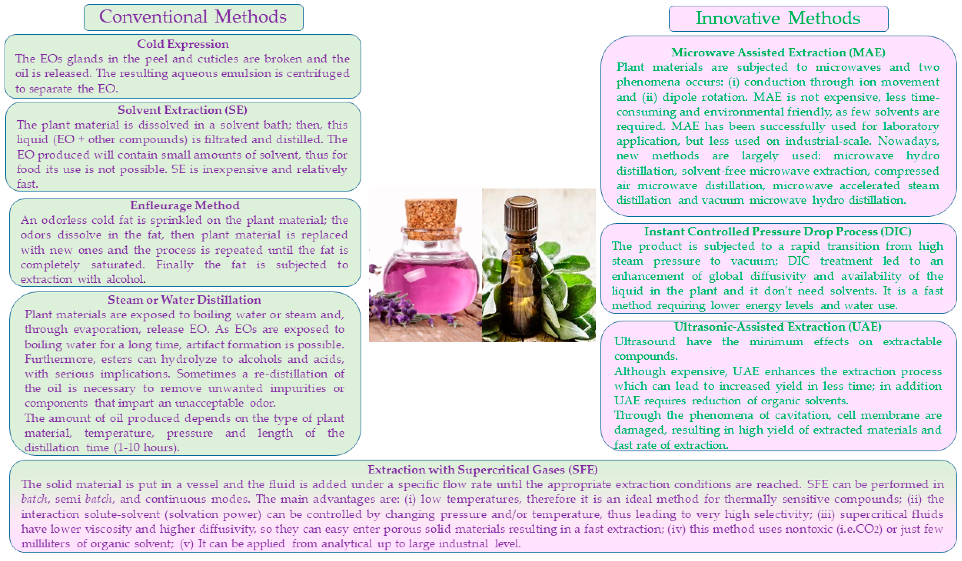
5. New Approaches for EOs Application

As is well recognized, EOs are volatile substances very sensitive to oxygen, light, moisture and heat; these characteristics could diminish their application in food and pharmaceutical industries. Thus, new technological and engineering approaches have been recently proposed to increase EOs’ chemical stability and solubility, decrease rapid evaporation and minimize the degradation of active compounds; one of these is the application of nanotechnologies. These last are considered a revolutionary strategy involving the control of matter at the atomic and molecular scales, usually below 100 nanometers (the term “nano” is referred to as a magnitude of 10−9 m): nanostructures, such as nano-emulsions, nanoliposomes and nanoparticles, can exhibit different physicochemical properties than their normal-scale analogues, such as a higher chemical reactivity due to a larger surface area [93,94]. Nanotechnologies have greatly contributed to the industrial application of EOs in fermentation processes and in food, textile, cosmetic, pharmaceutical, agricultural and environmental industries [95,96,97,98].
Recent studies have demonstrated that nanoparticles functionalized with EOs could be successfully used as antimicrobials against multidrug-resistant pathogens [1,95], whereas nano-emulsions could efficiently contribute to support the use of EOs in foods by increasing their dispersibility in the food areas where microorganisms grow and proliferate by reducing the impact on the quality attributes of the product, as well as by enhancing their antimicrobial activity [99]. Through nano-encapsulation, small capsules are formed by coating or embedding droplets of the bioactive compound in a matrix [99]: this physical barrier protects EOs from environmental factors, counteracts the high volatility of their components, facilitates their controlled release and improves bioavailability and efficiency [100]. Lavender oil encapsulated in chitosan nano-spheres demonstrated significant antimicrobial activity against B. cereus, B. subtilis, A. fumigatus and Macrophomina phaseolina in a study carried out by Velmurugan et al. [97]. In 2021, Pilicheva and co-authors [101] proposed the encapsulation of lavender oil in Arabic gum/maltodextrin microparticles by spray-drying, underlining that this feasible approach could be a useful tool for the conversion of liquid materials into solids with satisfactory flowability for further use in powder technology.
An interesting application of nanotechnologies applied to EOs is also found in the study by Dogan et al. [102], who tried to fabricate a novel active food packaging based on centrifugally spun nanofibers containing lavender essential oil: PVP, a non-toxic biocompatible polymer with GRAS (generally recognized as safe) status, was loaded with three different concentrations of LEO and used to preserve minced lamb meat. The in-vitro antioxidant effect of nanofiber mats increased with the loaded LEO concentration and the nanofibers positively affected the shelf life by suppressing the growth of aerobic mesophilic bacteria, psychotropic bacteria, yeast and molds that cause spoilage in meat.
The nanotechnology techniques have also been applied to EOs from Salvia by loading them in various matrices and green-synthesized metal or metal oxide nanoparticles (NPs). A good overview of these novel strategies related to both the nano-encapsulation of sage oils and related methods of preparation and application as antimicrobials can be found in the review by Zaccardelli et al. [103].
In recent years, new alternative technologies and green engineering approaches have also been proposed to improve EO extraction since conventional extraction technologies are characterized by several drawbacks, such as long extraction time, high energy consumption, low efficiency and reliance on hazardous solvents, which could limit their use in foods. Increasing energy costs and the more environmentally friendly approach being adopted (i.e., reduction of CO2 emissions) have moved researchers to seek alternative technologies that are cost-effective, sustainable and capable of producing products with the same or improved characteristics. Figure 5 provides a brief summary of both conventional and innovative methods proposed for EO extraction.

6. Concluding Remarks

Sage and lavender EOs have attracted researchers’ interest for a long time because of their numerous biological and antimicrobial properties that suggest their potential use as health enhancers and food preservatives.
However, to promote their widespread application in both the food and pharmaceuticals industries, some important issues should be yet addressed.
One aspect is the lack of consistency in their phytochemical composition since it depends on numerous factors such as plant species (sometimes, intra-species variation has also been observed), origin, plant part used for extraction, phenological stage and the agronomical practices used. All these factors are able to affect the profile of volatile compounds, underling that a standardization of the essential oils is mandatory before their addition to foods or use as therapeutic agents.
Another aspect to be considered is that relatively higher concentrations of EOs are generally needed to exert a beneficial effect, and this may adversely affect the organoleptic characteristics of foods. To minimize this aspect, it would be desirable to study the synergistic effects of EOs with other preservative techniques, including other natural antimicrobials and essential oils from other plants as well as with other non-thermal preservation techniques.
Finally, further clinical studies should be performed to evaluate their toxicity and assess with certainty their safety.

Author Contributions

Conceptualization, B.S. and M.R.C.; investigation, B.S., A.G., A.R. and D.C.; resources, A.B. and M.R.C.; writing—original draft preparation, B.S., A.G. and D.C.; writing—review and editing, B.S., A.G. and D.C.; supervision, B.S. and M.R.C.; funding acquisition, A.B. and M.R.C. All authors have read and agreed to the published version of the manuscript.

Funding

This study was carried out within the Agritech National Research Center and received funding from the European Union Next-Generation EU (PIANO NAZIONALE DI RIPRESA E RESILIENZA (PNRR)—MISSIONE 4 COMPONENTE 2, INVESTIMENTO 1.4—D.D. 1032 17/06/2022, CN00000022). This manuscript reflects only the authors’ views and opinions, neither the European Union nor the European Commission can be considered responsible for them.

Institutional Review Board Statement

Not applicable.

Informed Consent Statement

Not applicable.

Data Availability Statement

No new data were created for the production of this manuscript. All of the data here discussed and presented are available in the relative references here cited and listed.

Conflicts of Interest

The authors declare no conflict of interest.

References

  1. Chouhan, S.; Sharma, K.; Guleria, S. Antimicrobial activity of some Essential Oils-present status and future perspectives. Medicines 2017, 4, 58. [Google Scholar] [CrossRef] [PubMed] [Green Version]
  2. Mérillon, J.-M.; Rivière, C. Natural Antimicrobial Agents; Springer International Publishing AG: Cham, Switzerland, 2018. [Google Scholar]
  3. Markets and Markets. Available online: https://www.marketsandmarkets.com/Market-Reports/essential-oil-market-119674487.html (accessed on 20 May 2023).
  4. Swamy, M.K.; Sinniah, U.R. Patchouli (Pogostemon cablin Benth.): Botany, agrotechnology and biotechnological aspects. Ind. Crops Prod. 2016, 87, 161–176. [Google Scholar] [CrossRef]
  5. Sharifi-Rad, M.; Ozcelik, B.; Altın, G.; Daşkaya-Dikmen, C.; Martorell, M.; Ramírez-Alarcón, K.; Alarcón-Zapata, P.; Morais-Braga, M.F.B.; Carneiro, J.N.P.; Borges Leal, A.L.A.; et al. Salvia spp. plants-from farm to food applications and phytopharmacotherapy. Trends Food Sci. Technol. 2018, 80, 242–263. [Google Scholar] [CrossRef]
  6. Shariatifar, N.; Mostaghim, T.; Afshar, A.; Mohammadpourfard, I.; Sayadi, M.; Rezaei, M. Antibacterial properties of essential oil of Heracleum per-sicum (Golpar) and food-borne pathogens. Int. J. Enteric Pathog. 2017, 5, 41–44. [Google Scholar] [CrossRef] [Green Version]
  7. Hashempour-Baltork, F.; Hosseini, H.; Shojaee-Aliabadi, S.; Torbati, M.; Alizadeh, A.M.; Alizadeh, M. Drug Resistance and the Prevention Strategies in Food Borne Bacteria: An Update Review. Adv. Pharm. Bull. 2019, 9, 335–347. [Google Scholar] [CrossRef] [Green Version]
  8. Stan, D.; Enciu, A.-M.; Mateescu, A.L.; Ion, A.C.; Brezeanu, A.C.; Stan, D.; Tanase, C. Natural compounds with antimicrobial and antiviral effect and nanocarriers used for their transportation. Front. Pharmacol. 2021, 12, 723233. [Google Scholar] [CrossRef] [PubMed]
  9. Brnawi, W.I.; Hettiarachchy, N.S.; Horax, R.; Kumar-Phillips, G.; Ricke, S. Antimicrobial activity of leaf and bark cinnamon essential oils against Listeria monocytogenes and Salmonella Typhimurium in broth system and on celery. J. Food Process Preserv. 2019, 43, e13888. [Google Scholar] [CrossRef]
  10. Krasniewska, K.; Gniewosz, M.; Kosakowska, O.; Pobiega, K. Chemical composition and antimicrobial properties of essential oil from lavender (Lavandula angustifolia L.) in commercial available preparation. Postępy Fitoter. 2017, 18, 113–118. [Google Scholar]
  11. Swamy, M.K.; Akhtar, M.S.; Sinniah, U.R. Antimicrobial Properties of Plant Essential Oils against Human Pathogens and Their Mode of Action: An Updated Review. Evid. Based Complement. Alternat. Med. 2016, 2016, 3012462.21. [Google Scholar] [CrossRef] [Green Version]
  12. Ghorbani, A.; Esmaeilizadeh, M. Pharmacological properties of Salvia officinalis and its components. J. Tradit. Complement. Med. 2017, 7, 433–440. [Google Scholar] [CrossRef]
  13. Carrasco, A.; Thomas, V.; Tudela, J.; Miguel, M.G. Comparative study of GC-MS characterization, antioxidant activity and hyaluronidase inhibition of different species of Lavandula and Thymus essential oils. Flavour Fragr. J. 2016, 31, 57–69. [Google Scholar] [CrossRef] [Green Version]
  14. de Rapper, S.; Viljoen, A.; van Vuuren, S. The in vitro antimicrobial effects of Lavandula angustifolia essential oil in combination with conventional antimicrobial agents. Evid. Based Complement. Alternat. Med. 2016, 2016, 2752739. [Google Scholar] [CrossRef] [PubMed] [Green Version]
  15. Benabdelkader, T.; Zitouni, A.; Guitton, Y.; Jullien, F.; Maitre, D.; Casabianca, H.; Legendre, L.; Kameli, A. Essential oils from wild populations of Algerian Lavandula stoechas L.: Composition, chemical variability, and in vitro biological properties. Chem. Biodivers. 2011, 8, 937–953. [Google Scholar] [CrossRef] [PubMed]
  16. Turgut, A.C.; Emen, F.M.; Canbay, H.S.; Demirdöğen, R.E.; Çam, N.; Kılıç, D.; Yeşilkaynak, T. Chemical characterization of Lavandula angustifolia Mill. as a phytocosmetic species and investigation of its antimicrobia in cosmetic products. J. Turk. Chem. Soc. Sect. Chem. 2017, 4, 283–298. [Google Scholar] [CrossRef] [Green Version]
  17. Lis-Balchin, M. Lavender. In Handbook of Herbs and Spices; Peter, K.V., Ed.; Woodhead Publishing Limited: Cambridge, UK, 2012; Volume 2, pp. 329–347. [Google Scholar]
  18. Fattahi, B.; Nazeri, V.; Kalantari, S.; Bonfill, M.; Fattahi, M. Essential oil variation in wild-growing populations of Salvia reuterana Boiss. collected from Iran, using GC–MS and multivariate analysis. Ind. Crops Prod. 2016, 81, 180–190. [Google Scholar] [CrossRef]
  19. Beheshti-Rouy, M.; Azarsina, M.; Rezaie-Soufi, L.; Alikhani, M.Y.; Roshanaie, G.; Komaki, S. The antibacterial effect of sage extract (Salvia officinalis) mouthwash against Streptococcus mutans in dental plaque: A randomized clinical trial. Iran. J. Microbiol. 2015, 7, 173–177. [Google Scholar]
  20. Mokhtari, R.; Fard, M.K.; Rezaei, M.; Moftakharzadeh, S.A.; Mohseni, A. Antioxidant, antimicrobial activities, and characterization of phenolic compounds of Thyme (Thymus vulgaris L.), Sage (Salvia officinalis L.), and Thyme–Sage mixture extracts. J. Food. Qual. 2023, 2023, 2602454. [Google Scholar] [CrossRef]
  21. Sulaiman, A.M.; Abdulaziz, A.S.; Almutawea, A.M.; Alansari, S.A.; Aldoseri, F.A.; Bekhit, S.A.; Al-Thawadi, S.M.; Alqallaf, S.M.; Bekh, A.A. Evaluation of the antibacterial effect of Salvia officinalis Essential Oil and its synergistic effect with Meropenem. Lett. Appl. NanoBioScience 2023, 12, 44. [Google Scholar]
  22. ISO 9909:1997; Oil of Dalmatian Sage (Salvia officinalis L.). International Organization for Standardization (ISO): Geneva, Switzerland, 1997.
  23. Baricevic, D.; Bartol, T. The biological/pharmacological activity of the Salvia genus. In Sage, the Genus Salvia; Kintzios, S.E., Ed.; Harwood Academic Publishers: Singapore, 2005; p. 290. [Google Scholar]
  24. Yilar, M.; Kadioǧlu, I.; Telci, I. Chemical composition and antifungal activity of Salvia officinalis (L.), S. cryptantha (montbret et aucher ex benth.), S. tomentosa (mill.) plant essential oils and extracts. Fresenius Environ. Bull. 2018, 27, 1695–1706. [Google Scholar]
  25. Cutillas, A.B.; Carrasco, A.; Martinez-Gutierrez, R.; Tomas, V.; Tudela, J. Salvia officinalis L. Essential Oils from Spain: Determination of Composition, Antioxidant Capacity, Antienzymatic, and Antimicrobial Bioactivities. Chem. Biodivers. 2017, 14, e1700102. [Google Scholar] [CrossRef]
  26. Russo, A.; Formisano, C.; Rigano, D.; Senatore, F.; Delfine, S.; Cardile, V.; Rosselli, S.; Bruno, M. Chemical composition and anticancer activity of essential oils of Mediterranean sage (Salvia officinalis L.) grown in different environmental conditions. Food Chem. Toxicol. 2013, 55, 42–47. [Google Scholar] [CrossRef]
  27. Durling, N.E.; Catchpole, O.J.; Grey, J.B.; Webby, R.F.; Mitchell, K.A.; Foo, L.Y.; Perry, N.B. Extraction of phenolics and essential oil from dried sage (Salvia officinalis) using ethanol–water mixtures. Food Chem. 2007, 101, 1417–1424. [Google Scholar] [CrossRef]
  28. Kamatou, G.P.P.; Makunga, N.P.; Ramogola, W.P.N.; Viljoen, A.M. South African Salvia species: A review of biological activities and phytochemistry. J. Ethnopharmacol. 2008, 119, 664–672. [Google Scholar] [CrossRef] [PubMed]
  29. Dweck, A.C. Introduction. The folklore and cosmetic use of various salvia species. In Sage, the Genus Salvia; Kintzios, S.E., Ed.; Harwood Academic Publishers: Singapore, 2005. [Google Scholar]
  30. Monsen, R.E.; Herlofson, B.B.; Gay, C.; Fjeld, K.G.; Hove, L.H.; Malterud, K.E.; Saghaug, E.; Slaaen, J.; Sundal, T.; Tollisen, A.; et al. A mouth rinse based on a tea solution of Salvia officinalis for oral discomfort in palliative cancer care: A randomized controlled trial. Support. Care Cancer 2021, 29, 4997–5007. [Google Scholar] [CrossRef] [PubMed]
  31. Lakhal, H.; Ghorab, H.; Chibani, S.; Kabouche, A.; Semra, Z.; Smati, F.; Abuhamdah, S.; Kabouche, Z. Chemical composition and biological activities of the essential oil of Salvia officinalis from Batna (Algeria). Der Pharm. Lett. 2013, 5, 310–314. [Google Scholar]
  32. Abu-Darwish, M.S.; Cabral, C.; Ferreira, I.V.; Gonçalves, M.J.; Cavaleiro, C.; Cruz, M.T.; Albdour, T.H.; Salgueiro, L. Essential oil of common sage (Salvia officinalis L.) from Jordon: Assessment of safety in mammalian cells and its antifungal and anti-inflammatory potential. Biomed Res. Int. 2013, 2013, 538940. [Google Scholar] [CrossRef] [Green Version]
  33. European Medicines Agency (EMA). Assessment Report on Salvia fruticosa Mill Folium. EMA/HMPC/599992/2014. 2016. Available online: https://www.ema.europa.eu/en/documents/herbal-report/final-assessment-report-salvia-fruticosa-mill-folium_en.pdf (accessed on 15 May 2023).
  34. European Medicines Agency (EMA). Assessment Report on Salvia officinalis L., folium and Salvia officinalis L., Aetheroleum EMA/HMPC/150801/2015. 2016. Available online: https://www.ema.europa.eu/en/documents/herbal-report/draft-assessment-report-salvia-officinalis-l-folium-salvia-officinalis-l-aetheroleum_en.pdf (accessed on 15 May 2023).
  35. Wińska, K.; Mączka, W.; Łyczko, J.; Grabarczyk, M.; Czubaszek, A.; Szumny, A. Essential Oils as Antimicrobial Agents—Myth or Real Alternative? Molecules 2019, 24, 2130. [Google Scholar] [CrossRef] [Green Version]
  36. Haziri, A.; Faiku, F.; Mehmeti, A.; Kurteshi, K.; Haziri, I.; Rudhani, I. In vitro antibacterial properties of ethanol extract from Salvia officinalis (L.) plant growing wild in Kosovo. Biomed. J. Sci. Tech. Res. 2018, 2, 2578–2580. [Google Scholar]
  37. Mohamed, A.Y.; Mustafa, A.A. In vitro anti-microbial activity of Essential Oils and other extracts from Salvia officinalis against some bacteria. Plant Sci. 2019; 2019040012, preprints. [Google Scholar]
  38. Medeiros de Almeida, C.; de Souza Sales Rocha, E.A.L.; Ponchet Alves, É.; de Freitas Lima, R.; Wanderley Cavalcanti, Y.; Barbosa Gomes, R.C.; Vieira Pereira, J.; Melo de Brito Costa, E.M. In vitro evaluation of the antimicrobial potential of Salvia officinalis L. against oral pathogens. J. Health Sci. 2019, 21, 129–133. [Google Scholar] [CrossRef] [Green Version]
  39. de Oliveira, J.R.; Vilela, P.G.D.F.; Almeida, R.B.A.; de Oliveira, F.E.; Carvalho, C.A.T.; Camargo, S.E.A.; Jorge, A.O.C.; de Oliveira, L.D. Antimicrobial activity of noncytotoxic concentrations of Salvia officinalis extract against bacterial and fungal species from the oral cavity. Gen. Dent. 2019, 67, 22–26. [Google Scholar] [PubMed]
  40. Steffens, N.A.; Zimmermann, E.S.; Nichelle, S.M.; Brucker, N. Meropenem use and therapeutic drug monitoring in clinical practice: A literature review. J. Clin. Pharm. Ther. 2021, 46, 610–621. [Google Scholar] [CrossRef] [PubMed]
  41. Cui, H.; Zhang, X.; Zhou, H.; Zhao, C.; Lin, L. Antimicrobial activity and mechanisms of Salvia sclarea Essential Oil. Bot. Stud. 2015, 56, 16. [Google Scholar] [CrossRef] [PubMed] [Green Version]
  42. Divyapriya, G.K.; Veeresh, D.J.; Yavagal, P.C. Evaluation of antibacterial efficacy of Chia (Salvia hispanica) seeds extract against Porphyromonas gingivalis, Fusobacterium nucleatum and Aggregatibacter actinomycetemcomitans. An in-vitro study. Int. J. Ayurveda Pharm. Res. 2016, 4, 22–24. [Google Scholar]
  43. Elshafie, H.S.; Aliberti, L.; Amato, M.; De Feo, V.; Camele, I. Chemical composition and antimicrobial activity of chia (Salvia hispanica L.) Essential Oil. Eur. Food Res. Technol. 2018, 244, 1675–1682. [Google Scholar] [CrossRef]
  44. Moo, C.L.; Yang, S.K.; Osman, M.A.; Yuswan, M.H.; Loh, J.Y.; Lim, W.M.; Lim, S.H.; Lai, K.S. Antibacterial activity and mode of action of β-caryophyllene on Bacillus cereus. Pol. J. Microbiol. 2020, 69, 1–6. [Google Scholar] [CrossRef] [Green Version]
  45. Moon, K.; Cha, J. Enhancement of antioxidant and antibacterial activities of Salvia miltiorrhiza roots fermented with Aspergillus oryzae. Foods 2020, 9, 34. [Google Scholar] [CrossRef] [Green Version]
  46. Chen, B.-C.; Ding, Z.-S.; Dai, J.-S.; Chen, N.-P.; Gong, X.-W.; Ma, L.-F.; Qian, C.-D. New insights into the antibacterial mechanism of cryptotanshinone, a representative diterpenoid quinone from Salvia miltiorrhiza Bunge. Front. Microbiol. 2021, 12, 647289. [Google Scholar] [CrossRef]
  47. Lim Ah Tock, M.; Combrinck, S.; Kamatou, G.; Chen, W.; Van Vuuren, S.; Viljoen, A. Antibacterial screening, biochemometric and bioautographic evaluation of the non-volatile bioactive components of three indigenous South African Salvia species. Antibiotics 2022, 11, 901. [Google Scholar] [CrossRef]
  48. Jordán, M.J.; Lax, V.; Rota, M.C.; Lorán, S.; Sotomayor, J.A. Relevance of carnosic acid, carnosol, and rosmarinic acid concentrations in the in vitro antioxidant and antimicrobial activities of Rosmarinus officinalis (L.) Methanolic Extracts. J. Agric. Food Chem. 2012, 60, 9603–9608. [Google Scholar] [CrossRef]
  49. Bensebia, O.; Benamani, A.; Issadi, H.M. Antibacterial activity of sage leaves against pathogenic bacteria as affected by different drying Temperature. Arab. J. Med. Aromat. Plants 2021, 7, 74–92. [Google Scholar]
  50. Aćimović, M.; Pezo, L.; Cabarkapa, I.; Trudić, A.; Stanković Jeremić, J.; Varga, A.; Lončar, B.; Šovljanski, O.; Tešević, V. Variation of Salvia officinalis L. Essential Oil and hydrolate composition and their antimicrobial activity. Processes 2022, 10, 1608. [Google Scholar] [CrossRef]
  51. Benabdesslem, Y.; Hachem, K.; Kahloula, K.; Belakredar, A.; Slimani, M. In vitro antimicrobial activity of aqueous, ethanolic and methanolic leaves extracts from Salvia argentea. Sys. Rev. Pharm. 2020, 11, 1683–1691. [Google Scholar]
  52. Pieracci, Y.; Ciccarelli, D.; Giovanelli, S.; Pistelli, L.; Flamini, G.; Cervelli, C.; Mancianti, F.; Nardoni, S.; Bertelloni, F.; Ebani, V.V. Antimicrobial activity and composition of five rosmarinus (Now Salvia spp. and Varieties) Essential Oils. Antibiotics 2021, 10, 1090. [Google Scholar] [CrossRef]
  53. Salehi, B.; Mnayer, D.; Özçelik, B.; Altin, G.; Kasapoğlu, K.N.; Daskaya-Dikmen, C.; Sharifi-Rad, M.; Selamoglu, Z.; Acharya, K.; Sen, S.; et al. Plants of the Genus Lavandula: From Farm to Pharmacy. Nat. Prod. Commun. 2018, 13, 1385–1402. [Google Scholar] [CrossRef] [Green Version]
  54. Aprotosoaie, A.C.; Gille, E.; Trifan, A.; Luca, V.S.; Miron, A. Oils of Lavandula genus: A systematic review of their chemistry. Phytochem. Rev. 2017, 16, 761–799. [Google Scholar] [CrossRef]
  55. Jianu, C.; Pop, G.; Gruia, A.T.; Horhat, F.G. Chemical composition and antimicrobial activity of essential oils of lavender (Lavandula angustifolia) and lavandin (Lavandula x intermedia) grown in Western Romania. Int. J. Agric. Biol. 2013, 15, 772–776. [Google Scholar]
  56. Blažeković, B.; Yang, W.; Wang, Y.; Li, C.; Kindl, M.; Pepeljnjak, S.; Vladimir-Knežević, S. Chemical composition, antimicrobial and antioxidant activities of essential oils of Lavandula × intermedia ‘Budrovka’ and L. angustifolia cultivated in Croatia. Ind. Crops Prod. 2018, 123, 173–182. [Google Scholar] [CrossRef]
  57. Pistelli, L.; Najar, B.; Giovanelli, S.; Lorenzini, L.; Tavarini, S.; Angelini, L.G. Agronomic and phytochemical evaluation of lavandin and lavender cultivars cultivated in the Tyrrhenian area of Tuscany (Italy). Ind. Crops Prod. 2017, 109, 37–44. [Google Scholar] [CrossRef]
  58. Başer, K.H.C.; Buchbauer, G. Handbook of Essential Oils, Science, Technology and Applications, 2nd ed.; Başer, K.H.C., Buchbauer, G., Eds.; CRC Press: Boca Raton, FL, USA, 2016. [Google Scholar]
  59. Lu, H.; Li, H.; Lu, H.; Li, X.; Zhou, A. Chemical composition of lavender essential oil and its antioxidant activity and inhibition against rhinitis-related bacteria. Afr. J. Microbiol. Res. 2010, 4, 309–313. [Google Scholar]
  60. Cardia, G.F.E.; Silva-Filho, S.E.; Silva, E.L.; Uchida, N.S.; Cavalcante, H.A.O.; Cassarotti, L.L.; Salvadego, V.E.C.; Spironello, R.A.; Bersani-Amado, C.A.; Cuman, R.K.N. Effect of lavender (Lavandula angustifolia) essential oil on acute inflammatory response. Evid. Based Complement. Altern. Med. 2018, 2018, 1413940. [Google Scholar] [CrossRef] [Green Version]
  61. Mori, H.M.; Kawanami, H.; Kawahata, H.; Aoki, M. Wound healing potential of lavender oil by acceleration of granulation and wound contraction through induction of TGF-beta in a rat model. BMC Complement. Altern. Med. 2016, 16, 144. [Google Scholar] [CrossRef] [Green Version]
  62. Di Sotto, A.; Mazzanti, G.; Carbone, F.; Hrelia, P.; Maffei, F. Genotoxicity of lavender oil, linalyl acetate, and linalool on human lymphocytes in vitro. Environ. Mol. Mutagen. 2011, 52, 69–71. [Google Scholar] [CrossRef]
  63. El-Said, H.; Ashgar, S.S.; Bader, A.; AlQathama, A.; Halwani, M.; Ascrizzi, R.; Flamini, G. Essential Oil Analysis and Antimicrobial Evaluation of Three Aromatic Plant Species Growing in Saudi Arabia. Molecules 2021, 26, 959. [Google Scholar] [CrossRef] [PubMed]
  64. Adaszyńska-Skwirzyńska, M.; Dzięcioł, M. Comparison of phenolic acids and flavonoids contents in various cultivars and parts of common lavender (Lavandula angustifolia) derived from Poland. Nat. Prod. Res. 2017, 31, 2575–2580. [Google Scholar] [CrossRef] [PubMed]
  65. Hossain, S.; Heo, H.; De Silva, B.C.J.; Wimalasena, S.H.M.P.; Pathirana, H.N.K.S.; Heo, G.J. Antibacterial activity of essential oil from lavender (Lavandula angustifolia) against pet turtle-borne pathogenic bacteria. Lab. Anim. Res. 2017, 33, 195–201. [Google Scholar] [CrossRef] [PubMed] [Green Version]
  66. Caprari, C.; Fantasma, F.; Divino, F.; Bucci, A.; Iorizzi, M.; Naclerio, G.; Ranalli, G.; Saviano, G. Chemical Profile, In Vitro Biological Activity and Comparison of Essential Oils from Fresh and Dried Flowers of Lavandula angustifolia L. Molecules 2021, 26, 5317. [Google Scholar] [CrossRef]
  67. Nafis, A.; Ouedrhiri, W.; Iriti, M.; Mezrioui, N.; Marraiki, N.; Elgorban, A.M.; Syed, A.; Hassani, L. Chemical composition and synergistic effect of three Moroccan lavender EOs with ciprofloxacin against foodborne bacteria: A promising approach to modulate antimicrobial resistance. Lett. Appl. Microbiol. 2021, 72, 698–705. [Google Scholar] [CrossRef]
  68. El Baaboua, A.; El Maadoudi, M.; Bouyahy, A.; Belmehdi, O.; Kounnoun, A.; Cheyadmi, S.; Ouzakar, S.; Skali Senhaji, N.; Abrini, J. Evaluation of the combined effect of antibiotics and essential oils against Campylobacter multidrug resistant strains and their biofilm formation. S. Afr. J. Bot. 2022, 150, 451–465. [Google Scholar] [CrossRef]
  69. Tardugno, R.; Serio, A.; Pellati, F.; D’Amato, S.; Chaves López, C.; Bellardi, M.G.; Di Vito, M.; Savini, V.; Paparella, A.; Benvenuti, S. Lavandula x intermedia and Lavandula angustifolia essential oils: Phytochemical composition and antimicrobial activity against foodborne pathogens. Nat. Prod. Res. 2019, 33, 3330–3335. [Google Scholar] [CrossRef]
  70. Boy, F.R.; Benito, M.J.; Córdoba, M.d.G.; Rodríguez, A.; Casquete, R. Antimicrobial Properties of Essential Oils Obtained from Autochthonous Aromatic Plants. Int. J. Environ. Res. Public Health 2023, 20, 1657. [Google Scholar] [CrossRef] [PubMed]
  71. Dianez, F.; Santos, M.; Parra, C.; Navarro, M.J.; Blanco, R.; Gea, F.J. Screening of antifungal activity of 12 essential oils against eight pathogenic fungi of vegetables and mushroom. Lett. Appl. Microbiol. 2018, 67, 400–410. [Google Scholar] [CrossRef] [PubMed]
  72. Insawang, S.; Pripdeevech, P.; Tanapichatsakul, C.; Khruengsai, S.; Monggoot, S.; Nakham, T.; Artrod, A.; D’Souza, P.E.; Panuwet, P. Essential Oil Compositions and Antibacterial and Antioxidant Activities of Five Lavandula stoechas Cultivars Grown in Thailand. Chem. Biodivers. 2019, 16, e1900371. [Google Scholar] [CrossRef] [PubMed]
  73. Sayout, A.; Ouarhach, A.; Rabie, R.; Dilagui, I.; Soraa, N.; Romane, A. Evaluation of Antibacterial Activity of Lavandula pedunculata subsp. atlantica (Braun-Blanq.) Romo Essential Oil and Selected Terpenoids against Resistant Bacteria Strains-Structure-Activity Relationships. Chem. Biodivers. 2020, 17, e1900496. [Google Scholar] [CrossRef] [PubMed] [Green Version]
  74. Falleh, H.; Ben Jemaa, M.; Djebali, K.; Abid, S.; Saada, M.; Ksouri, R. Application of the mixture design for optimum antimicrobial activity: Combined treatment of Syzygium aromaticum, Cinnamomum zeylanicum, Myrtus communis, and Lavandula stoechas essential oils against Escherichia coli. J. Food Process Preserv. 2019, 43, e14257. [Google Scholar] [CrossRef]
  75. Aladağ, M.O.; Özcan, M.M.; Ergin, S. Inhibitory effect of some spice essential oils on growth of some gram-negative and gram-positive bacteria and a yeast. J. Food Process Preserv. 2021, 45, 15264. [Google Scholar] [CrossRef]
  76. Mačionienė, I.; Čepukoit, D.; Šalomskienė, J.; Černauskas, D.; Burokienė, D.; Šalaševičienė, A. Effects of Natural Antimicrobials on Xanthomonas Strains Growth. Horticulturae 2022, 8, 7. [Google Scholar] [CrossRef]
  77. Adaszyńska-Skwirzyńska, M.; Dzięcioł, M.; Szczerbińska, D. Lavandula angustifolia Essential Oils as Effective Enhancers of Fluconazole Antifungal Activity against Candida albicans. Molecules 2023, 28, 1176. [Google Scholar] [CrossRef]
  78. Vasileva, I.; Denkova, R.; Chochkov, R.; Teneva, D.; Denkova, Z.; Dessev, T.; Denev, P.; Slavov, A. Effect of lavender (Lavandula angustifolia) and melissa (Melissa officinalis) waste on quality and shelf life of bread. Food Chem. 2018, 253, 13–21. [Google Scholar] [CrossRef]
  79. Btissam, R.; Fatima, E.M.; Kamal, E.; Hassane, G.; Mohamed, N. Composition and Antibacterial Activity of Hydro-Alcohol and Aqueous Extracts Obtained from the Lamiaceae Family. Pharmacog. J. 2018, 10, 81–91. [Google Scholar] [CrossRef] [Green Version]
  80. Sakkas, H.; Papadopoulou, C. Antimicrobial activity of basil, oregano, and thyme essential oils. J. Microbiol. Biotechnol. 2017, 27, 429–438. [Google Scholar] [CrossRef] [PubMed] [Green Version]
  81. Borges, R.S.; Ortiz, B.L.S.; Pereira, A.C.M.; Keita, H.; Carvalho, J.C.T. Rosmarinus officinalis essential oil: A review of its phytochemistry, anti-inflammatory activity, and mechanisms of action involved. J. Ethnopharmacol. 2019, 30, 29–45. [Google Scholar] [CrossRef] [PubMed]
  82. Rathod, N.B.; Ranveer, R.C.; Benjakul, S.; Kim, S.-K.; Pagarkar, A.U.; Patange, S.; Ozogul, F. Recent developments of natural antimicrobials and antioxidants on fish and fishery food products. Compr. Rev. Food Sci. Food Saf. 2021, 20, 4182–4210. [Google Scholar] [CrossRef]
  83. Azizkhani, M.; Tooryan, F.; Azizkhani, M. Inhibitory potential of Salvia sclarea and ocimum basilicum aganist chemical and microbial spoilage in cheese. J. Food Saf. 2016, 36, 109–119. [Google Scholar] [CrossRef]
  84. Šojić, B.; Pavlić, B.; Zeković, Z.; Tomović, V.; Ikonić, P.; Kocić-Tanackov, S.; Džinić, N. The effect of essential oil and extract from sage (Salvia officinalis L.) herbal dust (food industry by-product) on the oxidative and microbiological stability of fresh pork sausages. LWT—Food Sci. Technol. 2018, 89, 749–755. [Google Scholar] [CrossRef]
  85. Kačániová, M.; Terentjeva, M.; Kántor, A.; Tokár, M.; Puchalski, C.; Ivanišová, E. Antimicrobial effect of Sage (Salvia officinalis L.) and rosemary (Rosmarinus officinalis L.) Essential Oils on microbiota of chicken breast. Proc. Latv. Acad. Sci. 2017, 71, 461–467. [Google Scholar]
  86. Moura-Alves, M.; Gouveia, A.R.; de Almeida, J.M.M.M.; Monteiro-Silva, F.; Silva, J.A.; Saraiva, C. Behavior of Listeria monocytogenes in beef Sous vide cooking with Salvia officinalis L. essential oil, during storage at different temperatures. LWT—Food Sci. Technol. 2020, 132, 109896. [Google Scholar] [CrossRef]
  87. Gál, R.; Čmiková, N.; Prokopová, A.; Kačániová, M. Antilisterial and antimicrobial effect of Salvia officinalis Essential Oil in beef sous-vide meat during storage. Foods 2023, 12, 2201. [Google Scholar] [CrossRef] [PubMed]
  88. Ferreira, C.S.R.; Saqueti, B.H.F.; Santos, P.D.S.; Silva, J.M.; Matiucci, M.A.; Feihrmann, A.C.; Mikcha, J.M.G.; Santos, O.O. Effect of Salvia (Salvia officinalis) on the oxidative stability of salmon hamburgers. LWT—Food Sci. Technol. 2022, 154, 112867. [Google Scholar] [CrossRef]
  89. Mehdizadeh, T.; Tajik, H.; Jafarie, S.; Kaboudari, A. Effect of Salvia officinalis L. extract on chemical, microbial, sensory and shelf life of rainbow trout fillet. Food Sci. Biotechnol. 2019, 28, 1499–1506. [Google Scholar] [CrossRef]
  90. Dincoglu, A.H.; Caliskan, Z. Investigation of the effect of lavender (Lavandula angustifolia Mill.) essential oil on microbiological, physicochemical, and sensorial properties of meatballs during shelf-life, and its inhibitory effect on Escherichia coli O157:H7. Int. Food Res. J. 2022, 29, 991–1004. [Google Scholar] [CrossRef]
  91. Šimat, V.; Skroza, D.; Čagalj, M.; Soldo, B.; Generalíc Mekiníc, I. Effect of plant extracts on quality characteristics and shelf-life of cold-marinated shrimp (Parapenaeus longirostris, Lucas, 1846) under refrigerated storage. Food Biosci. 2023, 53, 102673. [Google Scholar] [CrossRef]
  92. Ramdan, G.M.; Khalafalla, F.A.; Hassan, A.H.A.; Abdel-Atty, N.S. Antibacterial efficacy of chemically and plant-synthesized zinc oxide nanocomposite against Staphylococcus aureus and Escherichia coli inoculated in Tilapia fillet. Malays. J. Microbiol. 2023, 19, 37–46. [Google Scholar]
  93. Tahir, A.; Ahmad, R.S.; Imran, M.; Ahmad, M.H.; Khan, M.K.; Muhammad, N.; Un Nisa, M.; Nadeem, M.T.; Yasmin, A.; Tahir, H.S.; et al. Recent approaches for utilization of food components as nano-encapsulation: A review. Int. J. Food Prop. 2017, 24, 1074–1096. [Google Scholar] [CrossRef]
  94. EFSA. Available online: www.efsa.europa.eu/it/topics/topic/nanotechnology. (accessed on 19 July 2023).
  95. Mandal, D.; Sarkar, T.; Chakraborty, R. Critical Review on nutritional, bioactive, and medicinal potential of spices and herbs and their application in food fortifcation and nanotechnology. Appl. Biochem. Biotechnol. 2023, 195, 1319–1513. [Google Scholar]
  96. Sana, S.S.; Li, H.; Zhang, Z.; Sharma, M.; Usmani, Z.; Hou, T.; Netala, V.R.; Wang, X.; Gupta, V.K. Recent advances in essential oils-based metal nanoparticles: A review on recent developments and biopharmaceutical applications. J. Mol. Liq. 2021, 333, 115951. [Google Scholar] [CrossRef]
  97. Velmurugan, P.; Ganeshan, V.; Nishter, N.F.; Jonnalagadda, R.R. Encapsulation of orange and lavender essential oils in chitosan nanospherical particles and its application in leather for aroma enrichment. Surf. Interfaces 2017, 9, 124–132. [Google Scholar] [CrossRef]
  98. Sangsuwan, J.; Pongsapakworawat, T.; Bangmo, P.; Sutthasupa, S. Effect of chitosan beads incorporated with lavender or red thyme essential oils in inhibiting Botrytis cinere and their application in strawberry packaging system. LWT—Food Sci. Technol. 2016, 74, 14–20. [Google Scholar] [CrossRef]
  99. Donsì, F.; Ferrari, G. Essential oil nanoemulsions as antimicrobial agents in food. J. Biotechnol. 2016, 233, 106–120. [Google Scholar] [CrossRef]
  100. Albuquerque, P.M.; Azevedo, S.G.; de Andrade, C.P.; D’Ambros, N.C.d.S.; Pérez, M.T.M.; Manzato, L. Biotechnological applications of nanoencapsulated Essential Oils: A Review. Polymers 2022, 14, 5495. [Google Scholar] [CrossRef]
  101. Pilicheva, B.; Uzunova, Y.; Katsarov, P. Comparative study on microencapsulation of Lavender (Lavandula angustifolia Mill.) and Peppermint (Mentha piperita L.) Essential Oils via Spray-Drying Technique. Molecules 2021, 26, 7467. [Google Scholar] [CrossRef] [PubMed]
  102. Doğan, C.; Doğan, N.; Gungor, M.; Eticha, A.K.; Akgul, Y. Novel active food packaging based on centrifugally spun nanofibers containing lavender essential oil: Rapid fabrication, characterization, and application to preserve of minced lamb meat. Food Packag. Shelf Life 2022, 34, 100942. [Google Scholar] [CrossRef]
  103. Zaccardelli, M.; Roscigno, G.; Pane, C.; Celano, G.; Di Matteo, M.; Mainente, M.; Vuotto, A.; Mencherini, T.; Esposito, T.; Vitti, A.; et al. Essential oils and quality composts sourced by recycling vegetable residues from the aromatic plant supply chain. Ind. Crops Prod. 2021, 162, 113255. [Google Scholar] [CrossRef]
  104. Stratakos, A.C.; Koidis, A. Methods for Extracting Essential Oils. In Essential Oils in Food Preservation, Flavor and Safety; Preedy, V.R., Ed.; Irish Academic Press: San Diego, CA, USA, 2016; pp. 31–38. [Google Scholar]
  105. Nirmala, M.J.; Sekar, P.C.; Johnson, A.; Kizhuveetil, U.; Shora, S.; Nagarajan, R. A comprehensive review of nanoadditives in Plant-based biodiesels with a special emphasis on essential oils. Fuel 2023, 351, 128934. [Google Scholar] [CrossRef]
Figure 1. Structures of the major components of Salvia officinalis Essential Oils (SOEOs).
Microbiolres 14 00073 g001
Figure 2. Main biological properties of Salvia officinalis Essential Oils (SOEOs).
Microbiolres 14 00073 g002
Figure 3. Structures of the major components of Lavandula Essential Oils (LEOs).
Microbiolres 14 00073 g003
Figure 4. Main biological properties of Lavandula Essential Oils (LEOs).
Microbiolres 14 00073 g004
Figure 5. Conventional and innovative methods proposed for EOs extraction [104,105].
Microbiolres 14 00073 g005
Disclaimer/Publisher’s Note: The statements, opinions and data contained in all publications are solely those of the individual author(s) and contributor(s) and not of MDPI and/or the editor(s). MDPI and/or the editor(s) disclaim responsibility for any injury to people or property resulting from any ideas, methods, instructions or products referred to in the content.

