Article

Prevalence and Diversity of Hepatitis Virus Markers among Patients with Acute Febrile Jaundice in Chad

by
Fissou Henry Yandai
1,2,3,†,
Kuan Abdoulaye Traore
2,4,*,†,
Ali Mahamat Moussa
5,
Bruno Lalidia Ouoba
2,
Jean Bienvenue Ouoba
2,
Mahamat Ali Bolti
5,
Mahamat Fayiz Abakar
3,
Mathieu Hota
1,
Kadidja Gamougam
6,
Bessimbaye Nadlao
6,
Jean-Claude Uwimbabazi
7,
Nadji Emmanuel Tao
8,
Bongo Nare Ngandolo
3,
Pierre Roques
9,10,11,12 and
Nicolas Barro
2
1
Laboratoire Mobile des Virus Hémorragiques et Respiratoires, Ministère de la Santé Publique, N’Djamena BP 480, Chad
2
Laboratoire de Biologie Moléculaire, d’Epidémiologie et de Surveillance des Bactéries et Virus Transmissibles par les Aliments (LaBESTA), Université Joseph KI-ZERBO, Ouagadougou BP 7021, Burkina Faso
3
Institut de Recherche en Elevage pour le Développement (IRED), N’Djamena BP 433, Chad
4
Laboratoire de Sciences de la Vie et de la Terre (LaSVT), Université Norbert ZONGO, Koudougou BP 376, Burkina Faso
5
Faculté des Sciences de la Santé Humaine (FSSH), Université de N’Djamena, N’Djamena BP 1117, Chad
6
Service des Laboratoires, Hôpital Général de Référence Nationale (HGRN) de N’Djamena, N’Djamena BP 130, Chad
7
Laboratoire de Microbiologie Clinique, CHU de Liège, Université de Liège, B-4000 Liège, Belgium
8
Institut National de la Statistique, des Etudes Economiques et Démographiques (INSEED), N’Djamena BP 453, Chad
9
IDMIT Département/IBFJ|CEA, 92265 Fontenay-aux-Roses, France
10
Immunology of Viral Infections and Autoimmune Diseases (IMVA-HB), U1184, INSERM, 92265 Fontenay-aux-Roses, France
11
UMR1184, IMVA-HB, Université Paris-Saclay, 91400 Orsay, France
12
Virology Unit, Institut Pasteur de Guinée, Conakry BP 4416, Guinea
*
Author to whom correspondence should be addressed.
These authors contribute equally to this work.
Microbiol. Res. 2021, 12(4), 878-887; https://doi.org/10.3390/microbiolres12040064
Submission received: 8 October 2021 / Revised: 6 November 2021 / Accepted: 8 November 2021 / Published: 12 November 2021

Abstract

:
Only a minority of the patients with acute febrile jaundice evaluated through the Yellow Fever surveillance program were found positive for antibodies against Yellow Fever Virus (YFV). In order to characterize patients with acute febrile jaundice negative for YFV, we collected 255 sera between January to December 2019. We screened for HBV antigens, and antibodies against HCV and HEV. The seroprevalences observed were 10.6% (27/255) for HBV, 2% (5/255) for HCV, 17.3% (44/255) for HEV IgG, 4.3% (11/255) for HEV IgM, and 12.5% (32/255) for both IgG and IgM HEV. Prevalence of HEV was significantly higher in females than males (p < 0.01). HEV IgG prevalence was highest in those 20–29 years old, but the highest incidence rate (IgM positive) was in children 0–9 years old. Exposure to HEV was higher in the Sahelian zone (55.8%, 95%CI: 40.97–70.66) than in the Sudanese zone (30.2%, 95% CI: 24.01–36.37, p = 0.003). The high prevalence rates and hepatitis virus diversity underline the challenge of routine clinical diagnosis in Chad’s Yellow Fever surveillance program.

1. Introduction

Yellow Fever (YF) is a viral hemorrhagic disease transmitted by infected Aedes mosquitoes [1,2]. A total of 47 countries in Africa and Central and South America are considered endemic for YF, and the WHO estimates an annual burden of 200,000 severe Yellow Fever cases with a case-fatality rate of up to 60,000 deaths [3,4]. Since the beginning of the 21st century, outbreaks have been reported in five African countries, including Nigeria, Côte d’Ivoire, Liberia, Senegal, and Guinea with a total of 840 cases, including 216 deaths [5]. More recently, in 2016–2017 large outbreaks occurred in Angola and the neighboring RDC with a total of 4306 suspected cases and 376 deaths, producing a case fatality rate (CFR) of 8.8%. This epidemic spilled over to China where 11 cases were reported [6]. YF surveillance in Eastern Senegal detected a small number of cases at the end of 2020 [7]. In Chad, as requested by WHO, YF is a disease notifiable to the Ministry of Public Health. Clinical cases of acute febrile jaundice or suspected of YF are routinely screened annually for specific immune responses. Between 2013 and 2020, hundreds of samples were sent to N’Djamena for confirmatory testing. Among these samples, only 4 cases of YF were confirmed by biological tests [8].
YF causes jaundice which stems from an accumulation of bilirubin, a degradation product of heme metabolism in the blood [9,10]. This clinical syndrome is common to several other endemic diseases affecting the liver, particularly viral infections, such as hepatitis A virus (HAV), hepatitis B virus (HBV), hepatitis C (HCV), hepatitis D virus (HDV), and hepatitis E virus (HEV) [11,12]. HBV and HCV are transmitted by parenteral, sexual, or mother-to-child routes. They usually evolve into chronic hepatitis, liver cirrhosis, and hepatocellular carcinoma resulting in high mortality and morbidity rates [12]. HEV is spread mainly via fecal–oral transmission route [13]. Thus, although acute febrile jaundice strongly suggests infection with hepatitis viruses in medical practice, the patients’ sera that are retrospectively tested negative for yellow fever are never screened for these viruses. Because of the very different impact of hepatitis virus on public health management, we investigated the burden of other pathogens associated with jaundice. This retrospective, cross-sectional study was performed to show the diversity of hepatitis viruses associated with acute febrile jaundice so as to inform and potentially modify the YF surveillance program and to improve patient referral care.

2. Materials and Methods

2.1. Study Setting and Samples

The study was performed between January and December 2019 in Chad. Four hundred and five patients with acute fever followed by jaundice within 2 weeks of showing the first symptoms who were suspected of yellow fever, were investigated in all the 23 regions of Chad. Among these patients, some were under anti-malarial treatment with no improvement of their health condition. A volume of 2–3 mL of blood from each patient was collected by venepuncture using a dry tube and an EDTA tube. The samples were then centrifuged at 3000× g for 10 min. The sera collected were kept at a temperature of 2–8 °C and sent to the laboratory of the General Hospital of N’Djamena (GHN). Each serum was initially screened for yellow fever using an IgM Enzyme Linked Immunosorbent Assay (YF MAC-HD ELISA (BioMARC, Fort Collins, CO, USA), according to the CDC protocol [14]. The information collected from the patient demographic were age, sex, locality or city, region, and YF symptoms such as yellowing of the skin and eyes, dark urine, and abdominal pain with vomiting.

2.2. Laboratory Analysis

All sera that tested negative for YF were transferred to the Laboratory of Hemorrhagic Fever and Respiratory Viruses, and the presence of HBV, HCV, and HEV was investigated in 255 randomly selected sera. HBV antigen was first assayed using Abbott Determine TM HBsAg and total antibodies against HCV were tested using the Abbott SD BIOLINE test (STANDARD DIAGNOSTIC inc., Yongin-si, Korea). We then tested for the presence of IgG and IgM anti-HEV using the commercial kit HEV ELISA EUROIMMUN (Medizinische Labordiagnostika AG, Lübeck, Germany). All analyses were performed in accordance with the manufacturers’ instructions.

2.3. Statistical Analysis

Analyses were performed using IBM SPSS statistical software(IBM SPSS, Chicago, IL, USA), version 20. Prevalences were described with 95% confidence intervals (CIs). χ2 test of Pearson were performed to evaluate the difference in the prevalence of viral markers among sex, age, region, and geographical zone. Univariate analysis using the χ2 test and multivariate logistic regression analysis was performed to identify potential risk factors for HEV infection by calculating odds ratios (ORs) and 95% CI. For all analyses, p-value < 0.05 was considered statistically significant.

3. Results

3.1. Seroprevalence of HEV, HBV and HCV

The overall seroprevalence of positive IgG and/or IgM anti-HEV was 34.1% (87/255; CI 95% [28.3–39.9]). Of these, 16.9% (43/255; CI 95% [12.3–21.5]) and 17.3% (44/255; CI 95% [12.6–21.9]) were indicative of recent infection (positive for IgM anti-HEV) and past infection (positive for IgG anti-HEV only), respectively. The other hepatitis virus infections detected were HBV and HCV. We found that 10.6% (27/255 CI 95% [6.8–14.36]) of patients were exposed to HBV and 2.0% (5/255 CI 95% [0.3–3.7]) were exposed to HCV. Of the 168 patients that were HEV negative, 13 were infected with HBV and 3 were infected with HCV (Table 1). There were no samples positive for either HBV or HCV. There were 5 HBV positive sera, among which one was positive for HEV IgM, and 4 were both HEV IgM and IgG positive, indicating ongoing HBV infection and recent HEV exposure. Nine sera with HEV IgG+ only were also positive for HBV antigen.

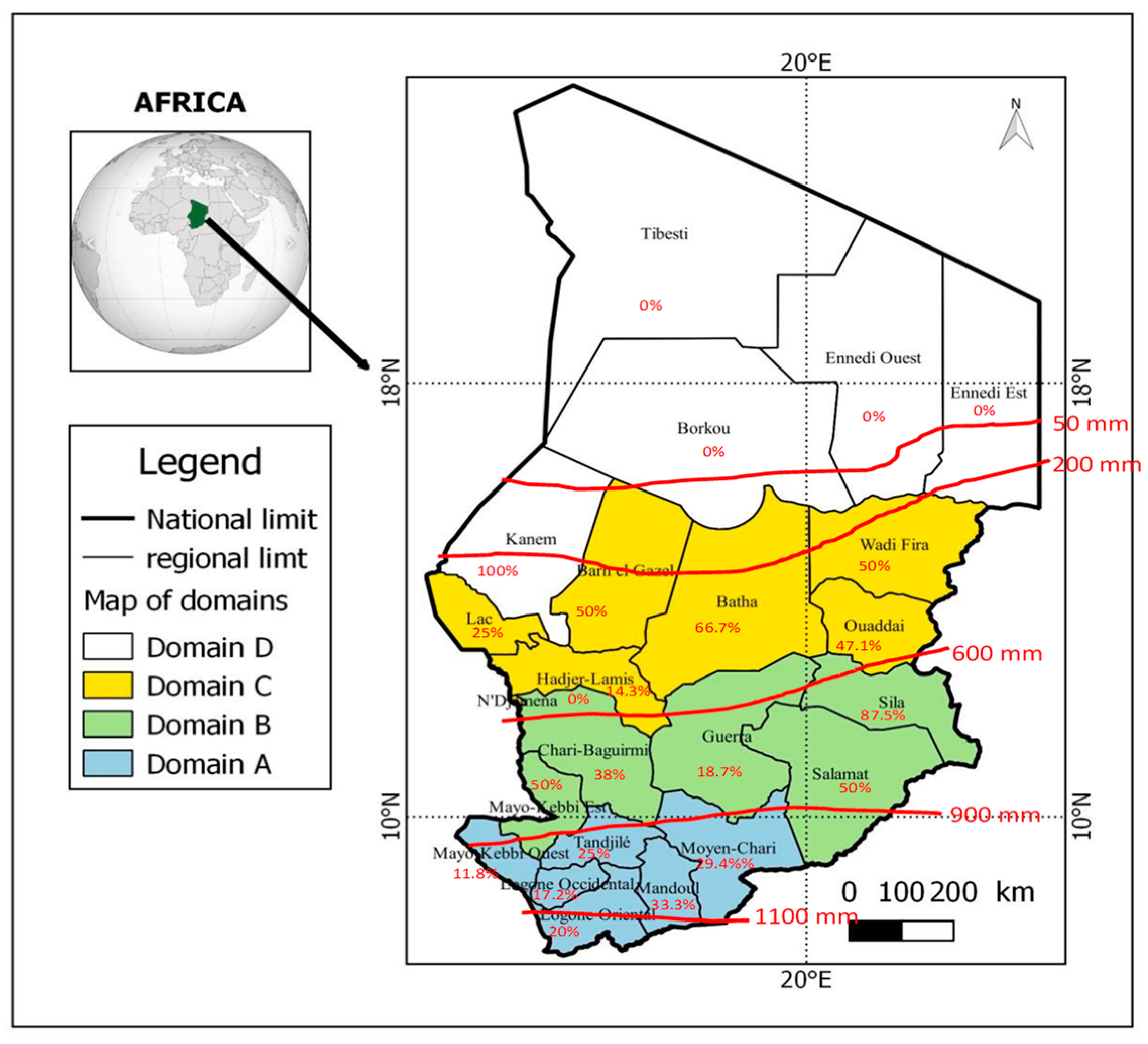
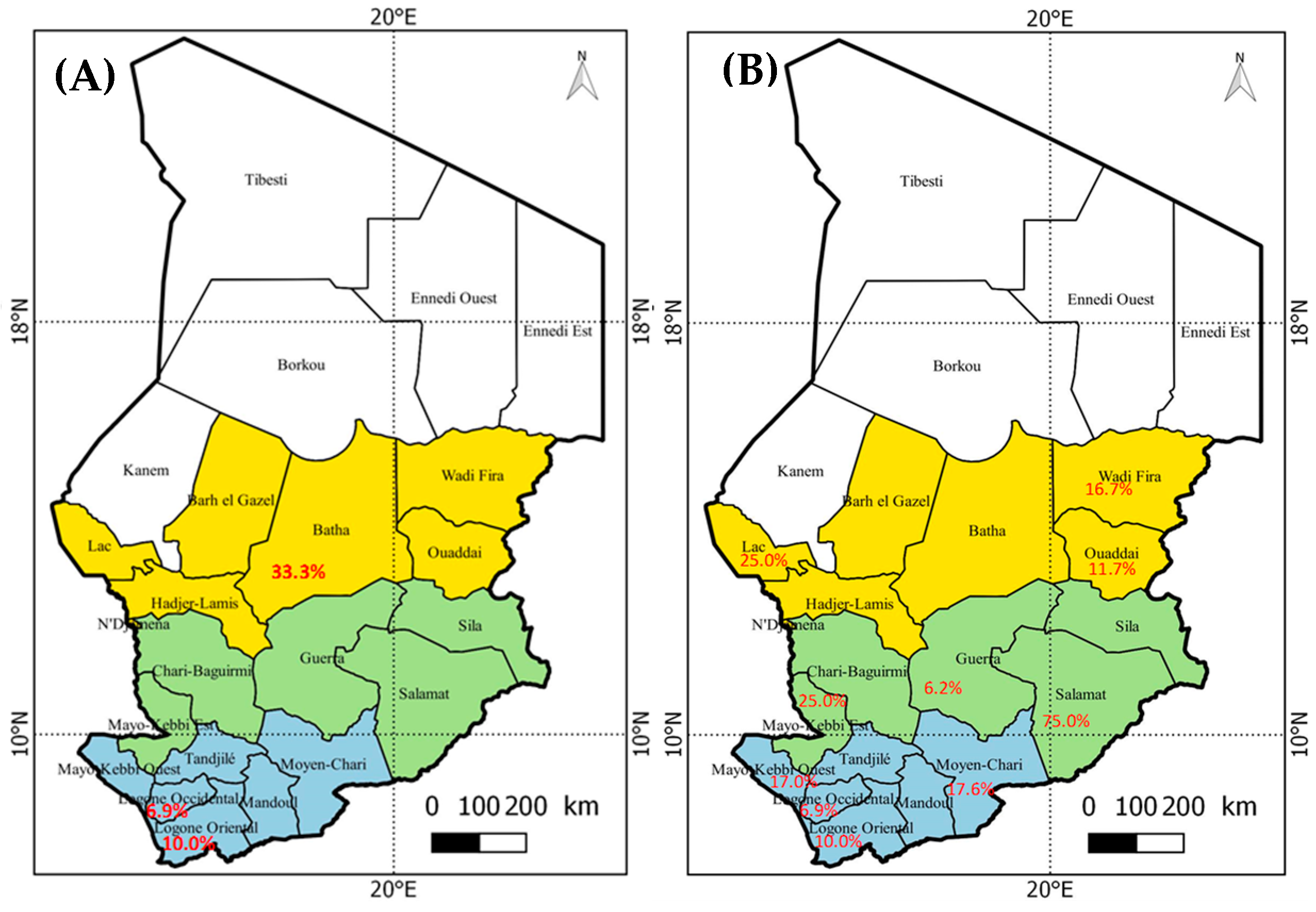
3.2. Geographical Distribution of Hepatitis Viruses

Hepatitis viruses were detected in patients from almost all the sampled provinces in Chad except the regions of Boukou, Ennedi East, Ennedi West, and Tibesti (Figure 1). Most of the cases recorded in this study originated from the provinces of south and center of the country. Hepatitis E markers were most often detected with various level of Anti-HEV IgG depending on the province. HBV antigen was detected in 10 out of 23 regions (Figure 2A). However, anti-HCV antibodies were rare and found only in the regions of Batha and Logone. Taken together, the data show that hepatitis E is a national infection occurring in almost all regions of Chad (Figure 2B).
The seroprevalences of hepatitis markers in the 23 regions of Chad were grouped into 4 domains roughly defined according to the amount of rainfall per year (Figure 1, Table 2). Domain A has 1100 to 900 mm/year, domain B has 900 to 600 mm/year, domain C has 600 to 200 mm/year, and domain D less than 200 mm/year (Figure 1). The observed level of anti-HEV IgG or IgM was high in domain D (but only 4 patients were tested), followed by domains C (18/55; 32.7%, CI 95% [20.3–45.1]), B (29/61; 47.5%, CI 95% [35.0–60.1]), and A (37/135; 27.41%, CI 95% [19.9–34.9]), but the variation between the domains was not significant. The rate of HBsAg positive samples was around 10% in domains A, B, C, and 0% in domains D. HCV antibodies were rare and found only in domains A and C. Details of the statistical comparison are given in Table 3.

3.3. Distribution of Hepatitis Markers in Geographical Zones

The distribution of hepatitis markers by geographical zone is presented in Table 4. These results show that the observed level of double positive anti-HEV IgG and IgM was higher in the Sahelian zone (26.2%; IC95% [14.9–34.5]) as compared to the Sudanese zone (9.9%; IC95% [5.9–13.9]) (p = 0.003) but the 11 patients with single anti-HEV IgM+ were found only in Sudanese zone. There was no correlation for HBV exposure with the geographical zone as 23 (10.8% of patients tested in this zone) were in the Sudanese zone and 4 (9.5% of this zone tested patients) in the Sahelian zone (p > 0.5). Regarding HCV antibodies, the 5 positive patients (2.0%) were all found in the Sudanese zone.

3.4. Potential Risk Factors for Hepatitis Infection

To investigate the potential risk factors for hepatitis virus exposure, the samples positive for anti-HEV antibodies (IgG and IgM), HBV antigen, and HCV antibodies were analyzed. Univariate analysis revealed that sex, age, and geographical zones were significantly associated with hepatitis viruses. HEV IgM and IgG positivity was significantly higher in women (19.4%) than in men (7.5%) (OR: 2.8. 95% CI: 1.39–6.7, p = 0.004). Both IgM and IgG anti-HEV positivity were also higher in 20–29-year-olds as compared to other age groups (OR: 3.8, 95% CI: 1.24–13.41, p = 0.02). However, the 0–9 years age group was significantly associated with HEV IgM level. It is also interesting to note that the geographical zone significantly associated with double HEV IgG and IgM positivity was the Sahelian zone (OR: 3.24, 95% CI: 1.39–7.29, p = 0.003). For HCV infection, the prevalence was only high in those aged 30 years and older compared to other age groups (p = 0.006). However, no statistically significant risk factors were observed between HBV antigen prevalence and the other analyzed variables (p > 0.05).

4. Discussion

This study brought to light a number of samples with evidence of exposure to different hepatitis viruses in patients sampled as part of Yellow Fever surveillance program. All three hepatitis viruses tested for were detected by serology. Of these, HEV antibodies were the most prevalent. This result indicates that hepatitis E virus was the primary pathogen associated with acute febrile jaundice in the YF negative cohort. The crude rate positive for IgM anti-HEV (16.9%) was higher than that reported by Vernier et al. [13] at the end of the 2017 HEV epidemic (7.7%) and lower than that recorded by Spina et al. [15] for HEV infections in the urban area in Am Timan, Chad (40%). An older study showed that 3.3% of patients hospitalized without evidence of acute hepatitis between 1993 and 1994 had IgM anti-HEV infections [16]. These different studies show that HEV infections are endemic in the country and that the prevalence of infections can vary depending on the year, the types of patients studied, and the study sites. Other similar studies have shown that HEV IgM seroprevalence is generally between 0–34% in sub-Saharan Africa [17]. The HEV IgM rates detected in other endemic countries were 2.6% in Burkina Faso [18], 10.4% in the Demographic Republic of Congo [12], 38.4% in Niger [19], 22% in Cameroon [20], 0.34% in Algeria [21], and 4.4% in Sudan [22].
According to literature data, numerous factors favor the transmission of HEV to humans. The most common are poor hygiene measures such as open defecation [23]; washing hands together [24]; not washing utensils [25]; water storage in open-mouth containers [24]; eating roadside food [25]; and eating unwashed vegetables [23]. Zoonotic links of this disease have been documented. The people most at risk were those who had regular contact with animals such as domestic pigs and wild boars, which are the main reservoirs of the virus [26,27]. Geographically, IgG and IgM antibodies to hepatitis E were found in samples from 20 out of 23 regions in Chad. The three regions where they were absent, Borkou, Ennedi East, and Ennedi West, are located in far north of Chad and have a true desert climate with hot and arid conditions with almost no rain. The absence of hepatitis virus markers we observed in these regions, can be explained by the small number of samples collected in these localities as part of the YF surveillance program. Mosquitoes are very rare and there are no cases of local malaria. Furthermore, the distribution of HEV markers by geographical zone showed that the Sahelian zone had a higher prevalence of HEV infection (IgG and IgM positive) as compared to that of the Sudanese zone (p = 0.03).
This zone, arid in the north and semi-arid in the south, which is characterized by average annual rainfall ranging from 200 to 900 mm, constitutes a favorable environment and may account for the endemicity of HEV infections. Indeed, the Sahelian zone is an area where livestock breeding is highly developed, and where pastoralists regularly move livestock between the north and the south [28]. In the rainy season, this area becomes the preferred area for livestock grazing and a refuge for wild animals fleeing floods in the southern regions. The animals mainly share the surface water with the human population [28]. Water from different sources, especially untreated water, is commonly reported as a source of human HEV exposure [25,29,30,31,32,33,34]. These conditions and practices could explain the high level of risk of HEV infection in the Sahelian zone, and thus support the relatively high level of antibodies (IgG and IgM) in domains B and C as compared to that in domain A (p = 0.01).
Regarding the sex of the patients, the seroprevalence of IgM and IgG anti-HEV was significantly higher in women than in men (p = 0.004). One explanation may be associated that in Chad, as in most countries in Central Africa, women are more engaged in different activities such as water transport from at risk sources.
HBV and HCV markers were also detected in this study. As expected, the rate of HCV infection increases with age (p = 0.006) though the number of positive patients we detected was low. The test we employed is more specific for older exposure and less suited to the detection of a potential relationship with recent jaundice.
The HCV prevalence obtained corroborates the results of a meta-analysis which reported an overall HCV seroprevalence of 2.98% in sub-Saharan Africa [35]. HBV cases are more numerous and were detected in samples from several provinces showing a distribution unrelated to the climate. To note, previous studies targeting HIV-infected individuals and populations have shown that among HIV-infected individuals in N’Djamena, 13.5% were infected with HBV [36]. This HIV+HBV+ seroprevalence was similar to our results but lower than the rate of 22.9% obtained in the population of Southern Chad [37]. These geographic and temporal variations in prevalence could be related to differences in the number of samples analyzed and the type of patients sampled that are rarely representative of the same population and that dependent of population clusters. Finally, co-infections with hepatitis B and E, or C and E viruses have been observed. These co-infections can accelerate the risk of rapid destruction of the liver which can quickly progress to cirrhosis or liver cancer. This result was similar to that reported by Makiala Mandanda et al. [12] in the Democratic Republic of Congo where co-infections with HAV and HBV, HEV and HBV, and HBV and HCV were noted. Similarly pregnant women seropositive for HEV in Burkina Faso in, the co-infection rate reached 21% with HIV, HBV, and HCV [38].
Fifty-three per cent (53%) of the jaundiced patients we tested were negative for hepatitis B, C, and E. In sub-Saharan Africa, one of the first cause for jaundice might be malaria infection, which we did not test for. Indeed, some patients in the study were under antimalarial treatment with no improvement of their health condition. Also, in this study, we did not evaluate hepatitis A virus (HAV), a virus transmitted via fecal-oral route like HEV. Taking into account that in Chad HAV seroprevalence was estimated to be at a saturation level (≥80%) as infection occurred in young infant, we did not examine the prevalence of recent HAV infection. However, a recent meta-analysis questioned the epidemiological status of HAV in Africa and even indicated a higher prevalence of anti-HAV IgM in adults than in children in some countries [39]. This recent observation in other African countries questioned the importance to evaluate HAV in our samples.

5. Conclusions

This study, despite its limitations, showed that viral hepatitis may explain up to 47% of acute febrile jaundice outside YF in Chad. We showed that there is a higher risk of acquiring HEV infection in the subtropical and Sahelian zones compared to the Saharan zone. These high hepatitis prevalence rates highlight the necessity to include screening for hepatitis viruses in the YF surveillance program in Chad.

Author Contributions

All authors contributed to the concept of this study. F.H.Y., K.A.T., A.M.M., K.G., J.B.O. and J.-C.U. designed the study and wrote the initial manuscript. N.E.T., B.L.O., K.A.T., P.R. and M.H. analysed data. B.L.O. performed SPSS statistical analysis. P.R., K.A.T. and F.H.Y. revised the manuscript, B.N., M.F.A., M.A.B., B.N.N., K.A.T., P.R. and N.B. approved the final version. All authors have read and agreed to the published version of the manuscript.

Funding

No specific founding was obtained to this study.

Institutional Review Board Statement

The study was conducted in accordance with the guidelines of the Declaration of Helsinki, and the ethical approval provided to the Yellow Fever surveillance program. The study protocol was approved by the National Hepatitis Committee in Chad (protocol code 029 approved 27 January 2020) and the Chadian Ministry of Health.

Informed Consent Statement

Informed consent was obtained from all subjects involved in the study. Written informed consent has been obtained from the patient(s) to publish this paper.

Data Availability Statement

All data are described in this article.

Acknowledgments

We sincerely thank all the participants who participated in this study, particularly the head of “Direction de lutte contre les Maladies” and the medical staff of “Hôpital Général de Référence Nationale” for allowing us to work on samples from the Yellow Fever surveillance program. We are also grateful to the medical staff of the Yellow Fever surveillance program in Chad who helped to conduct investigations. Particularly, we thank Narbe Maxim, Sangba Alexis, Adam Abba Alkhali, Djamal Hachim, Mahamat Adoum, and Boubacar Hassan for technical assistance. Dr Georges Snounou for reviewing the English.

Conflicts of Interest

The authors declare no competing interest.

References

  1. Staples, J.E. Yellow Fever: 100 Years of Discovery. JAMA 2008, 300, 960–962. [Google Scholar] [CrossRef] [PubMed]
  2. Yaro, S.; Ouoba, A.R.; Zango, A.; Rouamba, J.; Drabo, A.; Ouangraoua, S.; Samandoulougou-Kirakoya, F.; Macq, J.; Robert, A.; Ouedraogo, J.B. Management Problems of Trans-Frontier Yellow Fever Cases in Burkina Faso 2010. Adv. Infect. Dis. 2013, 3, 84–88. [Google Scholar] [CrossRef] [Green Version]
  3. WHO. Vaccines and vaccination against yellow fever. WHO position paper—June 2013. Wkly. Epidemiol. Rec. 2013, 88, 269–283. [Google Scholar]
  4. Chen, L.H.; Wilson, M.E. Yellow fever control: Current epidemiology and vaccination strategies. Trop. Dis. Travel Med. Vaccines 2020, 6, 1–10. [Google Scholar] [CrossRef]
  5. Mutebi, J.-P.; Barrett, A.D. The epidemiology of yellow fever in Africa. Microbes Infect. 2002, 4, 1459–1468. [Google Scholar] [CrossRef]
  6. Li, C.; Li, D.; Smart, S.J.; Zhou, L.; Yang, P.; Ou, J.; He, Y.; Ren, R.; Ma, T.; Xiang, N.; et al. Evaluating the importation of yellow fever cases into China in 2016 and strategies used to prevent and control the spread of the disease. West. Pac. Surveill. Response J. 2020, 11, 5–10. [Google Scholar] [CrossRef]
  7. Diagne, M.; Ndione, M.; Gaye, A.; Barry, M.; Diallo, D.; Diallo, A.; Mwakibete, L.; Diop, M.; Ndiaye, E.; Ahyong, V.; et al. Yellow Fever Outbreak in Eastern Senegal, 2020–2021. Viruses 2021, 13, 1475. [Google Scholar] [CrossRef]
  8. Djarma, O.M.; Elisee, D.; Bolti, M.A.; Sougoudi, D.A.; Diop, A.B.; Haggar, F.A.; Hidjab, A.; Chatté, A.; Mad-Toingue, J.; Fissou, H. Recrudescence of yellow fever in Chad: Case report of the last confirmed case in the health district of Lai-Chad. Pan. Afr. Med. J. 2021, 38, 248. [Google Scholar]
  9. Bissell, D.M. Formation and Elimination of Bilirubin. Gastroenterology 1975, 69, 519–538. [Google Scholar] [CrossRef]
  10. Billing, B.H. Twenty-five years of progress in bilirubin metabolism (1952-77). Gut 1978, 19, 481–491. [Google Scholar] [CrossRef] [Green Version]
  11. Adungo, F.; Yu, F.; Kamau, D.; Inoue, S.; Hayasaka, D.; Posadas-Herrera, G.; Sang, R.; Mwau, M.; Morita, K. Development and Characterization of Monoclonal Antibodies to Yellow Fever Virus and Application in Antigen Detection and IgM Capture Enzyme-Linked Immunosorbent Assay. Clin. Vaccine Immunol. 2016, 23, 689–697. [Google Scholar] [CrossRef] [Green Version]
  12. Makiala-Mandanda, S.; Le Gal, F.; Ngwaka-Matsung, N.; Ahuka-Mundeke, S.; Onanga, R.; Bivigou-Mboumba, B.; Pukuta-Simbu, E.; Gerber, A.; Abbate, J.L.; Mwamba, D.; et al. High Prevalence and Diversity of Hepatitis Viruses in Suspected Cases of Yellow Fever in the Democratic Republic of Congo. J. Clin. Microbiol. 2017, 55, 1299–1312. [Google Scholar] [CrossRef] [PubMed] [Green Version]
  13. Vernier, L.; Lenglet, A.; Hogema, B.M.; Moussa, A.M.; Ariti, C.; Vollmer, S.; Irwin, A.; Alfani, P.; Sang, S.; Kamau, C. Sero-prevalence and risk factors of recent infection with hepatitis E virus during an acute outbreak in an urban setting in Chad, 2017. BMC Infect. Dis. 2018, 18, 287. [Google Scholar] [CrossRef] [PubMed]
  14. Goodman, C.H.; Demanou, M.; Mulders, M.; Mendez-Rico, J.; Basile, A.J. Technical viability of the YF MAC-HD ELISA kit for use in yellow fever-endemic regions. PLoS Negl. Trop. Dis. 2021, 15, e0009417. [Google Scholar] [CrossRef]
  15. Spina, A.; Lenglet, A.; Beversluis, D.; De Jong, M.; Vernier, L.; Spencer, C.; Andayi, F.; Kamau, C.; Vollmer, S.; Hogema, B.; et al. A large outbreak of Hepatitis E virus genotype 1 infection in an urban setting in Chad likely linked to household level transmission factors, 2016-2017. PLoS ONE 2017, 12, e0188240. [Google Scholar] [CrossRef] [PubMed] [Green Version]
  16. Coursaget, P.; Buisson, Y.; N′Gawara, M.N.; Van Cuyck-Gandre, H.; Roue, R. Role of hepatitis E virus in sporadic cases of acute and fulminant hepatitis in an endemic area (Chad). Am. J. Trop. Med. Hyg. 1998, 58, 330–334. [Google Scholar] [CrossRef]
  17. Bagulo, H.; Majekodunmi, A.O.; Welburn, S.C. Hepatitis E in Sub Saharan Africa—A significant emerging disease. One Health 2020, 11, 100186. [Google Scholar] [CrossRef] [PubMed]
  18. DiMeglio, C.; Kania, D.; Mantono, J.M.; Kagoné, T.; Zida, S.; Tassembedo, S.; Dicko, A.; Tinto, B.; Yaro, S.; Hien, H.; et al. Hepatitis E Virus Infections among Patients with Acute Febrile Jaundice in Burkina Faso. Viruses 2019, 11, 554. [Google Scholar] [CrossRef] [Green Version]
  19. Lagare, A.; Testa, J.; Kadadé, G.; Zaneidou, M.; Ibrahim, A.; Issaka, B.; Ousmane, S.; Ibrahim, A. Outbreak of Hepatitis E Virus Infection in Displaced Persons Camps in Diffa Region, Niger, 2017. Am. J. Trop. Med. Hyg. 2018, 99, 1055–1057. [Google Scholar] [CrossRef]
  20. Modiyinji, A.F.; Amougou-Atsama, M.; Monamele, C.G.; Nola, M.; Njouom, R. Seroprevalence of hepatitis E virus antibodies in different human populations of Cameroon. J. Med. Virol. 2019, 91, 1989–1994. [Google Scholar] [CrossRef]
  21. Behloul, N.; Zhang, M.; Meng, J. Binding Preference of Anti-HEV Antibodies in Sera Collected in Algeria for Antigens Derived From HEV Genotype 1. Zahedan J. Res. Med. Sci. 2016, 16, e35312. [Google Scholar] [CrossRef] [Green Version]
  22. Azman, A.S.; Bouhenia, M.; Iyer, A.S.; Rumunu, J.; Laku, R.L.; Wamala, J.F.; Rodriguez-Barraquer, I.; Lessler, J.; Gignoux, E.; Luquero, F.J.; et al. High Hepatitis E Seroprevalence Among Displaced Persons in South Sudan. Am. J. Trop. Med. Hyg. 2017, 96, 1296–1301. [Google Scholar] [CrossRef]
  23. Junaid, S.A.; Agina, S.E.; Abubakar, K.A. Epidemiology and Associated Risk Factors of Hepatitis E Virus Infection in Plateau State, Nigeria. Virol. Res. Treat. 2014, 5, VRT.S15422–26. [Google Scholar] [CrossRef] [Green Version]
  24. Howard, C.M.; Handzel, T.; Hill, V.R.; Grytdal, S.P.; Blanton, C.; Kamili, S.; Drobeniuc, J.; Hu, D.; Teshale, E. Novel risk factors associated with hepatitis E virus infection in a large outbreak in northern Uganda: Results from a case-control study and en-vironmental analysis. Am. J. Trop. Med. Hyg. 2010, 83, 1170–1173. [Google Scholar] [CrossRef] [PubMed] [Green Version]
  25. Amanya, G.; Kizito, S.; Nabukenya, I.; Kalyango, J.; Atuheire, C.; Nansumba, H.; Abwoye, S.A.; Opio, D.N.; Kibuuka, E.; Karamagi, C. Risk factors, person, place and time characteristics associated with Hepatitis E Virus outbreak in Napak District, Uganda. BMC Infect. Dis. 2017, 17, 451. [Google Scholar] [CrossRef] [PubMed] [Green Version]
  26. Traoré, K.A.; Roques, P.; Huot, N.; Ouoba, J.B.; Barro, N.; Pavio, N.; Traoré, A.S.; Dumarest, M.; Rogée, S. Hepatitis E Virus Exposure is Increased in Pork Butchers from Burkina Faso. Am. J. Trop. Med. Hyg. 2015, 93, 1356–1359. [Google Scholar] [CrossRef] [PubMed] [Green Version]
  27. Schielke, A.; Sachs, K.; Lierz, M.; Appel, B.; Jansen, A.; Johne, R. Detection of hepatitis E virus in wild boars of rural and urban regions in Germany and whole genome characterization of an endemic strain. Virol. J. 2009, 6, 58. [Google Scholar] [CrossRef] [Green Version]
  28. Ministère de l’Élevage et des Productions Animales (MEPA). Plan National de Développement de L’Élevage, PNDE 2: 2017–2021. 2017. pp. 1–103. Available online: http://www.plateforme-pastorale-tchad.org/classified/PNDE_2__version_finale.pdf (accessed on 7 November 2021).
  29. Guthmann, J.-P.; Klovstad, H.; Boccia, D.; Hamid, N.; Pinoges, L.; Nizou, J.-Y.; Tatay, M.; Diaz, F.; Moren, A.; Grais, R.F.; et al. A Large Outbreak of Hepatitis E among a Displaced Population in Darfur, Sudan, 2004: The Role of Water Treatment Methods. Clin. Infect. Dis. 2006, 42, 1685–1691. [Google Scholar] [CrossRef] [Green Version]
  30. Tucker, T.J.; Kirsch, R.E.; Louw, S.J.; Isaacs, S.; Kannemeyer, J.; Robson, S.C. Hepatitis E in South Africa: Evidence for sporadic spread and increased seroprevalence in rural areas. J. Med. Virol. 1996, 50, 117–119. [Google Scholar] [CrossRef]
  31. Teshale, E.H.; Grytdal, S.P.; Howard, C.; Barry, V.; Kamili, S.; Drobeniuc, J.; Hill, V.; Okware, S.; Hu, D.J.; Holmberg, S.D. Evidence of Person-to-Person Transmission of Hepatitis E Virus during a Large Outbreak in Northern Uganda. Clin. Infect. Dis. 2010, 50, 1006–1010. [Google Scholar] [CrossRef]
  32. Muchiri, I.; Okoth, F.A.; Ngaira, J.; Tuei, S. Seroprevalence of HAV, HBV, HCV, and HEV among acute hepatitis patients at kenyatta national hospital in Nairobi, Kenya. East Afr. Med. J. 2012, 89, 199–205. [Google Scholar]
  33. Shimakawa, Y.; Njai, H.F.; Takahashi, K.; Berg, L.; Ndow, G.; Jeng-Barry, A.; Ceesay, A.; Tamba, S.; Opoku, E.; Taal, M.; et al. Hepatitis E virus infection and acute-on-chronic liver failure in West Africa: A case-control study from The Gambia. Aliment. Pharmacol. Ther. 2015, 43, 375–384. [Google Scholar] [CrossRef] [PubMed] [Green Version]
  34. Adjei, A.A.; Aviyase, J.T.; Tettey, Y.; Adu-Gyamfi, C.; Mingle, J.A.; Ayeh-Kumi, P.F.; Adiku, T.K.; Gyasi, R.K. Hepatitis E virus infection among pig handlers in Accra, Ghana. East Afr. Med. J. 2009, 86, 359–363. [Google Scholar]
  35. Rao, V.B.; Johari, N.; du Cros, P.; Messina, J.; Ford, N.; Cooke, G.S. Hepatitis C seroprevalence and HIV co-infection in sub-Saharan Africa: A systematic review and meta-analysis. Lancet Infect. Dis. 2015, 15, 819–824. [Google Scholar] [CrossRef]
  36. Bessimbaye, N.; Moussa, A.M.; Mbanga, D.; Tidjani, A.; Mahamat, S.O.; Ngawara, M.N.; Ngarnayal, G.; Fissou, H.Y.; Sangare, L.; Ndoutamia, G. Séroprévalence de l’Ag HBs et de l’anticorps Anti VHC chez les personnes infectées par le VIH1 à N’Djamena, Tchad. Bull. De La Société De Pathol. Exot. 2014, 107, 327–331. [Google Scholar] [CrossRef]
  37. Suesstrunk, J.; Djongali, F.B. Hepatitis B virus prevalence in rural areas in south-west Chad. Trop. Dr. 2017, 47, 374–377. [Google Scholar] [CrossRef] [PubMed]
  38. Florence, K.; Djeneba, O.; Charlemagne, G.; Djigma, F.; Obiri-Yeboah, D.; Compaore, T.R.; Théodora, Z.; Marius, B.; Paul, O.; Simpore, J. Hepatitis e in pregnant women at the saint camille hospital of ouagadougou in burkina faso: Prevalence and infection risk factors. Int. J. Recent Adv. Multidiscip. Res. 2016, 3, 15–79. [Google Scholar]
  39. Patterson, J.; Abdullahi, L.; Hussey, G.D.; Muloiwa, R.; Kagina, B.M. A systematic review of the epidemiology of hepatitis A in Africa. BMC Infect. Dis. 2019, 19, 651. [Google Scholar] [CrossRef]
Figure 1. Map of anti-HEV IgG prevalence in different provinces of Chad; left insert: definition of geographic domains.
Microbiolres 12 00064 g001
Figure 2. Map of Hepatitis markers prevalence in the Chad provinces. (A) HCV, (B) HBV.
Microbiolres 12 00064 g002
Table 1. Prevalence of anti-HEV markers and co-detection of HBV antigen (HBsAg) and antibody against HCV (HCV IgG).
Markers of HEVTotal HEV Tested %
n = 255
AgHBsHCV IgG
Positive for anti-HEV IgM11 (4.3%)10
Positive for anti-HEV IgM and IgG32 (12.5%)41
Positive for anti-HEV IgG44 (17.3%)91
HEV positive total87 (34.1%)142
Negative for HEV168 (65.9%)133
Table 2. Distribution of hepatitis markers in the geographic domains.
DomainsTotalHBsAg (%)Ab HCV (%)HEV (%)Neg of All (%)
IgM+ and IgG−IgM+, IgG+IgM− and IgG+
Domain A13515(11.1)4(3.0)8(5.9)9(6.7)20(14.8)79(58.5)
Domain B617(11.5)0(-)3(4.9)13(21.3)13(21.3)25(41.0)
Domain C555(9.1)1(1.8)0(-)9(16.4)9(16.4)31(56.4)
Domain D40(-)0(-)0(-)1(25.0)2(50.0)1(25.0)
Total25527(10.6)5(2.0)11(4.3)32(12.5)44(17.3)136(53.3)
Table 3. Characteristics of study subjects and comparison of different variables and hepatitis markers.
Variables Anti-HEV IgG+ IgM−Anti-HEV IgM+ IgG+Anti-HEV IgM+ IgG−HBsAg +Antibodies HCV
TotalN (Rate%)OR [CI 95%]p-ValueN (Rate%)OR [CI 95%]p-ValueN (Rate%)OR [CI 95%]p-ValueN (Rate%)OR [CI 95%]p-ValueN (Rate%)OR [CI 95%]p-Value
Sex 0.90 0.004 0.83 0.41 0.91
Male14736 (24.49) 11 (7.48) 6 (4.08) 14 (9.52) 3 (2.04)
Female10840 (37.04)2.0 [1.10–3.64] 21 (19.44)2.98 [1.39–6.7] 5 (4.63)1.14 [0.32–3.87] 13 (12.03)1.39 [0.61–3.18] 2 (1.85)
Age 0.12 0.03 0.29 0.01
≤9 years11313 (11,50) 10 (8.85) 10 (8.85) 0.027 (6.19) 0 (0.0)
[10–20]5911 (18.64)1.50 [0.68–3.32] 5 (8.47)3.85 [1.24–13.41] 1 (1.69) 9 (15.52)3.14 [1.06–9.35] 1 (1.72)
[20–30]3810 (26.31)4.04 [1.77–9.23] 10 (26.31)1.04 [0.35–3.51] (0) 5 (12.82)2.36 [0.69–8.09] 1 (2.56)1.65 [0.06–40.4]
≥30 years4510 (22.22)2.69 [1.21–6.02] 7 (15.56)1.99 [0.59–7.17] (0) 6 (13.33)2.56 [0.79–8.22] 3 (6.66)1.75 [0.06–45.43]
Ggeograpical zone 0.09 0.003 0.22 0.81 0.59
Soudanese21233 (15.56) 20 (9.43)3.24 [1.39–7.29] 11 (5.16) 23 (10.95) 4 (1.9)
Sahelian4312 (27.90)1.93 [0.86–4.15] 12 (27.90) (0) 4 (9.52)0.61 [0.15–2.60] 1 (2.3)
Domains 0.21 0.01 0.31 0,95 0,46
A13520 (14.8) 9 (6. 7) 8 (5.9) 15 (11.1) 4 (3.0)
B6113 (21.3) 13 (21.3)3.79 [1.53–9.74] 3 (4.9) 7 (11.5) 0 (0)
C559 (16.4) 9 (16.4)2.73 [1.01–7.43] 0 (0) 5 (9.1) 1 (1.8)
D42 (50) 1 (25)4.67 [0.22–41] 0 (0) 0 (0) 0 (0)
Table 4. Distribution of hepatitis markers in Sahelian and Sudanese area.
Number Per ZoneTotal TestedHBsAg (%)Ab HCV (%)Positive for Anti-HEV (%)Neg of All (%)
IgM+ and IgG−IgM+, IgG+IgM− and IgG+
Soudanian21223(10.8)4(1.9)11(5.2)20(9.4)33(15.6)121(57.1)
Sahelian434(9.3)1(2.3)0(-)12(27.9)12(27.9)14(32.6)
Both25527(10.6)5(2.0)11(4.3)32(12.5)45(17.6)135(52.9)
Publisher’s Note: MDPI stays neutral with regard to jurisdictional claims in published maps and institutional affiliations.

