Article

Culturable Endophytic Fungi from Glycyrrhiza inflata Distributed in Xinjiang, China with Antifungal Activity

1
Department of Plant Pathology, College of Plant Protection, China Agricultural University, Beijing 100193, China
2
Institute of Applied Microbiology, Xinjiang Academy of Agricultural Sciences, Urumqi 830091, China
*
Authors to whom correspondence should be addressed.
Microbiol. Res. 2021, 12(4), 829-839; https://doi.org/10.3390/microbiolres12040060
Submission received: 10 October 2021 / Revised: 27 October 2021 / Accepted: 1 November 2021 / Published: 4 November 2021

Abstract

:
A total of 99 endophytic fungal isolates were obtained from the roots of Glycyrrhiza inflata, which was a traditional medicinal plant mainly distributed in Xinjiang, China. Twenty-two distinct isolates were selected for further taxonomical identification by morphological traits and internal transcribed spacer (ITS) rRNA gene sequence analysis. Eleven genera were identified, among which Aspergillus, Alternaria and Fusarium were dominant. The crude extracts of 22 distinct identified fungi were successively evaluated for their antifungal activities on three rice fungal pathogens using the method of hyphal radial growth rate. Among them, the crude extract of Alternaria an-gustiovoidea Glinf007 showed the significantly mycelial growth inhibitory activity. The results demonstrated that G. inflata contained a diversity of culturable endophytic fungi, which could produce natural antimicrobial compounds that might be of great value to the agriculture and pharmaceutical industries.

1. Introduction

Endophytic fungi are microorganisms that live in plant tissues during part or all of their life history without causing obvious disease symptoms. They are widespread in nature and have been isolated from many herbs and woody plants [1,2]. During the internal growth of plant tissues, endophytic fungi establish a variety of relationships with their host plants, such as symbiosis, mutual benefit or parasitism. The colonization of endophytic fungi in host plants can help hosts adapt to biotic and abiotic stresses, which were usually associated with the secondary metabolite biosynthesis in endophytic fungi to provide protection and survival value for the hosts [3,4,5,6]. It has been reported that the metabolites produced by certain endophytic fungi were the same or similar as the metabolites produced by their host plants [7]. The genetic recombination between the host and the endophytic fungus during the evolutionary process was believed to be the cause of this phenomenon [8,9]. The endophytic fungi from medicinal plants have been paid attention to for their ability to produce various bioactive secondary metabolites [10,11,12,13,14,15,16,17]. Licorice is the general name for the genus Glycyrrhiza (Leguminosae) that includes 30 species in the world. It is one of the most important medicinal herbs used in traditional Chinese medicine. Licorice was used to treat gastric or duodenal ulcers, bronchitis, cough, arthritis, adrenal insufficiency and allergies. The active substances from licorice may be associated with specific microbes such as the endophytic fungi. Only the following three Glycyrrhiza species: G. uralensis, G. inflata, and G. glabra are officially recognized and used as traditional Chinese medicinal plants according to the Chinese Pharmacopoeia [18]. A variety of bioactive compounds including triterpenoids [19], flavonoids [20] and polysaccharides [21] have been isolated and identified from licorice plants [18].
The endophytic fungi and their secondary metabolites of G. glabra have been studied previously [22,23]. However, the endophytic fungi of G. infalta and G. uralensis have rarely been reported [24]. In this study, we studied the diversity of endophytic fungi of G. inflata after strict surface disinfection, and evaluated the antifungal activity of the extracts from 22 representative endophytic fungi. The purpose of this work was to study the diversity of the culturable endophytic fungi of G. inflata, as well as to reveal the antifungal activities of the isolated fungi in order to provide endophytic fungal candidates for the development of antifungal compounds such as agrochemicals and pharmaceuticals [25].

2. Materials and Methods

2.1. Plant Materials

The eight-year-old healthy plants of Glycyrrhiza inflata were collected in Xinjiang (86°34′27″ E, 42°03′32″ N), Northwest China, on 2 October 2020. All samples were placed in an ice box and immediately transported to the laboratory for further study. The plants were authenticated by Prof. Jizhao Zhang at Xinjiang Institute of Chinese Materia Medica and Ethnical Materia. A voucher specimen was deposited in the Department of Plant Pathology, China Agricultural University.

2.2. Separation and Purification of G. inflata Endophytic Fungi

A total of nine healthy roots from three G. inflata plants (three plants were randomly sampled, and three roots were randomly collected from each plant) were rinsed thoroughly with tap water to remove soil residue and dust, and then washed twice with deionized water. The clean root samples were sterilized with 70% ethanol for 2 min and immersed successively in 1% sodium hypochlorite for 20 min, then rinsed in sterile distilled water three times. Finally, the root samples were dried on sterile absorbent paper. The surface-dried root explants were cut into small pieces of 0.5 cm × 0.5 cm with a scalpel, and 2–3 pieces were placed on each potato dextrose agar (PDA) plate containing 500 μg/mL of streptomycin sulfate. Inoculated plates were incubated at 25 °C for 7–14 days, in the dark, until mycelial growth from the root tips were apparent. The pure cultures were isolated by hyphal tip isolation on PDA plates until the colony morphology was stable and consistent. The obtained single colony was either stored at 4 °C or kept in 20–30% glycerol/water at −80 °C for preservation.

2.3. Taxonomic Identification of Endophytic Fungi

The growth of each fungal strain was observed and recorded, including morphological characteristics such as colony shape, color and size. During the mature period of colony growth, the microscopic characteristics such as hyphal thickness, branching, separation and spore morphology were observed using an IX71 inverted microscope (Olympus, Tokyo, Japan) [10,26,27].
Genomic DNA was extracted using the CTAB method [28]. About 500 mg of fresh mycelia was scraped with a toothpick, and was put in a 2-milliliter Eppendorf (EP) tube with a few magnetic beads, and shaken on the breaker for 60 s; then, 700 μL of 2% CTAB extract was added, mixed well, and incubated at 65 °C for 30 min. The sample was removed, cooled to room temperature, and 600 μL was transferred to a 1.5-milliliter EP tube. An equal volume of 600 μL of phenol:chloroform:isoamyl alcohol (25:24:1, v/v) was added, mixed well, and centrifuged at 16,099 cf. for 15 min. Next, 450 μL of the supernatant was aspirated, 2 volumes of 900 μL of anhydrous ethanol was added, and DNA was precipitated at −20 °C for 30 min. After 30 min, each EP tube was centrifuged at 16,099 cf. for 15 min, the supernatant was discarded, and the pellet was washed twice with 70% ethanol. Finally, the pellet was washed once with anhydrous ethanol, dried, and dissolved in 30 μL of ddH2O.
Both internal transcribed spacer (ITS) primers ITS1 (5′-TCCGTAGGTGAACCTGCGG-3′) and ITS4 (5′-TCCTCCGCTTATTGATATGC-3′) were used to amplify the ITS rRNA gene sequences of endophytic fungi [10,11,14]. Polymerase chain reaction (PCR) was as follows. The reaction mixture contained 1 µL of 5 µmol/L forward primer, 1 µL of 5 µmol/L reverse primer, 25 µL of high-fidelity DNA polymerase, and 1 µL of template DNA, and 22 µL of ddH2O. The thermal cycling conditions were pre-denaturation at 95 °C for 4 min, then denaturation at 95 °C for 15 s, annealing at 55 °C for 15 s, and extension at 72 °C for 20 s, with a total of 35 cycles. At the end of the cycle, the reaction mixture was held at 72 °C for 10 min and then cooled to 16 °C. Gel electrophoresis (1%) was used to detect PCR products, and the results were analyzed by Tsingke Biotech: Tiangen Biotech Co., Ltd. (Beijing, China). Each ITS sequence was subjected to BLAST search against the GenBank database, and the strain with the highest similarity was found and downloaded. The phylogenetic tree was constructed using the Clustal 2.0 program and the Neighbor-Joining method. Phylogeny analysis was computed by MEGA 5.0.

2.4. Preparation of the Crude Extraction of Endophytic Fungi

Each fungal strain was inoculated on solid rice medium in 100-milliliter Fernbach flasks at 28 °C for 15–30 days (for each flask, 11 mL of distilled water was added to 10 g of rice and incubated overnight before autoclaving). The medium and mycelia were poured out, and secondary metabolites were extracted with ethyl acetate (EtOAc). The solvent was evaporated in vacuo to obtain the final crude EtOAc extract. This assay was repeated three times per fungal strain.

2.5. Antifungal Activity Determination

The antifungal activity of the EtOAc extracts of endophytic fungi was assessed on the basis of hyphal radial growth rate of filamentous fungi [29]. Three rice pathogenic fungi, including Villosicalva virens strains P1 and LN02, Magnaporthe oryzae and Rhizoctonia solani were used as the test fungi. Each endophytic fungal EtOAc extract was dissolved in 7% DMSO to prepare a solution with the concentration of 10 mg/mL, and then an appropriate amount was added into the potato dextrose agar (PDA) medium to make the final concentration of 100 μg/mL, the diameter of the Petri dish was 60 mm. The assay was performed by placing a 5-millimeter diameter plug of growing mycelia onto the center of a Petri dish containing EtOAc extract in medium. Carbendazim (100 μg/mL) and DMSO (0.7 μL/mL) were used as the positive control and negative (solvent) control, respectively. The pathogen hyphae of each negative control grew to 80% of the Petri dish diameter to be calculated for the growth diameter of hyphae, and the diameter of each colony was measured by two measurements at right angles. The following formula was used to calculate the fungal growth inhibition rate:
Mycelial growth inhibition rate (%) = [1 − (D − 5)/(Dck − 5)] × 100
where D represents the average colony diameter (in mm) of the test strain, Dck represents the average colony diameter (in mm) of the solvent control, and 5 represents the diameter of the original hypha agar plate (in mm).

2.6. Data Analysis

All experimental data were obtained by using Excel 2016 software to perform statistical analysis and expressed as mean ± SD from three separate observations. The data were submitted to an analysis of variance (one-way ANOVA) to detect significant differences by PROC ANOVA in SAS version 8.2. The term significant has been used to denote the differences for which p ≤ 0.05.

3. Results and Discussion

3.1. Identification of the Endophytic Fungi in G. inflata Roots

A total of 99 endophytic fungal isolates were separated from the tissues of nine roots of three G. inflata plants. According to their morphological characters (i.e., the shape of conidia, type of conidiophores, mycelial growth rate, colony color and texture, etc.), 22 representative fungal isolates were selected for further macro and microscopic identification. They were identified as 11 genera including Aspergillus (Glinf001, Glinf002, and Glinf003), Alternaria (Glinf004, Glinf005, Glinf006, Glinf007, and Glinf008), Fusarium (Glinf009, Glinf010, Glinf011, Glinf012, and Glinf013), Penicillium (Glinf014 and Glinf15), Acrocalymma (Glinf016), Athelia (Glinf017), Acremonium (Glinf018), Botryotrichum (Glinf019), Earliella (Glinf020), Rosellinia (Glinf021), and Trichothecium (Glinf022) (Table 1). Among them, the Fusarium species had the most isolation, with a colonization frequency (CF) of 39%, followed by the species of Aspergillus, Alternaria, Penicillium, and Earliella with CF values of 21, 19, 5, and 5%, respectively, and the remaining isolation with CF was in the range of 1–3%. The species of dominant genera such as Fusarium, Aspergillus, and Alternaria were almost equally isolated from all the roots detected.
The ITS1-5.8S-ITS2 partial sequences (Figure S2) of 22 distinct isolates were submitted to the GenBank (accession numbers: MW563907–MW563928), and the closest related species from BLASTn analysis was reported in Table 1. The expect values (E values) of 22 fungal species were all less than 10−5, indicating that the sequence alignment was highly reliable. Except for Glinf016 (identity, 98%), the other isolated endophytic fungi had homology greater than or equal to 99% to their closest related species. Twenty-two isolates were identified on the basis of morphological traits and ITS rRNA gene sequence analysis. The molecular identification results of the endophytic fungi were in agreement with the observed morphological characters. For example, isolate Glinf009 had a purple colony with aerial mycelia. Macroconidia were from falcate to straight, usually 3–5 septates with tips at two ends. It was tentatively identified as Fusarium sp. [27]. The closest sequence similarity of isolate Glinf009 was 100% to the fungus Fusarium proliferatum (MT560212.1) in GenBank (Table 1). In agreement with the morphology-based diagnosis, isolate Glinf009 was clustered in the clade containing Fusarium proliferatum (MT560212.1) with 99% NJ bootstrap support (Figure 1). On the basis of the ITS sequences and morphological traits, isolate Glinf009 was considered as the member of the genus Fusarium, and identified as Fusarium proliferatum [26,27]. In the future, a multi-gene phylogenetic analysis should be completed to confirm species level identification for all the isolates within the genera Fusarium, Penicillium, and Aspergillus in addition to morphological characteristics [30]. Except for the isolate Glinf016, the ITS sequence identity of the other 21 fungi reached more than 99%.
The phylogenetic analysis of the ITS-rDNA region was completed using our isolates and ITS sequences available in GenBank. The GenBank sequences were selected and based on the previous BLASTn results (Figure 1). The phylogenic relationship demonstrated that the isolates could be sorted into six groups (clades). The first group was composed of isolates Glinf009–Glinf013, Glinf018, Glinf020, and Glinf022, which all belonged to the Order Hypocreales. The second group was composed of isolate Glinf019, which belonged to the Order Sordariales. The third group was composed of isolate Glinf021, which belonged to the Order Xylariales. The fourth group was composed of isolates Glinf001–Glinf003, Glinf014 and Glinf015, which all belonged to the Order Eurotiales. The fifth group was composed of isolates Glinf004–Glinf008 and Glinf016, which all belonged to the Order Pleosporales. The sixth group was composed of isolate Glinf017, which belonged to the Order Atheliales. All the members of the Ascomycota produce an ascus that contains ascospores, and the members of Basidiomycota comprise fungi bearing the spores on a basidium. All the isolates belonged to the phylum Ascomycota with the exception of the isolate Glinf017, which belonged to the phylum Basidiomycota.

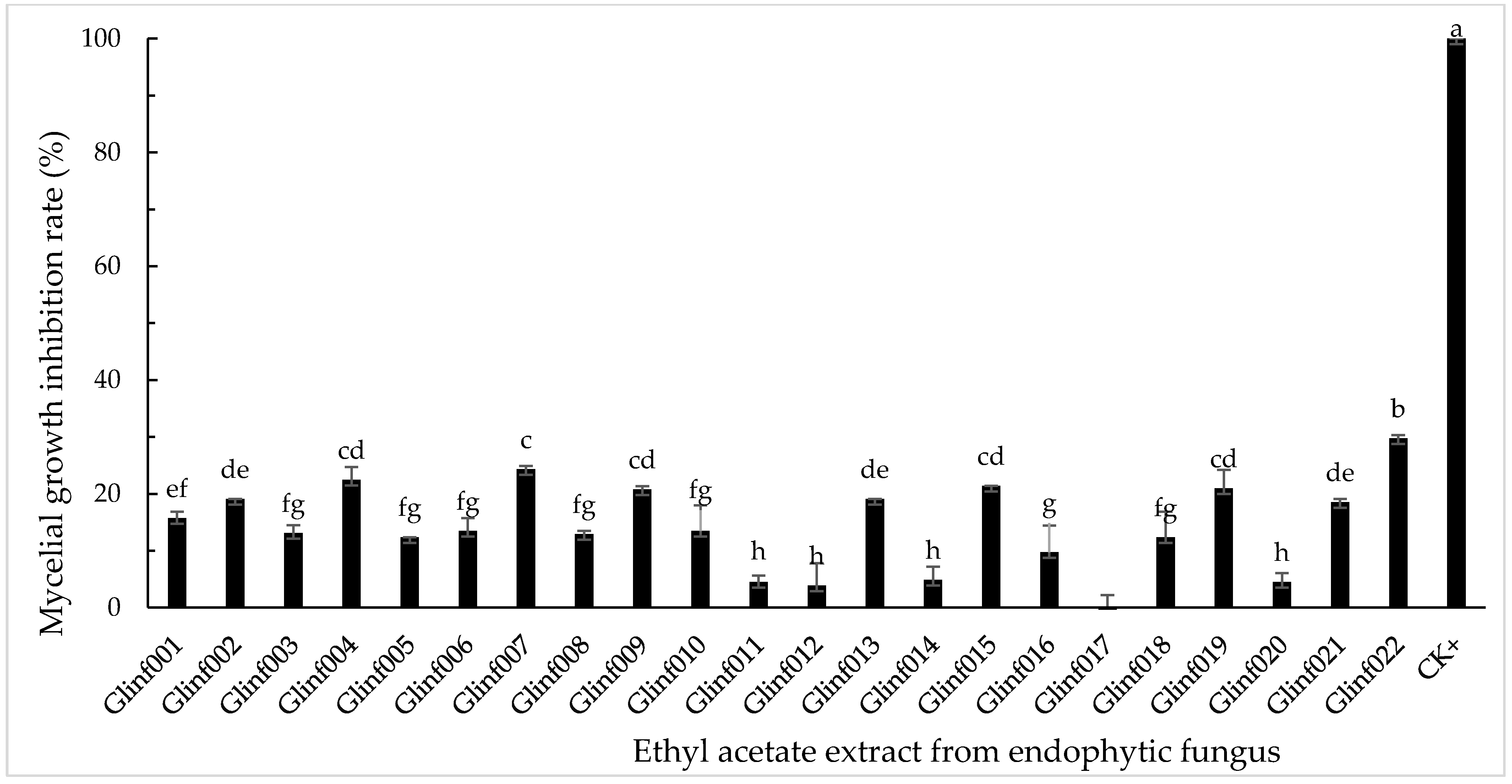
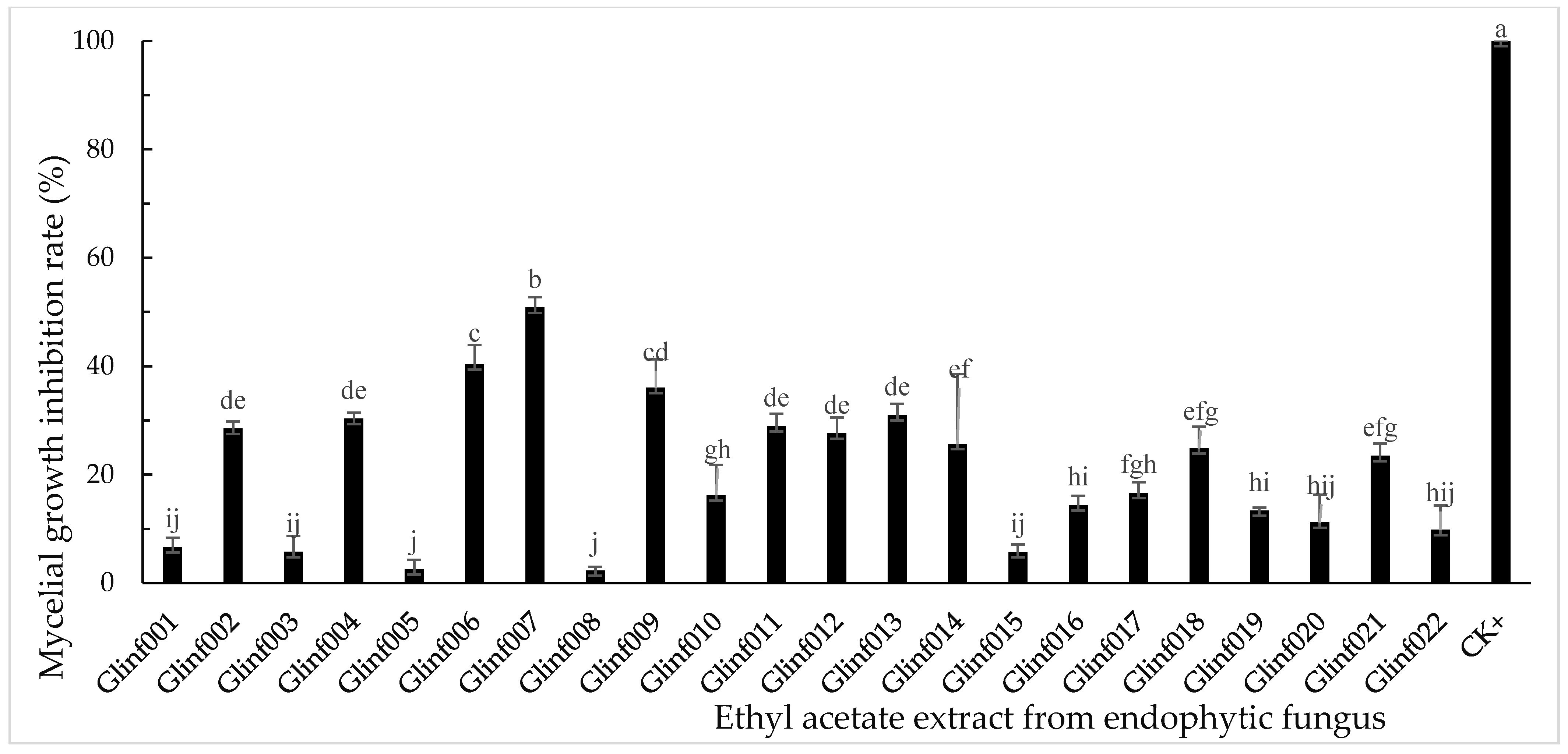
3.2. Inhibitory Activity of Endophytic Fungal Extracts on Mycelial Growth

The mycelial growth inhibition rates of the ethyl acetate (EtOAc) extracts at their concentration of 100 μg/mL in medium from 22 endophytic fungal strains were determined with mycelial growth inhibitions on rice sheath blight pathogen Rhizoctonia solani shown in Figure 2, on rice blast pathogen Magnaporthe oryzae shown in Figure 3, and on rice false smut pathogen Ustilaginoidea virens P1 and LN02 shown in Figure 4 and Figure 5, respectively. The reports of the statistical analysis using the one-way ANOVA method were shown in Tables S1–S4.
As shown in Figure 2, among all the test extracts, the crude EtOAc extract of Aspergillus keveii Glinf002 showed its strongest mycelial growth inhibition on R. solani with its inhibitory rate as 80.76%, which was equivalent to the positive control (carbendazim). In addition, the EtOAc extracts of Alternaria angustiovoidea Glinf007, Fusarium proliferatum Glinf009, and Botryotrichum murorum Glinf019 also showed significant mycelial growth inhibition on R. solani with their inhibitory rates as 69.30, 58.58, and 46.39%, respectively.
As shown in Figure 3, all the crude EtOAc extracts from endophytic fungi had weak inhibitions on M. oryzae mycelial growth. Only the crude EtOAc extract of Trichothecium roseum Glinf022 exhibited its relatively strongest inhibition on M. oryzae mycelial growth with its inhibitory rate as 29.78%.
Most of the crude extracts from endophytic fungi showed moderate inhibitions on U. virens P1 (Figure 4). Among them, the extract of Alternaria angustiovoidea Glinf007 had its relatively strongest inhibitory rate (50.82%) on U. virens P1. For the second U. virens strain LN02, most of the crude extracts showed significant inhibitions (Figure 5). Among them, the EtOAc extracts of Alternaria angustiovoidea Glinf007 and Fusarium fujikuroi Glinf011 exhibited the strongest inhibitions with their inhibitory rates as 71.10 and 71.49%, respectively.
This study described the culturable endophytic fungi from the roots of G. inflata. However, most endophytic fungi are currently considered unculturable [31], and further culture-independent techniques, such as the metagenomic library-based technique [32], should be completed to elucidate the unculturable fungi in G. inflata. Previous studies showed that endophytic microorganisms varied according to the location, age, and collection time of the host plants as well as the cultivation methods [33,34]. More detailed investigations should be necessary to increase the diversity of G. inflata materials (i.e., plant materials with different collection places and collection seasons, plant parts, and plant ages) as well as the cultivation media used. In addition, the fungal diversity of the soils around the roots as well as the fungi from the root surface are also worth further study. In this work, we only screened the ability of the endophytic fungal crude extracts for their potential to limit the growth of three rice fungal pathogens, other biological activities, such as cytotoxic, insecticidal, and antioxidant activities of the endophytic fungi from G. inflata, also should be studied in detail. A thin-layer chromatography (TLC) bioautography assay, which was considered as a valid method to qualitatively evaluate antimicrobial components, should be used to identify antimicrobial compounds in the crude extracts of fungi [35,36]. Furthermore, the bioactivity-guided discovery of antimicrobial compounds from endophytic fungi is also an effective strategy [37].

4. Conclusions

In this study, we reported the culturable endophytic fungi from the roots of the traditional medicinal plant G. inflata as well as the antifungal activities of ethyl acetate extracts from the distinct fungal isolates. A total of 99 endophytic fungal isolates were obtained. Eleven genera were identified among which Aspergillus, Alternaria, and Fusarium were dominant endophytes. Twenty-two representative isolates were selected and identified using both morphological and molecular methods. The crude extracts of Aspergillus keveii Glinf002, Alternaria angustiovoidea Glinf007, and Fusarium proliferatum Glinf009 showed obvious inhibitory activities against rice sheath blight pathogen R. solani, and the extracts of Alternaria angustiovoidea Glinf007 and Fusarium fujikuroi Glinf011 showed obvious inhibitory activities on rice false smut pathogen U. virens LN02. The results indicated that these endophytic fungi have great potential as producers of natural antimicrobial compounds. The following study will focus on the isolation of the antimicrobial compounds from these fungi as well as on their applications as biocontrol agents.

Supplementary Materials

The following are available online at https://www.mdpi.com/article/10.3390/microbiolres12040060/s1, Figure S1: The aerial parts (a), and roots (b) of Glycyrrhiza inflata collected in Xinjiang (86°34′27″ E, 42°03′32″ N), Northwest China, in 2 October 2020, Figure S2: The ITS1-5.8S-ITS2 partial sequences of 22 fungal isolates (i.e., Glinf001–Glinf022), Figure S3: Mycelial growth inhibition of the ethyl acetate extracts from 22 endophytic fungal strains on Rhizoctonia solani, Figure S4: Mycelial growth inhibition of the ethyl acetate extracts from 22 endophytic fungal strains on Magnaporthe oryzae, Figure S5: Mycelial growth inhibition of the ethyl acetate extracts from 22 endophytic fungal strains on Ustilaginoidea virens strain P1, Figure S6: Mycelial growth inhibition of the ethyl acetate extracts from 22 endophytic fungal strains on Ustilaginoidea virens strain LN02, Table S1: One-way ANOVA method was employed for statistical analysis of the inhibitions of 23 treatments on the pathogen Rhizoctonia solani. Nos. 1–22 were EtOAc extracts of endophytes and No. 23 was the positive control carbendazim, Table S2: One-way ANOVA method was employed for statistical analysis of the inhibitions of 23 treatments on the pathogen Magnaporthe oryzae. Nos. 1–22 were EtOAc extracts of endophytes and No. 23 was the positive control carbendazim, Table S3: One-way ANOVA method was employed for statistical analysis of the inhibitions of 23 treatments on the pathogen Villosicalva virens P1. Nos. 1–22 were EtOAc extracts of endophytes and No. 23 was the positive control carbendazim, Table S4: One-way ANOVA method was employed for statistical analysis of the inhibitions of 23 treatments on the pathogen Villosicalva virens LN-02. Nos. 1–22 were EtOAc extracts of endophytes and No. 23 was the positive control carbendazim.

Author Contributions

L.Z. and S.S. designed the study. S.S. collected the plant materials. G.G. and X.J. isolated and identified the plant endophytic fungi. G.G., X.J., W.W., P.L., S.Z., Z.Z. and R.Y. isolated the endophytic fungi and performed the antifungal activity evaluation of the extracts. G.G. and L.Z. interpreted the data and wrote the manuscript. D.L. reviewed and edited the manuscript. All authors have read and agreed to the published version of the manuscript.

Funding

This research was funded by the National Key R&D Program of China (2017YFD0201105).

Institutional Review Board Statement

Not applicable.

Informed Consent Statement

Not applicable.

Data Availability Statement

Not applicable.

Acknowledgments

The authors are grateful to Jizhao Zhang at Xinjiang Institute of Chinese Materia Medica and Ethnical Materia for plant material collection and authentication.

Conflicts of Interest

The authors declare no conflict of interest.

References

  1. Saikkonen, K.; Faeth, S.H.; Helander, M.; Sullivan, T.J. Fungal endophytes: A continuum of interactions with host plants. Annu. Rev. Ecol. Syst. 1998, 29, 319–343. [Google Scholar] [CrossRef]
  2. Hyde, K.D.; Soytong, K. The fungal endophyte dilemma. Fungal Divers. 2008, 33, 163–173. [Google Scholar]
  3. Bae, H.; Sicher, R.C.; Kim, M.S.; Kim, S.H.; Strem, M.D.; Melnick, R.L.; Bailey, B.A. The beneficial endophyte Trichoderma hamatum isolate DIS 219b promotes growth and delays the onset of the drought response in Theobroma cacao. J. Exp. Bot. 2009, 60, 3279–3295. [Google Scholar] [CrossRef] [PubMed]
  4. Deshmukh, S.K.; Verekar, S.A.; Bhave, S.V. Endophytic fungi: A reservoir of antibacterials. Front. Microbiol. 2015, 5, 715. [Google Scholar] [CrossRef] [PubMed] [Green Version]
  5. Nisa, H.; Kamili, A.N.; Nawchoo, I.A.; Shafi, S.; Shameem, N.; Bandh, S.A. Fungal endophytes as prolific source of phytochemicals and other bioactive natural products: A review. Microb. Pathog. 2015, 82, 50–59. [Google Scholar] [CrossRef] [PubMed]
  6. Venugopalan, A.; Srivastava, S. Endophytes as in vitro production platforms of high value plant secondary metabolites. Biotechnol. Adv. 2015, 33, 873–887. [Google Scholar] [CrossRef] [PubMed]
  7. Zhao, J.; Shan, T.; Mou, Y.; Zhou, L. Plant-derived bioactive compounds produced by endophytic fungi. Mini-Rev. Med. Chem. 2011, 11, 159–168. [Google Scholar] [CrossRef]
  8. Stierle, A.; Strobel, G.; Stierle, D. Taxol and taxane production by Taxomyces andreanae, an endophytic fungus of Pacific yew. Science 1993, 260, 214–216. [Google Scholar] [CrossRef]
  9. Tan, R.X.; Zou, W.X. Endophytes: A rich source of functional metabolites (1987 to 2000). Nat. Prod. Rep. 2001, 18, 448–459. [Google Scholar] [CrossRef]
  10. Li, J.; Zhao, J.; Xu, L.; Zhou, L.; Li, X.; Wang, J. Endophytic fungi from rhizomes of Paris polyphylla var. yunnanensis. World J. Microbiol. Biotechnol. 2008, 24, 733–737. [Google Scholar] [CrossRef]
  11. Xu, L.; Zhou, L.; Zhao, J.; Li, J.; Li, X.; Wang, J. Fungal endophytes from Dioscorea zingiberensis rhizomes and their antibacterial activity. Lett. Appl. Microbiol. 2008, 46, 68–72. [Google Scholar] [CrossRef]
  12. Huang, Y.; Zhao, J.; Zhou, L.; Wang, M.; Wang, J.; Li, X.; Chen, Q. Antimicrobial compounds from the endophytic fungus Fusarium sp. Ppf4 isolated from the medicinal plant Paris polyphylla var. yunnanensis. Nat. Prod. Commun. 2009, 4, 1455–1458. [Google Scholar] [CrossRef] [Green Version]
  13. Zhao, J.; Mou, Y.; Shan, T.; Li, Y.; Zhou, L.; Wang, M.; Wang, J. Antimicrobial metabolites from the endophytic fungus Pichia guilliermondii isolated from Paris polyphylla var. yunnanensis. Molecules 2010, 15, 7961–7970. [Google Scholar] [CrossRef] [Green Version]
  14. Lou, J.; Fu, L.; Luo, R.; Wang, X.; Luo, H.; Zhou, L. Endophytic fungi from medicinal herb Salvia miltiorrhiza Bunge and their antimicrobial activity. Afr. J. Microbiol. Res. 2013, 7, 5343–5349. [Google Scholar]
  15. Cui, J.-L.; Zhang, Y.-Y.; Vijayakumar, V.; Zhang, G.; Wang, M.-L.; Wang, J.-H. Secondary metabolite accumulation associates with ecological succession of endophytic fungi in Cynomorium songaricum Rupr. J. Agric. Food Chem. 2018, 66, 5499–5509. [Google Scholar] [CrossRef]
  16. Lai, D.; Mao, Z.; Zhou, Z.; Zhao, S.; Xue, M.; Dai, J.; Zhou, L.; Li, D. New chlamydosporol derivatives from the endophytic fungus Pleosporales sp. Sigrf05 and their cytotoxic and antimicrobial activities. Sci. Rep. 2020, 10, 8193. [Google Scholar] [CrossRef] [PubMed]
  17. Mao, Z.; Xue, M.; Gu, G.; Wang, W.; Li, D.; Lai, D.; Zhou, L. Lophiostomins A–D, new 3,4-dihydroisocoumarin derivatives from the endophytic fungus Lophiostoma sp. Sigrf10. RSC Adv. 2020, 10, 6985–6991. [Google Scholar] [CrossRef] [Green Version]
  18. Kao, T.-C.; Wu, C.-H.; Yen, G.-C. Bioactivity and potential health benefits of licorice. J. Agric. Food Chem. 2014, 62, 542–553. [Google Scholar] [CrossRef] [PubMed]
  19. Baltina, L.; Kndratenko, R. Glycyrrhizic acid derivatives as new antiviral and immune modulating agents. Curr. Bioact. Compd. 2021, 17, 41–58. [Google Scholar] [CrossRef]
  20. Zhang, Z.; Yang, L.; Hou, J.; Tian, S.; Liu, Y. Molecular mechanisms underlying the anticancer activities of licorice flafonoids. J. Ethnopharmacol. 2021, 267, 113635. [Google Scholar] [CrossRef] [PubMed]
  21. Simayi, Z.; Rozi, P.; Yang, X.; Ababaikeri, G.; Maimaitituoheti, W.; Bao, X.; Ma, S.; Askar, G.; Yadikar, N. Isolation, structural characterization, biological activity, and application of Glycyrrhiza polysaccharides: Systematic review. Int. J. Biol. Macromol. 2021, 183, 387–398. [Google Scholar] [CrossRef]
  22. Arora, P.; Wani, Z.A.; Ahmad, T.; Sultan, P.; Gupta, S.; Riyaz-Ul-Hssan, S. Community structure, spatial distribution, diversity and functional characterization of culturable endophytic fungi associated with Glycyrrhiza glabra L. Fungal Biol. 2019, 123, 373–383. [Google Scholar] [CrossRef]
  23. Nalli, Y.; Arora, P.; Khan, S.; Malik, F.; Riyaz-Ul-Hassan, S.; Gupta, V.; Ali, A. Isolation, structural modification of macrophin from endophytic fungus Phoma macrostoma and their cytotoxic potential. Med. Chem. Res. 2019, 28, 260–266. [Google Scholar] [CrossRef]
  24. Dang, H.; Zhang, T.; Wang, Z.; Li, G.; Zhao, W.; Lv, X.; Zhuang, L. Differences in the endophytic fungal community and effective ingredients in root of three Glycyrrhiza species in Xinjiang, China. PeerJ 2021, 9, e11047. [Google Scholar] [CrossRef] [PubMed]
  25. White, J.R.; Nagarajan, N.; Pop, M. Statistical methods for detecting differentially abundant features in clinical metagenomic samples. PLoS Comput. Biol. 2009, 5, e1000352. [Google Scholar] [CrossRef]
  26. Ainworth, G.C.; Sparrow, F.K.; Sussman, A.S. The Fungi, An Advanced Treatise. A Taxonomic Review with Keys Ascomycetes and Fungi Imperfecti; Academic Press: New York, NY, USA, 1973; Volume IV(A). [Google Scholar]
  27. Sun, S.; Lui, Q.; Han, L.; Ma, Q.; He, S.; Li, X.; Zhang, H.; Zhang, J.; Liu, X.; Wang, L. Identification and characterization of Fusarium proliferatum, a new species of fungi that cause fungal keratitis. Sci. Rep. 2018, 8, 4859. [Google Scholar] [CrossRef] [PubMed]
  28. Raeder, U.; Broda, P. Rapid preparation of DNA from filamentous fungi. Lett. Appl. Microbiol. 1985, 1, 17–20. [Google Scholar] [CrossRef]
  29. Quiroga, E.N.; Sampietro, A.R.; Vattuone, M.A. Screening antifungal activities of selected medicinal plants. J. Ethnopharmcol. 2001, 74, 89–96. [Google Scholar] [CrossRef]
  30. Raja, H.A.; Oberlier, N.H.; Stadler, M. Occasional comment: Fungal identification to species-level can be challenging. Phytochemistry 2021, 190, 112855. [Google Scholar] [CrossRef]
  31. Jiang, Z.-D.; An, Z. Bioactive fungal natural products through classic and biocombinatorial approaches. Stud. Nat. Prod. Chem. 2000, 22, 245–272. [Google Scholar]
  32. Singh, D.K.; Sharma, V.K.; Kumar, J.; Mishra, A.; Verma, S.K.; Sieber, T.N.; Kharwar, R.N. Diversity of endophytic mycobiota of tropical tree Tectona grandis Linn. f.: Spatiotemporal and tissue type effects. Sci. Rep. 2017, 7, 14. [Google Scholar]
  33. Hoffman, M.T.; Arnold, A.E. Geographic locality and host identity shape fungal endophyte communities in cupressaceous trees. Mycol. Res. 2008, 112, 331–344. [Google Scholar] [CrossRef]
  34. Persoh, D. Plant-associated fungal communities in the light of meta’omics. Fungal Divers. 2015, 75, 1–25. [Google Scholar] [CrossRef]
  35. Zhao, J.; Xu, L.; Huang, Y.; Zhou, L. Detection of antimicrobial components from extracts of the endophytic fungi associated with Paris polyphylla var. yunnanensis using TLC-bioautography-MTT assay. Nat. Prod. Res. Dev. 2008, 20, 28–32. [Google Scholar]
  36. Okusa, P.N.; Stevigny, C.; Devleeschouwer, M.; Duez, P. Optimization of the culture medium used for direct TLC–bioautography. Application to the detection of antimicrobial compounds from Cordia gilletii De Wild (Boraginaceae). J. Planar Chromat. 2010, 4, 245–249. [Google Scholar] [CrossRef]
  37. Shan, T.; Tian, J.; Wang, X.; Mou, Y.; Mao, Z.; Lai, D.; Dai, J.; Peng, Y.; Zhou, L.; Wang, M. Bioactive spiro-bisnaphthalenes from the endophytic fungus Berkleasmium sp. J. Nat. Prod. 2013, 77, 2151–2160. [Google Scholar] [CrossRef] [PubMed]
Figure 1. ITS rDNA phylogenetic relationship analyses of the fungal isolates from G. inflata. The numbers at the branches indicated the percentages of trees from 1000 bootstrap replication. The unrooted tree was generated using Clustal × 2.0 program using the Neighbor-Joining method. Phylogeny test was computed by MEGA 5.0. Additional taxa represent top BLASTn hits from the NCBI GenBank database.
Microbiolres 12 00060 g001
Figure 2. Mycelial growth inhibition of the ethyl acetate extracts from 22 endophytic fungal strains on R. solani. The error bars represent standard deviations from three independent samples. Different letters indicate statistically significant differences (p ≤ 0.05).
Microbiolres 12 00060 g002
Figure 3. Mycelial growth inhibition of the ethyl acetate extracts from 22 endophytic fungal strains on M. oryzae. The error bars represent standard deviations from three independent samples. Different letters indicate statistically significant differences (p ≤ 0.05).
Microbiolres 12 00060 g003
Figure 4. Mycelial growth inhibition of the ethyl acetate extracts from 22 endophytic fungal strains on U. virens P1. The error bars represent standard deviations from three independent samples. Different letters indicate statistically significant differences (p ≤ 0.05).
Microbiolres 12 00060 g004
Figure 5. Mycelial growth inhibition of the ethyl acetate extracts from 22 endophytic fungal strains on U. virens LN02. The error bars represent standard deviations from three independent samples. Different letters indicate statistically significant differences (p ≤ 0.05).
Microbiolres 12 00060 g005
Table 1. The endophytic fungi isolated from G. inflata roots.
Fungal
Isolate
CF
(%)
GenBank
Accession
Number
Closest Related Species
(Accession Number)
Identity
(%)
Macro- and Microscopic
Identification
Glinf00117MW563907Aspergillus ustus
(AY373874.1)
99Aspergillus ustus
Glinf0023MW563908Aspergillus keveii
(MN542353.1)
100Aspergillus keveii
Glinf0031MW563909Aspergillus germanicus
(MN650837.1)
100Aspergillus germanicus
Glinf00415MW563910Alternaria alternata
(MN615420.1)
100Alternaria alternata
Glinf0051MW563911Alternaria sp.
(MW220839.1)
99Alternaria sp.
Glinf0061MW563912Alternaria tenuissima
(MK616250.1)
100Alternaria tenuissima
Glinf0071MW563913Alternaria angustiovoidea
(MK910070.1)
99Alternaria angustiovoidea
Glinf0081MW563914Alternaria brassicae
(JF439450.1)
99Alternaria brassicae
Glinf00929MW563915Fusarium proliferatum
(MT560212.1)
100Fusarium proliferatum
Glinf0103MW563916Fusarium annulatum
(MT434005.1)
100Fusarium annulatum
Glinf0113MW563917Fusarium fujikuroi
(MT603302.1)
100Fusarium fujikuroi
Glinf0122MW563918Fusarium solani
(MN013858.1)
100Fusarium solani
Glinf0132MW563919Fusarium sp.
(MT252004.1)
99Fusarium sp.
Glinf0143MW563920Penicillium sizovae
(MN858522.1)
99Penicillium sizovae
Glinf0152MW563921Penicillium bilaiae
(LN901118.1)
99Penicillium bilaiae
Glinf0161MW563922Acrocalymma sp.
(KP170636)
98Acrocalymma sp.
Glinf0171MW563923Athelia bombacina
(MH201277.1)
99Athelia bombacina
Glinf0182MW563924Acremonium sclerotigenum
(MF077221.1)
99Acremonium sclerotigenum
Glinf0192MW563925Botryotrichum murorum
(MG228407.1)
100Botryotrichum murorum
Glinf0205MW563926Earliella scabrosa
(MF077243.1)
99Earliella scabrosa
Glinf0213MW563927Rosellinia sp.
(KU375680.1)
100Rosellinia sp.
Glinf0222MW563928Trichothecium roseum
(MN372207.1)
99Trichothecium roseum
Publisher’s Note: MDPI stays neutral with regard to jurisdictional claims in published maps and institutional affiliations.

