Article

Population-Based Distribution of Mycobacterium avium and Mycobacterium intracellulare in Japan

1
Division of Clinical Research, Fukujuji Hospital, Japan Anti-Tuberculosis Association, Tokyo 204-8522, Japan
2
Respiratory Disease Center, Fukujuji Hospital, Japan Anti-Tuberculosis Association, Tokyo 204-8522, Japan
3
Leprosy Research Center, National Institute of Infectious Diseases, Tokyo 189-0002, Japan
4
Center for Infectious Diseases and Infection Control, Keio University School of Medicine, Tokyo 160-8582, Japan
5
Graduate School of Biomedical Sciences, Nagasaki University, Nagasaki 852-8523, Japan
6
Department of Mycobacterium Reference and Research, The Research Institute of Tuberculosis, Japan Anti-Tuberculosis Association, Tokyo 204-0022, Japan
*
Author to whom correspondence should be addressed.
Microbiol. Res. 2021, 12(3), 739-743; https://doi.org/10.3390/microbiolres12030053
Submission received: 12 July 2021 / Revised: 16 September 2021 / Accepted: 17 September 2021 / Published: 18 September 2021

Abstract

:
This study aimed to clarify the population-based distributions of Mycobacterium avium and Mycobacterium intracellulare in Japan. We conducted a combined analysis of the national insurance claim and microbiological databases. The incidence rates of M. avium by province were similar throughout the country, with some exceptions, such as in Okinawa, probably because the bathing customs are different from those in mainland Japan. In contrast, M. intracellulare showed a gradual increase from the central part of the country to the southwestern region, with precise gradation, which may indicate infection sources in the natural environment. This study found that assessment of the infection route of M. intracellulare in the natural environment, which is similar to the distribution of M. intracellular patients, is warranted. In conclusion, improvement of the household environment could decrease the incidence of M. avium, while environmental countermeasures will be required to decrease the incidence of M. intracellulare.

1. Introduction

The prevalence of nontuberculous mycobacterial pulmonary disease (NTM-PD) has increased worldwide, and NTM-PD is recognized as a critical public health issue [1,2,3]. The incidence of NTM-PD has surpassed that of tuberculosis in industrialized countries, and surprisingly, even in countries with a high tuberculosis burden, it has been reported that the rate of NTM-PD cases misdiagnosed as multidrug-resistant cases or refractory tuberculosis is over 10% [4,5]. Given the interrelation between tuberculosis and NTM-PD, the decreasing tuberculosis incidence and the increasing elderly population, NTM-PD prevalence is expected to increase in many countries in the future.
Several population-based epidemiological studies that have been conducted in recent years in Japan have overlooked the situation of NTM-PD in the country [6,7,8,9]. The incidence of NTM-PD surpassed that of culture-positive tuberculosis for the first time in 2014, and the proportion of Mycobacterium avium complex (MAC) cases was revealed to be higher, reaching as high as 90%, than those in other countries [7,8].
The MAC includes more than ten species, and M. avium and M. intracellulare are the most prevalent species [10]. Several reports have revealed that there are clinical differences between the species [11,12,13]. Interestingly, the proportion of M. avium in the MAC gradually increases from southwestern regions to the northeastern part of the country. Conversely, the proportion of M. intracellulare is highest in the southwestern part of the country [8].
Several previous studies isolated M. avium from the interior household environment, mostly from the bathroom [14,15,16]. Moreover, the hot-water faucet of the bath system was the main location [17]. As bathing in bathtubs is a common practice in Japan, we speculated that the incidence of M. avium should be evenly distributed throughout the whole country, while that of M. intracellulare would be uneven. Accordingly, the present study aimed to clarify the population-based distributions of M. avium and M. intracellulare.

2. Materials and Methods

The incidence of MAC-related disease from 2011–2013, which was obtained using national health insurance claim data [9], and the proportion of M. avium among MAC cases in 2014, obtained via a questionnaire survey [7], were analyzed. The treatment incidence rate was calculated for 344 medical regions, and the proportional data were stratified by eight geographical regions: Kyushu–Okinawa, Chugoku, Shikoku, Kinki, Chubu, Kanto, Tohoku and Hokkaido (from southwest to northeast). The treatment incidences for M. avium and M. intracellulare were calculated by multiplying these data and are shown for each medical region.

3. Results

The treatment incidences of M. avium and M. intracellulare are shown in Figure 1. The treatment rate of M. intracellulare was higher in the southwestern regions than in the other regions. Conversely, the incidence rate of M. avium, though its proportions were higher in northeastern regions than in other regions, showed a relatively even distribution, except in Hokkaido, Kyushu and Okinawa, which included several low-incidence locations. The incidence rate of M. avium was higher than that of M. intracellulare in most regions except for the Kyushu and Okinawa regions.

4. Discussion

In the present study, we found that M. avium was relatively evenly distributed throughout the country, consistent with our speculation that the bath system is the primary infection source. Recently, an environmental survey focusing on the bathrooms of healthy volunteers was conducted across the country, and the researchers found that the overall detection rate of M. avium was approximately 16% [18]. Therefore, if people with risk factors, such as middle-aged to elderly females with low body weight, underlying lung disease or immune-suppressed status, used a contaminated bathroom, they could be infected by M. avium. The data support this hypothesis, as the detection rates were lowest in the Kyushu and Hokkaido regions, where the incidence rates were also low, except in Tohoku [18,19].
Interestingly, Okinawa had a low incidence of M. avium. Nagano et al. conducted an epidemiological study in Okinawa and found that the proportion of M. avium was lower than those of the M. abscessus complex and M. intracellulare [19]. Okinawa is located in the extreme southern part of Japan, and it has a subtropical climate. Due to this, residents do not soak in a bath, rather, they use a shower. The data indicated that the bath system was the source of M. avium and suggest that taking baths is associated with a higher risk of infection than showering.
The incidence rate of M. intracellulare was higher in the southwestern part of the country than in the other areas of the country. As M. intracellulare has not been isolated from the interior household environment, it is assumed that the infection source is the natural environment. Morimoto and Koh reviewed NTM epidemiology in Asia [20]. Interestingly, the proportion of M. intracellulare in NTM-PD in Nagasaki prefecture, located in the west side of the Kyushu region, was similar to that in South Korea [21,22,23,24]. Furthermore, a systematic review of Chinese epidemiological analyses showed that the proportion of M. intracellulare was higher in the northern part of the country near Korea; conversely, M. avium was highest in the southern part of the country [25]. These data indicate that regions with a higher proportion of M. intracellulare throughout Japan, South Korea and China exist, and there are natural environmental niches shared among these regions. The incidence rate of M. intracellulare gradually decreased in a northeasterly direction in Japan. This might indicate that the environmental niche is less prevalent in the northeastern regions. Searching for factors with such characteristics could identify environmental niches. We speculate that dusts, such as yellow dust or soot originating from China, as well as rain and soil, are potential sources, and air and soil sampling should be conducted.
There are some limitations of this study. First, although the present study used data originating from the two databases, ideally, primary population-based mycobacterial data should be analyzed. Second, the present analysis used the data from approximately ten years ago. The results may not show the current epidemiological situation. Therefore, it is crucial to construct a surveillance system to grasp the fluctuation of NTM epidemiological status (which may be impacted by the recent Olympic and Paralympic games) for future research. Lastly, we could not analyze the data of M. intracellulare subsp. chimaera, which could have been identified as M. intracellulare. However, because the proportion of the subspecies is quite low in Japan and M. intracellulare has never been isolated from either interior household environments or humans in previous studies, the influence of M. intracellulare subsp. M. chimaera is likely minimal [26].

5. Conclusions

M. avium evenly exists throughout the country, most likely due to soaking customs, with some exceptions, such as in Okinawa. As M. avium is a major cause of NTM-PD in Japan, improvement of the household environment is an essential strategy to reduce the risk of infection. The infection source of M. intracellulare in the natural environment should be identified by combining data from Japan, China and South Korea.

Author Contributions

K.M. designed and wrote the manuscript. S.M. supervised the project and assisted writing the manuscript. All authors reviewed the current version of the manuscript. M.A. and N.H. critically reviewed the manuscript. All authors have read and agreed to the published version of the manuscript.

Funding

This study was supported by the Japan Agency for Medical Research and Development (JP20fk0108129, 21fk0108129h0802), and the Kurozumi Medical Foundation.

Institutional Review Board Statement

The study was conducted according to the guidelines of the Declaration of Helsinki, and approved by the Institutional Review Board of Fukujuji Hospital (#14046, 3/12/2020).

Informed Consent Statement

Not applicable.

Data Availability Statement

The datasets supporting the conclusion of this study were provided by the government, and the original data are not available due to the assigned time and regulation.

Acknowledgments

We would like to thank Izumi, who contributed to this work.

Conflicts of Interest

There is no conflict of interest to disclose with respect to this study.

References

  1. Prevots, D.R.; Marras, T.K. Epidemiology of human pulmonary infection with nontuberculous mycobacteria: A review. Clin. Chest. Med. 2015, 36, 13–34. [Google Scholar] [CrossRef] [Green Version]
  2. Thomson, R.; Donnan, E.; Konstantinos, A. Notification of Nontuberculous Mycobacteria: An Australian Perspective. Ann. Am. Thorac. Soc. 2017, 14, 318–323. [Google Scholar] [CrossRef] [PubMed]
  3. Winthrop, K.L.; Henkle, E.; Walker, A.; Cassidy, M.; Hedberg, K.; Schafer, S. On the Reportability of Nontuberculous Mycobacterial Disease to Public Health Authorities. Ann. Am. Thorac. Soc. 2017, 14, 314–317. [Google Scholar] [CrossRef] [PubMed]
  4. Raju, R.M.; Raju, S.M.; Zhao, Y.; Rubin, E.J. Leveraging Advances in Tuberculosis Diagnosis and Treatment to Address Nontuberculous Mycobacterial Disease. Emerg. Infect. Dis. 2016, 22, 365–369. [Google Scholar] [CrossRef] [PubMed] [Green Version]
  5. Xu, K.; Bi, S.; Ji, Z.; Hu, H.; Hu, F.; Zheng, B.; Wang, B.; Ren, J.; Yang, S.; Deng, M.; et al. Distinguishing nontuberculous mycobacteria from multidrug-resistant Mycobacterium tuberculosis, China. Emerg. Infect. Dis. 2014, 20, 1060–1062. [Google Scholar] [CrossRef] [PubMed]
  6. Morimoto, K.; Iwai, K.; Uchimura, K.; Okumura, M.; Yoshiyama, T.; Yoshimori, K.; Ogata, H.; Kurashima, A.; Gemma, A.; Kudoh, S. A steady increase in nontuberculous mycobacteriosis mortality and estimated prevalence in Japan. Ann. Am. Thorac. Soc. 2014, 11, 1–8. [Google Scholar] [CrossRef] [Green Version]
  7. Namkoong, H.; Kurashima, A.; Morimoto, K.; Hoshino, Y.; Hasegawa, N.; Ato, M.; Mitarai, S. Epidemiology of Pulmonary Nontuberculous Mycobacterial Disease, Japan. Emerg. Infect. Dis. 2016, 22, 1116–1117. [Google Scholar] [CrossRef] [Green Version]
  8. Morimoto, K.; Hasegawa, N.; Izumi, K.; Hamkoong, H.; Uchimura, K.; Yoshiyama, T.; Hoshino, Y.; Kurashima, A.; Sokunaga, J.; Shibuya, S.; et al. A laboratory-based analysis of nontuberculous mycobacterial lung disease in Japan from 2012 to 2013. Ann. Am. Thorac. Soc. 2016, 14. [Google Scholar] [CrossRef]
  9. Izumi, K.; Morimoto, K.; Hasegawa, N.; Uchimura, K.; Kawatsu, L.; Ato, M.; Mitarai, S. Epidemiology of Adults and Children Treated for Nontuberculous Mycobacterial Pulmonary Disease in Japan. Ann. Am. Thorac. Soc. 2019, 16, 341–347. [Google Scholar] [CrossRef]
  10. Daley, C.L.; Iaccarino, J.M.; Lange, C.; Cambau, E.; Wallace, R.J., Jr.; Andrejek, C.; Bottger, E.C.; Brozek, J.; Griffith, D.E.; Guglielmetti, L.; et al. Treatment of Nontuberculous Mycobacterial Pulmonary Disease: An Official ATS/ERS/ESCMID/IDSA Clinical Practice Guideline. Clinical infectious diseases: An official publication of the Infectious Diseases Society of America. Clin. Inf. Dis. 2020, 71, 905–913. [Google Scholar] [CrossRef]
  11. Jhun, B.W.; Moon, S.M.; Jeon, K.; Kwon, O.J.; Yoo, H.; Carriere, K.C.; Huh, H.J.; Lee, N.Y.; Shin, S.J.; Daley, C.L.; et al. Prognostic factors associated with long-term mortality in 1445 patients with nontuberculous mycobacterial pulmonary disease: A 15-year follow-up study. Eur. Respir. J. 2020, 55. [Google Scholar] [CrossRef]
  12. Cho, E.H.; Huh, H.J.; Song, D.J.; Moon, S.M.; Lee, S.-H.; Shin, S.Y.; Kim, C.K.; Ki, C.-S.; Koh, W.-J.; Lee, N.Y. Differences in drug susceptibility pattern between Mycobacterium avium and Mycobacterium intracellulare isolated in respiratory specimens. Journal of infection and chemotherapy. Off. J. Jpn. Soc. Chemother. 2018, 24, 315–318. [Google Scholar]
  13. Koh, W.J.; Jeong, B.H.; Jeon, K.; Lee, N.Y.; Lee, K.S.; Woo, S.Y.; Shin, S.J.; Kwon, O.J. Clinical significance of the differentiation between Mycobacterium avium and Mycobacterium intracellulare in M avium complex lung disease. Chest 2012, 142, 1482–1488. [Google Scholar] [CrossRef] [Green Version]
  14. Nishiuchi, Y.; Maekura, R.; Kitada, S.; Tamaru, A.; Taguri, T.; Kira, Y.; Hiraga, T.; Hirotani, A.; Yoshimura, K.; Miki, M.; et al. The recovery of Mycobacterium avium-intracellulare complex (MAC) from the residential bathrooms of patients with pulmonary MAC. Clinical infectious diseases: An official publication of the Infectious Diseases Society of America. Clin. Inf. Dis. 2007, 45, 347–351. [Google Scholar] [CrossRef] [Green Version]
  15. Nishiuchi, Y.; Iwamoto, T.; Maruyama, F. Infection Sources of a Common Non-tuberculous Mycobacterial Pathogen, Mycobacterium avium Complex. Front. Med. 2017, 4, 27. [Google Scholar] [CrossRef] [PubMed] [Green Version]
  16. Morimoto, K.; Aono, A.; Murase, Y.; Sekizuka, T.; Kurashima, A.; Takaki, A.; Sasaki, Y.; Igarashi, Y.; Chikamatsu, K.; Goto, H.; et al. Prevention of aerosol isolation of nontuberculous mycobacterium from the patient′s bathroom. ERJ Open Res. 2018, 4. [Google Scholar] [CrossRef] [PubMed] [Green Version]
  17. Nishiuchi, Y.; Tamaru, A.; Kitada, S.; Taguri, T.; Matsomoto, S.; Tateishi, Y.; Yoshimura, M.; Ozeki, Y.; Matsumoto, N.; Ogara, H.; et al. Mycobacterium avium complex organisms predominantly colonize in the bathtub inlets of patients′ bathrooms. Jpn. J. Inf. Dis. 2009, 62, 182–186. [Google Scholar]
  18. Arikawa, K.; Ichijo, T.; Nakajima, S.; Nishiuchi, Y.; Yano, H.; Tamaru, A.; Yoshida, S.; Maruyama, F.; Ota, A.; Nasu, M.; et al. Genetic relatedness of Mycobacterium avium subsp. hominissuis isolates from bathrooms of healthy volunteers, rivers, and soils in Japan with human clinical isolates from different geographical areas. Infection, genetics and evolution. J. Mol. Epidemiol. Evol. Gene. Inf. Dis. 2019, 74, 103923. [Google Scholar] [CrossRef] [PubMed]
  19. Nagano, H.; Kinjo, T.; Nei, Y.; Yamashiro, S.; Fujita, J.; Kishaba, T. Causative species of nontuberculous mycobacterial lung disease and comparative investigation on clinical features of Mycobacterium abscessus complex disease: A retrospective analysis for two major hospitals in a subtropical region of Japan. PLoS ONE 2017, 12, e0186826. [Google Scholar] [CrossRef] [Green Version]
  20. Morimoto, K.; Koh, W. Asia, Global Epidemiology of NTM Disease (Except Northern America); Humana Press: Totowa, NJ, USA, 2019. [Google Scholar]
  21. Kim, H.S.; Lee, Y.; Lee, S.; Kim, Y.A.; Sun, Y.K. Recent trends in clinically significant nontuberculous Mycobacteria isolates at a Korean general hospital. Ann. Lab. Med. 2014, 34, 56–59. [Google Scholar] [CrossRef] [Green Version]
  22. Koh, W.J.; Chang, B.; Jeong, B.H.; Jeon, K.; Kim, S.-Y.; Lee, N.Y.; Ki, C.-S.; Kwon, O.J. Increasing Recovery of Nontuberculous Mycobacteria from Respiratory Specimens over a 10-Year Period in a Tertiary Referral Hospital in South Korea. Tuberc. Respir. Dis. 2013, 75, 199–204. [Google Scholar] [CrossRef] [PubMed]
  23. Koh, W.J.; Kwon, O.J.; Jeon, K.; Kim, T.S.; Lee, K.S.; Park, Y.K.; Bai, G.H. Clinical significance of nontuberculous mycobacteria isolated from respiratory specimens in Korea. Chest 2006, 129, 341–348. [Google Scholar] [CrossRef] [PubMed] [Green Version]
  24. Ide, S.; Nakamura, S.; Yamamoto, Y.; Kohno, Y.; Fukuda, Y.; Ikeda, H.; Sasaki, E.; Yanagihara, K.; Higashiyama, Y.; Hashiguchi, K.; et al. Epidemiology and clinical features of pulmonary nontuberculous mycobacteriosis in Nagasaki, Japan. PLoS ONE 2015, 10, e0128304. [Google Scholar]
  25. Yu, X.; Liu, P.; Liu, G.; Zhao, L.; Hu, Y.; Wai, G.; Luo, J.; Huang, H. The prevalence of non-tuberculous mycobacterial infections in mainland China: Systematic review and meta-analysis. J. Inf. 2016, 73, 558–567. [Google Scholar] [CrossRef]
  26. Yoshida, S.; Tomita, M. Pathogenic characterization of Mycobacterium intracellulare sequevar and susceptibility to clarithromycin. J. Jpn. Soc. Clin. Microbiol. 2013, 23, 29–38. [Google Scholar]
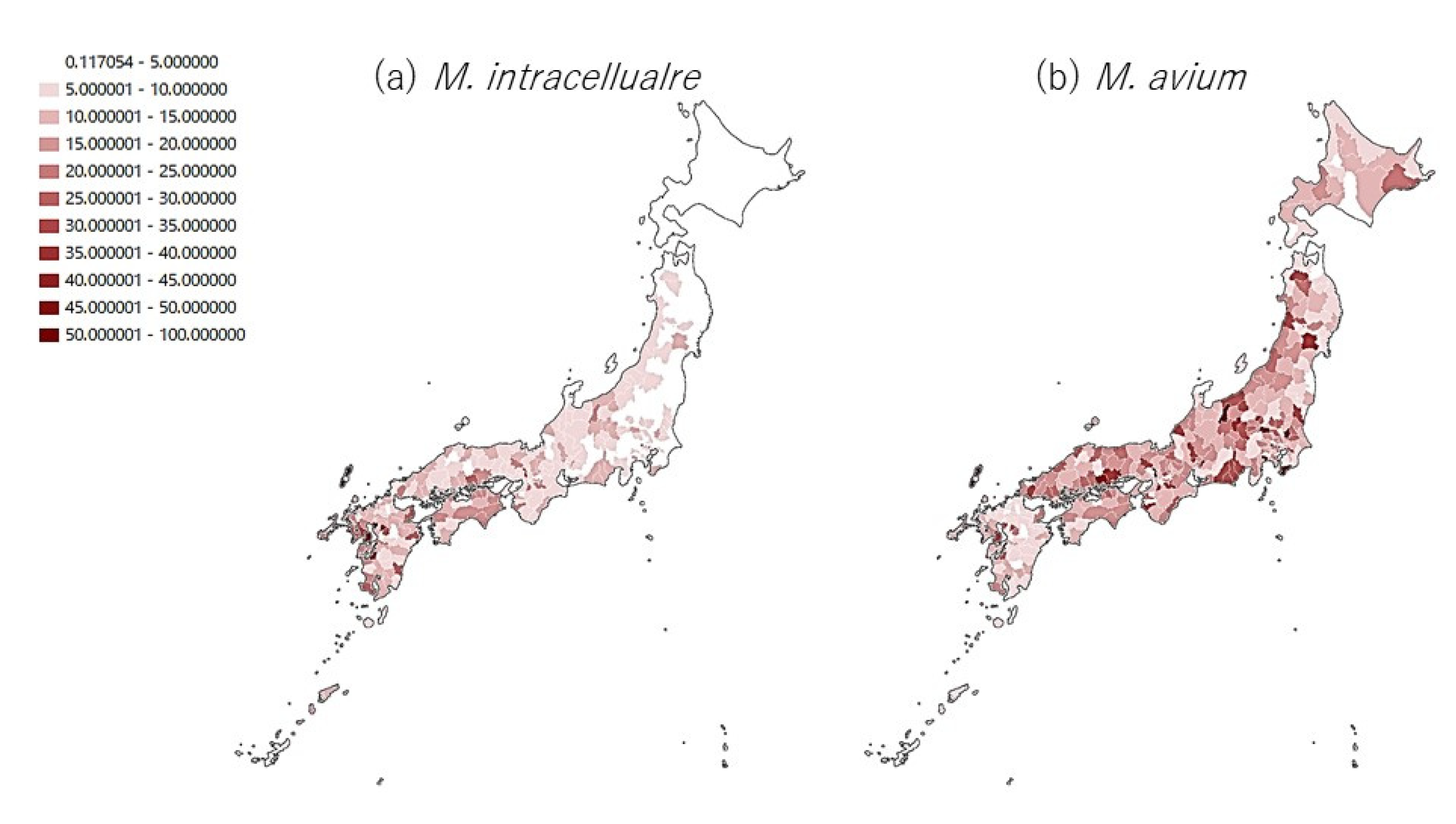
Figure 1. Incidence rates of NTM-PD cases (per 100,000 population) in 344 medical regions 2011–2013.
Microbiolres 12 00053 g001
Publisher’s Note: MDPI stays neutral with regard to jurisdictional claims in published maps and institutional affiliations.