Review

Manipulating the Microbiome: An Alternative Treatment for Bile Acid Diarrhoea

by
Evette B. M. Hillman
1,2,*,
Sjoerd Rijpkema
1,
Danielle Carson
1,
Ramesh P. Arasaradnam
3,4,
Elizabeth M. H. Wellington
2 and
Gregory C. A. Amos
1,*
1
Division of Bacteriology, National Institute for Biological Standards and Control, South Mimms, Potters Bar, Hertfordshire EN6 3QG, UK
2
School of Life Sciences, The University of Warwick, Coventry CV4 7AL, UK
3
Warwick Medical School, The University of Warwick, Coventry CV4 7AL, UK
4
University Hospital Coventry & Warwickshire, Coventry CV2 2DX, UK
*
Authors to whom correspondence should be addressed.
Microbiol. Res. 2021, 12(2), 335-353; https://doi.org/10.3390/microbiolres12020023
Submission received: 19 February 2021 / Revised: 2 April 2021 / Accepted: 3 April 2021 / Published: 9 April 2021

Abstract

:
Bile acid diarrhoea (BAD) is a widespread gastrointestinal disease that is often misdiagnosed as irritable bowel syndrome and is estimated to affect 1% of the United Kingdom (UK) population alone. BAD is associated with excessive bile acid synthesis secondary to a gastrointestinal or idiopathic disorder (also known as primary BAD). Current licensed treatment in the UK has undesirable effects and has been the same since BAD was first discovered in the 1960s. Bacteria are essential in transforming primary bile acids into secondary bile acids. The profile of an individual’s bile acid pool is central in bile acid homeostasis as bile acids regulate their own synthesis. Therefore, microbiome dysbiosis incurred through changes in diet, stress levels and the introduction of antibiotics may contribute to or be the cause of primary BAD. This literature review focuses on primary BAD, providing an overview of bile acid metabolism, the role of the human gut microbiome in BAD and the potential options for therapeutic intervention in primary BAD through manipulation of the microbiome.

1. Introduction

Bile acid diarrhoea (BAD) (previously referred to as bile acid malabsorption or bile salt malabsorption) is a condition that predominantly presents as chronic watery diarrhoea as well as bloating and abdominal pain [1]. A recent survey by Walters et al. (2020) observed that 86% of participants experienced frequent bowel movements, often more than 6 times a day [2]. In a different survey, Bannaga et al. (2017) reported that many BAD patients often experience tiredness, low energy levels and a lack of concentration [3]. Sufferers reported the negative impact BAD had on their social life and ability to work (which could lead to the loss of employment), and over 90% also felt a negative impact on their mental health. Although this condition is not life-threatening, the survey revealed that depression, isolation, helplessness and low self-esteem are very common among patients living with BAD, often leaving individuals housebound [3]. While the exact figures are not known, it is estimated that up to 1% of the population in the United Kingdom (UK) have primary BAD [4]. If non-primary BAD patients are included, this potentially results in an excess of 700,000 individuals in the UK alone [5]. Smith et al. (2000) estimated that over one-third of patients diagnosed with irritable bowel syndrome (IBS) actually suffer from BAD [6]. Consequently, BAD could have a wide-reaching economic impact due to absence from work through sickness and the cost of continued care.
BAD has several different causes that fall under one of three types [7]:
  • Type I, when the ileum is damaged due to inflammation or surgical removal.
  • Type II is idiopathic and is also known as “primary BAD”.
  • Type III, results from another disease or condition (such as gallbladder removal).
In 2009, Walters et al. proposed that primary BAD (type II) was not a result of reduced or impaired absorption but in fact resulted from unregulated bile acid synthesis [8]. Studies demonstrated that patients with BAD had significantly higher levels of 7α-hydroxy-4-cholesten-3-one ((C4), a bile acid precursor) and significantly lower levels of fibroblast growth factor 19 ((FGF19), a hormone that down regulates bile acid synthesis) compared with healthy individuals [8]. Similarly, the authors also showed that patients with secondary BAD (type I and III) had lower levels of FGF19 in their blood serum compared with that in healthy subjects [8,9,10]. This excessive bile acid synthesis results in the insufficient absorption of bile acids in the ileum, causing the increased transit of bile acids into the colon. High levels of bile acid in the colon trigger a rise in water secretion to combat irritation, which prevents stool from properly forming in the lumen, leading to diarrhoea. Several studies have reported the importance of bile acids in a number of gut related diseases [11]. Despite these observations, the cause of low serum levels of FGF19 and the consequent excess bile acid synthesis remains widely unknown.
The selenium-75-homocholic acid taurine (SeHCAT) scan (a nuclear medicine test) is considered the gold standard for diagnosing BAD, as it has a high sensitivity and specificity [12]. The parameters for diagnosing BAD are: less than 5% bile acid retention indicates severe BAD; between 5 to 10% bile acid retention indicates moderate BAD; and between 10 to 15% bile acid retention indicates mild BAD [4]. Despite the SeHCAT scan giving the highest diagnostic accuracy for BAD, it is not widely used or licensed in many countries (including the United States of America (USA)). In the absence of a SeHCAT scan, doctors often prescribe bile acid sequestrant medication as the response to treatment can provide a diagnosis. Bile acid sequestrants bind to excess bile acids with a high affinity preventing irritation in the large intestine. Frequently treatment for BAD is not administrated because it is often misdiagnosed as diarrhoea predominant IBS (IBS-D) due to the similarity in clinical presentation of both diseases. Indeed, Bannaga et al. (2017) reported that 44% of respondents had experienced symptoms for more than five years before diagnosis, with a range of between one and thirty years [3]. IBS is a multifactorial spectrum disease, which can be triggered by both environmental and genetic factors, although the exact cause of IBS is widely unknown [13]. There is no accepted diagnostic test for IBS; however, as mentioned, there is one for identifying BAD. IBS is not to be confused with inflammatory bowel disease (IBD), which presents as chronic inflammation on the GI tract.
Current treatment for BAD includes diet changes and medication; however, these only treat the symptoms and do not treat the cause of the disease. Physicians advise a diet with a reduced fat intake of 40 g of fat per day [14]. To date, there have been no randomised trials that have evaluated different dietary regimes with BAD sufferers. Instead, observational studies seem to suggest that low fat diets are beneficial. Low fat diets are thought to result in reduced bile acid synthesis and could possibly alter the microbiome, all leading to less severe symptoms in some cases [15,16,17]. Current treatment guidelines for BAD have not significantly improved since the 1960s, and sequestrants can have adverse effects such as constipation, bloating, nausea and abdominal pain (especially when combined with other medications) [18,19]. The pathophysiology of BAD is not fully understood, and, to date, a clinically effective cure remains elusive. This review will cover the biochemical structure of bile acids, the regulation of bile acid synthesis and the presentation of recent evidence towards the manipulation of the microbiome as a possible alternative treatment for primary BAD.

2. Structure and Function of Bile Acids

Bile acids are cyclo-pentane phenanthrene sterol molecules and are the main component of bile. The chemical composition of bile varies between individuals but usually consists of 61% bile acids, 12% fatty acids, 9% cholesterol, 7% proteins and small amounts of bilirubin [20]. Bile acids are amphiphilic molecules that possess a hydrophobic aliphatic steroid nucleus with 24 carbon atoms, which are attached to hydrophilic hydroxyl groups and an acidic side chain. The steroidal core of bile acids consists of a saturated four ring skeleton, consisting of three six-membered rings (A, B, and C) and one five-membered ring (D) (Figure 1).
The covalently bonded hydroxyl groups are in one of either two configurations: α, a downwards orientation; or β, an upwards orientation. Almost all bile acids have an α-hydroxyl group at position C-3, which is derived from the parent molecule cholesterol, which has the 3-hydroxyl group in the β orientation. There are two types of bile acids: primary and secondary bile acid. Primary bile acids in humans can be further divided between 3α, 7α, 12α-trihydroxy-5β-cholan-24-oic acid (or cholic) acid (CA) and 3α, 7α-dihydroxy-5β-cholan-24-oic acid (or chenodeoxycholic) acid (CDCA). These bile acids differ only at position C-12, wherein CA possess a hydroxyl (OH) group whereas CDCA does not. Primary bile acids are often conjugated via N-acyl amidation with an amino acid, as 95% of these conjugated bile acids are bound to either glycine or taurine at position C-24 (see Figure 1) [21]. Of this 95%, approximately 75% are conjugated to glycine and 25% are conjugated to taurine. Unconjugated bile acids have a pKa value range of 5–6. The addition of an amino acid lowers the pKa value of glycine and taurine conjugated bile acids from 5–6 to 4–5 and 1–2, respectively. At the physiological pH of 7.0–7.7, bile consists predominantly of conjugated bile acids, which are ionised and are alternatively known as bile salts or strong bile acids [22]. Bile salts are more water-soluble and are able to fulfil one of their physiologic functions of solubilising dietary lipids due to their increased amphipathic nature. The added solubility prevents the passive reabsorption of conjugated bile acid in the small intestine, causing a reliance on active reabsorption via the apical sodium–bile acid transporter (ASBT) in the ileum.
Bile acids have a number of functions in the gastrointestinal (GI) tract: conjugated bile acids at the right concentration form micelles, which function as biological detergents through the emulsification and solubilisation of fats in the small intestine [23]; they are essential for the absorption of dietary lipids (including lipoproteins and fat-soluble vitamins such as vitamin D); they act as a signalling molecules that regulate their own expression; they play a key role in immune homeostasis and the metabolism of glucose [24,25]. Due to their amphipathic detergent nature, bile acids are toxic to microbes and act as antimicrobial agents through membrane damage, thus helping to maintain homeostasis between bile acid-tolerant and bile acid-sensitive bacteria in the GI tract [26]. Even at sub-micellar concentrations, bile acids can alter bacterial membrane lipid composition [27]. In one of the few studies that has investigated the bile acid profile in SeHCAT diagnosed BAD patients, Sagar et al. (2020) reported an increase in the concentrations of bile acids (particularly CDCA) in the faeces and serum of BAD patients compared to those in IBS-D patients [28].

3. The Enterohepatic Circulation and Primary Bile Acid Synthesis

Bile acids are synthesised primarily by hepatocytes [29,30]. On average, the gallbladder secretes 12–18 g of bile acids per day into the intestine, typically following meals. However, in healthy individuals there are approximately 4–6 g of bile acids present in the intestine at any given time due to the enterohepatic circulation (EHC) of bile acids [31]. Approximately 95% of all intestinal bile acids are reabsorbed by passive diffusion in the intestine or, in the case of conjugated bile acids, by active transport. Following reabsorption from the intestine, bile acids are recycled by the liver, with any unabsorbed bile acids remaining in the intestine (~400–800 mg, or 5%) and passing to the colon for excretion [32].
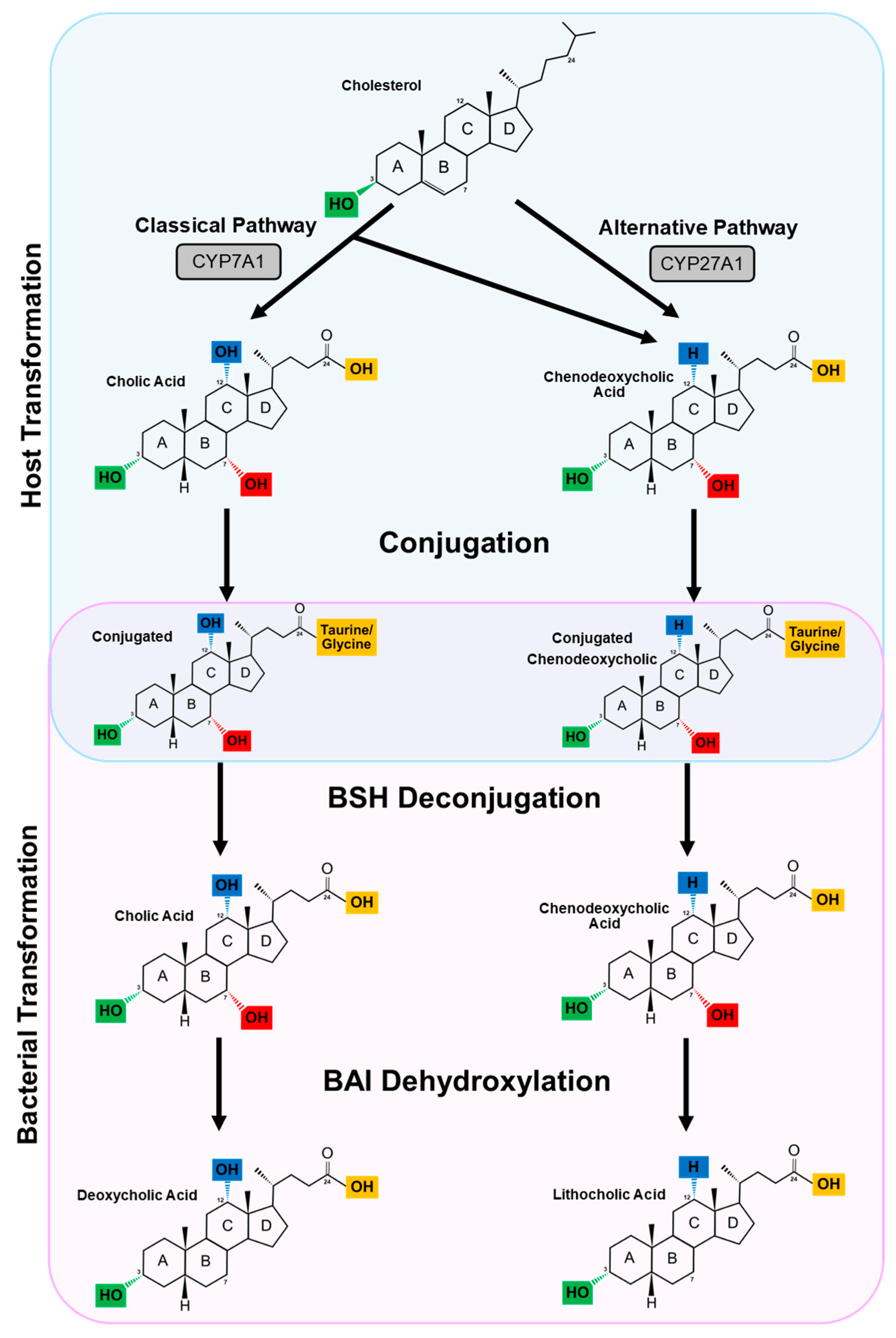
Cholesterol is an obligatory precursor of bile acids (Figure 2). The conversion involves several enzymatic processes; hepatocytes express all 17 enzymes necessary for modifying the steroid core, removing the side chain and conjugating with an amino acid, resulting in the synthesis of primary conjugated bile acids [33]. This occurs through one of two pathways: the classic (neutral) pathway (which occurs ∼90% of the time in humans and ∼75% of the time in mice) or the alternative (acidic) pathway (occurs ∼10% of the time in humans and ∼25% of the time in mice) [33,34,35]. The principal rate-limiting enzyme (cholesterol-7α-hydroxylase of the cytochrome P450 family 7 subfamily A member 1 (CYP7A1)) is required for bile acid biosynthesis in the classic pathway. CYP7A1 catalyses the hydroxylation of cholesterol at C-7, creating 7α-hydroxycholesterol [36]. There are several intermediates in the production of bile acids, including C4, which is sometimes used as a biomarker in diagnosing BAD [37,38]. Sterol-27-hydroxylase (CYP27A1, cytochrome P450 family 27 subfamily A member 1) is a mitochondrial enzyme that is responsible for facilitating bile acid synthesis in the alternative pathway. However, unlike in the classic pathway, the hydroxylation of cholesterol takes place at C-27 [34,35].

4. Secondary Bile Acids

In both the small intestine and in the colon (particularly in the colon), unabsorbed bile acids are deconjugated and further transformed into secondary bile acids by bacteria (Figure 2). Bacterial enzymes can manipulate bile acids at four main sites on the sterol core: C-3, C-7, C-12 and C-24 (see Figure 1 and Figure 2). C-24 is the site for deconjugation (the removal of the amino acid), C-7 is the site for dehydroxylation (the removal of hydroxyl group) or dehydrogenation (the removal of a hydrogen atom) and C-3, C-7 and C-12 are the sites for dehydrogenation. Bacterial dehydrogenation is a reversible process, as dehydrogenases catalyse the oxidative reactions in both directions.

4.1. Deconjugation

The conversion of conjugated bile acids by bacteria in the GI tract requires the bile salt hydrolase (BSH, EC 3.5.1.24) enzyme, a member of the choloylglycine hydrolase family that catalyse the hydrolysis of glycine or taurine from the C-24 position of the sterol core, resulting in deconjugated bile acids [39,40]. The active site of BSHs share several conserved amino acids (such as cysteine 2 (Cys2)) [41]. However, bacterial BSHs have widely different catalytic efficiencies as well as substrate specificities. In addition, bacterial BSHs differ in: genetic properties (gene organisation and regulation); physical properties (composition, size and weight of each subunit); and kinetic properties (Vmax, optimum pH’s and optimum temperatures). BSHs have been observed in numerous microbial genera and are predominantly described among Gram-positive bacteria, including Lactobacillus [42] and Bifidobacterium [43] which are often used as probiotics, as well as Enterococcus [44] and Clostridium spp. [45,46]. Furthermore, BSHs have been identified in Gram-negative bacteria such as and Bacteroides [45]. Recently, Song et al. (2019) elucidated the abundance of bacterial genes that encode BSHs among 11 human populations from geographically diverse origins [47]. After screening the Human Microbiome Project (HMP) database, they reported a total of 26% of bacteria strains carrying genes that encode BSHs [47]. These genes were widely distributed across 117 bacterial genera of the human microbiota, with the majority (71%) observed in Bacteroides, Blautia, Eubacterium, Clostridium and Roseburia [47]. How enzyme activity and specificity differ across strains is yet to be characterised; however, only Lactobacillus possessed the variant of BSH (BSH-T3) which has the highest known enzymatic activity amongst BSHs [47]. Studies suggest that BSHs have a preference for glycine conjugated bile acids, with the accumulation of taurine conjugated bile acids observed following the reduction of BSH-expressing bacteria in humans [48,49]. There are a number of benefits that BSHs confer to the bacterial host, including providing a source of nutrients [43,50,51]. Previously, it was proposed that deconjugation could protect bacteria from bile salt toxicity. However, recent studies have demonstrated unconjugated bile acids possess more potent antibacterial activity on Staphylococcus aureus, Ruminococcus bromii, Bacteriodes fragilis, Lactobacillus paracasei, Bifidobacterium longum, and Clostridium scindens than conjugated bile acids [52,53]. This is potentially due to unconjugated bile acids passively crossing membranes, resulting in intra-cellular toxicity while conjugated bile acids require a transporter and are thus more limited in their activity.

4.2. Dehydroxylation and Dehydrogenation

Bacterial genes that regulate the dehydroxylation and dehydrogenation of bile acids are encoded in the bile acid-inducible (bai) operon [41]. The first step of dehydroxylation requires baiB that encodes bile acid CoA ligase, resulting in a bile acid-CoA conjugate. Other key genes include baiE (which encodes 7α-de-hydratase) and baiA (which encodes 3α-hydroxysteroid dehydrogenases), resulting in the removal of a 7α hydroxyl group and a 3α hydrogen atom, respectively.

4.2.1. Dehydroxylation

Only a limited number of bacterial species possess the bai operon required for the 7α-dehydroxylation of bile acid [54]. In the human intestine, 7α-dehydroxylation results in secondary bile acids: deoxycholic acid ((DCA); 3α,12α-dihydroxy-5β-cholan-24-oic acid) and lithocholic acid ((LCA); 3α-hydroxy-5β-cholan-24-oic acid) from CA and CDCA, respectively. DCA and LCA are the most abundant bile acids found in human faeces, making up 34% and 29%, respectively [41]. Therefore, 7α-dehydroxylation is very quantitatively important in the bio-transformation of bile acid in the human colon. Genes encoding for dehydroxylation are most commonly observed in Clostridium spp. [55]. Recently, Eubacterium spp. have also been characterised to rapidly produce DCA from CDCA, and metagenomic approaches have observed the presence of 7α-dehydroxylation in Ruminococcaceae, Lachnospiraceae, and Peptostreptococcaceae [56,57]. Unlike bile acid C-3, C-7 and C-12 dehydrogenation, 7α/β-dehydroxylation is restricted to free, unconjugated bile acids. Therefore, the removal of the glycine or taurine via a BSH is an essential prerequisite for 7α/β-dehydroxylation by the intestinal microbiota [58,59].

4.2.2. Dehydrogenation

Dehydrogenation by hydroxysteroid dehydrogenases (HSDHs) leads to the oxidation, reduction and, often, epimerisation of the C-3-, C-7- and C-12-hydroxy groups of bile acids by 3α-HSDHs, 7α/β-HSDHs and 12α/β-HSDHs, respectively. Epimerisation is the reversible change in stereochemistry from an α → β or β → α configuration with the generation of a stable carbonyl group bile acid intermediate. 7-keto-lithocholic acid (7-keto-LCA) is the intermediate for the oxidation and reduction at C-7 of epimers CDCA and ursodeoxycholic acid (UDCA). UDCA epimerisation requires the combined effort of two position-specific, stereochemically distinct HSDHs. Such HSDHs can originate from the same or different species demonstrating the importance of synergy in the microbiome. For example, the presence of both 7α- and 7β-HSDH in C. absonum allows for epimerisation by a single bacterial species [60,61,62]. Meanwhile, epimerisation has been demonstrated in co-cultures of intestinal bacteria, with one possessing 7α-HSDH and the other 7β-HSDH [63]. To date, it has been difficult to quantify the degree of α/β-dehydroxylation that occurs in the gut due to the competing and reversible α/β-dehydrogenation of bile acids. Oxidation of the bile acid hydroxyl groups is believed to generate NADH, an important enzyme in making ATP that can be used by the bacterium [64].
3α-HSDHs have been detected in a number of Clostridium species, including C. perfringens [65], C. scindens [66] and C. hiranonis [67]. They have also been observed in other intestinal bacterial species, such as Eubacterium lentum [68] and Peptostreptococcus productus [69]. 3β-HSDH activity has been described in species of Clostridium, Rumminococcus and Peptostreptococcus productus [69,70,71,72]. Most 3α/β-HSDH genes described to date are expressed constitutively, though 3α/β-HSDHs from C. scindens and C. hiranonis are induced by CA and CDCA. [41].
7α/β-HSDHs are thought to be constitutively produced and are common among gut microbiota species [41]. Many intestinal clostridia express genes responsible for the production of both 7α- and 7β-HSDHs that epimerise the 7α/β-hydroxy group [62,73]. Several species with 7α/β-HSDH activity (such as Ba. fragilis [74], C. sordellii [75], C. perfringens [76] and C. innocuum [71]) also contain genes that encode for BSHs. The physiological significance of the bile acid 7α-hydroxy group epimerisation to gut bacteria is thought to decrease the toxicity of CDCA, as UDCA is less hydrophobic [77,78].
12α/β-HSDHs have been detected mainly among members of the genus Clostridium. 12α-HSDHs has been observed in C. perfringens [65], C. leptum [79], Clostridium group P [80] and E. lentum [68]. 12β-HSDHs have been reported in C. tertium, C. difficile, and C. paraputrificum [81]. Like 3 and 7α/β-HSDHs, most 12α/β-HSDHs are constitutively produced; however, the 12β-HSDH produced by C. paraputrificum is induced by 12-carboxyl-bile acid substrates [82].

4.3. H+ Bile Acid Transporter

7α/β-dehydroxylation activity and, therefore, the transformation of primary bile acids into the most commonly found secondary bile acids in human stool, requires the transport of free primary bile acids into the bacterial cell. This is carried out by the proton-dependent bile acid transporter coded for this purpose by baiG, which facilitates the transport of the unconjugated primary bile acids CA and CDCA but not of the secondary bile acids DCA and LCA [83]. Bacterial species capable of primary bile acids 7α-dehydroxylation possess baiG [84].

5. Bile Acid Receptors and Downstream Targets

Bile acids can act as signalling molecules which bind to receptors that are involved in several vital signalling networks [24,25]. Bile acids are not only ligands for the farnesoid X receptor (FXR), which is the nuclear receptor superfamily 1 group H member 4 (NR1H4), but also other members of the nuclear receptor superfamily, such as the androstane receptor (NR1H3 or CAR), the pregnane X receptor (NR1H2 or PXR) and the vitamin D receptor (NR1H1 or VDR) [85]. In addition, secondary bile acids interact with a number of membrane receptors, including the G-protein-coupled bile acid receptor 1 (GP-BAR1; also known as TGR5). Of these receptors, FXR and TGR5 have been implicated as therapeutic targets to treat BAD.

5.1. Bile Acid Regulate Their Own Synthesis

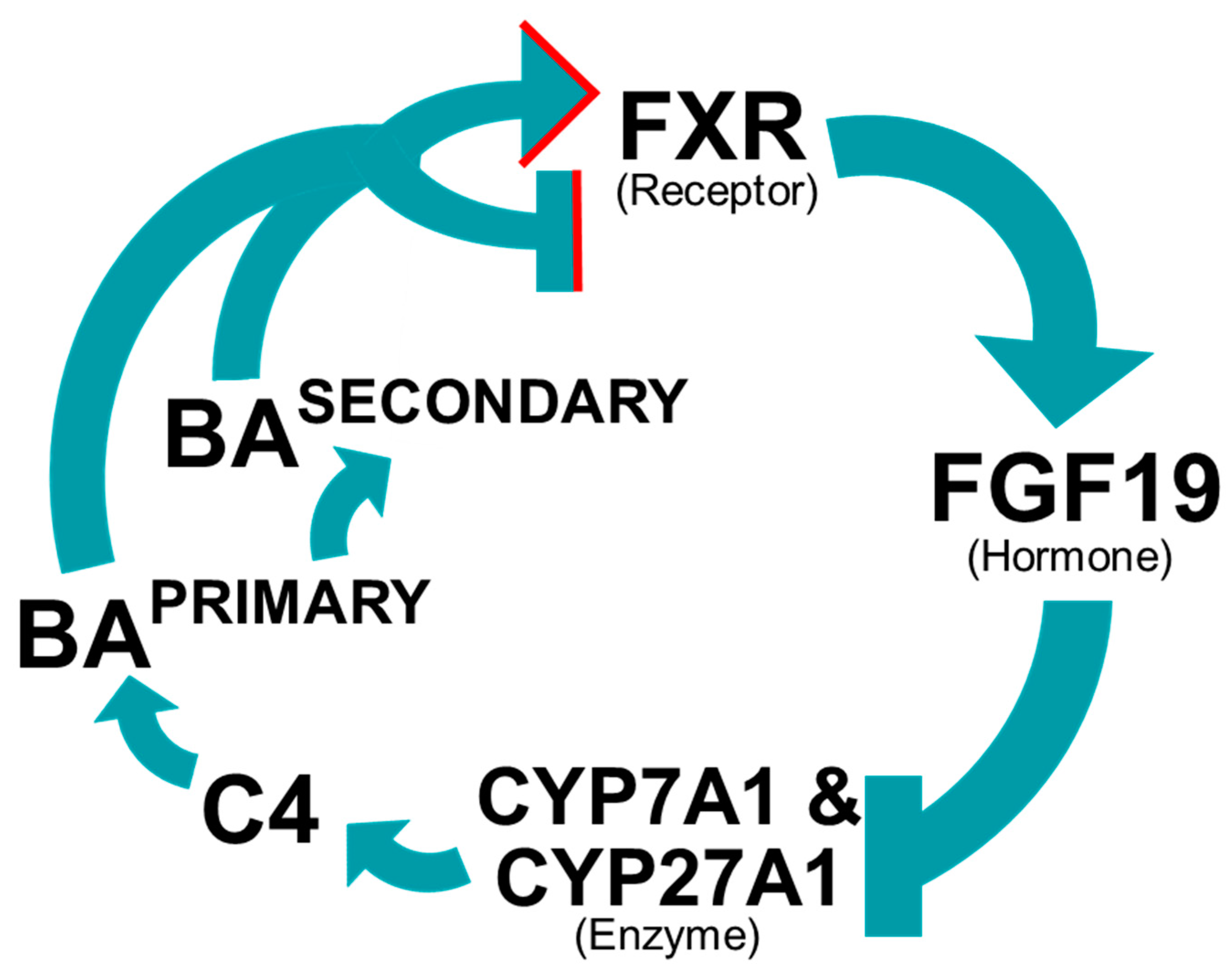
The enterohepatic circulation of bile acids is tightly regulated. FGF19 is a hormone produced in the liver and ileum that suppresses the expression of the rate-limiting enzymes CYP7A1 and CYP27A1 that are central to bile acid synthesis (see Figure 3). FXR is a bile acid sensor, which is responsible for the regulation of FGF19 and is primarily expressed by the ileum, liver, adrenal glands and kidneys [86,87]. Bile acids (particularly the primary bile acids CDCA and CA) are natural agonists for FXR. Consequently, in healthy individuals, high concentrations of bile acid activate FXR, which upregulates FGF19, thus inhibiting bile acid production through the hepatic coreceptors FGF receptor 4 and β klotho [30]. A range of bile acids can bind to FXR, with differing binding affinities and ligand efficacies [88,89,90]. Wang et al. (1999) reported that UDCA did not activate FXR [90]; instead, Mueller et al. (2015) revealed that it inhibited FXR activation [91], in agreement with the in silico FXR binding data [92]. Recently, Zhao et al. (2020) observed a number of bile acids (glycochenodeoxycholic acid (GCDCA), glycoursodeoxycholic acid (GUDCA), glycocholic acid (GCA), UDCA and 7-ketodeoxycholic acid (7-KDCA)) isolated from human stool could efficiently antagonise CDCA-induced FXR activation, while taurine conjugated bile acids (specifically tauroursodeoxycholic acid (TUDCA) and taurine-conjugated-mix) can attenuate cellular FGF19 expression in vitro [93]. UDCA was one of the bile acids that antagonised CDCA-induced FXR activation and was shown to be significantly elevated in the stool of BAD and IBS-D patients with elevated total faecal bile acids [28,93]. Indeed, studies comparing the bile acid pool in vegan and omnivore diets report significantly more bile acids (including UDCA, glycine and taurine conjugated bile acids) in participants who consumed meat [94]. These observations indicate that diet may influence the bile acid pool. By elucidating the antagonist effects of bile acids, this data could explain the increase in bile acid synthesis in BAD patients. Furthermore, the antagonist effects of bile acids on FXR are well documented in mice, which express fibroblast growth factor 15 (FGF15), the murine homologue of FGF19 [95].

5.2. Farnesoid X Receptor

FXR is a transcriptional activator which binds to form a hetero-dimeric complex with a retinoid X receptor (RXR) before binding to specific DNA response elements upstream of promoters [96]. The target DNA sequences are composed of two inverted repeats separated by one nucleotide. Most ligands that bind to RXR differ from those of FXR; for example, the natural agonist 9-cis-retinoic acid [97]. Interestingly, the ligands for both receptors can activate the transcriptional hetero-dimer, thus regulating bile acid synthesis. One of several FXR target genes is the small heterodimer partner (shp). SHP is a nuclear receptor that lacks a DNA binding domain; instead, it forms a hetero-dimer with both liver receptor homolog 1 and liver X receptor α. This complex, like FGF19, can inhibit CYP7A1 expression, thus inhibiting the classical pathway of bile acid synthesis [98]. To prevent toxification in the liver caused by bile acid, FXR negatively regulates bile acid uptake by hepatocytes via sodium taurocholate co-transporting polypeptide (NTCP) [99]. In addition, FXR activation represses human ASBT in the intestinal tissue. This is important, as ASBT transports bile acid from the lumen into enterocytes and induces basolateral organic solute transporters (OST) α and β that transport bile acids from enterocytes into sera [100].

5.3. Diet1 Protein

Vergnes et al. (2013) demonstrated that protein Diet1 regulates the production of FGF15/19 in mouse and human intestinal enterocytes [101]. The authors also observed that Diet1-deficient mice had reduced Fgf15 mRNA and protein levels in ileum, as well as increased plasma levels of bile acids particularly free and taurine conjugated CA [101]. Diet1 does not regulate transcription of FGF15/19, as it lacks the required DNA binding domains. It is hypothesised to physically interact with the protein [101]. Mutations in diet1 lead to differences in FGF15/19 expression and, subsequently, could contribute to variations in bile acid metabolism that may play a role in the development of BAD.

5.4. TGR5 Receptor

Present in the plasma membrane, TGR5 is expressed in many tissues, such as the gallbladder, spleen, liver and intestines, including the ileum and the colon [102,103,104]. Both primary and secondary conjugated and unconjugated bile acids constitute natural ligands for TGR5 [103]. LCA are thought to be the most potent natural ligand followed by DCA [103,104]. Inhibitory motor neurons form part of the enteric nervous system (ENS) and are responsible for inducing relaxations of intestinal smooth muscle. Studies have demonstrated that the activation of TGR5 by secondary bile acid agonist DCA and LCA inhibits spontaneous, phasic contractions of isolated colonic muscle in vitro [105]. In the intestinal tract, the G-protein-coupled receptor TGR5 is predominantly expressed by enteric neurons and enterochromaffin cells. Within the myenteric plexus, TGR5 is localised in ~50% of all neurons, mainly in the majority of inhibitory motor neurons and descending interneurons, which express nitric oxide (NO) synthase. NO is an important neurotransmitter and is also an anti-microbial agent important in host defence against enteric infection in murines and humans [105]. Moreover, in mice gavaged with DCA, delays in both gastric emptying and transit through the small intestine were observed [105]. Alemi et al. (2013) revealed that the activation of TGR5 by bile acids stimulated the release of neurotransmitters 5-hydroxytryptamine and the calcitonin gene-related peptide, which are both involved in peristalsis and cause involuntary movements of muscles cells of the intestine [106]. Furthermore, tgr5 (-/-) mice did not experience bile acid activated peristalsis or neurotransmitter release and had a whole-gut transit time 1.4-fold slower compared to that of wildtype. Thus, TGR5 activation by bile acids results in the BAD-like symptoms of diarrhoea and abdominal pain.

6. Microbiome

The term microbiome was first coined in the 1988 book Fungi in Biological Control Systems [107] and is currently broadly recognised as “the entire community of micro-organisms (bacteria, archaea, lower and higher eukaryotes, and viruses), their genomes and the surrounding environmental conditions” [108]. The metazoan body hosts several microbiomes; a medley of cross species physico-chemical and chemical interactions that have a marked effect on the balance between host health and disease [109,110]. The role of the microbiome in health is one of the most researched areas of the 21st century, due in part to advances in microbial detection [111]. The human microbiome is separated and named depending on the site of the body they occupy, with the most well studied being the intestinal, skin, oral and vaginal microbiomes. This review focuses on the gut microbiome. The human microbiota consists mostly of bacterial cells (around 3.8 × 10¹³) which have a marginally higher order compared to the number of human cells (around 3.0 × 10¹³) [112]. Therefore, the ratio is around 1:1 which is quite different to previous values which estimated the human microbiome contained 10 times as many cells as the host [113]. Notably, this highlights an issue of microbiome research which will benefit from standardisation of analytical techniques [114].

6.1. Gut Microbiome

The intestinal tract is home to 70–80% of the human body’s immune cells, which are shaped by the microbiome [115,116]. Moreover, around 200–600 million neurons capable of acting independently from the sympathetic and parasympathetic nervous systems (known as ENS) are embedded in the lining of the human GI tract [117,118]. Along the intestinal tract, the microbiota varies according to the anatomical region and organ they colonise. This is due to variations in pH, O2 concentration, digesta flow rates (also known as trans time), nutrients and substrate availability [119]. Given that colon harbours the densest number of microbes out of all the organs [112]; the gut microbiome is central in the development and function of physical and mental, health and disease.
Tierney et al. (2019) analysed 3500 faecal samples and detected the presence of 22 million microbial genes in the gut microbiome [120]. As the gut microbiota co-evolved with the host over thousands of years, these genes often have mutual and added benefits. Microbial genes play a critical role in facilitating host metabolism and the regulation of host physiology (such as in the removal, synthesis, and absorption of many fundamental nutrients and metabolites, including bile acids, lipids, amino acids, vitamins and short-chain fatty acids). Commensal bacteria have also been shown to protect the host against pathogens by consuming available nutrients, and often produce bacteriocin to inhibiting pathogenic growth and subsequent epithelium colonisation. Deficiencies or dysbiosis in the gut microbiome has been implicated in a wide range of diseases such as inflammatory bowel disease, celiac disease, colorectal cancer and BAD [121]. Disease is thought to result from imbalance in the host–microbe interaction when there is either a depletion or an increase of microbiota-derived metabolites such as short-chain fatty acids or bile acids. These metabolites interact with the host signalling and immune system to contribute, exacerbate or cause disease.

6.2. Microbiome and Bile Acid Profile in Mice

By observing and altering the microbiome as well as the bile acid pool in mice, scientists have demonstrated that intestinal bacterial homeostasis is maintained by bile acids and vice versa [95,122,123,124,125,126]. Studies in germ free (GF) mice revealed that the bile acid pool consists almost exclusively of primary conjugated bile acids as compared to that of conventionally raised mice [95,122]. These results verify that, in the absence of bacteria, deconjugation and secondary bile acid formation is greatly perturbed. Bile acids α and β muricholic acids (αMCA and βMCA) are derived from CDCA and, unlike those in humans, primary bile acids are almost exclusively conjugated with taurine in mice. The antagonistic effects of primary bile acids, taurine conjugated αMCA (TαMCA), taurine conjugated βMCA (TβMCA), glycine conjugated βMCA (GβMCA) and UDCA have been observed for FXR [91,95,127]. As bile acid deconjugation requires bacteria, Sayin et al. (2013) reported an accumulation of TβMCA in GF mice, reduced FXR signalling and a subsequently increased bile acid synthesis [95]. Interestingly, changes in the microbial community structure of mice following a gavage with FXR antagonists TαMCA, TβMCA and GβMCA, have been observed [123,124]. These observations confirm the importance of the microbiome in shaping the bile acid pool and these infer that manipulating the microbiome can be used as a treatment for BAD.

6.3. The Gut Microbiome and Bile Acid Diarrhoea

There have been very few studies to date on the gut microbiota of BAD patients (who were confirmed by SeHCAT scan). Sagar et al. (2020) revealed that the gut microbiome of BAD patients had evidence of dysbiosis, with significantly reduced bacterial diversity compared with IBS-D [28]. Indeed, despite IBS-D being a distinct and separate disease, BAD is often misdiagnosed as IBS-D; thus, much of the presumed knowledge on the microbiome of BAD patients is inferred from IBS-D studies. This is problematic when trying to evaluate an evidence-based role for the microbiome in BAD and highlights the importance of accurate clinical metadata in microbiome studies. For IBS-D, Duboc et al. (2012) observed reduced concentrations of the probiotic Bifidobacterium and the anti-inflammatory commensal C. leptum bacteria in the faecal microbiota of IBS-D patients [128]. In addition, the authors observed an overall decrease in the abundance of bacteria possessing the bsh gene required for bile acid transformation [128]. Consistent with these findings, a recent study demonstrated that individuals suffering from severe IBS-D, with elevated levels of bile acids in their stool, possessed a significantly reduced abundance of bsh and an elevated abundance of genes encoded by the bai operon [93]. Taken together, this suggests a lower level of bile acid transformation by the gut microbiota for individuals with IBS-D and potentially BAD, as the authors of these papers did not rule out BAD. Interestingly, although functional metagenomic analysis of the microbiota of BAD patients has yet to be performed, metabolomic analysis revealed that BAD patients have a lower proportion of secondary bile acids as compared to IBS-D patients [28]. Elevated concentrations of bile acids are linked to accelerated colonic transit time which could subsequently result in reduced time for bile acid transformation to secondary bile acids by gut bacteria, thus exacerbating BAD symptoms. Indeed, the gut microbiome and its subsequent transformation of bile acids is central to maintaining the balance between FXR and TGR5 receptor activation via the transformation of primary bile acids to secondary bile acids. Therefore, manipulating the gut microbiome could be a permanent treatment for BAD. Moreover, changes in the gut microbiome due to external factors such as stress, antibiotics or diet could be the primary cause of BAD, which would explain why treatment to date by bile sequestrants does not alleviate the underpinning cause of the disease.

7. Manipulating the Microbiome

The multifaceted gut microbiota is dominated by bacteria from 12 different phyla that predominantly belong to four major phyla (Firmicutes, Bacteroidetes, Actinobacteria, and Proteobacteria) [129,130,131]. With millions of genes and subsequent proteins performing beneficial and vital functions, it is clear why many scientists view the microbiome as a symbiotic organ. Diet, the environment, stress and drugs such as antibiotics can all disrupt homeostasis of the gut by disrupting the diversity of the microbiome [132,133,134]. Manipulating the microbiome can result in favourable changes in the structure and function of the intestinal microbiota and their metabolites. Indeed, microbiome dysbiosis and subsequent disease symptoms have been effectively treated by altering the microbiome [135,136]. The following therapies could be used to treat BAD as bacteria are responsible for transforming primary bile acids into secondary bile acids which have been shown to antagonise FXR activation and possibly increase bile acid synthesis, resulting in BAD symptoms [91,92,93].
To date, faecal microbiota transplantation (FMT) is the only licensed microbiome based therapeutic. FMT involves the transfer of stool from a healthy donor to the recipient and it is used to treat diarrhoea; this practice in different forms has been utilised for over 1700 years [137]. Clinical studies, particularly on patients with a C. difficile infection and inflammatory bowel disease, have demonstrated this to result in very high success rates [135,136,138]. Recently El-Salhy et al. (2020) published promising results on the efficacy of FMT in IBS patients [139]. A therapeutic gain was seen in 89.1% of donors that received 60 g FMT from a single super-donor. However, Lahtinen et al. (2020) concluded a single transplant via colonoscopy is not a recommended treatment for IBS. It is likely that FMT differs in its mode of action for different diseases. Consequently, FMT therapy lacks control of dose and duration of exposure to the microbiome or its constituents.
Following on from the success of FMT, live bio-therapeutic products (LBPs) are being trialled for the treatment of disease by modulating the microbiome [140]. “An LBP is a manufactured biological product that contains live organisms used for prevention, treatment, or cure of a disease or condition in humans” [140]. LBPs are sometimes termed next-generation probiotics, as they function in much the same way as classical probiotics but are rationally designed based on microbiome composition rather than containing specific probiotic strains which use specific food groups as substrates. Examples of bacterial species commonly observed in food-based probiotics include the Lactobacillus genus (L. acidophilus, L. plantarum and L. paracasei), the Bifidobacterium genus (B. breve, B. longum and B. infantis) and Streptococcus thermophilus [141,142].
A study employing sterile faecal filtrate transfer (FFT) showed interesting results by successfully treating recurrent C. difficile infection in all five patients, suggesting that live microbes are not required to restore healthy stool habits [143]. The authors of this study concluded that bacterial components, metabolites (such as bile acids) and/or bacteriophages are vital in a successful FMT [143]. Prebiotics are bacterial substrates (such as fructo-oligosaccharides and galacto-oligosaccharides) that can be used in parallel with probiotics and LBPs (synbiotics) or as a standalone strategy, and are another way of altering the microbiome.

8. Conclusions

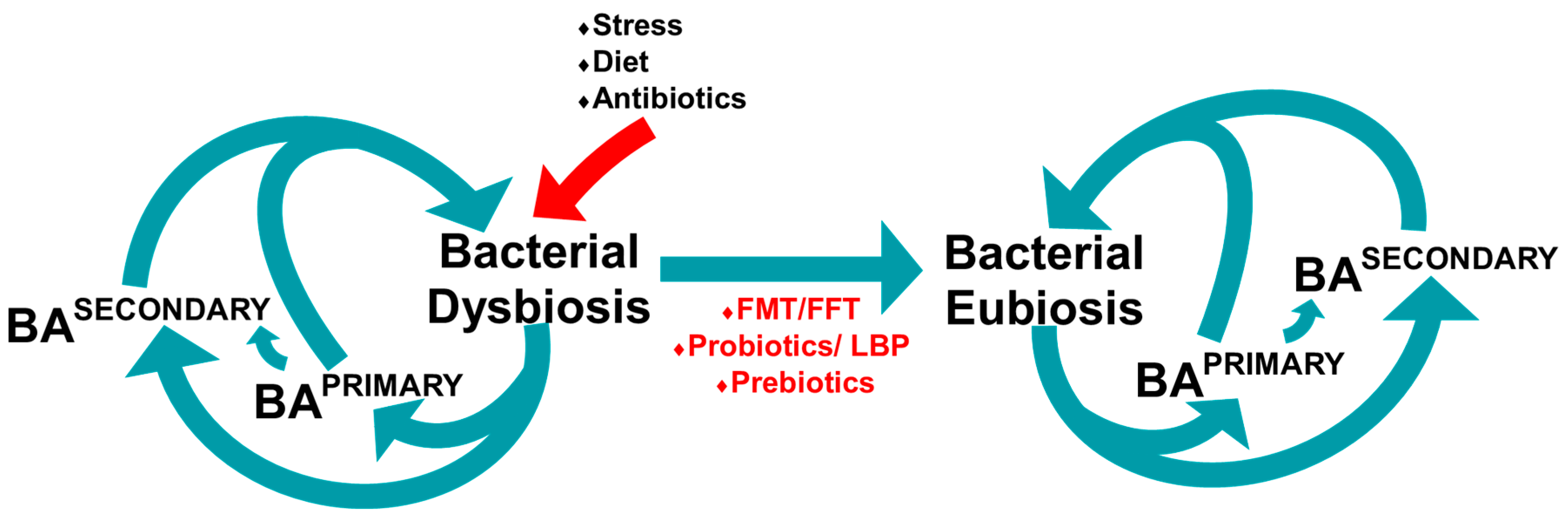
A vital way in which intestinal bacterial homeostasis is conserved in the GI tract is through bile acids that limit the growth of bile acid-sensitive bacteria and promote the growth of bile acid-tolerant bacteria. Data generated from IBS-D studies indicate that there is strong evidence that changes in the microbiome as a result of stress, and a change in diet or the taking of antibiotics could be responsible for bacterial dysbiosis and, subsequently, type II BAD (see Figure 4) [137,144,145,146]. High levels of taurine conjugation in bile acids have been observed in meat and seafood, suggesting that taurine conjugation is potentially associated with meat consumption [41,147]. As taurine conjugated bile acids have shown antagonistic effects on FXR activation, this may be a cause of dysbiosis [93].
The cause of primary BAD is unknown. Few studies have investigated the bile acid profile in SeHCAT diagnosed BAD patients. A potential hypothesis for the cause of BAD could be due to changes in the microbiome, as studies demonstrate that patients with BAD have reduced bacterial diversity [28]. Bacterial bile acid transformations shape the bile acids that are available for binding to FXR. Bile acids bind to FXR as both agonist and antagonist with differing affinity and efficacy levels, leading to various levels of the activation and expression of FGF19. This can result in an over-production of primary bile acids and may lead to primary BAD. Establishing the causality of microbiome constituents for health and disease remains a tremendous challenge. In addition, the bacterial species and strains that make up the microbiome are highly specific for the individual; no two individuals are colonised by the same assemblage of microbiota [148]. In order to accurately understand the role of the microbiome in BAD patients, functional genomic analysis of the microbiome of SeHCAT diagnosed patients coupled with metabolic analysis is needed.
Primary BAD is a common GI disorder, estimated to affect 1 in 100 individuals in the UK [4]. Despite this and advances in diagnosing BAD, the current treatment remains flawed, often having undesirable effects [19]. There has been increasing evidence of the promising role and therapeutic potential of the human gut microbiome and its potential for treating BAD. Further studies are needed to link gut microbiota and metabolic features in SeHCAT diagnosed BAD patients to better understand how the microbiome could influence this condition. In addition, further in vitro and in vivo studies would be beneficial in mapping out individual bile acids effects on FXR activation. Finally, clinical trials are necessary to investigate whether FMT, FFT, probiotics and other microbiota altering treatments are a viable therapeutic option to treat SeHCAT diagnosed BAD sufferers.

Author Contributions

Writing of manuscript and creation of figures, E.B.M.H.; reading and modifications of manuscript, S.R., D.C., R.P.A., E.M.H.W. and G.C.A.A. All authors have read and agreed to the published version of the manuscript.

Funding

This paper is based on independent research commissioned and funded by the NIHR Policy Research Programme (NIBSC Regulatory Science Research Unit).

Institutional Review Board Statement

The views expressed in the publication are those of the author(s) and not necessarily those of the NHS, the NIHR, the Department of Health, “arms” length bodies or other government departments.

Informed Consent Statement

Not applicable.

Data Availability Statement

No new data were created or analysed in this study. Data sharing is not applicable to this article.

Acknowledgments

Thank you to all the co-authors that supervise my PhD. Thank you to the microbiome group at NIBSC; Saba Anwar, Chrysi Sergaki and Alastair Logan.

Conflicts of Interest

The authors declare no conflict of interest.

References

  1. Arasaradnam, R.P.; Brown, S.; Forbes, A.; Fox, M.R.; Hungin, P.; Kelman, L.; Major, G.; O’Connor, M.; Sanders, D.S.; Sinha, R.; et al. Guidelines for the investigation of chronic diarrhoea in adults: British Society of Gastroenterology, 3rd edition. Gut 2018, 67, 1380–1399. [Google Scholar] [CrossRef] [PubMed]
  2. Walters, J.R.F.; Arasaradnam, R.; Andreyev, H.J.N. Diagnosis and management of bile acid diarrhoea: A survey of UK expert opinion and practice. Front. Gastroenterol. 2020, 11, 358–363. [Google Scholar] [CrossRef] [PubMed] [Green Version]
  3. Bannaga, A.; Kelman, L.; O’Connor, M.; Pitchford, C.; Walters, J.R.F.; Arasaradnam, R.P. How bad is bile acid diarrhoea: An online survey of patient-reported symptoms and outcomes. BMJ Open Gastroenterol. 2017, 4, e000116. [Google Scholar] [CrossRef]
  4. Wedlake, L.; A’Hern, R.; Russell, D.; Thomas, K.; Walters, J.R.F.; Andreyev, H.J.N. Systematic review: The prevalence of idiopathic bile acid malabsorption as diagnosed by SeHCAT scanning in patients with diarrhoea-predominant irritable bowel syndrome. Aliment. Pharmacol. Ther. 2009, 30, 707–717. [Google Scholar] [CrossRef]
  5. Slattery, S.A.; Niaz, O.; Aziz, Q.; Ford, A.C.; Farmer, A.D. Systematic review with meta-analysis: The prevalence of bile acid malabsorption in the irritable bowel syndrome with diarrhoea. Aliment. Pharmacol. Ther. 2015, 42, 3–11. [Google Scholar] [CrossRef] [PubMed] [Green Version]
  6. Smith, M.J.; Cherian, P.; Raju, G.S.; Dawson, B.F.; Mahon, S.; Bardhan, K.D. Bile acid malabsorption in persistent diarrhoea. J. R. Coll. Physicians Lond. 2000, 34, 448–451. [Google Scholar] [PubMed]
  7. Williams, A.J.; Merrick, M.V.; Eastwood, M.A. Idiopathic bile acid malabsorption: A review of clinical presentation, diagnosis, and response to treatment. Gut 1991, 32, 1004–1006. [Google Scholar] [CrossRef]
  8. Walters, J.R.; Tasleem, A.M.; Omer, O.S.; Brydon, W.G.; Dew, T.; le Roux, C.W. A New Mechanism for Bile Acid Diarrhea: Defective Feedback Inhibition of Bile Acid Biosynthesis. Clin. Gastroenterol. Hepatol. 2009, 7, 1189–1194. [Google Scholar] [CrossRef] [PubMed] [Green Version]
  9. Hofmann, A.F.; Poley, J.R. Role of Bile Acid Malabsorption in Pathogenesis of Diarrhea and Steatorrhea in Patients with Ileal Resection. Gastroenterology 1972, 62, 918–934. [Google Scholar] [CrossRef]
  10. Aldini, R.; Roda, A.; Festi, D.; Sama, C.; Mazzella, G.; Bazzoli, F.; Morselli, A.M.; Roda, E.; Barbara, L. Bile acid malabsorption and bile acid diarrhea in intestinal resection. Dig. Dis. Sci. 1982, 27, 495–502. [Google Scholar] [CrossRef]
  11. Joyce, S.A.; Gahan, C.G. Disease-Associated Changes in Bile Acid Profiles and Links to Altered Gut Microbiota. Dig. Dis. 2017, 35, 169–177. [Google Scholar] [CrossRef] [PubMed]
  12. Riemsma, R.; Al, M.; Ramos, I.C.; Deshpande, S.N.; Armstrong, N.; Lee, Y.C.; Ryder, S.; Noake, C.; Krol, M.; Oppe, M.; et al. SeHCAT [tauroselcholic (selenium-75) acid] for the investigation of bile acid malabsorption and measurement of bile acid pool loss: A systematic review and cost-effectiveness analysis. Heal. Technol. Assess. 2013, 17, 1–236. [Google Scholar] [CrossRef] [Green Version]
  13. Vaiopoulou, A. Molecular basis of the irritable bowel syndrome. World J. Gastroenterol. 2014, 20, 376–383. [Google Scholar] [CrossRef]
  14. Bosaeus, I.; Andersson, H.; Nyström, C. Effect of a Low-Fat Diet on Bile Salt Excretion and Diarrhoea in the Gastrointestinal Radiation Syndrome. Acta Radiol. Oncol. Radiat. Phys. Biol. 1979, 18, 460–464. [Google Scholar] [CrossRef]
  15. Chiang, J.Y.L. Bile Acid Metabolism and Signaling. Compr. Physiol. 2013, 3, 1191–1212. [Google Scholar] [CrossRef] [Green Version]
  16. Turnbaugh, P.J.; Ridaura, V.K.; Faith, J.J.; Rey, F.E.; Knight, R.; Gordon, J.I. The Effect of Diet on the Human Gut Microbiome: A Metagenomic Analysis in Humanized Gnotobiotic Mice. Sci. Transl. Med. 2009, 1, 6ra14. [Google Scholar] [CrossRef] [PubMed] [Green Version]
  17. Devkota, S.; Chang, E.B. Interactions between Diet, Bile Acid Metabolism, Gut Microbiota, and Inflammatory Bowel Diseases. Dig. Dis. 2015, 33, 351–356. [Google Scholar] [CrossRef] [PubMed] [Green Version]
  18. Hofmann, A.F.; Poley, J.R. Cholestyramine Treatment of Diarrhea Associated with Ileal Resection. N. Engl. J. Med. 1969, 281, 397–402. [Google Scholar] [CrossRef]
  19. Rössel, P.; Sortsøe Jensen, H.; Qvist, P.; Arveschoug, A. Prognosis of Adult-Onset Idiopathic Bile Acid Malabsorption. Scand. J. Gastroenterol. 1999, 34, 587–590. [Google Scholar] [CrossRef]
  20. Farina, A.; Dumonceau, J.M.; Lescuyer, P. Proteomic analysis of human bile and potential applications for cancer diagnosis. Expert Rev. Proteom. 2009, 6, 285–301. [Google Scholar] [CrossRef]
  21. Falany, C.N.; Johnson, M.R.; Barnes, S.; Diasio, R.B. Glycine and taurine conjugation of bile acids by a single enzyme. Molecular cloning and expression of human liver bile acid CoA:amino acid N-acyltransferase. J. Biol. Chem. 1994, 269, 205–934. [Google Scholar] [CrossRef]
  22. Kullak-Ublick, G.A.; Stieger, B.; Hagenbuch, B.; Meier, P.J. Hepatic Transport of Bile Salts. Semin. Liver Dis. 2000, 20, 273–292. [Google Scholar] [CrossRef] [PubMed] [Green Version]
  23. Hofmann, A. The function of bile salts in fat absorption. Biochem. J. 1963, 89, 57–68. [Google Scholar] [CrossRef] [Green Version]
  24. Houten, S.M.; Watanabe, M.; Auwerx, J. Endocrine functions of bile acids. EMBO J. 2006, 25, 1419–1425. [Google Scholar] [CrossRef]
  25. Lefebvre, P.; Cariou, B.; Lien, F.; Kuipers, F.; Staels, B. Role of Bile Acids and Bile Acid Receptors in Metabolic Regulation. Physiol. Rev. 2009, 89, 147–191. [Google Scholar] [CrossRef] [PubMed] [Green Version]
  26. Coleman, R.; Lowe, P.J.; Billington, D. Membrane lipid composition and susceptibility to bile salt damage. Biochim. Biophys. Acta Biomembr. 1980, 599, 294–300. [Google Scholar] [CrossRef]
  27. Zavaglia, A.G.; Kociubinski, G.; Perez, P.; Disalvo, E.; De Antoni, G. Effect of bile on the lipid composition and surface properties of bifidobacteria. J. Appl. Microbiol. 2002, 93, 794–799. [Google Scholar] [CrossRef] [Green Version]
  28. Sagar, N.M.; Duboc, H.; Kay, G.L.; Tauqeer, A.; Wicaksono, A.N.; Covington, J.A.; Quince, C.; Kokkorou, M.; Svolos, V.; Palmieri, L.J.; et al. The Pathophysiology of Bile Acid Diarrhoea: Differences in the Colonic Microbiome, Metabolome and Bile Acids. SSRN Electron. J. 2020, 10. [Google Scholar] [CrossRef]
  29. Zheng, X.; Chen, T.; Zhao, A.; Wang, X.; Xie, G.; Huang, F.; Liu, J.; Zhao, Q.; Wang, S.; Wang, C.; et al. The Brain Metabolome of Male Rats across the Lifespan. Sci. Rep. 2016, 6, 1–12. [Google Scholar] [CrossRef] [Green Version]
  30. Chiang, J.Y. Bile acids: Regulation of synthesis. J. Lipid Res. 2009, 50, 1955–1966. [Google Scholar] [CrossRef] [Green Version]
  31. Hofmann, A.F. The Continuing Importance of Bile Acids in Liver and Intestinal Disease. Arch. Intern. Med. 1999, 159, 2647–2658. [Google Scholar] [CrossRef]
  32. Vlahcevic, Z.R.; Heuman, D.M.; Hylemon, P.B. Physiology and pathophysiology of enterohepatic circulation of bile acids. In Hepatology: A Textbook of Liver Disease, 3rd ed.; Zakim, D., Boyer, T., Eds.; Saunders: Philadelphia, PA, USA, 1996. [Google Scholar]
  33. Russell, D.W. The Enzymes, Regulation, and Genetics of Bile Acid Synthesis. Annu. Rev. Biochem. 2003, 72, 137–174. [Google Scholar] [CrossRef] [PubMed] [Green Version]
  34. Reihnér, E.; Björkhem, I.; Angelin, B.; Ewerth, S.; Einarsson, K. Bile acid synthesis in humans: Regulation of hepatic microsomal cholesterol 7α-hydroxylase activity. Gastroenterology 1989, 97, 1498–1505. [Google Scholar] [CrossRef]
  35. Schwarz, M.; Russell, D.W.; Dietschy, J.M.; Turley, S.D. Marked reduction in bile acid synthesis in cholesterol 7α-hydroxylase-deficient mice does not lead to diminished tissue cholesterol turnover or to hypercholesterolemia. J. Lipid Res. 1998, 39, 1833–1843. [Google Scholar] [CrossRef]
  36. Gälman, C.; Arvidsson, I.; Angelin, B.; Rudling, M. Monitoring hepatic cholesterol 7α-hydroxylase activity by assay of the stable bile acid intermediate 7α-hydroxy-4-cholesten-3-one in peripheral blood. J. Lipid Res. 2003, 44, 859–866. [Google Scholar] [CrossRef] [Green Version]
  37. Axelson, M.; Aly, A.; Sjövall, J. Levels of 7α-hydroxy-4-cholesten-3-one in plasma reflect rates of bile acid synthesis in man. FEBS Lett. 1988, 239, 324–328. [Google Scholar] [CrossRef] [Green Version]
  38. Camilleri, M.; Nadeau, A.; Tremaine, W.J.; Lamsam, J.; Burton, D.; Odunsi, S.; Sweetser, S.; Singh, R. Measurement of serum 7α-hydroxy-4-cholesten-3-one (or 7αC4), a surrogate test for bile acid malabsorption in health, ileal disease and irritable bowel syndrome using liquid chromatography-tandem mass spectrometry. Neurogastroenterol. Motil. 2009, 21, 734-e43. [Google Scholar] [CrossRef] [PubMed] [Green Version]
  39. Batta, A.K.; Salen, G.; Shefer, S. Substrate specificity of cholylglycine hydrolase for the hydrolysis of bile acid conjugates. J. Biol. Chem. 1984, 259, 15036–15039. [Google Scholar] [CrossRef]
  40. Kawamoto, K.; Horibe, I.; Uchida, K. Purification and Characterization of a New Hydrolase for Conjugated Bile Acids, Chenodeoxycholyltaurine Hydrolase, from Bacteroides vulgatus. J. Biochem. 1989, 106, 1049–1053. [Google Scholar] [CrossRef]
  41. Ridlon, J.M.; Kang, D.J.; Hylemon, P.B. Bile salt biotransformations by human intestinal bacteria. J. Lipid Res. 2006, 47, 241–259. [Google Scholar] [CrossRef] [Green Version]
  42. Corzo, G.; Gilliland, S. Bile Salt Hydrolase Activity of Three Strains of Lactobacillus acidophilus. J. Dairy Sci. 1999, 82, 472–480. [Google Scholar] [CrossRef]
  43. Tanaka, H.; Hashiba, H.; Kok, J.; Mierau, I. Bile Salt Hydrolase of Bifidobacterium longum—Biochemical and Genetic Characterization. Appl. Environ. Microbiol. 2000, 66, 2502–2512. [Google Scholar] [CrossRef] [PubMed] [Green Version]
  44. Franz, C.M.; Specht, I.; Haberer, P.; Holzapfel, W.H. Bile Salt Hydrolase Activity of Enterococci Isolated from Food: Screening and Quantitative Determination. J. Food Prot. 2001, 64, 725–729. [Google Scholar] [CrossRef]
  45. Masuda, N. Deconjugation of Bile Salts by Bacteroides and Clostridium. Microbiol. Immunol. 1981, 25, 1–11. [Google Scholar] [CrossRef]
  46. Kishinaka, M.; Umeda, A.; Kuroki, S. High concentrations of conjugated bile acids inhibit bacterial growth of Clostridium perfringens and induce its extracellular cholyglycine hydrolase. Steroids 1994, 59, 485–489. [Google Scholar] [CrossRef]
  47. Song, Z.; Cai, Y.; Lao, X.; Wang, X.; Lin, X.; Cui, Y.; Kalavagunta, P.K.; Liao, J.; Jin, L.; Shang, J.; et al. Taxonomic profiling and populational patterns of bacterial bile salt hydrolase (BSH) genes based on worldwide human gut microbiome. Microbiome 2019, 7, 1–16. [Google Scholar] [CrossRef] [Green Version]
  48. Tian, Y.; Cai, J.; Gui, W.; Nichols, R.G.; Koo, I.; Zhang, J.; Anitha, M.; Patterson, A.D. Berberine Directly Affects the Gut Microbiota to Promote Intestinal Farnesoid X Receptor Activation. Drug Metab. Dispos. 2019, 47, 86–93. [Google Scholar] [CrossRef] [Green Version]
  49. Begley, M.; Hill, C.; Gahan, C.G.M. Bile Salt Hydrolase Activity in Probiotics. Appl. Environ. Microbiol. 2006, 72, 1729–1738. [Google Scholar] [CrossRef] [Green Version]
  50. De Smet, I.; Van Hoorde, L.; Woestyne, M.V.; Christiaens, H.; Verstraete, W. Significance of bile salt hydrolytic activities of lactobacilli. J. Appl. Bacteriol. 1995, 79, 292–301. [Google Scholar] [CrossRef]
  51. Dussurget, O.; Cabanes, D.; Dehoux, P.; Lecuit, M.; Buchrieser, C.; Glaser, P.; Cossart, P. The European Listeria Genome Consortium Listeria monocytogenes bile salt hydrolase is a PrfA-regulated virulence factor involved in the intestinal and hepatic phases of listeriosis. Mol. Microbiol. 2002, 45, 1095–1106. [Google Scholar] [CrossRef] [PubMed]
  52. Sannasiddappa, T.H.; Lund, P.A.; Clarke, S.R.; Sannasiddappa, T. In Vitro Antibacterial Activity of Unconjugated and Conjugated Bile Salts on Staphylococcus aureus. Front. Microbiol. 2017, 8, 1581. [Google Scholar] [CrossRef] [Green Version]
  53. Tian, Y.; Gui, W.; Koo, I.; Smith, P.B.; Allman, E.L.; Nichols, R.G.; Rimal, B.; Cai, J.; Liu, Q.; Patterson, A.D. The microbiome modulating activity of bile acids. Gut Microbes 2020, 11, 979–996. [Google Scholar] [CrossRef]
  54. Masuda, N.; Oda, H.; Hirano, S.; Masuda, M.; Tanaka, H. 7 α-Dehydroxylation of bile acids by resting cells of a Eubacterium lentum-like intestinal anaerobe, strain c-25. Appl. Environ. Microbiol. 1984, 47, 735–739. [Google Scholar] [CrossRef] [PubMed] [Green Version]
  55. Wells, J.E.; Berr, F.; A Thomas, L.; Dowling, R.; Hylemon, P.B. Isolation and characterization of cholic acid 7α-dehydroxylating fecal bacteria from cholesterol gallstone patients. J. Hepatol. 2000, 32, 4–10. [Google Scholar] [CrossRef]
  56. Björkhem, I.; Einarsson, K.; Melone, P.; Hylemon, P. Mechanism of intestinal formation of deoxycholic acid from cholic acid in humans: Evidence for a 3-oxo-delta 4-steroid intermediate. J. Lipid Res. 1989, 30, 1033–1039. [Google Scholar] [CrossRef]
  57. Vital, M.; Rud, T.; Rath, S.; Pieper, D.H.; Schlüter, D. Diversity of Bacteria Exhibiting Bile Acid-inducible 7α-dehydroxylation Genes in the Human Gut. Comput. Struct. Biotechnol. J. 2019, 17, 1016–1019. [Google Scholar] [CrossRef]
  58. Stellwag, E.J.; Hylemon, P.B. 7α Dehydroxylation of cholic acid and chenodeoxycholic acid by Clostridium leptum. J. Lipid Res. 1979, 20, 325–333. [Google Scholar] [CrossRef]
  59. Batta, A.K.; Salen, G.; Arora, R.; Shefer, S.; Batta, M.; Person, A. Side chain conjugation prevents bacterial 7-dehydroxylation of bile acids. J. Biol. Chem. 1990, 265, 10925–10928. [Google Scholar] [CrossRef]
  60. Sutherland, J.D.; Macdonald, I.A. The metabolism of primary, 7-oxo, and 7β-hydroxy bile acids by Clostridium absonum. J. Lipid Res. 1982, 23, 726–732. [Google Scholar] [CrossRef]
  61. Macdonald, I.A.; Hutchison, D.M.; Forrest, T.P. Formation of urso-and ursodeoxy-cholic acids from primary bile acids by Clostridium absonum. J. Lipid Res. 1981, 22, 458–466. [Google Scholar] [CrossRef]
  62. Macdonald, I.A.; Roach, P.D. Bile salt induction of 7α-and 7β-hydroxysteroid dehydrogenases in Clostridium absonum. Biochim. Biophys. Acta Lipids Lipid Metab. 1981, 665, 262–269. [Google Scholar] [CrossRef]
  63. Hirano, S.; Masuda, N. Epimerization of the 7-hydroxy group of bile acids by the combination of two kinds of microorganisms with 7 alpha- and 7 beta-hydroxysteroid dehydrogenase activity, respectively. J. Lipid Res. 1981, 22, 1060–1068. [Google Scholar] [CrossRef]
  64. Sherrod, J.; Hylemon, P. Partial purification and characterization of NAD-dependent 7α-hydroxysteroid dehydrogenase from Bacteroides thetaiotaomicron. Biochim. Biophys. Acta (BBA) Lipids Lipid Metab. 1977, 486, 351–358. [Google Scholar] [CrossRef]
  65. Macdonald, I.A.; Meier, E.C.; Mahony, D.E.; Costain, G.A. 3α-, 7α- And 12α-hydroxysteroid dehydrogenase activities from Clostridium perfringens. Biochim. Biophys. Acta Lipids Lipid Metab. 1976, 450, 142–153. [Google Scholar] [CrossRef]
  66. Bhowmik, S.; Jones, D.H.; Chiu, H.; Park, I.; Chiu, H.; Axelrod, H.L.; Farr, C.L.; Tien, H.J.; Agarwalla, S.; Lesley, S.A. Structural and functional characterization of BaiA, an enzyme involved in secondary bile acid synthesis in human gut microbe. Proteins Struct. Funct. Bioinform. 2014, 82, 216–229. [Google Scholar] [CrossRef] [PubMed] [Green Version]
  67. Wells, J.E.; Hylemon, P.B. Identification and Characterization of a Bile Acid 7α-Dehydroxylation Operon in Clostridium sp. Strain TO-931, a Highly Active 7α-Dehydroxylating Strain Isolated from Human Feces. Appl. Environ. Microbiol. 2000, 66, 1107–1113. [Google Scholar] [CrossRef] [PubMed] [Green Version]
  68. Macdonald, I.A.; Mahony, D.E.; Jellet, J.F.; Meier, C. Nad-dependent 3α- and 12α-hydroxysteroid dehydrogenase activities from eubacterwm lentum atcc no. 25559. Biochim. Biophys. Acta Lipids Lipid Metab. 1977, 489, 466–476. [Google Scholar] [CrossRef]
  69. Edenharder, R.; Pfützner, A.; Hammann, R. Characterization of NAD-dependent 3α- and 3β-hydroxysteroid dehydrogenase and of NADP-dependent 7β-hydroxysteroid dehydrogenase from Peptostreptococcus productus. Biochim. Biophys. Acta Lipids Lipid Metab. 1989, 1004, 230–238. [Google Scholar] [CrossRef]
  70. Edenharder, R.; Pfützner, M.; Hammann, R. NADP-dependent 3β-, 7α- and 7β-hydroxysteroid dehydrogenase activities from a lecithinase-lipase-negative Clostridium species 25.11.c. Biochim. Biophys. Acta Lipids Lipid Metab. 1989, 1002, 37–44. [Google Scholar] [CrossRef]
  71. Edenharder, R.; Pfützner, M. Partial purification and characterization of an NAD-dependent 3 beta-hydroxysteroid dehydrogenase from Clostridium innocuum. Appl. Environ. Microbiol. 1989, 55, 1656. [Google Scholar] [CrossRef] [Green Version]
  72. Teruaki, A.; Taiko, A.; Masao, H.; Tsuneo, N.; Kyoichi, K. Enzymes involved in the formation of 3β,7β-dihydroxy-12-oxo-5β-cholanic acid from dehydrocholic acid by Ruminococcus sp. obtained from human intestine. Biochim. Biophys. Acta Lipids Lipid Metab. 1987, 921, 275–280. [Google Scholar] [CrossRef]
  73. Sutherland, J.D.; Williams, C.N. Bile acid induction of 7 alpha- and 7 beta-hydroxysteroid dehydrogenases in Clostridium limosum. J. Lipid Res. 1985, 26, 344–350. [Google Scholar] [CrossRef]
  74. Stellwag, E.; Hylemon, P. Purification and characterization of bile salt hydrolase from Bacteroides fragilis subsp. fragilis. Biochim. Biophys. Acta Enzym. 1976, 452, 165–176. [Google Scholar] [CrossRef]
  75. Coleman, J.P.; Hudson, L.L.; Adams, M.J. Characterization and regulation of the NADP-linked 7 alpha-hydroxysteroid dehydrogenase gene from Clostridium sordellii. J. Bacteriol. 1994, 176, 4865–4874. [Google Scholar] [CrossRef] [Green Version]
  76. Hirano, S.; Masuda, N.; Oda, H.; Mukai, H. Transformation of bile acids by Clostridium perfringens. Appl. Environ. Microbiol. 1981, 42, 394–399. [Google Scholar] [CrossRef] [Green Version]
  77. Heuman, D.M. Quantitative estimation of the hydrophilic-hydrophobic balance of mixed bile salt solutions. J. Lipid Res. 1989, 30, 719–730. [Google Scholar] [CrossRef]
  78. Heuman, U.M.; Pandak, W.M.; Hylemon, P.B.; Vlahcevic, Z.R. Conjugates of ursodeoxycholate protect against cytotoxicity of more hydrophobic bile salts: In vitro studies in rat hepatocytes and human erythrocytes. Hepatology 1991, 14, 920–926. [Google Scholar] [CrossRef]
  79. Harris, J.; Hylemon, P. Partial purification and characterization of NADP-dependent 12α-hydroxysteroid dehydrogenase from clostridium leptum. Biochim. Biophys. Acta Lipids Lipid Metab. 1978, 528, 148–157. [Google Scholar] [CrossRef]
  80. Macdonald, I.A.; Jellett, J.F.; Mahony, D.E. 12alpha-Hydroxysteroid dehydrogenase from Clostridium group P strain C48-50 ATCC No. 29733: Partial purification and characterization. J. Lipid Res. 1979, 20, 234–239. [Google Scholar] [CrossRef]
  81. Edenharder, R.; Schneider, J. 12 beta-dehydrogenation of bile acids by Clostridium paraputrificum, C. tertium, and C. difficile and epimerization at carbon-12 of deoxycholic acid by cocultivation with 12 alpha-dehydrogenating Eubacterium lentum. Appl. Environ. Microbiol. 1985, 49, 964–968. [Google Scholar] [CrossRef] [PubMed] [Green Version]
  82. Edenharder, R.; Pfützner, A. Characterization of NADP-dependent 12β-hydroxysteroid dehydrogenase from Clostridium paraputrificum. Biochim. Biophys. Acta Lipids Lipid Metab. 1988, 962, 362–370. [Google Scholar] [CrossRef]
  83. Mallonee, D.H.; Hylemon, P.B. Sequencing and expression of a gene encoding a bile acid transporter from Eubacterium sp. strain VPI 12708. J. Bacteriol. 1996, 178, 7053–7058. [Google Scholar] [CrossRef] [PubMed] [Green Version]
  84. Funabashi, M.; Grove, T.L.; Wang, M.; Varma, Y.; McFadden, M.E.; Brown, L.C.; Guo, C.; Higginbottom, S.; Almo, S.C.; Fischbach, M.A. A metabolic pathway for bile acid dehydroxylation by the gut microbiome. Nat. Cell Biol. 2020, 582, 566–570. [Google Scholar] [CrossRef]
  85. Distrutti, E.; Santucci, L.; Cipriani, S.; Renga, B.; Schiaroli, E.; Ricci, P.; Donini, A.; Fiorucci, S. Bile acid activated receptors are targets for regulation of integrity of gastrointestinal mucosa. J. Gastroenterol. 2015, 50, 707–719. [Google Scholar] [CrossRef] [PubMed]
  86. Forman, B.M.; Goode, E.; Chen, J.; Oro, A.E.; Bradley, D.J.; Perlmann, T.; Noonan, D.J.; Burka, L.T.; McMorris, T.; Lamph, W.W.; et al. Identification of a nuclear receptor that is activated by farnesol metabolites. Cell 1995, 81, 687–693. [Google Scholar] [CrossRef] [Green Version]
  87. Seol, W.; Choi, H.S.; Moore, D.D. Isolation of proteins that interact specifically with the retinoid X receptor: Two novel orphan receptors. Mol. Endocrinol. 1995, 9, 72–85. [Google Scholar] [CrossRef]
  88. Makishima, M.; Okamoto, A.Y.; Repa, J.J.; Tu, H.; Learned, R.M.; Luk, A.; Hull, M.V.; Lustig, K.D.; Mangelsdorf, D.J.; Shan, B. Identification of a Nuclear Receptor for Bile Acids. Science 1999, 284, 1362–1365. [Google Scholar] [CrossRef]
  89. Parks, D.J.; Blanchard, S.G.; Bledsoe, R.K.; Chandra, G.; Consler, T.G.; Kliewer, S.A.; Stimmel, J.B.; Willson, T.M.; Zavacki, A.M.; Moore, D.D.; et al. Bile Acids: Natural Ligands for an Orphan Nuclear Receptor. Science 1999, 284, 1365–1368. [Google Scholar] [CrossRef]
  90. Wang, H.; Chen, J.; Hollister, K.; Sowers, L.C.; Forman, B.M. Endogenous Bile Acids Are Ligands for the Nuclear Receptor FXR/BAR. Mol. Cell 1999, 3, 543–553. [Google Scholar] [CrossRef]
  91. Mueller, M.; Thorell, A.; Claudel, T.; Jha, P.; Koefeler, H.; Lackner, C.; Hoesel, B.; Fauler, G.; Stojakovic, T.; Einarsson, C.; et al. Ursodeoxycholic acid exerts farnesoid X receptor-antagonistic effects on bile acid and lipid metabolism in morbid obesity. J. Hepatol. 2015, 62, 1398–1404. [Google Scholar] [CrossRef] [Green Version]
  92. Downes, M.; Verdecia, M.A.; Roecker, A.; Hughes, R.; HogenEsch, J.B.; Kast-Woelbern, H.R.; Bowman, M.E.; Ferrer, J.L.; Anisfeld, A.M.; Edwards, P.A.; et al. A Chemical, Genetic, and Structural Analysis of the Nuclear Bile Acid Receptor FXR. Mol. Cell 2003, 11, 1079–1092. [Google Scholar] [CrossRef]
  93. Zhao, L.; Yang, W.; Chen, Y.; Huang, F.; Lu, L.; Lin, C.; Huang, T.; Ning, Z.; Zhai, L.; Zhong, L.L.; et al. A Clostridia-rich microbiota enhances bile acid excretion in diarrhea-predominant irritable bowel syndrome. J. Clin. Investig. 2019, 130, 438–450. [Google Scholar] [CrossRef] [Green Version]
  94. Trefflich, I.; Marschall, H.-U.; di Giuseppe, R.; Ståhlman, M.; Michalsen, A.; Lampen, A.; Abraham, K.; Weikert, C. Associations between Dietary Patterns and Bile Acids: Results from a Cross-Sectional Study in Vegans and Omnivores. Nutrients 2019, 12, 47. [Google Scholar] [CrossRef] [PubMed] [Green Version]
  95. Sayin, S.I.; Wahlström, A.; Felin, J.; Jäntti, S.; Marschall, H.U.; Bamberg, K.; Angelin, B.; Hyötyläinen, T.; Orešič, M.; Bäckhed, F. Gut Microbiota Regulates Bile Acid Metabolism by Reducing the Levels of Tauro-beta-muricholic Acid, a Naturally Occurring FXR Antagonist. Cell Metab. 2013, 17, 225–235. [Google Scholar] [CrossRef] [PubMed] [Green Version]
  96. Mangelsdorf, D.J.; Evanst, R.M. The RXR Heterodimers and Orphan Receptors. Cell 1995, 83, 841–850. [Google Scholar] [CrossRef] [Green Version]
  97. Dawson, M.I.; Xia, Z. The retinoid X receptors and their ligands. Biochim. Biophys. Acta Mol. Cell Biol. Lipids 2012, 1821, 21–56. [Google Scholar] [CrossRef] [Green Version]
  98. Kong, B.; Wang, L.; Chiang, J.Y.L.; Zhang, Y.; Klaassen, C.D.; Guo, G.L. Mechanism of tissue-specific farnesoid X receptor in suppressing the expression of genes in bile-acid synthesis in mice. Hepatology 2012, 56, 1034–1043. [Google Scholar] [CrossRef] [Green Version]
  99. Zollner, G.; Marschall, H.U.; Wagner, A.M.; Trauner, M. Role of Nuclear Receptors in the Adaptive Response to Bile Acids and Cholestasis: Pathogenetic and Therapeutic Considerations. Mol. Pharm. 2006, 3, 231–251. [Google Scholar] [CrossRef]
  100. Ballatori, N.; Christian, W.V.; Lee, J.Y.; Dawson, P.A.; Soroka, C.J.; Boyer, J.L.; Madejczyk, M.S.; Li, N. OSTα-OSTβ: A major basolateral bile acid and steroid transporter in human intestinal, renal, and biliary epithelia. Hepatology 2005, 42, 1270–1279. [Google Scholar] [CrossRef] [PubMed]
  101. Vergnes, L.; Lee, J.M.; Chin, R.G.; Auwerx, J.; Reue, K. Diet1 Functions in the FGF15/19 Enterohepatic Signaling Axis to Modulate Bile Acid and Lipid Levels. Cell Metab. 2013, 17, 916–928. [Google Scholar] [CrossRef] [Green Version]
  102. Keitel, V.; Cupisti, K.; Ullmer, C.; Knoefel, W.T.; Kubitz, R.; Häussinger, D. The membrane-bound bile acid receptor TGR5 is localized in the epithelium of human gallbladders. Hepatology 2009, 50, 861–870. [Google Scholar] [CrossRef]
  103. Kawamata, Y.; Fujii, R.; Hosoya, M.; Harada, M.; Yoshida, H.; Miwa, M.; Fukusumi, S.; Habata, Y.; Itoh, T.; Shintani, Y.; et al. A G Protein-coupled Receptor Responsive to Bile Acids. J. Biol. Chem. 2003, 278, 9435–9440. [Google Scholar] [CrossRef] [Green Version]
  104. Maruyama, T.; Miyamoto, Y.; Nakamura, T.; Tamai, Y.; Okada, H.; Sugiyama, E.; Nakamura, T.; Itadani, H.; Tanaka, K. Identification of membrane-type receptor for bile acids (M-BAR). Biochem. Biophys. Res. Commun. 2002, 298, 714–719. [Google Scholar] [CrossRef]
  105. Poole, D.P.; Godfrey, C.; Cattaruzza, F.; Cottrell, G.S.; Kirkland, J.G.; Pelayo, J.C.; Bunnett, N.W.; Corvera, C.U. Expression and function of the bile acid receptor GpBAR1 (TGR5) in the murine enteric nervous system. Neurogastroenterol. Motil. 2010, 22, 814–825. [Google Scholar] [CrossRef] [PubMed] [Green Version]
  106. Alemi, F.; Poole, D.P.; Chiu, J.; Schoonjans, K.; Cattaruzza, F.; Grider, J.R.; Bunnett, N.W.; Corvera, C.U. The Receptor TGR5 Mediates the Prokinetic Actions of Intestinal Bile Acids and Is Required for Normal Defecation in Mice. Gastroenterology 2013, 144, 145–154. [Google Scholar] [CrossRef] [PubMed]
  107. Burge, M.N. Fungi in Biological Control Systems; Manchester University Press: Manchester, UK, 1988. [Google Scholar]
  108. Marchesi, J.R.; Ravel, J. The vocabulary of microbiome research: A proposal. Microbiome 2015, 3, 1–3. [Google Scholar] [CrossRef] [Green Version]
  109. Simon, G.L.; Gorbach, S.L. Intestinal flora in health and disease. Gastroenterology 1984, 86, 174–193. [Google Scholar] [CrossRef]
  110. Guarner, F.; Malagelada, J.R. Gut flora in health and disease. Lancet 2003, 361, 512–519. [Google Scholar] [CrossRef]
  111. Marchesi, J.R. The Microbiomes of Things. 2017. Available online: https://microbiologysociety.org/publication/past-issues/the-microbiome/article/the-microbiomes-of-things.html (accessed on 28 May 2020).
  112. Sender, R.; Fuchs, S.; Milo, R. Revised Estimates for the Number of Human and Bacteria Cells in the Body. PLoS Biol. 2016, 14, e1002533. [Google Scholar] [CrossRef] [Green Version]
  113. Savage, D.C. Microbial Ecology of the Gastrointestinal Tract. Annu. Rev. Microbiol. 1977, 31, 107–133. [Google Scholar] [CrossRef]
  114. Amos, G.C.A.; Logan, A.; Anwar, S.; Fritzsche, M.; Mate, R.; Bleazard, T.; Rijpkema, S. Developing standards for the microbiome field. Microbiome 2020, 8, 1–13. [Google Scholar] [CrossRef]
  115. Yoo, B.B.; Mazmanian, S.K. The Enteric Network: Interactions between the Immune and Nervous Systems of the Gut. Immunity 2017, 46, 910–926. [Google Scholar] [CrossRef] [Green Version]
  116. Lazar, V.; Ditu, L.M.; Pircalabioru, G.G.; Gheorghe, I.; Curutiu, C.; Holban, A.M.; Picu, A.; Petcu, L.; Chifiriuc, M.C. Aspects of Gut Microbiota and Immune System Interactions in Infectious Diseases, Immunopathology, and Cancer. Front. Immunol. 2018, 9, 1830. [Google Scholar] [CrossRef] [Green Version]
  117. Furness, J.B. The Enteric Nervous System; Wiley-Blackwell: Hoboken, NJ, USA, 2006. [Google Scholar]
  118. Furness, J.B.; Callaghan, B.P.; Rivera, L.R.; Cho, H.-J. The Enteric Nervous System and Gastrointestinal Innervation: Integrated Local and Central Control. Adv. Exp. Med. Biol. 2014, 817, 39–71. [Google Scholar] [CrossRef] [PubMed]
  119. Flint, H.J.; Scott, K.P.; Louis, P.; Duncan, S.H. The role of the gut microbiota in nutrition and health. Nat. Rev. Gastroenterol. Hepatol. 2012, 9, 577–589. [Google Scholar] [CrossRef]
  120. Tierney, B.T.; Yang, Z.; Luber, J.M.; Beaudin, M.; Wibowo, M.C.; Baek, C.; Mehlenbacher, E.; Patel, C.J.; Kostic, A.D. The Landscape of Genetic Content in the Gut and Oral Human Microbiome. Cell Host Microbe 2019, 26, 283–295.e8. [Google Scholar] [CrossRef] [PubMed]
  121. Carding, S.; Verbeke, K.; Vipond, D.T.; Corfe, B.M.; Owen, L.J. Dysbiosis of the gut microbiota in disease. Microb. Ecol. Health Dis. 2015, 26, 26191. [Google Scholar] [CrossRef] [PubMed]
  122. Selwyn, F.P.; Csanaky, I.L.; Zhang, Y.; Klaassen, C.D. Importance of Large Intestine in Regulating Bile Acids and Glucagon-Like Peptide-1 in Germ-Free Mice. Drug Metab. Dispos. 2015, 43, 1544–1556. [Google Scholar] [CrossRef] [Green Version]
  123. Zhang, L.; Xie, C.; Nichols, R.G.; Chan, S.H.J.; Jiang, C.; Hao, R.; Smith, P.B.; Cai, J.; Simons, M.N.; Hatzakis, E.; et al. Farnesoid X Receptor Signaling Shapes the Gut Microbiota and Controls Hepatic Lipid Metabolism. mSystems 2016, 1, e00070-16. [Google Scholar] [CrossRef] [Green Version]
  124. Just, S.; Mondot, S.; Ecker, J.; Wegner, K.; Rath, E.; Gau, L.; Streidl, T.; Hery-Arnaud, G.; Schmidt, S.; Lesker, T.R.; et al. The gut microbiota drives the impact of bile acids and fat source in diet on mouse metabolism. Microbiome 2018, 6, 134. [Google Scholar] [CrossRef]
  125. Nie, Y.F.; Hu, J.; Yan, X.H. Cross-talk between bile acids and intestinal microbiota in host metabolism and health. J. Zhejiang Univ. Sci. B 2015, 16, 436–446. [Google Scholar] [CrossRef] [PubMed] [Green Version]
  126. Wahlström, A.; Sayin, S.I.; Marschall, H.-U.; Bäckhed, F. Intestinal Crosstalk between Bile Acids and Microbiota and Its Impact on Host Metabolism. Cell Metab. 2016, 24, 41–50. [Google Scholar] [CrossRef] [Green Version]
  127. Jiang, C.; Xie, C.; Lv, Y.; Li, J.; Krausz, K.W.; Shi, J.; Brocker, C.N.; Desai, D.; Amin, S.G.; Bisson, W.H.; et al. Intestine-selective farnesoid X receptor inhibition improves obesity-related metabolic dysfunction. Nat. Commun. 2015, 6, 10166. [Google Scholar] [CrossRef] [Green Version]
  128. Duboc, H.; Rainteau, D.; Rajca, S.; Humbert, L.; Farabos, D.; Maubert, M.; Grondin, V.; Jouet, P.; Bouhassira, D.; Seksik, P.; et al. Increase in fecal primary bile acids and dysbiosis in patients with diarrhea-predominant irritable bowel syndrome. Neurogastroenterol. Motil. 2012, 24, 513-e247. [Google Scholar] [CrossRef]
  129. Hugon, P.; Dufour, J.C.; Colson, P.; Fournier, P.-E.; Sallah, K.; Raoult, D. A comprehensive repertoire of prokaryotic species identified in human beings. Lancet Infect. Dis. 2015, 15, 1211–1219. [Google Scholar] [CrossRef]
  130. Li, J.; Jia, H.; Cai, X.; Zhong, H.; Feng, Q.; Sunagawa, S.; Arumugam, M.; Kultima, J.R.; Prifti, E.; Nielsen, T.; et al. An integrated catalog of reference genes in the human gut microbiome. Nat. Biotechnol. 2014, 32, 834–841. [Google Scholar] [CrossRef]
  131. Thursby, E.; Juge, N. Introduction to the human gut microbiota. Biochem. J. 2017, 474, 1823–1836. [Google Scholar] [CrossRef] [PubMed]
  132. Levy, M.; Kolodziejczyk, A.A.; Thaiss, C.A.; Elinav, E. Dysbiosis and the immune system. Nat. Rev. Immunol. 2017, 17, 219–232. [Google Scholar] [CrossRef]
  133. Stecher, B.; Maier, L.; Hardt, W.D. ’Blooming’ in the gut: How dysbiosis might contribute to pathogen evolution. Nat. Rev. Genet. 2013, 11, 277–284. [Google Scholar] [CrossRef] [Green Version]
  134. Weiss, G.A.; Hennet, T. Mechanisms and consequences of intestinal dysbiosis. Cell. Mol. Life Sci. 2017, 74, 2959–2977. [Google Scholar] [CrossRef] [PubMed] [Green Version]
  135. Aas, J.; Gessert, C.E.; Bakken, J.S. RecurrentClostridium difficileColitis: Case Series Involving 18 Patients Treated with Donor Stool Administered via a Nasogastric Tube. Clin. Infect. Dis. 2003, 36, 580–585. [Google Scholar] [CrossRef] [Green Version]
  136. Lopez, J.; Grinspan, A. Fecal Microbiota Transplantation for Inflammatory Bowel Disease. Gastroenterol. Hepatol. 2016, 12, 374–379. [Google Scholar]
  137. Langdon, A.; Crook, N.; Dantas, G. The effects of antibiotics on the microbiome throughout development and alternative approaches for therapeutic modulation. Genome Med. 2016, 8, 1–16. [Google Scholar] [CrossRef] [PubMed] [Green Version]
  138. Garborg, K.; Waagsbø, B.; Stallemo, A.; Matre, J.; Sundøy, A. Results of faecal donor instillation therapy for recurrent Clostridium difficile-associated diarrhoea. Scand. J. Infect. Dis. 2010, 42, 857–861. [Google Scholar] [CrossRef] [PubMed]
  139. El-Salhy, M.; Hatlebakk, J.G.; Gilja, O.H.; Kristoffersen, A.B.; Hausken, T. Efficacy of faecal microbiota transplantation for patients with irritable bowel syndrome in a randomised, double-blind, placebo-controlled study. Gut 2019, 69, 859–867. [Google Scholar] [CrossRef] [PubMed] [Green Version]
  140. Food and Drug Administration. Early Clinical Trials with Live Biotherapeutic Products: Chemistry, Manufacturing, and Control Information; Guidance for Industry; Food and Drug Administration: White Oak, MD, USA, 2016. [Google Scholar]
  141. Fuller, R. Probiotics in human medicine. Gut 1991, 32, 439–442. [Google Scholar] [CrossRef] [PubMed] [Green Version]
  142. Afrc, R.F. Probiotics in man and animals. J. Appl. Bacteriol. 1989, 66, 365–378. [Google Scholar] [CrossRef]
  143. Ott, S.J.; Waetzig, G.H.; Rehman, A.; Moltzau-Anderson, J.; Bharti, R.; Grasis, J.A.; Cassidy, L.; Tholey, A.; Fickenscher, H.; Seegert, D.; et al. Efficacy of Sterile Fecal Filtrate Transfer for Treating Patients With Clostridium difficile Infection. Gastroenterology 2017, 152, 799–811.e7. [Google Scholar] [CrossRef] [Green Version]
  144. Foster, J.A.; Rinaman, L.; Cryan, J.F. Stress & the gut-brain axis: Regulation by the microbiome. Neurobiol. Stress 2017, 7, 124–136. [Google Scholar] [CrossRef] [Green Version]
  145. Madison, A.; Kiecolt-Glaser, J.K. Stress, depression, diet, and the gut microbiota: Human–bacteria interactions at the core of psychoneuroimmunology and nutrition. Curr. Opin. Behav. Sci. 2019, 28, 105–110. [Google Scholar] [CrossRef]
  146. Ianiro, G.; Tilg, H.; Gasbarrini, A. Antibiotics as deep modulators of gut microbiota: Between good and evil. Gut 2016, 65, 1906–1915. [Google Scholar] [CrossRef] [PubMed]
  147. Hardison, W.G. Hepatic Taurine Concentration and Dietary Taurine as Regulators of Bile Acid Conjugation with Taurine. Gastroenterology 1978, 75, 71–75. [Google Scholar] [CrossRef]
  148. Schloissnig, S.; Arumugam, M.; Sunagawa, S.; Mitreva, M.; Tap, J.; Zhu, A.; Waller, A.S.; Mende, D.R.; Kultima, J.R.; Martin, J.; et al. Genomic variation landscape of the human gut microbiome. Nat. Cell Biol. 2013, 493, 45–50. [Google Scholar] [CrossRef] [PubMed] [Green Version]
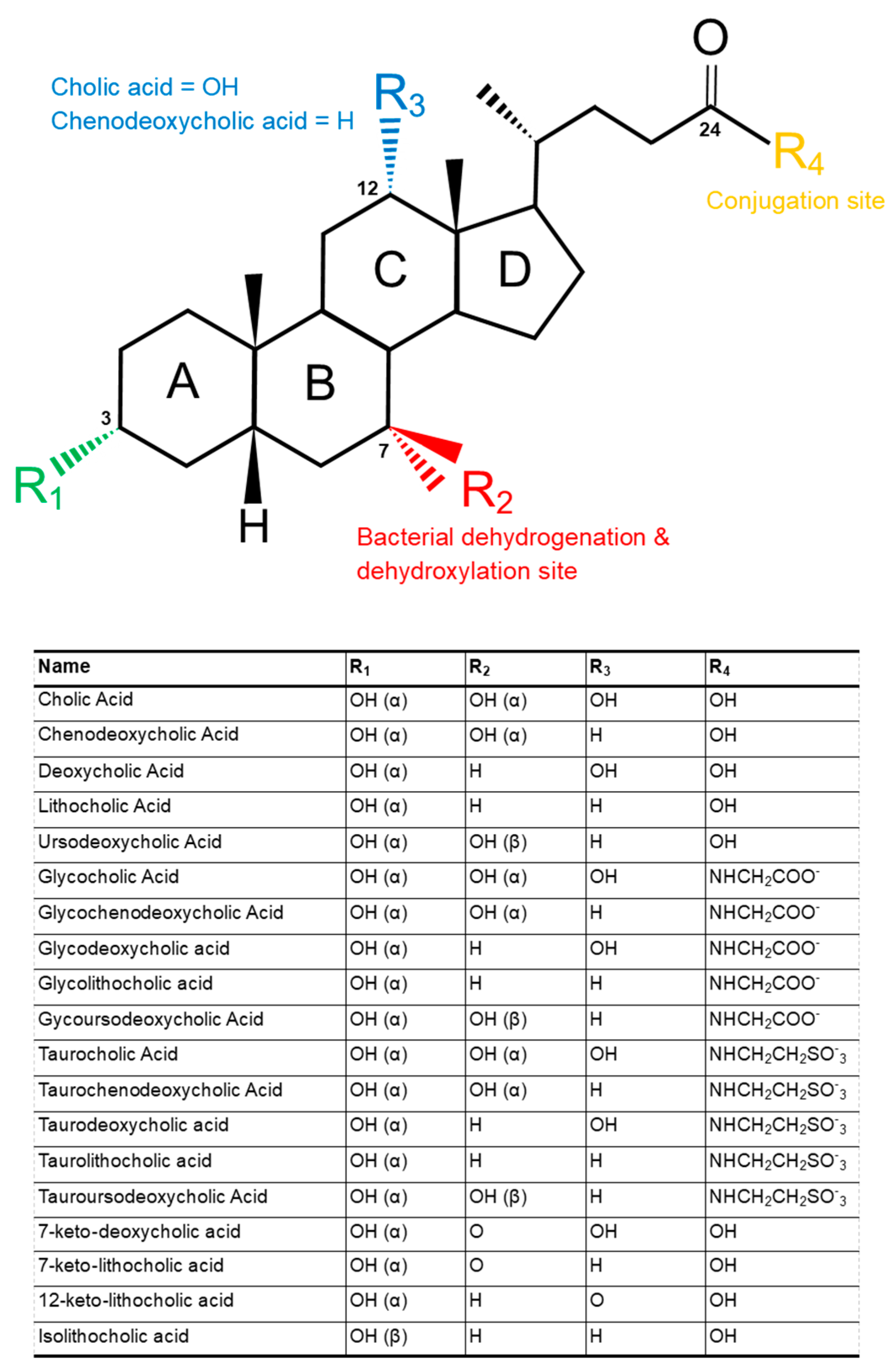
Figure 1. The schematic molecular structure of common bile acids found in humans annotated to show its relationship to other bile acids. α (represented by a dashed line) indicates a steric downwards orientation and β (represented by a solid line) indicates a steric upwards orientation.
Microbiolres 12 00023 g001
Figure 2. Host and bacterial primary and secondary bile acid metabolisms. Host transformation from cholesterol by enzymes (boxed) cytochrome P450 family 7 subfamily A member 1 (CYP7A1), in the classical pathway and CYP27A1 in the alternative pathway produces cholic acid and chenodeoxycholic acid, which are conjugated with taurine or glycine. Bacterial transformation often requires deconjugation via bile salt hydrolases (BSH), which exposes the bile acid to a range of further modifications. For simplicity, only deoxycholic acid and lithocholic acid are shown here, following dehydroxylation by enzymes encoded in the bile acid-inducible (bai) operon.
Microbiolres 12 00023 g002
Figure 3. Bile acids regulate their own expression as primary and secondary bile acids bind as agonists and antagonists to the nuclear receptor FXR, which transcriptionally activates the hormone FGF19 that inhibits CYP7A1 thus inhibiting bile acid synthesis. FXR, farnesoid X receptor; FGF19, fibroblast growth factor 19; CPY7A1, cholesterol-7α-hydroxylase; CYP27A1, Sterol-27-hydroxylase; C4, 7α-hydroxy-4-cholesten-3-one; BA, bile acid.
Microbiolres 12 00023 g003
Figure 4. Intestinal bacterial homeostasis maintained by primary and secondary bile acids. Stress, diet and antibiotics induced dysbiosis are treated by manipulating the microbiome via faecal microbiota transplantation (FMT); faecal filtrate transfer (FFT); and live biotherapeutic products (LBP).
Microbiolres 12 00023 g004
Publisher’s Note: MDPI stays neutral with regard to jurisdictional claims in published maps and institutional affiliations.