
North Italy: Welcome to the Tropics!

 ,
, 
 , and
, and 
Abstract
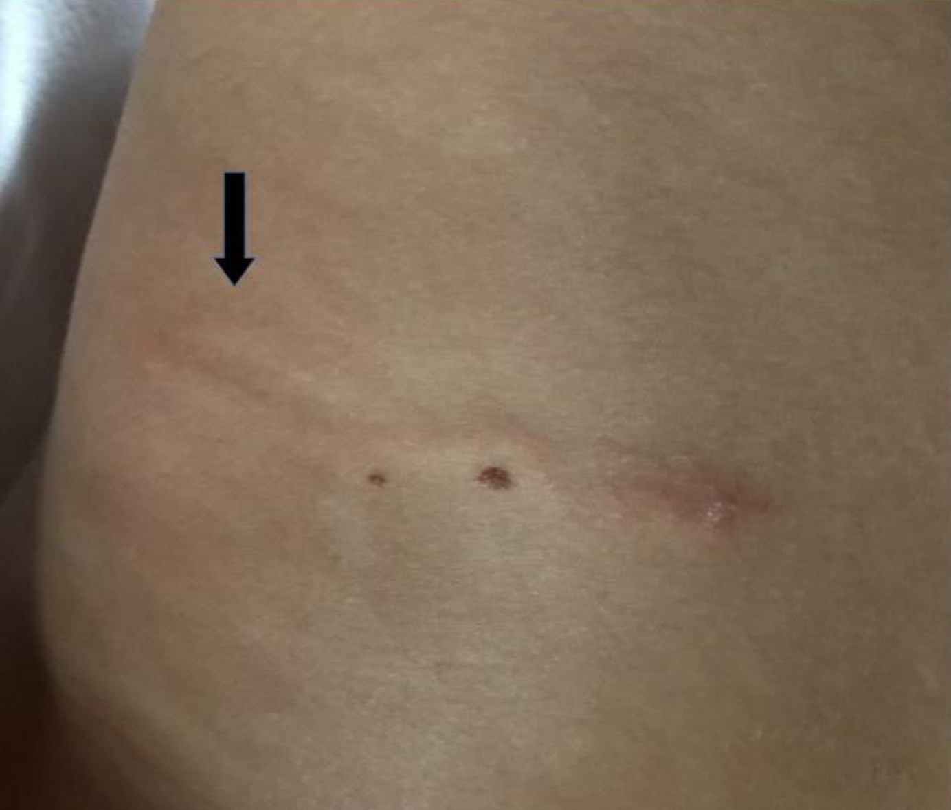
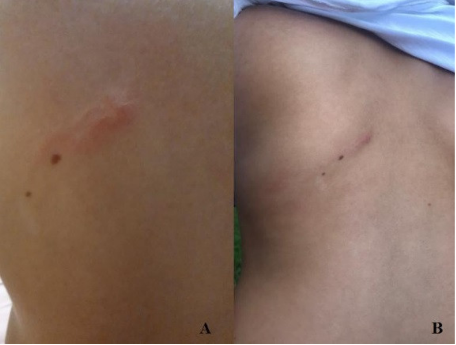
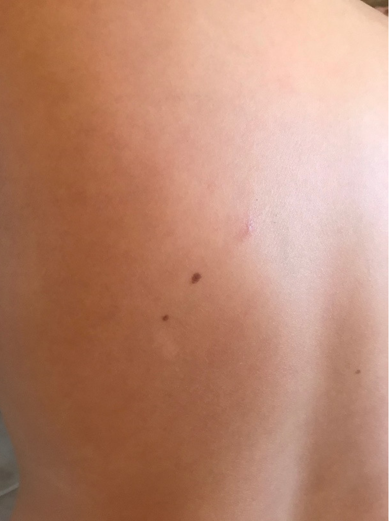
:1. Case Report
2. Discussion
3. Conclusions
Author Contributions
Funding
Institutional Review Board Statement
Informed Consent Statement
Data Availability Statement
Conflicts of Interest
References
- Bachmeyer, C.; Moreno-Sabater, A. Vesiculobullous cutaneous larva migrans in a 29-year-old man, diagnosed using teledermatology. Can. Med. Assoc. J. 2018, 190, E888. [Google Scholar] [CrossRef] [PubMed] [Green Version]
- Del Giudice, P.; Hakimi, S.; Vandenbos, F.; Magana, C.; Hubiche, T.; Giudice, P. Autochthonous Cutaneous Larva Migrans in France and Europe. Acta Derm. Venereol. 2019, 99, 805–808. [Google Scholar] [CrossRef] [PubMed] [Green Version]
- Howard, L.; Gibbs, S. A paediatric case of cutaneous larva migrans acquired in the UK. Clin. Exp. Dermatol. 2019, 44, 565–566. [Google Scholar] [CrossRef] [PubMed]
- Bowman, D.D.; Montgomery, S.P.; Zajac, A.M.; Eberhard, M.L.; Kazacos, K.R. Hookworms of dogs and cats as agents of cutaneous larva migrans. Trends Parasitol. 2010, 26, 162–167. [Google Scholar] [CrossRef] [PubMed]
- García-Rodrigo, C.G.; Romero, F.T.; Olivo, C.Z. Cutaneous larva migrans, welcome to a warmer Europe. J. Eur. Acad. Dermatol. Venereol. 2017, 31, e33–e35. [Google Scholar] [CrossRef] [PubMed]
- Herbener, D.; Borak, J. Cutaneous larva migrans in northern climates. Am. J. Emerg. Med. 1988, 6, 462–464. [Google Scholar] [CrossRef]
- Galanti, B.; Fusco, F.; Nardiello, S. Outbreak of cutaneous larva migrans in Naples, southern Italy. Trans. R. Soc. Trop. Med. Hyg. 2002, 96, 491–492. [Google Scholar] [CrossRef]
- Blaizot, R.; Goiset, A.; Caumes, E.; Gabriel, F.; Milpied, B. Cutaneous larva migrans: A case in Bordeaux, France and a systematic review of locally acquired cases in Europe. Eur. J. Dermatol. 2017, 27, 426–429. [Google Scholar] [CrossRef]

Publisher’s Note: MDPI stays neutral with regard to jurisdictional claims in published maps and institutional affiliations. |
© 2021 by the authors. Licensee MDPI, Basel, Switzerland. This article is an open access article distributed under the terms and conditions of the Creative Commons Attribution (CC BY) license (http://creativecommons.org/licenses/by/4.0/).
Share and Cite
Veronese, F.; Graziola, F.; Farinelli, P.; Zavattaro, E.; Tarantino, V.; Esposto, E.; Savoia, P. North Italy: Welcome to the Tropics! Infect. Dis. Rep. 2021, 13, 215-218. https://doi.org/10.3390/idr13010024
Veronese F, Graziola F, Farinelli P, Zavattaro E, Tarantino V, Esposto E, Savoia P. North Italy: Welcome to the Tropics! Infectious Disease Reports. 2021; 13(1):215-218. https://doi.org/10.3390/idr13010024
Chicago/Turabian StyleVeronese, Federica, Francesca Graziola, Pamela Farinelli, Elisa Zavattaro, Vanessa Tarantino, Elia Esposto, and Paola Savoia. 2021. "North Italy: Welcome to the Tropics!" Infectious Disease Reports 13, no. 1: 215-218. https://doi.org/10.3390/idr13010024
APA StyleVeronese, F., Graziola, F., Farinelli, P., Zavattaro, E., Tarantino, V., Esposto, E., & Savoia, P. (2021). North Italy: Welcome to the Tropics! Infectious Disease Reports, 13(1), 215-218. https://doi.org/10.3390/idr13010024