Review

Effects of Nutrients and Alcoholic Beverages on Gastrointestinal Tract Morphology

Department of Morphology, Institute of Anatomy and Anthropology, Riga Stradiņš University, LV-1007 Riga, Latvia
*
Author to whom correspondence should be addressed.
Gastroenterol. Insights 2025, 16(4), 42; https://doi.org/10.3390/gastroent16040042
Submission received: 17 September 2025 / Revised: 22 October 2025 / Accepted: 31 October 2025 / Published: 4 November 2025
(This article belongs to the Section Gastrointestinal Disease)

Abstract

This study aimed to review the effects of simple carbohydrates (SCs), fibre, proteins, fats, and alcoholic beverages on human gastrointestinal tract (GIT) morphology. Additional objectives included describing normal human GIT morphology, the mentioned dietary components, and their connection to GIT pathologies. An extensive literature review was conducted using PubMed, Scopus, ScienceDirect, and Google Scholar. This study revealed that excessive SC intake can increase intestinal permeability, modify gut microbiota, and cause tooth decay. Dietary fibre, through microbiota modulation, can enhance epithelium proliferation, improve intestinal barrier integrity, and prevent or manage GIT pathologies. Excessive protein consumption can decrease tight junction protein expression and increase inflammation, while insufficient intake can result in villi atrophy and increased permeability. A high-saturated-fat diet can increase intestinal permeability, increase inflammation, and promote gut dysbiosis, whereas omega-3 fatty acids can reduce inflammation and improve epithelial integrity. Immoderate alcohol use damages the GIT epithelium, causing inflammation and increasing the risk of cancer. The reviewed dietary components notably impact GIT morphology and are linked to various GIT pathologies. These findings highlight a balanced diet’s substantial role in preserving GIT health.

1. Introduction

Carbohydrates, proteins, fats, and water are integral components for the proper functioning of the human organism, yet modern society struggles with nutritional balance and moderation. The gastrointestinal tract (GIT) is the entrance for all consumed dietary components; thus, it is essential to investigate the latest discoveries on how nutrients and widely consumed beverages affect the morphology of the human GIT.
The simple carbohydrates (SCs), or monosaccharides, found in sweetened products are glucose and fructose. Glucose is typically the primary source of energy for cell metabolism, yet a correlation has been found between its consumption and intestinal permeability, which can lead to inflammation of the intestinal mucosa. Although there is not sufficient research on this issue yet, scientists theorise that it could contribute to inflammatory bowel disease (IBD) [1,2]. Fructose has similar effects, and an association exists between its consumption and irritable bowel syndrome (IBS) [3,4].
Dietary fibre (DF) is a nutrient that mostly consists of carbohydrate polymers with at least ten monomeric units that cannot be broken down by enzymes produced by the human body [5]. One of its most prominent roles is to promote the optimal metabolism of consumed foods. A broad spectrum of scientific research on DF’s influence on intestinal microbiota has been carried out, but only a few studies focus on its impact on GIT morphology. These studies have been performed on animals and humans using a broad spectrum of DF. Therefore, the reported results may vary and contradict one another. The conclusion that DF consumption undeniably has an impact on GIT morphology, such as villi length, crypt depth, mucosal integrity, and epitheliolytic proliferation, is prevalent in all of them [6,7,8]. DF intake impacts microbiota diversity, creating necessary conditions for many changes mentioned above [8].
Proteins are essential for the formation and functioning of an organism’s cells and structures [9]. In previous years, protein’s role in increasing and maintaining muscle mass has been widely discussed in the public domain; the global market value of isolated protein sources is increasing by 10.48% every year. Studies have shown the role of whey proteins in the regulation of inflammatory responses [10]. Despite the positive aspects, there is a necessity for a more thorough literature analysis, as previous studies are conflicting. Excess dietary protein intake has been shown to negatively affect the GIT by promoting the formation of ammonia and toxic amine compounds and stimulating gut dysbiosis [11,12].
Dietary fats supply the raw material necessary for each cell’s membrane and are the most significant energy reserve in the human organism. They provide protective functions, biologically active substance synthesis, and intestinal absorption of fat-soluble vitamins. However, in the present day, the increasing consumption of unhealthy fats has had adverse effects on the GIT. Most frequently occurring changes are increased intestinal permeability and dysbiosis, predisposing the person to the development of further illnesses [13]. High-fat diets (HFDs) also have an unfavourable effect on bile acid metabolism—increased production of secondary bile acids has been linked to colorectal cancer [14]. Fortunately, these alterations can be prevented with an appropriate and balanced diet. Thus, patients and healthcare professionals must be aware of the most recent discoveries regarding fats’ beneficial and detrimental effects on the human GIT.
The consumption of alcoholic beverages in Latvia continues to rise, increasing the relevance of research on its effects on the GIT. Alcohol directly irritates oesophageal tissues upon contact and contributes to the relaxation of the lower oesophageal sphincter [15]. Acetaldehyde, a by-product of ethanol metabolism, can induce faulty replication of GIT cells in its unmetabolized form [16]. To investigate the pre-existing information regarding these nutrients, this study aims to provide an extensive literature review on the effects of SCs, DF, proteins, fats, and alcoholic beverages on the morphology of the human GIT and their association with the development of GIT pathologies.
The objectives of the study are as follows:
1.
Describe the normal human GIT morphology.
2.
Describe the main characteristics of SCs, DF, proteins, fats, and alcoholic beverages.
3.
Describe the effects of consumption of SCs, DF, proteins, fats, and alcoholic beverages on the morphology of the human GIT.
4.
Describe the associations of the previously mentioned substances with the development of pathologies of the GIT.
Scientific databases such as PubMed, Scopus, ScienceDirect, and the academic search engine Google Scholar were used to write the literature review during the period from October 2024 to July 2025.
The publication search strategy included the following keywords: human gastrointestinal morphology, nutrition, simple carbohydrates, fibre, protein, fat, and alcoholic beverages. A complex keyword query was used, incorporating core study concepts and their synonyms (e.g., “human gastrointestinal morphology” OR “intestinal barrier”) AND (all studied nutrients and alcohol terms connected with OR).
The initial search produced 206,787 results; following the removal of duplicates and low-relevance records, a total of 205,637 items were excluded. A total of 1150 papers were screened by title and abstract. Only studies focusing on GIT structural changes as opposed to mere metabolism were included. Conclusively, 220 papers were selected for full-text assessment, and 99 scientific sources were included in the final review.
Studies were included if they met all the following criteria:
1.
Published in peer-reviewed scientific journals.
2.
Employed observational study designs (cohort, case–control, or cross-sectional), randomised controlled trials, or reviews.
3.
Published in the English language.
4.
Full-text articles available for review.
  • Studies were excluded if they did not fulfil any of the inclusion criteria.

2. Literature Review

2.1. Normal Morphology of the Human Gastrointestinal Tract

The gastrointestinal tract (GIT) is made up of the oral cavity, pharynx, oesophagus, stomach, and the small intestine, which is divided into the duodenum, jejunum, and ileum, and the large intestine, which includes the caecum; appendix; ascending, transverse, descending, and sigmoid colon; rectum; and anal canal. Accessory organs of digestion are the salivary glands, teeth and tongue, pancreas, liver, and gallbladder. The pancreas secretes enzymes to break down carbohydrates, proteins, and fats. The liver plays a vital role by producing bile for fat digestion, metabolising nutrients absorbed from the intestines, and detoxifying harmful substances [17,18]. The GIT wall consists of four layers: mucosa, submucosa, muscular layer, and serosa or adventitia. Mucosa is an essential part of the intestinal wall because of its direct contact with the lumen. It has three parts—epithelium, lamina propria, and muscularis mucosae. The stomach and intestines consist of a simple columnar epithelium, while a stratified squamous epithelium lines the oral cavity, oesophagus, and anus, providing superior resistance against mechanical stresses [17]. The main parts and structures of the GIT, as well as their functions, are presented in Figure 1 according to the order in which food moves through the GIT.
Normal and healthy GIT epithelium prevents unwanted substances and antigens from translocating into the lamina propria, where blood and lymphatic vessels are present. Therefore, preventing toxic substance dissemination systemically into deeper layers thus prevents inflammation. However, the selective permeability allows the vital absorption of nutrients. Tight junctions and adherent junctions together make the apical junctional complex, which is essential for maintaining selective molecular permeability by connecting the epithelial cells. Various proteins are involved in this action, such as occludins, claudins, and cadherins, as well as junctional adherent molecules. Furthermore, mucus secreted by goblet cells creates a protective lining against different antigens, together with defensins, immunoglobulin A immune responses, dendritic cells, and produced cytokines [13].

2.2. Description of Nutrients and Beverages

2.2.1. Simple Carbohydrates

Simple carbohydrates (SCs), or monosaccharides, comprise a single molecule with the formula Cx(H2O)n, where n is an integer between three and nine. They are composed of a chain of chiral hydroxy methylene units, terminating in a hydroxymethyl group at one end. At the other end, they contain either an aldehyde group, making an aldose, or an α-hydroxy ketone group, creating a ketose at the other end [19]. The most abundant monosaccharides in the diet are glucose, fructose, and galactose, absorbed in the small intestine. All three monosaccharides are found in fruit and vegetables. Glucose and fructose are found in honey, and galactose and glucose in dairy products [20]. When fructose is metabolised, part of it is utilised for synthesising fatty acids (FAs) and triacyl glycerides [21]. Fructose enters the enterocyte via GLUT receptors expressed in the apical membrane, often GLUT5. In turn, GLUT2 in the enterocyte basolateral membrane transports fructose to the blood vessels. The glucose and galactose pathways are similar, except they typically cross the enterocyte apical membrane via SGLT1 and GLUT2 transporters [3,22]. A summary of SC structure, digestion, and main sources is given in Table 1, together with other nutrients and beverages.

2.2.2. Fibre

By the definition of Codex Alimentarius, dietary fibre (DF) is a nutrient that mostly consists of carbohydrate polymers with at least ten monomeric units and that produce physiological benefits to health [5]. Enzymes produced by the human body cannot hydrolyse them, but they can be fermented by bacteria in the large intestine [23,24]. DF subgroups are non-starch polysaccharides and resistant starches. National authorities may also choose to include resistant oligosaccharides, i.e., non-digestible carbohydrates with 3–9 monomeric units, but in this study, we opted for the global DF definition provided by Codex Alimentarius [5,23]. DF is derived from plants and is primarily found in their cell walls. DFs differ by their physicochemical characteristics, like water solubility, fermentability, and viscosity, that is, their ability to bind water and adsorb ions and organic molecules. This nutrient is most often divided into two groups: soluble and insoluble. Soluble DFs, such as pectin, mixed-linkage glucans, hemicelluloses (e.g., xyloglucan and arabinoxylan), long-chain inulin, and others, are shorter and of a simpler structure. Most of them, upon entering GIT, bind water molecules and become gel-like. On the contrary, insoluble DFs, such as cellulose and lignin, are more complex, longer, and have less available surface area, which reduces their ability to bind water. Some DFs (e.g., hemicelluloses mentioned above), although being soluble in pure form, when consumed as plant cell walls from plant-based foods, because of their close proximity to cellulose, are found to be insoluble [24]. Soluble DFs are also fermentable by gut microbiota, but non-fermentable DFs are mostly insoluble; therefore, solubility can indicate, but does not equal, fermentability [23]. For example, resistant starch, which is insoluble, is efficiently fermented by gut bacteria [8,23]. During fermentation, short-chain fatty acids (SCFAs), such as acetic, propionic, and butyric acid; branched-chain fatty acids (BCFAs), like isobutyrate and isovalerate; and gases like hydrogen, carbon monoxide, and methane are produced [8,24]. World Health Organization’s (WHO) guidelines’ recommended DF consumption per day is > 20 g of non-starch polysaccharides and > 25 g of total DF. The preferred sources are wholegrain cereals, fruits, and vegetables [25].

2.2.3. Proteins

Proteins are high-molecular-weight compounds consisting of amino acids (AAs) linked by peptide bonds. Various animal products, like meat, eggs, dairy, seafood, and fish, are complete dietary protein sources since they contain all essential AAs. Most plant-derived proteins are limited in one or more essential amino acids—for instance, legumes are low in methionine, grains in lysine, and nuts and seeds in lysine or threonine. However, a balanced plant-based diet can supply all essential amino acids required for human health [26]. First, dietary proteins enter the stomach, where hydrochloric acid denatures them, making proteins more accessible for enzymatic action. Pepsin hydrolyses proteins to form shorter oligopeptide chains that enter the duodenum. There, pancreatic enzymes degrade the oligopeptides, making free AAs, dipeptides, and tripeptides, which are absorbed into the enterocytes in the small intestine. Dipeptides and tripeptides are hydrolysed intracellularly to form free AAs, which may be used to synthesise intestinal enzymes and epithelial cells. However, the majority enter the bloodstream and are carried to various tissues [27].

2.2.4. Fats

Fats or triacyl glycerides are composed of a glycerol molecule and three fatty acid (FA) chains and are classified depending on the structure of the FAs. Saturated FAs have zero double bonds and are commonly found in animal-derived products such as red meat, butter, dairy products, and refined oils. Trans-fats contain a double bond in a trans orientation and are most commonly synthetic and found in processed food [13]. Monounsaturated FAs have one double bond and are found in vegetable oils, dairy products, nuts, and red meat. Polyunsaturated FAs consist of at least two double bonds and comprise essential FAs such as omega-3 in fatty fish and omega-6 in animal fats and vegetable oils [28,29]. WHO recommends maintaining a daily dietary fat intake below 30% with saturated fats below 10% and trans-fats being no more than 1% of the total daily energy requirements for adults [30].
Digestion of fats begins in the duodenum, where bile and digestive enzymes are secreted. Dietary fat stimulates cholecystokinin secretion in the duodenum, which in turn stimulates bile elimination from the gallbladder. Most bile acids are reabsorbed in the ileum and returned to the liver [13]. Bile acids emulsify fat globules and increase the total surface area available to the lipases released from the pancreas. This results in the formation of fat micelles that serve as transport vehicles for the products of lipolysis to the surface of the enterocytes. The free FAs and monoacylglycerols are released from the micelle and diffuse across the apical membrane into the epithelial cells. Lipolysis products from short- and medium-chain triacyl glycerides with less than 12 carbon atoms diffuse through the enterocyte and into the portal vein. FAs and monoacylglycerols from long-chain triacyl glycerides with more than 12 carbon atoms are re-esterified and incorporated into chylomicrons, which are delivered into the bloodstream via the lymphatic system [13,31]. To examine the effects fats have on human health, researchers feed mice diets with different fat contents, measured by percentage of total daily caloric intake [13].

2.2.5. Alcoholic Beverages

Alcoholic beverages contain ethanol, which is obtained from fermentation processes involving yeast or bacteria. Ethanol (C2H5OH) is hydrophilic, yet it also has a slight ability to bind with nonpolar lipid molecules. Ethanol’s ability to dissolve in both water and lipids allows it to easily penetrate and damage the cell membranes of the GIT and absorb deeper into the mucosal tissues. It is primarily absorbed into the bloodstream through the stomach and small intestine by passive diffusion [32].
Ethanol is metabolised in the liver by alcohol dehydrogenase, microsomal ethanol oxidation system (MEOS), and catalase. The alcohol dehydrogenase pathway converts ethanol into acetaldehyde, a highly reactive and toxic metabolite, using NAD+ as a coenzyme, which accepts hydrogen ions released during the reaction. This process increases the NADH/NAD+ ratio, which disrupts FA oxidation and contributes to steatosis. Acetaldehyde can bind directly to the deoxyribonucleic acid (DNA) of GIT cells in its unconverted form, triggering faulty cell replication and causing oxidative stress. Simultaneously, the MEOS pathway, particularly CYP2E1, becomes more active with chronic alcohol exposure, using NADPH to oxidise ethanol while producing reactive oxygen species (ROS) that cause cellular injury [33].
WHO has stated in the Global status report on alcohol and health and treatment of substance use disorders 2024 that there is no safe amount of alcohol consumption that does not affect health. To define a safe limit for alcohol consumption, there would need to be scientific evidence demonstrating a threshold below which alcohol causes no harm. However, current evidence does not demonstrate any level of alcohol intake that could be considered completely safe. Available research consistently shows a dose–response relationship, indicating that the health risks increase with both the amount and duration of alcohol consumption [34].

2.3. Effects of Nutrients and Beverages on Gastrointestinal Morphology

2.3.1. Simple Carbohydrates

A diet rich in fructose and glucose may increase intestinal wall permeability. However, the studies are ongoing, and this question should be perceived as open. For now, most of the research is conducted on mice. An experiment in which mice were orally fed such a diet showed higher plasma levels of FITC–dextran, indicating higher intestinal wall permeability in mice. They also had fewer tight junction proteins in the colon, such as occludin and ZO-1, responsible for controlling gut wall permeability [35]. Diet-induced increased gut permeability has been shown to correlate positively with inflammation, which was assessed in the study using inflammatory cytokines. Also, a state called metabolic endotoxemia can develop, meaning that endotoxins from intestinal bacteria can travel through the blood, causing an inflammatory response in tissue elsewhere. Mice that were fed a high-fructose or high-glucose diet had significantly higher expression of TNF-α and IL-1β in the colon. Similar results were seen in the liver, joined by increased expression of MCP1 and TLR4 proteins. The histological analysis of liver tissue showed increased lipid accumulation, leading to hepatic steatosis (more severe in mice fed with a high-fructose diet). In both diet models, similar adipocyte hypertrophy was observed, as well as glucose intolerance and increased fasting blood glucose concentration. Thus, high glucose and fructose consumption can increase intestinal permeability, in turn stimulating inflammation [2,36]. A schematic representation of this process of alterations in mice caused by GIT morphology due to a high-fructose and -glucose diet is shown in Figure 2.
There is an established positive correlation between irritable bowel syndrome (IBS) and dysplasia in the intestinal mucosa, which in several studies has been linked to cancer, most commonly colorectal. It is also possible to come to a state called “mucosa indefinite for dysplasia”. In this case, the biopsy material contains a certain number of atypical cells, but not enough to be classified as dysplasia [37]. While high simple carbohydrate (SC) consumption contributes to mucosal inflammation and increased gut wall permeability, the precise association between these effects and the diagnosis of inflammatory bowel disease (IBD) is still under investigation. However, it is not possible to state irrefutably that there is an association between monosaccharide intake and the development of cancer in the GIT. It is even possible that the opposite effect is present—a decrease in the metabolic activity of cancer cells. Monosaccharides such as ᴅ-mannose, ᴅ-galactose, ᴅ-glucosamine, and xylitol can inhibit glycolytic enzymes when they enter tumour cells in copious quantities via highly expressed GLUT transporters, thereby reducing energy production and inducing tumour cell apoptosis [36].

2.3.2. Fibre

Dietary fibre (DF) can impact GIT morphology directly and indirectly, i.e., through microbiota [7,8,38,39,40,41]. The composition of microbiota can be altered by the foods consumed, but it can take up to six weeks for microbiota to change and stabilise after a switch in diet [8,24]. Research performed on DF supplementation’s impact on weaning piglets revealed that if at least 1% of food intake consists of DF, a favourable effect on microbiota diversity can be observed. Most noticeable results occur when intake of both soluble and insoluble DF is increased, as well as after the dietary changes have been implemented long-term. One of the possible mechanisms as to how these changes happen might be related to transit time. Soluble DF makes the content of the intestines more viscous, prolonging the time spent in the intestines and supporting the multiplication of some of the bacteria. It also has the potential to reduce enterotoxigenic Escherichia coli colonisation. Soluble DF alone or in combination with insoluble DF also lowers the incidence of diarrhoea and stimulates SGLT1, PePT1, and GLUT2 gene expression. On the other hand, an increase in insoluble DF intake alone is associated with more active peristalsis, which in turn shortens the transit time of digesta, reducing the proliferation of pathogens in the GIT. It is also associated with decreased inflammatory marker (IL-1β, TNF-α, IL-10) expression [42]. The comparative effects of soluble and insoluble DF on GIT morphology are presented in Table 2.
Furthermore, diet can affect microbiota by supplying specific bacteria with needed metabolites for their catabolic processes. For instance, when the substrate flow of fermentable DFs that are rich in β-glucan is increased, it partially changes the location of nutrient metabolism from the small intestine to the large intestine, thereby promoting the Firmicutes phylum diversity [43]. On the contrary, lack of DF intake may lead not only to fewer beneficial bacteria in microbiota but also to a change in enterocyte metabolism. Under normal conditions, i.e., when DF is ingested and butyrate, which is one of the short-chain fatty acids (SCFAs), is produced, intestinal epithelial cells use it for energy via β-oxidation. That supplies colonocytes with 60–70% of the required energy and stimulates their proliferation [8,24]. β-oxidation consumes large amounts of oxygen and maintains anaerobic conditions (containing less than 1% oxygen, i.e., pO2 < 7.6 mmHg, on the surface of colonocytes) in the gut; therefore, microbiota is dominated by obligate anaerobes. In butyrate deficiency, the intestinal epithelium produces energy by anaerobic glycolysis. In this case, the oxygen concentration in the intestine increases, creating suitable conditions for the growth of facultative pathogens such as Salmonella spp. [44].
Similarly to butyrate, propionate, which is another SCFA, is also metabolised by intestinal epithelial cells via a β-oxidation-like pathway, therefore creating a hypoxic environment in the intestinal lumen. This causes the release of hypoxia-inducible factor, especially HIF-2α. This factor upregulates MUC2 expression, stimulating mucin production and secretion from goblet cells, thus improving the state of the mucus layer [40]. The mucus layer’s viscosity may also be increased due to β-glucans, which can be found in some soluble DFs and penetrate the intestinal mucus layer. They also reduce the average pore size of the mucus layer, therefore lowering permeability [38]. In intestinal crypts, metabolites from DF fermentation not only promote goblet cell function, but also stem cell differentiation into goblet cells [41]. Stem cells in the intestinal crypts have the potential to become secretory cells—Paneth cells, goblet cells, enteroendocrine cells, and enterocytes. The development of these cells is mediated by enhanced activation of the Wnt signalling pathway, e.g., by bacteria such as Akkermanisa muciniphila in the microbiota. These bacteria promote the production of the Wnt3 signalling molecule by Paneth cells, thereby promoting the development of stem cells into secretory cells, including goblet cells. These bacteria also indirectly promote epithelial renewal and increase the production of acetate and propionate. Through its beneficial impact on the GIT, A. municiphila may lessen damage during radiation and chemotherapy treatments [41]. As for Lactobacillus spp. and Bifidobacterium spp., which are part of the microbiota and have been discovered to have a positive correlation with DF consumption, they can stimulate the expression of genes responsible for tight junction protein synthesis (CLDN-1, OCLN) [7,8]. These changes improve mucosal integrity, which in turn more effectively protects enterocytes and the organism from harmful pathogens and substances from the GIT.
On a more macroscopic level, villi length and crypt depth may increase or decrease. The changes vary depending on the type of DF and the amount of intake. A study on the effect of fibre source and concentration on morphological characteristics in the GIT of pigs revealed that soluble DF (in this case, pectin) was associated with shorter villi and crypts; on the other hand, a high high-insoluble (in this case, barley hull)-DF diet increased villi length [7]. In a different study, sodium carboxymethylcellulose (CMC), a non-fermentable viscous compound, was added to a low-fibre diet in weaning pigs. The largest values for small intestinal villi length and width of the muscle layers underneath them were seen in pigs fed with a low-viscosity CMC diet, but the smallest were in those fed with a high-viscosity CMC diet. The results of the control group without added CMC were between those of the low-viscosity CMC and high-viscosity CMC diet [45]. This DF, because of its polymers’ size and charge, may create reversible compression of the mucus layer [39].

2.3.3. Proteins

An organism’s ability to consume and digest substantial amounts of protein without causing damage to health varies depending on an individual’s age, biological sex, daily physical activities, and other factors. Therefore, it is difficult to name a universal maximal safe daily protein intake. Although the World Health Organisation (WHO)-recommended minimum daily intake for adults is 0.83 g per kilogram of body weight, the human organism can digest 3 to 4.4 g per kilogram a day for prolonged periods [46,47,48].
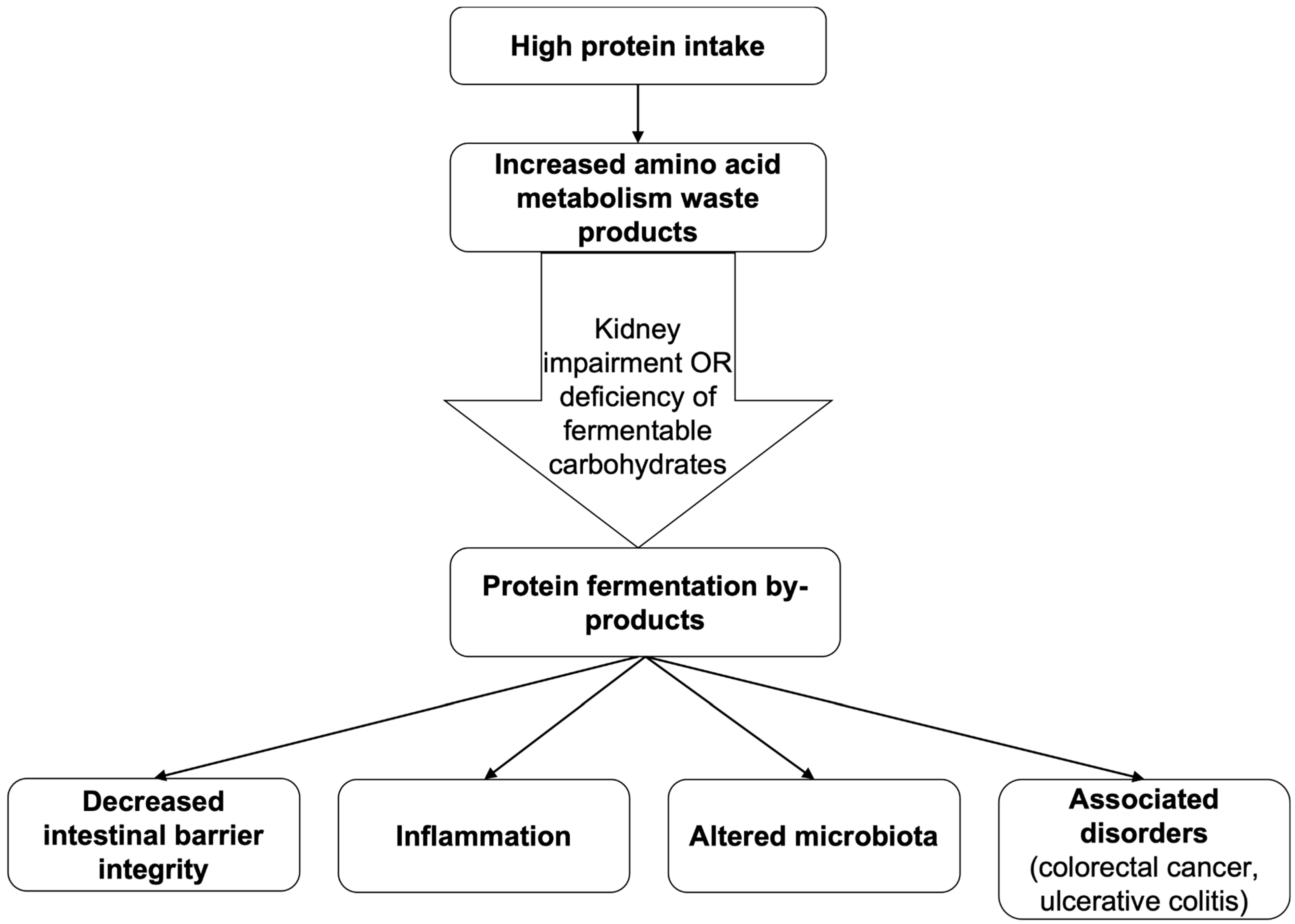
Changes caused by increased protein intake mostly manifest if the kidneys cannot effectively discharge waste products, such as urea and ammonia, from amino acid (AA) metabolism, as presented in the schematic illustration in Figure 3. This state is predominantly seen in people with chronic kidney disease, leading to urea accumulation and passive diffusion into the GIT. Increased levels of ammonia deplete the number of tight junction proteins in enterocytes, causing a decline in intestinal barrier integrity and increasing its permeability [12]. On the contrary, if consumption of a well-balanced diet is present, the metabolism of the essential AA tryptophan would increase the number of tight junction proteins in enterocytes [49]. Furthermore, elevated levels of urea are associated with a substantial number of inflammatory cells in the mucosa. An intestinal enzyme, microbial urease, converts urea into ammonia, which can inhibit cell mitochondrial respiration and cause necrosis of the GIT mucosa only at a concentration of 0.02% [50]. Moreover, the metabolism of urea leads to a slight pH increase in the intestinal lumen, causing changes in microbiota [12]. High-protein diets (at least 30% of caloric intake comes from proteins) can alter microbiota even in people without kidney function disorders [11]. Protein, similarly to DF, can be fermented by gut microbiota, producing ammonia, branched-chain fatty acids (BCFAs), amines, phenols, sulphides, and thiols. An excess of these metabolites (except BCFAs) has been associated with colorectal cancer, ulcerative colitis, and other bowel disorders. In most conditions, protein fermentation is insignificant when compared to carbohydrate, like DF, fermentation, and therefore does not cause detrimental effects on the body. However, the fermentation rate may increase when there is a shortage of fermentable carbohydrates [24].
The classification of protein–energy undernutrition includes primary and secondary forms. The primary form is caused by inadequate uptake of proteins in children and older people and most commonly manifests as marasmus or kwashiorkor. The secondary form is the result of some pathologies affecting the ability of the GIT to digest, uptake, or transport proteins with the lymphatic system, for example, pancreatic insufficiency and enteropathy. The other instance is catabolism-increasing disorders, such as cancer and acquired immune deficiency syndrome (AIDS) [51]. In the jejunum mucosa of a child affected by kwashiorkor and marasmus, various villi, both normal and almost atrophied, are observed. However, histologically, partial villus atrophy, an increase in the number of deep crypts and T-lymphocytes, infiltration into the mucosal connective tissue, as well as numerous eosinophils and plasma cells are most frequently found. On the other hand, the less affected mucosa is characterised by short enterocytes with pronounced vacuolization. The morphologies of the microvilli are also variable: some are long and densely arranged, others short and sparse [52].
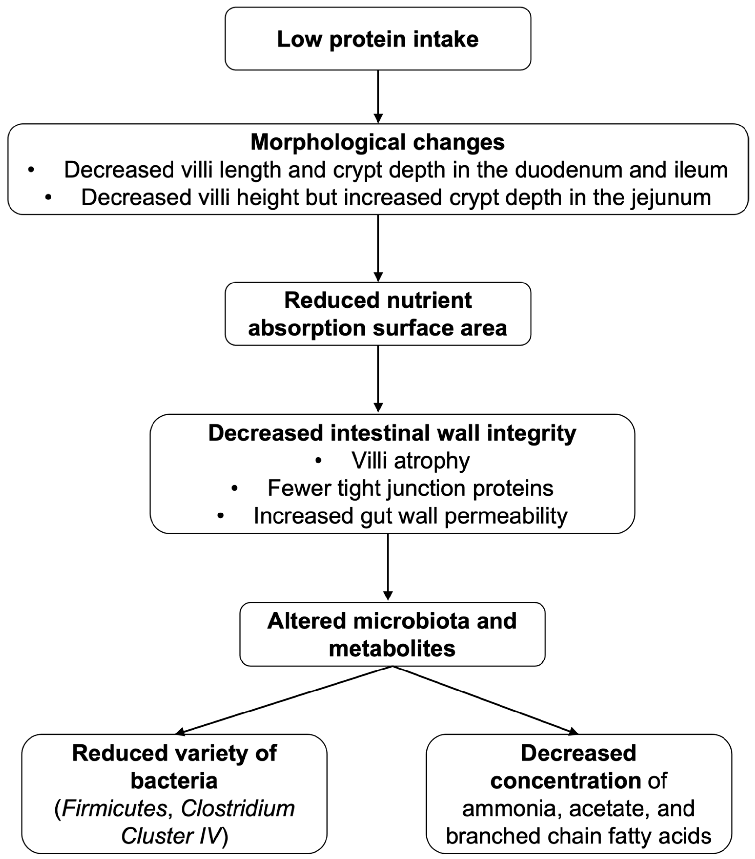
A Chinese study published in 2019 concluded that the length of the intestinal villi in the duodenum and jejunum of piglets decreased after reducing dietary crude protein intake from 23.1% to 17.2% while still ensuring the intake of eight essential fatty acids (FAs). The low-protein diet lowered the villi height and crypt depth, leaving their ratio in the duodenum and ileum unchanged. In contrast, the crypts deepened in the jejunum region, thereby reducing the ratio between the villous height and crypt depth, with a consequent decrease in the surface area of nutrient absorption [52]. Another group of researchers found that in piglets, lower protein consumption was positively correlated with a decrease in ileum wall integrity and villi atrophy. In this part of the intestine, the group with the highest protein consumption showed the longest villi. At lower protein contents in the diet (12%), fewer tight junction proteins (claudin-3 and claudin-7) were observed. Meanwhile, in the colon, occludin, claudin-1, claudin-7, and ZO-3 counts decreased in the group with 15% protein content in the diet. This tendency was observed even more in the 12% group [53]. Regarding microbiota, reducing crude protein content in the diet of pigs from 20% to 14% reduced the variety of bacteria, especially Firmicutes and Clostridium Cluster IV in the caecal chyme, as well as ammonia, acetate, and BCFA concentration [52]. The various effects of protein deficiency in the diet are shown in the schematic representation in Figure 4.

2.3.4. Fats

Dietary fats can have a different impact on the morphology of the GIT depending on their proportion, the type of FA, and the duration of consumption in the diet. Research conducted both on humans and on animals, analysing the effect of a prolonged high-fat diet (HFD) on GIT, has reported associations with gut dysbiosis, inflammation, and increased intestinal permeability when fat intake was 40% to 60% of the daily energy requirement [54,55,56].
Alterations in the morphology of the GIT due to dietary fats are achieved through complex mechanisms, one of them being indirect changes mediated by intestinal microbiota. During a six-month HFD, where fat comprised 40% of total daily caloric intake, the count of butyrate-producing and anti-inflammatory bacteria genera, Faecalibacterium, decreased, whereas the count of Bacteroides, Alistipes, Firmicutes, and Enterobacteriaceae genera increased [56]. The elevated presence of these bacteria enhances the production of lipopolysaccharides (LPSs), which can promote inflammatory processes and stimulate the release of arachidonic acid and its related metabolites. This leads to inflammation and oxidative stress that damage the intestinal mucosal tissue. An increase in LPS concentration downregulates the expression of tight junction proteins, leading to increased intestinal permeability [54]. Consequently, the HFD group exhibited an increased concentration of arachidonic acid in their faecal samples [54,57]. Intestinal dysbiosis also causes a reduction in SCFA, especially in butyrate, due to an imbalance between butyrate-producing and -degrading bacteria. Butyrate is an essential energy source for enterocytes. Thus, the mentioned alterations in the bacterial count can exacerbate atrophic processes and increase the permeability of the enterocyte monolayer [57].
Several studies have focused on the specific effects of FAs on gastrointestinal morphology. Consumption of saturated FAs has been shown to raise the number of hydrogen sulphide-producing bacteria of the genus Desulfovibrio, which in turn decreases the transepithelial resistance. The produced hydrogen sulphide impairs the butyrate oxidation process, which can lead to cellular dysfunctions. Research has shown that a diet rich in saturated FAs can cause decreased intestinal barrier resistance in the large intestine, increased inflammatory cell infiltration, and higher bacterial DNA count in mesenteric adipose tissue, which indicates enhanced intestinal permeability [57]. Another trial showed a substantial increase in faecal palmitic and stearic acid concentration in the HFD group. These saturated FAs have been shown to increase the inflammatory signalling pathways in macrophages, adipocytes, and myocytes [58].
The mentioned changes regarding saturated FAs were not present in mice fed with a diet high in omega-3 and omega-6 FAs. Omega-6-rich diets did not have any effect on the intestinal barrier [57]. However, increased consumption of omega-6 FA-containing products such as soybean oil is associated with pro-inflammatory effects. This is due to increased production of arachidonic acid, resulting in increased biosynthesis of prostaglandin E2 and thromboxane B2 [58].
An HFD has also been linked to elevated levels and altered profiles of secondary bile acids in faeces. An increase in deoxycholic acid’s concentration activates the nuclear factor-κB inflammatory pathways, causing increased levels of tumour necrosis factor in faecal samples. Therefore, these bile acids can damage the enterocyte monolayer and decrease the intestinal cells’ ability to regenerate. This study also found that intestinal permeability alterations due to an HFD are associated with increased expression of the farnesoid X receptor because of its involvement in tight junction protein function. As a result, intestinal permeability was increased in the jejunum, as well as in the large intestine. Interestingly, the only secondary bile acid whose concentration decreased due to the HFD was ursodeoxycholic acid, which is thought to be cytoprotective. Therefore, this decrease correlated with decreased intestinal barrier function, supporting the fact that not only the amount but also the bile acid profile are essential to consider [55]. However, a single high-fat meal in healthy participants did not cause significant changes in intestinal permeability markers and did not induce LPS translocation acutely and after 24 h, despite previous accusations [58].
Omega-3 FAs, compared to others, provide many beneficial effects and are essential for developing healthy gut microbiota and regulating inflammatory processes. Fish oil supplementation has been shown to restore gut permeability to the control group state after a saturated HFD and decrease the quantity of macrophages by 72% in the colon compared to a saturated HFD [57]. The consumption of Brazil nuts and perilla seed oil, which are filled with omega-3 FAs, can increase the number of beneficial bacteria, such as Bifidobacterium and Ruminococcus, whilst lowering the number of potentially harmful bacteria genera, for instance Enterobacteriaceae [54,59]. Prevention of dysbiosis promotes decreased pro-inflammatory cytokine production and oxidative stress, increased expression of tight junction proteins, and protection of goblet cells in the ileum [54]. Moreover, omega-3 FAs comprising 60% of the dietary caloric intake did not cause any adverse effects on the GIT, suggesting that an HFD consisting of omega-3 FAs is the only healthy instance based on the FA profile [57]. Furthermore, omega-3 FAs within a minimum of two months reverse HFD-induced dysbiosis and inflammation, as well as increase intestinal permeability [54,57]. Comparison between the contrasting effects of a diet filled with saturated or omega-3 FAs can be seen in Table 3.

2.3.5. Alcoholic Beverages

Following oral ingestion, alcoholic beverages encounter the oral cavity and oesophagus. The mucosa of the oral cavity consists of the non-keratinised or partially keratinised stratified squamous epithelium, serving as a mechanical barrier [60]. Ethanol is partially absorbed through these tissues, and due to its lipophilicity, it can disrupt cell membranes by dissolving lipid components, as shown in Table 4. As a result, alcohol increases cellular permeability, weakening the protective barrier function. Prolonged exposure may induce inflammation, leading to cellular damage and cellular dysplasia [61]. Reactive oxygen species (ROS) production, acetaldehyde toxicity, and the lipophilic nature of ethanol associated with prolonged alcohol exposure impair the structure and function of the salivary glands, therefore reducing saliva production in the oral cavity. Chronic alcohol use further increases TNF-α expression, leading to apoptosis of acinar cells, the main secretory units of the glands. In addition, alcohol causes fat accumulation, acinar cell swelling and atrophy, and alterations in salivary flow rate, protein synthesis, and electrolyte composition, all of which contribute to reduced salivary secretion. As a result, the oral cavity’s defence mechanisms are weakened, with reduced antimicrobial and buffering capacity, as well as decreased lubrication, increasing the risk of mucosal injury, infections, and dental caries [62]. Upon entering the oesophagus, ethanol encounters a tissue structure that lacks robust protective mechanisms against potent chemical irritants. As a result of its effect, ions or water can enter the cells unhindered, and ROS levels may rise. That can reversibly damage membrane lipids, further affecting cell shape and protective functions, increasing the likelihood of apoptosis or dysplasia [63].
Within the stomach, ethanol reduces the number of mucus-producing cells and their ability to secrete mucus by directly damaging the cells, promoting oxidative stress, triggering inflammatory responses, and reducing the level of prostaglandins necessary for mucus production. These cells produce a thick, gel-like mucus which forms a highly viscous barrier and protects the stomach lining from self-digestion and abrasion. A reduction in mucus secretion intensifies tissue vulnerability and facilitates further gastric mucosal damage [63].
The liver, as the main organ responsible for metabolising ethanol, is particularly vulnerable to alcohol-induced damage. Alcohol dehydrogenase, the microsomal ethanol oxidation system (MEOS), as well as the catalase pathway convert ethanol into acetaldehyde, which forms adducts with proteins, lipids, and DNA, impairing their function and triggering immune responses. Additionally, a small portion of ethanol undergoes esterification with FAs via carboxylesterase or carboxyl ester lipase in the liver and pancreas, forming FA ethyl esters. These compounds can contribute to pancreatic inflammation and increase the risk of binge alcohol-related liver injury [33].
By disrupting FA oxidation and autophagy, ethanol metabolism promotes hepatic steatosis, defined by lipid accumulation in hepatocytes. Steatosis may progress to alcoholic steatohepatitis, a more severe inflammatory stage within the broader spectrum of alcohol-associated liver disease (ALD). Hepatotoxicity is further intensified by the generation of ROS through the MEOS pathway, particularly via CYP2E1, whose loose association with cytochrome P450 reductase allows ROS formation even in the absence of substrate [64].
Alcohol increases gut permeability, allowing LPSs to enter the liver via the portal vein, which further activates Kupffer cells and hepatic stellate cells, promoting the release of pro-inflammatory cytokines, which enhance liver injury. Lastly, chronic alcohol exposure impairs hepatocyte regeneration by altering gene expression and disrupting cell cycle signalling [64].
In the intestines, ethanol promotes inflammatory processes and induces oxidative stress, resulting in an imbalance of ROS. One of the primary targets is the intestinal epithelial tight junctions, which maintain barrier integrity. Ethanol increases ROS production in the intestinal epithelium, damaging cellular proteins and lipids, which weakens intercellular connections and thereby increases intestinal epithelial permeability. Additionally, ethanol metabolites stimulate cytokine release (TNF-α, IL-1β), which negatively affects transmembrane proteins such as occludin and claudin, which are necessary for the formation of a tight and effective gap structure [61].
Chronic ethanol exposure induces structural and functional modifications in the intestinal epithelium, contributing to gut dysbiosis. Ethanol decreases the number of beneficial bacteria, promotes the proliferation of pathogenic species, and reduces microbial biodiversity. Chronic alcohol consumption does not induce significant changes in the population size of Lactobacillus species. However, it can lead to an increase in the population of bacteria from the Parabacteroides, Enterobacteriaceae, Streptococcaceae, Prevotellaceae, Veillonellaceae, Bacteroidaceae, and Clostridium spp. groups, or a reduction in the populations of Bacteroidetes, Lachnospiraceae, and Ruminococcaceae in the rat gut microbiota. Modifications in the microbiota can reduce the effectiveness of the intestinal barrier, compromise junction integrity, and activate inflammatory processes [65].
To gain a better understanding and summarise the comparative effects of simple carbohydrates, dietary fibre, saturated fats, and alcoholic beverages on gastrointestinal microbiota, inflammation, enterocyte integrity, and permeability, Table 5 is provided.

2.4. Nutrients and Beverages in Relation to Gastrointestinal Pathologies

2.4.1. Simple Carbohydrates

To assess how a diet rich in simple carbohydrates (SCs) affects the development of inflammation in the intestinal wall, three groups of mice were compared. One group received a normal diet, another was given a high-sugar diet, and the third was provided with a fibre-rich diet. When acute colitis was induced, it was found that the high-sugar-diet-fed mice developed acute colitis in a more severe form. Similar observations were also made regarding the exacerbation of chronic colitis. Microbiota analysis of faecal samples showed that high-sugar-diet-fed mice had an increase in Enterobacteriaceae and Turicibacter populations. In addition, the increase in E. coli in mice with colitis was 23.9% compared to healthy mice [2]. In the case of a high-sugar diet, the oral microbiota can metabolise SCs, producing acids, mainly lactic acid. These can contribute to the demineralisation of tooth enamel, thus increasing the risk of caries. Substitution of SCs with caloric sweeteners such as xylitol and mannitol can lead to osmotic diarrhoea, as they are only partially absorbed in the small intestine [66,67].
IBS is a multifactorial disease whose aetiology is not completely explored and whose pathogenesis involves many varied factors, one of which is the nutrient spectrum of the diet. As pharmacological treatment has not been very effective in treating IBS, lifestyle changes, especially diet, could be the way to improve the symptoms of this disease. The association of IBS with a carbohydrate-rich diet has been investigated. FODMAP (fermentable oligosaccharides, disaccharides, monosaccharides, and polyols)-rich diets are diets which contain an increased dietary fraction of these nutrients. A FODMAP-rich diet’s main components are wheat, dairy, some vegetables, and fruits. For example, wheat, barley, rye, couscous, white bread, croissants and muffins made from wheat, white pasta, crisp breads, apples, dates, cauliflowers, onions, beans and pulses, cashew and pistachio nuts, rum, fennel tea, buttermilk, Greek yoghurt, processed cheese, custard, dairy ice cream, etc. Therefore IBS patients could try to exclude these items from their diet and try replacing them with alternatives such as starchy foods (rice, oats), potatoes, polenta, corn, wheat-free or gluten-free bread or pasta, porridge, rice crackers, flapjacks, flourless cakes, unripe banana, strawberry, asparagus, aubergine, turnip, peanuts, pecans, non-caffeinated drinks, lactose-free milk or yoghurt, almond or hazelnut milk, cottage cheese, ricotta, dark chocolate, and others, thus practicing a low-FODMAP diet [68]. Over time, numerous studies have established a link between increased consumption of short-chain carbohydrates and their poorer absorption in the small intestine. They are then fermented by bacteria in the large intestine to produce various metabolites. This process contributes to the onset or worsening of IBS symptoms such as bloating, flatulence, and abdominal pain [1]. However, there are also studies that point to a multifactorial interaction between the microbiota and IBD, which makes the study of this association particularly challenging and does not prove the association of the microbiota with IBD pathogenesis, disease progression, or development [69]. Another prospective randomised study performed in India compared the IBS severity scores in two groups: low-FODMAP diet (LFD) and traditional dietary advice diet (TDA). The LFD group had a significantly lower IBS severity score index and showed markedly better results of IBS symptom relief than the TDA group after 4 weeks (62.7% and 40.8%, respectively) and after 16 weeks (52.9% and 30.6%). In both study groups, decrements in different IBS severity scoring component values were observed. Unlike in the case of TDA, LFD also showed a reduction in abdominal pain severity sub-score; stool frequency and consistency only significantly improved in the LFD group [70].
High intake of SCs has the potential to affect GIT morphology and lead to the development of various pathologies in the long term. For example, chronic inflammation in the gut wall can contribute to the development of GIT, IBD, and chronic colitis, as well as microbiota alterations. In the oral cavity, the tooth surface is at risk of caries [1].

2.4.2. Fibre

Dietary fibre (DF) consumption and the interlinked effect on intestinal microbiota are closely related to the prevention or improvement of many pathologies. A study showed that people who consume more high-fibre foods suffer from fewer non-infectious diseases (e.g., diabetes, cardiovascular disease, and in relation to the GIT—colon cancer) [8]. Generally, DF-rich diets may contribute to the prevention or management of GIT pathologies such as functional dyspepsia, diverticulitis, haemorrhoids, IBS, ulcerative colitis, and even colon cancer [7,71,72,73,74].
Inflammation is a major component in many of the pathologies. One of them is diverticulitis, which is an inflammation of the diverticula. In a 24-year-long prospective cohort study of 121,700 US women, higher DF (especially from fruits and cereals) intake was inversely associated with risk of diverticulitis. Fruits such as apples, pears, and prunes were the most associated with a lower risk, which may partly be because they were the most consumed fruits. DF might not be associated with the prevention of the development of diverticulosis, but it can reduce the risk of inflammation associated with this pathology [71].
Just as simple carbohydrates may influence the manifestation of IBS symptoms, so can DFs. In the case of IBS, transition to a low-FODMAP diet is advised because of rapid fermentation of these nutrients [23,70]. Because of FODMAPs’ wide presence in DF-containing products, lowering FODMAP intake negatively impacts DF consumption, causing changes to, for example, the composition of microbiota and epitheliocyte metabolism [8,24]. In a study performed on mice, models of specific probiotic strains (Bifidobacterium, Lactobacillus, Streptococcus, or a mixture of three) on post-infectious IBS (PI-IBS) were analysed. Probiotics containing Bifidobacterium, Lactobacillus, and a mixture increased nociceptive receptor threshold, therefore lowering visceral sensitivity, significantly reducing intestinal permeability by increasing tight junction protein expression and decreasing IL-6 and IL-17 expression. The Bifidobacterium strain and the mixture also decreased contractile responses to neurotransmitter acetylcholine [7]. The mechanisms of PI-IBS development are suggested to be associated with changes in intestinal permeability and persistent low-grade inflammation. Therefore, these observed effects that indicate improvement in intestinal hypersensitivity, intestinal barrier function, and inflammation point towards improvement in symptoms of PI-IBS. Resistant starches, especially type 2 and type 3, are associated with increased acetate and butyrate production as well as a rise in Bifidobacterium, therefore enabling the positive effects described above and providing epitheliocytes with metabolites needed for energy production [7,8]. Meanwhile, resistant starch is slow fermenting; therefore, its use in moderation should be tolerable, but it may also vary from patient to patient, and further research is needed [8,75]. A different study of a randomised controlled crossover trial looked at the impact of supplementing low-FODMAP diets with insoluble DF—sugarcane bagasse with/without resistant starch—for IBS patients. Supplementation of sugarcane bagasse improved symptoms for the patients with constipation-type IBS, as the DF normalised low stool water content and decreased colonic transit time [76]. Overall, global guidelines of IBS, including its management, advise a DF-rich diet with both soluble and insoluble DF combined with adequate amounts of water. At the same time, insoluble and easily fermentable DFs may exacerbate symptoms [77]. The bottom line is that DFs’ impact is immensely variable depending on DF type (as their properties can be different), IBS type, and each patient individually. For many years, in developed countries, high-energy and low-DF refined foods have been on the rise, leading to changes in microbiota, and a low diversity of abundance in gut flora has been linked not only to obesity, but also to IBD [78]. Lack of DF causes intestinal epitheliolytic metabolism to switch from aerobic β-oxidation to anaerobic glycolysis. This, in turn, increases oxygen saturation in the intestinal lumen and provides the right conditions for anaerobes, for example, Proteobacteria, to multiply. An increased abundance of Proteobacteria is characteristic of patients with intestinal inflammation, including IBD. It is also observed in conditions such as IBS, metabolic syndrome, necrotising enterocolitis, and colorectal cancer [44]. In the same way augmentation of Proteobacteria occurred, a depletion can also be caused by proper inclusion of DF in the daily diet. Maintaining the proper composition of the microbiota not only prevents undesirable bacteria from multiplying but also provides SCFAs. An experiment performed on mice that had artificially induced colitis revealed that butyrate has inflammation-factor—TNF-⍺, IL-6—reducing and IL-10-increasing qualities; therefore, it may alleviate symptoms of ulcerative colitis [73]. On the other hand, propionate has immunoregulatory effects on T cell-mediated cytokine production, which can protect against the development of IBD altogether. Moreover, propionate-producing bacteria may also stimulate MUC2 production, improving the condition of ulcerative colitis [40]. A few important pathophysiological factors in IBD are impaired mucus barrier integrity, low MUC2 expression, and inflammation. Intake of DF was previously explained in this review to be beneficial in all these areas [7,40,42]. Because of the many similarities in clinical symptoms between IBS and IBD, the same recommendations in IBS management in terms of DF consumption have been proved to be beneficial to IBD patients as well [79].
Similarly to IBD, colorectal cancer is increasing in industrialised countries, and one of the reasons is the higher prevalence of a low-DF diet. DF is shown to potentially reduce the risk of developing colorectal cancer by decreasing carcinogen transit time and by reducing faecal pH through DF fermentation, decreasing production of bacterial carcinogens attained from bile acid metabolism [74]. Furthermore, SCFAs have been shown to impact epigenetic regulation by inhibiting selected histone acetyltransferases, thereby blocking cancer stem cell proliferation, targeting genes and pathways that are mutated in tumours, and promoting expression of tumour suppressors [74]. As an example, butyrate has been reported to inhibit histone deacetylase and, through activating the Fas-receptor-mediated extrinsic death pathway, to induce apoptosis in colorectal cancer cells [74]. Previously mentioned anti-inflammatory and intestinal-barrier-integrity-strengthening properties of DF are also crucial in protection against colorectal cancer [7,8,40,42,74].
DF consumption is associated with a positive impact on functional disorders, diverticulitis, haemorrhoids, and ulcerative colitis, and colorectal cancer prevention and management, but it may also worsen symptoms of IBS and IBD. The impact of DF depends on the type of DF, the amount of intake, and a person’s parameters, such as baseline health status.

2.4.3. Proteins

Both increased or decreased intake of proteins and consumption of certain types of proteins can intensify or cause gastrointestinal issues, as well as help the healing processes in patients with IBD. A study conducted on mice fed a high-protein diet, where protein comprised 53% of caloric intake, showed exacerbated intensity and duration of inflammation in mice with induced colitis. A moderately high-protein diet (30%) compared to a low-protein diet (14%) improved healing processes and decreased inflammation. The moderately high-protein diet caused crypt hyperproliferation and enhanced the gene expressions responsible for repair and development of the tight junction protein, protective properties against damage, and anti-inflammatory effects. This resulted in restoration of intestinal permeability, which is necessary in patients with IBD colitis. An increase in protein intake during a flare-up of the disease, but not exceeding the moderately high-protein threshold, could be beneficial to patients suffering from IBD colitis [80].
Moreover, a similar study on mice showed that only a diet rich in animal-derived proteins has been shown to aggravate inflammation in the case of ulcerative colitis, compared to a plant-based protein diet. These distinct effects are achieved through alterations in the intestinal microbiota and mucosa transcription. The study also found that a high-protein diet accelerates carcinogenesis of colon cancer because of the increased inflammatory processes [81].
A study performed on post-weaning piglets concluded that a high-protein diet for infants, which consisted of 30% casein, induced persistent diarrhoea and impaired growth in piglets throughout the two-week trial period [82]. However, in mice with induced ulcerative colitis, the consumption of the protein in milk casein did not cause inflammation [81].
Specific proteins can cause extreme changes in the GIT morphology. Celiac disease is triggered by an excessive immune response against gluten, a group of proteins found in cereals. The immune response, shown by human intestinal ex vivo organoid cultures, usually leads to villous atrophy and crypt hyperplasia, particularly in the duodenum and jejunum, which can lead to protein and other nutrient malabsorption [83].
Previously mentioned marasmus clinically manifests itself with weight loss and reduction in muscle and adipose tissue, whereas kwashiorkor can be seen as impaired growth, unusually with blond, thin, and fragile hair, along with areas of discoloured skin. Regarding pathogenesis, in the case of kwashiorkor, peripheral oedema occurs because cellular membranes leak; therefore, intravascular fluid enters the intercellular space [51].

2.4.4. Fats

An increasing amount of research has shown a link between a high-fat diet (HFD) and pathological changes in the GIT. A diet filled with fried foods, saturated fatty acids (FAs), and monounsaturated FAs has been associated with the development of gastroesophageal reflux disease (GERD) [84]. Studies have also investigated the link between HFD and IBD and found that only genetically predisposed mice developed IBD during the influence of an HFD, suggesting that an HFD is not a direct cause of the disease, but instead a risk factor [85]. Moreover, excessive intake of dietary fats; polyunsaturated FAs, especially omega-6 FAs; and high meat consumption are associated with increased risk of developing IBD [86].
An HFD has also been shown to increase the risk of colorectal cancer. High-risk products are animal fats, particularly red meat, filled with saturated FAs and industrial trans-FAs [87]. Furthermore, a high dietary omega-6-to-omega-3 FA ratio is linked to an elevated risk of colorectal cancer [88]. Cancer development is often linked to dysbiosis and increased production of secondary bile acids, which promote epithelial damage, inflammation, enhanced cell proliferation, and the activation of oncogenic signalling pathways [89,90]. Tumour-promoting activity is associated with alterations in the activity of the farnesoid X receptor, which provides a balance between bile acid synthesis in the liver and its transport in the intestine. A decrease in farnesoid X receptor activity enhances bile acid synthesis and reduces bile acid excretion [14].
On the contrary, omega-3 FAs can reduce T-lymphocyte and macrophage activity, which contribute to chronic inflammation, thereby reducing the exaggerated immune response characteristic of IBD [91]. Medium-chain and odd-chained saturated FAs and highly unsaturated FAs, including omega-3 FAs, are protective against developing colorectal cancer [88].

2.4.5. Alcoholic Beverages

The consumption of alcoholic beverages induces oxidative stress in the body’s cells, both through its metabolic processing in the liver, releasing ROS, and through direct contact with the gastrointestinal mucosa. Excessive alcohol intake can contribute to dehydration of the oral cavity, increase the risk of GIT infections, reduce the production of the gastric mucus layer, enhance intestinal epithelial permeability, and disrupt the balance of the gut microbiota. These changes may negatively affect intestinal health, weaken the immune function, and increase the risk of inflammation, as well as lead to progression of alcohol-associated liver disease (ALD)—a range of conditions from steatosis to progressive steatohepatitis, fibrosis, cirrhosis, and hepatocellular carcinoma [64,92].
Prolonged alcohol exposure may promote relaxation of the lower oesophageal sphincter, potentially causing Barrett’s oesophagus—a precancerous condition that occurs in 5% to 10% of patients with continuing gastroesophageal reflux disease. In GERD, pepsin and gastric acid flow in the oesophagus, irritating and damaging the oesophageal epithelium, which over time may lead to the development of Barrett’s oesophagus [93].
Barrett’s oesophagus is characterised by cellular metaplasia, a protective mechanism in which normal cells are replaced by a cell type that is better able to survive under conditions of irritation. In this process, the non-keratinised squamous epithelium is replaced by intestinal-type columnar epithelia containing goblet cells. These new cells lose certain specific functions, and under prolonged exposure to irritants such as stomach acid, a malignant tumour such as adenocarcinoma may develop [93].
The risk of developing ALD rises with increasing alcohol intake. Additionally, alcoholic hepatitis, a life-threatening condition presenting with signs of liver failure such as jaundice, infections, bleeding from oesophageal varices, ascites, and hepatic encephalopathy, could develop at any stage of ALD [64].
Steatosis is the earliest, most common response that develops in more than 90% of drinkers who consume 4–5 standard drinks per day [94]. In alcoholic steatosis, hepatocyte cytoplasm contains numerous differently sized lipid droplets in the hepatocyte cytosol. These histological changes can reverse after 4–6 weeks of abstinence. Excessive lipid accumulation in hepatocytes can disrupt cellular metabolism, promote oxidative stress, promote inflammation, and lead to steatohepatitis [64].
Progression from alcoholic steatosis to steatohepatitis and fibrosis occurs in about 20–40% of heavy alcohol users. Alcoholic steatohepatitis and fibrosis are histologically defined by neutrophilic infiltration, necrosis, hepatocyte ballooning, Mallory–Denk body formation, signs of cholestasis, megamitochondria, and fibrosis in perivenular and pericellular regions. Most patients with alcoholic steatosis do not develop cirrhosis—development of regenerative nodules surrounded by fibrous bands—despite long-term alcohol consumption. Progression depends on factors like gender, ethnicity, genetics, viral hepatitis, and obesity. Although alcoholic steatosis is common among heavy drinkers, only 8% to 20% progress to cirrhosis, a condition marked by regenerative nodules surrounded by fibrous bands. Cirrhosis can lead to serious complications, including portal hypertension, ascites, variceal bleeding, hepatic encephalopathy, coagulopathy, increased risk of infections, and hepatocellular carcinoma [64].
Alcoholic cirrhosis is a major risk factor for hepatocellular carcinoma, particularly in individuals with prolonged high alcohol consumption, with a reported 10-year cumulative incidence ranging from 6.8% to 28.7% [64]. Hepatocellular carcinoma accounts for approximately 75% of liver cancers and is the most common type of primary liver malignancy. It is characterised histologically by loss of normal liver architecture; atypical hepatocytes arranged in trabecular, solid, or pseudo glandular patterns; nuclear pleomorphism; increased mitotic activity; and frequent vascular invasion [95].
A summative illustration of the nutrients and beverages contributing to positive or negative effects on GIT health has been provided in Figure 5.

3. Discussion

Investigations on the effects of simple carbohydrates (SCs) on the morphology of the GIT have mainly been conducted in experimental studies on animals, mostly mice [2,35]. The pathogenesis of irritable bowel syndrome (IBS) and its relationship to subsequent cancer development is not fully explained, and all the influential factors have not been identified yet. Therefore, studies on human tissue might be the best option to obtain conclusive results (however, ethical considerations preclude this). It is mainly cohort and case–control studies that have been conducted in humans, but they do not have high reliability or include enough participants to draw reliable conclusions about the development of IBS [96]. Another limiting factor is that IBS is a slowly progressive disease, so a prospective cohort study requires extended follow-up of participants. In contrast, a retrospective design requires ancient data from participants’ medical histories, which may be unavailable or incomplete due to the methods used to collect them. Study results on the contributory effect of IBS on cancer development are not always clear-cut, as the individual is exposed to other carcinogenic factors during the individual’s lifetime. The number of study participants is insufficient because of several reasons, such as failure to attend colonoscopy, biopsy, and examination, and the revelation that the participant does not meet the selection criteria [37,97,98].
The conclusion reached by several research groups is that a low-FODMAP diet is positively correlated with a reduction in IBS symptoms. However, the influence of microbiota and other confounding factors on GIT morphology is not always understood [1,69,96]. Regarding carcinogenicity, it is complicated to evaluate the impact of different SCs on the enamel, because the diet of research participants may include all kinds of monosaccharides. Also, another limiting factor is time—the effect on the dental surface can usually only be seen in the long term. Therefore, while differentiating the damage various SCs can cause, it is important to maximally equalise study participants’ diets. As this is very complicated in long-term studies with humans, mice models could be considered as a better solution. This brings up another question: how much do mice and human teeth’s structural composition differ, and can they be compared [66,67]?
A broad spectrum of studies is available on dietary fibre (DF)’s impact on the body’s physiology, especially on microbiota, but much less on DF’s impact on GIT morphological changes. Out of the available studies on the effect of DF consumption on morphology, many date back to the 20th century. Most studies reviewed in this paper have been performed on monogastric animals, like pigs and mice, instead of humans [6,7,39,42,45,73]. Although human GIT morphology is similar enough to other monogastric animals to gain valuable insights from these types of studies, the observed results may not be entirely applicable to humans [8]. Moreover, the wide variety of DFs complicates the interpretation of their impact on the human organism since their structural differences influence their solubility and fermentability. Therefore, each kind of DF should be analysed separately, considering the amount of intake, how long the diet was implemented for, and the individual’s parameters. Also, the definition of DF poses some challenges since there is not one universal definition, but instead each national authority may decide whether to include resistant oligosaccharides or not [5]. This may affect the interpretation of results if inclusion/exclusion of resistant oligosaccharides has not been clearly stated in the study [6,38,39,43,71,72,76]. Further studies on DF’s impact on GIT morphology, including parts less covered in previous studies, such as the oral cavity, oesophagus, and stomach, should be considered.
Increased dietary protein intake leads to a reduced number of villi, increased gut wall permeability, and altered gut microbiota composition associated with increased urea levels [11,12]. Such a diet, especially one rich in animal protein, may amplify existing inflammatory processes [80,81]. Conversely, insufficient protein content in the diet may also cause increased permeability of the intestinal wall, intestinal villi atrophy, shortening of the crypts in the duodenum and ileum but deepening in the jejunum [52,53], and impaired tissue regeneration in the GIT [80]. However, certain factors could have affected the accuracy and comparability of the results. In one study, participants received nutritional counselling rather than a complete meal replacement, and the cohort of this study was predominantly male; thus, the results may not apply to women [11]. Several studies have been conducted on piglets [52,53], yet this model is considered successful in studying human GIT morphology [99]. The feeding length of piglets should be accounted for when evaluating the results [52]. Several studies reviewed are from the last century [48,50]; thus, more studies must be conducted exploring the effects dietary proteins have on GIT morphology based on their type and amount.
Research consistently demonstrates that a high-fat diet (HFD) induces gut dysbiosis, intestinal epithelium damage, and inflammation. These outcomes have been discovered in animal models [54,55,57] and human studies [56]. In contrast, omega-3 fatty acids (FAs) have demonstrated the ability to balance the intestinal microbiota and reduce inflammation [54,59,91]. Several studies have suggested that HFD increases the risk of developing colorectal cancer [88,89] as well as inflammatory bowel disease (IBD) [85,86]. However, the comparability of these findings across studies is limited by several factors, including differing definitions of what constitutes an HFD based on the caloric intake proportion, as well as variations in the FA profiles of the diets being tested. For instance, one study used lard to create a diet rich in saturated FA, but another relied on omega-6 FA-rich soybean oil [55,56].
Additionally, certain studies focused on the effects of individual dietary components, such as Brazil nuts or perilla seed oil, containing a high proportion of omega-3 FAs, together with other bioactive lipids that may have influenced the outcomes of the study [54,59]. The duration of dietary interventions also varied across studies and was not considered in the literature review. In addition, many studies were performed on mice whose gut microbiota composition and GIT anatomy differ from humans [100]. Other studies enrolled healthy young adults, which limits the generalizability of the findings [56]. Future studies should discover the optimal doses and ratios necessary for each patient to achieve the desirable outcome, either preventative or therapeutic, and investigate the mechanisms behind individual responses.
Studies on alcohol’s impact on the morphology of the GIT demonstrate that ethanol damages epithelial cells, induces inflammatory processes and oxidative stress, reduces the production of the protective mucus layer in the stomach, increases intestinal epithelial permeability, and disrupts the balance of the intestinal microbiota. These effects have been seen in clinical studies involving humans, animal studies, and in vitro experiments [65,101]. Many studies in this field are over a decade old, and there is a lack of systematic literature reviews and meta-analyses that would allow for comprehensive data synthesis. To enhance the understanding of the effects alcoholic beverages have on the GIT morphology, it would be advisable to conduct long-term cohort studies in humans with larger sample sizes and broader confounder analysis.

4. Conclusions

The aim to provide an extensive literature review on the effects of simple carbohydrates (SCs), dietary fibre (DF), protein, fat, and alcoholic beverages on the human GIT morphology and their link to GIT pathologies has been achieved.
Normal GIT morphology is crucial for effective digestion, absorption, immune defence, as well as the prevention of gastrointestinal pathologies. SCs, DF, protein, and fat are nutrients vital for the proper functioning of the human organism, and the GIT is adapted for their metabolism. Alcoholic beverages have the potential to affect the GIT directly or via their metabolic products.
Excessive intake of SCs, protein, fat, and alcohol, as well as insufficient protein intake, can increase intestinal permeability and inflammatory cell infiltration. These changes may occur directly or through alterations in the gut microbiota. Alcohol reduces the number of gastric mucous cells and damages the gastric wall tissue. In contrast, DF and omega-3 FAs have shown beneficial effects on intestinal microbiota and mucosal structure, as well as enhanced tight junction protein expression, helping to maintain epithelial cell integrity.
Increased SC intake or an HFD are linked to an elevated risk of developing IBD, while excessive SC or protein intake is linked to flare-ups of colitis. Both an HFD and prolonged, excessive consumption of alcoholic beverages increase the risk of developing GERD. Furthermore, an HFD increases the risk of colorectal cancer, while long-term alcohol use increases the risk of oesophageal cancer. Excessive intake of SCs can contribute to caries. In contrast, DF and omega-3 FAs may support the prevention and management of gastrointestinal diseases.

5. Recommendations

The recommendations regarding reviewed nutrient consumption are as follows:
(1)
A high SC consumption, especially fructose and glucose, should be perceived with caution, as it may be a factor contributing to intestinal wall permeability. Practising a low-FODMAP diet is one of the recommendations to help relieve the symptoms for patients suffering from IBD. A high-sugar diet can contribute to development of tooth caries; their substitution with caloric sweeteners should be performed carefully, as it can lead to an osmotic diarrhoea.
(2)
WHO guidelines suggest consuming > 20 g per day of non-starch polysaccharides and >25 g per day of total DF. Wholegrain cereals, fruits, and vegetables are highlighted as the preferred sources of the nutrient. For IBS patients, insoluble and fermentable DFs should be reduced or consumed with caution as they can cause bloating, distention, flatulence, and cramping. Resistant starches and soluble DFs, such as psyllium, are some DFs that may be beneficial or well-tolerated. Due to the common clinical overlap of IBS and IBD, IBS diet guidelines have proved to be beneficial for IBD patients too and are advisable for implementation. In any case, DFs should be gradually integrated and consumed in moderation, adjusting the intake to the body’s tolerance.
(3)
For healthy individuals, firstly, protein intake should be accompanied by sufficient fermentable carbohydrates (DFs), which promote beneficial bacterial metabolism and reduce harmful protein fermentation products such as ammonia and amines. Secondly, both animal and plant proteins can be included, but a higher proportion of plant-derived proteins may better support microbial diversity and lower inflammatory potential.
(4)
For individuals suffering from IBD or colitis, moderately increased protein intake could protect intestinal mucosa, while overconsumption of proteins could exacerbate inflammation. Emphasis should be put on fish and legumes, while avoiding processed or red meat. Chronic kidney disease patients also should be mindful about consuming proteins because protein intake above 0.6–0.8 g/kg/day can damage intestinal mucosa and increase permeability. Proteins with complete essential amino acid profiles, such as egg, dairy, soy, and lean meats, should be prioritised to support mucosal recovery. To prevent villous atrophy and restore intestinal integrity, patients with coeliac disease need to avoid gluten and substitute gluten-containing grains with alternative sources (legumes, quinoa, amaranth, buckwheat).
(5)
Daily fat intake should not surpass 30% of the total daily energy consumption, but intake of saturated fats should not exceed 10%. As for trans-fats, they should not be more than 1% of the total energy caloric intake for adults. Unsaturated fats, for example, in fish, nuts, avocados, sunflower, canola, soybean, and olive oil, are preferable and have more positive effects than saturated fats in lard, fatty meat, palm oil, coconut oil, butter, cheese, and trans-fats sourced, for instance, from baked, fried, and pre-packaged foods, as well as in the meat and dairy of ruminant species, including cows, sheep, and goats. The long-term effects of an HFD are increased intestinal permeability and dysbiosis, leading to chronic, low-grade inflammation. These changes are responsible for many metabolic disorders, including type 2 diabetes, obesity, cardiovascular diseases, as well as IBD. Furthermore, an HFD alters bile acid metabolism, elevating the risk for colorectal cancer.
(6)
There is no amount of alcohol consumption that is considered completely safe and that does not have adverse effects on health. The risks increase with both the amount and duration of alcohol consumption. Long-term gastrointestinal effects of alcohol consumption include alcoholic liver disease, Barrett’s oesophagus, oesophageal adenocarcinoma, gastric mucosal atrophy and ulceration, intestinal dysbiosis and malabsorption, salivary gland impairment, and oral mucosal dysplasia with cellular damage, among other alcohol-related pathologies.

Author Contributions

Conceptualization, M.E.V. and A.J.; methodology, A.J.; investigation, M.E.V., R.A.E., S.I. and K.A.K.; resources, M.E.V., R.A.E., S.I. and K.A.K.; data curation, M.E.V.; writing—original draft preparation, M.E.V., R.A.E., S.I. and K.A.K.; writing—review and editing, A.J., M.P., M.E.V., R.A.E., S.I. and K.A.K.; visualisation, M.E.V., R.A.E., S.I. and K.A.K.; supervision, M.E.V., A.J. and M.P.; project administration, A.J. and M.P. All authors have read and agreed to the published version of the manuscript.

Funding

This research received no external funding.

Institutional Review Board Statement

Not applicable.

Informed Consent Statement

Not applicable.

Data Availability Statement

No new data were created or analysed in this study. Data sharing is not applicable to this article.

Conflicts of Interest

The authors declare no conflicts of interest.

Abbreviations

The following abbreviations are used in this manuscript:
AIDSAcquired immune deficiency syndrome
AAAmino acid
ALDAlcohol-associated liver disease
BCFABranched-chain fatty acid
CMCCarboxymethylcellulose
DFDietary fibre
DNADeoxyribonucleic acid
FAFatty acid
FITC-dextranFluorescein isothiocyanate dextran
FODMAPFermentable oligosaccharides, disaccharides, monosaccharides, and polyols
GERDGastroesophageal reflux disease
GITGastrointestinal tract
HFDHigh-fat diet
IBDInflammatory bowel disease
IBSIrritable bowel syndrome
ILInterleukin
LPSLipopolysaccharide
LFDLow-FODMAP diet
MEOSMicrosomal ethanol oxidation system
NADNicotinamide adenine dinucleotide
PI-IBSPost-infectious irritable bowel syndrome
ROSReactive oxygen species
SCSimple carbohydrate
SCFAShort-chain fatty acid
TDATraditional dietary advice diet
TNF-αTumour necrosis factor alpha

References

  1. Bertin, L.; Zanconato, M.; Crepaldi, M.; Marasco, G.; Cremon, C.; Barbara, G.; Barberio, B.; Zingone, F.; Savarino, E.V. The Role of the FODMAP Diet in IBS. Nutrients 2024, 16, 370. [Google Scholar] [CrossRef]
  2. Fajstova, A.; Galanova, N.; Coufal, S.; Malkova, J.; Kostovcik, M.; Cermakova, M.; Pelantova, H.; Kuzma, M.; Sediva, B.; Hudcovic, T.; et al. Diet Rich in Simple Sugars Promotes Pro-Inflammatory Response via Gut Microbiota Alteration and TLR4 Signaling. Cells 2020, 9, 2701. [Google Scholar] [CrossRef]
  3. Merino, B.; Fernández-Díaz, C.M.; Cózar-Castellano, I.; Perdomo, G. Intestinal Fructose and Glucose Metabolism in Health and Disease. Nutrients 2019, 12, 94. [Google Scholar] [CrossRef]
  4. Melchior, C.; Douard, V.; Coëffier, M.; Gourcerol, G. Fructose and irritable bowel syndrome. Nutr. Res. Rev. 2020, 33, 235–243. [Google Scholar] [CrossRef]
  5. Codex Alimentarius Commission. Joint FAO/WHO Food Standards Programme; Report of the 30th Session of the Codex Committee on Nutrition and Foods for Special Dietary Uses; Food and Agriculture Organization of the United Nations (FAO): Cape Town, South Africa, 2008; p. 5. [Google Scholar]
  6. Hedemann, M.S.; Eskildsen, M.; Lærke, H.N.; Pedersen, C.; Lindberg, J.E.; Laurinen, P.; Knudsen, K.E.B. Intestinal morphology and enzymatic activity in newly weaned pigs fed contrasting fiber concentrations and fiber properties. J. Anim. Sci. 2006, 84, 1375–1386. [Google Scholar] [CrossRef]
  7. Wang, H.; Gong, J.; Wang, W.; Long, Y.; Fu, X.; Fu, Y.; Qian, W.; Hou, X. Are there any different effects of Bifidobacterium, Lactobacillus and Streptococcus on intestinal sensation, barrier function and intestinal immunity in PI-IBS mouse model? PLoS ONE 2014, 9, e90153. [Google Scholar] [CrossRef] [PubMed]
  8. Wang, M.; Wichienchot, S.; He, X.; Fu, X.; Huang, Q.; Zhang, B. In vitro colonic fermentation of dietary fibers: Fermentation rate, short-chain fatty acid production and changes in microbiota. Trends Food Sci. Technol. 2019, 88, 1–9. [Google Scholar] [CrossRef]
  9. Lim, M.T.; Pan, B.J.; Toh, D.W.K.; Sutanto, C.N.; Kim, J.E. Animal Protein versus Plant Protein in Supporting Lean Mass and Muscle Strength: A Systematic Review and Meta-Analysis of Randomized Controlled Trials. Nutrients 2021, 13, 661. [Google Scholar] [CrossRef] [PubMed]
  10. Pillai, A.T.; Morya, S.; Kasankala, L.M. Emerging trends in bioavailability and pharma-nutraceutical potential of whey bioactives. J. Nutr. Metab. 2024, 2024, 1–14. [Google Scholar] [CrossRef]
  11. Dong, T.S.; Luu, K.; Lagishetty, V.; Sedighian, F.; Woo, S.L.; Dreskin, B.W.; Katzka, W.; Chang, C.; Zhou, Y.; Arias-Jayo, N.; et al. A High Protein Calorie Restriction Diet Alters the Gut Microbiome in Obesity. Nutrients 2020, 12, 3221. [Google Scholar] [CrossRef]
  12. Vaziri, N.D.; Yuan, J.; Norris, K. Role of urea in intestinal barrier dysfunction and disruption of epithelial tight junction in chronic kidney disease. Am. J. Nephrol. 2013, 37, 1–6. [Google Scholar] [CrossRef]
  13. Rohr, M.W.; Narasimhulu, C.A.; Rudeski-Rohr, T.A.; Parthasarathy, S. Negative effects of a high-fat diet on intestinal permeability: A review. Adv. Nutr. 2019, 11, 77–91. [Google Scholar] [CrossRef]
  14. Ocvirk, S.; O’Keefe, S.J. Dietary fat, bile acid metabolism and colorectal cancer. Semin. Cancer Biol. 2021, 73, 347–355. [Google Scholar] [CrossRef]
  15. Chen, S.; Wang, J.; Li, Y. Is alcohol consumption associated with gastroesophageal reflux disease? J. Zhejiang Univ. Sci. B 2010, 11, 423–428. [Google Scholar] [CrossRef] [PubMed]
  16. Tamura, M.; Ito, H.; Matsui, H.; Hyodo, I. Acetaldehyde is an oxidative stressor for gastric epithelial cells. J. Clin. Biochem. Nutr. 2014, 55, 26–31. [Google Scholar] [CrossRef]
  17. Paulsen, D.F. Chapter 15: Digestive Tract. In Histology and Cell Biology: Examination & Board Review, 6th ed; McGraw-Hill Education: New York, NY, USA, 2022. [Google Scholar]
  18. Zhang, L.; Shao, H.; Alkan, S. (Eds.) Diagnostic Pathology of Hematopoietic Disorders of Spleen and Liver, 1st ed.; Springer: Cham, Switzerland, 2020. [Google Scholar]
  19. Seeberger, P.H. Monosaccharide Diversity. In Essentials of Glycobiology, 4th ed.; Varki, A., Cummings, R.D., Esko, J.D., Freeze, H.H., Stanley, P., Bertozzi, C.R., Hart, G.W., Etzler, M.E., Eds.; NCBI Bookshelf; Cold Spring Harbor Laboratory Press: New York, NY, USA, 2022. Available online: https://www.ncbi.nlm.nih.gov/books/NBK579981/ (accessed on 4 November 2024).
  20. Qi, X.; Tester, R.F. Fructose, galactose and glucose—In health and disease. Clin. Nutr. ESPEN 2019, 33, 18–28. [Google Scholar] [CrossRef] [PubMed]
  21. Johnson, R.J.; Lanaspa, M.A.; Sanchez-Lozada, L.G.; Tolan, D.; Nakagawa, T.; Ishimoto, T.; Andres-Hernando, A.; Rodriguez-Iturbe, B.; Stenvinkel, P. The fructose survival hypothesis for obesity. Philos. Trans. R Soc. Lond. B Biol. Sci. 2023, 378, 1885. [Google Scholar] [CrossRef] [PubMed]
  22. Koepsell, H. Glucose transporters in the small intestine in health and disease. Pflügers Arch. Eur. J. Physiol. 2020, 472, 1207–1248. [Google Scholar] [CrossRef]
  23. Stephen, A.M.; Champ, M.M.-J.; Cloran, S.J.; Fleith, M.; van Lieshout, L.; Mejborn, H.; Burley, V.J. Dietary fibre in Europe: Current state of knowledge on definitions, sources, recommendations, intakes and relationships to health. Nutr. Res. Rev. 2017, 30, 149–190. [Google Scholar] [CrossRef]
  24. Williams, B.A.; Grant, L.J.; Gidley, M.J.; Mikkelsen, D. Gut fermentation of dietary fibres: Physico-chemistry of plant cell walls and implications for health. Int. J. Mol. Sci. 2017, 18, 2203. [Google Scholar] [CrossRef]
  25. World Health Organization. Diet, Nutrition and the Prevention of Chronic Diseases: Report of Joint WHO/FAO Expert Consultation; WHO Technical Report Series 916; World Health Organization: Geneva, Switzerland, 2002; 58p. [Google Scholar]
  26. Mariotti, F.; Gardner, C.D. Dietary protein and amino acids in vegetarian diets: A review. Nutrients 2019, 11, 2661. [Google Scholar] [CrossRef] [PubMed]
  27. LaPelusa, A.; Kaushik, R. Physiology, Proteins; National Library of Medicine; StatPearls: Treasure Island, FL, USA, 2022. Available online: https://www.ncbi.nlm.nih.gov/books/NBK555990/ (accessed on 24 November 2024).
  28. Imamura, F.; Micha, R.; Wu, J.H.; de Oliveira Otto, M.C.; Otite, F.O.; Abioye, A.I.; Mozaffarian, D. Effects of Saturated Fat, Polyunsaturated Fat, Monounsaturated Fat, and Carbohydrate on Glucose-Insulin Homeostasis: A Systematic Review and Meta-analysis of Randomised Controlled Feeding Trials. PLoS Med. 2016, 13, e1002087. [Google Scholar] [CrossRef] [PubMed]
  29. Sevela, S.; Meisnerova, E.; Vecka, M.; Vavrova, L.; Rychlikova, J.; Lenicek, M.; Vitek, L.; Novakova, O.; Novak, F. High dose fish oil added to various lipid emulsions normalizes superoxide dismutase 1 activity in home parenteral nutrition patients. Nutrients 2024, 16, 485. [Google Scholar] [CrossRef]
  30. World Health Organization. Healthy Diet; World Health Organization: Geneva, Switzerland, 2020; Available online: https://www.who.int/news-room/fact-sheets/detail/healthy-diet (accessed on 19 October 2025).
  31. Ye, Z.; Li, R.; Cao, C.; Xu, Y.-J.; Cao, P.; Li, Q.; Liu, Y. Fatty acid profiles of typical dietary lipids after gastrointestinal digestion and absorption: A combination study between in-vitro and in-vivo. Food Chem. 2019, 280, 34–44. [Google Scholar] [CrossRef]
  32. Le Daré, B.; Lagente, V.; Gicquel, T. Ethanol and its metabolites: Update on toxicity, benefits, and focus on immunomodulatory effects. Drug Metab. Rev. 2019, 51, 545–561. [Google Scholar] [CrossRef]
  33. Lu, Y.; George, J. Interaction between fatty acid oxidation and ethanol metabolism in liver. Am. J. Physiol. Gastrointest. Liver Physiol. 2024, 326, G483–G494. [Google Scholar] [CrossRef]
  34. World Health Organization. Global Status Report on Alcohol and Health and Policy 2023; World Health Organization: Geneva, Switzerland, 2023. [Google Scholar]
  35. Do, M.H.; Lee, E.; Oh, M.J.; Kim, Y.; Park, H.Y. High-Glucose or -Fructose Diet Cause Changes of the Gut Microbiota and Metabolic Disorders in Mice without Body Weight Change. Nutrients 2018, 10, 761. [Google Scholar] [CrossRef] [PubMed]
  36. McCallum, N.; Najlah, M. The anticancer activity of Monosaccharides: Perspectives and outlooks. Cancers 2024, 16, 2775. [Google Scholar] [CrossRef]
  37. Lai, K.K.; Horvath, B.; Xie, H.; Wu, X.; Lewis, B.L.; Pai, R.K.; Plesec, T.; Patil, D.T.; Gordon, I.O.; Wang, Y.; et al. Risk for Colorectal Neoplasia in Patients with Inflammatory Bowel Disease and Mucosa Indefinite for Dysplasia. Inflamm. Bowel Dis. 2015, 21, 378–384. [Google Scholar] [CrossRef]
  38. Mackie, A.; Rigby, N.; Harvey, P.; Bajka, B. Increasing dietary oat fibre decreases the permeability of intestinal mucus. J. Func. Foods 2016, 26, 418–427. [Google Scholar] [CrossRef]
  39. Steinberg, A.P.; Wang, Z.-G.; Ismagilov, R.F. Food Polyelectrolytes Compress the Colonic Mucus Hydrogel by a Donnan Mechanism. Biomacromolecules 2019, 20, 2675–2683. [Google Scholar] [CrossRef]
  40. Ma, S.; Yeom, J.; Lim, Y.H. Specific activation of hypoxia-inducible factor-2α by propionate metabolism via a β-oxidation-like pathway stimulates MUC2 production in intestinal goblet cells. Biomed. Pharmacother. 2022, 155, 113672. [Google Scholar] [CrossRef]
  41. Kim, S.; Shin, Y.C.; Kim, T.Y.; Kim, Y.; Lee, Y.S.; Lee, S.H.; Kim, M.N.; O, E.; Kim, K.S.; Kweon, M.N. Mucin degrader Akkermansia muciniphila accelerates intestinal stem cell-mediated epithelial development. Gut Microbes 2021, 13, 1892441. [Google Scholar] [CrossRef]
  42. Chen, T.; Chen, D.; Tian, G.; Zheng, P.; Mao, X.; Yu, J.; He, J.; Huang, Z.; Luo, Y.; Luo, J.; et al. Effects of soluble and insoluble dietary fiber supplementation on growth performance, nutrient digestibility, intestinal microbe and barrier function in weaning piglet. Anim. Feed Sci. Technol. 2020, 260, 114335. [Google Scholar] [CrossRef]
  43. Fouhse, J.M.; Gänzle, M.G.; Beattie, A.D.; Vasanthan, T.; Zijlstra, R.T. Whole-Grain Starch and Fiber Composition Modifies Ileal Flow of Nutrients and Nutrient Availability in the Hindgut, Shifting Fecal Microbial Profiles in Pigs. J. Nutr. 2017, 11, 2031–2040. [Google Scholar] [CrossRef]
  44. Litvak, Y.; Byndloss, M.X.; Tsolis, R.M.; Bäumler, A.J. Dysbiotic Proteobacteria expansion: A microbial signature of epithelial dysfunction. Curr. Opin. Microbiol. 2017, 39, 1–6. [Google Scholar] [CrossRef] [PubMed]
  45. McDonald, D.E.; Pethick, D.W.; Mullan, B.P.; Hampson, D.J. Increasing viscosity of the intestinal contents alters small intestinal structure and intestinal growth and stimulates proliferation of enterotoxigenic Escherichia coli in newly weaned pigs. Br. J. Nutr. 2001, 86, 487–498. [Google Scholar] [CrossRef] [PubMed]
  46. World Health Organization. Protein and amino acid requirements in human nutrition: Report of a joint FAO/WHO/UNU expert consultation. WHO Tech. Rep. Ser. 2007, 935, 242–243. [Google Scholar]
  47. Witard, O.C.; Jackman, S.R.; Kies, A.K.; Jeukendrup, A.E.; Tipton, K.D. Effect of increased dietary protein on tolerance to intensified training. Med. Sci. Sports Exerc. 2011, 43, 598–607. [Google Scholar] [CrossRef] [PubMed]
  48. Antonio, J.; Peacock, C.A.; Ellerbroek, A.; Fromhoff, B.; Silver, T. The effects of consuming a high protein diet (4.4 g/kg/d) on body composition in resistance-trained individuals. J. Int. Soc. Sports Nutr. 2014, 11, 19. [Google Scholar] [CrossRef]
  49. Li, J.; Zhang, L.; Wu, T.; Li, Y.; Zhou, X.; Ruan, Z. Indole-3-propionic Acid Improved the Intestinal Barrier by Enhancing Epithelial Barrier and Mucus Barrier. J. Agric. Food Chem. 2021, 69, 1487–1495. [Google Scholar] [CrossRef] [PubMed]
  50. Tsujii, M.; Kawano, S.; Tsuji, S.; Fusamoto, H.; Kamada, T.; Sato, N. Mechanism of gastric mucosal damage induced by ammonia. Gastroenterology 1992, 102, 1881–1888. [Google Scholar] [CrossRef] [PubMed]
  51. Bhupathiraju, S.N.; Hu, F.; Protein-Energy Undernutrition (PEU). MSD Manual Professional Edition. Available online: https://www.msdmanuals.com/professional/nutritional-disorders/undernutrition/protein-energy-undernutrition-peu (accessed on 27 February 2025).
  52. Yu, D.; Zhu, W.; Hang, S. Effects of long-term dietary protein restriction on intestinal morphology, digestive enzymes, gut hormones, and colonic microbiota in pigs. Animals 2019, 9, 180. [Google Scholar] [CrossRef] [PubMed]
  53. Chen, X.; Song, P.; Fan, P.; He, T.; Jacobs, D.; Levesque, C.L.; Johnston, L.J.; Ji, L.; Ma, N.; Chen, Y.; et al. Moderate Dietary Protein Restriction Optimized Gut Microbiota and Mucosal Barrier in Growing Pig Model. Front. Cell. Infect. Microbiol. 2018, 8, 246. [Google Scholar] [CrossRef]
  54. Kangwan, N.; Pratchayasakul, W.; Kongkaew, A.; Pintha, K.; Chattipakorn, N.; Chattipakorn, S.C. Perilla Seed Oil Alleviates Gut Dysbiosis, Intestinal Inflammation and Metabolic Disturbance in Obese-Insulin-Resistant Rats. Nutrients 2021, 13, 3141. [Google Scholar] [CrossRef]
  55. Stenman, L.K. High-fat-induced intestinal permeability dysfunction associated with altered fecal bile acids. World J. Gastroenterol. 2012, 18, 923. [Google Scholar] [CrossRef]
  56. Wan, Y.; Wang, F.; Yuan, J.; Li, J.; Jiang, D.; Zhang, J.; Li, H.; Wang, R.; Tang, J.; Huang, T.; et al. Effects of dietary fat on gut microbiota and faecal metabolites, and their relationship with cardiometabolic risk factors: A 6-month randomised controlled-feeding trial. Gut 2019, 68, 1417–1429. [Google Scholar] [CrossRef]
  57. Lam, Y.Y.; Ha, C.W.Y.; Storlien, L.H.; Hoffmann, J.M.A.; Oscarsson, J.; Dinudom, A.; Mather, T.J.; Cook, D.I.; Hunt, N.H.; Caterson, I.D.; et al. Effects of dietary fat profile on gut permeability and microbiota and their relationships with metabolic changes in mice. Obesity 2015, 23, 1429–1439. [Google Scholar] [CrossRef]
  58. Mogilevski, T.; Rosella, S.; Nguyen, A.; Fitzpatrick, J.; Parker, F.; Halmos, E.P.; Gibson, P.R. Characterisation of biomarkers of intestinal barrier function in response to a high fat/high carbohydrate meal and corticotropin releasing hormone. PLoS ONE 2024, 19, e0294918. [Google Scholar] [CrossRef]
  59. Silveira, B.K.S.; Rocha, D.M.U.P.; Martino, H.S.D.; Grancieri, M.; Gomes, M.J.C.; Mantovani, H.C.; Bressan, J.; Hermsdorff, H.H.M. Daily cashew and Brazil nut consumption modifies intestinal health in overweight women on energy-restricted intervention: A randomized controlled trial (Brazilian Nuts Study). J. Nutr. 2024, 154, 962–977. [Google Scholar] [CrossRef]
  60. Brizuela, M.; Winters, R. Histology, Oral Mucosa; StatPearls Publishing: Treasure Island, FL, USA, 2025. Available online: https://www.ncbi.nlm.nih.gov/books/NBK572115/ (accessed on 24 November 2024).
  61. Goldstein, B.Y.; Chang, S.C.; Hashibe, M.; La Vecchia, C.; Zhang, Z.F. Alcohol consumption and cancers of the oral cavity and pharynx from 1988 to 2009: An update. Eur. J. Cancer Prev. 2010, 19, 431–465. [Google Scholar] [CrossRef]
  62. Inenaga, K.; Ono, K.; Hitomi, S.; Kuroki, A.; Ujihara, I. Thirst sensation and oral dryness following alcohol intake. Jpn. Dent. Sci. Rev. 2017, 53, 78–85. [Google Scholar] [CrossRef]
  63. Li, G.; Zhu, L.; Cao, Z.; Wang, J.; Zhou, F.; Wang, X.; Li, X.; Nie, G. A new participant in the pathogenesis of alcoholic gastritis: Pyroptosis. Cell. Physiol. Biochem. 2018, 49, 406–418. [Google Scholar] [CrossRef]
  64. Ohashi, K.; Pimienta, M.; Seki, E. Alcoholic liver disease: A current molecular and clinical perspective. Liver Res. 2018, 2, 161–172. [Google Scholar] [CrossRef]
  65. Kosnicki, K.L.; Penprase, J.C.; Cintora, P.; Torres, P.J.; Harris, G.L.; Brasser, S.M.; Kelley, S.T. Effects of moderate, voluntary ethanol consumption on the rat and human gut microbiome. Addict. Biol. 2018, 24, 617–630. [Google Scholar] [CrossRef]
  66. Benahmed, A.G.; Gasmi, A.; Dadar, M.; Arshad, M.; Bjørklund, G. The role of sugar-rich diet and salivary proteins in dental plaque formation and oral health. J. Oral Biosci. 2021, 63, 134–141. [Google Scholar] [CrossRef]
  67. Gupta, P.; Gupta, N.; Pawar, A.P.; Birajdar, S.S.; Natt, A.S.; Singh, H.P. Role of Sugar and Sugar Substitutes in Dental Caries: A Review. ISRN Dent. 2013, 2013, 1–5. [Google Scholar] [CrossRef]
  68. IBS and the Low FODMAP Diet. Gloucestershire Hospitals NHS Foundation Trust. Available online: https://www.gloshospitals.nhs.uk/our-services/services-we-offer/nutrition-dietetics/irritable-bowel-syndrome-ibs/ibs-and-low-fodmap-diet/ (accessed on 18 October 2025).
  69. Pittayanon, R.; Lau, J.T.; Yuan, Y.; Leontiadis, G.I.; Tse, F.; Surette, M.; Moayyedi, P. Gut microbiota in patients with irritable bowel syndrome—A systematic review. Gastroenterology 2019, 157, 97–108. [Google Scholar] [CrossRef] [PubMed]
  70. Goyal, O.; Batta, S.; Nohria, S.; Kishore, H.; Goyal, P.; Sehgal, R.; Sood, A. Low fermentable oligosaccharide, disaccharide, monosaccharide, and polyol diet in patients with diarrhea-predominant irritable bowel syndrome: A prospective, randomized trial. J. Gastroenterol. Hepatol. 2021, 36, 2107–2115. [Google Scholar] [CrossRef] [PubMed]
  71. Ma, W.; Nguyen, L.H.; Song, M.; Jovani, M.; Liu, P.H.; Cao, Y.; Tam, I.; Wu, K.; Giovannucci, E.L.; Strate, L.L.; et al. Intake of Dietary Fiber, Fruits, and Vegetables and Risk of Diverticulitis. Am. J. Gastroenterol. 2019, 114, 1531–1538. [Google Scholar] [CrossRef] [PubMed]
  72. Boughton, R.S.; Brophy, C.; Corbett, G.; Murphy, S.; Clifford, J.; Hanly, A.; Fitzpatrick, M.; O’Brien, L. Haemorrhoids and Anal Fissures in Pregnancy: Predictive Factors and Effective Treatments. Cureus 2024, 16, e53773. [Google Scholar] [CrossRef]
  73. Yu, W.; Su, X.; Chen, W.; Tian, X.; Zhang, K.; Guo, G.; Zhou, L.; Zeng, T.; Han, B. Three types of gut bacteria collaborating to improve Kui Jie’an enema treat DSS-induced colitis in mice. Biomed. Pharmacother. 2019, 113, 108751. [Google Scholar] [CrossRef]
  74. Vernia, F.; Longo, S.; Stefanelli, G.; Viscido, A.; Latella, G. Dietary factors modulating colorectal carcinogenesis. Nutrients 2021, 13, 143. [Google Scholar] [CrossRef]
  75. So, D.; Yao, C.K.; Gibson, P.R.; Muir, J.G. Evaluating tolerability of resistant starch 2, alone and in combination with minimally fermented fibre for patients with irritable bowel syndrome: A pilot randomised controlled cross-over trial. J. Nutr. Sci. 2022, 11, e15. [Google Scholar] [CrossRef] [PubMed]
  76. So, D.; Yao, C.K.; Ardalan, Z.S.; Thwaites, P.A.; Kalantar-Zadeh, K.; Gibson, P.R.; Muir, J.G. Supplementing Dietary Fibers with a Low FODMAP Diet in Irritable Bowel Syndrome: A Randomized Controlled Crossover Trial. Clin. Gastr. Hep. 2022, 20, 2112–2120.e7. [Google Scholar] [CrossRef] [PubMed]
  77. Quigley, E.M.; Fried, M.; Gwee, K.A.; Khalif, I.; Hungin, A.P.S.; Lindberg, G.; Abbas, Z.; Fernandez, L.B.; Bhatia, S.J.; Schmulson, M.; et al. World Gastroenterology Organisation global guidelines: Irritable bowel syndrome. J. Clin. Gastroenterol. 2016, 50, 704–713. [Google Scholar] [CrossRef]
  78. Huttenhower, C.; Gevers, D. Structure, function and diversity of the healthy human microbiome. Nature 2012, 486, 207–214. [Google Scholar] [CrossRef] [PubMed]
  79. Moran, G.W.; Gordon, M.; Sinopoulou, V.; Radford, S.J.; Darie, A.M.; Vuyyuru, S.K.; Alrubaiy, L.; Arebi, N.; Blackwell, J.; Butler, T.D.; et al. British Society of Gastroenterology guidelines on inflammatory bowel disease in adults: 2025. Gut 2025, 74, s1–s101. [Google Scholar] [CrossRef]
  80. Vidal-Lletjós, S.; Andriamihaja, M.; Blais, A.; Grauso, M.; Lepage, P.; Davila, A.-M.; Viel, R.; Gaudichon, C.; Leclerc, M.; Blachier, F.; et al. Dietary protein intake level modulates mucosal healing and mucosa-adherent microbiota in mouse model of colitis. Nutrients 2019, 11, 514. [Google Scholar] [CrossRef]
  81. Kostovcikova, K.; Coufal, S.; Galanova, N.; Fajstova, A.; Hudcovic, T.; Kostovcik, M.; Prochazkova, P.; Jiraskova Zakostelska, Z.; Cermakova, M.; Sediva, B.; et al. Diet Rich in Animal Protein Promotes Pro-inflammatory Macrophage Response and Exacerbates Colitis in Mice. Front. Immunol. 2019, 10, 919. [Google Scholar] [CrossRef]
  82. Gao, J.; Yin, J.; Xu, K.; Han, H.; Liu, Z.; Wang, C.; Li, T.; Yin, Y. Protein Level and Infantile Diarrhea in a Post Weaning Piglet Model. Mediat. Inflamm. 2020, 2020, 1937387. [Google Scholar] [CrossRef]
  83. Dieterich, W.; Neurath, M.F.; Zopf, Y. Intestinal ex vivo organoid culture reveals altered programmed crypt stem cells in patients with celiac disease. Sci. Rep. 2020, 10, 3535. [Google Scholar] [CrossRef]
  84. Zhang, M.; Hou, Z.K.; Huang, Z.B.; Chen, X.L.; Liu, F.B. Dietary and lifestyle factors related to gastroesophageal reflux disease: A systematic review. Ther. Clin. Risk Manag. 2021, 17, 305–323. [Google Scholar] [CrossRef]
  85. Paik, J.; Fierce, Y.; Treuting, P.M.; Brabb, T.; Maggio-Price, L. High-fat diet-induced obesity exacerbates inflammatory bowel disease in genetically susceptible Mdr1a−/− male mice. J. Nutr. 2013, 143, 1240–1247. [Google Scholar] [CrossRef] [PubMed]
  86. Hou, J.K.; Abraham, B.; El-Serag, H. Dietary intake and risk of developing inflammatory bowel disease: A systematic review of the literature. Am. J. Gastroenterol. 2011, 106, 563–573. [Google Scholar] [CrossRef] [PubMed]
  87. Kumar, A.; Chinnathambi, S.; Kumar, M.; Pandian, G.N. Food intake and colorectal cancer. Nutr. Cancer 2023, 75, 1710–1742. [Google Scholar] [CrossRef] [PubMed]
  88. Lu, Y.; Li, D.; Wang, L.; Zhang, H.; Jiang, F.; Zhang, R.; Xu, L.; Yang, N.; Dai, S.; Xu, X.; et al. Comprehensive investigation on associations between dietary intake and blood levels of fatty acids and colorectal cancer risk. Nutrients 2023, 15, 730. [Google Scholar] [CrossRef] [PubMed]
  89. Cao, H.; Xu, M.; Dong, W.; Deng, B.; Wang, S.; Zhang, Y.; Wang, S.; Luo, S.; Wang, W.; Qi, Y.; et al. Secondary bile acid-induced dysbiosis promotes intestinal carcinogenesis. Int. J. Cancer 2017, 140, 2545–2556. [Google Scholar] [CrossRef]
  90. Dermadi, D.; Valo, S.; Ollila, S.; Soliymani, R.; Sipari, N.; Pussila, M.; Sarantaus, L.; Linden, J.; Baumann, M.; Nyström, M. Western diet deregulates bile acid homeostasis, cell proliferation, and tumorigenesis in colon. Cancer Res. 2017, 77, 3352–3363. [Google Scholar] [CrossRef]
  91. Zhou, Y.; Zhou, Z. Unraveling the causal link: Fatty acids and inflammatory bowel disease. Front. Immunol. 2024, 15, 1405790. [Google Scholar] [CrossRef]
  92. Wu, D.; Cederbaum, A.I. Alcohol, oxidative stress, and free radical damage. Alcohol Res. Health 2013, 27, 277–284. [Google Scholar]
  93. Giroux, V.; Rustgi, A.K. Metaplasia: Tissue injury adaptation and a precursor to the dysplasia-cancer sequence. Nat. Rev. Cancer 2017, 17, 594–604. [Google Scholar] [CrossRef]
  94. Subramaniyan, V.; Chakravarthi, S.; Jegasothy, R.; Seng, W.Y.; Fuloria, N.K.; Fuloria, S.; Hazarika, I.; Das, A. Alcohol-associated liver disease: A review on its pathophysiology, diagnosis and drug therapy. Toxicol. Rep. 2021, 8, 376–385. [Google Scholar] [CrossRef]
  95. McGlynn, K.A.; Petrick, J.L.; El-Serag, H.B. Epidemiology of Hepatocellular Carcinoma. Hepatology 2020, 73, 4–13. [Google Scholar] [CrossRef]
  96. McIntosh, K.; Reed, D.E.; Schneider, T.; Dang, F.; Keshteli, A.H.; De Palma, G.; Madsen, K.; Bercik, P.; Vanner, S. FODMAPs alter symptoms and the metabolome of patients with IBS: A randomised controlled trial. Gut 2016, 66, 1241–1251. [Google Scholar] [CrossRef] [PubMed]
  97. Loosen, S.H.; Jördens, M.S.; Luedde, M.; Modest, D.P.; Labuhn, S.; Luedde, T.; Kostev, K.; Roderburg, C. Incidence of cancer in patients with irritable bowel syndrome. J. Clin. Med. 2021, 10, 5911. [Google Scholar] [CrossRef] [PubMed]
  98. Nørgaard, M.; Farkas, D.K.; Pedersen, L.; Erichsen, R.; de la Cour, Z.D.; Gregersen, H.; Sørensen, H.T. Irritable bowel syndrome and risk of colorectal cancer: A Danish nationwide cohort study. Br. J. Cancer 2011, 104, 1202–1206. [Google Scholar] [CrossRef]
  99. Gonzalez, L.M.; Moeser, A.J.; Blikslager, A.T. Porcine models of digestive disease: The future of large animal translational research. Transl. Res. 2015, 166, 12–27. [Google Scholar] [CrossRef]
  100. Nguyen, T.L.A.; Vieira-Silva, S.; Liston, A.; Raes, J. How informative is the mouse for human gut microbiota research? Dis. Model. Mech. 2015, 8, 1–16. [Google Scholar] [CrossRef]
  101. Elamin, E.; Jonkers, D.; Juuti-Uusitalo, K.; van Ijzendoorn, S.; Troost, F.; Duimel, H.; Broers, J.; Verheyen, F.; Dekker, J.; Masclee, A. Effects of ethanol and acetaldehyde on tight junction integrity: In vitro study in a three-dimensional intestinal epithelial cell culture model. PLoS ONE 2012, 7, e35008. [Google Scholar] [CrossRef] [PubMed]
Figure 1. Main structures and their functions in the gastrointestinal tract. A schematic illustration of the human gastrointestinal tract showing the passage of food starting from the oral cavity and ending at the anal canal. This schematic illustration emphasises the epithelium type, specific histological characteristics, and main physiological functions related to each part of the gastrointestinal tract.
Gastroent 16 00042 g001
Figure 2. Effects of a high-fructose and -glucose diet on the gastrointestinal tract morphology. A schematic representation of a high-fructose and -glucose diet causing inflammation and systemic adverse effects. Excess dietary carbohydrates reduce the expression of intestinal tight junction proteins (occludin and ZO-1), which results in increased intestinal wall permeability. This in turn allows intestinal bacterial endotoxins to enter the bloodstream (metabolic endotoxemia) and travel through the blood, causing an inflammatory response in tissue elsewhere, particularly in the liver and locally in the colon. These effects can cause more serious changes, such as hepatic steatosis, adipocyte hypertrophy, and glucose intolerance.
Gastroent 16 00042 g002
Figure 3. Effects of excessive protein consumption on the gastrointestinal tract. An illustration of the proposed mechanisms linking excessive protein consumption to intestinal dysfunction. Changes caused by increased protein intake mostly manifest if the kidneys cannot effectively discharge waste products, such as urea and ammonia, from amino acid metabolism or when a shortage of fermentable carbohydrates is present. In this case, protein fermentation and production of ammonia and other by-products increase. This results in a decline in intestinal barrier integrity, inflammation, lowered pH, and subsequent changes in microbiota, as well as pathogenesis of colorectal cancer, ulcerative colitis, and other gastrointestinal disorders.
Gastroent 16 00042 g003
Figure 4. Effects of protein deficiency on the gastrointestinal tract. A visual representation of the effects of low protein intake on the gastrointestinal tract. Protein deficiency in the diet leads to morphologic changes in the small intestine, including decreased villi height and crypt depth in the duodenum and ileum, but decreased villi height and increased crypt depth in the jejunum. This is followed by a decrease in nutrient absorption surface area and a decrease in intestinal wall integrity, characterised by villi atrophy, fewer tight junction proteins, and increased intestinal wall permeability. In the protein-deficient environment, the intestinal microbiota and their metabolites become altered, leading to a reduced variety of bacteria (e.g., Firmicutes and Clostridium Cluster IV) and a decreased concentration of ammonia, acetate, and branched-chain fatty acids.
Gastroent 16 00042 g004
Figure 5. Positive and negative impacts of nutrients and alcoholic beverages on gastrointestinal health. A summary of the positive and negative impacts of simple carbohydrates, dietary fibre, proteins, fats, and alcoholic beverages on gastrointestinal health and their relation to pathologies. Dietary fibre, moderate protein intake, and omega-3 fatty acids have shown positive effects, such as protecting against inflammatory diseases like IBD, carcinogenesis to colorectal cancer, and improving healing processes in people with inflammatory diseases. On the contrary, simple carbohydrates, animal-based protein, a high-fat diet, and beverages in excessive amounts impact the GIT negatively by exacerbating inflammation and increasing the risk of cancer, infections, and inflammatory diseases.
Gastroent 16 00042 g005
Table 1. Summary of nutrient and beverage structure, digestion, and main sources.
Nutrients/BeveragesStructureDigestion/AbsorptionSources
Simple carbohydratesMonosaccharides (e.g., glucose and fructose).
Single molecule with formula Cx(H2O)n.
Absorbed in the small intestine via SGLT1, GLUT5, and GLUT2 transporters.Fruit, vegetables, honey, dairy products
FibreCarbohydrate polymers with at least 10 monomers.Not hydrolysed by human enzymes. Fermented by bacteria in the large intestine.Plant-based foods
ProteinsHigh-molecular-weight compounds of amino acids linked by peptide bonds.Denatured in the stomach by hydrochloric acid. Pepsin and pancreatic enzymes break them into amino acids, dipeptides, and tripeptides in the duodenum.Animal products (meat, eggs, dairy, seafood, fish) and plant-derived products (legumes, nuts, seeds, cereals, soy).
FatsTriacyl glycerides are composed of glycerol and three fatty acid chains.Digested in the duodenum with bile and lipases. Form micelles, which are absorbed into enterocytes.Animal-derived products, red meat, butter, dairy, fish, vegetable oils, nuts
Alcoholic beveragesContain ethanol (C2H5OH)Absorbed in the stomach and small intestine by passive diffusion.Obtained from fermentation processes involving yeast or bacteria
Table 2. Comparative effects of soluble and insoluble dietary fibre on gastrointestinal tract morphology.
Effects OnSoluble Dietary FibreInsoluble Dietary Fibre
Intestinal contentMakes more viscousIncreases peristalsis
Transit timeProlongsShortens
MicrobiotaSupports the multiplication of some bacteria; reduces E. coli colonisationReduces the proliferation of pathogenic microorganisms
Table 3. Comparison between saturated and omega-3 fatty acid effects.
Fatty AcidsMicrobiotaInflammationPermeability
Saturated 1 hydrogen sulphide-producing bacteria (Desulfovibrio)
2 butyrate-producing bacteria (Faecalibacterium)
Impairs butyrate oxidation, decreases intestinal barrier resistance.
Omega-3↑ beneficial bacteria (Bifidobacteria, Ruminococcus)
↓ harmful bacteria (Enterobacteriaceae)
reverses HFD-induced dysbiosis
Increases expression of tight junction proteins, protects goblet cells
Restores gut permeability
1 decrease; 2 increase.
Table 4. Effects of alcoholic beverages depending on the part of the GIT.
Gastrointestinal OrganEffects of Alcoholic Beverages
Oral cavityCell membrane disruption, inflammation, reduced saliva production, and partial ethanol absorption
OesophagusTissue damage and increased risk of apoptosis or dysplasia
StomachReduced mucus secretion and increased tissue vulnerability
IntestinesInflammation, oxidative stress, weakened tight junctions, and gut dysbiosis
LiverMetabolism into toxic acetaldehyde, hepatic steatosis, oxidative stress, and inflammation
Table 5. Comparative effects of nutrients and alcoholic beverages on the gastrointestinal tract.
Nutrients/BeveragesEpithelium IntegrityInflammationPermeabilityMicrobiota
Simple carbohydrates1Dysbiosis
Fibre2Beneficial effect
Saturated fatsDysbiosis
Alcoholic beveragesDysbiosis
1 decrease; 2 increase.
Disclaimer/Publisher’s Note: The statements, opinions and data contained in all publications are solely those of the individual author(s) and contributor(s) and not of MDPI and/or the editor(s). MDPI and/or the editor(s) disclaim responsibility for any injury to people or property resulting from any ideas, methods, instructions or products referred to in the content.