Abstract
Background: Short-chain fatty acids (SCFAs) are produced by the colon microbiome and bind to specific G-protein coupled receptors GPR 41 and GPR 43. Leptin and glucagon-like peptide 1 (GLP-1) are produced mainly in the intestinal lumen as a result of SCFAs binding to their receptors at this level. Inflammatory bowel diseases (IBD) such as Crohn’s disease (CD) and ulcerative colitis (UC), and their major complication, colorectal cancer (CRC), can disturb the dynamics of the colonic microenvironment thus influencing SCFAs production and effects. Our study aimed to investigate serum levels of SCFAs and SCFAs-mediated production of circulating leptin, GLP-1, and Nesfatin-1 in patients with IBD and CRC. Methods: A total of 88 subjects (29 with CRC, 29 with IBD, and 30 controls) were included in this pilot study. Serum SCFAs, leptin, Nesfatin-1, and GLP-1 levels were analyzed. Results: Nesfatin-1 levels were significantly higher in CRC patients (p < 0.05) compared to IBD and controls. Leptin levels were positively correlated with Nesfatin-1 levels in CRC, IBD, and control groups (CRC: R2 = 0.6585, p < 0.01; IBD: R2 = 0.2984, p < 0.01; Control: R2 = 0.2087, p < 0.05). Serum SCFAs levels were negatively correlated with GLP-1 levels in CRC and IBD (CRC: R2 = 0.3324, p < 0.01; IBD: R2 = 0.1756, p < 0.05) and negatively correlated with Nesfatin-1 levels in CRC (R2 = 0.2375, p < 0.05). Conclusions: These findings suggest that alterations in gut microenvironment may influence systemic metabolic regulators involved in appetite control and inflammation, potentially influencing IBD and CRC pathogenesis. This is the first study to evaluate the relationships between Nesfatin-1, leptin, GLP-1, and SCFAs in CRC and IBD patients; further research is needed to clarify their mechanistic links and therapeutic potential.
1. Introduction
1.1. Short-Chain Fatty Acids, Leptin, Glucagon-like Peptide-1, and Nesfatin-1
Short-chain fatty acids (SCFAs) are organic acids that contain up to five carbon atoms. The three main SCFAs produced in the human colon through microbial fermentation of indigestible fibers are acetate, propionate, and butyrate [1]. While acetate and propionate are absorbed almost entirely from the colon lumen, butyrate remains largely at its production site, where it is used as an energy substrate by colonocytes [2].
There are two main specific G-protein coupled receptors for SCFAs, GPR 41 or free fatty acid receptor 3 (FFAR 3), and GPR 43 or free fatty acid receptor 1 (FFAR 2), expressed mainly by adipose and intestinal tissue [3,4]. Binding of SCFAs to intestinal enteroendocrine L cells increases the secretion of incretins, such as glucagon-like peptide 1 (GLP-1) or peptide YY, leads to decreased blood glucose levels and increased satiety [5,6].
Inflammatory bowel disease (IBD) is a category of gastrointestinal autoimmune diseases that mainly comprise Crohn’s disease (CD) and ulcerative colitis (UC). The health and normal functioning of the gut epithelium is maintained by SCFAs, both directly, by enhancing tight intercellular junctions, and via their G-protein coupled receptors [7,8]. SCFAs also promote gut epithelial cell proliferation and differentiation [7]. The gut microbiome in IBD becomes altered, decreasing the production of SCFAs [9]. This might contribute to the sustained immune response maintained by the poor functioning of the intestinal barrier and might also lead to the appearance of metaplasia and subsequent malignant transformation of colon epithelial cells, one of the complications of IBD [10]. Leptin and GLP-1 are secreted by colon mucosal enteroendocrine cells as a result of the binding of SCFAs to GPR 41 and GPR 43 at this level; consequently, any disturbance in the production of SCFAs by the colon microbiome can potentially have an impact on leptin and GLP-1 secretion [6,11,12,13,14].
Leptin is one of the first anorexigenic proteins discovered. Circulating leptin levels were found to be positively correlated with the amount of fat tissue in humans, leptin secretion being also stimulated by SCFAs binding to GPR 41 [11,12]. Leptin secretion is mediated by the effects of the composition and function of the gut microbiome via the production and binding of SCFAs to their receptors. The composition of the gut microbiome is directly correlated to the general health of the digestive system [15]. Leptin was shown to act as a growth factor for colorectal cancer (CRC), with mitogenic and antiapoptotic effects [16,17]. High leptin levels were found to be a significant risk factor for developing CRC in men; however, it is unclear whether this is a secondary effect of obesity which leads to elevated leptin levels [18]. Low serum leptin levels were shown in IBD patients and were associated with disease activity [19,20].
Glucagon-like peptide 1 is an incretin secreted by the distal ileal and colonic epithelium as a result of SCFAs binding to GPR 43 [6,13,14]. Similarly to Nesfatin-1 and leptin, the hypothalamus also expresses GLP-1 receptors, with higher levels of GLP-1 shown to increase satiety and decrease energy intake [21]. Previous research has also shown a Nesfatin-1-mediated increase in GLP-1 expression in intestinal enteroendocrine cells [22]. A recent study has highlighted a potential protective effect against CRC of GLP-1 agonist treatment, regardless of whether patients were obese or normal weight [23]. The majority of the literature focuses on the effects of GLP-1 agonist treatment in IBD, compared to endogenous production, with GLP-1 agonist use being associated with better disease outcome in IBD [24].
Nesfatin-1 was first identified and characterized in 2006 by Oh-I et al. [25] as an energy uptake inhibitor in rats. Nesfatin-1 was shown to be mainly secreted in the hypothalamus, but also to some extent by enteroendocrine cells from the gastrointestinal tract. It is an anorexigenic neuropeptide, similar to leptin, which exerts its effects by binding to specific G-protein coupled receptors, especially in the arcuate and paraventricular nucleus of the hypothalamus. While its main effect is to reduce energy intake, similarly to leptin, Nesfatin-1 also reduces blood glucose levels by increasing insulin secretion and gastric emptying [22,26]. Besides these effects, it also functions as a proinflammatory cytokine, which was found to be elevated in IBD [27]. Nesfatin-1 levels were found to be higher in the case of CRC patients and similarly to leptin, were found to enhance cell migration and invasion [16,17,28].
The production and the mechanisms by which Nesfatin-1 exerts its effects are still poorly understood, and whether SCFAs regulate Nesfatin-1 secretion from enteroendocrine cells is yet to be determined.
1.2. Inflammatory Bowel Disease and Colorectal Cancer
While both CD and UC share similar symptoms (including, but not limited to diarrhea, rectal bleeding, abdominal pain, and weight loss) and affect both men and women equally, there are several key differences between the two [29].
While CD occurs in relatively young patients aged 15–35 years and mainly affects the lower gastrointestinal (GI) tract, it is not limited to the large intestine and can occur in any part of the GI tract from the mouth to the anus. The characteristic ulcerative lesions in CD are the result of an increase in the permeability of the mucosal layer, leading to the invasion of the gut microbiome and subsequent inflammatory response [30].
Compared to CD, UC only affects the large intestine, with the rectum almost always being affected [31]. While the lesions in the case of CD are transmural, UC lesions affect only the mucosa [32]. The disease is the result of T-cell infiltration of the colon wall; however, the triggering cause for this is still unknown [33]. One proposed pathophysiological pathway is the impairment of beta oxidation of butyrate by hydrogen sulfide, thus depriving the colonic epithelium of one of its main energy sources [34]. There is evidence to suggest a colonic dysbiosis in the case of UC [35].
Colorectal cancer is one of the leading causes of global cancer-related hospitalization and death [36]. The vast majority (>90%) of CRC histologically is adenocarcinoma (AC) originating in the glandular epithelium, with the rest made up of adenosquamous, mucinous, signet ring cell, medullary, micropapillary, serrated, cribriform, spindle cell, and completely undifferentiated CRC [37,38]. The main course of treatment for CRC, regardless of histology, is hemicolectomy or total colectomy, depending on the stage and localization of the tumor [39].
Patients with IBD are at risk of developing multiple complications and present a significant increase in the risk of the disease progressing to CRC, due to multiple factors such as sustained chronic inflammation and increased epithelial cell turnover [10]. When associated with IBD, CRC tends to present more aggressive invasive characteristics, poorer cellular differentiation, and lower curative surgery rates [40,41].
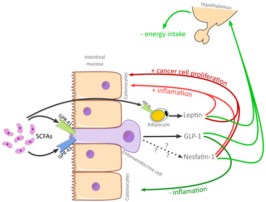
A summary of the relationship between SCFAs and Leptin, GLP-1 and Nesfatin-1, and their effects on the intestinal mucosa is presented in Figure 1.

Figure 1.
Summary of the relationship between SCFAs and leptin, GLP-1 and Nesfatin-1, and their effects on the intestinal mucosa. Abbreviations: SCFAs, short chain fatty acids; GLP-1, glucagon-like peptide 1; GPR 41, G-protein coupled receptor 41; GPR 43, G-protein coupled receptor 43.
The relationship between SCFAs, leptin, and GLP-1 secretion has previously been analyzed [6,11,12,14]; however, the interplay of SCFAs with other anorexigenic signaling molecules such as Nesfatin-1 in the context of modified colon mucosa has not yet been studied. Therefore, this pilot study aimed to investigate the influence of IBD and CRC, as one of its main complications, on SCFA production, and indirectly on SCFA-mediated production of leptin, GLP-1, and Nesfatin-1 in the context of the modified intestinal mucosa which arises in these diseases.
2. Materials and Methods
2.1. Sample Selection and Inclusion
A total of 88 subjects were included in this study: 29 with CRC, 29 with IBD, and 30 healthy controls. Subjects were selected from patients presenting in 2024 to “Prof. Dr. Octavian Fodor”, Regional Institute for Gastroenterology and Hepatology Cluj-Napoca, Romania, and to the gastroenterology department of the County Emergency Clinical Hospital of Târgu Mureș, Romania. Patients with other digestive system comorbidities were not included. All patients with CRC were admitted for scheduled colectomy, while patients with IBD were presented for routine disease check-up. Crohn’s Disease Activity Index (CDAI) and Mayo Clinic Score for ulcerative colitis (MAYO) were determined for IBD patients. Of the included patients, all except two CD and two UC patients were either at remission or with mild disease, with the exceptions having moderate disease according to CDAI and MAYO scores, respectively. In the case of CRC, only patients without any previous treatment and without metastatic disease were included. Sample collection for CRC patients took place pre-operatively, and for IBD patients during routine outpatient blood drawing. Healthy controls were selected from otherwise healthy individuals with no recent medical history.
2.2. Laboratory Analysis
Venous blood was collected into additive free vacuum tubes and left for 30 min at room temperature to clot. After clotting, the tubes were centrifuged at 3000× g for 10 min. The resulting serum was aliquoted into 1.5 mL microcentrifuge tubes, labeled, and stored at −80 °C until the time of analysis. Blood count results for CRC and IBD patients were obtained from routine lab work carried out during presentation, with samples being collected at the same time as the previously mentioned venous blood collection.
Nesfatin-1 (Fine-Test®, FN-Test, Wuhan, China), leptin, GLP-1 (Fine-Test®, FN-Test, Wuhan, China), and total SCFA (Elabscience®, Elabscience Bionovation, Houston, TX, USA) levels were analyzed using commercial Enzyme-Linked Immunosorbent Assay (ELISA) kits. Plates were processed according to the individual kit’s included protocols and were read using an automated ELISA microplate reader (TECAN Sunrise™, Tecan Trading AG, Männedorf, Switzerland). Intra- and inter-assay coefficients of variation were under 10% for all parameters.
2.3. Statistical Analysis
The data was statistically analyzed using RStudio Desktop (RStudio©, PBC v1.4.1106, Boston, MA, USA) with a value of p ≤ 0.05 considered statistically significant. Analysis of normality was carried out using the Shapiro–Wilk test with Holm’s correction. Data found not to be normally distributed was represented as the median (minimum–maximum), while data following a Gaussian distribution was represented as mean ± standard deviation. Difference in means were analyzed using the Student t test or ANOVA with post hoc Tukey’s analysis with Bonferroni’s correction, while medians were analyzed using the Kruskal–Wallis test with Dunn post hoc analysis and Bonferroni’s correction or the Mann–Whitney U test. Correlation of parameters was analyzed using Spearman’s coefficient. Multiple regression was also utilized to test whether the age of participants had any effect on the measured parameters.
3. Results
A total of 88 subjects were included in the study. The comparison of parameters between CRC, IBD, and control groups is presented in Table 1 and Table 2.

Table 1.
Comparison of quantitative variables between groups (significant p values for Kruskal–Wallis tests are bolded).

Table 2.
Comparison of blood count parameters (according to distribution, significant p values for Kruskal–Wallis and ANOVA are bolded).
Post hoc Dunn analysis showed a significant difference (p < 0.05) in Nesfatin-1 levels between CRC and IBD groups. However, the difference between CRC and controls was borderline insignificant (p = 0.06) and there were no differences (p = 0.275) between IBD and control groups.
Post hoc Tukey’s analysis showed significant differences in hemoglobin (p < 0.01), hematocrit (p < 0.01), mean corpuscular volume (p < 0.01), and mean corpuscular hemoglobin concentrations (p < 0.01) between CRC and IBD, CRC and CD, and CRC and UC, respectively; however, there were no significant differences in any of the parameters between CD and UC.
Nesfatin-1 levels were significantly higher in women than in men in both the CRC (p < 0.01) and the IBD (p < 0.01) group, but not the control (p = 0.073) group. On the other hand, men in the IBD group had significantly higher SCFAs levels (p < 0.05) than women; however, this was not present in the CRC (p = 0.272) or control (p = 0.496) groups.
In the case of the CRC group, tumor localization (ascending- vs. transverse- vs. descending- vs. sigmoid-colon) showed no statistically significant differences for any of the analyzed parameters.
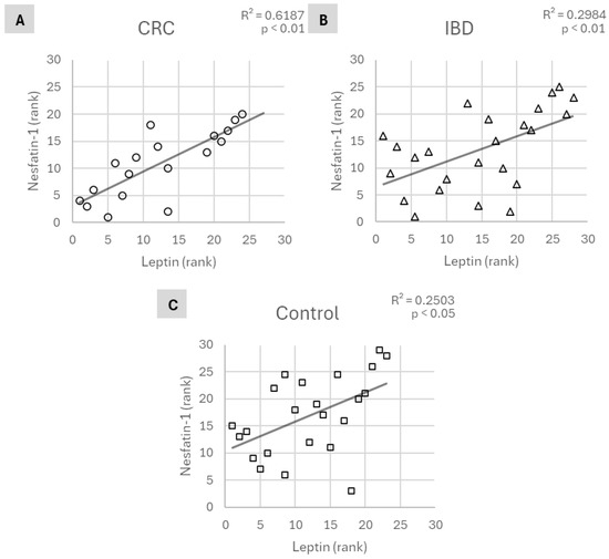
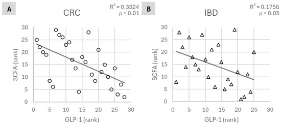
Leptin levels showed a significant positive correlation with Nesfatin-1 levels in all three groups (Figure 2), while SCFAs were significantly negatively correlated with GLP-1 levels in the CRC and IBD groups but not the controls (Figure 3).

Figure 2.
Correlation of Nesfatin-1 and leptin levels in (A): CRC, (B): IBD, and (C): control groups. Abbreviations: CRC, colorectal cancer; IBD, inflammatory bowel disease.

Figure 3.
Correlation of SCFA and GLP-1 levels in (A): CRC and (B): IBD groups. Abbreviations: SCFA, short-chain fatty acids; CRC, colorectal cancer; IBD, inflammatory bowel disease.
SCFAs levels were significantly negatively correlated (R2 = 0.2375, p < 0.05) with Nesfatin-1 levels, and leptin was significantly negatively correlated (R2 = 0.2326, p < 0.05) with neutrophil count in CRC patients; however, neither was present in IBD patients or controls.
Other parameters, including disease activity scores, showed no statistically significant correlations. Multivariate regression analysis showed no significant effect of age on any of the parameters studied.
4. Discussion
Nesfatin-1 levels in CRC patients were significantly higher than in the IBD group and borderline insignificantly higher than in the control group. Multiple studies have shown higher Nesfatin-1 levels in cancer to be a negative prognostic factor, including for CRC [28,42]. In the current study, Nesfatin-1 levels were consistently elevated in CRC, with neoplastic diseases showing the same trend in the previously published literature as well [28]. There is conflicting evidence regarding its role in tumor progression, with both pro- and anti-metastatic effects [43]. Nesfatin-1 also acts as an anorexigenic signaling molecule in the hypothalamus, reducing energy intake [26]. One of the main symptoms associated with neoplastic disease is severe and rapid weight loss, as a result of an increase in energy demand by the growth of the tumor, as well as a reduction in appetite [44]. It is possible that the characteristic reduction in appetite in the case of cancer is an effect of the increased Nesfatin-1 production in these conditions. In our study, Nesfatin-1 was not elevated in comparison with the control group, being in contradiction with this theory. However, Nesfatin-1 is a relatively recently discovered molecule, and research and understanding regarding its effects are still evolving rapidly. It is still too early to draw strong conclusions related to its roles, especially in cancer, without further research.
Nesfatin-1 levels in all three groups were positively correlated with leptin levels. Both leptin and Nesfatin-1 are anorexigenic proteins that reduce energy intake by binding to specific receptors in the hypothalamus [26,45]. It is possible that the mechanism by which the characteristic appetite suppression accompanying different types of intestinal inflammation [46,47] is the result of increased leptin and Nesfatin-1 production. Several studies were published on the individual levels of both Nesfatin-1 and leptin in various diseases; however, the relationship between them has not been elucidated. Energy intake regulation should be analyzed as the combined effect of multiple signaling molecules, as opposed to their individual effects, because their secretion and effects are combined. This hypothesis would also explain why attempts to devise weight loss medication based on individually targeting molecules have generally failed or have presented reduced efficacy. Further research should focus on the combined effects of these molecules rather than analyzing them individually.
In the case of CRC and IBD patients, SCFAs levels were negatively correlated with GLP-1 levels; however, this was not present in the control group. Treatment with GLP-1 agonists in UC showed a reduction in proinflammatory mediators and an alleviation of the gut microbiome dysbiosis [48] and decreased hospitalization rates for IBD [24]. The intrinsic levels of GLP-1 produced by the intestinal wall have been observed to be elevated in the case of IBD [49]. A possible hypothesis would be that, similarly to Nesfatin-1 levels, higher GLP-1 levels indicate a more severe disease, correlated with a higher degree of dysbiosis and subsequently lower SCFAs production. The increase in GLP-1 production might be an attempt to normalize inflammation; however, the body’s capacity to achieve this goal is limited. This would explain why treatment with GLP-1 agonists shows promising results on the reduction in inflammation in patients with IBD. Chronic inflammation, an important preneoplastic condition in the case of IBD, is a significant factor in the appearance and progression of many types of cancers [50]. The binding of SCFAs to GPR 43 on the surface of enteroendocrine L cells was shown to increase GLP-1 production [51], contradicting the negative correlation between SCFAs levels and GLP-1 observed in our study. This might suggest that in patients with CRC and IBD, SCFAs have a limited effect on the secretion of GLP-1, being most likely regulated by other pathways.
Nesfatin-1 levels were negatively correlated with serum SCFA levels in the case of the CRC group. This was not observed in patients with IBD or the control group. Gut microbiome composition is almost always affected by IBD, leading to dysbiosis, with a decrease in prebiotic production, including SCFAs [52]. Patients with both CD and UC have been shown to have higher levels of Nesfatin-1 compared to healthy individuals, both in the remission stage and in the case of flare-ups [27]. In the case of patients with higher levels of Nesfatin-1, the significantly lower levels of SCFAs could be a result of more severe alteration of the gut microbiome in the case of more severe disease. The current study is the first to analyze the relationship between circulating Nesfatin-1 and SCFAs in patients with IBD, in the context of only a few studies that analyze the behavior of Nesfatin-1 in IBD. Further research is needed to clarify the exact nature of this relationship.
Hemoglobin, Ht, and MCV were all significantly lower in CRC in the IBD group. This is most probably a result of more significant gastrointestinal bleeding that accompanies CRC, compared to IBD. It would be useful to carry out and compare blood counts for the control group as well; however, due to technical reasons, this was not possible in our research.
This is the first study to analyze SCFAs and their relationship to leptin, Nesfatin-1, and GLP-1 in CRC and IBD patients. Fully understanding the effects of the colon dynamics in the case of these diseases, which have previously been shown to influence gut microbiome health and composition, warrants further studies on larger sample sizes than the current study. The included IBD patients were all in remission, following individualized treatment plans according to their needs. Whether or not the type of treatment affects gut microbiome composition or serum leptin, Nesfatin-1, or GLP-1 levels, however, is unknown and should be the subject of further studies.
The current pilot study, however, presents a few weaknesses which should be addressed in future studies. Firstly, the number of patients included needs to be increased, in order to solidify the observations and decrease the likelihood of any false positive or negative observations. Secondly, the nutritional status of patients, especially in the case of IBD, needs to be analyzed in the context of SCFA production. The need to directly analyze gut microbiome composition also arises, and so does the necessity to correlate the direct changes with indirect results such as SCFA production and its effects on leptin, Nesfatin-1, and GLP-1 as well as other parameters linked to SCFA production. The method used in this article measures total SCFA concentrations, and the measurement of the individual concentrations of acetate, propionate, and butyrate using gas-chromatography coupled with mass-spectrometry should also be taken into consideration in the future.
5. Conclusions
This is the first study to evaluate circulating Nesfatin-1, leptin, GLP-1, and SCFAs in patients with CRC and IBD. Increased Nesfatin-1 and GLP-1 levels were shown to be correlated with lower levels of SCFAs in CRC patients, and GLP-1 levels were shown to be correlated with lower levels of SCFAs in IBD patients. The negative correlation between Nesfatin-1 and SCFAs levels in CRC and IBD patients underscores a possible relationship between altered gut microbiota function and systemic metabolic changes in cancer progression. Nesfatin-1 levels were significantly elevated in CRC patients, sustaining previously published evidence; however, the exact role and/or effect of this increase remains poorly understood.
Nesfatin-1 and leptin levels were positively correlated in all three groups, suggesting a potentially coordinated role in energy homeostasis and appetite regulation. Future research should analyze these anorexigenic proteins and their effects as a whole, rather than individually, which would lead to a better understanding of the regulation of energy intake. The current article highlights the importance of considering multiple gut-derived and systemic factors when investigating the pathophysiology of gastrointestinal diseases such as CRC and IBD. Future studies on larger sample sizes and comprehensive gut microbiome analyses are needed to clarify the mechanistic links and potential therapeutic implications of these findings.
Author Contributions
Conceptualization, T.I. and P.G.; methodology, T.I.; software, T.I.; validation, C.N.S., S.R.G. and A.M.C.; formal analysis, S.R.G.; investigation, T.I., resources, V.A.; data curation, P.G.; writing—original draft preparation, T.I.; writing—review and editing, C.N.S.; visualization, V.A.; supervision, A.M.C.; project administration, A.M.C. All authors have read and agreed to the published version of the manuscript.
Funding
This research received no external funding.
Institutional Review Board Statement
The study was conducted in accordance with the Declaration of Helsinki and approved by the Institutional Review Board of the Medical Ethics Committee of the “Iuliu Hațieganu” University of Medicine and Pharmacy, Cluj-Napoca, with the approval code AVZ 206/26 July 2023 and the Ethics Committee of Târgu Mureș Emergency Clinical County Hospital, with the approval code Ad.15239/4 July 2024.
Informed Consent Statement
Informed consent was obtained from all subjects involved in the study.
Data Availability Statement
Data is contained in the article.
Conflicts of Interest
The authors declare no conflicts of interest.
Abbreviations
The following abbreviations are used in this manuscript:
| CRC | Colorectal Cancer |
| IBD | Inflammatory Bowel Disease |
| CD | Crohn’s Disease |
| UC | Ulcerative Colitis |
| SCFAs | Short-Chain Fatty Acids |
| GLP-1 | Glucagon-like Peptide 1 |
| Hb | Hemoglobin |
| Ht | Hematocrit |
| MCV | Mean Corpuscular Volume |
| MCHC | Mean Corpuscular Hemoglobin Concentration |
| PLT | Platelets |
| WBC | White Blood Cells |
| Neu | Neutrophils |
| Eo | Eosinophils |
| Ba | Basophils |
References
- Ilyés, T.; Pop, M.; Surcel, M.; Pop, D.M.; Rusu, R.; Silaghi, C.N.; Zaharie, G.C.; Crăciun, A.M. First Comparative Evaluation of Short-Chain Fatty Acids and Vitamin-K-Dependent Proteins Levels in Mother–Newborn Pairs at Birth. Life 2023, 13, 847. [Google Scholar] [CrossRef]
- Ilyés, T.; Silaghi, C.N.; Crăciun, A.M. Diet-Related Changes of Short-Chain Fatty Acids in Blood and Feces in Obesity and Metabolic Syndrome. Biology 2022, 11, 1556. [Google Scholar] [CrossRef]
- Xiong, Y.; Miyamoto, N.; Shibata, K.; Valasek, M.A.; Motoike, T.; Kedzierski, R.M.; Yanagisawa, M. Short-Chain Fatty Acids Stimulate Leptin Production in Adipocytes through the G Protein-Coupled Receptor GPR41. Proc. Natl. Acad. Sci. USA 2004, 101, 1045–1050. [Google Scholar] [CrossRef]
- Karaki, S.-I.; Tazoe, H.; Hayashi, H.; Kashiwabara, H.; Tooyama, K.; Suzuki, Y.; Kuwahara, A. Expression of the Short-Chain Fatty Acid Receptor, GPR43, in the Human Colon. J. Mol. Histol. 2008, 39, 135–142. [Google Scholar] [CrossRef] [PubMed]
- Priyadarshini, M.; Lednovich, K.; Xu, K.; Gough, S.; Wicksteed, B.; Layden, B.T. FFAR from the Gut Microbiome Crowd: SCFA Receptors in T1D Pathology. Metabolites 2021, 11, 302. [Google Scholar] [CrossRef] [PubMed]
- Psichas, A.; Sleeth, M.L.; Murphy, K.G.; Brooks, L.; Bewick, G.A.; Hanyaloglu, A.C.; Ghatei, M.A.; Bloom, S.R.; Frost, G. The Short Chain Fatty Acid Propionate Stimulates GLP-1 and PYY Secretion via Free Fatty Acid Receptor 2 in Rodents. Int. J. Obes. 2015, 39, 424–429. [Google Scholar] [CrossRef]
- Shin, Y.; Han, S.; Kwon, J.; Ju, S.; Choi, T.G.; Kang, I.; Kim, S.S. Roles of Short-Chain Fatty Acids in Inflammatory Bowel Disease. Nutrients 2023, 15, 4466. [Google Scholar] [CrossRef]
- Parada Venegas, D.; De la Fuente, M.K.; Landskron, G.; González, M.J.; Quera, R.; Dijkstra, G.; Harmsen, H.J.M.; Faber, K.N.; Hermoso, M.A. Short Chain Fatty Acids (SCFAs)-Mediated Gut Epithelial and Immune Regulation and Its Relevance for Inflammatory Bowel Diseases. Front. Immunol. 2019, 10, 277. [Google Scholar] [CrossRef] [PubMed]
- Deleu, S.; Machiels, K.; Raes, J.; Verbeke, K.; Vermeire, S. Short Chain Fatty Acids and Its Producing Organisms: An Overlooked Therapy for IBD? eBioMedicine 2021, 66, 103293. [Google Scholar] [CrossRef]
- Stidham, R.W.; Higgins, P.D.R. Colorectal Cancer in Inflammatory Bowel Disease. Clin. Colon Rectal Surg. 2018, 31, 168–178. [Google Scholar] [CrossRef]
- Considine, R.V.; Sinha, M.K.; Heiman, M.L.; Kriauciunas, A.; Stephens, T.W.; Nyce, M.R.; Ohannesian, J.P.; Marco, C.C.; McKee, L.J.; Bauer, T.L.; et al. Serum Immunoreactive-Leptin Concentrations in Normal-Weight and Obese Humans. N. Engl. J. Med. 1996, 334, 292–295. [Google Scholar] [CrossRef] [PubMed]
- Gabriel, F.C.; Fantuzzi, G. The Association of Short-Chain Fatty Acids and Leptin Metabolism: A Systematic Review. Nutr. Res. 2019, 72, 18–35. [Google Scholar] [CrossRef]
- Baggio, L.L.; Drucker, D.J. Biology of Incretins: GLP-1 and GIP. Gastroenterology 2007, 132, 2131–2157. [Google Scholar] [CrossRef]
- Tolhurst, G.; Heffron, H.; Lam, Y.S.; Parker, H.E.; Habib, A.M.; Diakogiannaki, E.; Cameron, J.; Grosse, J.; Reimann, F.; Gribble, F.M. Short-Chain Fatty Acids Stimulate Glucagon-Like Peptide-1 Secretion via the G-Protein–Coupled Receptor FFAR2. Diabetes 2012, 61, 364–371. [Google Scholar] [CrossRef] [PubMed]
- Hills, R.D.; Pontefract, B.A.; Mishcon, H.R.; Black, C.A.; Sutton, S.C.; Theberge, C.R. Gut Microbiome: Profound Implications for Diet and Disease. Nutrients 2019, 11, 1613. [Google Scholar] [CrossRef]
- Kim, H.R. Obesity-Related Colorectal Cancer: The Role of Leptin. Ann. Coloproctol. 2015, 31, 209–210. [Google Scholar] [CrossRef][Green Version]
- Socol, C.T.; Chira, A.; Martinez-Sanchez, M.A.; Nuñez-Sanchez, M.A.; Maerescu, C.M.; Mierlita, D.; Rusu, A.V.; Ruiz-Alcaraz, A.J.; Trif, M.; Ramos-Molina, B. Leptin Signaling in Obesity and Colorectal Cancer. Int. J. Mol. Sci. 2022, 23, 4713. [Google Scholar] [CrossRef]
- Stattin, P.; Palmqvist, R.; Söderberg, S.; Biessy, C.; Ardnor, B.; Hallmans, G.; Kaaks, R.; Olsson, T. Plasma Leptin and Colorectal Cancer Risk: A Prospective Study in Northern Sweden. Oncol. Rep. 2003, 10, 2015–2021. [Google Scholar] [CrossRef]
- Trejo-Vazquez, F.; Garza-Veloz, I.; Villela-Ramirez, G.A.; Ortiz-Castro, Y.; Mauricio-Saucedo, P.; Cardenas-Vargas, E.; Diaz-Baez, M.; Cid-Baez, M.A.; Castañeda-Miranda, R.; Ortiz-Rodriguez, J.M.; et al. Positive Association Between Leptin Serum Levels and Disease Activity on Endoscopy in Inflammatory Bowel Disease: A Case-Control Study. Exp. Ther. Med. 2018, 15, 3336–3344. [Google Scholar] [CrossRef]
- Chouliaras, G.; Panayotou, I.; Margoni, D.; Mantzou, E.; Pervanidou, P.; Manios, Y.; Chrousos, G.P.; Roma, E. Circulating Leptin and Adiponectin and Their Relation to Glucose Metabolism in Children with Crohn’s Disease and Ulcerative Colitis. Pediatr. Res. 2013, 74, 420–426. [Google Scholar] [CrossRef] [PubMed]
- Seino, Y.; Fukushima, M.; Yabe, D. GIP and GLP-1, the Two Incretin Hormones: Similarities and Differences. J. Diabetes Investig. 2010, 1, 8–23. [Google Scholar] [CrossRef]
- Dotania, K.; Tripathy, M.; Rai, U. Nesfatin-1 as a Crucial Mediator of Glucose Homeostasis in the Reptile, Hemidactylus Flaviviridis. Sci. Rep. 2024, 14, 31565. [Google Scholar] [CrossRef] [PubMed]
- Wang, L.; Wang, W.; Kaelber, D.C.; Xu, R.; Berger, N.A. GLP-1 Receptor Agonists and Colorectal Cancer Risk in Drug-Naive Patients with Type 2 Diabetes, with and without Overweight/Obesity. JAMA Oncol. 2024, 10, 256–258. [Google Scholar] [CrossRef]
- Gorelik, Y.; Ghersin, I.; Lujan, R.; Shlon, D.; Loewenberg Weisband, Y.; Ben-Tov, A.; Matz, E.; Zacay, G.; Dotan, I.; Turner, D.; et al. GLP-1 Analog Use Is Associated with Improved Disease Course in Inflammatory Bowel Disease: A Report from the Epi-IIRN. J. Crohn’s Colitis 2024, 19, jjae160. [Google Scholar] [CrossRef] [PubMed]
- Oh-I, S.; Shimizu, H.; Satoh, T.; Okada, S.; Adachi, S.; Inoue, K.; Eguchi, H.; Yamamoto, M.; Imaki, T.; Hashimoto, K.; et al. Identification of Nesfatin-1 as a Satiety Molecule in the Hypothalamus. Nature 2006, 443, 709–712. [Google Scholar] [CrossRef] [PubMed]
- Schalla, M.A.; Stengel, A. Current Understanding of the Role of Nesfatin-1. J. Endocr. Soc. 2018, 2, 1188–1206. [Google Scholar] [CrossRef]
- Beyaz, Ş.; Akbal, E. Increased Serum Nesfatin-1 Levels in Patients with Inflammatory Bowel Diseases. Postgrad. Med. J. 2022, 98, 446–449. [Google Scholar] [CrossRef]
- Kan, J.-Y.; Yen, M.-C.; Wang, J.-Y.; Wu, D.-C.; Chiu, Y.-J.; Ho, Y.-W.; Kuo, P.-L. Nesfatin-1/Nucleobindin-2 Enhances Cell Migration, Invasion, and Epithelial-Mesenchymal Transition via LKB1/AMPK/TORC1/ZEB1 Pathways in Colon Cancer. Oncotarget 2016, 7, 31336–31349. [Google Scholar] [CrossRef]
- Seyedian, S.S.; Nokhostin, F.; Malamir, M.D. A Review of the Diagnosis, Prevention, and Treatment Methods of Inflammatory Bowel Disease. J. Med. Life 2019, 12, 113–122. [Google Scholar] [CrossRef]
- Torres, J.; Mehandru, S.; Colombel, J.-F.; Peyrin-Biroulet, L. Crohn’s Disease. Lancet 2017, 389, 1741–1755. [Google Scholar] [CrossRef]
- Magro, F.; Gionchetti, P.; Eliakim, R.; Ardizzone, S.; Armuzzi, A.; Barreiro-de Acosta, M.; Burisch, J.; Gecse, K.B.; Hart, A.L.; Hindryckx, P.; et al. Third European Evidence-Based Consensus on Diagnosis and Management of Ulcerative Colitis. Part 1: Definitions, Diagnosis, Extra-Intestinal Manifestations, Pregnancy, Cancer Surveillance, Surgery, and Ileo-Anal Pouch Disorders. J. Crohn’s Colitis 2017, 11, 649–670. [Google Scholar] [CrossRef]
- Feuerstein, J.D.; Moss, A.C.; Farraye, F.A. Ulcerative Colitis. Mayo Clin. Proc. 2019, 94, 1357–1373. [Google Scholar] [CrossRef]
- Ko, I.K.; Kim, B.-G.; Awadallah, A.; Mikulan, J.; Lin, P.; Letterio, J.J.; Dennis, J.E. Targeting Improves MSC Treatment of Inflammatory Bowel Disease. Mol. Ther. 2010, 18, 1365–1372. [Google Scholar] [CrossRef] [PubMed]
- Jørgensen, J.; Mortensen, P.B. Hydrogen Sulfide and Colonic Epithelial Metabolism: Implications for Ulcerative Colitis. Dig. Dis. Sci. 2001, 46, 1722–1732. [Google Scholar] [CrossRef]
- Levine, J.; Ellis, C.J.; Furne, J.K.; Springfield, J.; Levitt, M.D. Fecal Hydrogen Sulfide Production in Ulcerative Colitis. Am. J. Gastroenterol. 1998, 93, 83–87. [Google Scholar] [CrossRef]
- Duan, B.; Zhao, Y.; Bai, J.; Wang, J.; Duan, X.; Luo, X.; Zhang, R.; Pu, Y.; Kou, M.; Lei, J.; et al. Colorectal Cancer: An Overview. In Gastrointestinal Cancers; Morgado-Diaz, J.A., Ed.; Exon Publications: Brisbane, Australia, 2022; ISBN 978-0-6453320-6-3. [Google Scholar]
- Fleming, M.; Ravula, S.; Tatishchev, S.F.; Wang, H.L. Colorectal Carcinoma: Pathologic Aspects. J. Gastrointest. Oncol. 2012, 3, 3. [Google Scholar] [CrossRef]
- Remo, A.; Fassan, M.; Vanoli, A.; Bonetti, L.R.; Barresi, V.; Tatangelo, F.; Gafà, R.; Giordano, G.; Pancione, M.; Grillo, F.; et al. Morphology and Molecular Features of Rare Colorectal Carcinoma Histotypes. Cancers 2019, 11, 1036. [Google Scholar] [CrossRef]
- Crippa, J.; Grass, F.; Achilli, P.; Behm, K.T.; Mathis, K.L.; Day, C.N.; Harmsen, W.S.; Mari, G.M.; Larson, D.W. Surgical Approach to Transverse Colon Cancer: Analysis of Current Practice and Oncological Outcomes Using the National Cancer Database. Dis. Colon Rectum 2021, 64, 284–292. [Google Scholar] [CrossRef] [PubMed]
- Sato, Y.; Tsujinaka, S.; Miura, T.; Kitamura, Y.; Suzuki, H.; Shibata, C. Inflammatory Bowel Disease and Colorectal Cancer: Epidemiology, Etiology, Surveillance, and Management. Cancers 2023, 15, 4154. [Google Scholar] [CrossRef] [PubMed]
- Mattar, M.C.; Lough, D.; Pishvaian, M.J.; Charabaty, A. Current Management of Inflammatory Bowel Disease and Colorectal Cancer. Gastrointest. Cancer Res. 2011, 4, 53–61. [Google Scholar]
- Liang, Y.; Ma, Y.; Wang, K.; Xiang, M.; Yi, B. NUCB-2/Nesfatin-1 Promotes the Proliferation of Nasopharyngeal Carcinoma Cells. Cancer Cell Int. 2023, 23, 181. [Google Scholar] [CrossRef]
- Skorupska, A.; Lenda, R.; Ożyhar, A.; Bystranowska, D. The Multifaceted Nature of Nucleobindin-2 in Carcinogenesis. Int. J. Mol. Sci. 2021, 22, 5687. [Google Scholar] [CrossRef]
- Haemmerle, R.J.; Jatoi, A. Loss of Appetite in Patients with Cancer: An Update on Characterization, Mechanisms, and Palliative Therapeutics. Curr. Opin. Support. Palliat. Care 2023, 17, 168–171. [Google Scholar] [CrossRef] [PubMed]
- Kelesidis, T.; Kelesidis, I.; Chou, S.; Mantzoros, C.S. Narrative Review: The Role of Leptin in Human Physiology: Emerging Clinical Applications. Ann. Intern. Med. 2010, 152, 93–100. [Google Scholar] [CrossRef] [PubMed]
- Bakal, U.; Saraç, M.; Ciftci, H.; Tartar, T.; Kazez, A.; Aydin, S. Leptin and NUCB2/Nesfatin-1 in Acute Appendicitis. Int. J. Clin. Med. 2015, 6, 919–927. [Google Scholar] [CrossRef]
- Moran, G.W.; Thapaliya, G. The Gut–Brain Axis and Its Role in Controlling Eating Behavior in Intestinal Inflammation. Nutrients 2021, 13, 981. [Google Scholar] [CrossRef] [PubMed]
- Wang, W.; Zhang, C.; Zhang, H.; Li, L.; Fan, T.; Jin, Z. The Alleviating Effect and Mechanism of GLP-1 on Ulcerative Colitis. Aging 2023, 15, 8044–8060. [Google Scholar] [CrossRef]
- Zatorski, H.; Sałaga, M.; Fichna, J. Role of Glucagon-like Peptides in Inflammatory Bowel Diseases—Current Knowledge and Future Perspectives. Naunyn-Schmiedeb. Arch. Pharmacol. 2019, 392, 1321–1330. [Google Scholar] [CrossRef]
- Singh, N.; Baby, D.; Rajguru, J.P.; Patil, P.B.; Thakkannavar, S.S.; Pujari, V.B. Inflammation and Cancer. Ann. Afr. Med. 2019, 18, 121–126. [Google Scholar] [CrossRef]
- Zeng, Y.; Wu, Y.; Zhang, Q.; Xiao, X. Crosstalk between Glucagon-like Peptide 1 and Gut Microbiota in Metabolic Diseases. mBio 2024, 15, e02032-23. [Google Scholar] [CrossRef]
- Zhang, Z.; Zhang, H.; Chen, T.; Shi, L.; Wang, D.; Tang, D. Regulatory Role of Short-Chain Fatty Acids in Inflammatory Bowel Disease. Cell Commun. Signal. 2022, 20, 64. [Google Scholar] [CrossRef] [PubMed]
Disclaimer/Publisher’s Note: The statements, opinions and data contained in all publications are solely those of the individual author(s) and contributor(s) and not of MDPI and/or the editor(s). MDPI and/or the editor(s) disclaim responsibility for any injury to people or property resulting from any ideas, methods, instructions or products referred to in the content. |
© 2025 by the authors. Licensee MDPI, Basel, Switzerland. This article is an open access article distributed under the terms and conditions of the Creative Commons Attribution (CC BY) license (https://creativecommons.org/licenses/by/4.0/).